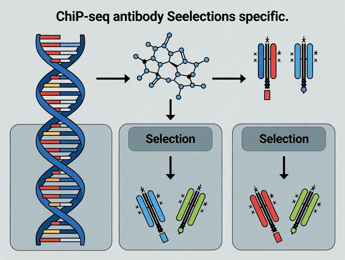
The Ultimate Guide to ChIP-seq Antibody Selection: Navigating Histone Modification Specificity for Robust Epigenetic Data
This comprehensive guide provides researchers, scientists, and drug development professionals with a strategic framework for selecting and validating ChIP-seq antibodies for histone modifications.
The Ultimate Guide to ChIP-seq Antibody Selection: Navigating Histone Modification Specificity for Robust Epigenetic Data
Abstract
This comprehensive guide provides researchers, scientists, and drug development professionals with a strategic framework for selecting and validating ChIP-seq antibodies for histone modifications. Covering foundational principles, methodological applications, troubleshooting strategies, and rigorous validation approaches, the article addresses the critical challenge of antibody specificity. We synthesize current best practices, leveraging recent community standards and technical insights to ensure the generation of reliable, reproducible epigenetic data essential for basic research and therapeutic discovery.
Histone Code 101: Understanding Modifications and the Critical Role of Antibody Specificity
Within the context of a thesis on ChIP-seq antibody selection for specific histone modifications, understanding the biological functions and genomic contexts of key histone marks is paramount. This document provides detailed application notes and protocols for investigating four cornerstone modifications: H3K4me3, H3K27ac, H3K9me3, and H3K27me3. The choice of a highly specific and validated antibody for each mark is the single most critical factor determining the success and interpretability of ChIP-seq experiments, as it directly dictates signal-to-noise ratio and the biological conclusions drawn.
Key Histone Modifications: Biological Functions and Genomic Contexts
The table below summarizes the core functions, genomic locations, and associated states for the four histone modifications.
Table 1: Core Characteristics of Key Histone Modifications
| Modification | Full Name | Associated State | Primary Genomic Location | Key Biological Function | Cross-reactivity Concerns in Antibody Selection |
|---|---|---|---|---|---|
| H3K4me3 | Histone H3 Lysine 4 trimethylation | Active Transcription | Transcriptional start sites (TSS) of active genes | Facilitates recruitment of transcriptional machinery, RNA Pol II. | Anti-H3K4me3 antibodies must not bind H3K4me2 or H3K4me1. |
| H3K27ac | Histone H3 Lysine 27 acetylation | Active Enhancers/Promoters | Active enhancers and promoters | Neutralizes lysine charge, loosens chromatin, promotes factor binding. | Must be distinguished from H3K27me3; acetylation-specific. |
| H3K9me3 | Histone H3 Lysine 9 trimethylation | Facultative & Constitutive Heterochromatin | Repetitive regions, telomeres, silenced genes | Recruits HP1 proteins, condenses chromatin, mediates transcriptional silencing. | Critical specificity against H3K9me1/2 and other methylated lysines. |
| H3K27me3 | Histone H3 Lysine 27 trimethylation | Facultative Heterochromatin | Promoters of developmentally silenced genes (Polycomb targets) | Deposited by PRC2, maintains gene repression during development/differentiation. | Must not recognize H3K27ac or H3K27me1/2. High specificity is essential. |
Application Notes & Protocols
Protocol 1: Chromatin Immunoprecipitation Followed by Sequencing (ChIP-seq) for Histone Modifications
Objective: To map the genome-wide distribution of a specific histone modification using an optimized ChIP-seq workflow, with emphasis on antibody validation.
Materials: See "The Scientist's Toolkit" below.
Method:
- Crosslinking & Cell Harvesting: Treat cells with 1% formaldehyde for 10 min at room temperature to crosslink proteins to DNA. Quench with 125 mM glycine. Wash cells with cold PBS containing protease inhibitors.
- Chromatin Preparation: Lyse cells sequentially with cytoplasmic and nuclear lysis buffers. Isolate nuclei and sonicate chromatin using a focused ultrasonicator (e.g., Covaris) to achieve fragments of 200-500 bp. Centrifuge to clear debris.
- Immunoprecipitation (The Critical Step):
- Pre-clear chromatin lysate with Protein A/G magnetic beads for 1 hour at 4°C.
- Split lysate: Input reference (10%) and IP sample (90%).
- To the IP sample, add the validated, high-specificity primary antibody (see Table 2). Use 1-5 µg of antibody per 10-50 µg of chromatin. Incubate overnight at 4°C with rotation.
- Add pre-blocked Protein A/G magnetic beads and incubate for 2 hours.
- Wash beads sequentially with: Low Salt Wash Buffer, High Salt Wash Buffer, LiCl Wash Buffer, and TE Buffer.
- Elution & Decrosslinking: Elute chromatin from beads with elution buffer (1% SDS, 100mM NaHCO3). Reverse crosslinks for both IP and Input samples by adding NaCl (200 mM final) and incubating at 65°C overnight.
- DNA Purification: Treat samples with RNase A and Proteinase K. Purify DNA using SPRI bead-based cleanup.
- Library Preparation & Sequencing: Use a commercial library prep kit for low-input ChIP-DNA. Perform size selection (~200-300 bp inserts). Validate library quality via Bioanalyzer and quantify by qPCR. Sequence on an appropriate platform (e.g., Illumina NovaSeq) to a minimum depth of 10-20 million non-duplicate reads for histone marks.
Antibody Validation Controls:
- Positive Control: Use a cell line with a well-established profile for the target mark (e.g., H3K4me3 at active housekeeping genes).
- Negative Control: Perform an IP with an IgG isotype control antibody.
- Specificity Check (Essential): Validate antibody by peptide competition (pre-incubation with target vs. non-target modified peptide) or using cell lines deficient for the modifying enzyme.
Protocol 2: Sequential ChIP (Re-ChIP) for Bivalent Domain Analysis
Objective: To identify genomic regions co-marked by opposing modifications, such as "bivalent domains" containing both H3K4me3 and H3K27me3, requiring two sequential immunoprecipitations.
Method:
- Perform the first ChIP as described in Protocol 1 (e.g., using anti-H3K27me3 antibody). Elute the bound chromatin not with standard elution buffer, but with 10 mM DTT at 37°C for 30 min.
- Dilute the eluate 1:50 in Re-ChIP Dilution Buffer and use it as the input for a second round of ChIP with the second antibody (e.g., anti-H3K4me3).
- Process the second IP through washes, elution, and decrosslinking as in Protocol 1.
- Analyze the final DNA by qPCR (for known bivalent promoters) or prepare a sequencing library for genome-wide analysis.
Signaling Pathways and Experimental Workflows
Diagram Title: Histone Modifications and Their Functional Outcomes
Diagram Title: ChIP-seq Experimental Workflow for Histone Modifications
The Scientist's Toolkit: Essential Research Reagent Solutions
Table 2: Key Reagents for Histone Modification ChIP-seq Research
| Item | Function & Importance in Thesis Context | Key Selection Criteria |
|---|---|---|
| High-Specificity Primary Antibodies | Binds specifically to the target histone modification. The most critical variable affecting ChIP-seq data quality. | Validation: Check for citations in key papers, vendor-provided WB/ChIP-seq data. Specificity: Must be verified by peptide array or competition assays. Low lot-to-lot variation. |
| Magnetic Protein A/G Beads | Solid support for antibody-antigen complex capture. Efficient washing reduces background. | Binding capacity for your antibody isotype. Low non-specific DNA binding. Consistent particle size. |
| Focus Ultrasonicator (e.g., Covaris) | Provides reproducible, controlled chromatin shearing to ideal fragment sizes (200-500 bp). | Ability to process multiple samples with consistent shear profiles. Minimizes heat generation. |
| ChIP-seq Grade Enzymatic Kits | For end-repair, A-tailing, adapter ligation, and PCR amplification of low-input ChIP DNA. | Optimized for low-input, high-efficiency conversion to sequencing library. Minimizes PCR bias. |
| Validated Positive Control Primer Sets | qPCR primers for genomic regions known to be enriched or depleted for the mark. Essential for antibody and protocol validation. | Published, sequence-verified primers for active (e.g., GAPDH promoter for H3K4me3) and silent loci. |
| Spike-in Control Chromatin (e.g., S. cerevisiae) | Normalizes for technical variation between samples, crucial for quantitative comparisons. | Chromatin from an organism absent in your sample, with species-specific antibodies. |
| Cell Line or Tissue with Defined Epigenetic State | Positive control biological material with known modification landscape. | Well-characterized lines (e.g., ES cells for bivalent domains, HeLa for active marks). Consistent culture conditions. |
Why Antibody Choice is the Linchpin of Successful ChIP-seq Experiments
Within the context of histone modification research, the selection of a ChIP-grade antibody is not merely a preliminary step; it is the foundational determinant of experimental validity. An antibody's specificity, affinity, and batch-to-batch consistency directly dictate the signal-to-noise ratio, impacting the biological interpretation of chromatin landscapes. This application note details protocols and considerations for rigorous antibody validation, a critical thesis for generating reproducible and meaningful ChIP-seq data.
The Critical Parameters: Antibody Validation Data
Successful ChIP-seq relies on antibodies with proven performance. The following table summarizes key validation benchmarks for common histone modifications.
Table 1: Key Validation Benchmarks for Histone Modification Antibodies
| Histone Mark | Recommended Validation Assay | Acceptable Signal-to-Noise (ChIP-qPCR) | Peak Enrichment over IgG (Fold) | Cross-Reactivity Check |
|---|---|---|---|---|
| H3K4me3 | Peptide/Histone Array, KO Validation | >10 (at active promoters) | >20 | H3K4me1, H3K4me2 |
| H3K27ac | Peptide/Histone Array, KO Validation | >10 (at enhancers/promoters) | >15 | H3K27me3 |
| H3K27me3 | Peptide Array, Cell KO (EZH2-) | >5 (at repressed regions) | >10 | H3K27ac |
| H3K9me3 | Cell KO (e.g., SUV39H1/2 dKO) | >8 (at heterochromatin) | >12 | H3K9me1, H3K9me2 |
| H3K36me3 | Peptide Array, enzymatic treatment | >10 (across gene bodies) | >15 | H3K36me1, H3K36me2 |
Detailed Experimental Protocols
Protocol 1: Pre-Use Antibody Specificity Verification by Peptide Dot Blot
Purpose: To assess antibody cross-reactivity against related modified peptides prior to ChIP.
Materials:
- Biotinylated histone peptides (e.g., target mod, unmodified, related mods).
- Nitrocellulose membrane.
- Blocking buffer (5% BSA in TBST).
- Antibody of interest.
- HRP-conjugated secondary antibody.
- Chemiluminescent substrate and imager.
Procedure:
- Spot 1 µL of each peptide (100 ng/µL) onto a nitrocellulose membrane. Air dry.
- Block membrane with 5% BSA in TBST for 1 hour at room temperature (RT).
- Incubate with primary antibody (at ChIP dilution) in blocking buffer overnight at 4°C.
- Wash membrane 3x with TBST for 5 minutes each.
- Incubate with appropriate HRP-conjugated secondary antibody for 1 hour at RT.
- Wash 3x with TBST. Develop using chemiluminescent substrate and image.
Protocol 2: Gold-Standard ChIP-qPCR Validation for New Antibody Lots
Purpose: To confirm antibody performance in the actual ChIP context using positive and negative control genomic loci.
Materials:
- Crosslinked chromatin from relevant cell line (1x10^6 cells per IP).
- Validated ChIP-grade antibody and species-matched IgG control.
- Protein A/G magnetic beads.
- ChIP elution buffer (1% SDS, 100mM NaHCO3).
- PCR primers for known positive and negative control genomic regions.
- qPCR instrument and SYBR Green master mix.
Procedure:
- Chromatin Preparation: Crosslink cells with 1% formaldehyde for 10 min. Quench with glycine. Sonicate chromatin to 200-500 bp fragments. Clarify by centrifugation.
- Immunoprecipitation: Aliquot chromatin. Pre-clear with beads for 1 hour. Incubate supernatant with 1-5 µg of antibody or IgG overnight at 4°C with rotation. Add beads and incubate for 2 hours.
- Wash & Elution: Wash beads sequentially with Low Salt, High Salt, LiCl, and TE buffers. Elute chromatin in elution buffer at 65°C for 15 min with shaking. Reverse crosslinks at 65°C overnight.
- DNA Recovery: Treat with RNase A and Proteinase K. Purify DNA using silica spin columns.
- qPCR Analysis: Perform SYBR Green qPCR using primers for 2-3 positive control loci (e.g., GAPDH promoter for H3K4me3) and 2-3 negative control loci (e.g., gene desert). Calculate % Input and fold enrichment over IgG.
Visualizing the Antibody-Centric Workflow
The following diagram illustrates the critical decision points and validation checkpoints in the antibody selection workflow for histone modification ChIP-seq.
ChIP-seq Antibody Selection and Validation Workflow
The Scientist's Toolkit: Essential Research Reagent Solutions
Table 2: Essential Toolkit for Histone Modification ChIP-seq
| Reagent/Material | Function & Selection Criteria |
|---|---|
| ChIP-Grade Antibody | Core reagent. Must be validated for ChIP-seq application, preferably with citations and KO/peptide array data available. |
| Protein A/G Magnetic Beads | For antibody-antigen complex pulldown. Choose mix of A/G for optimal species-specific binding. Magnetic beads simplify washing. |
| Cell Line with KO/KD for Target | Essential negative control for antibody validation (e.g., EZH2 KO for H3K27me3). Confirms loss of signal. |
| Validated qPCR Primers | For known positive/negative genomic loci. Critical for quantifying antibody performance via % Input and fold enrichment. |
| Sonicator (Covaris or Bioruptor) | For consistent chromatin shearing to 200-500 bp. Reproducible fragmentation is key for resolution and IP efficiency. |
| Histone Peptide Array | Comprehensive tool for mapping antibody specificity against a panel of modifications. Superior to single peptide blots. |
| High-Fidelity DNA Library Prep Kit | For constructing sequencing libraries from low-input ChIP DNA. Must minimize PCR bias and duplicate reads. |
| SPRI Beads (e.g., AMPure XP) | For post-library prep size selection and cleanup. Consistent bead-to-sample ratio is crucial for reproducible yield. |
Within the broader thesis on ChIP-seq antibody selection for histone modifications, the choice between polyclonal (pAb) and monoclonal (mAb) antibodies is foundational. This decision impacts specificity, sensitivity, reproducibility, and ultimately, the biological interpretation of chromatin landscapes in fundamental research and drug discovery targeting epigenetic regulators.
Table 1: Core Characteristics of Polyclonal vs. Monoclonal Antibodies in Histone ChIP-seq
| Characteristic | Polyclonal Antibody (pAb) | Monoclonal Antibody (mAb) |
|---|---|---|
| Epitope Recognition | Multiple, against various regions of the histone modification. | Single, against one specific epitope of the modification. |
| Typical Specificity | High affinity, but may include off-target binding to similar modifications. | Extremely high and defined; minimal cross-reactivity if well-validated. |
| Batch-to-Batch Consistency | Variable; requires rigorous lot testing. | Extremely consistent across lots and time. |
| Typical Sensitivity | Often higher due to signal amplification from multiple epitopes. | Can be lower if the single epitope is occluded in chromatin. |
| Common Cost | Lower per unit. | Higher initial development, consistent cost. |
| Optimal Use Case | Well-characterized modifications (e.g., H3K4me3); when signal amplification is needed. | Complex or closely related modifications (e.g., H3K27me3 vs. H3K27me2); multi-site studies. |
Table 2: Performance Metrics in Model System ChIP-seq Experiments
| Metric | Polyclonal (anti-H3K27ac) | Monoclonal (anti-H3K27ac) |
|---|---|---|
| Peak Calls | 12,450 ± 1,200 | 11,980 ± 350 |
| Signal-to-Noise Ratio | 8.5 ± 1.2 | 9.8 ± 0.6 |
| Inter-experiment Correlation (R²) | 0.89 ± 0.05 | 0.97 ± 0.02 |
| Non-specific Binding Events | 45 ± 15 | 12 ± 5 |
Detailed Experimental Protocols
Protocol 1: Cross-reactivity Validation for Antibody Selection (Immunoblot) This protocol is critical for screening antibodies prior to ChIP-seq.
- Prepare Nuclear Extracts: Isolate nuclei from target cells using a hypotonic lysis buffer (10 mM HEPES pH 7.9, 1.5 mM MgCl₂, 10 mM KCl) with protease inhibitors. Extract histones with 0.2M H₂SO₄ overnight at 4°C, then precipitate with 33% trichloroacetic acid.
- Acid-Urea-Triton (AUT) Gel Electrophoresis: Resolve 2 µg of histone extract on a 15% AUT gel at 100V for 18 hours. This gel system separates histones based on charge (modification state).
- Transfer & Blocking: Transfer to PVDF membrane and block with 5% BSA in TBST for 1 hour.
- Primary Antibody Incubation: Incubate with candidate pAb or mAb (typically 1:1000 dilution) in blocking buffer overnight at 4°C.
- Detection: Use appropriate HRP-conjugated secondary antibody and chemiluminescence. A specific antibody will detect only the band corresponding to the correct modified histone.
Protocol 2: Standardized Histone ChIP-seq Workflow Optimized for either pAb or mAb after validation.
- Crosslinking & Harvesting: Treat cells (~1x10⁶ per IP) with 1% formaldehyde for 10 min at room temperature. Quench with 125 mM glycine.
- Chromatin Preparation: Lyse cells sequentially with buffers (e.g., LB1: 50 mM HEPES-KOH pH 7.5, 140 mM NaCl, 1 mM EDTA, 10% Glycerol, 0.5% NP-40, 0.25% Triton X-100; LB2: 10 mM Tris-HCl pH 8.0, 200 mM NaCl, 1 mM EDTA, 0.5 mM EGTA). Pellet nuclei and resuspend in shearing buffer.
- Chromatin Shearing: Sonicate chromatin to ~200-500 bp fragments using a focused ultrasonicator (e.g., Covaris). Centrifuge to remove debris.
- Immunoprecipitation:
- Pre-clear chromatin with Protein A/G beads for 1 hour.
- For each IP, use 2-5 µg of validated antibody (pAb or mAb) and 25-50 µl of magnetic Protein A/G beads.
- Incubate antibody with chromatin (from ~0.5-1x10⁶ cells) overnight at 4°C with rotation.
- Capture immune complexes with beads, wash sequentially with: Low Salt Wash Buffer, High Salt Wash Buffer, LiCl Wash Buffer, and TE Buffer.
- Elution & Decrosslinking: Elute with freshly prepared elution buffer (1% SDS, 100 mM NaHCO₃). Add NaCl to 200 mM and reverse crosslinks at 65°C overnight.
- DNA Purification: Treat with RNase A and Proteinase K, then purify DNA using SPRI beads. Quantify by qPCR at positive and negative control genomic loci before library prep.
Visualizations
Histone Antibody Recognition Mechanism
Histone ChIP-seq Core Workflow
The Scientist's Toolkit: Research Reagent Solutions
Table 3: Essential Materials for Histone ChIP-seq Antibody Evaluation
| Reagent / Material | Function & Importance |
|---|---|
| Validated Histone Modification Antibodies (pAb & mAb) | Core reagent. Must be ChIP-seq grade, validated for specificity via immunoblot (e.g., AUT gel) and peptide competition. |
| Acid-Urea-Triton (AUT) Gels | Critical for separating histone variants and modification states to visually assess antibody specificity prior to ChIP. |
| Control Cell Lines (e.g., with KO/KD of specific histone methyltransferases) | Essential negative controls to confirm loss of ChIP signal and validate antibody specificity in a cellular context. |
| Sonicator (Focused Ultrasonicator) | For consistent chromatin shearing to 200-500 bp fragments, a key variable affecting resolution and background. |
| Magnetic Protein A/G Beads | Provide consistent, low-background capture of antibody-chromatin complexes versus slurry beads. |
| Spike-in Control Chromatin (e.g., from Drosophila, yeast) | Normalization control to correct for technical variation, especially crucial when comparing different antibody types or lots. |
| qPCR Primers for Validated Genomic Loci | Positive (known modified) and negative (unmodified) control regions to quantitatively assess IP efficiency before sequencing. |
| ChIP-seq Grade Proteinase K & RNase A | Ensure complete reversal of crosslinks and removal of RNA contamination for clean DNA recovery. |
Within Chromatin Immunoprecipitation followed by sequencing (ChIP-seq) for histone modification research, antibody performance is the single most critical variable determining data validity. This application note details the core characteristics—affinity, specificity, titer, and lot-to-lot consistency—that researchers must evaluate to select robust and reproducible antibodies, directly supporting the broader thesis that systematic antibody validation is fundamental to reliable epigenomic data.
Core Characteristics & Assessment Protocols
Affinity
Affinity measures the strength of the interaction between a single antibody paratope and its target epitope, expressed as the equilibrium dissociation constant (K_D). High-affinity antibodies are essential for ChIP-seq to withstand stringent wash conditions and efficiently capture low-abundance histone marks.
Quantitative Data Summary: Table 1: Typical Affinity Ranges for ChIP-grade Antibodies
| Affinity Category | Equilibrium K_D (M) | Suitability for ChIP-seq |
|---|---|---|
| Very High | < 1 x 10⁻¹¹ | Excellent for low-abundance marks (e.g., H3K27me3) |
| High | 1 x 10⁻¹¹ to 1 x 10⁻⁹ | Good for most common marks (e.g., H3K4me3) |
| Moderate | 1 x 10⁻⁹ to 1 x 10⁻⁷ | May require protocol optimization; risk of low signal |
| Low | > 1 x 10⁻⁷ | Generally unsuitable for standard ChIP-seq |
Protocol 1: Determining Apparent Affinity via ELISA
- Coat a 96-well plate with a histone peptide containing the target modification (e.g., H3K4me3) and an unmodified control (2 µg/mL in PBS, 100 µL/well, overnight at 4°C).
- Block with 200 µL/well of 5% BSA in PBST for 2 hours at room temperature (RT).
- Incubate with a serial dilution (e.g., 1 nM to 1 µM) of the test antibody in blocking buffer for 2 hours at RT.
- Wash (3x with PBST) and incubate with an HRP-conjugated secondary antibody (1:5000 in blocking buffer) for 1 hour at RT.
- Develop using TMB substrate for 10-15 minutes, stop with 1M H₂SO₄, and read absorbance at 450 nm.
- Analyze by fitting a 4-parameter logistic curve to the data. The half-maximal effective concentration (EC₅₀) serves as a comparative apparent affinity measurement.
Specificity
Specificity is the ability of an antibody to bind exclusively to its intended target epitope. In histone research, this requires discrimination between similar modifications (e.g., H3K4me1 vs. H3K4me3) and absence of cross-reactivity to unrelated proteins or unmodified histones.
Protocol 2: Specificity Assessment by Peptide Microarray (Dot Blot)
- Prepare a nitrocellulose membrane spotted with an array of synthesized histone peptides (e.g., target mod, related mods, unmodified).
- Block the membrane in 5% non-fat milk in TBST for 1 hour at RT.
- Probe with the primary antibody at a determined working concentration (see Titer) in blocking buffer overnight at 4°C.
- Wash (3x 10 mins with TBST) and incubate with an IRDye-labeled secondary antibody (1:15,000) for 1 hour at RT in the dark.
- Image using an infrared imaging system (e.g., LI-COR Odyssey).
- Analyze signal intensity. A specific antibody shows strong signal only at its target peptide spot.
Quantitative Data Summary: Table 2: Specificity Assessment Results for Candidate Anti-H3K27ac Antibodies
| Antibody Lot | Signal (Target H3K27ac) | Signal (H3K27me3) | Signal (Unmodified H3) | Specificity Ratio (Target/Next Highest) | Pass/Fail (Ratio >10) |
|---|---|---|---|---|---|
| Vendor A, Lot X | 45,200 | 280 | 150 | 161 | Pass |
| Vendor B, Lot Y | 38,500 | 4,100 | 320 | 9.4 | Fail |
Titer
Titer refers to the effective working concentration or dilution of an antibody. Determining the optimal titer maximizes signal-to-noise ratio and cost-effectiveness in ChIP-seq.
Protocol 3: Titer Optimization for ChIP-seq
- Cross-link and prepare chromatin from ~1x10⁶ cells per condition (e.g., using 1% formaldehyde for 10 mins).
- Shear chromatin to an average size of 200-500 bp via sonication.
- Aliquot sheared chromatin into equal fractions.
- Immunoprecipitate each fraction with a serial dilution of the primary antibody (e.g., 1 µg, 2 µg, 5 µg per reaction) alongside a no-antibody control. Use constant protein A/G bead volume.
- Reverse cross-links, purify DNA, and analyze yield via qPCR at positive and negative control genomic regions.
- Plot % input recovery vs. antibody amount. The optimal titer is the point just before the plateau of the curve, balancing yield and cost.
Lot-to-Lot Consistency
Variation between production lots can invalidate longitudinal studies. Consistency must be verified across key performance metrics.
Protocol 4: Validating Lot-to-Lot Consistency
- Test new lot alongside previous, validated lot using Protocol 2 (Dot Blot) and Protocol 3 (ChIP-qPCR).
- Perform small-scale pilot ChIP-seq for the top two antibody concentrations from the titer curve.
- Sequence libraries on a low-throughput flow cell (e.g., 10% of a lane).
- Analyze key metrics: library complexity, enrichment at known positive/negative regions (e.g., spike-in normalized), and correlation of read density profiles (Pearson's R). A correlation coefficient R > 0.9 is typically acceptable.
Quantitative Data Summary: Table 3: Lot-to-Lot Consistency Metrics for Anti-H3K9me3
| Performance Metric | Validated Lot #12345 | New Lot #67890 | % Variation | Acceptance Threshold |
|---|---|---|---|---|
| ChIP-qPCR (% Input @ Positive Locus) | 12.5% | 11.8% | 5.6% | < 20% |
| Dot Blot Specificity Ratio | 85 | 79 | 7.1% | < 25% |
| Pilot ChIP-seq Pearson's R (vs. Lot #12345) | 1.00 | 0.96 | - | > 0.90 |
The Scientist's Toolkit: Research Reagent Solutions
Table 4: Essential Materials for ChIP-seq Antibody Validation
| Item | Function in Validation |
|---|---|
| Modified & Unmodified Histone Peptide Arrays | Definitive assessment of antibody specificity against a panel of related epitopes. |
| Recombinant Nucleosomes (with specific modifications) | Provides a native chromatin context for affinity and specificity testing beyond linear peptides. |
| ChIP-seq Spike-in Controls (e.g., Drosophila chromatin, modified nucleosomes) | Allows quantitative normalization between experiments and antibody lots for accurate comparative analysis. |
| Isotype Control Antibodies | Critical negative controls for non-specific binding during ChIP-seq optimization. |
| Validated Positive Control Cell Lines (e.g., known high expression of target mark) | Provides a consistent biological substrate for titer determination and lot-to-lot testing. |
| ChIP-Grade Protein A/G Magnetic Beads | Standardized solid-phase matrix for reproducible immunoprecipitation. |
Visualizations
Title: Antibody Validation Workflow for ChIP-seq
Title: Antibody Traits Impact on ChIP-seq Protocol
Navigating Vendor Landscapes and Reputation for Epigenetic Reagents
In the context of ChIP-seq antibody selection for histone modification research, the choice of vendor and specific reagent is a critical, yet often under-scrutinized, parameter. The reproducibility crisis in epigenetics underscores that not all antibodies marketed for ChIP-seq perform as advertised. This application note provides a structured approach to evaluating vendors and their products, supported by protocols for empirical validation.
Vendor & Reagent Landscape Analysis
A comparative analysis of leading vendors offering histone modification antibodies for ChIP-seq reveals significant differences in validation criteria, which correlate with reputation in the literature.
Table 1: Vendor Comparison for Histone Modification Antibodies (e.g., H3K27me3, H3K4me3)
| Vendor | Validation Grade Offered | Key Validation Data Provided (for ChIP) | Typical Cost per 100 µg | Common User-Rated Pros | Common User-Rated Cons |
|---|---|---|---|---|---|
| Active Motif | ChIP-seq Grade | ChIP-seq data (peaks), genome browser tracks, siRNA/knockout validation for some. | $$$$ | Highly trusted, extensive application-specific data. | Premium pricing, smaller catalog for some modifications. |
| Cell Signaling Technology (CST) | ChIP Validated, PathScan | ChIP-qPCR data, knockout/knockdown validation (CUT&Tag, IHC). | $$$ | Rigorous validation against genetic controls, high specificity. | Validation may focus more on IHC; ChIP-seq data less comprehensive than Active Motif. |
| Abcam | ChIP Guaranteed | ChIP-qPCR data, comparison to knockout cell line via WB. | $$ | Broad catalog, competitive pricing, "guarantee" program. | Batch-to-batch variability noted in some user reviews. |
| Diagenode | pico-CHIP Grade | ChIP-seq data, high sensitivity advertised for low cell numbers. | $$$ | Specialized in epigenetics, robust protocols for low-input. | Brand recognition slightly lower in some fields. |
| MilliporeSigma (Upstate) | CHIP Grade | Historical gold standard, often with published ChIP-qPCR data. | $$ - $$$ | Long-standing reputation, widely cited. | Older lots may lack modern genomic validation. |
Essential Research Reagent Solutions Toolkit
Table 2: Core Reagents for ChIP-seq Antibody Validation Workflow
| Item | Function & Selection Criteria |
|---|---|
| Validated Positive Control Antibody | Benchmark for assay performance. E.g., H3K4me3 (strong promoters) or H3K27me3 (Polycomb targets) from a top-tier vendor. |
| Validated Negative Control IgG | Species-matched, non-immune immunoglobulin for background assessment. Must be from same host species as test antibody. |
| Cross-linked Chromatin | Prepared from a well-characterized cell line (e.g., HeLa, K562) known to possess the target histone mark. |
| Magnetic Protein A/G Beads | For antibody-chromatin complex pulldown. Consistent bead size and low non-specific binding are critical. |
| qPCR Primers for Validated Loci | Primer sets for known positive and negative genomic regions for the target mark. Essential for initial specificity check. |
| High-Sensitivity DNA Assay Kit | For accurate quantification of low-concentration ChIP DNA libraries prior to sequencing. |
| Spike-in Control Chromatin (e.g., D. melanogaster, SNAP-ChIP) | Normalizes for technical variation between samples, allowing quantitative comparisons between experiments. |
Core Validation Protocol: Pre-Sequencing Antibody Qualification
This protocol provides a cost-effective method to qualify an antibody for ChIP-seq before committing to full library preparation and sequencing.
Protocol: ChIP-qPCR Cross-Vendor Antibody Qualification
Objective: To compare the signal-to-noise ratio of a new/test antibody against a benchmark antibody for the same histone mark.
Materials:
- Cultured cells (≥ 1x10^6 per IP)
- Formaldehyde (37%)
- Glycine (2.5M)
- Cell lysis buffers (with protease inhibitors)
- Sonication device (e.g., Bioruptor)
- Antibodies: Test antibody, Benchmark antibody, Control IgG
- Magnetic Protein A/G Beads
- ChIP Elution Buffer
- Reverse Cross-linking Buffer (e.g., NaCl + Proteinase K)
- DNA Purification Kit (PCR clean-up)
- SYBR Green qPCR Master Mix
- Validated qPCR Primers (3 positive control loci, 2 negative control loci)
Method:
- Cross-linking & Quenching: Fix cells with 1% formaldehyde for 10 min at RT. Quench with 125mM glycine for 5 min.
- Chromatin Preparation: Wash cells, lyse in SDS Lysis Buffer. Pellet nuclei, resuspend in Sonication Buffer. Sonicate to shear DNA to 200-500 bp fragments. Centrifuge to clear debris.
- Immunoprecipitation: For each IP, aliquot chromatin (equivalent to ~1-2x10^5 cells). Dilute in IP Dilution Buffer. Set up three parallel IPs per antibody comparison: a) Test Antibody (1-5 µg), b) Benchmark Antibody, c) Control IgG.
- Beads Incubation: Pre-clear chromatin with beads for 1 hr. Incubate chromatin with antibodies overnight at 4°C. Add pre-washed magnetic beads for 2 hours.
- Washes & Elution: Wash beads sequentially with Low Salt, High Salt, LiCl, and TE buffers. Elute complexes in fresh elution buffer.
- Reverse Cross-linking & Purification: Add NaCl and incubate at 65°C overnight to reverse crosslinks. Treat with RNase A and Proteinase K. Purify DNA using a spin column kit.
- qPCR Analysis: Run triplicate qPCR reactions for each primer set on all IP samples and a 1% Input reference sample.
- Data Analysis: Calculate % Input for each locus: % Input = 2^(Ct[Input] - Ct[IP] - log2(Input Dilution Factor)) * 100%. Plot % Input for test vs. benchmark antibody at each locus. A qualified antibody should show strong enrichment (>1% Input) at positive loci and minimal signal at negative loci, comparable to the benchmark.
Visualization of Decision and Validation Workflows
Title: Vendor Selection Decision Tree
Title: Antibody Validation Triage Pipeline
Title: Specific Antibody Binding in ChIP-seq
From Catalog to Chromatin: A Step-by-Step Protocol for Antibody Integration and ChIP-seq Success
Within a thesis on ChIP-seq for histone modifications, antibody selection is the most critical variable determining data validity. This protocol provides a systematic, pre-purchase checklist to align reagent selection with your specific experimental model (e.g., primary cells, disease models) and research goal (e.g., mapping broad domains vs. pinpointing sharp peaks).
Application Notes: Key Selection Criteria
1. Model-Specific Epitope Considerations Histone sequences are highly conserved, but post-translational modifications (PTMs) can exhibit model-specific nuances. For example, H3K27me3 patterns in Drosophila vs. mammalian cells may differ in flanking sequences.
2. Antibody Validation Landscape A 2023 survey of 200 peer-reviewed papers using histone ChIP-seq revealed that only 35% cited independent antibody validation data beyond the manufacturer's claims. This underscores the need for rigorous personal checklists.
Table 1: Critical Pre-Purchase Antibody Assessment Criteria
| Criterion | Key Question | Optimal Source/Evidence |
|---|---|---|
| Immunogen | Is the modified peptide sequence identical to your target in your model organism? | Manufacturer datasheet; compare to model's protein sequence. |
| Application Validation | Is ChIP-seq specifically demonstrated? | Peer-reviewed references, preferably in a similar model (e.g., primary neurons). |
| Specificity Data | Is there evidence against cross-reactivity with similar PTMs (e.g., H3K27me3 vs. H3K27me2)? | Western blot on histone extracts, peptide spot arrays. |
| Species Reactivity | Is reactivity confirmed for your model species (e.g., mouse, human, zebrafish)? | Datasheet; independent validation databases (e.g., Histone Antibody Specificity Database). |
| Lot Consistency | Does the vendor provide lot-specific validation data? | Requestable QC certificates; prior user feedback. |
3. Aligning with Experimental Goals
- Mapping Broad Domains (e.g., H3K9me3): Requires antibodies with high affinity but may tolerate minor cross-reactivity.
- Pinpointing Sharp Peaks (e.g., H3K4me3): Demands extreme specificity to avoid background noise.
Protocol 1: In Silico Pre-Validation Workflow
Purpose: To computationally assess antibody suitability before purchase.
Materials:
- Target protein sequence (from UniProt).
- Vendor antibody datasheets.
- Public validation databases (CiteAb, Antibodypedia, Histone Antibody Specificity Database).
Methodology:
- Retrieve Target Sequence: Obtain the full amino acid sequence of your target histone (e.g., Human HIST1H3A) from a trusted database.
- Analyze Immunogen: Extract the immunogen sequence from the vendor datasheet. Perform a strict alignment with your target sequence, focusing on the PTM site and +/- 5 amino acids.
- Cross-Reference Databases: Search the antibody clone/catalog number in public databases. Prioritize entries with data from independent reviews or published ChIP-seq datasets.
- Check for Citations: Use PubMed to find papers using the exact antibody clone in a context similar to yours (model system, goal).
Diagram 1: Antibody Selection Decision Workflow
Protocol 2: In-House Specificity Verification (Peptide Competition Assay)
Purpose: To confirm antibody specificity upon receipt, prior to full-scale ChIP-seq.
Research Reagent Solutions Toolkit
| Item | Function |
|---|---|
| Target PTM Peptide | Synthetic peptide with identical modification. Serves as competitive inhibitor for specific binding. |
| Non-Modified Peptide | Control peptide lacking the PTM. Tests for non-specific/sequence-based antibody binding. |
| Cross-Reactive PTM Peptide | Peptide with a similar, but different, modification (e.g., H3K4me2 for a H3K4me3 Ab). Tests for cross-reactivity. |
| Chromatin Extract | Pre-ChIP input material from your experimental model system. |
| Dot Blot Apparatus | Platform for immobilizing peptides and chromatin for antibody probing. |
Methodology:
- Prepare Antigen Dots: Spot 1 µg of each peptide (target, non-modified, cross-reactive) and 2 µL of chromatin extract onto a nitrocellulose membrane. Let dry.
- Antibody Pre-Incubation: Aliquot the antibody at working dilution. Pre-incubate separate aliquots for 1 hour at 4°C with:
- a) No peptide (control).
- b) 10x molar excess of target PTM peptide.
- c) 10x molar excess of non-modified peptide.
- Probing: Follow standard dot blot protocol. Probe separate membrane strips with each pre-incubated antibody aliquot.
- Analysis:
- Specific Antibody: Signal at target peptide and chromatin dots is abolished only by pre-incubation with the target PTM peptide.
- Non-Specific Antibody: Signal is reduced by both target and non-modified peptides.
- Cross-Reactive Antibody: Signal at chromatin is reduced by both target and cross-reactive PTM peptides.
Diagram 2: Peptide Competition Assay Interpretation
Protocol 3: Pilot Micro-ChIP-qPCR Validation
Purpose: To functionally validate antibody performance in your specific ChIP protocol before genome-wide sequencing.
Methodology:
- Perform Micro-ChIP: Conduct a small-scale ChIP (using 1/10 typical chromatin amount) with the new antibody and a validated positive control antibody (e.g., H3 antibody).
- qPCR Analysis: Design primers for:
- Positive Control Region: A genomic locus known to be enriched for your PTM in your model (from literature).
- Negative Control Region: A known unmodified/gene-desert region (e.g., GAPDH promoter for active marks).
- Calculate % Input: Analyze enrichment. A valid antibody should show >5-fold enrichment at the positive locus over the negative locus and a robust signal over the negative control antibody (e.g., IgG).
Table 2: Pilot ChIP-qPCR Acceptance Thresholds (Example for H3K4me3)
| Metric | Threshold for Proceeding to Seq | Rationale |
|---|---|---|
| Fold-Enrichment (Positive/Negative locus) | ≥ 10-fold | Ensures sufficient signal-to-noise for peak calling. |
| Signal vs. IgG Control | ≥ 5-fold higher at positive locus | Confirms specific immunoprecipitation. |
| Reproducibility (Technical replicates) | CV < 20% | Indicates robust and consistent performance. |
Optimal Antibody Dilution and Bead Coupling Strategies for Histone ChIP
Application Note: Within a thesis investigating ChIP-seq antibody selection for histone modifications, achieving high signal-to-noise ratios is paramount. This document details optimized protocols for two critical, interdependent steps: determining the optimal antibody dilution for specific histone marks and preparing high-performance bead-antibody complexes.
Optimizing Antibody Dilution for Histone Modifications
The ideal antibody concentration maximizes specific enrichment while minimizing non-specific background. A checkerboard titration against a range of chromatin inputs is essential.
Protocol: Checkerboard Titration for Antibody Optimization
Materials:
- Cross-linked chromatin (from ~1x10⁶ cells per IP)
- Candidate ChIP-validated antibody for target histone mark (e.g., H3K4me3, H3K27me3)
- IP Dilution Buffer (20 mM Tris-HCl pH 8.0, 150 mM NaCl, 2 mM EDTA, 1% Triton X-100)
- Protein A/G Magnetic Beads
- Protease Inhibitors
Method:
- Chromatin Input Matrix: Prepare a dilution series of sheared chromatin equivalent to 50,000, 100,000, 250,000, and 500,000 cells per IP reaction in 500 µL of IP Dilution Buffer.
- Antibody Dilution Matrix: Prepare a dilution series of the antibody in IP Dilution Buffer. Typical starting points: 1:50, 1:100, 1:250, 1:500, 1:1000.
- Incubation: For each chromatin input point, incubate aliquots with each antibody dilution overnight at 4°C with rotation.
- Bead Capture: Add a constant, pre-blocked volume of Protein A/G beads to each reaction. Incubate for 2 hours.
- Wash, Elute, and Quantify: Perform standard ChIP washes, reverse cross-links, and purify DNA. Quantify enrichment at a known positive genomic locus and a negative control region via qPCR.
- Calculate Signal-to-Noise (S/N): S/N = (2^(-ΔCt[IP]) / (2^(-ΔCt[Input])) for positive locus, normalized to the negative control.
Table 1: Example Titration Data for an H3K4me3 Antibody
| Chromatin Input (Cells/IP) | Antibody Dilution | % Input (Positive Locus) | % Input (Negative Locus) | Signal-to-Noise Ratio |
|---|---|---|---|---|
| 100,000 | 1:50 | 2.5 | 0.08 | 31.25 |
| 100,000 | 1:100 | 2.1 | 0.05 | 42.00 |
| 100,000 | 1:250 | 1.8 | 0.04 | 45.00 |
| 100,000 | 1:500 | 1.2 | 0.03 | 40.00 |
| 250,000 | 1:100 | 1.8 | 0.07 | 25.71 |
| 250,000 | 1:250 | 1.5 | 0.05 | 30.00 |
Interpretation: For this H3K4me3 antibody, the optimal condition is 100,000 cells with a 1:250 dilution, yielding the highest S/N (45).
Bead Coupling and Blocking Strategies
Efficient, clean bead coupling reduces background. Magnetic Protein A/G bead mixtures are standard, but pre-coupling and crosslinking can improve reproducibility.
Protocol: Antibody-Bead Pre-Coupling and Crosslinking
Materials:
- Protein A/G Magnetic Beads
- PBS + 0.5% BSA (Blocking Buffer)
- Dimethyl pimelimidate (DMP) or similar crosslinker
- Triethanolamine, pH 8.2
- 50 mM Glycine, pH 3.0 (for elution pre-crosslinking)
Method:
- Wash and Block: Wash 50 µL bead slurry per IP 3x with PBS. Resuspend in 1 mL PBS + 0.5% BSA. Rotate for 1 hour at 4°C.
- Antibody Binding: Wash beads 2x with IP Dilution Buffer. Resuspend beads in IP Dilution Buffer containing the optimized amount of antibody (determined in Section 1). Incubate with rotation for 6 hours at 4°C.
- Wash Off Unbound Antibody: Pellet beads, collect supernatant (can be re-used for more coupling). Wash beads 2x with IP Dilution Buffer.
- (Optional but Recommended) Crosslinking:
- Resuspend beads in 1 mL Triethanolamine, pH 8.2.
- Add DMP to a final concentration of 5-10 mM. Incubate with rotation for 45 min at room temperature.
- Quench the reaction by adding Tris-HCl, pH 7.5 to 50 mM final concentration. Incubate 15 min.
- Wash beads 3x with IP Dilution Buffer. The crosslinked beads are now stable and can be stored at 4°C for several weeks.
- Use in ChIP: Add the pre-coupled, potentially crosslinked beads directly to the chromatin-antibody mixture (if not pre-coupled) or to chromatin alone (if antibody is already on beads). Proceed with standard IP.
Table 2: Comparison of Bead Coupling Strategies
| Strategy | Advantages | Disadvantages | Recommended For |
|---|---|---|---|
| Standard In-Solution IP | Flexible, easy to titrate antibody freshly. | Higher non-specific binding, less reproducible. | Initial antibody titration and validation. |
| Pre-Coupling (No Crosslink) | Reduces bead-induced background, faster IP day. | Antibody may leach off beads during IP. | High-affinity antibodies, routine protocols. |
| Pre-Coupling with Crosslink | Minimal antibody leaching, highly reproducible. | Irreversible; cannot recover antibody. | Large-scale or core facility workflows. |
The Scientist's Toolkit: Key Research Reagent Solutions
Table 3: Essential Materials for Histone ChIP Optimization
| Item | Function/Application |
|---|---|
| ChIP-Validated Histone Modification Antibodies | Primary antibodies specifically validated for chromatin immunoprecipitation, targeting modifications like H3K4me3, H3K27ac, H3K9me3. |
| Protein A/G Magnetic Bead Mix | Binds a broad range of antibody Fc regions for efficient capture and easy magnetic separation. |
| Dimethyl Pimelimidate (DMP) | Homobifunctional crosslinker for amine groups; used to covalently crosslink antibodies to Protein A/G beads. |
| Micrococcal Nuclease (MNase) | For native ChIP (nChIP); digests linker DNA to yield mononucleosomes, preserving histone modifications. |
| Sonication Shearing Equipment | For crosslinked ChIP (XChIP); fragments chromatin to 200-500 bp via acoustic energy. |
| ChIP-Seq Grade Proteinase K | Efficiently digests proteins and reverses crosslinks after immunoprecipitation. |
| qPCR Primers for Positive/Negative Genomic Loci | Essential for titration analysis to quantify specific enrichment versus background. |
| SPRI Beads (e.g., AMPure) | For efficient clean-up and size selection of ChIP-DNA libraries prior to sequencing. |
Title: Antibody Titration Optimization Workflow
Title: Bead Coupling Strategy Comparison
Within the broader thesis on ChIP-seq antibody selection for specific histone modifications, the decision to use cross-linking is fundamental. This choice profoundly impacts data quality, signal-to-noise ratio, and biological interpretation. Cross-linking covalently binds proteins to DNA, preserving in vivo interactions but potentially introducing epitope masking. Native (non-cross-linked) ChIP offers higher resolution for stable histone-DNA interactions but may lose transient or indirect binding events. This application note evaluates the critical considerations, supported by current data and detailed protocols.
Quantitative Comparison: Cross-linked vs. Native ChIP for Histone Modifications
Table 1: Performance Metrics for H3K4me3 and H3K27me3 Analysis
| Parameter | Cross-linked ChIP (X-ChIP) | Native ChIP (N-ChIP) | Notes |
|---|---|---|---|
| Typical Yield (DNA ng) | 1-10 | 5-50 | N-ChIP typically yields more DNA due to less shearing loss. |
| Peak Resolution (bp) | 200-500 | 50-200 | N-ChIP provides sharper peaks due to absence of cross-link reversal artifacts. |
| Background Signal | Higher | Lower | X-ChIP may have increased non-specific background. |
| Protocol Duration | 2-3 days | 1-2 days | X-ChIP requires cross-linking and reversal steps. |
| Epitope Accessibility Risk | Moderate to High | Low | Formaldehyde can mask epitopes, affecting antibody efficacy. |
| Suitability for Low-Abundance Marks | Moderate | High | Higher yield and lower noise benefit marks like H3K36me2. |
Table 2: Recommended Application by Histone Modification Type
| Histone Modification | Recommended Method | Key Rationale | Key Antibody Consideration |
|---|---|---|---|
| H3K4me3 | Native ChIP | Very stable, promoter-proximal mark; benefits from high resolution. | Antibodies perform well in native conditions; high specificity required for sharp TSS peaks. |
| H3K27me3 | Cross-linked ChIP | Broad domains; cross-linking helps preserve complex integrity in facultative heterochromatin. | Antibody must recognize epitope despite cross-linking; validated for X-ChIP essential. |
| H3K9me3 | Cross-linked ChIP | Associated with constitutive heterochromatin; cross-linking aids in precipitation. | Must be validated for pericentric regions; potential for high background. |
| H3K36me3 | Either (Leaning Native) | Elongation mark; stable association. Native gives clean signal; X-ChIP can capture transcription complexes. | Select antibody with low cross-reactivity to H3K36me1/2. |
| H3K27ac | Cross-linked ChIP | Dynamic, enhancer-associated mark; cross-links transient co-activator interactions. | Sensitivity is critical due to lower abundance at specific enhancers. |
Detailed Experimental Protocols
Protocol 1: Cross-linked ChIP-seq for Histone Modifications (e.g., H3K27me3)
Principle: Formaldehyde cross-links proteins to DNA in vivo, chromatin is sheared by sonication, and specific histone modifications are immunoprecipitated.
Reagents & Solutions:
- PBS-G: PBS with protease inhibitors.
- Fixation Solution: 1% Formaldehyde in PBS.
- Quenching Solution: 2.5M Glycine.
- Lysis Buffer 1: 50mM HEPES-KOH pH7.5, 140mM NaCl, 1mM EDTA, 10% Glycerol, 0.5% NP-40, 0.25% Triton X-100, protease inhibitors.
- Lysis Buffer 2: 10mM Tris-HCl pH8.0, 200mM NaCl, 1mM EDTA, 0.5mM EGTA, protease inhibitors.
- Shearing Buffer (RIPA-like): 10mM Tris-HCl pH8.0, 1mM EDTA, 0.1% SDS, 0.1% Na-Deoxycholate, 1% Triton X-100.
- ChIP Dilution Buffer: 0.01% SDS, 1.1% Triton X-100, 1.2mM EDTA, 16.7mM Tris-HCl pH8.0, 167mM NaCl.
- Low Salt Wash: 0.1% SDS, 1% Triton X-100, 2mM EDTA, 20mM Tris-HCl pH8.0, 150mM NaCl.
- High Salt Wash: 0.1% SDS, 1% Triton X-100, 2mM EDTA, 20mM Tris-HCl pH8.0, 500mM NaCl.
- LiCl Wash: 0.25M LiCl, 1% NP-40, 1% Na-Deoxycholate, 1mM EDTA, 10mM Tris-HCl pH8.0.
- TE Buffer: 10mM Tris-HCl pH8.0, 1mM EDTA.
- Elution Buffer: 1% SDS, 100mM NaHCO3.
- Proteinase K Solution: 20mg/mL.
Procedure:
- Cross-linking: Harvest ~1x10^7 cells. Resuspend in PBS-G. Add fixation solution to 1% final. Incubate 8-12 minutes at room temperature with gentle rotation.
- Quenching: Add glycine to 125mM final. Incubate 5 min at RT. Pellet cells, wash 2x with ice-cold PBS-G.
- Nuclear Extraction: Resuspend pellet in Lysis Buffer 1. Incubate 10 min, 4°C. Pellet, resuspend in Lysis Buffer 2. Incubate 10 min, 4°C. Pellet nuclei.
- Chromatin Shearing: Resuspend nuclei in Shearing Buffer. Sonicate to achieve 200-500 bp fragments (optimize for sonicator). Centrifuge at max speed, 10 min, 4°C. Collect supernatant (sheared chromatin). Take a 50μL aliquot for input control.
- Immunoprecipitation: Dilute chromatin 10-fold in ChIP Dilution Buffer. Pre-clear with Protein A/G beads for 1h, 4°C. Incubate with 1-5μg of validated histone modification antibody overnight at 4°C.
- Bead Capture: Add pre-blocked Protein A/G beads. Incubate 2h, 4°C.
- Washing: Pellet beads, wash sequentially (5 min each, 4°C) with: Low Salt Wash (1x), High Salt Wash (1x), LiCl Wash (1x), TE Buffer (2x).
- Elution & Reverse Cross-link: Add 100μL Elution Buffer to beads and 100μL to saved input. Incubate 30 min at 65°C with shaking. Centrifuge, transfer supernatant. Add 5μL Proteinase K solution to eluates and inputs. Incubate 2h to overnight at 65°C.
- DNA Purification: Purify DNA using silica column or phenol-chloroform. Proceed to library prep.
Protocol 2: Native ChIP-seq for Histone Modifications (e.g., H3K4me3)
Principle: Native chromatin is prepared by micrococcal nuclease (MNase) digestion, which releases primarily mononucleosomes. Histone-DNA interactions are native, and immunoprecipitation is performed without cross-link reversal.
Reagents & Solutions:
- Nuclei Buffer (NB): 10mM Tris-HCl pH7.5, 3mM CaCl2, 2mM MgAc2, 0.5mM DTT, 0.25M Sucrose, protease inhibitors.
- MNase Digestion Buffer: NB with 0.32M Sucrose.
- Micrococcal Nuclease (MNase).
- MNase Stop Solution: 10mM Tris-HCl pH7.5, 5mM EDTA, 0.5% SDS.
- Low Salt Wash/Elution Buffers (as in Protocol 1, but without SDS).
Procedure:
- Nuclei Preparation: Harvest ~1x10^7 cells. Wash in PBS. Resuspend in NB. Homogenize with Dounce (loose pestle). Layer over NB with 1.2M sucrose. Centrifuge. Pellet nuclei.
- MNase Digestion: Resuspend nuclei in MNase Digestion Buffer. Add MNase (titrate for ~80% mononucleosomes). Incubate 15-20 min, 37°C.
- Digestion Stop & Solubilization: Add MNase Stop Solution. Incubate 10 min on ice. Centrifuge 10 min, 4°C. Collect supernatant (soluble chromatin).
- Immunoprecipitation: Dilute chromatin in Native Dilution Buffer (ChIP Dilution Buffer without SDS). Pre-clear. Add antibody (0.5-2μg). Incubate overnight, 4°C.
- Bead Capture & Washing: Add pre-blocked beads. Incubate 2h. Wash with Native Low Salt, High Salt, and TE Buffers.
- DNA Elution: Elute DNA directly in TE buffer with 1% SDS or using a standard column elution buffer. No cross-link reversal is needed.
- DNA Purification: Purify DNA. Proceed to library prep.
Visualizations
Decision Tree for ChIP Method Selection
Comparison of X-ChIP and N-ChIP Workflows
The Scientist's Toolkit: Key Research Reagent Solutions
Table 3: Essential Materials for Histone Modification ChIP
| Reagent / Material | Function & Importance | Example / Note |
|---|---|---|
| Validated ChIP-grade Antibodies | Primary driver of specificity; must be validated for the chosen method (X- or N-ChIP) and species. | Check CUT&Tag/ChIP-seq citations on vendor sites (e.g., Cell Signaling, Abcam, Active Motif). |
| Protein A/G Magnetic Beads | Efficient capture of antibody-chromatin complexes; reduce background vs. agarose. | Facilitate wash steps; crucial for low-input protocols. |
| Ultra-Pure Formaldehyde | For X-ChIP; creates reversible protein-DNA cross-links. Concentration and time are critical. | Use fresh 1% solutions; over-fixation increases epitope masking. |
| Micrococcal Nuclease (MNase) | For N-ChIP; digests linker DNA to yield mononucleosomes. Titration is essential. | Requires Ca2+ for activity; use high-purity, RNase-free grade. |
| Sonication Device | For X-ChIP; shears cross-linked chromatin to optimal fragment size (200-500bp). | Focused ultrasonicator or bath; optimize power/time to avoid heating. |
| Protease Inhibitor Cocktails | Prevent histone degradation during chromatin preparation. | Use broad-spectrum, EDTA-free cocktails. |
| DNA Purification Kits | Clean recovery of ChIP DNA for library prep. Silica columns are standard. | Ensure high recovery for low-yield experiments. |
| qPCR Primers | For positive/negative control genomic loci to validate ChIP efficiency prior to sequencing. | Design for known enriched and depleted regions for your target mark. |
Sonication and Fragmentation Optimization for Nucleosomal DNA Recovery
This protocol is a critical methodological component of a broader thesis investigating antibody selection for specific histone modification ChIP-seq. The efficiency and specificity of chromatin immunoprecipitation (ChIP) are fundamentally dependent on the optimal fragmentation of chromatin into mononucleosomal particles. Inadequate sonication leads to incomplete shearing, resulting in high background noise and reduced resolution, while over-sonication can damage epitopes and compromise antibody binding, directly confounding comparisons of antibody efficacy. Therefore, precise optimization of sonication for nucleosomal DNA recovery is a prerequisite for robust, reproducible data in histone modification research.
Key Research Reagent Solutions
| Reagent/Material | Function in Protocol |
|---|---|
| Covaris truChIP Chromatin Shearing Kit | Standardized reagents including buffers and protease inhibitors for optimized chromatin shearing and stability. |
| Diagenode Bioruptor Pico | Ultrasonic device designed for high-sensitivity chromatin shearing in microcentrifuge tubes, minimizing sample loss. |
| Covaris S220/E220 Focused-ultrasonicator | Instrument using adaptive focused acoustics (AFA) for highly controlled, water-based, reproducible shearing. |
| MNase (Micrococcal Nuclease) | Enzymatic alternative to sonication; digests linker DNA between nucleosomes. |
| Dynabeads Protein A/G | Magnetic beads for efficient antibody and chromatin complex pulldown, used post-sonication. |
| QIAGEN MinElute PCR Purification Kit | For purification and concentration of low-abundance ChIP-DNA post-recovery. |
| Bioanalyzer High Sensitivity DNA Assay | Microfluidic electrophoresis for precise sizing and quantification of sheared chromatin (100-500 bp target). |
| SimpleChIP Enzymatic Chromatin IP Kits | Commercial kits providing a complete, optimized system from fragmentation to DNA purification. |
Table 1: Sonication Parameter Optimization for a 200 bp Target
| Instrument | Sample Volume | Duty Cycle/Intensity | Cycles/Time | Peak Size (bp) | % in 100-500 bp Range |
|---|---|---|---|---|---|
| Bioruptor Pico | 130 µL | 30 sec ON / 30 sec OFF | 8 cycles | 205 | 85% |
| Bioruptor Pico | 130 µL | 30 sec ON / 30 sec OFF | 12 cycles | 150 | 92% |
| Covaris S220 | 130 µL | 5% Duty Factor, 140 PIP, 200 cycles/burst | 4 min | 250 | 78% |
| Covaris S220 | 130 µL | 10% Duty Factor, 140 PIP, 200 cycles/burst | 6 min | 190 | 90% |
| Bath Sonicator (generic) | 500 µL | Constant | 15 min | Broad (100-1000) | 65% |
Table 2: Comparison of Fragmentation Methods for Nucleosomal Recovery
| Method | Principle | Optimal Size | Pros | Cons | Ideal for Thesis Application? |
|---|---|---|---|---|---|
| Ultrasonic Shearing (Covaris) | Focused acoustic energy | 100-500 bp | High reproducibility, tunable, low heat | Equipment cost, requires optimization | Yes - Primary method |
| Ultrasonic Shearing (Bath) | Acoustic cavitation | Variable | Low cost, high throughput | High variability, heat generation, cross-contamination risk | No - High variability |
| MNase Digestion | Enzymatic cleavage | ~147 bp (mononucleosome) | Native nucleosome boundaries, no epitope damage | Digestion bias, over-digestion risk, requires titration | Yes - Complementary method |
| Combined (MNase + Sonication) | Enzymatic then mechanical | 150-300 bp | Very precise size control, efficient | Complex protocol, additional step | Yes - For high-resolution mapping |
Detailed Experimental Protocols
Protocol 1: Covaris S220 Focused-Ultrasonication for Chromatin Shearing
Objective: To reproducibly shear cross-linked chromatin to a peak size of 200-300 bp for histone ChIP-seq.
Materials:
- Covaris S220 with microTUBE AFA Fiber Screw-Cap (130 µL)
- truChIP Chromatin Shearing Kit
- Fixed chromatin from ~1x10⁶ cells
- Bioanalyzer or TapeStation
Procedure:
- Chromatin Preparation: Harvest cells, cross-link with 1% formaldehyde for 10 min at RT. Quench with glycine. Pellet, wash with cold PBS. Lyse cells using the truChIP lysis buffers according to kit instructions. Pellet nuclei.
- Resuspension: Resuspend pelleted nuclei in 130 µL of shearing buffer (provided in kit). Transfer suspension to a pre-chilled Covaris microTUBE. Avoid bubbles.
- Instrument Setup: Initialize Covaris S220 and set the following parameters in the software:
- Peak Incident Power (W): 140
- Duty Factor: 10%
- Cycles per Burst: 200
- Treatment Time: 6 minutes
- Temperature: 4°C (maintained by water bath)
- Sonication: Place the microTUBE in the instrument and start the run.
- Post-Sonication: Transfer sheared chromatin to a fresh 1.5 mL microcentrifuge tube. Pellet debris at 16,000 x g for 10 min at 4°C. Transfer supernatant (sheared chromatin) to a new tube.
- Analysis: Take a 10 µL aliquot. Reverse cross-links by incubating with 5M NaCl and Proteinase K at 65°C for 2 hours. Purify DNA using a MinElute column. Analyze 1 µL on a Bioanalyzer High Sensitivity DNA chip to verify shearing profile. Aim for a peak between 200-300 bp.
- Titration: If the fragment size is too large (>500 bp), increase treatment time by 1-2 minute increments. If too small (<150 bp), decrease duty factor to 5% or reduce time.
Protocol 2: MNase Titration for Enzymatic Fragmentation
Objective: To determine the optimal MNase concentration for generating >70% mononucleosomes, as a complementary or validation method.
Materials:
- MNase (e.g., Worthington)
- Nuclei from ~5x10⁵ cells
- MNase Digestion Buffer (10 mM Tris-HCl pH 7.5, 15 mM NaCl, 60 mM KCl, 1 mM CaCl₂)
- 0.5 M EDTA (stop solution)
- Thermonixer
Procedure:
- Nuclei Preparation: Prepare nuclei from cross-linked cells as in Protocol 1, Step 1. Resuspend nuclei pellet in 500 µL of pre-warmed MNase Digestion Buffer.
- Aliquot: Divide the suspension into 5 x 100 µL aliquots in PCR tubes.
- Titration: Add MNase to each tube to achieve final concentrations of 0, 0.5, 1, 2, and 4 units per 100 µL. Mix gently.
- Digestion: Incubate tubes in a thermomixer at 37°C for 10 minutes with gentle agitation (300 rpm).
- Stop Reaction: Add 10 µL of 0.5 M EDTA (pH 8.0) to each tube to chelate Ca²⁺ and stop digestion. Place on ice.
- Reverse Cross-links & Analyze: Add 2 µL of 10% SDS and 2.5 µL of 20 mg/mL Proteinase K to each. Incubate at 65°C for 2 hours. Purify DNA and analyze on a Bioanalyzer.
- Selection: Identify the concentration yielding a sharp ~147 bp mononucleosome band with minimal sub-nucleosomal fragments (<147 bp). This is the optimal condition for native nucleosome mapping.
Visualization: Experimental Workflows and Logical Framework
Title: Sonication Optimization & Validation Workflow
Title: Role of Fragmentation in ChIP-seq Antibody Thesis
Best Practices for Antibium y-Chromatin Incubation and Wash Stringency
1. Introduction
This document presents detailed application notes and protocols for two critical steps in the Chromatin Immunoprecipitation sequencing (ChIP-seq) workflow: antibody-chromatin incubation and post-immunoprecipitation washing. These protocols are framed within a broader thesis on ChIP-seq antibody selection for specific histone modifications research, where the success of the entire experiment hinges on the specificity and sensitivity of the antibody-antigen interaction. Optimizing incubation conditions and wash stringency is essential to maximize true signal while minimizing non-specific background and false-positive peaks.
2. Key Parameters & Quantitative Data Summary
The optimal conditions for incubation and washing are interdependent and must be calibrated based on the antibody's affinity and the abundance of the target epitope. The following table summarizes key quantitative parameters from current literature and best practices.
Table 1: Optimization Parameters for Incubation and Wash Steps
| Parameter | Typical Range / Options | Impact on Specificity/Sensitivity | Recommendation for High-Abundance Marks (e.g., H3K4me3) | Recommendation for Low-Abundance Marks (e.g., H3K27me3) |
|---|---|---|---|---|
| Incubation Temperature | 4°C, Room Temp (22-25°C) | Higher temp increases kinetics but may increase non-specific binding. | 4°C overnight (12-16 hrs) for maximum specificity. | 4°C overnight (12-16 hrs) is standard; can extend to 24 hrs. |
| Incubation Duration | 2 hrs - 24 hrs | Longer incubation increases yield but may increase background. | 4-6 hours may be sufficient. | Overnight (12-16 hrs) is critical for sufficient yield. |
| Wash Buffer Ionic Strength | Low Salt (150 mM NaCl), High Salt (500 mM NaCl), LiCl Detergent (250 mM LiCl) | Higher salt reduces non-ionic/electrostatic interactions. | Series ending with a single High Salt (500 mM NaCl) wash. | Series including High Salt (500 mM NaCl) and often LiCl (250 mM) wash. |
| Number of Washes | 3-6 washes per buffer | More washes reduce background but risk eluting specific complexes. | 3-5 washes total, adjusting based on antibody performance. | 4-6 washes total, often including stringent final steps. |
| Detergent Type & Concentration | 0.1% SDS, 0.1-1% Triton X-100, 0.1% Na-Deoxycholate | SDS is more stringent, disrupting protein-protein interactions. | Final wash may contain 0.1% SDS. | Commonly use buffers with 0.1% SDS throughout series. |
3. Detailed Experimental Protocols
Protocol A: Standard Antibody-Chromatin Incubation for Histone Modifications
Reagents: Diluted chromatin, validated primary antibody, Protein A/G magnetic beads, ChIP Incubation Buffer (20 mM Tris-HCl pH 8.0, 2 mM EDTA, 150 mM NaCl, 0.1% SDS, 1% Triton X-100, 1x protease inhibitors). Procedure:
- Pre-clear chromatin by adding 20 µl of equilibrated Protein A/G beads to the diluted chromatin sample. Rotate for 1 hour at 4°C. Pellet beads and transfer supernatant to a new tube.
- Add the recommended amount of specific histone modification antibody (typically 1-5 µg) to the pre-cleared chromatin. For an isotype control, add an equivalent amount of control IgG.
- Incubate with rotation at 4°C for 12-16 hours (overnight). For very robust marks, a 4-6 hour incubation can be tested.
- Following incubation, add 30 µl of pre-equilibrated Protein A/G magnetic beads.
- Incubate with rotation for 2 hours at 4°C to capture the antibody-chromatin complexes.
Protocol B: Stringency-Adjusted Wash Protocol Post-Immunoprecipitation
Reagents: Low Salt Wash Buffer (20 mM Tris-HCl pH 8.0, 2 mM EDTA, 150 mM NaCl, 0.1% SDS, 1% Triton X-100), High Salt Wash Buffer (20 mM Tris-HCl pH 8.0, 2 mM EDTA, 500 mM NaCl, 0.1% SDS, 1% Triton X-100), LiCl Wash Buffer (10 mM Tris-HCl pH 8.0, 1 mM EDTA, 250 mM LiCl, 1% NP-40, 1% Na-Deoxycholate), TE Buffer (10 mM Tris-HCl pH 8.0, 1 mM EDTA). Procedure:
- Pellet the bead-antibody-chromatin complexes using a magnetic rack. Carefully remove and discard the supernatant.
- Wash sequentially with rotation for 5 minutes per wash at 4°C: a. Low Salt Wash Buffer: One wash. b. High Salt Wash Buffer: One wash. Critical for reducing non-specific DNA binding. c. LiCl Wash Buffer: One wash. Disrupts non-covalent protein aggregates. d. TE Buffer: Two quick washes to remove detergents and salts before elution.
- After the final TE wash, remove all residual supernatant. The complexes are now ready for elution and reverse-crosslinking.
4. Visualizing the Workflow and Decision Logic
ChIP-seq Wash Stringency Decision Workflow
5. The Scientist's Toolkit: Key Research Reagent Solutions
Table 2: Essential Materials for ChIP-seq Incubation & Wash Steps
| Item | Function & Importance |
|---|---|
| Validated ChIP-seq Grade Antibody | The core reagent. Specificity is paramount; antibodies validated for ChIP-seq reduce false positives. |
| Protein A/G Magnetic Beads | Facilitate efficient capture and washing of antibody complexes. Magnetic separation minimizes mechanical damage. |
| ChIP Incubation Buffer (with Protease Inhibitors) | Maintains chromatin integrity and protein complexes while allowing specific antibody binding. |
| Stringent Wash Buffers (High Salt, LiCl) | Critical for removing loosely bound and non-specifically associated chromatin fragments. |
| Low-Binding Microcentrifuge Tubes | Minimizes loss of material and beads during incubation and wash steps. |
| Rotating Mixer at 4°C | Ensures constant, gentle agitation for even incubation and washing without foaming. |
| Magnetic Separation Rack | Allows for efficient, non-destructive supernatant removal during wash steps. |
Diagnosing Disaster: Solving Common ChIP-seq Antibody Problems and Enhancing Signal-to-Noise
Within the critical framework of ChIP-seq antibody selection for histone modifications research, identifying a high-quality, specific antibody is paramount. The integrity of chromatin immunoprecipitation followed by sequencing (ChIP-seq) data hinges entirely on the antibody's performance. A suboptimal antibody can lead to erroneous biological conclusions, wasted resources, and failed drug development pipelines. This application note details the five primary signs of a deficient antibody, providing protocols for their assessment and tools for mitigation.
The Five Signs: Assessment and Protocols
Low Enrichment in ChIP-qPCR
Low enrichment indicates poor antibody affinity or specificity for the target epitope, resulting in insufficient pulldown of the chromatin of interest.
Assessment Protocol: ChIP-qPCR at Positive/Negative Control Genomic Loci
- Cell Fixation: Cross-link ~1x10^6 cells using 1% formaldehyde for 10 min at room temperature. Quench with 125 mM glycine.
- Chromatin Preparation: Lyse cells and shear chromatin via sonication to an average fragment size of 200-500 bp. Use a validated sonicator (e.g., Covaris).
- Immunoprecipitation: Incubate 5-10 µg of chromatin with 1-5 µg of the test antibody overnight at 4°C. Use antibody-matched beads for capture.
- Wash & Elution: Wash beads stringently (e.g., with high-salt and LiCl buffers). Elute chromatin and reverse cross-links.
- qPCR Analysis: Perform qPCR on the immunoprecipitated DNA using primers for well-characterized positive control loci (e.g., active promoters for H3K4me3) and negative control loci (e.g., gene deserts for H3K4me3). Include an input DNA sample (1%) for normalization.
- Calculation: Calculate % Input for each locus. A good antibody should show high enrichment (% Input >1%) at positive loci and minimal signal (<0.1%) at negative loci.
High Background Signal
High background arises from non-specific antibody binding, leading to a noisy, uninterpretable ChIP-seq signal across the genome.
Assessment Protocol: Sequencing Library Complexity Analysis
- Perform ChIP-seq: Conduct a full ChIP-seq experiment using the test antibody and a matched IgG control.
- Sequence: Generate at least 10 million paired-end reads per sample.
- Bioinformatic Analysis:
- Map reads to the reference genome (e.g., using Bowtie2/BWA).
- Call peaks (e.g., using MACS2).
- Calculate the Fraction of Reads in Peaks (FRiP). FRiP is the proportion of all mapped reads that fall within called peak regions. A low FRiP score indicates high background noise.
- Interpretation: Compare the FRiP score of the test antibody to published benchmarks or a known positive control antibody.
Table 1: Quantitative Metrics for Antibody Quality Assessment
| Metric | Calculation | Target for a Good Antibody | Indicator of a Bad Antibody |
|---|---|---|---|
| ChIP-qPCR Enrichment | % Input at Positive Control Locus | > 1% (histone mod-dependent) | < 0.5% |
| Signal-to-Noise (qPCR) | (Positive Locus % Input) / (Negative Locus % Input) | > 10-fold | < 5-fold |
| FRiP Score | (Reads in Peaks) / (Total Mapped Reads) | > 1-5% (varies by target) | < 1% |
| Peak Number | Count from ChIP-seq (e.g., MACS2) | Consistent with literature | Extremely high (>100k) or low (<100) |
| Correlation with IgG | Pearson correlation of genome-wide signal | Low (R² < 0.3) | High (R² > 0.5) |
Off-Target Binding and Cross-Reactivity
The antibody binds to unrelated epitopes or different histone modifications with similar sequences, generating false-positive signals.
Assessment Protocol: Peptide Microarray or Western Blot Competition
- Peptide Array: Incubate the antibody on a microarray containing the target histone peptide (e.g., H3K27me3) and a panel of closely related peptides (e.g., H3K27me1, H3K9me3, unmodified H3). Measure fluorescence.
- Analysis: Quantify binding intensity. A specific antibody will bind only to the target peptide. Significant binding to other peptides indicates cross-reactivity.
- Alternative - Competitive Western: Perform a western blot with histone extracts. Pre-incubate the antibody with an excess of the target peptide vs. a non-target peptide. Only target peptide competition should abolish the signal.
Inconsistent or Non-Reproducible Results
Results vary between experiments, lots, or users, indicating poor antibody manufacturing validation or stability.
Assessment Protocol: Inter-lot and Inter-experiment Reproducibility Test
- Acquire Lots: Secure at least two different lot numbers of the same antibody.
- Parallel ChIP-qPCR: Using the same cell line and chromatin batch, perform the ChIP-qPCR protocol (as in Sign 1) in triplicate for each antibody lot.
- Statistical Analysis: Calculate the coefficient of variation (CV) for the enrichment (% Input) across replicates and between lots. A CV > 20-25% indicates poor reproducibility.
Failure in Orthogonal Validation
The ChIP-seq data cannot be confirmed by an independent method, casting doubt on its biological validity.
Assessment Protocol: CUT&Tag Correlation
- Perform CUT&Tag: Using the same cell line, perform CUT&Tag for the same histone mark. This method uses a protein A-Tn5 fusion and is less reliant on antibody quality in the later steps.
- Sequencing & Analysis: Generate sequencing libraries and map reads.
- Correlation Analysis: Compare the genome-wide signal profiles (e.g., over promoter regions or genome-wide bins) between the ChIP-seq and CUT&Tag datasets using Pearson correlation. A strong correlation (R > 0.7) supports the ChIP-seq antibody's specificity.
The Scientist's Toolkit: Key Research Reagent Solutions
Table 2: Essential Materials for Antibody Validation in Histone ChIP-seq
| Item | Function & Importance |
|---|---|
| Validated Positive Control Antibody | Gold-standard antibody (e.g., from Abcam, Cell Signaling) for the same target. Serves as a critical benchmark for enrichment and specificity. |
| Species-Matched IgG | Non-immune immunoglobulin from the same host species as the test antibody. The essential negative control for assessing non-specific background. |
| Control Cell Lines | Well-characterized lines with known expression/absence of the target histone mark (e.g., HeLa for many marks, engineered KO lines). |
| ChIP-Grade Protein A/G Magnetic Beads | Provide efficient, low-background capture of antibody-chromatin complexes. Magnetic beads simplify washing steps. |
| Sonication System (e.g., Covaris) | Provides consistent, tunable chromatin shearing to the ideal 200-500 bp fragment size, critical for resolution and efficiency. |
| qPCR Primers for Validated Genomic Loci | Pre-designed, published primers for known enriched and non-enriched regions for your histone mark. Essential for quantitative enrichment assays. |
| Blocking Reagents (e.g., BSA, Salmon Sperm DNA) | Used in ChIP buffers to reduce non-specific binding of the antibody or beads to chromatin, lowering background. |
| Peptide for Competition | Synthetic peptide identical to the target epitope. Used to confirm antibody specificity by competing away the ChIP signal. |
Visualization of Workflows and Relationships
Title: Decision Pathway for Identifying a Bad Antibody
Title: Core ChIP-seq Validation Experimental Workflow
Within the broader thesis on ChIP-seq antibody selection for specific histone modifications, a critical roadblock is obtaining sufficient immunoprecipitated DNA for sequencing. Low yield compromises data quality, statistical power, and reproducibility. This application note systematically addresses the three primary culprits: antibody efficacy, chromatin quality, and protocol stringency, providing diagnostic experiments and optimized protocols.
The following table consolidates key metrics from recent studies (2023-2024) investigating factors affecting ChIP-seq yield for histone modifications like H3K4me3 and H3K27ac.
Table 1: Quantitative Impact of Variables on ChIP-seq Yield
| Variable | Optimal Condition/Agent | Suboptimal Condition/Agent | Typical Yield Impact (DNA ng per 10⁶ Cells) | Key Diagnostic Readout | |
|---|---|---|---|---|---|
| Antibody Specificity | Validated, ChIP-grade polyclonal | Non-validated antibody or inappropriate host | Optimal: 2-10 ng | Suboptimal: <0.5 ng | Signal-to-Noise Ratio (Peak Call) |
| Chromatin Fragmentation | 200-500 bp fragments (optimized sonication) | Under-shearing (>1000 bp) or over-shearing (<150 bp) | Optimal: 5-15 ng | Suboptimal: 1-3 ng | Fragment size distribution (Bioanalyzer) |
| Fixation Time | 10 min for histones | Prolonged fixation (>15 min) | 10 min: 8 ng | 30 min: 1.5 ng | IP efficiency (qPCR at positive control locus) |
| Cell Count Input | 1x10⁶ - 5x10⁶ cells | Low input (<1x10⁵ cells) | 1x10⁶ cells: 5 ng | 1x10⁵ cells: <0.2 ng | Total DNA recovered post-IP |
| Magnetic Bead Type | Protein A/G beads with low non-specific binding | Beads with high DNA binding | High-quality: 4 ng | Low-quality: 1 ng | % Input in negative control region |
Detailed Experimental Protocols
Protocol 1: Chromatin Quality Control & Fragmentation Assessment
Objective: Diagnose whether chromatin preparation is the yield-limiting factor.
- Cross-linking & Harvesting: Treat 1x10⁶ cells with 1% formaldehyde for 10 min at RT. Quench with 125 mM glycine.
- Lysis & Sonication: Lyse cells in SDS Lysis Buffer. Sonicate using a focused ultrasonicator (e.g., Covaris) or bath sonicator. CRITICAL: Perform a time-course sonication (e.g., 2, 4, 8, 12 min).
- Decrosslink & Quantify: Reverse cross-links for a 50 µl aliquot of each sonicated sample at 65°C overnight with 200 mM NaCl. Purify with RNAse/Proteinase K treatment and phenol-chloroform extraction.
- Analysis: Run purified DNA on a 2% agarose gel or Agilent Bioanalyzer High Sensitivity DNA chip. The ideal smear should center between 200-500 bp.
Protocol 2: Antibody Validation & Titration Experiment
Objective: Determine the optimal amount and efficacy of the histone modification antibody.
- Prepare Aliquots: Using a single batch of optimally sheared chromatin (from Protocol 1), prepare 100 µl aliquots (equivalent to ~1x10⁵ cells each).
- Antibody Titration: To each aliquot, add the ChIP antibody at varying concentrations (e.g., 0.5 µg, 1 µg, 2 µg, 5 µg). Include a species-matched IgG control.
- Immunoprecipitation: Follow standard IP with magnetic Protein A/G beads, washes, and elution.
- Quantitative PCR (qPCR) Analysis: Analyze immunoprecipitated DNA by qPCR using primers for a known positive genomic locus (e.g., active promoter for H3K4me3) and a negative control locus (e.g., gene desert).
- Calculation: Determine the % Input and Signal-to-Noise Ratio (Positive Locus % Input / Negative Locus % Input). The antibody concentration yielding the highest ratio is optimal.
Protocol 3: Stringent Wash Optimization to Reduce Background
Objective: Increase specific yield by reducing non-specific background DNA carryover.
- Standard IP: Perform immunoprecipitation as per Protocol 2 with optimized antibody.
- Modified Wash Series: After binding, wash beads sequentially with:
- Wash Buffer A (Low Salt): 20 mM Tris-HCl (pH 8.0), 150 mM NaCl, 2 mM EDTA, 1% Triton X-100, 0.1% SDS.
- Wash Buffer B (High Salt): 20 mM Tris-HCl (pH 8.0), 500 mM NaCl, 2 mM EDTA, 1% Triton X-100, 0.1% SDS.
- Wash Buffer C (LiCl Wash): 10 mM Tris-HCl (pH 8.0), 250 mM LiCl, 1 mM EDTA, 1% NP-40, 1% Na-Deoxycholate.
- Wash Buffer D (TE): 10 mM Tris-HCl (pH 8.0), 1 mM EDTA.
- Elution & Analysis: Elute in ChIP Elution Buffer. Compare yield (by qPCR at positive/negative loci) and specificity to standard wash protocols.
Diagnostic Decision Pathways
Title: ChIP Yield Troubleshooting Diagnostic Tree
The Scientist's Toolkit: Key Research Reagent Solutions
Table 2: Essential Reagents for Robust Histone Modification ChIP-seq
| Item | Function & Rationale | Example/Note |
|---|---|---|
| Validated ChIP-grade Antibodies | High specificity and affinity for the target histone modification under fixed conditions. The single most critical reagent. | Suppliers: Cell Signaling Technology, Abcam, Active Motif. Check for cited ChIP-seq data. |
| Magnetic Protein A/G Beads | Efficient capture of antibody-antigen complexes with low non-specific DNA binding. | Mixtures of Protein A & G bind a broader range of antibody species/isotypes. |
| Formaldehyde (37%) | Reversible cross-linker to fix protein-DNA interactions. Over-fixation is a common cause of low yield. | Use fresh aliquots. Quench precisely with glycine. |
| Protease/Phosphatase Inhibitor Cocktails | Preserve histone modifications and protein integrity during cell lysis and chromatin prep. | Add fresh to all buffers before lysis and IP. |
| Covaris AFA Tubes | Ensure consistent, tunable acoustic shearing to optimal chromatin fragment size. | Alternative: Bioruptor bath sonicator with precise temperature control. |
| RNAse A & Proteinase K | Essential for complete digestion of RNA and protein during DNA purification post-IP. | Required for clean, sequencable DNA library prep. |
| SPRIselect Beads | For post-IP DNA clean-up and size selection; more consistent than column purification. | Critical for removing primers and selecting library fragments. |
| Control qPCR Primers | Validate experiment: Positive locus (known enrichment) and Negative locus (no enrichment). | Enables quantitative troubleshooting pre-sequencing. |
Optimized Integrated ChIP-seq Protocol for Histone Modifications
- Cell Fixation: Cross-link 1-4x10⁶ cells with 1% formaldehyde for 10 min at RT. Quench.
- Chromatin Prep: Lyse cells. Sonicate (e.g., Covaris: 105s, Duty Factor 5%, 140 PIP, 200 cycles/burst) to achieve 200-500 bp fragments. Centrifuge to clear debris.
- Immunoprecipitation: Per 100 µl chromatin, add 1-2 µg validated antibody. Incubate 4°C overnight. Add 25 µl pre-blocked Protein A/G magnetic beads, incubate 2 hours.
- Stringent Washes: Wash sequentially with Buffers A, B, C, D (see Protocol 3), 5 min each on rotator at 4°C.
- Elution & Decrosslinking: Elute in 100 µl fresh elution buffer (1% SDS, 100 mM NaHCO3). Add 200 mM NaCl and reverse cross-links at 65°C overnight.
- DNA Purification: Treat with RNAse A, then Proteinase K. Purify using SPRIselect beads (1.8x ratio). Elute in 20 µl TE.
- QC & Library Prep: Quantify yield by fluorometry (e.g., Qubit). Verify enrichment by qPCR at control loci. Proceed to library preparation for sequencing.
Optimizing Input Controls and Normalization Strategies for Quantitative Accuracy
This application note details protocols and considerations for optimizing quantitative accuracy in chromatin immunoprecipitation sequencing (ChIP-seq), specifically within the context of a thesis focusing on antibody selection for histone modification research. Accurate quantification of histone modification signals is critical for understanding epigenetic regulation in development, disease, and drug discovery.
The Role of Input Controls in ChIP-seq
The Input DNA sample (a non-immunoprecipitated, sonicated chromatin control) is a mandatory experimental component. It serves as a control for:
- Background DNA accessibility: Corrects for regions of the genome that are more susceptible to fragmentation and sequencing due to open chromatin.
- Mappability and GC bias: Accounts for technical sequencing biases inherent to the genome's nucleotide composition.
- Copy number variation: Normalizes for genomic amplifications or deletions. Failure to use an Input control can lead to false-positive peak calling, especially in open chromatin regions.
Normalization Strategies for Quantitative Comparison
When comparing samples (e.g., different antibodies, conditions, or time points), robust normalization is required. Common strategies are summarized in Table 1.
Table 1: Common ChIP-seq Normalization Strategies
| Method | Description | Best Use Case | Key Consideration |
|---|---|---|---|
| Total Read Count | Normalizes all samples to the same total (e.g., million) mapped reads. | Preliminary, quick assessment. | Highly sensitive to a small number of very strong peaks. |
| Peak-based (e.g., DESeq2) | Normalizes based on read counts within consensus peak regions. | Comparing signal strength at defined, shared genomic loci. | Requires a set of reproducible peaks across samples. |
| Background Region (e.g., MAnorm) | Normalizes using read counts in non-peak background regions of the genome. | Comparing samples with potentially different peak sets. | Assumes background signal is constant, which may not hold in dynamic systems. |
| Spike-in Normalization | Normalizes to a constant amount of chromatin from a different species (e.g., D. melanogaster) added to each sample. | Absolute quantification; comparing samples where global histone occupancy may change (e.g., drug treatment). | Requires careful handling and sequencing depth allocation. |
Experimental Protocols
Protocol for Input Control Preparation
Materials: Crosslinked cells, Lysis Buffer, Sonicator, Proteinase K, RNase A, Phenol-Chloroform, Ethanol.
- Cell Lysis & Sonication: Take an aliquot of crosslinked cell suspension (representing 1-10% of the material used for each IP). Process it in parallel with IP samples through the cell lysis and chromatin shearing steps.
- Reverse Crosslinks: To the sheared chromatin, add NaCl to a final concentration of 200 mM and incubate at 65°C for 4-6 hours (or overnight).
- DNA Purification: Treat sample with RNase A (30 min, 37°C) followed by Proteinase K (2 hours, 55°C). Purify DNA using phenol-chloroform extraction and ethanol precipitation.
- Quantification & Storage: Resuspend DNA in TE buffer, quantify by Qubit, and store at -20°C. This is your Input DNA library, to be processed for sequencing alongside your IP samples.
Protocol for Spike-in Controlled ChIP-seq
Materials: Drosophila melanogaster S2 cells, Antibody against D. melanogaster histone (e.g., H2Av), Species-specific ChIP reagents.
- Spike-in Chromatin Preparation: Fix D. melanogaster S2 cells with 1% formaldehyde. Prepare sheared chromatin as per standard protocol.
- Spike-in Addition: Add a fixed, small amount (typically 2-10%) of D. melanogaster chromatin to each of your human (or other model organism) chromatin samples before the immunoprecipitation step.
- Co-immunoprecipitation: Perform the IP reaction with your antibody of interest. The antibody will precipitate chromatin from both species.
- Sequencing & Analysis: Sequence the library. Use reads mapping to the D. melanogaster genome as an internal control for normalization across samples, as the amount of spike-in chromatin is constant.
The Scientist's Toolkit: Research Reagent Solutions
Table 2: Essential Reagents for Histone Modification ChIP-seq
| Item | Function | Key Consideration |
|---|---|---|
| High-Quality, Validated Antibody | Specifically enriches target histone modification. | Critical for success. Use antibodies with published ChIP-seq data or validated in-house. |
| Magna ChIP Protein A/G Beads | Magnetic beads for antibody-antigen complex pulldown. | Low non-specific binding is essential for clean background. |
| Covaris S220 Ultrasonicator | Provides consistent, tunable chromatin shearing to desired fragment size (200-600 bp). | Reproducible shearing is key for resolution and library prep. |
| NEBNext Ultra II DNA Library Prep Kit | Prepares sequencing libraries from low-input ChIP DNA. | Optimized for ChIP-seq workflows, includes size selection. |
| Drosophila melanogaster S2 Cells & Chromatin | Source of spike-in chromatin for normalization. | Ensures consistency across experiments for quantitative comparisons. |
| QIAGEN MinElute PCR Purification Kit | Purifies DNA from enzymatic reactions and size selection. | Efficient recovery of small DNA fragments (ChIP DNA). |
| Cell Counting Kit-8 (CCK-8) | Accurately quantifies viable cell number before crosslinking. | Normalizing by cell number improves cross-experiment reproducibility. |
Visualizations
ChIP-seq Workflow with Controls
Normalization Decision Pathway
Mitigating Cross-Reactivity with Similar Histone Marks or Isoforms
Within chromatin immunoprecipitation followed by sequencing (ChIP-seq), antibody specificity is the cornerstone of data validity. A principal challenge is cross-reactivity, where an antibody intended for a specific histone modification (e.g., H3K4me3) also recognizes similar marks (e.g., H3K4me1/2) or histone isoforms (e.g., H3.3 vs. H3.1). This application note, framed within a thesis on rigorous ChIP-seq antibody selection, details protocols and strategies to identify and mitigate such cross-reactivity, ensuring target-specific signal interpretation.
Identifying Cross-Reactivity: Peptide Competition Assay (Dot Blot)
This initial validation screen assesses antibody specificity against a panel of modified peptides.
Protocol:
- Peptide Array Preparation: Spot 1 µL (0.5 µg/µL in PBS) of biotinylated histone peptides onto a nitrocellulose membrane. Include the target peptide, unmodified core histone, and known similar modifications (e.g., for H3K27me3, include H3K27me2, H3K27me1, H3K27unmod, H3K9me3).
- Blocking: Air-dry membrane, then block with 5% BSA in TBST for 1 hour.
- Primary Antibody Incubation: Pre-incubate the ChIP-grade antibody (1:1000 dilution) with or without a 10-fold molar excess of the target unlabeled peptide competitor in blocking buffer for 30 minutes at 4°C. Apply solutions to separate membranes for 2 hours.
- Detection: Wash, incubate with HRP-conjugated secondary antibody (1:5000), and develop with chemiluminescent substrate. Signal specific to the target spot that is abolished by competitor confirms specificity. Persistent signal at non-target spots indicates cross-reactivity.
Data Presentation: Table 1: Example Results from Peptide Dot Blot for Anti-H3K4me3 Antibody (Clone CMA303)
| Peptide Spot | Signal Intensity (No Competitor) | Signal Intensity (With Target Peptide Competitor) | Interpretation |
|---|---|---|---|
| H3K4me3 | Strong | Absent | Specific binding |
| H3K4me2 | Moderate | Strong | Cross-reactive |
| H3K4me1 | Weak | Strong | Cross-reactive |
| H3 unmodified | Absent | Absent | No non-specific binding |
| H3K9me3 | Absent | Absent | Specific to K4 modification |
Validating in a Complex Background: Western Blot of Histone Extracts
A more stringent test using full-length histones in a complex mixture.
Protocol:
- Sample Preparation: Acid-extract core histones from HeLa or another suitable cell line.
- Gel Electrophoresis: Load 2-5 µg of histone extract on a 15% SDS-PAGE gel. Include a lane for recombinant histone with the target modification if available.
- Transfer and Blocking: Transfer to PVDF membrane and block with 5% non-fat milk.
- Antibody Probing: Probe with the candidate antibody (1:1000) overnight at 4°C.
- Analysis: After detection, assess for a single, sharp band at the correct molecular weight (~15 kDa for H3). Multiple bands or smearing suggest cross-reactivity with other modified isoforms or proteins.
Definitive Functional Test: Knockdown/Inhibition Control ChIP-seq
The most biologically relevant validation uses genetic or pharmacological perturbation of the modifying enzyme.
Protocol:
- Perturbation: Use siRNA to knock down the histone methyltransferase (HMT) or demethylase (HDM) responsible for depositing or removing the target mark. Alternatively, use a specific small-molecule inhibitor (e.g., GSK343 for EZH2/H3K27me3).
- ChIP-seq: Perform parallel ChIP-seq experiments with the test antibody in treated and control cells.
- Bioinformatic Analysis: Compare peak calls and signal intensities genome-wide. A specific antibody will show significant loss of peaks in perturbed samples. Persistent peaks may indicate cross-reactive signal.
Data Presentation: Table 2: Key Metrics from Knockdown Control ChIP-seq for an Anti-H3K27me3 Antibody
| Metric | Control siRNA | EZH2 siRNA | Fold Change | Interpretation |
|---|---|---|---|---|
| Total Peaks Called | 15,842 | 2,105 | -7.5x | High specificity |
| Average Peak Signal | 145.2 RPM | 18.7 RPM | -7.8x | Target-specific signal loss |
| Peaks at Promoters | 12% | 55% | +4.6x | Residual peaks are non-specific background |
| Correlation with H3K9me3 peaks | Low (r=0.1) | High (r=0.8) | N/A | Residual signal correlates with a different mark, suggesting cross-reactivity upon EZH2 loss |
The Scientist's Toolkit: Research Reagent Solutions
Table 3: Essential Reagents for Cross-Reactivity Mitigation
| Reagent/Solution | Function & Importance |
|---|---|
| Biotinylated Modified Histone Peptide Libraries | Essential for peptide competition assays. Allows systematic testing against numerous similar modifications in a high-throughput dot/slot blot format. |
| Recombinant Mononucleosomes | Defined substrates containing specific modifications for in vitro pull-down assays. More physiologically relevant than peptides alone. |
| Isogenic Cell Lines (e.g., ΔSETD2) | Genetically engineered cells lacking specific histone modifiers. Provide a clean, in vivo negative control for antibody validation by ChIP. |
| High-Stringency Wash Buffers (e.g., RIPA + 500mM NaCl) | Critical for ChIP protocols. Increased salt concentration can wash away weakly bound, cross-reactive antibodies, enriching for specific interactions. |
| Cross-linking Reversal & Protease K | Standard ChIP reagents. Inefficient reversal or digestion can leave epitopes masked, leading to false negatives, but does not cause false-positive cross-reactivity. |
| Validated Positive Control Antibodies | Antibodies with well-documented specificity (e.g., from public databases like CAPP-seq). Serve as a benchmark for experimental performance and normalization. |
| Spike-in Control Chromatin (e.g., D. melanogaster, S. pombe*) | Foreign chromatin added in fixed amounts during ChIP. Normalization to spike-in signals corrects for technical variability and reveals global changes in mark abundance post-perturbation. |
Diagrams
Title: Cross-Reactivity Validation & Mitigation Workflow
Title: Antibody Cross-Reactivity in Peptide Assay
The Role of Spike-in Controls (e.g., S. pombe, Drosophila) for Antibody Performance Assessment
Within a thesis focused on the critical selection of ChIP-seq antibodies for specific histone modification research, the objective assessment of antibody performance is paramount. A primary challenge is the normalization of ChIP-seq data to account for technical variability, including differences in cell number, lysis efficiency, and DNA recovery. Spike-in controls, involving the addition of chromatin from a genetically distinct organism (e.g., S. pombe or Drosophila), provide an external reference that enables the quantitative comparison of histone modification enrichment across experiments and conditions. This application note details the use of these spike-in controls to rigorously assess and validate antibody performance in histone ChIP-seq workflows.
The Principle of Spike-in Normalization
Spike-in normalization relies on adding a fixed, known amount of chromatin from an evolutionarily distant species to the experimental (e.g., human or mouse) chromatin sample prior to immunoprecipitation. Following sequencing, reads are mapped to both the primary and spike-in genomes. The spike-in signal serves as an internal control for the IP efficiency of the antibody itself. For a high-quality antibody against a conserved histone mark, the enrichment of the mark on the spike-in chromatin should be consistent across experiments. Significant deviations indicate technical variability or, critically, differences in antibody performance between lots or suppliers.
Key Quantitative Metrics for Assessment
The performance of an antibody using spike-in controls is typically evaluated using the following metrics:
Table 1: Key Quantitative Metrics for Spike-in Antibody Assessment
| Metric | Formula/Description | Interpretation for Antibody Performance |
|---|---|---|
| Spike-in Recovery Rate | (Spike-in IP DNA / Total Spike-in DNA) / (Experimental IP DNA / Total Experimental DNA) | A consistent ratio across experiments indicates stable antibody affinity. High variability suggests inconsistent performance. |
| Normalized Enrichment (Spike-in Adjusted) | Experimental IP reads * (Spike-in Total reads / Experimental Total reads) | Enables direct quantitative comparison of histone mark levels between different biological samples or conditions, independent of IP efficiency. |
| Spike-in Percent Alignment | (Reads mapping to spike-in genome / Total passed filter reads) * 100 | Typically targeted at 0.5-2%. Verifies correct spike-in chromatin addition and library complexity. |
| Cross-reactivity Index | Enrichment at conserved vs. non-conserved regions in the spike-in genome | High enrichment at evolutionarily conserved histone mark loci confirms specificity for the intended epitope. |
Experimental Protocols
Protocol A: Standard ChIP-seq withDrosophilaS2 Chromatin Spike-in
Research Reagent Solutions & Essential Materials:
- Primary Cells/Tissue: Experimental sample (e.g., human cells).
- Spike-in Chromatin: Fixed Drosophila melanogaster S2 cell chromatin (commercially available, e.g., Active Motif #53083).
- Antibody: Candidate antibody against specific histone modification (e.g., H3K27ac).
- Protein A/G Magnetic Beads: For antibody immobilization.
- Cell Lysis Buffers: SDS Lysis Buffer, IP Dilution Buffer.
- Chromatin Shearing Device: Sonication system (e.g., Covaris, Bioruptor).
- DNA Clean-up Kit: For purifying immunoprecipitated and input DNA.
- Library Prep Kit: For next-generation sequencing library construction.
- qPCR Reagents & Primers: For spike-in and target locus validation.
Detailed Methodology:
- Cell Fixation & Harvest: Fix experimental cells with 1% formaldehyde for 10 min. Quench with 125 mM glycine.
- Spike-in Addition: Resuspend the fixed experimental cell pellet in lysis buffer. Add a pre-determined, fixed volume (e.g., 5 µl) of fixed Drosophila S2 chromatin directly to the experimental lysate. This is a critical step.
- Chromatin Shearing: Sonicate the combined lysate to achieve DNA fragments of 200–500 bp. Verify fragment size by agarose gel electrophoresis.
- Immunoprecipitation: Aliquot sheared chromatin. Incubate overnight at 4°C with:
- Test antibody for the histone mark.
- Positive control antibody (e.g., H3K4me3).
- Negative control IgG.
- Bead Capture & Washes: Add Protein A/G magnetic beads, capture, and perform a series of stringent washes (Low Salt, High Salt, LiCl, TE buffers).
- Elution & Decrosslinking: Elute complexes, add NaCl, and heat at 65°C overnight to reverse crosslinks.
- DNA Purification: Treat with RNase A and Proteinase K, then purify DNA using a clean-up kit.
- Quality Control: Analyze DNA yield. Perform qPCR on both experimental target loci and Drosophila spike-in specific loci to confirm enrichment.
- Library Prep & Sequencing: Prepare sequencing libraries from IP and Input samples. Sequence on an appropriate platform to obtain sufficient depth (e.g., 20-40 million reads per sample).
Protocol B: Post-IPS. pombeDNA Spike-in for Library Normalization
This alternative method adds spike-in DNA after IP, primarily controlling for library preparation and sequencing depth variation.
Detailed Methodology:
- Standard ChIP: Perform ChIP on experimental samples without initial spike-in chromatin, following steps 1 and 3-7 of Protocol A.
- Post-IP Spike-in Addition: After purifying the immunoprecipitated DNA, add a defined amount of commercially available S. pombe genomic DNA (e.g., ERCC ExFold RNA Spike-in mixes can be adapted for DNA) or a pre-constructed S. pombe DNA library.
- Library Preparation: Proceed with simultaneous library preparation of the experimental ChIP DNA and the added S. pombe DNA.
- Sequencing & Analysis: Sequence and map reads to the combined experimental and S. pombe genomes. Normalize experimental signal based on the read count from the post-IP spike-in.
Table 2: Comparison of Spike-in Protocols
| Feature | Chromatin Spike-in (Protocol A) | DNA Spike-in (Protocol B) |
|---|---|---|
| Control Stage | Cell lysis, IP efficiency, library prep, sequencing. | Library prep and sequencing only. |
| Assesses Antibody Performance | Yes. Directly measures antibody's IP efficiency on a controlled chromatin substrate. | No. Only controls for technical steps after IP. |
| Best For | Primary antibody comparison, lot-to-lot validation, quantitative differential mark analysis across samples. | Normalizing sample-to-sample variation when the same antibody is used under identical IP conditions. |
| Commonly Used Spike-in | Drosophila S2 cells, S. pombe cells. | S. pombe genomic DNA, synthetic DNA oligos. |
Data Analysis Workflow
Title: ChIP-seq Data Analysis with Spike-in Controls
Decision Pathway for Antibody Assessment
Title: Antibody Validation Decision Tree Using Spike-ins
Beyond the Vendor Datasheet: Rigorous Validation and Comparative Frameworks for Antibody Confidence
In ChIP-seq studies of histone modifications, the specificity of the antibody is the single most critical variable determining data reliability. Non-specific antibody binding can lead to false-positive peaks, misassignment of biological function, and irreproducible results. Within the broader thesis of rigorous ChIP-seq antibody selection, two orthogonal validation methods stand as the gold standard: the use of genetically engineered knockout/knockdown (KO/KD) cell lines and peptide competition assays (PCAs). These methods provide direct evidence of an antibody's on-target specificity.
Quantitative Comparison of Validation Methods
The table below summarizes the key characteristics, outputs, and appropriate use cases for the two gold-standard validation methods.
Table 1: Comparative Analysis of Gold-Standard Antibody Validation Methods
| Aspect | Knockout/Knockdown (KO/KD) Validation | Peptide Competition Assay (PCA) |
|---|---|---|
| Principle | Genetic removal or reduction of the target epitope. | Competitive binding of free peptide vs. chromatin-bound epitope. |
| Primary Readout | Loss of ChIP-seq signal in KO/KD vs. Wild-Type (WT). | Dose-dependent reduction of ChIP-seq signal with competing peptide. |
| Specificity Evidence | Direct; confirms antibody binding is dependent on the target. | Circumstantial; confirms antibody recognizes the peptide sequence. |
| Epitope Context | Native, chromatin-embedded epitope. | Free peptide in solution. |
| Key Quantitative Metric | % reduction in peak calls or read counts in KO/KD. | IC50 (concentration of competitor giving 50% signal reduction). |
| Technical Complexity | High (requires cell line engineering). | Moderate (requires synthetic peptides). |
| Time Investment | Long (weeks to months). | Short (days). |
| Best For | Final, definitive validation for critical reagents. | Rapid screening of multiple antibodies or lot-to-lot testing. |
| Major Caveat | Compensatory epigenetic changes possible; expensive. | Does not guarantee specificity in chromatin context. |
Detailed Protocols
Protocol 1: Validation Using CRISPR-Cas9 Knockout Cell Lines
Objective: To generate an isogenic cell line lacking the histone modification of interest and perform comparative ChIP-seq.
Materials (Research Reagent Solutions):
- Cell Line: Relevant, genetically stable parental cell line (e.g., HEK293, K562).
- CRISPR-Cas9 System: sgRNA targeting the histone-modifying enzyme (e.g., EZH2 for H3K27me3) or the histone gene itself.
- Selection & Cloning Reagents: Puromycin or appropriate antibiotic; 96-well plates for single-cell cloning.
- Validation Antibodies: Antibody against target histone mod; antibody for a different, unchanged mod (loading control).
- ChIP-seq Kit: Validated kit for high-sensitivity ChIP (e.g., Magna ChIP, SimpleChIP).
- Analysis Software: Peak calling (MACS2), differential analysis tools (DiffBind).
Workflow:
- Design & Transfection: Design sgRNAs to knock out the gene encoding the histone-modifying enzyme. Transfect cells with Cas9 and sgRNA constructs.
- Single-Cell Cloning: After antibiotic selection, dilute cells to ~0.5 cells/well in a 96-well plate. Expand clones for 3-4 weeks.
- Genotypic Screening: Isolate genomic DNA from clones. Confirm knockout via Sanger sequencing and T7E1 assay.
- Phenotypic Screening (Western Blot): Confirm loss of the target histone mark by western blot using cell lysates from candidate KO and WT clones. Use an antibody against an unrelated histone mark (e.g., H3K9me3) as a control.
- ChIP-seq Validation: a. Perform parallel ChIP-seq experiments on KO and WT cells using the antibody under validation. b. Also perform ChIP-seq for a control histone mark (e.g., H3K4me3) and an Input sample for each genotype. c. Sequence libraries and map reads.
- Data Analysis: Call peaks for WT and KO samples. The definitive result is a near-total absence of called peaks in the KO sample. Quantify as the percentage of WT peaks absent in the KO.
Validation of Antibody Specificity Using KO Cell Lines
Protocol 2: Peptide Competition Assay for ChIP-seq
Objective: To demonstrate that the ChIP signal is specifically blocked by the peptide corresponding to the target epitope.
Materials (Research Reagent Solutions):
- Target Peptide: Biotinylated or unbiotinylated peptide with the exact histone modification (e.g., H3K4me3).
- Control Peptide: Same sequence with a different modification (e.g., H3K4me1) or unmodified.
- Antibody: The ChIP-grade antibody under validation.
- ChIP Buffer: Standard ChIP lysis/wash buffers.
- qPCR Reagents: For quantitative analysis of immunoprecipitated DNA.
Workflow:
- Peptide-Antibody Pre-incubation: Prior to adding to chromatin, pre-incubate a constant amount of antibody with a titration series of competing peptide (e.g., 0, 0.1, 1, 10 µg) for 1-2 hours on ice in ChIP buffer. Set up parallel reactions with the target peptide and the control peptide.
- Standard ChIP Procedure: Add the pre-incubated antibody-peptide mixtures to fixed, sheared chromatin samples from your cell line. Complete the standard ChIP protocol (overnight IP, washes, elution, reverse cross-linking).
- Quantitative Analysis: Analyze the immunoprecipitated DNA by qPCR at known positive and negative genomic loci. Calculate the % recovery relative to the no-competitor (0 µg) control.
- Interpretation: A specific antibody will show a dose-dependent decrease in signal with the target peptide, but not with the control peptide. Calculate an approximate IC50.
Peptide Competition Assay for ChIP Antibody Validation
The Scientist's Toolkit: Essential Research Reagent Solutions
Table 2: Key Reagents for Gold-Standard Antibody Validation
| Reagent / Solution | Function & Importance in Validation |
|---|---|
| Isogenic KO/KD Cell Pairs | Provides the biologically relevant null background; essential for demonstrating on-target dependency of ChIP signal. |
| Validated sgRNA & Cas9 System | Enables efficient generation of KO cell lines targeting histone writers/erasers or histone genes themselves. |
| Modified & Unmodified Histone Peptides | Synthetic peptides are the core of PCAs; must be high-purity with exact modification states. |
| High-Sensitivity ChIP-seq Kit | Maximizes signal-to-noise for reliable comparison between conditions (WT/KO, +/- peptide). |
| ChIP-Grade Control Antibodies | Antibodies for stable marks (e.g., H3) or different marks are critical for normalizing and assessing assay quality. |
| Next-Generation Sequencing Service/Platform | Required for genome-wide assessment of peak loss or retention in validation experiments. |
| Bioinformatics Pipeline (Peak Caller, DiffBind) | Essential for the quantitative, unbiased analysis of ChIP-seq data from validation experiments. |
Application Notes
Within a thesis on ChIP-seq antibody selection for specific histone modifications, a critical chapter involves the comparative evaluation of antibodies targeting the same mark. Publicly available datasets from consortia like ENCODE and Cistrome DB are invaluable resources for this pre-experimental screening. The following analysis focuses on evaluating antibodies for the histone modification H3K27ac, a mark of active enhancers and promoters, using these repositories.
The primary metrics for comparison include ChIP-seq data quality (measured by ENCODE's standards), signal-to-noise ratio, and reproducibility across cell types. The analysis below summarizes a comparative evaluation of three widely used H3K27ac antibodies from public data.
Table 1: Comparative Analysis of H3K27ac Antibodies from Public Repository Data (e.g., ENCODE/Cistrome)
| Antibody ID (Provider) | Reported Target | ENCODE Quality Metrics (Avg. across cell lines) | Typical Peak Number (in HeLa) | FRiP Score Range | Cross-Reactivity Notes (from provider data) |
|---|---|---|---|---|---|
| A1 (Millipore, 07-360) | H3K27ac | Gold (Passing IDR) | ~45,000 | 0.15 - 0.25 | Low for H3K27me3 |
| A2 (Abcam, ab4729) | H3K27ac | Silver (Replicates consistent) | ~38,000 | 0.12 - 0.22 | Not detected for H3K9ac |
| A3 (Active Motif, 39133) | H3K27ac | Gold (Passing IDR) | ~50,000 | 0.18 - 0.28 | Minimal for H3K27me3 & H3K9ac |
Key: IDR (Irreproducible Discovery Rate); FRiP (Fraction of Reads in Peaks).
The data suggests that while all antibodies are specific for H3K27ac, Antibody A3 consistently yields a higher FRiP and peak number, indicating robust enrichment. Antibody A1 also performs well with high reproducibility. Antibody A2, while reliable, shows slightly lower sensitivity in these aggregated metrics.
Protocol: In-House Validation of Candidate Antibodies Following Database Analysis
Based on the public data analysis, this protocol details the subsequent wet-lab validation for thesis research.
Objective: To empirically validate the performance of two candidate H3K27ac antibodies (A1 and A3 from Table 1) in a specific cell line of interest (e.g., MCF-7) for ChIP-seq.
I. Materials & Reagents (The Scientist's Toolkit) Table 2: Key Research Reagent Solutions for ChIP-seq Validation
| Reagent/Material | Function/Brief Explanation |
|---|---|
| Crosslinking Solution | 1% Formaldehyde in PBS. Fixes protein-DNA complexes in situ. |
| Glycine Quenching Solution | 1.25M Glycine. Stops the crosslinking reaction. |
| ChIP-Validated Antibodies | A1 (Millipore 07-360) and A3 (Active Motif 39133). Target-specific enrichment agents. |
| Protein A/G Magnetic Beads | For immunoprecipitation of antibody-bound complexes. |
| ChIP Sonication Buffer | Lysis buffer with protease inhibitors. Maintains complex integrity during chromatin shearing. |
| DNA Clean & Concentrator-5 | For post-IP DNA purification and concentration prior to library prep. |
| qPCR Primers (Positive/Negative Control Genomic Loci) | Validate enrichment specificity at known marked vs. unmarked regions. |
| High-Sensitivity DNA Assay Kit | Accurately quantifies low-concentration DNA post-IP for library preparation. |
II. Detailed Methodology
A. Chromatin Preparation & Immunoprecipitation
- Crosslinking: For 10^7 MCF-7 cells, add 1mL of 1% formaldehyde directly to culture medium. Incubate 10 min at room temperature (RT) with gentle shaking.
- Quenching: Add 100μL of 1.25M glycine. Incubate 5 min at RT. Place cells on ice.
- Lysis & Shearing: Wash cells twice with cold PBS. Lyse cells in 1mL ChIP Sonication Buffer on ice for 10 min. Pellet nuclei. Resuspend pellet in 500μL sonication buffer. Sonicate using a Covaris S220 (or equivalent) to achieve DNA fragments of 200-500 bp. Centrifuge at 12,000g for 10 min at 4°C to clear debris.
- Immunoprecipitation: Dilute 50μL of sheared chromatin (per IP) with 450μL Dilution Buffer. Add 1-5μg of test antibody (A1 or A3) or IgG control. Incubate overnight at 4°C with rotation.
- Bead Capture: Add 50μL of pre-washed Protein A/G Magnetic Beads. Incubate for 2 hours at 4°C.
- Washing: Wash beads sequentially with: Low Salt Wash Buffer (once), High Salt Wash Buffer (once), LiCl Wash Buffer (once), and TE Buffer (twice). Perform all washes on ice for 5 min each.
- Elution: Elute chromatin from beads with 100μL Fresh Elution Buffer (1% SDS, 0.1M NaHCO3) at 65°C for 15 min with shaking. Centrifuge and collect supernatant.
- Reverse Crosslinking: Add 5μL of 5M NaCl to eluate and incubate at 65°C overnight.
B. DNA Purification & Analysis
- Digestion: Add 2μL of RNase A (10mg/mL), incubate 30 min at 37°C. Then add 2μL Proteinase K (20mg/mL), incubate 2 hours at 55°C.
- Purification: Purify DNA using a DNA Clean & Concentrator-5 kit. Elute in 20μL nuclease-free water.
- qPCR Validation: Perform qPCR on 2μL of purified DNA using primers for a known H3K27ac-positive locus (e.g., GAPDH promoter) and a negative locus (e.g., gene desert). Calculate % Input and fold-enrichment over IgG for each antibody.
- Library Prep & Sequencing: Quantify DNA using a High-Sensitivity DNA Assay Kit. Prepare sequencing libraries from 1-10ng of ChIP DNA using a commercial kit (e.g., NEBNext Ultra II DNA). Sequence on an Illumina platform.
Visualizations
Title: Antibody Validation Workflow for ChIP-seq Thesis Chapter
Title: Logical Flow from DB Analysis to Thesis Conclusion
Leveraging Public Benchmarking Data and Community Consortia Recommendations
Within the broader thesis on optimizing chromatin immunoprecipitation followed by sequencing (ChIP-seq) for histone modification research, antibody selection is the single most critical experimental variable. Inconsistent antibody performance remains a primary source of irreproducibility, leading to wasted resources and ambiguous data. This application note details a systematic, evidence-based protocol for selecting and validating ChIP-seq antibodies for specific histone marks by leveraging public benchmarking data and adhering to community consortia recommendations.
Key Public Benchmarking Data Repositories & Consortia
The following table summarizes essential public resources providing quantitative antibody performance data and standardized guidelines.
Table 1: Primary Public Resources for Histone Modification Antibody Evaluation
| Resource / Consortia | Primary Focus | Key Deliverable | Direct URL / Identifier |
|---|---|---|---|
| ENCODE Consortium (modERN) | Genome-wide profiling standards | Antibody lot-specific validation guidelines, approved antibody list for histone marks. | https://www.encodeproject.org/ |
| CʰIP-atlas | Integrative data mining | Meta-analysis of public ChIP-seq data; shows success rates and typical signal profiles per antibody. | https://chip-atlas.org |
| Histone Antibody Specificity Database (HASD) | Specificity benchmarking (Peptide & SNAP-ChIP) | Quantitative specificity scores (e.g., % cross-reactivity) for many commercial antibodies. | PMID: 34718731 |
| Benchmarking data fromNature Methods (2011) | Side-by-side comparison | Gold-standard dataset comparing 246 antibodies to 57 histone modifications. | PMID: 21706014 |
| IHEC (International Human Epigenome Consortium) | Data quality standards | Quality metrics (SPOT score) for public datasets, informing antibody performance. | http://ihec-epigenomes.org/ |
Core Protocol: A Tiered Strategy for Antibody Selection & Validation
This protocol outlines a multi-tiered approach integrating public data with in-house validation.
Tier 1: In Silico Selection Using Public Data
Objective: Create a shortlist of candidate antibodies.
Consult Consortia Recommendations:
- Access the ENCODE portal. Navigate to "Antibodies" and search for your target histone modification (e.g., "H3K4me3").
- Table 2: ENCODE Recommendations for H3K4me3 (Example):
Vendor Catalog # Lot # (Example) Characterization Grade Use in Publications Cell Signaling Technology 9751S 5 Exemplary (>2 labs) >100 Millipore 07-473 LV1965011 Exemplary (>2 labs) >50 Abcam ab8580 GR3365004-1 Suitable (1-2 labs) >20
Interrogate CʰIP-atlas for Performance Metrics:
- Query the target histone mark and antibody vendor. Record the "Antigen effectiveness" score and review aligned reads from multiple experiments to assess signal-to-noise consistency.
Check Specificity Benchmarks:
- Consult the HASD or primary literature (e.g., the Nature Methods 2011 study) for peptide array or SNAP-ChIP data. Prioritize antibodies with low cross-reactivity scores against similar histone motifs.
Tier 2: In-House Validation (Essential Steps)
Objective: Confirm antibody specificity and enrichment efficiency for your specific model system.
Protocol 3.2.1: Cross-Correlation Analysis for ChIP-seq Quality Assessment
- Method: After sequencing and alignment, calculate the normalized strand cross-correlation coefficient (NSC) and relative strand cross-correlation (RSC) using tools like
phantompeakqualtools. - Interpretation: For histone marks, an RSC ≥ 1 and NSC ≥ 1.05 are minimal thresholds. An RSC ≥ 1.5 indicates high-quality enrichment. Compare these metrics to those found in ENCODE datasets for the same antibody.
Protocol 3.2.2: Orthogonal Validation by Knockdown/Inhibition
- Principle: Treat cells with specific inhibitors of histone writers or erasers (e.g., UNC1999 for EZH2/H3K27me3) or use siRNA against the modifying enzyme.
- Method: Perform ChIP-qPCR on positive control genomic regions 72-96 hours post-treatment. A successful antibody should show a significant reduction (≥50%) in enrichment compared to untreated controls, confirming target specificity.
Tier 3: Community Reporting
- Action: Report validation data (NSC/RSC scores, orthogonal validation plots) to public repositories or in manuscript supplementary materials, citing the antibody lot number explicitly.
The Scientist's Toolkit: Essential Reagent Solutions
Table 3: Key Research Reagents for Histone Modification ChIP-seq
| Reagent / Material | Function & Importance | Example Product / Note |
|---|---|---|
| Validated Primary Antibody | Specifically enriches the target histone modification; the critical variable. | Selected via Table 1 & 2. Always note LOT number. |
| Protein A/G Magnetic Beads | Efficient capture of antibody-chromatin complexes; reduce background. | Dynabeads, ChIP-grade. |
| Micrococcal Nuclease (MNase) | Digests chromatin to yield mononucleosomes for higher resolution profiling. | Worthington Biochemical, ChIP-grade. |
| Spike-in Control Chromatin & Antibody | Normalizes for technical variation between samples; essential for quantitative comparisons. | Drosophila S2 chromatin & anti-H2Av (Active Motif, #61686). |
| Cell Line with Known Histone Mark Profile | Positive control for validation (e.g., H1-hESC for broad H3K27me3 domains). | ENCODE recommended cell lines. |
| Library Prep Kit for Low DNA Input | Optimized for sub-nanogram DNA from ChIP; maintains complexity. | NEBNext Ultra II DNA Library Prep. |
| HDAC/Histone Methyltransferase Inhibitors | For orthogonal validation experiments (Protocol 3.2.2). | e.g., Trichostatin A (HDACi), UNC0638 (G9a/GLP inhibitor). |
Visualization of the Tiered Selection and Validation Workflow
Diagram 1: 3-Tier Antibody Selection & Validation Workflow
Diagram 2: Orthogonal Validation via Enzyme Inhibition
Correlative Validation with Other Epigenetic Assays (CUT&Tag, ATAC-seq, RNA-seq)
Within the context of a broader thesis on ChIP-seq antibody selection for specific histone modifications, orthogonal validation is paramount. Primary ChIP-seq data for histone marks (e.g., H3K27ac, H3K4me3, H3K9me3) must be corroborated with other functional genomic assays to confirm biological relevance and antibody specificity. This Application Note details protocols and analytical frameworks for correlative validation using CUT&Tag, ATAC-seq, and RNA-seq.
Rationale for Multi-Assay Correlation
Each assay provides a complementary view of chromatin state and function. Correlation between these datasets strengthens conclusions drawn from ChIP-seq alone.
- CUT&Tag: Validates histone mark localization with a fundamentally different biochemistry (tagmentation vs. immunoprecipitation).
- ATAC-seq: Identifies accessible chromatin regions; active histone marks (e.g., H3K27ac) should correlate with accessibility.
- RNA-seq: Links histone modifications to transcriptional output; active marks at promoters/enhancers should correlate with gene expression.
Table 1: Expected Correlation Coefficients (Pearson's r) Between Epigenetic Assays for Validated Histone Marks
| Histone Modification | ChIP-seq vs. CUT&Tag (Peak Overlap) | ChIP-seq vs. ATAC-seq (Signal at Promoters) | ChIP-seq vs. RNA-seq (Promoter Mark & Expression) |
|---|---|---|---|
| H3K4me3 (Active Promoter) | High (>0.85) | High (>0.75) | Moderate-High (0.6-0.8) |
| H3K27ac (Active Enhancer/Promoter) | High (>0.80) | High (>0.70) | Moderate (0.5-0.7) |
| H3K9me3 (Heterochromatin) | High (>0.80) | Strong Negative Anti-Correlation (< -0.6) | Low/None (≈0) |
| H3K36me3 (Transcribed Bodies) | Moderate-High (>0.75) | Low | Moderate (0.5-0.65) |
Detailed Experimental Protocols
Protocol A: CUT&Tag for Orthogonal Histone Mark Validation
Purpose: To independently map the same histone modification targeted by ChIP-seq using an antibody-directed tagmentation approach.
Key Reagents:
- Concanavalin A-coated magnetic beads
- Digitonin permeabilization buffer
- Primary antibody (same specificity as ChIP-seq)
- pA-Tn5 adapter complex
- Magnesium chloride for tagmentation activation
Procedure:
- Cell Preparation: Harvest ~100,000 cells, wash, and bind to Concanavalin A beads.
- Permeabilization: Incubate bead-bound cells in Digitonin buffer.
- Antibody Binding: Incubate with validated primary antibody against target histone mark (1:50 dilution) overnight at 4°C.
- pA-Tn5 Binding: Wash and incubate with pA-Tn5 adapter complex (1:100) for 1 hour at room temperature.
- Tagmentation: Wash and resuspend in tagmentation buffer with MgCl₂. Incubate at 37°C for 1 hour.
- DNA Extraction & PCR: Stop reaction, extract DNA with Phenol-Chloroform, and amplify with indexed primers.
- Sequencing: Purify library and sequence on an Illumina platform (≥ 5M reads).
Protocol B: ATAC-seq for Chromatin Accessibility Correlation
Purpose: To map open chromatin regions and correlate with active histone mark signal from ChIP-seq.
Key Reagents:
- Transposase (Tn5) loaded with sequencing adapters
- NP-40 and Digitonin lysis buffers
- MinElute PCR Purification Kit
Procedure:
- Nuclei Isolation: Lyse ~50,000 cells in cold lysis buffer (NP-40 + Digitonin). Pellet nuclei.
- Tagmentation: Resuspend nuclei in transposase reaction mix. Incubate at 37°C for 30 minutes.
- DNA Purification: Purify tagmented DNA using a MinElute column.
- Library Amplification: Amplify library with limited-cycle PCR (5-12 cycles).
- Size Selection & Sequencing: Purify library, select fragments < 800 bp, and sequence.
Protocol C: RNA-seq for Transcriptional Correlation
Purpose: To measure gene expression levels and correlate with promoter/enhancer histone mark occupancy.
Key Reagents:
- TRIzol or equivalent RNA isolation reagent
- DNase I
- Poly-A selection beads or rRNA depletion kit
- Reverse transcriptase and strand-specific library prep kit
Procedure:
- RNA Extraction: Isolate total RNA from matched cell sample using TRIzol.
- RNA Purification: Treat with DNase I and purify. Assess integrity (RIN > 8).
- Library Preparation: Enrich for mRNA using poly-A selection or perform rRNA depletion. Synthesize cDNA and construct stranded libraries.
- Sequencing: Sequence on Illumina platform to a depth of 20-40 million paired-end reads.
The Scientist's Toolkit: Research Reagent Solutions
Table 2: Essential Materials for Correlative Epigenetic Analysis
| Item | Function in Validation | Example Product/Catalog |
|---|---|---|
| Validated ChIP-seq Grade Antibody | Primary reagent for both ChIP-seq and CUT&Tag; specificity is critical. | Cell Signaling Technology, Active Motif, Abcam |
| pA-Tn5 Fusion Protein | Enzyme for antibody-directed tagmentation in CUT&Tag. | EpiCypher 15-1017 |
| Hyperactive Tn5 Transposase | Enzyme for fragmenting accessible chromatin in ATAC-seq. | Illumina FC-121-1030 |
| Concanavalin A Beads | Binds glycoproteins to immobilize cells for CUT&Tag. | Bangs Laboratories BP531 |
| Digitonin | Permeabilizes cell and nuclear membranes for enzyme access. | Millipore Sigma D141 |
| Nextera XT DNA Library Prep Kit | Used for ATAC-seq and CUT&Tag library amplification and indexing. | Illumina FC-131-1096 |
| Stranded mRNA Library Prep Kit | For RNA-seq library construction preserving strand information. | Illumina 20040529 |
| High-Sensitivity DNA Assay Kit | Quantifies low-concentration sequencing libraries. | Agilent 5067-4626 |
| Dual Index Kit (Unique) | Allows multiplexing of samples from different assays. | Illumina 20040529 |
Analytical Workflow & Pathway Visualizations
Diagram Title: Multi-Assay Validation Workflow for ChIP-seq
Diagram Title: H3K27ac Pathway & Assay Measurement Points
Within the context of a broader thesis on ChIP-seq antibody selection for specific histone modifications, establishing a rigorous, multi-tiered in-house quality control (QC) pipeline is paramount. The selection of a poorly performing antibody is a primary cause of failed or irreproducible chromatin immunoprecipitation followed by sequencing (ChIP-seq) experiments. This application note details a sequential QC workflow, from initial antigen-specificity screening via dot blot to functional validation through pilot ChIP-qPCR, ensuring that only antibodies with proven specificity and efficacy are used for large-scale, costly ChIP-seq studies.
Initial Screening: Antigen-Specific Dot Blot
Purpose: To rapidly screen multiple candidate antibodies for basic antigen recognition and rule out those with no detectable specificity.
Protocol:
- Antigen Preparation: Dilute the unmodified and modified histone peptides (e.g., H3, H3K4me3, H3K27me3, H3K9ac) in PBS to a concentration of 1 µg/µL.
- Membrane Spotting: Using a pipette, spot 1 µL (1 µg) of each peptide onto a nitrocellulose membrane in a defined grid. Let the spots air dry completely.
- Blocking: Block the membrane with 5% non-fat milk in TBST (Tris-buffered saline with 0.1% Tween-20) for 1 hour at room temperature (RT) with gentle agitation.
- Primary Antibody Incubation: Dilute the candidate antibody per manufacturer's recommendation in blocking solution. Incubate the membrane with the antibody solution for 1 hour at RT or overnight at 4°C.
- Washing: Wash the membrane 3 times for 5 minutes each with TBST.
- Secondary Antibody Incubation: Incubate with an appropriate HRP-conjugated secondary antibody (e.g., anti-rabbit IgG-HRP) in blocking solution for 1 hour at RT.
- Washing: Repeat step 5.
- Detection: Develop the membrane using a chemiluminescent substrate and image using a digital imaging system.
Data Presentation: Table 1: Example Dot Blot Results for Anti-H3K4me3 Antibody Candidates
| Antibody Candidate | H3 Peptide | H3K4me3 Peptide | H3K27me3 Peptide | Result |
|---|---|---|---|---|
| Vendor A, Lot X | No Signal | Strong Signal | No Signal | Pass |
| Vendor B, Lot Y | Weak Signal | Strong Signal | Weak Signal | Fail (Cross-reactivity) |
| Vendor C, Lot Z | No Signal | No Signal | No Signal | Fail (No binding) |
Confirmatory Assay: Western Blot with Nuclear Extracts
Purpose: To verify antibody specificity in a more complex biological matrix, assessing cross-reactivity with other nuclear proteins or modified histone isoforms.
Protocol:
- Sample Preparation: Prepare nuclear extracts from your target cell line (e.g., HeLa, MCF-7) using a standard protocol involving cell lysis, nuclei isolation, and acid extraction for histones.
- Gel Electrophoresis: Load 2-5 µg of nuclear extract per lane on a 4-20% gradient SDS-polyacrylamide gel. Include a pre-stained protein ladder.
- Transfer: Transfer proteins from the gel to a PVDF membrane using a wet or semi-dry transfer system.
- Blocking & Antibody Incubation: Follow steps 3-7 from the Dot Blot protocol above.
- Analysis: A specific antibody should produce a single strong band at the expected molecular weight (~15-17 kDa for core histones). Multiple bands or a smear indicates non-specific binding.
Functional Validation: Pilot Chromatin Immunoprecipitation and qPCR (ChIP-qPCR)
Purpose: The definitive test. Assesses the antibody's ability to specifically immunoprecipitate its target epitope from cross-linked chromatin at genomic regions known to be enriched or depleted for the mark.
Detailed Protocol:
A. Chromatin Preparation & Immunoprecipitation:
- Cross-linking: Fix ~1-2 x 10^6 cells per IP in 1% formaldehyde for 10 minutes at RT. Quench with 125 mM glycine.
- Cell Lysis: Wash cells and resuspend in SDS Lysis Buffer. Incubate on ice for 10 minutes.
- Sonication: Sonicate lysate to shear chromatin to an average fragment size of 200-500 bp. Optimize conditions for your sonicator. Centrifuge to clear debris.
- Immunoprecipitation Setup: Dilute the sonicated supernatant 10-fold in ChIP Dilution Buffer. Reserve a 1% aliquot as "Input" control.
- Pre-clear chromatin with protein A/G beads for 1 hour at 4°C.
- Add the pre-cleared chromatin to tubes containing the candidate antibody (typically 1-5 µg). Include a matched species IgG as a negative control. Incubate overnight at 4°C with rotation.
- Bead Capture & Washes: Add protein A/G beads and incubate for 2 hours. Pellet beads and wash sequentially with:
- Low Salt Wash Buffer (once)
- High Salt Wash Buffer (once)
- LiCl Wash Buffer (once)
- TE Buffer (twice)
- Elution & Reverse Cross-linking: Elute chromatin from beads twice with Fresh Elution Buffer (1% SDS, 0.1M NaHCO3). Combine eluates. Add 5M NaCl to Input and IP samples and heat at 65°C for 4-6 hours to reverse cross-links.
- DNA Purification: Treat samples with Proteinase K, then purify DNA using a spin column kit. Elute in TE buffer or nuclease-free water.
B. Quantitative PCR (qPCR) Analysis:
- Primer Design: Design SYBR Green qPCR primers for known positive and negative control genomic loci. For H3K4me3, target active gene promoters (e.g., GAPDH, ACTB). For H3K27me3, target silenced gene promoters.
- qPCR Reaction: Set up reactions with 2-5 µL of purified ChIP or Input DNA, SYBR Green Master Mix, and primers. Run in triplicate.
- Data Analysis: Calculate % Input for each IP sample: % Input = 2^(Ct[Input] - Ct[IP]) * DF * 100, where DF is the Input dilution factor (typically 100). Enrichment is calculated as fold-change over the IgG negative control.
Data Presentation: Table 2: Example Pilot ChIP-qPCR Results for H3K4me3 Antibody Validation
| Genomic Locus | Expected Status | % Input (Test Antibody) | % Input (IgG Control) | Fold Enrichment | Verdict |
|---|---|---|---|---|---|
| GAPDH Promoter | Positive | 5.2% | 0.08% | 65x | Pass |
| ACTB Promoter | Positive | 3.8% | 0.07% | 54x | Pass |
| Gene Desert Region | Negative | 0.1% | 0.09% | 1.1x | Pass (Specific) |
The Scientist's Toolkit
Table 3: Essential Research Reagent Solutions for ChIP Antibody QC
| Item | Function & Importance |
|---|---|
| Modified & Unmodified Histone Peptides | Pure antigens for initial dot blot screening of antibody specificity. |
| Validated Positive Control Antibody | A benchmark antibody (e.g., from a published study) for comparison in Western and ChIP. |
| Normal IgG (Species-Matched) | Critical negative control for ChIP-qPCR to establish background signal. |
| Cell Line with Defined Epigenetic State | A model system (e.g., HeLa for active marks, differentiated cells for repressive marks) with known histone modification landscapes. |
| Validated qPCR Primer Sets | Primers for genomic loci with established enrichment/absence of the target modification, essential for interpreting ChIP efficiency. |
| Magnetic Protein A/G Beads | Enable efficient capture of antibody-chromatin complexes with low non-specific binding. |
| Sonication System (e.g., Bioruptor) | For consistent and efficient chromatin shearing to optimal fragment sizes. |
| SYBR Green qPCR Master Mix | For sensitive and quantitative measurement of DNA enrichment from ChIP samples. |
Workflow and Pathway Diagrams
Title: Three-Tier Antibody QC Workflow for ChIP-seq
Title: Key Steps in Pilot ChIP-qPCR Validation Protocol
Conclusion
Selecting the optimal ChIP-seq antibody for a specific histone modification is a multidimensional decision that balances foundational knowledge, methodological precision, proactive troubleshooting, and rigorous validation. Mastery of this process is not merely a technical step but a fundamental determinant of data integrity, influencing downstream biological interpretations. As the field progresses towards single-cell and ultra-low input applications, antibody specificity and performance will become even more critical. By adopting the community-driven standards and validation frameworks outlined here, researchers can generate robust, reproducible epigenetic datasets. This rigor is essential for advancing our understanding of disease mechanisms, identifying novel therapeutic targets, and ultimately translating epigenetic insights into clinical applications in drug development and personalized medicine.