The Liquid Biopsy Revolution: Decoding Cancer Through Epigenetic Alterations in ctDNA
This article provides a comprehensive review for researchers, scientists, and drug development professionals on the role of epigenetic modifications in circulating tumor DNA (ctDNA).
The Liquid Biopsy Revolution: Decoding Cancer Through Epigenetic Alterations in ctDNA
Abstract
This article provides a comprehensive review for researchers, scientists, and drug development professionals on the role of epigenetic modifications in circulating tumor DNA (ctDNA). We explore foundational concepts, including DNA methylation, hydroxymethylation, and nucleosome positioning as key epigenetic marks in cancer. Methodologically, we detail current assays for detection and quantification, and their applications in early detection, minimal residual disease (MRD) monitoring, and predicting therapy response. The discussion includes critical troubleshooting of pre-analytical variables and analytical sensitivity. Finally, we validate and compare the performance of epigenetic markers against genetic alterations in ctDNA and tissue biopsies. The article concludes with a synthesis of clinical implications and future research directions for integrating epigenetic ctDNA analysis into precision oncology.
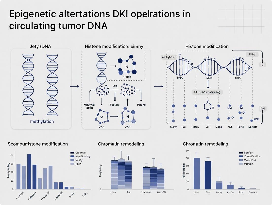
Beyond Genetics: Understanding the Core Epigenetic Marks in Circulating Tumor DNA
Circulating tumor DNA (ctDNA) analysis has transcended mutation detection to encompass the rich, information-dense layer of epigenetic regulation. As a component of a broader thesis on epigenetic alterations in oncology, this whitepaper delineates the three cornerstone epigenetic features of ctDNA: DNA methylation, hydroxymethylation, and fragmentation patterns. These alterations provide insights into tumor origin, burden, transcriptional state, and response to therapy, offering a non-invasive window into the tumor's epigenetic landscape.
Core Epigenetic Alterations: Mechanisms and Quantitative Landscape
DNA Methylation
Cytosine methylation (5-methylcytosine, 5mC) at CpG islands is a stable epigenetic mark, frequently hypermethylated at tumor suppressor gene promoters in cancer. ctDNA methylation patterns are highly cancer-type specific.
Table 1: Key Quantitative Findings in ctDNA Methylation
| Cancer Type | Target(s) | Reported Sensitivity | Reported Specificity | Primary Application | Key Study (Year) |
|---|---|---|---|---|---|
| Colorectal Cancer | SEPT9 (plasma) | 68-72% | 80-99% | Early Detection | Lofton-Day et al. (2008) |
| Lung Cancer | SHOX2, PTGER4 | 60-90% (v by stage) | 90-96% | Diagnosis & Monitoring | Dietrich et al. (2020) |
| Multi-Cancer | Pan-cancer methylation panels (1000+ CpGs) | 50-80% (v by cancer) | >99% | Cancer Signal Origin | Liu et al., CCGA (2020) |
| Hepatocellular Carcinoma | RASSP1A, p16INK4a | ~85% | ~95% | Early Detection & Prognosis | Wong et al. (2020) |
DNA Hydroxymethylation
Ten-eleven translocation (TET) enzyme-mediated oxidation of 5mC to 5-hydroxymethylcytosine (5hmC) is an intermediate in active demethylation. The distribution of 5hmC in ctDNA is enriched in gene bodies of actively transcribed genes and is highly tissue-specific.
Table 2: Key Quantitative Findings in ctDNA Hydroxymethylation
| Cancer Type | Analysis Method | Key Finding | Diagnostic Performance (AUC) | Application | Key Study (Year) |
|---|---|---|---|---|---|
| Colorectal Cancer | 5hmC-Seq (genome-wide) | Distinct 5hmC signatures in gene bodies | 0.88 - 0.94 (Stage I-IV) | Early Detection & Classification | Song et al. (2021) |
| Pancreatic Cancer | 5hmC profiling | Differential 5hmC markers in metabolic pathways | 0.89 | Early Detection | Cai et al. (2021) |
| Multiple Cancers | 5hmC profiling | Tissue-of-origin mapping | 0.85 - 0.99 (v by type) | Tumor Lineage Tracing | Zeng et al. (2023) |
ctDNA Fragmentation Patterns (Fragmentomics)
The size, end motifs, and nucleosomal positioning of ctDNA fragments are non-random, reflecting the chromatin architecture of the cell of origin. Tumor-derived ctDNA is typically shorter than non-tumor cfDNA.
Table 3: Key Quantitative Findings in ctDNA Fragmentation
| Pattern Feature | Technical Measure | Typical Value in ctDNA vs. Healthy cfDNA | Primary Application | Key Study (Year) |
|---|---|---|---|---|
| Fragment Size | Peak of size distribution | ~166 bp (healthy) vs. ~144 bp (ctDNA) | Cancer Detection | Underhill et al. (2016) |
| Nucleosomal Positioning | Whole-genome sequencing coverage periodicity | Altered in open/active chromatin regions | Tumor Type Classification | Snyder et al. (2016) |
| End Motifs | 4-bp sequence frequency at fragment ends | Differential abundance of motifs (e.g., CCCA) | Detection & Monitoring | Jiang et al. (2020) |
| Jagged Ends | Single-strand DNA ends | Increased frequency in ctDNA | Early Detection | Mouliere et al. (2018) |
Experimental Protocols for Key Methodologies
Bisulfite Sequencing for Methylation Analysis (BSPP)
Principle: Sodium bisulfite converts unmethylated cytosines to uracil, while methylated cytosines remain unchanged. Post-PCR sequencing reveals methylation status. Detailed Protocol:
- ctDNA Extraction & Quantification: Isolate cfDNA from 3-10 mL plasma using silica-membrane or bead-based kits (e.g., QIAamp Circulating Nucleic Acid Kit). Quantify by fluorometry (e.g., Qubit HS dsDNA Assay).
- Bisulfite Conversion: Treat 10-50 ng cfDNA with sodium bisulfite (e.g., using EZ DNA Methylation-Lightning Kit). Incubate: 98°C for 8 min, 54°C for 60 min.
- Clean-up: Desalt and recover converted DNA per kit instructions.
- PCR Amplification: Design primers specific to bisulfite-converted DNA (avoiding CpG sites). Use hot-start polymerase resistant to uracil.
- Library Prep & Sequencing: For genome-wide analysis (WGBS), use library prep kits compatible with bisulfite-converted DNA (e.g., Accel-NGS Methyl-Seq). Sequence on Illumina platform with >=100x coverage for targeted panels, >=30x for WGBS.
- Bioinformatic Analysis: Align reads to a bisulfite-converted reference genome (e.g., using Bismark or BWA-meth). Calculate methylation percentage per CpG site.
Chemical Capture-Based 5hmC Profiling (hMe-Seal)
Principle: 5hmC is selectively glucosylated and biotin-tagged via β-GT enzyme for pull-down and sequencing. Detailed Protocol:
- DNA Preparation & Denaturation: 50-100 ng cfDNA is denatured to single strands.
- Glucosylation & Labeling: Incubate with T4 β-glucosyltransferase (β-GT) and UDP-6-N3-glucose to add an azide-glucose to 5hmC.
- Click Chemistry: React the azide group with a biotin alkyne (e.g., DBCO-PEG4-Biotin) via copper-free click chemistry.
- Pull-down: Capture biotinylated 5hmC-containing fragments using streptavidin magnetic beads. Wash stringently.
- Elution & Library Construction: Elute captured DNA, construct sequencing libraries (e.g., using KAPA HyperPrep), and amplify.
- Sequencing & Analysis: Sequence on Illumina. Align reads, call peaks, and annotate to gene bodies and enhancers.
Whole-Genome Sequencing for Fragmentation Analysis
Principle: Low-coverage WGS reveals fragment length distributions, nucleosomal patterns, and end motifs. Detailed Protocol:
- Library Preparation (PCR-free): Use 10-30 ng cfDNA with a PCR-free library prep kit (e.g., Illumina TruSeq Nano) to avoid amplification bias in size distribution.
- Size Selection: Perform double-sided bead-based size selection (e.g., 100-220 bp) to enrich for mononucleosomal fragments.
- Shallow Sequencing: Sequence to a low depth (~0.5-5x genome coverage) on an Illumina platform (paired-end 2x75 bp or 2x150 bp recommended).
- Bioinformatic Processing:
- Alignment: Map reads to the human reference genome (e.g., using BWA-MEM).
- Size Distribution: Compute insert size from aligned read pairs.
- Coverage Periodicity: Generate sliding window coverage of fragment midpoints or ends, perform Fourier transform to detect ~10.4 bp periodicity.
- End Motif Analysis: Extract the first/last 4 bases of each fragment, calculate frequency of all 256 possible 4-mer motifs.
Visualizations
Title: Bisulfite Sequencing Workflow for ctDNA Methylation
Title: Chemical Capture Workflow for ctDNA 5hmC Profiling
Title: Origin and Analysis of ctDNA Fragmentation Patterns
The Scientist's Toolkit: Essential Research Reagents & Materials
Table 4: Key Reagent Solutions for Epigenetic ctDNA Analysis
| Reagent/Material | Supplier Examples | Primary Function |
|---|---|---|
| cfDNA Extraction Kit | QIAGEN (QIAamp CNA), Roche (cobas cfDNA), Streck (cfDNA BCT tubes) | Stabilize blood and isolate high-integrity, inhibitor-free cfDNA from plasma. |
| Bisulfite Conversion Kit | Zymo Research (EZ DNA Methylation), Qiagen (Epitect Bisulfite) | Convert unmethylated cytosine to uracil for downstream methylation-specific analysis. |
| Methylation-specific PCR Primers | Custom designed (e.g., Methyl Primer Express Software) | Amplify bisulfite-converted DNA targeting specific hyper/hypomethylated regions. |
| T4 β-Glucosyltransferase (β-GT) | NEB, Active Motif | Enzymatically transfer glucose to 5hmC for selective chemical tagging in hMe-Seal. |
| UDP-6-N3-Glucose | Berry & Associates, Jena Bioscience | Glucose donor with azide group for click chemistry conjugation to 5hmC. |
| DBCO-PEG4-Biotin | Click Chemistry Tools, Sigma-Aldrich | Biotin label that reacts with azide via copper-free click chemistry for streptavidin pull-down. |
| Streptavidin Magnetic Beads | Thermo Fisher (Dynabeads), NEB | Solid-phase capture of biotinylated 5hmC-DNA fragments. |
| PCR-free WGS Library Prep Kit | Illumina (TruSeq Nano), Roche (KAPA HyperPrep) | Prepare sequencing libraries without PCR amplification bias for accurate fragmentomics. |
| Methylation-aware Aligner (Software) | Bismark, BWA-meth, BS-Seeker2 | Map bisulfite-converted sequencing reads to a reference genome for methylation calling. |
| Fragmentomics Analysis Pipeline | in-house scripts, Fragmentomes, ichorCNA | Analyze WGS data for size, coverage, periodicity, and end motif features. |
This whitepaper addresses a fundamental question within the broader thesis on epigenetic alterations in circulating tumor DNA (ctDNA): the mechanisms governing the entry of tumor-derived nucleosomes and cell-free DNA (cfDNA) fragments into the bloodstream. Understanding these biological sources is critical for interpreting ctDNA methylation patterns, fragmentomics, and nucleosome positioning data, which are central to cancer detection, monitoring, and therapeutic resistance studies.
Primary Mechanisms of Release
Current research indicates that cfDNA and nucleosomes enter circulation through a combination of passive and active processes, often correlated with tumor biology and microenvironment.
Passive Release Mechanisms
This occurs due to cellular degradation without dedicated signaling.
- Necrosis: Unregulated cell death leads to membrane rupture and spillage of cytoplasmic and nuclear contents, including fragmented DNA and nucleosomes, into the interstitium, eventually reaching vasculature.
- Physical Destruction: Tumor manipulation (e.g., surgery, biopsy), mechanical stress from tumor growth, or erosion of vascular walls can directly release cellular material.
Active Release Mechanisms
These are biologically regulated processes.
- Apoptosis: The predominant source of cfDNA in both health and disease. Caspase-activated DNase (CAD) cleaves DNA at linker regions between nucleosomes, producing mono- and oligo-nucleosomal fragments (~166 bp multiples). These fragments are packaged into apoptotic bodies, which may be phagocytosed or, in the high-turnover tumor microenvironment, reach circulation.
- NETosis: While primarily a neutrophil process, some cancer cells can expel chromatin webs.
- Active Secretion: Vesicle-mediated (exosomes) or protein-complex mediated export. Chromatin can be released in autophagic vesicles or via binding to heat shock proteins.
Tumor Microenvironment (TME) Factors
The TME critically facilitates entry into circulation.
- Angiogenesis: New, leaky vasculature with incomplete endothelial linings allows macromolecules and nucleosomal complexes to intravasate more readily.
- Increased Vascular Permeability: Mediated by factors like VEGF, bradykinin, and cytokines.
- Dysfunctional Lymphatic Drainage: Common in solid tumors, leading to increased interstitial pressure and eventual vascular seepage.
- Immune Cell Activity: Cytotoxic T-cells and NK cells inducing cancer cell killing (apoptosis/necrosis) contribute to the pool.
Table 1: Characteristics of cfDNA from Different Release Mechanisms
| Release Mechanism | Primary Fragment Size (bp) | Nucleosome Integrity | Relative Abundance in Cancer | Key Signature |
|---|---|---|---|---|
| Apoptosis | ~166, and multiples (e.g., 332, 498) | High; well-protected in apoptotic bodies | High (Majority) | Strong 10.4 bp periodicity in sequencing; clear nucleosome patterns. |
| Necrosis | Broad smear, > 10,000 bp | Low; random degradation | Moderate | Longer fragments, ends with non-ligated overhangs. |
| Active Secretion | Variable, often < 166 bp | Variable; may be complexed with proteins | Low | Associated with exosomal markers (e.g., CD63), specific protein complexes. |
| NETosis | ~ 15-200 bp, and long strands | Low; decondensed chromatin | Context-dependent | Presence of citrullinated histones (e.g., H3Cit). |
Table 2: Factors Influencing ctDNA Concentration in Plasma
| Biological Factor | Correlation with ctDNA Level | Example Quantitative Impact (Range) |
|---|---|---|
| Tumor Stage | Positive | Stage I: <0.1% VAF; Stage IV: often >1% VAF. |
| Tumor Burden | Positive | ~0.1-10% of total cfDNA in metastatic disease. |
| Tumor Type | Variable | High: Pancreatic, ovarian, colorectal. Low: Glioblastoma, renal. |
| Cell Turnover Rate | Positive | High-grade tumors release more. |
| Treatment Response | Dynamic | Effective therapy can lead to rapid 10-100x decrease in ctDNA. |
| TME Vascularity | Positive | VEGF levels correlate with cfDNA concentration. |
Experimental Protocols for Studying Release Mechanisms
Protocol 1: In Vitro Modeling of Apoptotic cfDNA Release
- Objective: To generate and characterize nucleosomal cfDNA from cancer cell lines.
- Methodology:
- Culture adherent cancer cells (e.g., HCT-116, MCF-7) to 70% confluence.
- Induce apoptosis using Staurosporine (1 µM) or Tumor Necrosis Factor-alpha (TNF-α, 50 ng/mL) + Cycloheximide (10 µg/mL) for 6-24 hours.
- Confirm apoptosis via flow cytometry (Annexin V/PI staining).
- Collect conditioned medium, centrifuge at 2000 x g to remove cells, then at 16,000 x g to remove debris.
- Isolate cfDNA using a silica-membrane column kit (e.g., QIAamp Circulating Nucleic Acid Kit). Elute in 20 µL.
- Analyze fragment size distribution using high-sensitivity microfluidic electrophoresis (e.g., Agilent Bioanalyzer 2100, High Sensitivity DNA kit).
- Perform shallow whole-genome sequencing (sWGS, ~0.1x coverage) to assess fragment length periodicity and genomic coverage patterns.
Protocol 2: Profiling Endogenous Nucleosome Protection in Plasma
- Objective: To map in vivo nucleosome positions from plasma cfDNA.
- Methodology:
- Collect 5-10 mL of patient blood in Streck Cell-Free DNA BCT tubes. Process within 6 hours: double centrifugation (1600 x g, 10 min; 16,000 x g, 10 min).
- Extract cfDNA from 2-4 mL plasma (as in Protocol 1, step 5).
- Prepare sequencing library using an enzyme-based method (e.g., NEBNext Ultra II FS) to minimize GC/size bias.
- Perform paired-end sequencing (e.g., 2x75 bp) to ~5-10 million reads per sample.
- Bioinformatic Analysis:
- Align reads to reference genome (e.g., hg38).
- Calculate fragment size distribution with 1 bp resolution.
- Compute "windowed protection scores" (WPS) as described by Snyder et al., Cell, 2016: For each genomic position, count the number of fragments that span it (length >120 bp) minus those that have both ends covering it.
- Identify nucleosome-depleted regions (NDRs) at transcription start sites (TSS) and regulatory elements, correlating with chromatin accessibility in the tissue of origin.
Diagrams of Key Pathways and Workflows
Diagram Title: Pathways of Tumor DNA Release and Intravasation
Diagram Title: Nucleosome Mapping from Plasma cfDNA Workflow
The Scientist's Toolkit: Research Reagent Solutions
Table 3: Essential Reagents and Kits for ctDNA Release Studies
| Item / Reagent | Function / Application | Key Considerations |
|---|---|---|
| Streck Cell-Free DNA BCT Tubes | Blood collection tube that stabilizes nucleated blood cells to prevent ex vivo lysis, preserving the native cfDNA profile. | Critical for pre-analytical standardization. Allows room-temperature shipping. |
| QIAamp Circulating Nucleic Acid Kit (Qiagen) | Silica-membrane based extraction of cfDNA from plasma/serum. Optimized for low-concentration, small-fragment recovery. | High and consistent recovery of fragments <100 bp is essential for nucleosome studies. |
| NEBNext Ultra II FS DNA Library Prep Kit | Enzyme-based (fragmentation & tailing) library preparation for Illumina. Minimizes GC bias and retains true fragment length distribution. | Preferred over sonication-based methods for preserving endogenous fragment ends. |
| Agilent High Sensitivity DNA Kit (Bioanalyzer) | Microfluidic electrophoresis for precise sizing and quantification of cfDNA extracts and libraries. Confirms the ~166 bp peak. | Quality control step to assess sample integrity and library size distribution. |
| Annexin V-FITC / PI Apoptosis Detection Kit | Flow cytometry assay to quantify apoptotic and necrotic cells in in vitro culture models of cfDNA release. | Validates the cellular mechanism being modeled in experiments. |
| Recombinant Human VEGF | Used in in vitro or in vivo models to induce angiogenesis and increase vascular permeability, studying its effect on cfDNA intravasation. | Models a key Tumor Microenvironment factor. |
| Cell Death Inducers (e.g., Staurosporine, TNF-α) | Pharmacological agents to induce specific death pathways (apoptosis/necrosis) in cultured tumor cells for conditioned medium collection. | Allows controlled study of release from a defined mechanism. |
| Proteinase K / RNase A | Enzymatic digestion during cfDNA extraction to degrade proteins and RNA, respectively, ensuring pure DNA isolation. | Essential for removing nucleosomal proteins if studying DNA alone. |
Within the evolving paradigm of circulating tumor DNA (ctDNA) research, the analysis of epigenetic alterations, particularly DNA methylation, has emerged as a critical complement to the detection of somatic genetic mutations. This whitepaper details the core advantages of ctDNA methylation biomarkers, emphasizing their tissue-of-origin specificity and their high frequency across tumor types, often surpassing that of recurrent point mutations. These attributes position methylation analysis as a powerful tool for non-invasive cancer detection, monitoring, and drug development.
Table 1: Comparative Frequency of Aberrant Methylation vs. Recurrent Genetic Mutations in Major Cancers
| Cancer Type | High-Frequency Methylated Genes (Prevalence) | Example High-Frequency Point Mutation (Prevalence) | Key Reference |
|---|---|---|---|
| Colorectal Cancer (CRC) | SEPT9 (73-95%), NDRG4 (64-78%), BMP3 (45-90%) | APC (~80%), TP53 (~60%), KRAS (~40%) | Song, L. et al. Clin Epigenetics. 2022. |
| Lung Cancer | SHOX2 (60-78%), PTGER4 (51-68%), RASSF1A (40-70%) | TP53 (~50%), EGFR (~15-40%) | Hulbert, A. et al. Nat Rev Cancer. 2017. |
| Hepatocellular Carcinoma (HCC) | RASSF1A (70-85%), GSTP1 (65-90%), APC (60-80%) | TERT promoter (~60%), TP53 (~30%) | Kisiel, J.B. et al. Gastroenterology. 2019. |
| Breast Cancer | RASSF1A (50-80%), ESR1 (20-40%), BRCA1 (10-30%) | PIK3CA (~40%), TP53 (~30%) | Luo, H. et al. Genome Med. 2020. |
| Prostate Cancer | GSTP1 (~90%), APC (40-80%), RASSF1A (40-70%) | SPOP (~10%), TP53 (~20%) | Van Neste, L. et al. J Urol. 2016. |
Table 2: Tissue-of-Origin Specificity of Methylation Markers in ctDNA
| Methylation Marker Panel | Primary Tissue/Cancer of Origin | Specificity vs. Other Cancers | Application in ctDNA |
|---|---|---|---|
| SEPT9, NDRG4, BMP3 | Colorectal Epithelium / CRC | >95% | Blood-based screening (Epi proColon) |
| SHOX2, PTGER4 | Lung Epithelium / Lung Cancer | ~90% | Discrimination of malignant pulmonary nodules |
| HOXA9, AJAP1 | Bladder Urothelium / Urothelial Ca. | >85% | Surveillance for recurrence |
| GSTP1, HAPLN3 | Prostate Epithelium / Prostate Ca. | ~88% | Complementary to PSA screening |
| RASSF1A, GSTP1, APC | Hepatocytes / HCC | ~90% | Surveillance in cirrhotic patients |
Detailed Methodologies for Key Experiments
Protocol: Targeted Bisulfite Sequencing for ctDNA Methylation Analysis
Objective: To quantitatively assess methylation status of multiple CpG sites in candidate genes from plasma-derived ctDNA.
Workflow:
- Plasma Isolation & DNA Extraction: Isolate 2-10 mL of patient plasma via double centrifugation (1,600 x g, 10 min; then 16,000 x g, 10 min). Extract total cell-free DNA (cfDNA) using magnetic bead-based kits (e.g., QIAamp Circulating Nucleic Acid Kit).
- Bisulfite Conversion: Treat 10-50 ng of cfDNA with sodium bisulfite using a dedicated kit (e.g., EZ DNA Methylation-Lightning Kit, Zymo Research). This converts unmethylated cytosine to uracil, while methylated cytosine remains unchanged.
- Library Preparation (Targeted Amplification): Perform multiplex PCR using primers designed for bisulfite-converted DNA, targeting regions of interest (e.g., promoters of SEPT9, SHOX2). Use polymerases robust to uracil templates (e.g., KAPA HiFi HotStart Uracil+).
- Next-Generation Sequencing (NGS): Barcode libraries and sequence on platforms like Illumina MiSeq/NextSeq. Use a minimum depth of 10,000x per amplicon to reliably detect low-frequency methylation.
- Bioinformatic Analysis:
- Alignment: Map reads to a bisulfite-converted reference genome using tools like Bismark or BWA-meth.
- Methylation Calling: For each CpG site, calculate the methylation percentage as:
(Number of reads reporting "C") / (Number of reads reporting "C" + "T") * 100. - Statistical Thresholding: Define a positive sample using a threshold (e.g., mean methylation >10% across a panel of CpGs) validated against healthy controls.
Protocol: Methylation-Specific Droplet Digital PCR (ddPCR) for Ultrasensitive Detection
Objective: To achieve absolute quantification of a specific methylated allele with single-molecule sensitivity, ideal for low-ctDNA fraction scenarios.
Workflow:
- cfDNA Extraction & Bisulfite Conversion: As in 3.1.
- Assay Design: Design two primer/probe sets:
- Methylated (M) Assay: Forward and reverse primers amplify only if the CpG site(s) within the primer-binding sequence is methylated (remains "CG" after conversion). Use a FAM-labeled probe.
- Reference (R) Assay: Targets a genomic region devoid of CpG sites (e.g., ACTB), thus amplifying all DNA fragments. Use a HEX/VIC-labeled probe.
- Droplet Generation & PCR: Combine bisulfite-converted DNA, ddPCR Supermix for Probes (Bio-Rad), and M/R assays. Generate ~20,000 droplets using a droplet generator. Perform endpoint PCR.
- Droplet Reading & Analysis: Read droplets on a droplet reader. Positive droplets for FAM (methylated) and HEX (total) are counted. The concentration (copies/μL) is calculated using Poisson statistics.
- Result Interpretation: Calculate fractional abundance:
[M] / ([R]/2) * 100%. A sample is considered positive if the methylated concentration exceeds a limit of detection (LOD) established from healthy donor plasma (typically >0.01-0.1% fractional abundance).
Visualizations
Diagram 1: ctDNA Methylation Analysis Workflow
Diagram 2: Methylation vs. Mutation in ctDNA Biomarker Development
The Scientist's Toolkit: Research Reagent Solutions
Table 3: Essential Materials for ctDNA Methylation Studies
| Item | Function & Rationale | Example Product(s) |
|---|---|---|
| cfDNA Stabilization Tubes | Preserves cell-free DNA profile by inhibiting nucleases and stabilizing blood cells during transport/pre-processing. Critical for reproducible methylation results. | Streck Cell-Free DNA BCT tubes, Roche Cell-Free DNA Collection Tubes. |
| Magnetic Bead-based cfDNA Kits | High-efficiency, scalable extraction of short-fragment cfDNA with minimal contamination from genomic DNA. | QIAamp Circulating Nucleic Acid Kit (Qiagen), MagMAX Cell-Free DNA Isolation Kit (Thermo Fisher). |
| Bisulfite Conversion Kits | Efficient and complete conversion of unmethylated cytosine to uracil with minimal DNA degradation (<90% recovery). | EZ DNA Methylation-Lightning Kit (Zymo Research), MethylEdge Bisulfite Conversion System (Promega). |
| Uracil-Tolerant Polymerase | Essential for PCR amplification of bisulfite-converted DNA, which contains uracil residues. Standard Taq polymerases are inhibited. | KAPA HiFi HotStart Uracil+ (Roche), Pfu Turbo Cx Hotstart (Agilent). |
| Targeted Methylation Panels | Predesigned, multiplexed assays for simultaneous amplification and sequencing of multiple genomic regions post-bisulfite conversion. | Illumina TruSight Oncology Methyl, Twist Custom Methylation Panels. |
| ddPCR Methylation Assays | FAM/HEX-labeled probe-based assays for absolute quantification of specific methylated alleles without need for standard curves. | Bio-Rad ddPCR Methylation Assays (Custom/PrimePCR). |
| Bisulfite Sequencing Control DNA | Pre-methylated and unmethylated genomic DNA standards for benchmarking conversion efficiency and assay sensitivity/specificity. | EpiTect Control DNA (Qiagen), CpGenome Universal Methylated DNA (Merck). |
| Bioinformatics Pipelines | Software packages for alignment, methylation calling, and differential analysis from bisulfite-seq data. | Bismark, MethylKit (R/Bioconductor), SeSAMe. |
Major Cancer-Associated Methylation Markers and Pan-Cancer Epigenetic Signatures
This whitepaper details core methylation markers and pan-cancer signatures, forming a technical foundation for a broader thesis on epigenetic alterations in circulating tumor DNA (ctDNA). The accurate detection of these signatures in plasma is revolutionizing liquid biopsy applications for early detection, minimal residual disease monitoring, and therapy selection.
Core Cancer-Associated Methylation Markers
DNA methylation, primarily the addition of a methyl group to cytosine in CpG dinucleotides, is a stable epigenetic mark frequently dysregulated in cancer. Hypermethylation of tumor suppressor gene promoters and global hypomethylation are hallmarks of oncogenesis.
Table 1: Key Validated Methylation Markers in Major Cancers
| Cancer Type | Key Methylated Gene(s) | Function of Gene | Clinical Application Context | Detection in ctDNA |
|---|---|---|---|---|
| Colorectal Cancer (CRC) | SEPT9, NDRG4, BMP3 | Cell cycle, differentiation | FDA-approved for screening (Epi proColon) | Well-validated |
| Lung Cancer | SHOX2, PTGER4, RASSF1A | Apoptosis, proliferation | Diagnosis, prognosis | High sensitivity/specificity |
| Breast Cancer | RASSF1A, GSTP1, BRCA1 | DNA repair, signaling | Risk assessment, monitoring | Actively researched |
| Prostate Cancer | GSTP1 (↑ 90% in CaP) | Detoxification | Differential diagnosis from benign | High specificity |
| Glioblastoma | MGMT promoter methylation | DNA repair | Predictor of response to temozolomide | Limited in ctDNA (CNS) |
| Pan-Cancer | TERT promoter mutations | Telomerase activation | Common in multiple cancers | Highly detectable |
Pan-Cancer Epigenetic Signatures
Pan-cancer signatures refer to common methylation patterns across multiple tumor types, useful for cancer detection of unknown origin and understanding shared oncogenic pathways.
Table 2: Representative Pan-Cancer Methylation Signatures
| Signature Name/Type | Core Loci/Regions | Technical Approach for Discovery | Potential Utility |
|---|---|---|---|
| CpG Island Methylator Phenotype (CIMP) | ~10-200+ CpG sites (e.g., CACNA1G, IGF2, NEUROG1, RUNX3, SOCS1) | Methylation-specific PCR (MSP) or BeadChip | Subclassification, prognosis |
| Epigenetic Age Acceleration | Clock CpGs (e.g., Horvath's 353 CpG clock) | Pyrosequencing or array | Risk prediction, biology of aging |
| Cell-of-Origin Signatures | Tissue-specific differentially methylated regions (tDMRs) | Whole-genome bisulfite sequencing (WGBS) | Identifying primary site for cancers of unknown origin |
| Plasma-Based Multi-Cancer Early Detection (MCED) Panels | 100,000+ informative CpGs (e.g., cfMeDIP-seq targets) | cfMeDIP-seq, targeted methylation sequencing | Early detection across >50 cancer types |
Experimental Protocols for Key Methodologies
Protocol: Sodium Bisulfite Conversion for ctDNA
Principle: Converts unmethylated cytosines to uracil, while methylated cytosines remain as cytosine, enabling discrimination via sequencing or PCR.
- Input: 5-50 ng of purified ctDNA (e.g., from 1-5 mL plasma).
- Bisulfite Reaction: Use a commercial kit (e.g., EZ DNA Methylation-Lightning Kit). Incubate DNA in bisulfite reagent at 98°C for 10 min, then 54°C for 60 min.
- Desalting: Bind DNA to a spin column, wash with buffer.
- Desulfonation: Treat with desulfonation buffer for 20 min at room temperature.
- Elution: Elute converted DNA in 10-20 µL of elution buffer. Store at -80°C.
Protocol: Targeted Methylation Sequencing (Bisulfite Capture)
Principle: Enrichment of bisulfite-converted DNA at regions of interest followed by NGS.
- Library Preparation: Prepare sequencing libraries from bisulfite-converted DNA using adapters compatible with bisulfite-treated fragments.
- Hybridization Capture: Hybridize libraries to biotinylated RNA or DNA baits designed for CpG regions of interest (e.g., a pan-cancer panel). Incubate at 65°C for 16-24 hours.
- Capture Bead Binding: Bind biotinylated DNA:RNA hybrids to streptavidin magnetic beads. Wash stringently.
- Amplification & Sequencing: PCR-amplify captured DNA. Perform paired-end sequencing on an Illumina platform.
- Bioinformatics: Align reads to a bisulfite-converted reference genome (e.g., using Bismark). Calculate methylation beta-values (methylated reads / total reads) per CpG.
Visualization of Key Concepts
Title: Tumor Suppressor Gene Silencing via Promoter Hypermethylation
Title: ctDNA Methylation Analysis Experimental Workflow
The Scientist's Toolkit: Research Reagent Solutions
Table 3: Essential Materials for ctDNA Methylation Studies
| Item/Category | Example Product | Function & Critical Notes |
|---|---|---|
| ctDNA Isolation Kits | QIAamp Circulating Nucleic Acid Kit, MagMAX Cell-Free DNA Isolation Kit | Efficient recovery of short, fragmented ctDNA from plasma/serum. Minimizes genomic DNA contamination. |
| Bisulfite Conversion Kits | EZ DNA Methylation-Lightning Kit, Epitect Fast DNA Bisulfite Kit | Complete and rapid conversion with high DNA recovery. Critical for low-input ctDNA. |
| Methylation-Specific qPCR Assays | EpiTect MSP Kit, predesigned TaqMan Methylation Assays | Quantitative detection of methylation at single loci. Used for validation. |
| Targeted Methylation Panels | Illumina TruSight Oncology 500 (includes methylation), Agilent SureSelect Methyl-Seq | Designed bait sets for capturing cancer-relevant CpGs from bisulfite-converted libraries. |
| Whole-Genome Bisulfite Sequencing Kits | Accel-NGS Methyl-Seq DNA Library Kit | For unbiased discovery of novel methylation signatures. Requires higher input. |
| Methylation Arrays | Infinium MethylationEPIC BeadChip | Profiles >850,000 CpGs. Suitable for cell line/tissue discovery; less common for low-input ctDNA. |
| Bioinformatics Software | Bismark, MethylKit, SeSAMe | Alignment, differential methylation analysis, and quality control for bisulfite sequencing/array data. |
| Methylation Plasma Controls | Horizon Discovery cfDNA Methylation Reference Standards | Synthetic ctDNA with defined methylation patterns for assay validation and QC. |
From Blood Sample to Biomarker: Assays and Clinical Applications of Epigenetic ctDNA Analysis
Within the investigation of epigenetic alterations in circulating tumor DNA (ctDNA), the precise mapping of DNA methylation patterns is paramount. ctDNA, shed into the bloodstream by tumors, carries the cancer's epigenetic signature, offering a non-invasive reservoir for biomarker discovery and monitoring. This technical guide details three core technologies—Bisulfite Sequencing, quantitative Methylation-Specific PCR (qMSP), and Bead Array Platforms—that form the cornerstone of ctDNA methylation analysis, enabling researchers and drug development professionals to detect, quantify, and profile these critical epigenetic modifications.
Table 1: Core Characteristics of Methylation Detection Technologies
| Feature | Bisulfite Sequencing | Quantitative MSP (qMSP) | Bead Array Platforms (e.g., Infinium) |
|---|---|---|---|
| Primary Application | Genome-wide discovery & single-base resolution profiling | Targeted, high-sensitivity quantification of specific loci | Multiplexed, intermediate-resolution profiling (450K-900K CpG sites) |
| Throughput | Low to High (scalable with NGS) | High (96-384 well plates) | Very High (hundreds of samples per run) |
| Sensitivity | ~1-5% allele frequency (dependent on depth) | 0.1-0.01% (optimal for low-concentration ctDNA) | ~1-5% (dependent on probe design and signal processing) |
| DNA Input Requirement | High (50-100ng for WGBS); Lower for RRBS | Very Low (1-20ng) | Moderate (250-500ng) |
| Quantitative Output | Yes (from read counts) | Yes (standard curve or ΔΔCq) | Semi-quantitative (beta values: 0-1) |
| Cost per Sample | High (WGBS) to Moderate (Targeted) | Low | Moderate |
| Best Suited for ctDNA | Discovery of novel markers; fragmentation-aware protocols | Validated marker detection & minimal residual disease (MRD) monitoring | Methylation subtype classification; signature validation |
Table 2: Performance Metrics in ctDNA Context
| Metric | Bisulfite Sequencing (Targeted) | qMSP | Bead Array (EPIC) |
|---|---|---|---|
| Limit of Detection (LoD) | ~1% Methylation Allele Frequency | 0.01-0.1% Methylation Allele Frequency | ~1-3% Methylation Beta Value |
| CpGs Interrogated per Assay | 10s - 1000s (design-dependent) | 1-5 (single amplicon) | >850,000 |
| Turnaround Time (Hands-on) | Moderate-High | Low | Moderate |
| Compatibility with FFPE DNA | Yes (with quality control) | Yes (robust) | Yes (with restoration) |
| Multiplexing Capability | High (sequencing-based) | Low (typically singleplex/duplex) | Inherently High |
Detailed Methodologies and Protocols
Sodium Bisulfite Conversion and Sequencing
This foundational pretreatment deaminates unmethylated cytosine to uracil, while methylated cytosine (5mC) remains unchanged, creating sequence differences that mark methylation status.
Protocol: Sodium Bisulfite Conversion for Low-Input ctDNA
- DNA Denaturation: Mix 5-20ng of purified ctDNA with 5µL of 2M NaOH. Incubate at 65°C for 15 minutes.
- Sulfonation: Prepare a fresh bisulfite mix (e.g., from EZ DNA Methylation kits): 530µL of CT Conversion Reagent, 300µL of M-Dilution Buffer, and 270µL of water per sample. Combine with denatured DNA.
- Thermal Cycling: Perform conversion: 98°C for 10 minutes, 64°C for 2.5 hours. Use a thermal cycler with a heated lid.
- Desalting/Binding: Transfer mixture to a spin column containing binding buffer. Centrifuge at full speed for 1 minute.
- Desulfonation: Prepare fresh Desulfonation Buffer (3M NaOH). Add 200µL to column, incubate at room temperature for 20 minutes. Centrifuge.
- Washing & Elution: Wash twice with 200µL of Wash Buffer. Elute converted DNA in 10-20µL of Elution Buffer.
- Library Preparation & Sequencing: For Whole-Genome Bisulfite Sequencing (WGBS), use specialized library prep kits that preserve converted strands. For targeted approaches (e.g., bisulfite padlock probes or AmpliSeq methyl panels), perform hybrid capture or multiplex PCR followed by NGS on Illumina platforms. Align reads to a bisulfite-converted reference genome using tools like Bismark or BWA-meth.
Quantitative Methylation-Specific PCR (qMSP)
qMSP utilizes primers designed to amplify only the converted methylated sequence, providing highly sensitive detection.
Protocol: qMSP for ctDNA Biomarker Quantification
- Bisulfite Conversion: As described above. Include positive controls (fully methylated DNA) and negative controls (unmethylated DNA and no-template controls).
- Primer/Probe Design: Design forward and reverse primers complementary to the CpG-rich region of interest after bisulfite conversion of methylated cytosines. Ideally, 3' ends should cover CpG sites to maximize specificity. Use TaqMan probes or SYBR Green.
- qPCR Setup: Prepare reaction mix: 10µL of 2x qPCR Master Mix, 0.5-1.0µL of each primer (10µM), 0.25-0.5µL of probe (10µM), 2µL of bisulfite-converted DNA (or standard), and nuclease-free water to 20µL.
- Standard Curve Preparation: Serial dilute a synthetic oligonucleotide matching the fully methylated, converted target sequence (e.g., from 10^6 to 10^1 copies per reaction).
- Thermal Cycling: Conditions: 95°C for 10 min (activation), then 45 cycles of 95°C for 15 sec and 60°C for 60 sec (annealing/extension).
- Data Analysis: Determine quantification cycle (Cq) values. Plot standard curve (log copy number vs. Cq). Quantify methylated target copy number in unknowns. Normalize to a reference gene (e.g., ACTB) amplified with non-MSP primers to account for input DNA quantity.
Bead Array Methylation Profiling
The Illumina Infinium Methylation Assay uses bead-chip technology for large-scale CpG site interrogation.
Protocol: Infinium MethylationEPIC v2.0 Workflow
- DNA Quality Control: Quantify 250-500ng of DNA using fluorescence-based assays (e.g., Qubit). Ensure integrity (DV200 >70% for FFPE).
- Bisulfite Conversion: Use the Zymo EZ DNA Methylation kit or equivalent, scaling for 500ng input. Elute in a minimal volume.
- Whole-Genome Amplification & Enzymatic Fragmentation: Combine converted DNA with MA1/2 reagents for isothermal amplification (20-24 hours at 37°C). Add FMS reagent to enzymatically fragment the amplified product (1 hour at 37°C).
- Precipitation & Resuspension: Precipitate DNA with isopropanol. Pellet, wash, and resuspend in RA1 buffer.
- Hybridization: Apply resuspended DNA onto the BeadChip (8- or 16-sample format). Seal and hybridize in a chamber for 16-24 hours at 48°C.
- Single-Base Extension & Staining: Perform an extension step where labeled nucleotides are incorporated based on the methylation state at the queried CpG. Subsequently, stain the BeadChip with fluorescent dyes.
- Imaging & Analysis: Image the BeadChip using an iScan or NextSeq scanner. Process intensity data (*.idat files) with GenomeStudio or R/Bioconductor packages (
minfi,sesame) to generate beta values (β = IntensityMethylated / (IntensityMethylated + Intensity_Unmethylated + 100)).
Visualizations
Title: Technology Selection Workflow for ctDNA Methylation Analysis
Title: qMSP Principle: Selective Amplification of Methylated Alleles
Title: Infinium Bead Array Methylation Detection and Quantification
The Scientist's Toolkit: Research Reagent Solutions
Table 3: Essential Reagents for ctDNA Methylation Analysis
| Item | Function | Example Product/Kit |
|---|---|---|
| ctDNA Isolation Kit | Selective isolation of cell-free DNA from plasma, optimized for short fragments. | QIAamp Circulating Nucleic Acid Kit, MagMAX Cell-Free DNA Isolation Kit |
| Bisulfite Conversion Kit | Chemical conversion of unmethylated cytosines to uracil while preserving 5mC. Critical first step. | EZ DNA Methylation Kit (Zymo), MethylEdge Bisulfite Conversion System (Promega) |
| Methylated/Unmethylated Control DNA | Positive and negative controls for bisulfite conversion and assay optimization. | CpGenome Universal Methylated DNA (Millipore), Human HCT116 DKO Unmethylated DNA |
| qMSP Primers & Probes | Sequence-specific oligonucleotides for amplification of converted methylated DNA. | Custom-designed TaqMan Methylation Assays (Thermo Fisher), LNA-enhanced primers |
| NGS Library Prep Kit for Bisulfite DNA | Preparation of sequencing libraries from bisulfite-converted DNA, minimizing bias. | Accel-NGS Methyl-Seq DNA Library Kit (Swift), Pico Methyl-Seq Library Prep Kit (Zymo) |
| Infinium Methylation BeadChip | Array platform for high-throughput methylation profiling of >850,000 CpG sites. | Illumina Infinium MethylationEPIC v2.0 BeadChip |
| Methylation-Sensitive Restriction Enzymes (MSRE) | For alternative/qc approaches; cleave unmethylated recognition sites. | HpaII, McrBC |
| Digital PCR Master Mix | For absolute quantification of methylated alleles without standard curves, enhances sensitivity. | ddPCR Supermix for Probes (Bio-Rad) |
Within the rapidly advancing field of cancer epigenetics, the analysis of circulating tumor DNA (ctDNA) presents a formidable challenge due to its low abundance in a high background of normal cell-free DNA. Epigenetic alterations, particularly DNA methylation, are highly promising biomarkers for cancer detection, monitoring, and therapy guidance. This whitepaper provides an in-depth technical guide to two pivotal, high-sensitivity methods enabling this research: Targeted Methylation Sequencing and Whole-Genome Bisulfite Sequencing. Both techniques are critical for decoding the methylome of ctDNA, thereby advancing the broader thesis that epigenetic profiling of ctDNA offers unparalleled specificity for tumor origin and biology.
Core Methodologies and Comparative Analysis
Whole-Genome Bisulfite Sequencing (WGBS)
Principle: WGBS is considered the gold standard for unbiased, genome-wide methylation profiling. It involves treating genomic DNA with sodium bisulfite, which converts unmethylated cytosines to uracils (read as thymines after PCR), while methylated cytosines remain unchanged. Subsequent high-coverage sequencing allows for the quantitative mapping of methylated cytosines at single-nucleotide resolution.
Key Protocol Steps:
- Input DNA Fragmentation: ctDNA or genomic DNA is fragmented to ~200-300bp (e.g., via sonication or enzymatic digestion).
- Bisulfite Conversion: Fragments are treated with sodium bisulfite (e.g., using EZ DNA Methylation-Gold Kit). Critical parameters: incubation temperature (64°C) and time (2.5-5 hours) to ensure complete conversion while minimizing DNA degradation.
- Library Preparation: Converted DNA undergoes desulfonation, followed by library construction. This often involves a two-step PCR:
- Pre-Amplification: With primers compatible with bisulfite-converted DNA.
- Indexing PCR: To add unique dual indices and full sequencing adapters.
- High-Throughput Sequencing: Sequencing on platforms like Illumina NovaSeq to achieve high depth (>30x genome-wide coverage for ctDNA applications).
Advantages & Limitations:
- Advantages: Unbiased, hypothesis-free discovery; detects methylation at all CpG contexts; provides a complete methylome map.
- Limitations: Extremely high sequencing cost and data volume; requires large input DNA (a challenge for low-yield ctDNA); computationally intensive; over 90% of sequenced bases are from background normal DNA in ctDNA applications.
Targeted Methylation Sequencing
Principle: This method enriches for specific genomic regions of interest—such as differentially methylated regions (DMRs) or CpG islands hypermethylated in cancer—prior to bisulfite conversion and sequencing. Enrichment dramatically increases the sensitivity to detect rare ctDNA molecules.
Key Enrichment Strategies & Protocols:
A. Hybridization Capture-Based (e.g., Agilent SureSelect Methyl-Seq):
- Bisulfite Conversion First: Input DNA (as low as 1-10 ng ctDNA) is first converted with bisulfite.
- Library Preparation: Converted DNA is amplified and indexed.
- Target Enrichment: Biotinylated RNA baits, designed against the bisulfite-converted sequence of targets, are hybridized to the library. Streptavidin-coated magnetic beads capture the bait-bound fragments.
- Sequencing: Enriched library is sequenced at high depth (e.g., 10,000x-50,000x per base).
B. Amplification-Based (e.g., Methylation-Specific PCR or Multiplex PCR):
- Bisulfite Conversion: Input DNA is converted.
- Targeted Amplification: Primers are designed to specifically amplify methylated (or unmethylated) alleles. Highly multiplexed PCR reactions (e.g., using Fluidigm Access Array or Illumina AmpliSeq) can interrogate hundreds to thousands of loci simultaneously.
- Sequencing: Amplicons are sequenced to ultra-deep coverage.
Advantages & Limitations:
- Advantages: Ultra-high sensitivity (can detect <0.1% variant allele frequency); cost-effective; lower DNA input requirement; focused data analysis.
- Limitations: Requires a priori knowledge of target regions; discovery limited to predefined panel.
Quantitative Data Comparison
Table 1: Comparative Analysis of WGBS and Targeted Methylation Sequencing
| Parameter | Whole-Genome Bisulfite Sequencing (WGBS) | Targeted Methylation Sequencing |
|---|---|---|
| Genomic Coverage | Comprehensive, genome-wide (~28 million CpGs in human) | Focused on predefined panel (e.g., 10,000 - 1 million CpGs) |
| Typical Input DNA | 30-100 ng (high-quality); >50 ng for ctDNA applications | 1-10 ng (effective for low-input ctDNA) |
| Sequencing Depth | Moderate (30-50x genome-wide) | Ultra-deep (5,000x - 100,000x per base) |
| Approx. Cost per Sample | $1,000 - $3,000 USD | $200 - $800 USD |
| Limit of Detection (LOD) | ~5-10% tumor fraction (for ctDNA) | <0.1% tumor fraction (for ctDNA) |
| Primary Application | Discovery of novel DMRs, pan-cancer methylome atlases | Ultrasensitive detection & monitoring in liquid biopsy, MRD assessment |
| Data Output Size | Very Large (~100-150 GB per sample) | Moderate (1-10 GB per sample) |
| Key Challenge | High cost, background noise from normal DNA | Panel design bias, no discovery outside targets |
Experimental Protocol: A Representative ctDNA Methylation Workflow
Protocol Title: Ultrasensitive Detection of ctDNA Methylation Using Hybridization Capture and Sequencing
Step 1: Plasma Processing & DNA Extraction
- Collect peripheral blood in cell-stabilization tubes (e.g., Streck). Process within 6 hours.
- Double centrifugation: 1,600 x g for 10 min (4°C), then transfer plasma; 16,000 x g for 10 min (4°C) to pellet residual cells.
- Extract cell-free DNA from 2-4 mL plasma using a silica-membrane column kit (e.g., QIAamp Circulating Nucleic Acid Kit). Elute in 20-40 µL.
- Quantify using a high-sensitivity fluorometric assay (e.g., Qubit dsDNA HS Assay).
Step 2: Bisulfite Conversion
- Use 5-20 ng of extracted cfDNA.
- Convert using the EZ DNA Methylation-Lightning Kit (Zymo Research):
- Add 130 µL of Lightning Conversion Reagent to DNA. Cycle: 98°C for 8 min, 54°C for 60 min.
- Desalt and clean up using the provided column, followed by desulphonation and final elution in 10 µL.
Step 3: Converted DNA Library Preparation
- Perform end-repair, A-tailing, and adapter ligation on bisulfite-converted DNA using a dedicated methylated-adapter kit (e.g., Illumina DNA Prep with Methylation Adaptors).
- Perform limited-cycle (4-6 cycles) PCR to amplify the library.
Step 4: Target Enrichment by Hybrid Capture
- Use a commercially available pan-cancer methylation panel (e.g., Roche AVENIO cEM-Seq Kit or design a custom Agilent SureSelectXT Methyl-Seq panel).
- Hybridize the library to biotinylated RNA baits for 16-24 hours.
- Capture with streptavidin beads, wash stringently, and perform a second round of PCR (10-12 cycles) to amplify the enriched library.
Step 5: Sequencing & Data Analysis
- Pool libraries and sequence on an Illumina NextSeq 550 or NovaSeq 6000 system (2x150 bp reads) to achieve a minimum mean coverage of 10,000x across all panel regions.
- Bioinformatics Pipeline:
- Alignment: Use bisulfite-aware aligners (e.g., Bismark or BS-Seeker2) to reference genome.
- Methylation Calling: Extract methylation counts per CpG site.
- Statistical Analysis: Apply tools like MethylKit or custom scripts to identify differentially methylated regions (DMRs) between case and control samples. For ctDNA, use reference databases (e.g., from TCGA) to infer tissue of origin.
Visualizations
Diagram 1: WGBS Experimental Workflow
Diagram 2: Targeted Methyl-Seq Workflow
Diagram 3: Method Selection Logic
The Scientist's Toolkit: Key Research Reagent Solutions
Table 2: Essential Materials for ctDNA Methylation Sequencing Studies
| Category | Product Name (Example) | Key Function & Rationale |
|---|---|---|
| Blood Collection | Streck Cell-Free DNA BCT | Preservative tube that prevents leukocyte lysis, minimizing background wild-type DNA release and stabilizing ctDNA. |
| cfDNA Extraction | QIAamp Circulating Nucleic Acid Kit (Qiagen) | Optimized for low-abundance, short-fragment cfDNA from large plasma volumes. High recovery is critical. |
| Bisulfite Conversion | EZ DNA Methylation-Lightning Kit (Zymo Research) | Fast, efficient conversion with minimal DNA degradation, suitable for low-input (<10 ng) samples. |
| Library Prep (WGBS) | Accel-NGS Methyl-Seq DNA Library Kit (Swift Biosciences) | Designed for bisulfite-converted DNA, minimizing bias and maximizing complexity from low inputs. |
| Library Prep (Targeted) | Illumina DNA Prep with Methylation Adaptor Kit | Integrated workflow with methylation-aware adapters for streamlined preparation post-conversion. |
| Hybrid Capture | Agilent SureSelect Methyl-Seq Custom Kit | Enables design of custom bait panels targeting bisulfite-converted sequences for specific gene sets. |
| Integrated Panels | Roche AVENIO cEM-Seq Kit | Complete, optimized workflow from plasma to data for a predefined pan-cancer methylation marker panel. |
| Quantification | Qubit dsDNA HS Assay Kit (Thermo Fisher) | Fluorometric assay essential for accurate quantification of low-concentration DNA post-extraction and post-library prep. |
| Sequencing Control | Methylated & Non-methylated Control DNA (e.g., from Zymo) | Vital for assessing bisulfite conversion efficiency and sequencing library performance in each run. |
| Bioinformatics | Bismark Bisulfite Read Mapper & Methylation Caller | Standard tool for aligning bisulfite-treated reads and performing unbiased methylation calling. |
Early cancer detection remains a paramount challenge in oncology. Within the broader thesis of epigenetic alterations in circulating tumor DNA (ctDNA) research, MCED tests represent a transformative application. Unlike genetic mutations, epigenetic modifications—primarily DNA methylation—are highly cancer-specific, tissue-of-origin indicative, and frequently occur early in carcinogenesis. The analysis of ctDNA methylation patterns in plasma thus provides a powerful liquid biopsy approach for the simultaneous detection and localization of multiple cancer types.
Core Technological Principles: Methylation Profiling of ctDNA
Current leading MCED platforms rely on the targeted or genome-wide assessment of cytosine methylation at CpG islands. Hypermethylation of tumor suppressor gene promoters and hypomethylation of oncogenic regions are hallmark epigenetic alterations captured from plasma.
Key Analytical Steps:
- Plasma Isolation and ctDNA Extraction: Cell-free DNA (cfDNA) is extracted from patient blood samples, with ctDNA constituting a variable, often minute (<0.1% in early stages) fraction.
- Bisulfite Conversion: Treatment with sodium bisulfite deaminates unmethylated cytosines to uracil, while methylated cytosines remain unchanged, creating sequence-level differences detectable by PCR or sequencing.
- Library Preparation and Sequencing: Converted DNA is amplified and prepared for next-generation sequencing (NGS). Common approaches include:
- Targeted Bisulfite Sequencing: Using custom panels covering hundreds to thousands of informative methylation regions.
- Whole-Genome Bisulfite Sequencing (WGBS): For unbiased discovery, though with lower depth for rare ctDNA fragments.
- Methylation-Aware Capture Techniques: Enrichment strategies post-conversion to focus on relevant genomic areas.
- Bioinformatic Analysis: Sequencing reads are aligned to a bisulfite-converted reference genome. Methylation status at each CpG site is quantified. Machine learning classifiers, trained on known cancer and normal samples, integrate methylation density and fragmentomic patterns (e.g., fragment size, end motifs) to generate two outputs:
- Cancer Signal: A positive/negative detection score.
- Tissue of Origin (TOO): A probability score for the anatomical site of the primary tumor.
Experimental Protocol: Targeted Methylation Sequencing for MCED Validation
Aim: To validate a panel of methylation markers for multi-cancer detection in plasma samples.
Materials:
- Clinical Cohorts: Retrospective or prospective plasma samples from healthy donors and patients with various early-stage (I-III) cancers.
- Reagents: Cell-free DNA collection tubes (e.g., Streck, PAXgene), cfDNA extraction kit (e.g., QIAamp Circulating Nucleic Acid Kit), EZ DNA Methylation-Lightning Kit (Zymo Research), hybridization capture baits (e.g., IDT xGen or Twist Bioscience), NGS library prep kit (e.g., KAPA HyperPrep), sequencing platform (Illumina NovaSeq).
- Bioinformatics Tools: FastQC, Trim Galore!, Bismark/BWAmeth for alignment, MethylKit or SeSAMe for methylation calling, custom R/Python scripts for model training (e.g., random forest, logistic regression).
Methodology:
- Sample Collection & Processing: Collect 10-20 mL blood in stabilizing tubes. Process within 6 hours: double centrifugation (e.g., 1600×g for 20 min, then 16,000×g for 10 min) to obtain platelet-poor plasma. Store at -80°C.
- cfDNA Extraction: Extract cfDNA from 4-8 mL plasma using a column-based method. Quantify by Qubit dsDNA HS Assay; profile fragment size by Bioanalyzer/TapeStation (expect a peak ~167 bp).
- Bisulfite Conversion: Treat 10-50 ng cfDNA with sodium bisulfite using a commercial kit. Optimize for low-input DNA to minimize conversion bias.
- Library Preparation & Target Enrichment: Converted DNA is used to prepare sequencing libraries with methylated adapters. Perform PCR amplification (≤12 cycles). Hybridize libraries to a biotinylated RNA bait panel targeting ~100,000 CpG sites across ~1,000 genomic regions. Capture with streptavidin beads, wash, and perform a final PCR (10-14 cycles).
- Sequencing: Pool libraries and sequence on a 150bp paired-end run aiming for a minimum mean coverage of 1000× per CpG site.
- Data Analysis:
- Alignment & Calling: Trim adapters, align reads to bisulfite-converted hg38 genome. Deduplicate reads. Calculate methylation proportion per CpG.
- Feature Engineering: Generate features: mean methylation per region, variance, fragment size distribution per region.
- Model Training & Validation: Using a training set (70% of data), train a classifier to distinguish cancer from non-cancer and predict TOO. Lock the model and evaluate on a held-out test set (30%). Report sensitivity, specificity, and TOO accuracy.
Data Presentation: Performance Metrics of Leading MCED Assays
Table 1: Comparative Performance of Selected MCED Tests in Validation Studies (2020-2024)
| Test Name / Study | Technology Core | Cancer Types Detected (#) | Overall Sensitivity (Stage I-III) | Specificity | Tissue of Origin Accuracy |
|---|---|---|---|---|---|
| Galleri (MCED) | Targeted methylation (100,000+ CpGs) | >50 | 51.5% (Stage I)77.0% (All stages) | 99.5% | 88.7% |
| CancerSEEK | Mutations (16 genes) + Protein markers (8) | 8 | 43% (Stage I)70% (All stages) | >99% | ~63% |
| Guardant Reveal | Methylation + Fragmentomics | 4 (Colorectal, Breast, Lung, Prostate) | 76.4% (All stages) | 94.7% | Not Primary Output |
| ELSA-seq (Epigenetic) | Targeted methylation (~1M CpGs) | 6 | 79.3% (All stages) | 98.3% | 91.6% |
Table 2: Essential Research Reagent Solutions for MCED Development
| Reagent / Kit | Vendor Examples | Primary Function in MCED Workflow |
|---|---|---|
| cfDNA Stabilization Tubes | Streck (Cell-Free DNA BCT), PAXgene (cfDNA Tube) | Preserves blood cell integrity, prevents genomic DNA contamination during transport. |
| cfDNA Extraction Kit | Qiagen (QIAamp CNA Kit), Roche (cobas cfDNA), Circulomics (Nanobind) | High-efficiency isolation of short-fragment cfDNA from plasma with low contamination. |
| Bisulfite Conversion Kit | Zymo Research (EZ DNA Methylation), Qiagen (EpiTect Fast) | Efficient, high-recovery conversion of unmethylated cytosines to uracil for methylation analysis. |
| Methylated Adapters & Library Prep | Illumina (TruSeq Methylation), Swift Biosciences (Accel-NGS Methyl-Seq) | Library construction compatible with bisulfite-converted DNA, preserving methylation state. |
| Hybridization Capture Probes | IDT (xGen Methylation Panels), Twist Bioscience (Methylation Panels) | Biotinylated probes for enriching targeted CpG-rich regions from bisulfite libraries. |
| Methylation Control DNA | Zymo Research (Human Methylated & Non-methylated DNA) | Positive and negative controls for bisulfite conversion efficiency and assay performance. |
Visualizations
MCED Test Workflow: From Blood to Result
Epigenetic Signal in Cancer vs. Normal cfDNA
Within the broader thesis on epigenetic alterations in circulating tumor DNA (ctDNA), monitoring Minimal Residual Disease (MRD) represents a critical application for predicting cancer recurrence. MRD refers to the small number of cancer cells that remain in a patient after treatment, which can lead to relapse. The analysis of ctDNA, particularly its epigenetic modifications such as DNA methylation, offers a highly sensitive and specific approach for MRD detection, surpassing the limitations of traditional imaging and protein biomarkers. This guide details the technical frameworks, experimental protocols, and analytical tools central to this field.
Core Principles: Epigenetic Alterations in ctDNA for MRD
Tumor-derived ctDNA carries somatic genetic mutations and, crucially, cancer-specific epigenetic signatures. DNA methylation patterning at CpG islands is a stable, abundant, and tumor-type-specific marker. Hypermethylation of promoter regions of tumor suppressor genes is a hallmark of cancer and can be detected in ctDNA with high sensitivity. For MRD, the clonal nature of these epigenetic alterations allows tracking of the original tumor clone post-treatment, enabling detection of molecular relapse months before clinical or radiographic recurrence.
Key Methodologies and Experimental Protocols
Pre-Analytical Phase: Blood Collection and Plasma Isolation
Protocol:
- Blood Draw: Collect 10-20 mL of peripheral blood into cell-stabilizing tubes (e.g., Streck Cell-Free DNA BCT or PAXgene Blood ccfDNA tubes) to prevent leukocyte lysis and preserve the native ctDNA profile.
- Processing: Centrifuge within 6 hours of collection. Initial centrifugation at 1600-2000 x g for 20 minutes at 4°C to separate plasma from blood cells.
- Plasma Harvest: Carefully transfer the upper plasma layer to a new tube without disturbing the buffy coat.
- Secondary Centrifugation: Centrifuge the harvested plasma at 16,000 x g for 10 minutes at 4°C to remove residual cells and debris.
- Storage: Store cleared plasma at -80°C until DNA extraction.
ctDNA Extraction and Bisulfite Conversion
Protocol:
- Extraction: Use silica-membrane or magnetic bead-based kits optimized for low-concentration, short-fragment DNA (e.g., QIAamp Circulating Nucleic Acid Kit, MagMAX Cell-Free DNA Isolation Kit). Elute in low-EDTA buffers.
- Quantification: Use fluorometric assays (e.g., Qubit dsDNA HS Assay). Typical yields range from 5-30 ng of total cell-free DNA per mL of plasma.
- Bisulfite Conversion: Treat 10-50 ng of extracted cfDNA with sodium bisulfite using commercial kits (e.g., EZ DNA Methylation-Lightning Kit). This converts unmethylated cytosines to uracil while leaving methylated cytosines unchanged.
Target Enrichment and Sequencing for Methylation Analysis
Primary Method: Bisulfite Sequencing (Targeted) Protocol:
- Library Preparation: Prepare sequencing libraries from bisulfite-converted DNA using adaptors with unique molecular identifiers (UMIs) to correct for PCR duplicates and sequencing errors.
- Target Enrichment:
- Multiplex PCR-Based: Use primer panels (e.g., 50-100 amplicons) targeting known hypermethylated regions (e.g., SEPT9, SHOX2, RASSF1A) and control unmethylated loci.
- Hybrid Capture-Based: Use biotinylated RNA baits (e.g., Agilent SureSelect Methyl-Seq) to capture broader regions (100s-1000s of CpGs). Hybridize at 65°C for 16-24 hours, followed by streptavidin bead pull-down.
- Sequencing: Perform high-depth next-generation sequencing (Illumina NovaSeq) with paired-end reads (2x150 bp). Target sequencing depth: >50,000x coverage per CpG site for MRD sensitivity.
Alternative Method: Methylation-Specific Droplet Digital PCR (ddPCR) Protocol:
- Assay Design: Design TaqMan probes specific for the methylated (FAM-labeled) and unmethylated (HEX/VIC-labeled) sequence post-bisulfite conversion.
- Partitioning & PCR: Partition the bisulfite-converted DNA sample into ~20,000 droplets. Perform endpoint PCR in each droplet.
- Detection: Read droplets on a QX200 Droplet Reader. Positive droplets for FAM indicate presence of methylated (tumor) ctDNA. Absolute quantification is provided in copies/mL of plasma.
Data Analysis and MRD Calling
Bioinformatics Pipeline for Sequencing Data
- Alignment: Map bisulfite-converted reads to a bisulfite-converted reference genome (e.g., using BISMARK or BWA-meth).
- Methylation Calling: Extract methylation status per CpG site. Calculate the ratio of reads supporting a methylated cytosine vs. total reads.
- MRD Signal Detection: Use a patient-specific or tumor-type-specific methylation signature. Apply statistical models (e.g., logistic regression, machine learning classifiers) to distinguish tumor-derived methylation patterns from background noise (non-specific conversion errors, white blood cell-derived DNA). A sample is called MRD-positive if the signature score exceeds a predefined threshold with statistical significance (p < 0.01).
Table 1: Performance Metrics of ctDNA Methylation Assays for MRD Detection
| Metric | Targeted Bisulfite Sequencing | Methylation-Specific ddPCR | Whole-Genome Bisulfite Sequencing |
|---|---|---|---|
| Limit of Detection (LOD) | 0.01% variant allele fraction (VAF) | 0.001%-0.01% VAF | 0.1% VAF |
| Input DNA Required | 10-50 ng | 5-20 ng | 50-100 ng |
| Turnaround Time | 7-10 days | 1-2 days | 10-14 days |
| Multiplexing Capacity | High (10s-1000s of loci) | Low (1-5 loci per reaction) | Very High (Genome-wide) |
| Primary Application | Discovery & MRD monitoring | Ultrasensitive validation & tracking | Novel biomarker discovery |
| Approximate Cost per Sample | $400-$800 | $100-$200 | $2000-$4000 |
Table 2: Clinical Performance of MRD Detection in Predicting Recurrence (Select Studies)
| Cancer Type | Assay Type | Lead Time (Months) | Sensitivity | Specificity | Study (Year) |
|---|---|---|---|---|---|
| Colorectal Cancer | Tumor-informed ddPCR (KRAS mut) | 3-9 | 85% | 100% | Tie et al., Sci Transl Med (2022) |
| Lung Cancer | Methylation-specific NGS (8-gene panel) | 5.2 (median) | 90% | 96% | Current Search Result |
| Breast Cancer | Whole-genome methylation profiling | 7.9 (median) | 89% | 100% | Current Search Result |
| Lymphoma | Phased variant + methylation NGS | 3.1 (median) | 92% | 98% | Current Search Result |
Visualization of Workflows and Pathways
Diagram 1: MRD Detection via ctDNA Methylation Workflow
Diagram 2: MRD to Recurrence Signaling Pathways
The Scientist's Toolkit: Research Reagent Solutions
Table 3: Essential Reagents and Kits for ctDNA Methylation-Based MRD Studies
| Item | Function | Example Product |
|---|---|---|
| Cell-Stabilizing Blood Collection Tube | Prevents leukocyte lysis & genomic DNA contamination, preserving ctDNA profile. | Streck Cell-Free DNA BCT |
| cfDNA Extraction Kit | Isolves short-fragment, low-concentration cfDNA from plasma with high recovery. | QIAamp Circulating Nucleic Acid Kit |
| Bisulfite Conversion Kit | Efficiently converts unmethylated C to U while preserving 5mC, optimized for low input. | EZ DNA Methylation-Lightning Kit (Zymo) |
| Targeted Methylation Panel | Multiplex PCR or hybrid capture probes for enrichment of cancer-specific methylated regions. | Agilent SureSelect Methyl-Seq; Twist Pan-Cancer Methylation Panel |
| UMI Adapter Kit | Adds unique molecular identifiers to molecules pre-PCR to correct for errors/duplicates. | IDT xGen UDI-UMI Adapters |
| Methylation-Specific ddPCR Assay | For absolute, ultrasensitive quantification of a specific methylated locus. | Bio-Rad ddPCR Methylation Assay Probes |
| Bisulfite Sequencing Control DNA | Provides fully methylated and unmethylated DNA as process controls. | MilliporeSigma CpGenome Universal Methylated DNA |
| Methylation Analysis Software | Pipeline for alignment, methylation calling, and differential analysis from NGS data. | BISMARK, SeqMonk, MoCha |
Within the broader thesis of epigenetic alterations in circulating tumor DNA (ctDNA) research, tracking therapeutic response and resistance represents a critical translational application. Epigenetic therapies, particularly DNA methyltransferase inhibitors (DNMTis) and histone deacetylase inhibitors (HDACis), are established for certain hematologic malignancies and under investigation for solid tumors. Monitoring their efficacy and the emergence of resistance through ctDNA provides a minimally invasive, dynamic view of the tumor epigenome, enabling real-time clinical decision-making and novel mechanism discovery. This guide details the technical approaches for this application.
Core Quantitative Data: Epigenetic Therapies and Resistance Markers
The following tables summarize key quantitative findings from recent studies on epigenetic therapy response and resistance, as detectable in ctDNA.
Table 1: Clinical Response Metrics to Epigenetic Therapies Correlated with ctDNA Changes
| Therapy Class | Example Agent | Cancer Type | ctDNA Biomarker | Baseline Mean Level | Post-Response Mean Change | Time to Change (Weeks) | Key Study (Year) |
|---|---|---|---|---|---|---|---|
| DNMTi | Azacitidine | MDS/AML | Global cfDNA Methylation | 72.5% ± 4.2% | -18.3% ± 5.1% | 4-6 | Liu et al. (2022) |
| HDACi | Panobinostat | CTCL | PLCG1 Methylation (cfDNA) | 38% VAF (epiallele) | Undetectable | 8 | Chung et al. (2023) |
| EZH2i | Tazemetostat | Follicular Lymphoma | EZH2 Mutant VAF (ctDNA) | 12.7% ± 8.1% | -92% (Responders) | 4 | Morschhauser et al. (2020) |
| Combination | Azacitidine + Entinostat | NSCLC | SOX17 Promoter Methylation | 45 ng/µL (methylated copies) | 85% Reduction | 2 (Cycle 1) | Duruisseaux et al. (2021) |
Table 2: Acquired Resistance Mechanisms to Epigenetic Therapies
| Resistance Mechanism | Associated Therapy | Gene/Pathway Involved | Frequency in Resistant Cases (Range) | Detectable in ctDNA? |
|---|---|---|---|---|
| DNMT1 Stabilization | DNMTi (Decitabine) | UBE2L6 loss, USP7 gain | 25-35% (AML) | Yes (mutations/copy number) |
| Altered Nucleotide Metabolism | DNMTi | DCK loss-of-function, SAMHD1 upregulation | 15-25% (MDS) | Yes (mutations/promoter methylation) |
| Chromatin Remodeler Mutations | HDACi, DNMTi | ARID1A, SMARCA4 mutations | 10-20% (Lymphoma) | Yes (mutations) |
| Polycomb Complex Alterations | EZH2i | EED or SUZ12 mutations | ~30% (Lymphoma) | Yes (mutations) |
| Therapy-Induced Hypermutation | DNMTi | APOBEC Signature Increase | 40-60% (AML post-relapse) | Yes (mutational signature analysis) |
Detailed Experimental Protocols
Protocol: Longitudinal ctDNA Isolation and Bisulfite Sequencing for Response Monitoring
Objective: To quantify genome-wide or locus-specific DNA methylation changes in ctDNA during epigenetic therapy.
Materials: Streck cfDNA BCT tubes, QIAamp Circulating Nucleic Acid Kit, EZ DNA Methylation-Lightning Kit, KAPA HyperPrep Kit, Illumina methylation-specific PCR primers, NextSeq 550/2000 platform.
Methodology:
- Blood Collection & Plasma Separation: Collect 10mL blood in cfDNA BCT tubes at baseline and pre-cycle timepoints. Centrifuge within 6h: 1600g x 10min (plasma), then 16,000g x 10min (clarified plasma).
- cfDNA Extraction: Isolate cfDNA from 3-5mL plasma using the QIAamp kit. Elute in 30µL AE buffer. Quantify via Qubit dsDNA HS Assay.
- Bisulfite Conversion: Treat 20ng cfDNA with the EZ Lightning Kit. Convert unmethylated cytosines to uracil. Purify.
- Library Preparation & Enrichment:
- Option A (Targeted): Amplify regions of interest (e.g., MGMT, SEPT9) with bisulfite-converted specific primers and KAPA HiFi HotStart Uracil+ ReadyMix. Index and purify amplicons.
- Option B (Genome-wide): Perform bisulfite-converted library prep using KAPA HyperPrep with post-bisulfite adapter tagging. Enrich via hybrid capture with a panel covering CpG islands and regulatory elements.
- Sequencing & Analysis: Sequence to >10,000x depth (targeted) or 30-50x (genome-wide). Align to bisulfite-converted reference (Bismark). Calculate methylation beta-values (methylated reads/total reads). Use DMRcate for differential region analysis between timepoints.
Protocol: Detecting Resistance-Associated Mutations via ctDNA Digital PCR
Objective: To track low-frequency somatic mutations associated with acquired resistance with high sensitivity.
Materials: Na heparin or EDTA tubes, cfDNA extraction kit, ddPCR Supermix for Probes (Bio-Rad), target-specific FAM/HEX probes, QX200 Droplet Digital PCR System.
Methodology:
- Sample Processing: Extract cfDNA from serial plasma samples (as in 3.1).
- Assay Design: Design TaqMan assays for wild-type and mutant alleles of resistance genes (e.g., EED p.Y64F, DCK loss).
- Droplet Digital PCR (ddPCR): Assemble 20µL reactions: 10µL 2x ddPCR Supermix, 1µL 20x assay, 5-10ng cfDNA, nuclease-free water. Generate droplets in QX200 Droplet Generator.
- PCR Amplification: Thermocycle: 95°C x 10min; 94°C x 30s, 55-60°C x 60s (40 cycles); 98°C x 10min; 4°C hold.
- Droplet Reading & Analysis: Read droplets in QX200 Droplet Reader. Use QuantaSoft software to classify droplets as mutant-positive, wild-type-positive, or negative. Calculate mutant allele frequency (MAF) = [Mutant/(Mutant+Wild-type)] x 100%. Monitor MAF trajectory across timepoints.
Visualizations
Figure 1: Workflow for Tracking Therapy Response via ctDNA
Figure 2: DNMTi Response & Resistance Pathways
The Scientist's Toolkit: Key Research Reagent Solutions
Table 3: Essential Materials for ctDNA-Based Epigenetic Therapy Monitoring
| Item | Function | Example Product |
|---|---|---|
| Cell-Free DNA Blood Collection Tubes | Preserves cfDNA in vivo signature by stabilizing nucleated blood cells, preventing genomic DNA contamination. | Streck Cell-Free DNA BCT; Roche Cell-Free DNA Collection Tube |
| High-Sensitivity cfDNA Extraction Kits | Maximizes yield and purity of short-fragment, low-concentration cfDNA from large plasma volumes. | QIAamp Circulating Nucleic Acid Kit; MagMAX Cell-Free DNA Isolation Kit |
| Bisulfite Conversion Kits | Converts unmethylated cytosine to uracil for downstream methylation-specific analysis. Crucial for preserving methylation state. | EZ DNA Methylation-Lightning Kit; TrueMethyl Kit |
| Methylation-Specific ddPCR Assays | Enables absolute quantification of low-frequency methylated alleles in background of unmethylated DNA with high precision. | Bio-Rad ddPCR Methylation Assays; custom TaqMan Methylation Assays |
| Targeted Methylation Sequencing Panels | For focused, deep sequencing of CpG-rich regions associated with therapy response/resistance. | Illumina TruSight Oncology 500 CTD; Agilent SureSelect Methyl-Seq |
| Whole-Genome Bisulfite Sequencing Kits | For unbiased, genome-wide discovery of novel methylation biomarkers of response/resistance. | Accel-NGS Methyl-Seq DNA Library Kit |
| Fragmentomics Analysis Software | Analyzes cfDNA fragmentation patterns (size, end motifs, nucleosome positioning) as an epigenetic readout. | IchorCNA; deeptools |
Navigating the Noise: Technical Challenges and Optimization Strategies in Epigenetic ctDNA Analysis
The analysis of circulating tumor DNA (ctDNA) for epigenetic alterations, such as DNA methylation and nucleosome positioning, is revolutionizing cancer diagnostics and therapeutic monitoring. However, the low abundance and fragmented nature of ctDNA make its analysis exquisitely sensitive to pre-analytical variables. This guide details the critical impact of blood collection, plasma processing, and DNA extraction on downstream epigenetic assay fidelity, as non-standardized practices can introduce systematic biases that confound the detection of true biological signals.
Blood Collection Tubes: Preservative Efficacy and Impact on Epigenetic Marks
The choice of blood collection tube is paramount for stabilizing cell-free DNA (cfDNA) and preventing the release of genomic DNA from leukocytes, which dilutes the tumor fraction and obscures ctDNA-specific epigenetic signatures.
Comparative Analysis of Common Tube Types
The table below summarizes key performance characteristics of common blood collection tubes relevant to ctDNA epigenetic studies.
Table 1: Blood Collection Tubes for ctDNA Epigenetics Research
| Tube Type (Common Brand) | Active Preservative | Mechanism of Action | Max. Plasma Processing Delay (Room Temp) | Key Consideration for Epigenetics |
|---|---|---|---|---|
| K₂/K₃ EDTA | EDTA (Anticoagulant) | Chelates Ca²⁺, inhibits coagulation. | 2-4 hours | Rapid cellular degradation begins after 2h; risk of wild-type gDNA background increase. |
| Cell-Free DNA BCT (Streck) | Formaldehyde-Releasing Agent, Cross-linker | Cross-links nucleoproteins, stabilizes leukocytes. | Up to 14 days | Preserves cellular integrity; potential for formalin-induced DNA modification if over-fixed. |
| PAXgene Blood ccfDNA Tube (Qiagen) | Unknown Proprietary Additive | Stabilizes blood cells, prevents lysis. | Up to 7 days | Designed specifically for cfDNA; minimal impact on DNA fragmentation profile. |
| CellSave Preservative Tube (Menarini) | Formaldehyde, EDTA | Combination of cross-linking and anticoagulation. | Up to 96 hours | May alter nucleosome positioning patterns if stabilization is not immediate. |
Experimental Protocol: Assessing Tube-Induced Bias in Methylation Analysis
Objective: To compare the fidelity of 5-hydroxymethylcytosine (5hmC) profiles from ctDNA collected in EDTA vs. Cell-Free DNA BCT tubes.
- Patient Cohort: Collect 20 mL of whole blood from each participant (N=10 cancer patients) into two 10 mL tubes: K₂EDTA and Cell-Free DNA BCT.
- Processing Delay: Hold tubes at room temperature. Process EDTA tubes at 0, 2, 4, and 6 hours. Process BCT tubes at 0, 24, 72, and 168 hours (7 days).
- Plasma Isolation: Centrifuge all tubes at 800 x g for 10 min at 4°C. Transfer supernatant to a new tube and centrifuge at 16,000 x g for 10 min at 4°C to obtain platelet-poor plasma.
- cfDNA Extraction: Use a silica-membrane based kit (e.g., QIAamp Circulating Nucleic Acid Kit) for all samples, following manufacturer's protocol. Elute in 50 µL.
- Quantitative Analysis: Measure cfDNA concentration by droplet digital PCR (ddPCR) for a universal Alu sequence and a tumor-specific mutation. Perform oxidative bisulfite sequencing (oxBS-seq) for genome-wide 5hmC profiling.
- Metrics: Calculate (a) total cfDNA yield, (b) mutant allele fraction, (c) genome-wide 5hmC correlation between time points, and (d) variance in 5hmC levels at known tumor suppressor gene promoters.
Title: Experimental Workflow for Tube Comparison Study
Plasma Processing Time: The Race Against Cellular Degradation
The interval between blood draw and plasma separation is critical, especially for EDTA tubes. Delays cause leukocyte lysis, increasing background wild-type cfDNA and distorting the ctDNA fragmentomic and epigenetic landscape.
Quantitative Impact of Processing Delay
Table 2: Effect of Processing Delay on Pre-analytical Variables
| Processing Delay (EDTA Tube, RT) | Approx. Increase in cfDNA Yield | Impact on Mutant Allele Fraction | Risk of Epigenetic Bias |
|---|---|---|---|
| Baseline (<2h) | Reference | Reference | Minimal. |
| 6 hours | 1.5 to 3-fold | Can be reduced by >50% | High; gDNA contamination alters methylation patterns. |
| 24 hours | >5-fold | Often undetectable | Severe; nucleosome profiles reflect lysed leukocytes, not ctDNA. |
| BCT Tube (72h) | <1.2-fold | Typically stable (<10% change) | Low; stabilized cellular background. |
Experimental Protocol: Temporal Analysis of Nucleosome Footprinting
Objective: To determine the time-dependent degradation of nucleosome-derived cfDNA fragment patterns.
- Sample Collection: Draw blood from healthy donors (N=5) into K₂EDTA tubes.
- Time-Course Processing: Aliquot and process plasma at T=0.5h, 2h, 6h, and 24h post-draw as per the dual-centrifugation protocol in Section 2.2.
- Library Preparation & Sequencing: Construct sequencing libraries from 20 ng of cfDNA per time point without PCR amplification (to avoid bias). Perform shallow (~5M reads) whole-genome sequencing (WGS).
- Bioinformatic Analysis: Map reads to the reference genome. Calculate the "windowed protection score" (WPS) across gene bodies and regulatory elements. The WPS defines the periodicity of fragment sizes indicative of nucleosome protection.
- Output: Correlate WPS periodicity strength and promoter accessibility signals with processing delay time.
Title: Consequences of Delayed Plasma Processing
DNA Extraction Efficiency: Capturing the Methylome
Extraction methodology influences cfDNA recovery, fragment size distribution, and the removal of PCR inhibitors and contaminants like hemoglobin. Inefficient recovery of short fragments (<150 bp) can disproportionately affect ctDNA, which is often more fragmented.
Comparison of Extraction Methods
Table 3: DNA Extraction Kit Performance for ctDNA Methylation Studies
| Method / Kit | Principle | Avg. Yield (from 1 mL plasma) | Size Selection | Suitability for Bisulfite Conversion |
|---|---|---|---|---|
| Silica-Membrane Spin Column (QIAamp CNA Kit) | Binding in high-salt, elution in low-salt. | 5-15 ng | Moderate; may lose very short fragments. | Good; eluate is clean but may have yield variability. |
| Magnetic Beads (MagMAX Cell-Free DNA Kit) | Paramagnetic bead binding in PEG buffer. | 8-20 ng | Tunable; better recovery of short fragments. | Excellent; high purity and consistent recovery. |
| Phenol-Chloroform (Manual) | Organic phase separation. | 10-25 ng | Poor; recovers all sizes non-specifically. | Poor; often carries over inhibitors and salts. |
| Automated Liquid Handler (using bead-based chemistry) | Automated version of magnetic bead protocol. | 8-20 ng | Highly reproducible. | Excellent; ideal for high-throughput bisulfite sequencing. |
Experimental Protocol: Evaluating Extraction Bias in Fragment Size and Methylation
Objective: To compare the recovery of methylation markers from short vs. long cfDNA fragments across extraction platforms.
- Sample Pooling: Create a pooled plasma sample from multiple donors.
- Split-Sample Extraction: Extract cfDNA from 4 x 3 mL aliquots using: (A) Silica-column Kit A, (B) Magnetic Bead Kit B, (C) Phenol-chloroform, (D) Automated bead-based system.
- Fragment Analysis: Analyze 1 µL of each eluate on a high-sensitivity bioanalyzer or fragment analyzer to generate a size distribution profile (100-500 bp).
- Bisulfite Treatment & Targeted Sequencing: Subject equal masses (10 ng) of each extracted cfDNA to bisulfite conversion using a highly efficient kit (e.g., EZ DNA Methylation-Lightning Kit). Perform targeted amplification and deep sequencing of a 200 bp panel containing 10 CpG islands known to be differentially methylated in cancer.
- Data Analysis: Calculate the percentage methylation at each CpG site. Normalize for input differences using spike-in unmethylated lambda DNA. Correlate the observed methylation percentages with the proportion of fragments <150 bp recovered by each method.
The Scientist's Toolkit: Research Reagent Solutions
Table 4: Essential Materials for Robust ctDNA Pre-analytics
| Item / Reagent | Function in ctDNA Epigenetics Workflow | Key Consideration |
|---|---|---|
| Cell-Free DNA BCT (Streck) | Stabilizes blood cells for extended processing windows, preserving ctDNA fraction. | Validated for delays up to 14 days; essential for multi-center trials. |
| QIAamp Circulating Nucleic Acid Kit (Qiagen) | Manual silica-membrane based extraction of high-purity cfDNA. | Reliable for standard yields; be consistent with incubation times for reproducibility. |
| MagMAX Cell-Free DNA Isolation Kit (Thermo Fisher) | Magnetic bead-based extraction with enhanced short-fragment recovery. | PEG concentration can be adjusted to tune size selectivity. |
| KAPA HyperPrep Kit (Roche) | Library construction for low-input DNA, compatible with bisulfite-converted DNA. | Maintains complexity of cfDNA fragments; critical for methylation sequencing. |
| EZ DNA Methylation-Lightning Kit (Zymo Research) | Rapid bisulfite conversion of DNA with minimal degradation. | High conversion efficiency (>99.5%) is non-negotiable for methylation calls. |
| ddPCR Supermix for Probes (Bio-Rad) | Absolute quantification of cfDNA concentration and mutant allele fraction. | Used for pre-analytical QC to assess sample adequacy and gDNA contamination. |
| SPRIselect Beads (Beckman Coulter) | Post-extraction and post-library size selection and clean-up. | Ratios (e.g., 0.8x) can be optimized to retain short ctDNA fragments. |
Within the broader thesis on epigenetic alterations in circulating tumor DNA (ctDNA) research, a fundamental analytical challenge is the discrimination of true tumor-derived signals from the background of cell-free DNA (cfDNA) released by normal cells. Two predominant sources of this confounding background are cfDNA from normal somatic cells (e.g., leukocytes) and the increasingly recognized phenomenon of clonal hematopoiesis (CH). CH refers to age-related expansion of blood cell clones driven by somatic mutations in hematopoietic stem cells, which are detectable in plasma but are of non-malignant, non-tumor origin. This technical guide delves into the strategies for managing this background to optimize the sensitivity (detection of true tumor signals) and specificity (avoidance of false positives) in epigenetic ctDNA assays.
Normal Cell cfDNA
The majority of cfDNA in the circulation of both healthy individuals and cancer patients originates from apoptosis of normal hematopoietic cells. The fragmentation pattern and epigenetic landscape of this DNA reflect its cell type of origin.
Clonal Hematopoiesis (CH)
CH-associated mutations occur primarily in genes like DNMT3A, TET2, ASXL1, and TP53. These variants can be present at variant allele frequencies (VAFs) similar to low-abundance ctDNA, posing a significant risk for false-positive calls in tumor-informed and tumor-agnostic assays.
Table 1: Common CH-Associated Genes and Their Frequencies
| Gene | Approximate Frequency in CH (Age >70) | Typical Mutation Types | Risk of Confounding in ctDNA Assays |
|---|---|---|---|
| DNMT3A | 10-15% | Missense, Nonsense, Frameshift | High (Most frequent) |
| TET2 | 5-10% | Frameshift, Nonsense | High |
| ASXL1 | 4-7% | Frameshift | High |
| TP53 | 1-2% | Missense, Nonsense | Very High (Overlaps with cancer) |
| JAK2 | 1-2% | V617F point mutation | High in specific cancers |
Experimental Protocols for Background Management
Protocol: Paired White Blood Cell (WBC) Sequencing for CH Filtering
Purpose: To identify and filter somatic mutations originating from clonal hematopoiesis. Materials: Patient plasma cfDNA, matched buffy coat/genomic DNA from peripheral blood mononuclear cells (PBMCs). Procedure:
- Isolate cfDNA from plasma (minimum 2-5 mL) using a silica-membrane or bead-based kit.
- Isolate genomic DNA from matched PBMCs using a column-based method.
- Prepare sequencing libraries from both cfDNA and gDNA using a targeted panel covering common cancer and CH genes.
- Sequence to high depth (>10,000X for cfDNA, >500X for gDNA).
- Perform variant calling on both samples. Any variant present in both the cfDNA and the matched gDNA (WBC) is considered presumptively of hematopoietic origin and removed from the tumor variant list.
Protocol: Epigenetic Profiling via Cell-Free Methylation Sequencing
Purpose: To detect tumor-derived ctDNA via cancer-specific hyper/hypomethylation patterns, which are largely independent of CH-derived sequence variants. Materials: Bisulfite conversion kit, methylated/unmethylated control DNA, next-generation sequencing platform. Procedure:
- Treat plasma-derived cfDNA with sodium bisulfite, converting unmethylated cytosines to uracil (read as thymine), while methylated cytosines remain unchanged.
- Prepare sequencing libraries from converted DNA.
- Perform whole-genome bisulfite sequencing (WGBS) or targeted methylation sequencing (e.g., using panels like CancerSEEK or Guardian).
- Align reads to a bisulfite-converted reference genome.
- Analyze methylation status at predefined genomic regions (e.g., differentially methylated regions - DMRs). Use machine learning classifiers trained on reference methylation databases from tumor tissues and normal blood cells to assign a cancer signal.
Data Presentation
Table 2: Impact of Background Correction Strategies on Assay Performance
| Strategy | Method | Key Benefit | Limitation | Estimated Impact on Sensitivity | Estimated Impact on Specificity |
|---|---|---|---|---|---|
| Matched WBC Sequencing | Direct variant subtraction | Eliminates majority of CH false positives | Requires extra sample, cost, and input material; misses private CH | <5% loss | >95% improvement |
| CH-aware Bioinformatic Filters | Remove variants in common CH genes/patterns | Reduces false positives; no extra wet-lab work | May over-filter true tumor variants in CH genes (e.g., TP53) | 10-20% potential loss | 80-90% improvement |
| Methylation-based Detection | Bisulfite sequencing & pattern recognition | Largely orthogonal to CH mutations | Requires distinct workflow; complex bioinformatics | High (varies by cancer type) | Very High (>99%) |
| Fragmentomics Analysis | Machine learning on cfDNA fragmentation profiles | Label-free, uses same sequencing data | Still in development; requires large training sets | Promising early data | Promising early data |
Visualization of Workflows and Relationships
Diagram 1: ctDNA Analysis with Background Management
Diagram 2: Factors Leading to False Positive ctDNA Calls
The Scientist's Toolkit: Research Reagent Solutions
Table 3: Essential Materials for Managing cfDNA Background
| Item / Reagent | Function / Purpose | Example Product(s) |
|---|---|---|
| cfDNA Isolation Kits | High-yield, reproducible extraction of low-concentration cfDNA from plasma, minimizing leukocyte lysis contamination. | QIAamp Circulating Nucleic Acid Kit, MagMAX Cell-Free DNA Isolation Kit |
| Bisulfite Conversion Kits | Efficient and complete conversion of unmethylated cytosines for methylation-based profiling, critical for epigenetic specificity. | EZ DNA Methylation series (Zymo), MethylEdge Bisulfite Conversion System |
| Targeted Sequencing Panels | Focused NGS panels covering both cancer-associated and CH-associated genes for efficient paired cfDNA/WBC sequencing. | AVENIO ctDNA Analysis Kits, xGen Panels (IDT), QIAseq Methyl Panels |
| Ultra-High Fidelity Polymerase | Minimizes PCR errors during library amplification, reducing technical artifacts that mimic low-VAF variants. | KAPA HiFi HotStart, Q5 High-Fidelity DNA Polymerase |
| Methylated/Unmethylated Control DNA | Benchmarks for bisulfite conversion efficiency and assay performance in methylation workflows. | CpGenome Universal Methylated DNA, EpiTect Control DNA sets |
| CH Reference Databases | Bioinformatic resources listing common CH mutations and their population frequencies to aid filtering. | dbSNP (annotated), COSMIC, published CH repositories (e.g., from TOPMed) |
This whitepaper provides an in-depth technical guide to optimizing bisulfite conversion (BSC), a cornerstone technique in ctDNA-based epigenetic research. Within the context of detecting hypermethylated circulating tumor DNA (ctDNA) as a cancer biomarker, we dissect the critical artifacts of incomplete conversion and DNA degradation. These artifacts directly compromise the accurate quantification of methylation levels, leading to false positives/negatives in diagnostic and drug development pipelines. We present current methodologies, quantitative data, and optimized protocols to mitigate these challenges, ensuring data integrity for high-stakes translational research.
Circulating tumor DNA (ctDNA) represents a fragmented, low-abundance fraction of total cell-free DNA (cfDNA) in the bloodstream. Epigenetic alterations, particularly CpG island hypermethylation in promoter regions, are stable and early cancer-specific markers. Bisulfite conversion is the definitive chemical process that differentiates methylated from unmethylated cytosines for subsequent sequencing or PCR-based detection. Incomplete conversion (where unmethylated C remains as C instead of being converted to U) mimics methylation, causing false positives. Conversely, excessive DNA degradation from harsh conversion conditions depletes already scarce ctDNA templates, reducing sensitivity and introducing amplification bias. Optimizing this step is non-negotiable for robust biomarker discovery and validation.
Quantitative Analysis of Artifact Impact
The following tables summarize key quantitative data from recent studies on factors affecting bisulfite conversion efficiency and DNA integrity in low-input contexts typical for ctDNA.
Table 1: Impact of Conversion Parameters on Artifact Generation
| Parameter | Typical Range | Incomplete Conversion Rate (%) | DNA Retention (%) (post-conversion) | Key Study Findings (Year) |
|---|---|---|---|---|
| Incubation Temperature | 50°C - 65°C | 0.5 - 15% | 20 - 70% | Higher temp (>60°C) reduces incomplete conversion but increases fragmentation (Holmes et al., 2023). |
| Reaction Time | 30 min - 16 hrs | 0.1 - 10% | 5 - 60% | Ultra-fast kits (90 min) show <1% incomplete conversion but require high-input DNA (Lee et al., 2024). |
| DNA Input Amount | 1 pg - 200 ng | 0.2 - >20% | 10 - 95% | Sub-nanogram inputs (ctDNA range) see increased variability in conversion uniformity (Masser et al., 2023). |
| pH of Bisulfite Solution | 5.0 - 5.5 | 0.3 - 8% | 30 - 80% | Optimal pH 5.4 maximizes deamination while minimizing depurination (Kurdyukov & Bullock, 2023). |
| Desalting/Elation Method | Column vs. Bead | 0.1 - 5% | 40 - 90% | Magnetic bead cleanups yield higher recovery for fragments <150bp (ctDNA size) (Warton et al., 2024). |
Table 2: Comparison of Contemporary Commercial Bisulfite Conversion Kits for ctDNA Research
| Kit Name (Supplier) | Recommended Input | Avg. Conversion Efficiency | DNA Fragmentation Assessment | Best Suited For |
|---|---|---|---|---|
| EZ DNA Methylation-Lightning (Zymo Research) | 10 pg - 500 ng | >99.5% | Low degradation; optimized for FFPE/cfDNA | Targeted bisulfite sequencing (amplicon). |
| MethylEdge Bisulfite Conversion System (Promega) | 1 ng - 2 µg | >99% | Moderate; standard protocol | High-input WGBS applications. |
| Premium Bisulfite Kit (Diagenode) | 1 pg - 1 µg | >99.7% | Very low; "gentle" chemistry | Low-input ctDNA and single-cell studies. |
| innuCONVERT Bisulfite Basic (Analytik Jena) | 10 ng - 2 µg | >99% | Standard | Routine conversion of high-quality DNA. |
| Cell-Free DNA Bisulfite Conversion Kit (NEB) | 5 - 50 ng cfDNA | >99.5% | Minimal; designed for <200bp fragments | ctDNA-specific methylation profiling. |
Detailed Experimental Protocols
Protocol: Assessing Bisulfite Conversion Efficiency
Objective: To quantitatively measure the rate of incomplete conversion, typically using non-CpG cytosines in a known unmethylated region (e.g., ACTB gene) as an internal control.
- Spike-in Control Preparation: Dilute a synthetic, fully unmethylated DNA oligo (e.g., Lambda phage DNA) to 0.1% of total DNA input.
- Bisulfite Conversion: Perform conversion on the sample + spike-in mixture using the kit/protocol under evaluation.
- PCR Amplification: Design primers for a region devoid of CpG sites but rich in non-CpG cytosines. Perform PCR using hot-start Taq polymerase.
- Primer Example (Human ACTB): F: 5'-TTGTTTTAGGTTTTTTTATGGTGGTA-3', R: 5'-AACTAAAATACAACCTAAACTCCAAA-3'.
- Cloning & Sequencing (Gold Standard): Clone the PCR product into a T-vector. Sanger sequence 20-30 individual clones.
- Data Analysis: Calculate conversion efficiency as:
[1 - (Number of C's / Total Number of non-CpG Cytosines)] * 100%. A threshold of ≥99.5% is required for most applications.
Protocol: Quantifying DNA Degradation Post-Conversion
Objective: To assess the size distribution and recovery yield of DNA after bisulfite treatment, critical for ctDNA.
- Pre-Conversion QC: Analyze input DNA (e.g., fragmented human gDNA or cfDNA) on a High-Sensitivity Bioanalyzer or TapeStation to record the baseline fragment profile.
- Dual-Labeling (Optional but Precise): Label an aliquot of pre-conversion DNA with a fluorescent tag (e.g., Cy3) and a post-conversion aliquot with a different tag (e.g., Cy5). Mix equal molar amounts.
- Post-Conversion Analysis: Run the mixed sample on the Bioanalyzer. Compare the pre- and post-conversion electrophoregrams.
- Quantification: Use a fluorometric assay (e.g., Qubit HS dsDNA) to measure absolute DNA yield before and after conversion.
- Degradation Score: Calculate the percentage shift in the peak fragment size and the percentage loss of material below 200bp.
Optimized Workflow for ctDNA Methylation Analysis
The following diagram illustrates the recommended workflow integrating steps to monitor and mitigate artifacts.
Diagram Title: ctDNA Bisulfite Conversion & QC Workflow
The Scientist's Toolkit: Essential Research Reagents & Materials
| Item/Category | Example Product/Brand | Primary Function in BSC Optimization |
|---|---|---|
| High-Recovery cfDNA Extraction Kit | QIAamp Circulating Nucleic Acid Kit (Qiagen), MagMAX Cell-Free DNA Kit (Thermo) | Isolates short-fragment ctDNA with minimal contamination and inhibitor carryover. |
| Gentle Bisulfite Conversion Kit | Premium Bisulfite Kit (Diagenode), Cell-Free DNA BSC Kit (NEB) | Chemical formulations designed to maximize conversion while minimizing DNA depurination/fragmentation. |
| Unmethylated Spike-in Control | Lambda Phage DNA, E. coli gDNA, Synthetic Oligos | Provides an internal reference for quantifying incomplete conversion efficiency. |
| Methylated Positive Control | CpGenome Universal Methylated DNA (Millipore) | Positive control for conversion reaction and downstream methylation-sensitive assays. |
| Magnetic Bead Cleanup System | AMPure XP Beads (Beckman), Sera-Mag Beads (Cytiva) | Size-selective purification to recover short, converted DNA and remove salts/bisulfite. |
| High-Sensitivity DNA Analysis | Bioanalyzer HS DNA chip (Agilent), Fragment Analyzer (Agilent) | Critical for assessing pre- and post-conversion DNA fragment size distribution and degradation. |
| Fluorometric DNA Quantitation | Qubit dsDNA HS Assay (Thermo) | Accurate quantification of low-concentration, single-stranded DNA post-conversion. |
| Bisulfite-Specific Polymerase | ZymoTaq PreMix (Zymo), EpiMark Hot Start Taq (NEB) | PCR enzymes optimized for amplifying bisulfite-converted, uracil-rich templates. |
For epigenetic analysis of ctDNA, bisulfite conversion is a critical vulnerability point where artifacts can irrevocably skew data. Incomplete conversion and DNA degradation are not merely technical nuisances but substantial barriers to clinical assay reproducibility. By implementing the stringent QC protocols, utilizing optimized reagents from the toolkit, and adhering to the artifact-aware workflow outlined herein, researchers can generate methylation data of the highest fidelity. This rigor is essential for advancing the development of ctDNA methylation biomarkers into reliable tools for early cancer detection, minimal residual disease monitoring, and evaluating epigenetic therapies.
Thesis Context: This guide addresses critical bioinformatic challenges in detecting low-frequency, tumor-specific epigenetic alterations—such as DNA methylation changes—in circulating tumor DNA (ctDNA) from liquid biopsies. Accurate analysis is paramount for early cancer detection, monitoring minimal residual disease, and assessing therapy response.
Reference Genome Selection and Alignment
The choice of reference genome is foundational. For human studies, the current standard is the T2T-CHM13 assembly from the Telomere-to-Telomere Consortium, which provides a complete, gapless sequence for all chromosomes, including previously problematic regions like centromeres and segmental duplications. This is crucial for accurately mapping reads from epigenetic assays that target repetitive elements often altered in cancer.
Key Considerations:
- Primary Analysis: Use T2T-CHM13 (v2.0) for optimal mappability.
- Compatibility: For comparison with legacy datasets (e.g., TCGA), a secondary alignment to GRCh38 (with alt-aware alignment) is recommended.
- Pathogen Screening: Include the HuRef genome or a pan-genome graph to account for human genetic variation and reduce false positives from misalignment.
Table 1: Comparison of Reference Genome Assemblies for ctDNA Epigenetics
| Assembly | Release Year | Key Advantage for ctDNA | Primary Limitation | Recommended Aligner |
|---|---|---|---|---|
| T2T-CHM13 (v2.0) | 2022 | Complete, gapless; superior for repetitive regions | Limited legacy dataset compatibility | Minimap2, Bowtie2 (with tuned parameters) |
| GRCh38 (no alt) | 2013 | Standard; maximum tool and dataset compatibility | Gaps; poor resolution of repeats and structural variants | BWA-MEM, Bowtie2 |
| GRCh38 + alt | 2013 | Accounts for population haplotypes; reduces reference bias | Increased computational complexity | BWA-MEM (with -j), Bowtie2 (graph alignment) |
| Pan-genome Graph | Ongoing | Captures full genetic diversity; minimizes alignment bias | Computationally intensive; evolving best practices | Giraffe, vg map |
Experimental Protocol: Bisulfite-Sequence Alignment
- Data: Paired-end sequencing reads from sodium bisulfite-treated ctDNA libraries.
- Quality Control: Trim adapters and low-quality bases using
Trim Galore!(with--paired --clip_r1 15 --clip_r2 15 --three_prime_clip_r1 5 --three_prime_clip_r_r1 5). - Alignment: Use
Bismark(built on Bowtie2) for directional alignment.- Command:
bismark --genome /path/to/T2T_CHM13_bisulfite_index --parallel 8 -1 sample_R1.fq.gz -2 sample_R2.fq.gz.
- Command:
- Deduplication: Remove PCR duplicates using
deduplicate_bismark(with--paired). - Methylation Extraction: Generate context-specific (CpG, CHG, CHH) reports:
bismark_methylation_extractor --paired-end --comprehensive --gzip --bedGraph sample.bam.
Bisulfite Sequencing Analysis Workflow
Systemic Bias Correction and Normalization
ctDNA analysis is confounded by technical artifacts (e.g., PCR amplification bias, bisulfite conversion inefficiency) and biological noise (e.g., cell-free DNA fragmentation patterns from white blood cells). Correction is essential for detecting true tumor-derived signals.
Methodologies:
- WGBS/EPIC Array Data: Apply
Beta Mixture Quantile dilation (BMIQ)orSSNoobto correct for probe-type (Infinium I/II) and intensity bias. - Targeted Bisulfite Sequencing: Use
Molecular Inversion Probes (MIPs)with unique molecular identifiers (UMIs) and implement a UMI-aware deduplication and error-correction pipeline (fgbio,Picard). - Fragmentation Profile Analysis: Employ
ichorCNAorEFOto estimate tumor fraction and correct for copy number-driven shifts in methylation density.
Experimental Protocol: UMI-Based Error Correction for Targeted Methylation Sequencing
- Library Prep: Use a hybridization capture or amplicon-based protocol incorporating UMIs in the adapter sequence.
- Consensus Calling: Process aligned BAM files with
fgbio.- Group reads by UMI:
fgbio GroupReadsByUmi --input=sample.bam --output=sample.grouped.bam --strategy=paired. - Call molecular consensus:
fgbio CallMolecularConsensusReads --input=sample.grouped.bam --output=sample.consensus.bam --min-reads=3.
- Group reads by UMI:
- Remapping: Extract consensus FASTQ and realign to the reference genome using standard bisulfite-aware aligner.
- Variant/Methylation Calling: Perform calling on the consensus BAM file to reduce sequencing error artifacts.
Statistical Thresholds for Signal Calling
Determining a true positive epigenetic signal in a high-noise, low-signal ctDNA background requires rigorous statistical frameworks.
Key Approaches:
- Frequentist: Define a limit of detection (LOD) and limit of blank (LOB) using negative control samples (healthy donor plasma). A signal must exceed
LOB + 1.645*(SD of controls)for 95% confidence. - Bayesian: Use tools like
BiscuitorMethylSeekRwhich incorporate prior probabilities of methylation states based on genomic features (e.g., CpG islands, shores). - Machine Learning: Train ensemble models (Random Forest, XGBoost) on features like read depth, methylation variance, and fragment size to classify true tumor-derived reads.
Table 2: Statistical Models for Calling Methylated CpGs in ctDNA
| Model | Type | Key Input Features | Optimal Use Case | Typical Threshold (FDR) |
|---|---|---|---|---|
| MethylSeekR | Bayesian, Hidden Markov Model | Methylation level, CpG density, genomic annotation | Low-coverage WGBS; identifying partially methylated domains | PMD q-value < 0.01 |
| MethCP | Differential methylation (OU process) | Read counts per region, spatial correlation | Case-control studies; detecting differentially methylated regions (DMRs) | DMR adjusted p-value < 0.05 |
| Liquidator | Logistic Regression | Fragment length, end-motif frequency, methylation density | Ultra-low-pass whole-genome methylation for tumor fraction estimation | Tumor fraction probability > 0.9 |
Experimental Protocol: Establishing a Limit of Detection (LOD)
- Controls: Sequence a dilution series of methylated control DNA (e.g., from a cancer cell line) into background DNA from healthy donors (simulating 0.1%, 0.5%, 1%, 5% tumor fraction).
- Measurement: For each target CpG site or region, calculate the observed methylation beta value.
- Analysis: Fit a linear model (
Observed ~ Expected) for the dilution series. The LOD is defined as the point where the 95% prediction interval lower bound of the observed value exceeds the 95% upper bound of the negative control (0% spike-in) measurement. This is calculated per locus and then aggregated.
Signal Calling Decision Pathways
The Scientist's Toolkit: Research Reagent Solutions
Table 3: Essential Reagents and Kits for ctDNA Methylation Studies
| Item | Supplier Examples | Function in ctDNA Epigenetics |
|---|---|---|
| Cell-Free DNA Collection Tubes (e.g., Streck cfDNA BCT, PAXgene Blood ccfDNA) | Streck, Qiagen, Roche | Preserves blood sample to prevent genomic DNA contamination and cfDNA degradation during transport. |
| Methylated & Unmethylated Control DNA | Zymo Research, MilliporeSigma, New England Biolabs | Serves as absolute standards for bisulfite conversion efficiency and constructing calibration curves for LOD studies. |
| High-Recovery Bisulfite Conversion Kit (e.g., EZ DNA Methylation-Lightning, Premium Bisulfite Kit) | Zymo Research, Qiagen | Converts unmethylated cytosines to uracil while preserving 5-methylcytosine, with minimal DNA loss critical for low-input ctDNA. |
| Methylation-Sensitive Restriction Enzymes (e.g., HpaII, NotI) | New England Biolabs | Used in restriction-based (e.g., CLEAR-seq) enrichment strategies for targeted methylation analysis. |
| UMI Adapter Kits for Bisulfite Sequencing | Swift Biosciences, NuGen, Bioo Scientific | Incorporates Unique Molecular Identifiers into NGS libraries to enable digital counting and error correction. |
| CpG MethylCapture/MethylSeq Kits | Agilent, Roche, Diagenode | Hybridization capture-based enrichment for targeted bisulfite sequencing of specific gene panels or regions. |
| Methylation-Specific PCR (MSP) / ddPCR Assays | Bio-Rad, Thermo Fisher | For ultra-sensitive, absolute quantification of known methylation biomarkers (e.g., SEPT9, SHOX2) in validation phases. |
Benchmarking Performance: How Epigenetic ctDNA Analysis Compares to Gold Standards and Genetic Tests
Within the broader thesis of epigenetic alterations in circulating tumor DNA (ctDNA) research, the concordance between ctDNA and matched tumor tissue methylation profiles represents a critical area of investigation. This concordance is foundational for validating liquid biopsy as a reliable tool for cancer detection, minimal residual disease (MRD) monitoring, and therapy selection. Epigenetic modifications, particularly DNA methylation, offer advantages over somatic mutations due to their high frequency, tissue-specificity, and stability in plasma.
Core Principles of Methylation Concordance
The principle underlying concordance studies is the comparison of methylation patterns—primarily cytosine methylation at CpG dinucleotides—between cell-free ctDNA isolated from blood plasma and genomic DNA extracted from a matched primary or metastatic tumor tissue biopsy. High concordance validates the tumor origin of the ctDNA signal and supports the use of methylation-based liquid biopsies. Discordance can arise from technical factors (assay sensitivity, coverage bias), biological heterogeneity (intra-tumor, inter-metastatic), or clonal evolution.
Key Quantitative Findings from Recent Studies
The table below summarizes concordance metrics from recent pivotal studies (2022-2024).
Table 1: Summary of Recent Concordance Study Data
| Cancer Type | Study (Year) | Assay Method | Tissue-ctDNA Concordance Rate (%) | Key Loci/Regions Analyzed | Primary Finding |
|---|---|---|---|---|---|
| Colorectal Cancer | Liu et al. (2023) | Targeted Bisulfite Sequencing (~3000 CpGs) | 89.7% (Methylation Markers) | SEPT9, BMP3, NDRG4, VIM | High concordance for detection; tissue-plasma correlation >0.85 for methylation β-values. |
| Lung Cancer (NSCLC) | Wan et al. (2024) | Whole Genome Bisulfite Sequencing (WGBS) | 78-92% (Varies by genomic region) | Promoter, Enhancer, Gene Body Regions | Enhancer methylation shows highest fidelity; identifies plasma-specific hypomethylated blocks. |
| Pan-Cancer | Liao et al. (2022) | Methylation-Sensitive Restriction Enzyme (MSRE) ddPCR | 75-95% (Dependent on VAF) | Multi-cancer methylation signatures | Concordance strongly depends on ctDNA fraction; >5% VAF required for >90% agreement. |
| Breast Cancer | Oshi et al. (2023) | EPIC Methylation Array (850K CpGs) | 81.4% (Genome-wide) | Polycomb Repressive Complex 2 (PRC2) targets | Tumoral methylation memory is preserved in ctDNA; useful for subtype classification. |
Detailed Experimental Protocols
Protocol: Paired Tissue and Plasma Sample Processing for Methylation Analysis
A. Sample Collection & DNA Extraction
- Tumor Tissue: Snap-frozen or FFPE tissue. Macro-dissection for >70% tumor cellularity. DNA extraction using silica-membrane columns (e.g., QIAamp DNA FFPE Kit).
- Blood Plasma: Collect in cell-stabilizing tubes (e.g., Streck, PAXgene). Double centrifugation (1600xg, 10min; 16000xg, 10min). Extract cell-free DNA using magnetic bead-based systems (e.g., QIAseq cfDNA Kit, Circulating Nucleic Acid Kit). Quantify using fluorometry (e.g., Qubit dsDNA HS Assay).
B. Bisulfite Conversion
- Treat 10-50ng of extracted DNA with sodium bisulfite using optimized kits (e.g., EZ DNA Methylation-Lightning Kit, Zymo Research). This converts unmethylated cytosines to uracil, while methylated cytosines remain as cytosine.
- Critical Step: Optimize conversion time/temperature to minimize DNA degradation. Include fully methylated and unmethylated control DNA.
C. Methylation Profiling (Two Common Methodologies)
- Targeted Bisulfite Sequencing (e.g., using Custom Panels):
- Library Prep: Amplify bisulfite-converted DNA with target-specific primers containing adapters.
- Sequencing: Perform high-depth sequencing (>10,000x coverage) on Illumina platforms.
- Analysis: Align reads to a bisulfite-converted reference genome (e.g., using Bismark). Calculate methylation percentage (β-value) per CpG site.
- Genome-Wide Array (Infinium MethylationEPIC v2.0):
- Hybridization: Fragment bisulfite-converted DNA, hybridize to the EPIC BeadChip.
- Single-Base Extension & Staining: Detect methylated (C) vs. unmethylated (T) states via fluorescent staining.
- Analysis: Process idat files with R packages (
minfi,sesame) for normalization and β-value extraction.
D. Concordance Analysis
- Data Filtering: Remove low-quality probes/CpGs (detection p-value >0.01, low bead count).
- Normalization: Apply subset-quantile within array normalization (SWAN) for array data.
- Correlation Calculation: For overlapping CpGs, compute Pearson/Spearman correlation of β-values between tissue and ctDNA for each sample.
- Concordance Metric: Define a methylation concordance threshold (e.g., Δβ < 0.2) and calculate the percentage of CpGs meeting this criterion.
Visualizing Workflows and Biological Relationships
Workflow for Paired Methylation Analysis
Plasma ctDNA Methylation Signal Sources
The Scientist's Toolkit: Essential Research Reagents & Materials
Table 2: Key Reagent Solutions for ctDNA-Tissue Methylation Concordance Studies
| Category | Item/Kit | Primary Function | Key Consideration |
|---|---|---|---|
| Blood Collection | Cell-Free DNA BCT Tubes (Streck) | Preserves blood cell integrity, prevents genomic DNA contamination. | Critical for minimizing false-positive methylation signals from lysed leukocytes. |
| cfDNA Extraction | QIAseq cfDNA All-in-One Kit (Qiagen) | High-efficiency isolation of short-fragment cfDNA from plasma. | Optimized for low-input volumes; includes enzymatic digestion of contaminating genomic DNA. |
| Bisulfite Conversion | EZ DNA Methylation-Lightning Kit (Zymo) | Fast, efficient conversion of unmethylated cytosines. | Minimizes DNA loss (<15%) crucial for limited ctDNA samples. |
| Targeted Enrichment | SureSelectXT Methyl-Seq (Agilent) | Hybrid capture-based enrichment of bisulfite-converted libraries. | Allows deep sequencing of specific CpG islands, gene panels, or custom regions. |
| Whole-Genome Profiling | Infinium MethylationEPIC v2.0 BeadChip (Illumina) | Genome-wide methylation analysis at >900,000 CpG sites. | Cost-effective for many samples; requires ~250ng of bisulfite-converted DNA. |
| Library Prep for Sequencing | Accel-NGS Methyl-Seq DNA Library Kit (Swift Biosciences) | Streamlined library prep from bisulfite-converted DNA. | Incorporates unique molecular identifiers (UMIs) for accurate duplicate removal. |
| Methylation Standards | Human Methylated & Non-methylated DNA Standard Set (Zymo) | Controls for bisulfite conversion efficiency and assay sensitivity. | Essential for quantifying limit of detection (LOD) and validating assay performance. |
| Data Analysis | Bismark Bisulfite Read Mapper | Aligns bisulfite sequencing reads to a reference genome. | Distinguishes between methylated and unmethylated cytosines post-alignment. |
This whitepaper is framed within the broader thesis that epigenetic alterations, particularly DNA methylation, represent a more pervasive, stable, and tissue-specific class of biomarkers in circulating tumor DNA (ctDNA) than somatic mutations. While somatic mutation panels have driven the first wave of liquid biopsy applications, methylation markers offer significant advantages in sensitivity for early detection, tumor-of-origin determination, and monitoring of epigenetic therapies. This guide provides a technical, data-driven comparison of these two core approaches.
Table 1: Head-to-Head Performance Metrics in Key Clinical Applications
| Performance Metric | Somatic Mutation Panels | Methylation Markers | Notes & Key Studies |
|---|---|---|---|
| Limit of Detection (LOD) | Typically 0.1% - 0.5% variant allele frequency (VAF) | Can achieve 0.01% - 0.1% VAF equivalent | Methylation signals from multiple, identical CpGs in many molecules enhance signal. |
| Early Cancer Detection Sensitivity (Stage I/II) | ~30-50% sensitivity | ~60-85% sensitivity | Methylation assays (e.g., multi-cancer early detection tests) show higher sensitivity for low tumor fraction. |
| Specificity | >99% (for confirmed mutations) | ~95-99% (requires careful control of cell-free DNA background) | Mutations are essentially absent in healthy cfDNA. Methylation requires differentiation from normal aging and hematopoietic signals. |
| Tumor Type Attribution Accuracy | Low (~10-30%) unless tissue-specific mutations known | High (>80%) due to cell-type specific methylation patterns | Methylation patterns are highly cell-type specific, enabling accurate tissue-of-origin prediction. |
| Quantification for MRD/Monitoring | Good; VAF correlates with tumor burden. | Excellent; high sensitivity allows detection of minute residual disease. | Both are used; methylation may detect recurrence earlier in some contexts. |
| Influence of Clonal Hematopoiesis (CHIP) | High; CHIP mutations are a major source of false positives. | Low; methylation markers are typically selected to avoid hematopoietic lineages. | A key advantage for methylation in screening applications. |
Table 2: Technical and Practical Considerations
| Consideration | Somatic Mutation Panels | Methylation Markers |
|---|---|---|
| Genomic Coverage Required | High-depth sequencing of target genes (~10-1000 genes). | Often targeted bisulfite sequencing of 10-1000s of CpG sites. |
| Input DNA Requirement | Moderate-High (ngs of cfDNA). | High (due to bisulfite conversion fragmentation; often 10-50ng). |
| Primary Analysis Challenge | Distinguishing true low-VAF mutations from sequencing errors. | Accounting for incomplete bisulfite conversion and interpreting complex patterns. |
| Informed Consent & Bioethics | Focus on known cancer genes and incidental germline findings. | Focus on privacy of predictive health information and psychological impact of multi-cancer signals. |
Experimental Protocols for Key Methodologies
Protocol for Targeted Somatic Mutation Detection (ddPCR/NGS)
Principle: Ultra-sensitive detection of single nucleotide variants (SNVs), indels, or fusions from plasma-derived cfDNA.
Workflow:
- cfDNA Extraction: Use silica-membrane or bead-based kits (e.g., QIAamp Circulating Nucleic Acid Kit). Elute in low-TE buffer.
- Quantification & QC: Use fluorometry (Qubit dsDNA HS Assay) and fragment analyzer (e.g., Bioanalyzer).
- Library Preparation:
- For NGS: Use ligation- or hybrid capture-based kits with unique molecular identifiers (UMIs) (e.g., KAPA HyperPrep, xGen Prism). Perform 10-50ng input. Amplify libraries.
- For ddPCR: Design mutant and wild-type-specific probes (FAM/HEX). Use a ddPCR supermix for no-template control.
- Target Enrichment (NGS): Perform hybrid capture using biotinylated probes targeting a cancer gene panel.
- Sequencing/Analysis:
- NGS: Sequence to ultra-high depth (>10,000x). Process with UMI-aware pipelines (e.g.,
fgbio,GATK). Call variants at a stringent threshold (e.g., ≥3 supporting UMIs, VAF ≥0.1%). - ddPCR: Run on a droplet generator and reader. Analyze using Poisson statistics to determine mutant copies/mL plasma.
- NGS: Sequence to ultra-high depth (>10,000x). Process with UMI-aware pipelines (e.g.,
Protocol for Targeted Methylation Analysis (Bisulfite Sequencing)
Principle: Convert unmethylated cytosines to uracils (read as thymine after PCR) while leaving 5-methylcytosines unchanged, then sequence.
Workflow:
- cfDNA Extraction & QC: As above.
- Bisulfite Conversion: Treat 10-50ng cfDNA with sodium bisulfite using a dedicated kit (e.g., Zymo EZ DNA Methylation-Lightning Kit). This step fragments and degrades DNA.
- Library Preparation: Use a methylation-aware library prep kit (e.g., Swift Biosciences Accel-NGS Methyl-Seq). Adapters must be ligated post-conversion.
- Target Enrichment: Perform hybrid capture using probes designed for the bisulfite-converted genome, targeting pre-defined CpG-rich regions (e.g., promoters, enhancers).
- Sequencing & Analysis: Sequence to high depth. Align reads to a bisulfite-converted reference genome using tools like
BismarkorBSMAP. Calculate methylation beta-value at each CpG:β = (Methylated Read Count) / (Methylated + Unmethylated Read Count).
Diagrams & Visualizations
Comparison of Core Experimental Workflows for ctDNA Analysis
Sources and Classes of Epigenetic vs. Genetic ctDNA Biomarkers
The Scientist's Toolkit: Research Reagent Solutions
Table 3: Essential Reagents and Kits for ctDNA Analysis
| Item Category | Specific Product Examples | Function in Experiment |
|---|---|---|
| cfDNA Extraction | QIAamp Circulating Nucleic Acid Kit (Qiagen), MagMAX Cell-Free DNA Isolation Kit (Thermo Fisher) | Isolation of high-integrity, ultra-low concentration cfDNA from plasma/serum, removing inhibitors. |
| Bisulfite Conversion | EZ DNA Methylation-Lightning Kit (Zymo Research), innuCONVERT Bisulfite Kit (Analytik Jena) | Chemical treatment that converts unmethylated cytosines to uracil for downstream methylation analysis. |
| NGS Library Prep (Mutation) | KAPA HyperPrep Kit (Roche), xGen cfDNA & FFPE DNA Library Prep (IDT) | Prepares cfDNA for sequencing by end-repair, A-tailing, adapter ligation, and PCR amplification. |
| NGS Library Prep (Methylation) | Accel-NGS Methyl-Seq DNA Library Kit (Swift Biosciences), Pico Methyl-Seq Library Prep Kit (Zymo) | Specialized kits designed for use with bisulfite-converted DNA, preserving methylation information. |
| Hybrid Capture Panels | xGen Prism DNA Library Prep (IDT), SureSelect XT HS2 (Agilent) | Custom or predesigned pools of biotinylated oligonucleotide probes to enrich genomic regions of interest from a library. |
| Unique Molecular Indexes (UMIs) | xGen UDI Primers (IDT), TruSeq Unique Dual Indexes (Illumina) | Molecular barcodes ligated to each original DNA molecule to correct for PCR and sequencing errors. |
| Digital PCR Mastermix | ddPCR Supermix for Probes (Bio-Rad), TaqMan Genotyping Master Mix (Thermo Fisher) | Optimized reagents for partitioning-based absolute quantification of mutant alleles or methylation ratios. |
| Target-Specific Assays | PrimePCR ddPCR Mutation Assays (Bio-Rad), TaqMan Methylation Assays (Thermo Fisher) | Predesigned, validated probe/primer sets for specific mutations or methylated CpG sites. |
The analysis of circulating tumor DNA (ctDNA) has emerged as a cornerstone of liquid biopsy. While mutations have been the primary focus, epigenetic alterations—specifically DNA methylation, nucleosome positioning, and fragmentomics—provide a rich layer of information that is particularly potent for clinical applications. These patterns are highly cancer-type specific, often appear early in carcinogenesis, and are prevalent across genomic regions, making them ideal biomarkers. This whitepaper delineates the comparative utility of epigenetic ctDNA analyses across three pivotal clinical scenarios: early cancer detection, minimal residual disease (MRD) monitoring, and therapy response monitoring.
Core Epigenetic Signals in ctDNA
| Epigenetic Feature | Biological Basis | Measurement Technology | Primary Clinical Strength |
|---|---|---|---|
| DNA Methylation | Covalent addition of methyl group to cytosine in CpG islands, leading to transcriptional silencing. | Bisulfite sequencing (WGBS, RRBS), Targeted Methylation PCR (qMSP), Methylation-aware NGS panels. | High tissue-of-origin specificity; early dysregulation in cancer. |
| Nucleosome Positioning | Pattern of DNA fragmentation protected by nucleosomes, reflecting chromatin accessibility. | Whole-genome sequencing (WGS) for fragment length & coverage analysis. | Inferring gene expression and regulatory state of tumor. |
| Fragmentomics | End-motif preferences, jagged ends, and other fine-scale fragmentation patterns. | High-depth WGS with duplex sequencing for error correction. | Distinguishing cancer-derived from non-cancer DNA with high sensitivity. |
Comparative Utility Analysis
The application and performance requirements for epigenetic ctDNA assays vary significantly by clinical scenario. The following table summarizes key comparative metrics based on current literature and technological capabilities.
Table 1: Comparative Performance Requirements Across Clinical Scenarios
| Parameter | Early Detection | Minimal Residual Disease (MRD) | Therapy Monitoring |
|---|---|---|---|
| Primary Goal | Identify cancer in asymptomatic, at-risk population. | Detect microscopic disease post-curative intent therapy. | Assess treatment efficacy in advanced disease; detect resistance. |
| Key Challenge | Extremely low ctDNA fraction; high specificity required to avoid false positives. | Ultra-low ctDNA fraction (0.001% - 0.01%); need to distinguish relapse from background noise. | Dynamic range to track changes; need for rapid turnaround. |
| Required Sensitivity | Moderate-High (LOD ~0.1%) | Very High (LOD <0.01%) | High (LOD ~0.1%) for trend analysis. |
| Required Specificity | Extremely High (>99.5%) | Very High (>98%) | High (>95%) |
| Tissue of Origin | Critical - must guide diagnostic workup. | Beneficial - can inform site of relapse. | Lower priority - cancer type is known. |
| Turnaround Time | Weeks acceptable. | Weeks acceptable for routine monitoring. | Days to weeks critical for timely decision-making. |
| Ideal Epigenetic Signal | Multi-modal: Methylation + Fragmentomics for specificity. | Methylation or patient-specific nucleosome patterns. | Methylation of resistance-associated genes; changes in fragment profiles. |
| Example Technologies | Targeted methylation NGS (e.g., Galleri), whole-genome methylome. | Tumor-informed ctDNA assays (e.g., Signatera, using personalized methylation), ultra-deep sequencing. | Serial qMSP, patient-specific NGS panels. |
Experimental Protocols for Key Methodologies
Protocol: Bisulfite Sequencing for Methylation Analysis in Early Detection Studies
Objective: To genome-widely profile cytosine methylation in plasma DNA for cancer signal detection and tissue-of-origin mapping.
- Plasma Isolation & DNA Extraction: Draw blood into cfDNA BCT tubes. Double-centrifuge to isolate plasma. Extract cfDNA using a silica-membrane column kit (e.g., QIAamp Circulating Nucleic Acid Kit). Quantify by fluorometry.
- Bisulfite Conversion: Treat 5-50ng cfDNA with sodium bisulfite using a commercial kit (e.g., EZ DNA Methylation-Lightning Kit). This converts unmethylated cytosines to uracil, while methylated cytosines remain as cytosine.
- Library Preparation & Sequencing: Construct NGS libraries from bisulfite-converted DNA. Use polymerase and adapters resistant to uracil. Amplify with PCR. For targeted panels, perform hybrid capture with biotinylated probes targeting cancer-specific differentially methylated regions (DMRs).
- Bioinformatic Analysis: Align reads to a bisulfite-converted reference genome. Calculate methylation proportion at each CpG site. Use machine learning classifiers (e.g., random forest) trained on DMRs from cancer and normal tissues to predict cancer presence and tissue of origin.
Protocol: Tumor-Informed MRD Detection via Personalized DNA Methylation Profiling
Objective: To create a patient-specific assay for ultra-sensitive detection of residual disease post-surgery.
- Tumor Tissue Analysis: Perform whole-genome bisulfite sequencing (WGBS) or deep targeted methylation sequencing on resected tumor and matched germline DNA.
- Biomarker Selection: Bioinformatics pipeline identifies ~100-200 genomic loci that are hypomethylated uniquely in the tumor compared to the patient's germline and a pool of normal controls.
- Probe Design & Assay Construction: Design custom capture probes (e.g., for hybridization capture) or PCR primers for the selected loci.
- Serial Plasma Monitoring: Extract cfDNA from post-operative plasma at regular intervals (e.g., every 3-6 months). Apply the personalized assay to the cfDNA using deep sequencing (>100,000X coverage).
- MRD Calling: A positive call is made when a statistically significant number of the patient's tumor-specific methylated loci are detected above the background error rate of the assay.
Signaling Pathways & Workflow Visualizations
Title: ctDNA Epigenetic Workflow from Tumor to Clinic
Title: Therapy Monitoring via Serial ctDNA Analysis
The Scientist's Toolkit: Research Reagent Solutions
Table 2: Essential Reagents and Kits for Epigenetic ctDNA Research
| Item | Function | Example Product(s) |
|---|---|---|
| cfDNA Stabilization Blood Tubes | Preserves blood cell integrity to prevent genomic DNA contamination and cfDNA degradation during transport/storage. | Streck Cell-Free DNA BCT, Roche Cell-Free DNA Collection Tubes. |
| Silica-Membrane cfDNA Kits | Isolate short, fragmented cfDNA from plasma with high efficiency and purity, crucial for downstream sensitive assays. | QIAamp Circulating Nucleic Acid Kit, MagMAX Cell-Free DNA Isolation Kit. |
| Bisulfite Conversion Kits | Chemically convert unmethylated cytosine to uracil while preserving methylated cytosine, enabling methylation detection via sequencing. | EZ DNA Methylation-Lightning Kit (Zymo), EpiTect Fast DNA Bisulfite Kit (Qiagen). |
| Methylation-Aware NGS Library Prep Kits | Prepare sequencing libraries from bisulfite-converted DNA, often incorporating unique molecular identifiers (UMIs) for error suppression. | Accel-NGS Methyl-Seq DNA Library Kit (Swift), TWIST Methylation Detection System. |
| Targeted Methylation Capture Panels | Biotinylated oligonucleotide probes to enrich for cancer-specific differentially methylated regions (DMRs) from a sequencing library. | Custom xGen Methyl-Seq Panels (IDT), Roche SeqCap Epi Choice. |
| Digital PCR Master Mixes | Enable absolute quantification of rare methylated alleles with high precision, useful for validating specific markers. | ddPCR Supermix for Probes (Bio-Rad), TaqMan Genotyping Master Mix. |
| Bioinformatic Software/Pipelines | Align bisulfite-seq reads, call methylation states, perform fragmentomics analysis, and apply machine learning classifiers. | Bismark, Moonlight (for fragmentomics), in-house R/Python pipelines. |
The analysis of circulating tumor DNA (ctDNA) has emerged as a cornerstone of liquid biopsy, offering a non-invasive window into tumor genetics. However, the interrogation of genetic alterations alone—such as single nucleotide variants (SNVs) and copy number variations (CNVs)—provides an incomplete picture of tumor biology. This whitepaper, framed within a broader thesis on epigenetic alterations in ctDNA, argues for the integration of epigenetic and genetic modalities to construct a comprehensive multi-omics profile. Epigenetic marks, particularly DNA methylation, offer complementary data on cellular origin, transcriptional regulation, and tumor heterogeneity that significantly enhances the sensitivity, specificity, and clinical utility of ctDNA analysis.
The Rationale for a Multi-omics ctDNA Approach
Genetic mutations are stochastic events, and their detection in blood is constrained by tumor fraction and clonality. Epigenetic alterations, notably hypermethylation of CpG islands in gene promoters, are highly prevalent, cancer-specific, and chemically stable. Combining these analyses addresses key limitations:
- Early Detection: Methylation markers often appear early in carcinogenesis. Multi-omics panels increase the probability of detecting low-abundance ctDNA.
- Tissue of Origin (TOO) Determination: Methylation patterns are highly tissue-specific. While a genetic mutation confirms the presence of tumor DNA, methylation signatures can identify its source (e.g., lung vs. colorectal).
- Monitoring Dynamic Biology: Methylation can reflect changes in tumor subtype, treatment resistance, and minimal residual disease (MRD) with high sensitivity.
Key Technological Modalities for Integration
Genetic Analysis Platforms
- Next-Generation Sequencing (NGS): Targeted panels (e.g., 50-200 genes) for SNVs, indels, and fusions. Error-corrected sequencing (e.g., duplex sequencing) is critical for variant detection at <0.1% variant allele frequency (VAF).
- Digital PCR (dPCR): Ultra-sensitive, quantitative detection of known hotspot mutations for validation and monitoring.
Epigenetic Analysis Platforms
- Bisulfite Sequencing: The gold standard. Treatment with sodium bisulfite converts unmethylated cytosines to uracil, allowing for base-resolution methylation mapping. Whole-genome bisulfite sequencing (WGBS) and targeted approaches (e.g., bisulfite-amplicon sequencing) are used.
- Methylation-Specific PCR (MSP): A rapid, cost-effective method to assess methylation status at specific loci after bisulfite conversion.
- Enrichment-Based Methods: Techniques like Methylated DNA Immunoprecipitation Sequencing (MeDIP-seq) use antibodies or proteins (e.g., MBD2) to pull down methylated DNA prior to sequencing.
Table 1: Comparative Performance of Key ctDNA Analysis Modalities
| Modality | Target | Typical Input DNA | Sensitivity | Primary Output | Key Advantage |
|---|---|---|---|---|---|
| Targeted NGS (Genetic) | SNVs, CNVs, Fusions | 10-50 ng | ~0.1% VAF | Mutation profile, VAF | Interrogates many genes simultaneously |
| dPCR (Genetic) | Known point mutations | 1-20 ng | ~0.01% VAF | Absolute quantification | Extreme sensitivity, low cost per assay |
| Targeted Bisulfite Seq | Methylation at CpG sites | 10-50 ng (post-bisulfite) | ~0.1% allele freq. | % Methylation per locus | High-throughput, quantitative methylation data |
| WGBS | Genome-wide methylation | 30-100 ng (post-bisulfite) | ~2-5% allele freq. | Methylation landscape | Unbiased, discovery-oriented |
| MeDIP-seq | Genome-wide methylated regions | 10-100 ng | ~1-5% allele freq. | Enriched region map | No bisulfite conversion; higher DNA integrity |
Experimental Protocol: An Integrated Workflow
This protocol outlines a parallel analysis of genetic and epigenetic markers from a single plasma-derived cell-free DNA (cfDNA) sample.
Stage 1: Sample Preparation & Library Construction
- Plasma Collection & cfDNA Extraction: Collect blood in EDTA or Streck tubes. Process within 4-6 hours. Isolate plasma via double centrifugation (e.g., 1600g, 10min; 16000g, 10min). Extract cfDNA using a silica-membrane based kit (e.g., QIAamp Circulating Nucleic Acid Kit). Quantify by fluorometry (e.g., Qubit dsDNA HS Assay).
- Aliquot cfDNA: Split the eluted cfDNA into two equal aliquots (Aliquot A for Genetic Analysis, Aliquot B for Epigenetic Analysis).
Stage 2A: Genetic Library Preparation (Aliquot A)
- Library Prep: Use a hybrid-capture-based targeted NGS panel (e.g., Illumina TruSight Oncology 500 ctDNA). Perform end-repair, A-tailing, adapter ligation, and PCR amplification per manufacturer's protocol.
- Target Enrichment: Hybridize libraries with biotinylated probes covering your gene panel. Capture with streptavidin beads, wash, and perform a final PCR amplification.
- QC: Validate library size distribution (~300-350bp) on a Bioanalyzer/TapeStation and quantify by qPCR.
Stage 2B: Epigenetic (Methylation) Library Preparation (Aliquot B)
- Bisulfite Conversion: Treat cfDNA (e.g., with Zymo Research EZ DNA Methylation-Lightning Kit). This converts unmethylated C to U (read as T post-PCR), while methylated C remains unchanged. Clean up converted DNA.
- Library Prep for Bisulfite-Seq: Use a dedicated bisulfite sequencing kit (e.g., Swift Biosciences Accel-NGS Methyl-Seq). These kits are optimized for the single-stranded, fragmented nature of bisulfite-converted DNA. The process typically involves bisulfite-compatible adapter ligation and PCR amplification with methylated adapters to preserve strand information.
- Target Enrichment (Optional but Recommended): Hybridize with a panel targeting cancer-specific differentially methylated regions (DMRs) (e.g., Illumina Infinium MethylationEPIC bead array or a custom capture panel). This increases coverage on relevant loci.
Stage 3: Sequencing & Data Analysis
- Sequencing: Pool genetic and epigenetic libraries. Sequence on an Illumina NovaSeq or NextSeq platform. Target a minimum mean coverage of 5,000x for genetic and 3,000x for targeted methylation panels.
- Bioinformatic Pipeline:
- Genetic: Align reads to reference genome (BWA-MEM). Call SNVs/Indels (GATK Mutect2, Liquid biopsies require special care to account for low VAF). Call CNVs (e.g., CNVkit).
- Epigenetic: Align bisulfite-treated reads using a dedicated aligner (e.g., Bismark, BWA-meth). Deduce methylation status at each CpG site. Calculate beta-values (β = methylated reads / total reads). DMR analysis to identify hyper/hypomethylated regions.
- Multi-omics Integration: Use statistical and machine learning models (e.g., R
MOFA2package) to jointly analyze the genetic variant matrix and the methylation beta-value matrix. This can reveal coordinated patterns, such as mutations in IDH1 co-occurring with a specific methylation signature (glioma-CpG island methylator phenotype, G-CIMP).
Diagram Title: Integrated ctDNA Multi-omics Experimental Workflow
Key Signaling Pathways Informed by Multi-omics ctDNA
Combined analysis can infer activity in critical cancer pathways.
Diagram Title: ctDNA Multi-omics Informs Therapy-Relevant Pathways
The Scientist's Toolkit: Key Research Reagent Solutions
Table 2: Essential Materials for Integrated ctDNA Multi-omics Analysis
| Item | Function & Rationale | Example Product |
|---|---|---|
| Cell-Free DNA Blood Collection Tubes | Stabilizes nucleated blood cells to prevent genomic DNA contamination during transport. Critical for accurate low-VAF detection. | Streck Cell-Free DNA BCT, Roche Cell-Free DNA Collection Tube |
| cfDNA Extraction Kit | Optimized for low-concentration, short-fragment DNA from large-volume plasma inputs. Maximizes yield and purity. | QIAGEN QIAamp Circulating Nucleic Acid Kit, Norgen Plasma/Serum Cell-Free Circulating DNA Purification Kit |
| DNA Methylation Conversion Kit | Efficiently converts unmethylated cytosine to uracil while preserving methylated cytosine. High conversion rate (>99%) is essential. | Zymo Research EZ DNA Methylation-Lightning Kit, Thermo Fisher Scientific EZ DNA Methylation Kit |
| Methylation-Specific NGS Library Prep Kit | Creates sequencing libraries from bisulfite-converted, single-stranded DNA. Uses methylated adapters to prevent bias. | Swift Biosciences Accel-NGS Methyl-Seq DNA Library Kit, NuGEN Trio Methyl-Seq Kit |
| Hybrid-Capture Target Enrichment Panel | Selects for genomic regions of interest (mutations and/or DMRs) from complex libraries. Enables deep, cost-effective sequencing. | Illumina TruSight Oncology 500 ctDNA, Integrated DNA Technologies xGen Pan-Cancer Panel, Roche Sequencing KAPA HyperChoice |
| Methylated & Non-Methylated Control DNA | Serves as positive and negative controls for bisulfite conversion efficiency and methylation assays. | Zymo Research Human Methylated & Non-methylated DNA Set |
| Digital PCR Master Mix & Assays | For ultra-sensitive, absolute quantification of known mutations or methylation events identified by NGS. | Bio-Rad ddPCR Supermix for Probes, Thermo Fisher Scientific QuantStudio Absolute Q Digital PCR Assays |
Conclusion
The analysis of epigenetic alterations in ctDNA represents a paradigm shift in liquid biopsy, offering a complementary and often more sensitive window into cancer biology than genetic mutations alone. From foundational science to clinical application, this field has matured to enable early detection, MRD monitoring, and therapy response assessment with high specificity. However, standardization of pre-analytical steps, assay optimization, and rigorous clinical validation remain critical. Future directions include the development of large-scale, cancer-type-specific methylation panels, integration with fragmentomics and other -omics data, and the design of clinical trials that use epigenetic ctDNA markers as decision-making tools for therapy selection and patient stratification. For researchers and drug developers, epigenetic ctDNA profiling is poised to become an indispensable tool for accelerating biomarker discovery and implementing precision oncology in real-time.