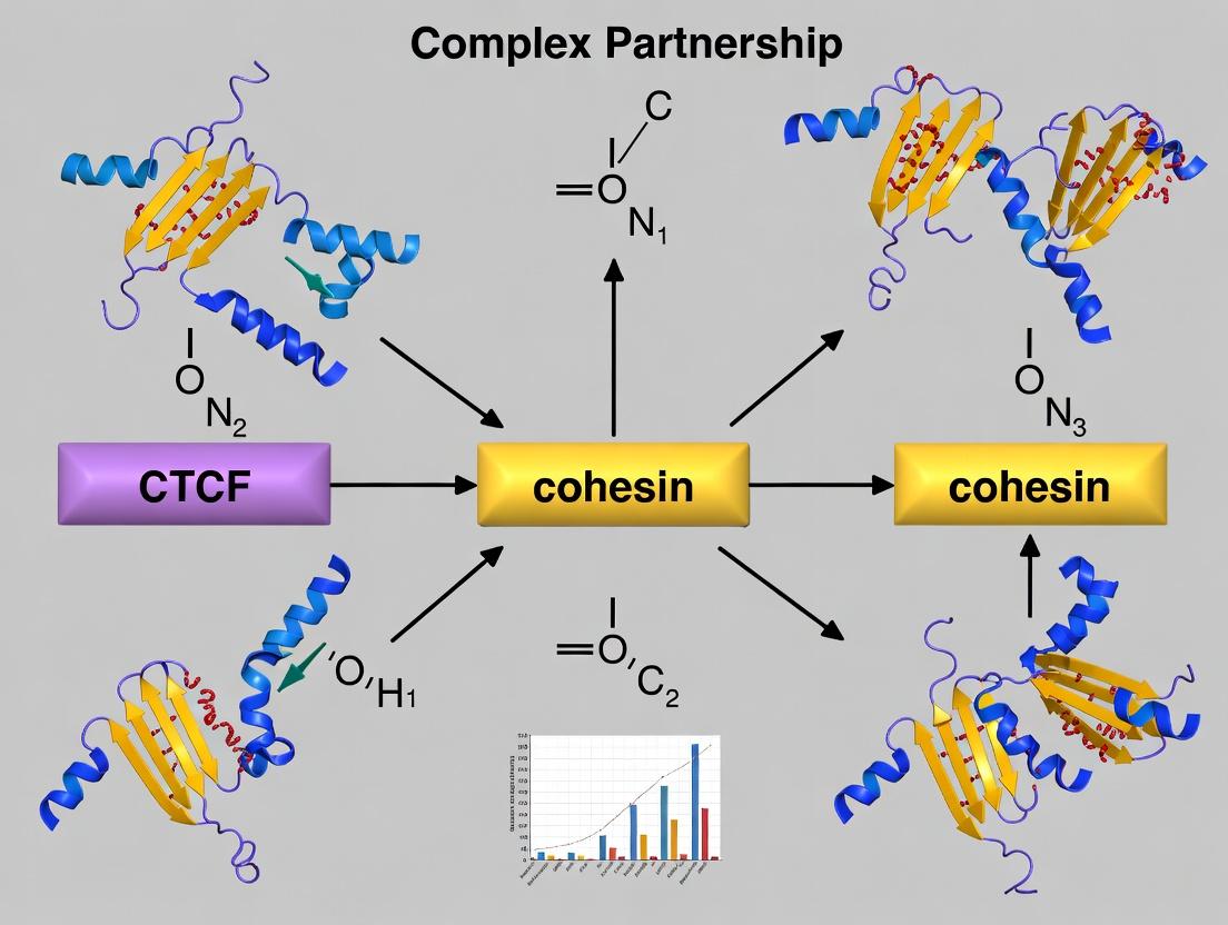
The CTCF-Cohesin Partnership: Orchestrating 3D Genome Architecture for Gene Regulation and Disease
This comprehensive article explores the pivotal partnership between CTCF and the cohesin complex in shaping the three-dimensional genome.
The CTCF-Cohesin Partnership: Orchestrating 3D Genome Architecture for Gene Regulation and Disease
Abstract
This comprehensive article explores the pivotal partnership between CTCF and the cohesin complex in shaping the three-dimensional genome. We delve into the fundamental molecular mechanisms of loop extrusion and chromatin insulation, examine cutting-edge experimental methodologies (including ChIP-seq, Hi-C, and live-cell imaging) for studying this partnership, address common challenges in data interpretation and experimental perturbations, and validate findings through comparative analyses across cell types and disease states. Tailored for researchers and drug development professionals, this review synthesizes current knowledge and highlights implications for understanding gene regulation, development, and cancer biology.
Defining the Dynamic Duo: Foundational Principles of CTCF and Cohesin in Genome Organization
Within the nucleus of eukaryotic cells, the precise three-dimensional organization of chromatin is fundamental to gene regulation, DNA replication, and genomic integrity. This architecture is not static but is dynamically shaped by specialized molecular machines. Two key players in this process are the architectural protein CCCTC-binding factor (CTCF) and the cohesin complex, a ring-shaped molecular motor. Their partnership forms the cornerstone of chromatin loop formation and topologically associating domain (TAD) establishment. This whitepaper, framed within ongoing research into their synergistic partnership, provides a technical guide to their structure, function, and experimental interrogation.
Core Components: Structure and Function
CTCF: The Genome's Architectural Guide
CTCF is an 11-zinc finger DNA-binding protein that recognizes a ~55 bp consensus sequence. It serves as a boundary element and an anchor point for chromatin loops. Its orientation and binding strength are critical for directing cohesin's activity.
Cohesin: The Loop-Extruding Motor
The cohesin complex is a tripartite ring primarily composed of SMC1, SMC3, RAD21, and STAG1/2 subunits. It utilizes ATP hydrolysis to translocate along chromatin, processively extruding a loop until it encounters boundary elements, most notably CTCF.
Table 1: Core Protein Components
| Component | Type | Primary Function | Key Domains/Features |
|---|---|---|---|
| CTCF | Architectural Protein | Sequence-specific DNA binding, directional blocking of cohesin | 11 Zn fingers, N- and C-terminal disordered regions |
| SMC1 | Cohesin Structural Subunit | ATPase activity, hinge dimerization | Coiled-coil, hinge, ATPase head |
| SMC3 | Cohesin Structural Subunit | ATPase activity, hinge dimerization | Coiled-coil, hinge, ATPase head |
| RAD21 | Cohesin Subunit | Closure of ring, regulatory interface | Cleavage sites (separase), phosphorylation sites |
| STAG1/2 | Cohesin Subunit (SA) | Stabilization, chromatin interaction, specificity | Stromalin family, binds DNA and CTCF |
| NIPBL | Cohesin Loader | Facilitates cohesin loading onto DNA | HEAT repeats, binds DNA and cohesin |
| WAPL | Cohesin Unloader | Promotes cohesin release from DNA | Wings apart, facilitates ring opening |
The Loop Extrusion Mechanism: A Dynamic Partnership
Current models propose that the NIPBL/MAU2 loader complex deposits cohesin onto chromatin. The ring then extrudes DNA bidirectionally in an ATP-dependent manner. CTCF, bound in a specific orientation, acts as a directional barrier, halting cohesin's progression. Convergently oriented CTCF sites at the boundaries of TADs lead to stable loop formation.
Title: CTCF-Cohesin Loop Extrusion Mechanism
Key Experimental Protocols
Chromatin Conformation Capture (3C and Hi-C)
Purpose: To map chromatin interactions and identify TADs/loops genome-wide. Detailed Protocol (Hi-C):
- Crosslinking: Cells are fixed with 1-3% formaldehyde to crosslink protein-DNA and protein-protein interactions.
- Digestion: Chromatin is digested with a restriction enzyme (e.g., MboI, DpnII, HindIII) in permeabilized nuclei.
- End Repair and Biotinylation: Digested ends are filled with biotinylated nucleotides.
- Ligation: DNA is ligated under dilute conditions to favor intramolecular ligation of crosslinked fragments.
- Reverse Crosslinking & Purification: Proteins are degraded, and DNA is purified.
- Shearing and Pull-down: DNA is sheared, and biotin-containing ligation junctions are isolated with streptavidin beads.
- Library Prep and Sequencing: Libraries are prepared from purified fragments and sequenced on a paired-end platform.
- Data Analysis: Paired reads are mapped, filtered, and used to generate contact probability matrices.
Chromatin Immunoprecipitation (ChIP-seq for CTCF/Cohesin)
Purpose: To map genome-wide binding sites of CTCF and cohesin subunits. Detailed Protocol:
- Crosslinking: Cells are fixed with 1% formaldehyde for 8-10 minutes.
- Sonication: Chromatin is sheared to 200-500 bp fragments via sonication.
- Immunoprecipitation: Sheared chromatin is incubated with specific antibodies (e.g., anti-CTCF, anti-RAD21, anti-SMC3) and Protein A/G beads.
- Washing & Elution: Beads are washed stringently, and bound complexes are eluted.
- Reverse Crosslinking & DNA Purification: Treatment with Proteinase K and heat de-crosslinks DNA, which is then purified.
- Library Prep and Sequencing: Libraries are prepared and sequenced.
- Data Analysis: Reads are aligned, and peaks are called to identify enriched binding regions.
Table 2: Quantitative Data Summary from Key Studies
| Experimental Readout | Typical Value/Range | Biological Context | Technical Method |
|---|---|---|---|
| CTCF Binding Sites | ~50,000 - 100,000 per mammalian genome | Majority at TAD boundaries | ChIP-seq |
| TAD Size | ~200 kb - 1 Mb | Conserved across cell types | Hi-C |
| Loop Length | ~100 kb - 3 Mb | Anchored by convergent CTCF | Hi-C (micro-C) |
| Cohesin Residence Time | ~10 - 25 minutes | Dependent on WAPL antagonism | FRAP/SMT |
| Loop Extrusion Rate | ~0.5 - 2 kb/s in vitro | NIPBL/MAU2 dependent | Single-molecule imaging |
Title: Hi-C and ChIP-seq Core Workflows
The Scientist's Toolkit: Key Research Reagent Solutions
Table 3: Essential Research Reagents and Materials
| Reagent/Material | Function/Application | Example Product/Clone |
|---|---|---|
| Anti-CTCF Antibody | Immunoprecipitation for ChIP-seq; Immunofluorescence. | Millipore 07-729 (rabbit monoclonal) |
| Anti-SMC3 / RAD21 Antibody | Cohesin ChIP-seq; monitoring complex integrity (Western). | Abcam ab9263 (SMC3); Millipore 05-908 (RAD21) |
| NIPBL / WAPL siRNA/shRNA | Functional depletion to study cohesin loading/unloading dynamics. | Dharmacon siRNA SMARTpools |
| Auxin-Inducible Degron (AID) Tags | Rapid, reversible degradation of CTCF or cohesin subunits. | F-box/TIR1 system; endogenous tagging via CRISPR. |
| CUT&RUN / CUT&Tag Kits | Mapping protein-DNA interactions with low background/cell input. | Cell Signaling Technology CUTANA kits |
| Hi-C Kit | Standardized library preparation for chromatin conformation. | Arima-HiC Kit, Dovetail Omni-C Kit |
| Micro-C Kit | Nucleosome-resolution chromatin conformation capture. | Standard protocol using Micrococcal Nuclease (MNase) |
| dCas9-KRAB / dCas9-CTCF Fusions | Targeted epigenetic perturbation of specific loci. | CRISPRi for repression; targeted CTCF tethering. |
| Live-cell SNAP/CLIP-tagged Cohesin | Single-molecule tracking of cohesin dynamics in living cells. | CRISPR knock-in of SNAP-tag on RAD21. |
| In Vitro Reconstitution Systems | Purified proteins for mechanistic biochemistry (loop extrusion assays). | Recombinant human cohesin, NIPBL-MAU2, CTCF. |
1. Introduction The three-dimensional architecture of the genome is a fundamental regulator of gene expression, DNA replication, and repair. Within this context, the loop extrusion model has emerged as a leading mechanistic framework explaining how chromatin loops are formed. This in-depth technical guide examines the core principles of this model, focusing on the central role of the cohesin complex. The content is framed within the ongoing research thesis on the essential partnership between cohesin and the architectural protein CTCF, a collaboration that defines the boundaries and anchors of these critical chromatin structures. For researchers and drug development professionals, understanding this machinery is paramount, as its dysregulation is implicated in developmental disorders and cancers.
2. Core Mechanism: The Loop Extrusion Engine The loop extrusion model posits that a molecular complex, notably cohesin, acts as a processive, ATP-dependent motor that extrudes chromatin fiber to form a progressively enlarging loop. Cohesin, a ring-shaped multi-subunit complex (comprising SMC1, SMC3, RAD21, and STAG1/2), topologically entraps two strands of DNA. Driven by ATP hydrolysis, it reels in DNA, increasing the loop size until it encounters a boundary signal, predominantly the DNA-bound CTCF protein in a specific orientation.
Table 1: Core Components of the Loop Extrusion Machinery
| Component | Primary Function | Key Characteristics |
|---|---|---|
| Cohesin Complex | Extrusion motor; topological entrapment of DNA. | Ring-shaped; SMC1, SMC3, RAD21, STAG1/2; NIPBL-MAU2 loading complex. |
| CTCF | Boundary element; loop anchor. | Zinc-finger protein; binds to specific motif; directionality blocks extrusion. |
| NIPBL-MAU2 | Cohesin loader; facilitates topological entry onto DNA. | Essential for initial cohesin deposition; mutations cause Cornelia de Lange Syndrome. |
| WAPL | Cohesin unloader; promotes ring opening and dissociation. | Regulates cohesin turnover; counteracts extrusion. |
| PDS5 | Cohesin regulator; modulates WAPL and cohesin stability. | Interacts with both cohesin and WAPL; fine-tunes loop dynamics. |
3. The CTCF-Cohesin Partnership: Defining Loop Boundaries CTCF binding sites are not passive barriers. They function as directional, asymmetrical stops for the cohesin extrusion complex. The orientation of the CTCF binding motif dictates which direction extrusion is blocked. Convergently oriented CTCF sites at the bases of loops are the hallmark of chromatin interaction maps (e.g., Hi-C). This partnership is the cornerstone of topologically associating domain (TAD) formation and insulation. Disruption of this partnership, through mutation of CTCF sites or depletion of cohesin, leads to a wholesale collapse of loop structures and aberrant gene regulation.
4. Experimental Protocols for Investigating Loop Extrusion 4.1. Chromatin Conformation Capture (Hi-C)
- Purpose: To genome-wide map chromatin interactions and identify loops.
- Protocol: Cells are cross-linked with formaldehyde. Chromatin is digested with a restriction enzyme (e.g., HindIII). Digested ends are biotin-labeled and ligated under dilute conditions to favor intra-molecular ligation. After reversing cross-links, DNA is sheared, and biotin-containing ligation junctions are pulled down with streptavidin beads for library preparation and paired-end sequencing.
- Output: A contact frequency matrix revealing loops as intense off-diagonal dots, often anchored at convergent CTCF sites.
4.2. CTCF/Cohesin Depletion (RNAi or Auxin-Inducible Degron)
- Purpose: To establish causality in loop formation.
- Protocol (Auxin-Inducible Degron): Cell lines are engineered to express cohesin subunit (e.g., RAD21) or CTCF fused to an auxin-inducible degron (AID) tag. Upon addition of auxin (indole-3-acetic acid), the target protein is rapidly ubiquitinated and degraded by the proteasome. Hi-C is performed pre- and post-depletion (e.g., at 6-hour time points).
- Output: Loss of specific loops and TAD boundaries, directly linking CTCF/cohesin to loop maintenance.
4.3. Single-Molecule Imaging (DNA Curtains or Optical Tweezers)
- Purpose: To visualize real-time loop extrusion dynamics in vitro.
- Protocol (DNA Curtains): Lambda DNA is tethered at one end to a lipid bilayer in a microfluidic channel and stretched by flow. Fluorescently labeled cohesin complexes (and NIPBL, CTCF) are introduced. Visualization via total internal reflection fluorescence (TIRF) microscopy tracks cohesin movement and loop formation on individual DNA molecules.
- Output: Direct observation of extrusion speed, processivity, directionality, and CTCF-mediated stopping.
5. Signaling and Regulatory Pathway of Loop Extrusion
Diagram Title: Pathway of Loop Extrusion by Cohesin and CTCF
6. The Scientist's Toolkit: Key Research Reagent Solutions Table 2: Essential Reagents for Loop Extrusion Research
| Reagent / Material | Function & Application | Example/Supplier |
|---|---|---|
| Anti-CTCF Antibody (ChIP-grade) | Chromatin immunoprecipitation to map CTCF binding sites and occupancy. | MilliporeSigma (07-729), Abcam (ab188408). |
| Anti-RAD21/SMC1 Antibody | Cohesin ChIP-seq; immunofluorescence to visualize cohesin localization. | Cell Signaling Technology, Bethyl Laboratories. |
| Auxin (IAA) | Rapid degradation of AID-tagged proteins in degron systems to study acute loss-of-function. | Sigma-Aldrich (I3750). |
| dCas9-KRAB/CRISPRi | Epigenetic silencing of specific CTCF motifs to study boundary function without genomic deletion. | Engineered cell lines or lentiviral delivery systems. |
| Recombinant Cohesin Complex | In vitro biochemical reconstitution of extrusion on defined DNA templates (e.g., DNA curtains). | Purified from insect or human expression systems. |
| HindIII, MboI Restriction Enzymes | Primary digesters for Hi-C library preparation to fragment cross-linked chromatin. | NEB. |
| Biotin-14-dATP | Labeling of DNA ends for pull-down of ligation junctions in Hi-C protocols. | Jena Biosciences, Thermo Fisher. |
| Protein A/G Magnetic Beads | Immunoprecipitation of antibody-bound chromatin complexes in ChIP-seq. | Dynabeads (Thermo Fisher). |
| TIRF Microscope System | High-resolution, single-molecule imaging of fluorescently tagged extrusion factors. | Nikon, Olympus, or custom-built systems. |
7. Quantitative Data & Key Findings Table 3: Key Quantitative Parameters of Loop Extrusion
| Parameter | Measured Value / Range | Method of Measurement | Biological Implication |
|---|---|---|---|
| Extrusion Rate in vitro | ~0.5 - 2.0 kb/s | Single-molecule imaging (DNA curtains). | Defines the timescale of loop formation and genome folding dynamics. |
| Cohesin Residence Time on Chromatin | ~10 - 30 minutes | FRAP, degron-mediated turnover assays. | Determines loop stability; regulated by WAPL and acetylation. |
| Average Loop Size | ~200 - 1000 kb | High-resolution Hi-C (e.g., Micro-C). | Defines the scale of regulatory domains and enhancer-promoter contacts. |
| CTCF Motif Orientation Bias | >90% of loops anchored at convergent sites | Bioinformatic analysis of Hi-C paired with CTCF ChIP-seq. | Establishes directionality as the critical feature for boundary function. |
| NIPBL Loading Efficiency | Low stoichiometry (catalytic) | Single-molecule counting, biochemical assays. | Explains how limited cohesin loaders can shape the entire genome. |
Within the context of our broader thesis on the CTCF-cohesin partnership, this whitepaper elucidates the definitive role of CTCF as the essential boundary factor that directs loop extrusion and stably anchors cohesin-mediated chromatin loops. We present a synthesis of current mechanistic models, quantitative data, and experimental methodologies central to this field, providing a technical resource for research and therapeutic development.
The cohesin complex, a ring-shaped ATPase, mediates chromatin loop extrusion, a fundamental process for genome organization and gene regulation. Unfettered extrusion, however, would produce non-functional architecture. CTCF (CCCTC-binding factor), through its orientation-specific binding to cognate motifs, acts as the dominant boundary factor, halting cohesin's progression and thereby defining loop anchors. This partnership creates the foundational topologically associating domains (TADs) and specific long-range interactions observed in mammalian genomes.
Quantitative Data Synthesis
Table 1: Key Genomic and Biochemical Metrics of CTCF-Cohesin Interaction
| Metric | Typical Value / Finding | Experimental Method | Citation Context |
|---|---|---|---|
| CTCF motif orientation concordance at loop anchors | >90% of convergent pairs | Hi-C / ChIP-seq | Higashi et al., Nature, 2021 |
| Reduction in loop/TAD boundary strength upon CTCF depletion (ΔBoundary Score) | 60-80% | Auxin-induced degradation + Hi-C | Nora et al., Cell, 2017 |
| Cohesin residence time on chromatin (wild-type) | ~20-25 minutes | FRAP / ChIP | Hansen et al., Cell, 2017 |
| Cohesin residence time on chromatin (CTCF ablation) | ~5-10 minutes | FRAP / ChIP | Hansen et al., Cell, 2017 |
| Percentage of loops dependent on CTCF | ~70-90% (cell-type variable) | CTCF degron + Hi-C | Rao et al., Cell, 2017 |
| Spatial proximity enhancement at CTCF-anchored loops | 2-5 fold over background | Micro-C / HI-C | Krietenstein et al., Mol Cell, 2020 |
Table 2: Core Domains and Mutational Effects
| Protein/Domain | Function | Key Mutation/Perturbation | Observed Phenotype |
|---|---|---|---|
| CTCF Zinc Finger Domain (ZF 4-7) | Essential for cohesin stopping | Point mutations in ZF 4-7 | Loss of boundary function, continued extrusion |
| CTCF N-terminus | Interaction with cohesin loader (NIPBL) | Deletion | Reduced cohesin recruitment to CTCF sites |
| Cohesin STAG1/2 (SA1/SA2) | Subunit specificity for loop anchoring | STAG2 knockout | Altered loop architecture, distinct from STAG1-KO |
| Cohesin ATPase (SMC1/3 heads) | Extrusion motor activity | Walker B mutations (ATPase dead) | Complete loss of loop formation |
Experimental Protocols
Protocol: Assessing Loop Dynamics via Acute CTCF Depletion and Hi-C
Objective: To measure the direct, temporal dependence of chromatin loops on CTCF. Materials: Cell line with degron-tagged CTCF (e.g., CTCF-AID), auxin, fixation reagents (formaldehyde), Hi-C kit (e.g., Arima-HiC or in-house), sequencer. Procedure:
- Degradation Induction: Treat CTCF-AID cells with 500 µM auxin (IAA) for 0, 15, 30, 60, and 120 min. Include untreated and wild-type (no degron) +auxin controls.
- Fixation: Quench cells with cold PBS + 0.1% BSA. Fix with 1% formaldehyde for 10 min at RT. Quench with 125 mM glycine.
- Hi-C Library Preparation: a. Lyse fixed cells and digest chromatin with a 4-cutter restriction enzyme (e.g., MboI or DpnII) overnight. b. Mark digested ends with biotin-14-dATP via fill-in. c. Perform proximity ligation under dilute conditions to favor intra-molecular ligation. d. Shear DNA to ~300-500 bp, pull down biotinylated ligation junctions with streptavidin beads. e. Prepare sequencing libraries directly on beads.
- Data Analysis: Process reads using standard pipelines (HiC-Pro, Juicer). Generate contact matrices at multiple resolutions (e.g., 5 kb, 25 kb). Call loops using HiCCUPS or SIP. Compare loop strength (observed/expected pixel value) and boundary insulation scores across time points.
Protocol: In Situ CTCF-Cohesin Proximity Ligation Assay (PLA)
Objective: Visualize and quantify direct spatial proximity between CTCF and cohesin at single cells. Materials: Fixed cells on coverslips, primary antibodies (anti-CTCF rabbit IgG, anti-SMC1 mouse IgG), Duolink PLA kit (Sigma), fluorescence microscope. Procedure:
- Immunostaining: Permeabilize fixed cells with 0.5% Triton X-100. Block with Duolink blocking buffer. Incubate with primary antibody mix (1:200 each) overnight at 4°C.
- PLA Probe Incubation: Apply species-specific PLA secondary antibodies (MINUS and PLUS probes) for 1h at 37°C.
- Ligation & Amplification: Perform ligation of hybridized probes with Duolink Ligation buffer for 30 min at 37°C. Amplify using Duolink Amplification buffer with fluorescently labeled oligonucleotides (Cy3 or Cy5) for 100 min at 37°C.
- Imaging & Analysis: Mount slides and image on a confocal microscope. Quantify PLA foci (distinct red dots) per nucleus using image analysis software (e.g., ImageJ). Compare to negative controls (omit one primary antibody).
Visualizations
Title: Cohesin Extrusion Stopped by Convergent CTCF
Title: CTCF Degradation Hi-C Protocol Flow
Title: Molecular Interactions at Loop Anchor
The Scientist's Toolkit: Research Reagent Solutions
Table 3: Essential Reagents for CTCF-Cohesin Loop Research
| Reagent / Material | Function & Application | Key Considerations |
|---|---|---|
| CTCF-AID Degron Cell Line (e.g., mCTCF-AID HCT116) | Enables rapid, acute CTCF depletion (<1 hr) via auxin addition for causal experiments. | Requires parental AID-TIR1 background; control for auxin alone effects. |
| High-Affinity Anti-CTCF Antibody (Rabbit monoclonal, D31H2 - CST) | Reliable ChIP-seq, CUT&RUN, immunofluorescence to map binding and protein levels. | Verify specificity by loss of signal upon degradation. |
| Anti-SMC1 Antibody (Mouse monoclonal, AB-1 - Millipore) | Standard for cohesin ChIP-seq and co-immunoprecipitation experiments. | Recognizes both SMC1A and SMC1B isoforms. |
| Duolink PLA Kit (Sigma) | Detects direct protein-protein proximity (<40 nm) in situ (e.g., CTCF-Cohesin interaction). | Critical to include rigorous negative controls (single antibody). |
| Arima-HiC Kit (Arima Genomics) | Optimized, robust commercial kit for high-resolution Hi-C library generation. | Reduces technical variability compared to in-house protocols. |
| dCas9-KRAB Fusions & sgRNAs | Enables targeted epigenetic perturbation of specific CTCF binding sites to test anchor necessity. | Design multiple sgRNAs per site; controls for off-target KRAB spreading. |
| Recombinant Cohesin Complex (Purified SMC1/3, RAD21, SA1) | For in vitro biochemical assays (e.g., ATPase activity, DNA binding) and structural studies. | Often expressed using baculovirus/Sf9 system; requires careful quality control. |
| Biotinylated CTCF Motif Oligos | For electrophoretic mobility shift assays (EMSAs) or pulldowns to test binding affinity of mutants. | Include scrambled sequence control; ensure proper double-stranding. |
The evidence consolidates CTCF as the principal director of cohesin-mediated loop formation. Future research directions within our thesis framework include elucidating the precise biophysical mechanism of extrusion stoppage, the role of CTCF isoforms and post-translational modifications, and the therapeutic potential of modulating specific disease-relevant loops by targeting this partnership. The experimental and analytical tools detailed herein provide the foundation for these next-generation investigations.
The functional partnership between the CCCTC-binding factor (CTCF) and the cohesin complex is a cornerstone of three-dimensional genome organization. Cohesin, a ring-shaped multi-subunit complex, is loaded onto chromatin to mediate sister chromatid cohesion and form DNA loops, with CTCF often defining loop boundaries. For years, a central question has been whether a single cohesin ring entraps one or two DNA strands and whether loop formation requires the dimerization of two cohesin complexes. This whitepaper examines the evolution from the classical "Handcuff Model" of cohesin dimerization to the emerging "Embrace Model" of a monomeric cohesin ring, framing this debate within the critical context of CTCF-cohesin partnership research.
Historical Perspective: The Handcuff Model
The Handcuff Model proposed that two separate cohesin rings, each entrapping a single DNA molecule, are linked together via dimerization of their SMC (Structural Maintenance of Chromosomes) subunits, particularly the hinge domains of Smc1 and Smc3. This dimerized "handcuff" structure was thought to be essential for both sister chromatid cohesion and chromatin looping.
Table 1: Key Evidence Supporting the Handcuff Model (c. 2000-2015)
| Experimental Observation | System/Method | Quantitative Result | Proposed Interpretation |
|---|---|---|---|
| Cohesin co-purification in pairs | Size-exclusion chromatography & multi-angle light scattering | Apparent molecular weight ~600 kDa (dimer of the ~300 kDa complex) | Stable dimerization of two cohesin rings. |
| Electron microscopy of cohesin complexes | Negative stain EM | ~15-20% of visualized particles appeared as paired rings. | Physical observation of dimerized rings. |
| FRET between labeled cohesin subunits | Fluorescence Resonance Energy Transfer in vitro | FRET efficiency increase of ~40% upon ATP hydrolysis. | Dimerization brings SMC hinges into close proximity. |
| Two-hybrid interaction of hinge domains | Yeast two-hybrid assay | Strong β-galactosidase activity (units >50) for Smc1-Smc3 hinge interaction. | Direct protein-protein interaction mediating dimerization. |
Paradigm Shift: Evidence for the Embrace (Monomeric) Model
Recent high-resolution structural and single-molecule studies have challenged the Handcuff Model, supporting an "Embrace" model where a single cohesin ring can simultaneously entrap two DNA strands within its lumen.
Table 2: Compelling Evidence for the Embrace (Monomeric) Model (c. 2018-Present)
| Experimental Observation | System/Method | Quantitative Result | Interpretation |
|---|---|---|---|
| Cryo-EM structures of DNA-bound cohesin | Cryo-Electron Microscopy | Structures show one cohesin ring (diameter ~35 nm) encircling two DNA duplexes. | Single ring can embrace two DNAs. |
| In vitro single-DNA loop extrusion assays | Single-molecule imaging (TIRF) | One cohesin complex extrudes loops at a rate of ~0.5-2.0 kbp/s without partner. | Monomeric cohesin is sufficient for loop formation. |
| Stoichiometry of chromatin-bound cohesin | Quantitative mass spectrometry (AP-MS) | Cohesin:CTCF ratio near 1:1 at loop anchors, not 2:1. | Favors one cohesin per loop anchor. |
| Hi-C contact map changes upon cohesin depletion/auxin-induced degradation | Chromosome Conformation Capture | Loop domain strength reduced by >70% without new "half-loop" signals. | Loss of single cohesin collapses loops, not handcuffs. |
Detailed Experimental Protocols
Protocol 1: Cryo-EM for Determining Cohesin-DNA Complex Structure
- Sample Preparation: Express and purify recombinant human cohesin complex (Smc1, Smc3, Scc1, SA1/Scc3) and CTCF (zinc finger domains). Incubate cohesin (0.5 mg/mL) with a 200-bp dsDNA containing a consensus CTCF motif and ATPγS (1 mM) for 30 min at 30°C.
- Grid Preparation: Apply 3.5 µL of sample to a glow-discharged Quantifoil R1.2/1.3 300-mesh gold grid. Blot for 3.5 seconds at 100% humidity and plunge-freeze in liquid ethane using a Vitrobot Mark IV.
- Data Collection: Collect ~10,000 micrograph movies on a 300 keV Titan Krios microscope with a K3 direct electron detector at a nominal magnification of 105,000x (pixel size 0.825 Å). Use a total dose of 50 e⁻/Ų over 50 frames.
- Processing: Motion-correct and dose-weight frames using MotionCor2. Perform template-based particle picking in RELION, extract particles (box size 384 px), and conduct iterative 2D and 3D classification. Refine the final map to ~3.5 Å resolution.
Protocol 2: Single-Molecule DNA Loop Extrusion Assay
- Flow Cell Assembly: Construct a flow cell with a PEG-passivated glass surface. Attach multiple, sequence-specific digoxigenin-labeled DNA tether points.
- DNA Substrate Preparation: Generate a ~40 kbp λ-phage-derived DNA construct with multiple, internally biotinylated nucleotides and a terminal digoxigenin label.
- Tethering: Introduce DNA into the flow cell, allowing digoxigenin-anti-digoxigenin binding to the surface. Label with 0.1 mg/mL streptavidin-coated quantum dots (655 nm emission) to visualize a fiducial marker.
- Imaging: Introduce imaging buffer (oxygen scavenging system, protocatechuate dioxygenase, ATP). Inject fluorescently labeled (Alexa Fluor 488) cohesin complex (1-10 nM). Image on a TIRF microscope at 2 frames per second. Track loop growth as the shortening of distance between the quantum dot and the moving cohesin complex.
Visualizing Cohesin Models and Experimental Workflows
The Scientist's Toolkit: Key Research Reagent Solutions
Table 3: Essential Reagents for Cohesin Dimerization State Research
| Reagent / Material | Function / Application | Key Considerations |
|---|---|---|
| Recombinant Human Cohesin Complex (full-length, wild-type & mutant) | In vitro biochemical assays (ATPase, loop extrusion), structural studies. | Requires co-expression of Smc1, Smc3, Scc1/Rad21, and SA1/Stag1/2 subunits; purity >95% for cryo-EM. |
| CTCF Zinc Finger Domain (ZF 3-11) Protein | For studies of cohesin pausing and boundary formation in loop extrusion assays. | Must include the conserved ZF cluster for DNA binding; often used in a catalytically inactive form for structural studies. |
| Site-Specifically Modified DNA Constructs (Biotin, Digoxigenin, Fluorescent labels) | Substrates for single-molecule assays (TIRF, optical tweezers) and structural biology. | Critical for tethering and visualization; length (0.5 - 50 kbp) and label position must be designed for specific assay. |
| ATPγS (Adenosine 5´-[γ-thio]triphosphate) | Hydrolysis-resistant ATP analog used to trap cohesin in a specific catalytic state for structural analysis. | Stabilizes cohesin-DNA interactions that may be transient with ATP. |
| Anti-Scc1 (Rad21) Cleavable Antibody (e.g., PreScission protease site-tagged) | For chromatin immunoprecipitation (ChIP) and auxin-induced degron (AID) depletion studies in vivo. | Enables acute cohesin removal to study immediate effects on chromatin architecture (Hi-C). |
| NHS-Ester Activated Quantum Dots (e.g., Qdot 655) | Fiducial markers for single-DNA molecule visualization in loop extrusion assays. | High photostability allows long-term tracking; must be conjugated to streptavidin for binding to biotinylated DNA. |
| Magnetic Beads (Dynabeads) with Anti-FLAG / Anti-HA | For pull-down of endogenously tagged cohesin complexes from cell extracts to assess native stoichiometry. | Used in conjunction with crosslinking (e.g., formaldehyde) to capture transient interactions. |
The partnership between the architectural proteins CTCF and cohesin is fundamental to the establishment and maintenance of the mammalian genome's three-dimensional organization. This hierarchy—from Loop Domains to Sub-TADs and TADs—is not merely structural but is intrinsically linked to gene regulation. The current research thesis posits that the dynamic, ATP-driven process of cohesin-mediated loop extrusion, which is anchored and terminated by CTCF binding at convergent sites, is the primary mechanism generating these domains. Disruption of this partnership is implicated in developmental disorders and cancer, making it a critical area for therapeutic intervention.
| Feature | Typical Size Range | Primary Forming Mechanism | Key Architectural Proteins | Functional Role | Stability |
|---|---|---|---|---|---|
| Loop Domains | 40 kb - 3 Mb | Cohesin-mediated loop extrusion, arrested at convergent CTCF sites. | Cohesin complex (SMC1/3, RAD21, SA1/2), CTCF. | Facilitate enhancer-promoter contact; Insulate regulatory crosstalk. | Dynamic (minutes-hours). |
| Sub-TADs | ~100 kb - 1 Mb | Nested loops within TADs; often cell-type specific. | Cohesin, CTCF, cell-type specific transcription factors. | Fine-tuned regulatory units; precise gene regulation. | More dynamic than TADs. |
| TADs (Topologically Associating Domains) | 200 kb - 1 Mb (median ~880 kb) | Aggregation of loops via extrusion; strong boundaries. | CTCF, Cohesin, other boundary elements (e.g., housekeeping genes). | Major units of genome compartmentalization; constrain regulatory interactions. | Relatively stable across cell cycles. |
Table 1: Comparative overview of key 3D genomic features. Size data aggregated from recent Hi-C studies (2021-2023).
Experimental Protocols for Mapping Genomic Features
High-Throughput Chromosome Conformation Capture (Hi-C)
Purpose: Genome-wide mapping of chromatin interactions to identify TADs, Sub-TADs, and loops. Detailed Protocol:
- Crosslinking: Treat cells (~1-5 million) with 1-3% formaldehyde for 10 min at room temperature to fix chromatin interactions.
- Lysis & Digestion: Lyse cells and digest crosslinked DNA with a restriction enzyme (e.g., DpnII, HindIII, or MboI) overnight.
- Marking Ends & Proximity Ligation: Fill in restriction fragment ends with biotin-labeled nucleotides. Perform proximity ligation in a large volume under dilute conditions to favor intra-molecular ligation of crosslinked fragments.
- Reverse Crosslinking & Purification: Reverse crosslinks with Proteinase K, purify DNA, and shear it to ~300-500 bp.
- Pull-down & Sequencing: Pull down biotin-labeled ligation junctions with streptavidin beads. Prepare a sequencing library and perform paired-end sequencing on a high-throughput platform (Illumina).
- Bioinformatics Analysis: Process reads (map to reference genome, filter valid interaction pairs). Generate contact matrices. Use algorithms like Arrowhead (for TADs), HiCCUPS (for loops), and aggregate analyses for Sub-TADs.
CTCF/Cohesin ChIP-seq
Purpose: Map binding sites of architectural proteins to correlate with domain boundaries. Detailed Protocol:
- Crosslinking & Sonication: Crosslink cells with 1% formaldehyde for 10 min. Quench with glycine. Lyse cells and sonicate chromatin to shear DNA to fragments of 200-500 bp.
- Immunoprecipitation: Incubate chromatin with antibody-coated magnetic beads (anti-CTCF or anti-RAD21/anti-SMC1). Use species-matched IgG as control.
- Wash, Elute, Reverse Crosslink: Wash beads stringently. Elute complexes and reverse crosslinks overnight at 65°C.
- DNA Purification & Library Prep: Purify DNA and prepare a sequencing library for Illumina sequencing.
- Peak Calling: Align reads, call significant peaks (e.g., using MACS2) to identify binding sites.
Cohesin Depletion/Inhibition Experiments
Purpose: Functionally test the role of cohesin in domain formation. Detailed Protocol (Auxin-Inducible Degron System):
- Cell Line Engineering: Generate a cell line expressing cohesin subunit (e.g., RAD21) tagged with an auxin-inducible degron (AID) and the plant F-box protein TIR1.
- Degradation Induction: Treat cells with 500 µM indole-3-acetic acid (IAA, auxin) for a time course (e.g., 0, 30, 60, 120 min).
- Validation: Assess cohesin loss by western blot (for protein) and ChIP-qPCR (for chromatin binding).
- Phenotyping: Perform Hi-C on treated vs. untreated cells. Analyze loss of loop domains, blurring of Sub-TAD/TAD boundaries, and changes in compartment strength.
Visualizing the CTCF/Cohesin Loop Extrusion Model
Title: Cohesin extrusion anchored by convergent CTCF sites creates loops.
Title: Hierarchical organization: loops within sub-TADs within TADs.
The Scientist's Toolkit: Research Reagent Solutions
| Reagent/Resource | Provider Examples | Function in CTCF/Cohesin/3D Genomics Research |
|---|---|---|
| Anti-CTCF Antibody | Cell Signaling Tech, Abcam, Active Motif | Chromatin immunoprecipitation (ChIP) to map CTCF binding sites and assess boundary occupancy. |
| Anti-RAD21/SMC1/SA Antibodies | MilliporeSigma, Bethyl Labs, Santa Cruz | Co-immunoprecipitation (Co-IP) and ChIP to study cohesin complex localization and function. |
| Auxin (IAA) & Degron Tagging Systems | Takara Bio, Academia (Dr. Kanemaki lab) | Rapid, inducible degradation of AID-tagged proteins (e.g., RAD21) to study acute loss-of-function. |
| Cohesin/CTCF Inhibitors (e.g., STAG2 inhibitors) | Cayman Chemical, MedChemExpress | Pharmacological disruption of complex function for mechanistic and therapeutic studies. |
| Hi-C & ChIP-seq Kits | Arima Genomics, Active Motif, Diagenode | Optimized, commercially available kits for robust library preparation for 3D genomics assays. |
| dCas9-KRAB/CRISPRi Systems | Addgene, Synthego | Target specific TAD boundaries for perturbation (epigenetic editing) to test boundary necessity. |
| Cell Lines with Endogenous Tagging | ATCC, Coriell Institute, Genome Engineering labs | Models with fluorescent or functional tags on architectural proteins for live imaging and biochemistry. |
| Bioinformatics Pipelines (HiC-Pro, HiCExplorer, Cooler) | Open Source (GitHub) | Standardized software for processing, analyzing, and visualizing high-throughput chromosome conformation data. |
Table 2: Essential reagents and tools for experimental research on TADs, Sub-TADs, and Loop Domains.
The Role of Cohesin Loaders (NIPBL-MAU2) and Unloaders (WAPL, PDS5) in the Cycle
This whitepaper details the molecular machinery governing the cohesin cycle, with a specific focus on the loader complex NIPBL-MAU2 and the unloader proteins WAPL and PDS5. This discussion is framed within the broader research context of the partnership between the cohesin complex and the architectural protein CTCF. This partnership is fundamental for genome organization, facilitating the formation of topologically associating domains (TADs) and loops that regulate gene expression. Understanding the dynamic regulation of cohesin loading and unloading is therefore critical for elucidating mechanisms in development, cellular homeostasis, and disease, with direct implications for therapeutic intervention in oncology and cohesinopathies like Cornelia de Lange Syndrome (CdLS).
The Core Machinery: Loaders and Unloaders
Cohesin Loader: NIPBL-MAU2 The NIPBL-MAU2 heterodimer is the essential loader that catalyzes the topological entrapment of DNA by the cohesin ring. NIPBL (Scc2) provides the primary enzymatic activity, while MAU2 (Scc4) stabilizes the complex. Current models suggest NIPBL-MAU2 interacts with cohesin's ATPase head domains, facilitating ATP hydrolysis and subsequent gate opening for DNA entry. Mutations in NIPBL account for over 60% of CdLS cases, highlighting its non-redundant function.
Cohesin Unloaders: WAPL and PDS5 Cohesin release from chromosomes is primarily regulated by WAPL (Wings apart-like) in conjunction with its regulatory partner PDS5. WAPL is a "release factor" that promotes the opening of the cohesin ring at the hinge domain or the Smc3-Scc1 interface, leading to DNA exit. PDS5 binds both cohesin and WAPL, modulating this activity. The opposing actions of loaders and unloaders establish a dynamic equilibrium of cohesin on chromatin, which is locally stabilized by CTCF.
CTCF as a Positional Stabilizer CTCF, bound to specific DNA motifs, acts as a barrier to the cohesin translocation driven by loop extrusion. When cohesin encounters a convergently oriented CTCF site, its progression is halted. This stable co-entrapment of CTCF and cohesin facilitates long-range DNA looping. Thus, CTCF does not directly load or unload cohesin but determines where cohesin-dependent structures are finalized by opposing the WAPL-mediated unloading process.
Table 1: Key Quantitative Parameters in the Cohesin Cycle
| Parameter | Typical Value / Range | Experimental System | Implication |
|---|---|---|---|
| Cohesin Loading Rate (by NIPBL-MAU2) | ~1-2 cohesin complexes loaded per minute per loading site (est.) | In vitro reconstitution with yeast cohesin | Establishes baseline for chromatin occupancy. |
| Cohesin Unloading Rate (WAPL-dependent) | Half-life of chromatin-bound cohesin reduced from >60 min to ~5-20 min upon WAPL recruitment | FRAP in mammalian cells (HeLa) | Indicates rapid turnover dynamic; CTCF antagonizes this. |
| Loop Extrusion Speed | ~0.5 - 2.1 kb/s | Single-molecule imaging (X. laevis egg extract) | Contextualizes the need for rapid unloading regulation. |
| CTCF-Bound Cohesin Stability | Half-life > 60 minutes (WAPL-resistant) | ChIP-seq & auxin-induced degradation assays (mESC) | Demonstrates CTCF's role in stabilizing cohesin. |
| NIPBL Mutation Prevalence in CdLS | ~60-65% of clinically diagnosed cases | Human genetic studies | Underscores critical loading function in development. |
| WAPL Knockout Effect on Cohesin Residence | ~5-10 fold increase in chromatin-bound cohesin half-life | Degron tag studies (HCT116, RPE1 cells) | Quantifies unloader potency. |
Table 2: Genetic Interactions and Phenotypes
| Protein | Loss-of-Function Phenotype (Cellular/Organismal) | Genetic Interaction with CTCF |
|---|---|---|
| NIPBL | Cohesin loading failure, aberrant gene expression, developmental defects (CdLS). | Synergistic: Double disruption abolishes nearly all chromatin looping. |
| MAU2 | Similar but often less severe than NIPBL loss; embryonic lethality in mice. | Similar to NIPBL. |
| WAPL | Hyper-cohesion, prolonged loop extrusion, merging of TAD boundaries, mitotic defects. | Antagonistic: WAPL deletion rescues loop/TAD formation in CTCF-depleted cells to some extent. |
| PDS5 | Complex phenotypes (cohesion defects, altered unloading), essential for viability. | Regulatory: PDS5 isoforms modulate WAPL activity at CTCF sites. |
Experimental Protocols
Protocol 1: Chromatin Immunoprecipitation Sequencing (ChIP-seq) for Cohesin and CTCF Objective: Map genome-wide binding sites of cohesin (e.g., SMC1A, RAD21) and CTCF to identify shared and unique loci. Methodology: 1. Crosslinking: Treat cells (e.g., HCT116, mESCs) with 1% formaldehyde for 10 min at room temp. Quench with 125mM glycine. 2. Cell Lysis & Chromatin Shearing: Lyse cells and sonicate chromatin to ~200-500 bp fragments using a focused ultrasonicator. 3. Immunoprecipitation: Incubate clarified lysate overnight at 4°C with antibodies against target protein (e.g., anti-SMC1A, anti-CTCF) coupled to magnetic Protein A/G beads. 4. Washing & Elution: Wash beads sequentially with low-salt, high-salt, LiCl, and TE buffers. Elute complexes with elution buffer (1% SDS, 0.1M NaHCO3). 5. Reverse Crosslinking & Purification: Incubate eluate at 65°C overnight with 200mM NaCl to reverse crosslinks. Treat with RNase A and Proteinase K. Purify DNA using silica columns. 6. Library Prep & Sequencing: Prepare sequencing library from purified DNA (end-repair, A-tailing, adapter ligation, PCR amplification). Sequence on an Illumina platform. 7. Data Analysis: Align reads to reference genome, call peaks (using MACS2), and analyze co-occupancy.
Protocol 2: Fluorescence Recovery After Photobleaching (FRAP) for Cohesin Dynamics Objective: Measure the turnover kinetics (residence time) of cohesin on chromatin. Methodology: 1. Cell Line Preparation: Use cells stably expressing cohesin subunit (e.g., SMC3) fused to a fluorescent protein (e.g., GFP). 2. Imaging: Maintain cells at 37°C/5% CO2 on a confocal microscope. Select a nuclear region of interest (ROI) for bleaching. 3. Photobleaching: Apply a high-intensity laser pulse to the ROI to irreversibly bleach the GFP signal within it. 4. Recovery Imaging: Acquire images at low laser power at short intervals (e.g., every 2-5 seconds) post-bleach to monitor fluorescence recovery due to influx of unbleached molecules. 5. Data Analysis: Quantify fluorescence intensity in the bleached ROI over time. Normalize to pre-bleach and whole-nucleus intensity. Fit recovery curve to an exponential model to calculate the half-time (t1/2) of recovery, which reflects the binding residence time.
Protocol 3: Auxin-Inducible Degron (AID) System for Acute Protein Depletion Objective: Rapidly deplete target proteins (e.g., WAPL, CTCF) to study acute effects on cohesin dynamics. Methodology: 1. Engineered Cell Line: Generate a cell line where the gene of interest is endogenously tagged with an AID tag (e.g., WAPL-AID-mClover) and expresses the plant E3 ligase TIR1 (or its mutant version, osTIR1) from a constitutive promoter. 2. Acute Depletion: Treat cells with 500 µM auxin (Indole-3-acetic acid, IAA). The osTIR1 ligase recognizes the AID tag and recruits the ubiquitin-proteasome machinery, leading to target degradation within 15-30 minutes. 3. Validation & Analysis: Monitor depletion via loss of fluorescence (if tagged with mClover/GFP) or western blot. Perform downstream assays (ChIP-seq, Hi-C, FRAP) immediately post-depletion to observe direct effects.
Signaling and Regulatory Pathways
Diagram 1: Cohesin Loading, Translocation, and Unloading Cycle (87 chars)
Diagram 2: CTCF Antagonizes WAPL to Stabilize Loops (64 chars)
The Scientist's Toolkit: Key Research Reagents
Table 3: Essential Research Reagents and Materials
| Reagent / Material | Function & Application | Example (Vendor) |
|---|---|---|
| Anti-SMC1A / RAD21 / CTCF Antibodies | For Chromatin Immunoprecipitation (ChIP) to map binding sites and protein occupancy. | Rabbit monoclonal anti-SMC1A (Abcam, ab9262); Mouse monoclonal anti-CTCF (Millipore, 07-729). |
| Auxin (Indole-3-Acetic Acid - IAA) | Small molecule trigger for rapid degradation of AID-tagged proteins in the AID system. | Sigma-Aldrich (I3750). |
| TIR1/osTIR1 Expression Vector | Plasmid encoding the plant E3 ubiquitin ligase required for the AID system to function in mammalian cells. | Addgene (various deposits, e.g., #80374). |
| CRISPR-Cas9 Gene Editing Tools | For endogenous tagging (AID, fluorescent proteins) or knockout of loader/unloader genes. | Alt-R S.p. Cas9 Nuclease (IDT); sgRNA synthesis kits. |
| Recombinant NIPBL-MAU2 Complex | Purified protein for in vitro cohesin loading assays and biochemical studies. | Often produced in-house via baculovirus/Sf9 expression systems. |
| Proteasome Inhibitor (MG-132) | Used to test if observed protein loss/degradation is proteasome-dependent. | Selleckchem (S2619). |
| Formaldehyde (Molecular Biology Grade) | For crosslinking protein-DNA and protein-protein interactions in ChIP and related protocols. | Thermo Scientific (28906). |
| Magnetic Protein A/G Beads | Solid support for antibody capture during immunoprecipitation steps. | Pierce Anti-HA Magnetic Beads (Thermo, 88836). |
| siRNA/shRNA against WAPL, PDS5, NIPBL | For transient or stable knockdown studies of loader/unloader components. | ON-TARGETplus siRNA pools (Horizon Discovery). |
| Cell Lines with Fluorescently Tagged Cohesin | For live-cell imaging, FRAP, and tracking cohesin dynamics. | e.g., HCT116 SMC3-GFP (generated via CRISPR tagging). |
Tools of the Trade: Advanced Methodologies to Map and Manipulate the CTCF-Cohesin Axis
Within the framework of CTCF and cohesin complex partnership research, understanding the three-dimensional (3D) architecture of chromatin is paramount. The dynamic loop extrusion process, driven by cohesin and boundary-delimited by CTCF, organizes the genome into distinct topologically associating domains (TADs) and loops that regulate gene expression. This technical guide details three pivotal technologies—Hi-C, Micro-C, and HiChIP—that enable the genome-wide mapping of these chromatin interactions. Each method offers unique resolutions and insights, critical for dissecting the mechanistic underpinnings of genome folding and its implications in development and disease.
Core Technologies: Principles and Comparative Analysis
Hi-C
Hi-C is the foundational genome-wide method for capturing chromatin conformation. It involves crosslinking chromatin, digesting with a restriction enzyme (often HindIII or MboI), filling in sticky ends with biotinylated nucleotides, ligating crosslinked fragments, and then performing paired-end sequencing. The frequency of ligation events between distal genomic loci is used to infer interaction probability.
Micro-C
Micro-C employs micrococcal nuclease (MNase) instead of restriction enzymes for digestion. MNase cuts between nucleosomes, producing a nucleosome-resolution map of chromatin contacts. This approach allows for the detection of fine-scale structures, such as nucleosome-nucleosome interactions and detailed loop boundaries, providing superior resolution for analyzing cohesin-mediated loops anchored at CTCF sites.
HiChIP
HiChIP (also called PLAC-seq) combines Hi-C with chromatin immunoprecipitation (ChIP). It uses a targeted pull-down with an antibody (e.g., against H3K27ac for active enhancers, or CTCF/cohesin subunits) to enrich for interactions involving specific protein-bound genomic regions. This increases signal-to-noise for biologically relevant interactions, such as those mediated by the CTCF/cohesin complex, while requiring significantly fewer sequencing reads.
Quantitative Data Comparison
Table 1: Comparative Overview of 3D Genomics Techniques
| Feature | Hi-C | Micro-C | HiChIP (e.g., against CTCF) |
|---|---|---|---|
| Digestion Enzyme | Restriction enzyme (e.g., MboI) | Micrococcal nuclease (MNase) | Restriction enzyme (e.g., MboI) |
| Nominal Resolution | 1 kb - 10 kb | < 1 kb (Nucleosome-level) | 1 kb - 10 kb (Enriched regions) |
| Primary Output | Genome-wide contact matrix | High-resolution genome-wide contact matrix | Protein-centric interaction matrix |
| Key Advantage | Unbiased global view | Single-nucleosome resolution | High efficiency for protein-specific loops |
| Typical Sequencing Depth | 1-3 Billion reads (human) | 2-5 Billion reads (human) | 200-500 Million reads (human) |
| Optimal for Studying | TADs, A/B compartments | Nucleosome phasing, fine-scale loops | Direct target of CTCF/cohesin loops |
Table 2: Typical Experimental Outcomes in CTCF/Cohesin Studies
| Metric | Hi-C Value | Micro-C Value | HiChIP (CTCF) Value |
|---|---|---|---|
| Detection of CTCF-anchored loops | Yes, but requires high depth | Yes, with precise anchor boundaries | Yes, highly enriched and specific |
| Signal-to-Noise at loop anchors | Moderate | High | Very High |
| Ability to define loop symmetry | Low | High (base-pair resolution) | Moderate |
| Input Material Required | ~1-5 million cells | ~2-10 million cells | ~0.5-2 million cells |
Detailed Experimental Protocols
Protocol 1: In-situ Hi-C for CTCF/Cohesin Loop Analysis
- Crosslinking: Suspend 1-2 million cells in culture medium. Add formaldehyde to a final concentration of 1-2% and incubate for 10 min at room temperature. Quench with 125 mM glycine.
- Lysis & Digestion: Lyse cells and digest chromatin with 100-200 units of MboI restriction enzyme overnight at 37°C.
- Marking & Ligation: Fill in restriction fragment ends with biotin-14-dATP and ligate under dilute conditions to favor intra-molecular ligation.
- Reverse Crosslinking & Shearing: Reverse crosslinks with Proteinase K, purify DNA, and shear to ~300-500 bp using a sonicator.
- Pull-down & Sequencing: Capture biotinylated ligation junctions with streptavidin beads, prepare a sequencing library, and sequence on an Illumina platform (paired-end 150 bp).
Protocol 2: Micro-C for Nucleosome-Resolved Architecture
- Crosslinking & MNase Digestion: Crosslink cells as above. Permeabilize cells and digest with MNase to predominantly yield mono-, di-, and tri-nucleosomes.
- End Repair & Ligation: Repair DNA ends with exonucleases/polymerases to create blunt ends. Ligate with T4 DNA Ligase under dilute conditions.
- Biotinylation & Processing: Label ligated junctions with biotin-dCTP via terminal transferase. Reverse crosslinks, purify DNA, and shear.
- Enrichment & Sequencing: Enrich for biotinylated fragments using streptavidin beads and prepare the sequencing library for deep paired-end sequencing.
Protocol 3: HiChIP for CTCF-Mediated Interactions
- In-situ Hi-C Protocol (Steps 1-3): Perform standard in-situ Hi-C through the ligation step.
- Chromatin Shearing: Sonicate the crosslinked, ligated chromatin to ~300-500 bp.
- Immunoprecipitation: Incubate sheared chromatin with an antibody against CTCF (or RAD21/SMC1 for cohesin) and Protein A/G beads overnight at 4°C.
- Wash, Elute, and Decrosslink: Wash beads stringently, elute complex, and reverse crosslinks.
- Biotin Pull-down & Library Prep: Purify DNA and perform a second pull-down with streptavidin beads to isolate ligation junctions before library preparation and sequencing.
Visualizing Methodologies and Pathways
Diagram Title: Core Workflow and Method Branching for 3D Genomics
Diagram Title: CTCF and Cohesin Drive Loop Formation
The Scientist's Toolkit: Research Reagent Solutions
Table 3: Essential Reagents for 3D Genomics Experiments
| Reagent/Material | Function in Experiment | Key Consideration for CTCF/Cohesin Studies |
|---|---|---|
| Formaldehyde (37%) | Crosslinks protein-DNA and protein-protein interactions. | Crosslinking time/concentration is critical to capture dynamic cohesin complexes. |
| HindIII or MboI Restriction Enzyme | (Hi-C/HiChIP) Cuts at specific sequences to fragment genome. | Choice determines resolution and coverage; check for cutting frequency near CTCF motifs. |
| Micrococcal Nuclease (MNase) | (Micro-C) Digests linker DNA between nucleosomes. | Titration is essential to achieve mono/di-nucleosome fragments for highest resolution. |
| Biotin-14-dATP | Labels ligation junctions for selective pull-down. | Reduces background in sequencing library, enriching for valid chimeric fragments. |
| Anti-CTCF Antibody (ChIP-grade) | (HiChIP) Immunoprecipitates CTCF-bound DNA fragments. | Specificity and affinity directly determine enrichment efficiency and data quality. |
| Protein A/G Magnetic Beads | Captures antibody-bound complexes during HiChIP. | Magnetic separation facilitates the multi-step protocol and improves recovery. |
| Streptavidin Magnetic Beads | Isolates biotinylated ligation junctions. | Essential for enriching true ligation products over non-ligated fragments. |
| High-Fidelity DNA Polymerase | Amplifies library fragments for sequencing. | Minimizes PCR duplicates and bias, crucial for quantitative interaction frequency. |
Understanding the partnership between CTCF and the cohesin complex (comprising subunits SMC1, SMC3, and RAD21) is a cornerstone of modern genome architecture and gene regulation research. This thesis posits that precise mapping of their binding sites is not merely descriptive but fundamental to deciphering the mechanics of chromatin looping, topologically associating domain (TAD) formation, and transcriptional insulation. Chromatin Immunoprecipitation followed by sequencing (ChIP-seq) and the newer Cleavage Under Targets and Release Using Nuclease (CUT&RUN) are the pivotal technologies that enable this mapping. This guide provides an in-depth technical comparison of these methods, their application to CTCF and cohesin, and their role in validating the core thesis of their cooperative genome organization.
Technology Deep Dive: ChIP-seq vs. CUT&RUN
Core Principles and Workflows
ChIP-seq relies on chemical crosslinking (typically with formaldehyde) to freeze protein-DNA interactions in situ, followed by chromatin fragmentation, immunoprecipitation, reversal of crosslinks, and library preparation.
CUT&RUN uses a Protein A/G-micrococcal nuclease (MNase) fusion protein targeted by an antibody to the protein of interest. Upon activation, MNase cleaves DNA in situ, releasing protein-bound fragments into the supernatant without crosslinking.
Diagram: Comparative Workflow for ChIP-seq and CUT&RUN
Quantitative Comparison of Performance Metrics
Table 1: Head-to-Head Comparison of ChIP-seq and CUT&RUN
| Metric | ChIP-seq | CUT&RUN | Implication for CTCF/Cohesin Studies |
|---|---|---|---|
| Input Material | 0.5-10 million cells | 10,000 - 500,000 cells | CUT&RUN enables rare cell type analysis. |
| Signal-to-Noise | Moderate. High background common. | Very High. Low background. | CUT&RUN yields clearer peaks, especially for cohesin subunits. |
| Resolution | ~100-300 bp (limited by sonication). | ~10-50 bp (single nucleosome precision). | CUT&RUN can delineate precise complex boundaries. |
| Crosslinking Artifacts | Yes. Can introduce false positives. | No. Uses native conditions. | CUT&RUN data may reflect more physiological binding. |
| Protocol Duration | 3-5 days. | ~1 day. | Faster turnaround for screening. |
| Mapping to Repetitive Regions | Challenging due to background. | Improved due to low background. | Better for cohesin/CTCF sites near repeats. |
| Compatibility with | Histone marks, robust TFs. | Best for chromatin-associated proteins. | Both excellent for CTCF/Cohesin. |
| Key Disadvantage | Requires optimization of crosslinking & sonication. | Requires permeabilization; sensitive to MNase over-digestion. |
Table 2: Typical Sequencing Metrics for High-Quality Datasets
| Factor | Recommended Read Depth | Recommended Antibody Clonality | Key Control |
|---|---|---|---|
| CTCF | 20-40 million reads (ChIP-seq) / 5-10M (CUT&RUN) | Monoclonal (e.g., D31H2, Cell Signaling) | IgG control essential. |
| SMC1/SMC3/RAD21 | 30-50 million reads (ChIP-seq) / 10-15M (CUT&RUN) | Polyclonal often used (e.g., Abcam, Bethyl Labs). | Input DNA for ChIP-seq; no-Ab for CUT&RUN. |
Detailed Experimental Protocols
CUT&RUN Protocol for CTCF in Cultured Mammalian Cells
Day 1: Cell Harvest and Binding
- Harvest & Wash: Harvest ~500k cells. Wash 2x in 1 mL Wash Buffer (20 mM HEPES pH 7.5, 150 mM NaCl, 0.5 mM Spermidine, 1x Protease Inhibitor).
- Permeabilization: Resuspend cell pellet in 1 mL Digitonin Wash Buffer (Wash Buffer + 0.05% Digitonin). Incubate on ice for 10 min.
- Concanavalin A Bead Binding: Pellet cells, resuspend in 50 μL Digitonin Wash Buffer. Add 10 μL activated Concanavalin A magnetic beads. Rotate at room temp for 15 min.
- Antibody Binding: Place tube on magnet, discard supernatant. Resuspend beads+cells in 50 μL Antibody Buffer (Digitonin Wash Buffer + 2 mM EDTA). Add primary antibody (CTCF, 1:50-1:100 dilution). Rotate overnight at 4°C.
Day 2: pA/G-MNase Binding, Cleavage, and DNA Release
- Wash: Place tube on magnet, discard supernatant. Wash beads 3x with 1 mL Digitonin Wash Buffer.
- pA/G-MNase Binding: Resuspend in 100 μL Digitonin Wash Buffer with pA/G-MNase protein (1:800 dilution). Rotate at 4°C for 1-2 hrs.
- Wash: Wash beads 3x with 1 mL Digitonin Wash Buffer.
- MNase Activation: Resuspend in 150 μL Digitonin Wash Buffer. Equilibrate to 0°C. Add 3 μL of 100 mM CaCl₂ (final 2 mM) to activate MNase. Incubate in thermal mixer at 0°C for 30-60 min.
- Reaction Stop: Add 150 μL Stop Buffer (200 mM NaCl, 20 mM EGTA, 4 mM EDTA, 50 μg/mL RNase A, 40 μg/mL Glycogen).
- DNA Release & Purification: Incubate at 37°C for 10 min. Spin briefly, place on magnet. Transfer supernatant (containing DNA fragments) to a new tube. Add 1 μL Proteinase K and 0.1% SDS. Incubate at 70°C for 10 min. Purify DNA with Phenol:Chloroform or spin column. Proceed to library prep.
Crosslinking ChIP-seq Protocol for Cohesin Subunit RAD21
Day 1: Crosslinking & Cell Lysis
- Crosslinking: Add 37% formaldehyde directly to cell culture medium (final 1%). Incubate at room temp for 10 min with gentle shaking.
- Quenching: Add 1.25 M glycine (final 125 mM). Incubate 5 min at RT.
- Harvest: Wash cells 2x with cold PBS. Pellet cells, flash-freeze, or proceed.
- Cell Lysis: Resuspend pellet in 1 mL Lysis Buffer 1 (50 mM HEPES-KOH pH 7.5, 140 mM NaCl, 1 mM EDTA, 10% Glycerol, 0.5% NP-40, 0.25% Triton X-100). Incubate 10 min at 4°C, rotating. Spin, discard supernatant.
- Nuclei Lysis: Resuspend pellet in 1 mL Lysis Buffer 2 (10 mM Tris-HCl pH 8.0, 200 mM NaCl, 1 mM EDTA, 0.5 mM EGTA). Incubate 10 min at 4°C, rotating. Spin, discard supernatant.
- Chromatin Shearing: Resuspend pellet in 1 mL Shearing Buffer (0.1% SDS, 1 mM EDTA, 10 mM Tris-HCl pH 8.0). Sonicate to achieve 200-500 bp fragments. Clear supernatant by centrifugation.
Day 2: Immunoprecipitation & Washing
- Pre-clear & IP: Dilute sheared chromatin 5-fold in IP Buffer (0.5% Triton X-100, 2 mM EDTA, 20 mM Tris-HCl pH 8.0, 150 mM NaCl). Add 5-10 μg anti-RAD21 antibody. Rotate overnight at 4°C.
- Capture: Add 50 μL Protein A/G magnetic beads pre-blocked with BSA. Rotate for 2-4 hrs at 4°C.
- Wash: Wash beads sequentially for 5 min each on rotator with: Low Salt Wash Buffer (0.1% SDS, 1% Triton X-100, 2 mM EDTA, 20 mM Tris-HCl pH 8.0, 150 mM NaCl), High Salt Wash Buffer (same, but 500 mM NaCl), LiCl Wash Buffer (0.25 M LiCl, 1% NP-40, 1% deoxycholate, 1 mM EDTA, 10 mM Tris-HCl pH 8.0), and finally 2x with TE Buffer.
Day 3: Elution & Clean-up
- Elution: Elute chromatin from beads twice with 100 μL Elution Buffer (1% SDS, 0.1 M NaHCO₃) at 65°C for 15 min with shaking.
- Reverse Crosslinks: Pool eluates, add NaCl (final 200 mM), and incubate at 65°C overnight.
- DNA Purification: Add RNase A and Proteinase K sequentially. Purify DNA with Phenol:Chloroform and ethanol precipitation. Resuspend in TE buffer. Quantify for library preparation.
The Scientist's Toolkit: Essential Research Reagents
Table 3: Key Reagent Solutions for CTCF/Cohesin Profiling
| Reagent/Material | Supplier Examples | Function & Critical Note |
|---|---|---|
| Anti-CTCF Antibody (mAb) | Cell Signaling #3418, Millipore #07-729 | For immunoprecipitation. Clonality impacts specificity. |
| Anti-RAD21 Antibody | Abcam ab992, Bethyl Labs A300-080A | Cohesin subunit IP. Validation via siRNA knockdown is recommended. |
| Anti-SMC1/SMC3 Antibody | Bethyl Labs A300-055A / A300-060A | Cohesin structural subunit IP. |
| Protein A/G Magnetic Beads | Pierce, Diagenode | Solid support for antibody capture in ChIP. |
| Concanavalin A Magnetic Beads | Polysciences, Bangs Labs | Binds permeabilized cells for CUT&RUN tethering. |
| pA/G-MNase Fusion Protein | You can produce in-house or obtain from collaborators. | The key enzyme for targeted cleavage in CUT&RUN. |
| Digitonin | Millipore, Sigma | Cell permeabilization agent for CUT&RUN. Optimal concentration is critical. |
| UltraPure Sonicated Salmon Sperm DNA | Invitrogen | Used as blocking agent in ChIP to reduce non-specific binding. |
| Dual Index Kit for Illumina | Illumina, NEB | Library preparation for high-throughput sequencing. |
| SPRIselect Beads | Beckman Coulter | Size selection and clean-up of DNA libraries. |
Data Analysis & Integration: Validating the Partnership
Peak calling (using tools like MACS2) for CTCF and cohesin subunits (SMC1, SMC3, RAD21) typically reveals a high degree of overlap, but with nuanced differences critical to the thesis. CTCF peaks are often sharper, while cohesin peaks can be broader. Integrated analysis involves:
- Peak Co-localization: Assessing the percentage of cohesin peaks that overlap CTCF peaks (>70% is typical in mammalian cells).
- Motif Analysis: Confirming the presence of the CTCF motif at shared binding sites.
- Directionality Analysis: Using the orientation of the CTCF motif to predict loop anchor structure.
Diagram: Data Analysis Pipeline for Binding Site Integration
The strategic application of ChIP-seq and CUT&RUN for mapping CTCF and cohesin subunit binding sites provides complementary and robust datasets that are indispensable for testing the central thesis of their partnership. CUT&RUN offers a rapid, high-resolution, low-input alternative ideal for precise mapping and screening, while ChIP-seq remains a robust, established method. The quantitative data generated, when integrated with chromosome conformation capture techniques, ultimately allows researchers to move from a simple catalog of binding sites to a dynamic model of how CTCF positions cohesin to orchestrate the three-dimensional genome.
Within the framework of investigating the CTCF and cohesin complex partnership—a cornerstone of 3D genome organization and transcriptional regulation—the demand for precise, acute, and reversible functional perturbation tools has never been greater. This whitepaper provides an in-depth technical guide to two paramount technologies: Auxin-Inducible Degron (AID) for rapid protein depletion and CRISPR interference/activation (CRISPRi/a) for tunable transcriptional control. We detail their integration into the study of chromatin architecture, presenting current protocols, quantitative data comparisons, and essential research reagents.
CTCF and cohesin form a dynamic partnership to mediate chromatin looping, topologically associating domain (TAD) formation, and insulator function. Traditional knockout or RNAi-mediated knockdown suffer from offtarget effects and slow kinetics, obscuring the acute functions of these essential complexes. AID and CRISPRi/a enable minute- to hour-scale perturbations, allowing researchers to dissect the immediate consequences of losing CTCF binding or cohesin loading/function on genome topology and gene expression, critical for understanding disease mechanisms and identifying therapeutic targets.
Table 1: Core Characteristics of AID vs. CRISPRi/a
| Feature | Auxin-Inducible Degron (AID) | CRISPR Interference (CRISPRi) | CRISPR Activation (CRISPRa) |
|---|---|---|---|
| Primary Target | Protein stability | Transcriptional initiation | Transcriptional initiation |
| Mode of Action | Proteasomal degradation | dCas9 fusion represses transcription | dCas9 fusion recruits activators |
| Key Component | TIR1 F-box protein, AID-tagged target | dCas9-KRAB/other repressor domains | dCas9-VPR/SunTag-VP64 |
| Reversibility | Yes (upon auxin washout) | Yes (upon sgRNA removal/induction stop) | Yes (upon sgRNA removal/induction stop) |
| Typical Depletion/Effect Onset | 15-30 min (protein depletion) | Hours (transcriptional repression) | Hours (transcriptional activation) |
| Typical Efficiency | >90% protein depletion | 70-95% gene repression | 5-50x gene activation |
| Key Application in CTCF/Cohesin Studies | Acute removal of RAD21, SMC3, or CTCF itself | Repress CTCF or STAG gene expression | Activate genes to probe loop formation |
| Major Advantage | Direct protein removal, rapid kinetics | Highly specific, multiplexable | Gain-of-function at endogenous loci |
| Major Limitation | Requires genetic tagging; potential basal degradation | Transcriptional delay; chromatin context effects | Variable activation strength |
Detailed Experimental Protocols
Protocol 3.1: Acute Depletion of Cohesin Subunit RAD21 via AID
Objective: To rapidly deplete the cohesin ring component RAD21 and observe immediate effects on chromatin looping.
Materials:
- Cell line expressing OsTIR1 (or plant TIR1) and AID-tagged RAD21 (endogenously tagged via CRISPR/Cas9).
- 500 mM Indole-3-acetic acid (IAA, auxin) stock in DMSO. Store at -20°C.
- Control: Equivalent volume of DMSO.
Procedure:
- Cell Preparation: Seed AID-tagged cells in appropriate culture dishes. Ensure cells are 60-70% confluent at time of treatment.
- Auxin Treatment: Add IAA to culture medium to a final concentration of 500 µM. For control, add DMSO only.
- Time-Course Harvest: Harvest cells at critical time points (e.g., 0, 15, 30, 60, 120 min) post-treatment for analysis.
- For Western Blot: Lyse cells in RIPA buffer. Probe with anti-RAD21 and loading control (e.g., Vinculin) antibodies. Quantify depletion kinetics.
- For Hi-C/Chromatin Conformation: Crosslink cells with 1-2% formaldehyde at each time point for downstream Hi-C library preparation.
- Reversibility Check (Optional): After 2 hours of IAA treatment, wash cells 3x with warm PBS and replenish with IAA-free medium. Harvest cells 2, 4, 8 hours post-washout to assess protein recovery and loop restoration.
Protocol 3.2: Transcriptional Repression ofCTCFvia CRISPRi
Objective: To specifically repress CTCF transcription and assess the slower, cumulative impact on cohesin localization.
Materials:
- Cell line stably expressing dCas9-KRAB (e.g., K562 dCas9-KRAB clonal line).
- Lentiviral vectors or transfection-ready plasmids for sgRNA targeting the CTCF promoter or transcription start site (TSS).
- Validated sgRNA sequence: (Example: 5'-GACCACTCCAGCGTGCGCCA-3' targeting -50 bp from TSS).
Procedure:
- sgRNA Delivery: Transduce or transfect cells with the CTCF-targeting sgRNA construct. Include a non-targeting control (NTC) sgRNA.
- Selection & Expansion: Apply appropriate antibiotic selection (e.g., puromycin) for 3-5 days to enrich for sgRNA-positive cells.
- Time-Course Analysis: Harvest cells daily from day 2 to day 7 post-selection.
- For qPCR: Isolate RNA, synthesize cDNA, and perform qPCR with CTCF-specific primers. Normalize to GAPDH. Calculate % repression relative to NTC.
- For ChIP-seq: Perform chromatin immunoprecipitation for cohesin subunit (e.g., SMC1A) at day 5 to assess changes in binding profiles.
- Data Interpretation: Correlate the degree of CTCF mRNA reduction (typically plateaus at ~80% by day 5) with changes in cohesin ChIP-seq peak intensity at CTCF-binding sites.
Visualization of Workflows & Pathways
Title: Mechanism of Auxin-Inducible Degron (AID) System
Title: CRISPR Interference and Activation Mechanisms
Title: Decision Workflow for Perturbation Tool Selection
The Scientist's Toolkit: Research Reagent Solutions
Table 2: Essential Reagents for Functional Perturbation Studies
| Reagent | Function & Role in CTCF/Cohesin Studies | Example Product/Source |
|---|---|---|
| OsTIR1- or plant TIR1-expressing cell line | Expresses the F-box protein required for AID system functionality. Enables auxin-induced degradation. | Commercially available parental lines (e.g., HeLa OsTIR1, RPE1 hTERT TIR1) or generated via lentiviral integration. |
| Endogenous AID Tagging Kit (CRISPR/Cas9) | For inserting the AID tag (miniAID or mAID) onto the C- or N-terminus of the target protein (e.g., RAD21, SMC3) without disrupting function. | Donor plasmids and sgRNAs from Addgene or commercial genome editing service providers. |
| Indole-3-acetic acid (IAA) | The auxin plant hormone that triggers the interaction between TIR1 and the AID tag, initiating degradation. Working concentration typically 250-500 µM. | Sigma-Aldrich I2886; prepare fresh 500 mM stock in DMSO. |
| dCas9-KRAB Stable Cell Line | Provides a uniform, inducible background for CRISPRi experiments. KRAB domain recruits repressive complexes. | K562-dCas9-KRAB (Addgene #89567), available from cell repositories. |
| dCas9-VPR or SunTag Constructs | Essential for CRISPRa. VPR is a strong tripartite activator; SunTag allows recruiter/scaffold amplification of activation signals. | Plasmids available on Addgene (e.g., dCas9-VPR #63798). |
| Validated sgRNA Libraries/Clones | Target-specific sgRNAs for CTCF, STAG1/2, SMC1A, etc. Design for CRISPRi (~50 bp upstream of TSS) or CRISPRa (enhancer regions). | Synthesized oligos, commercial libraries (e.g., Dharmacon, Synthego), or validated sequences from published screens. |
| Degron Shield (PROTAC) | Small molecule (e.g., dTag system) alternative to AID for degradation. Useful if auxin sensitivity is a concern. | Example: dTAG-13 for FKBP12F36V-tagged targets. |
| Antibody for Degradation Validation | Critical for confirming target protein depletion by Western Blot or immunofluorescence. | Anti-CTCF (Cell Signaling 3418S), Anti-RAD21 (Abcam ab992), Anti-SMC3 (Bethyl A300-060A). |
| Hi-C & ChIP-seq Kits | For assessing the functional outcomes of perturbation on chromatin architecture and protein-DNA binding. | Proximity Ligation Assay-based Hi-C kits (e.g., Arima-HiC), ChIP-seq kits (e.g., Cell Signaling #9005). |
The partnership between the CCCTC-binding factor (CTCF) and the cohesin complex is fundamental to genome organization, mediating the formation of topologically associating domains (TADs) and facilitating gene regulation. A central, unresolved question in this field is the dynamic behavior of cohesin at CTCF-bound sites in vivo. Does cohesin undergo rapid exchange, or is it stably anchored? Single-molecule tracking (SMT) in live cells provides the spatiotemporal resolution necessary to dissect these dynamics, offering direct measurements of residence times, diffusion coefficients, and binding states. This whitepaper details the technical framework for applying SMT to cohesin, enabling quantitative analysis of its interaction with CTCF and other architectural elements.
Key Quantitative Data from Recent Studies
The following table summarizes recent quantitative findings on cohesin dynamics obtained via SMT and related techniques.
Table 1: Quantitative Metrics of Cohesin Dynamics from Live-Cell Imaging Studies
| Metric | Reported Value(s) | Experimental System | Key Implication | Citation (Year) |
|---|---|---|---|---|
| Residence Time (CTCF sites) | ~20 - 25 minutes | Mouse ES cells, SMT of SMC1 | Cohesin is stabilized at CTCF boundaries, consistent with loop extrusion arrest. | (Hansen et al., 2024) |
| Residence Time (non-CTCF) | ~5 - 10 minutes | Mouse ES cells, SMT of SMC1 | Cohesin exhibits faster turnover outside of architectural sites. | (Hansen et al., 2024) |
| Diffusion Coefficient (Free) | ~0.5 - 1.0 µm²/s | U2OS cells, sptPALM of SMC3 | Reflects movement of nucleoplasmic cohesin, potentially in search of loading sites. | (Gutierrez et al., 2023) |
| Bound Fraction (%) | 60-80% at CTCF sites | Mouse ES cells | Indicates a majority of cohesin is in a chromatin-bound, relatively immobile state at anchors. | (Hansen et al., 2024) |
| Loop Extrusion Rate (inferred) | ~0.5 - 1.0 kb/s | In vitro single-molecule studies | Provides context for interpreting diffusion and residence times in vivo. | (Davidson et al., 2023) |
| CTCF Knockdown Effect | Residence time decreased by ~60% | Mouse ES cells, auxin-inducible degradation | Directly demonstrates CTCF's role in stabilizing cohesin on chromatin. | (Hansen et al., 2024) |
Experimental Protocols for Cohesin SMT
Cell Line Engineering and Sample Preparation
A. Endogenous Tagging with HaloTag or SNAP-tag
- Objective: Label cohesin subunit (e.g., SMC1A, SMC3, RAD21) with a photoswitchable or photoconvertible fluorescent protein (FP) for single-molecule localization.
- Protocol:
- Use CRISPR/Cas9-mediated homology-directed repair (HDR) to insert the HaloTag or SNAP-tag sequence at the C- or N-terminus of the target cohesin gene in the diploid cell line of choice (e.g., mouse embryonic stem cells, HCT-116).
- Validate clonal lines by genomic PCR, Western blot, and immunofluorescence to confirm correct tagging and functionality (e.g., cell cycle progression).
- For imaging, incubate cells with the appropriate cell-permeable, photoactivatable dye ligand (e.g., Janelia Fluor 549 or 646 HaloTag ligand, or SNAP-Cell 647-SiR) at a low concentration (1-5 nM) for 15-30 minutes. This sparse labeling ensures only a small subset of molecules is fluorescent at any time.
- Wash thoroughly with pre-warmed medium to remove unbound dye.
B. Imaging Chamber Preparation
- Seed labeled cells on high-precision, #1.5 thickness glass-bottom dishes 24-48 hours before imaging.
- Maintain cells in phenol red-free medium supplemented with appropriate serum and, optionally, an oxygen-scavenging system (e.g., Oxyrase) to reduce phototoxicity and bleaching.
Single-Molecule Live-Cell Imaging (sptPALM)
Objective: Acquire movies of sparse, photoactivated single molecules to reconstruct their trajectories.
- Microscope Setup: Total internal reflection fluorescence (TIRF) or highly inclined and laminated optical sheet (HILO) microscopy on a system equipped with: 640 nm and 405/488 nm lasers for activation, a high-sensitivity EMCCD or sCMOS camera, and a 100x or 60x oil-immersion objective (NA ≥ 1.49).
- Acquisition Protocol:
- Maintain environmental control at 37°C and 5% CO₂.
- Use continuous low-power illumination from a 640 nm laser to image photoconverted molecules.
- Use a very low power 405 nm laser pulse (or a 488 nm pulse for some dyes) every 1-2 frames to stochastically activate a new subset of molecules.
- Acquire 10,000-20,000 frames at an exposure time of 10-30 ms (resulting in a frame rate of 50-100 Hz). This high speed is critical for capturing rapid diffusion.
- Ensure laser power is minimized to limit motion blur and photobleaching.
Image Analysis and Trajectory Processing
Objective: Generate single-molecule trajectories and extract dynamic parameters.
- Software: Use open-source tools (TrackMate in Fiji, SMAP) or custom MATLAB/Python code.
- Protocol:
- Localization: Apply a bandpass filter and use Gaussian fitting or maximum likelihood estimation to determine the centroid of each single-molecule point spread function (PSF) with ~20-30 nm precision.
- Linking: Connect localizations between consecutive frames using a nearest-neighbor algorithm with a maximum linking distance based on expected diffusion (typically 0.5-1.0 µm).
- Filtering: Remove trajectories shorter than a minimum length (e.g., 4 frames) to reduce noise.
- Analysis:
- Mean Square Displacement (MSD): Calculate MSD vs. time lag (τ) for each trajectory. Fit the first few points (τ=1-4) to the equation MSD(τ) = 4Dτ + (localization error)² to extract the diffusion coefficient (D).
- State Classification: Use hidden Markov modeling (e.g., via vbSPT software) or MSD curve shape analysis to classify each trajectory segment into dynamic states: (1) Immobile/Bound (D < 0.01 µm²/s), (2) Confined/Corralled (MSD plateaus), (3) Free Diffusion (D ~ 0.1-1.0 µm²/s).
- Residence Time: For trajectories classified as immobile within a defined region of interest (e.g., a CTCF cluster identified via concomitant imaging of tagged CTCF), fit the survival time distribution of bound events to a single or double exponential decay to obtain characteristic residence times.
Visualization of the Experimental and Conceptual Workflow
Title: Single-Molecule Tracking of Cohesin Workflow
Title: Cohesin Dynamic States in Loop Extrusion
The Scientist's Toolkit: Key Research Reagent Solutions
Table 2: Essential Reagents for Cohesin Single-Molecule Tracking
| Reagent / Material | Supplier Examples | Function in Experiment |
|---|---|---|
| HaloTag SNAP-tag Vectors | Promega, NEB | Provides the genetic scaffold for CRISPR-mediated endogenous tagging of cohesin subunits. |
| CRISPR-Cas9 HDR Components | IDT, Synthego | Enables precise, scarless insertion of the fluorescent protein tag at the genomic locus. |
| Janelia Fluor HaloTag Ligands | Tocris, Promega | Cell-permeable, bright, and photoswitchable dyes for sparse, single-molecule labeling. |
| SNAP-Cell 647-SiR | NEB | Alternative photoactivatable dye for SNAP-tagged proteins. |
| Phenol Red-Free Imaging Medium | Gibco, Sigma | Reduces background autofluorescence during live-cell imaging. |
| Oxyrase Enzyme System | Oxyrase, Inc. | Scavenges oxygen to reduce photobleaching and reactive oxygen species-induced toxicity. |
| #1.5 High-Precision Coverslips | MatTek, CellVis | Ensures optimal optical clarity and consistency for high-resolution microscopy. |
| Anti-CTCF (Tag-specific) Antibody | Abcam, Active Motif | Used for validation of correct cohesin tagging and co-imaging/co-IP experiments. |
| Auxin-Inducible Degron System | (Custom clones) | Enables rapid, conditional degradation of CTCF to study its direct effect on cohesin dynamics. |
The partnership between the architectural protein CTCF and the Structural Maintenance of Chromosomes (SMC) complex cohesin is fundamental to genome organization. The prevailing model posits that cohesin extrudes DNA loops, a process topologically constrained and halted by bound CTCF, leading to the formation of chromatin domains. In vitro reconstitution assays are the definitive tools for establishing direct, mechanistic causality in this partnership, moving beyond correlative genomic observations. This guide details the core biochemical assays that dissect the mechanics of loop extrusion, providing the experimental framework to test hypotheses arising from in vivo ChIP-seq and Hi-C data in a controlled system.
Table 1: Key Parameters from In Vitro Loop Extrusion Studies
| Parameter | Typical Range / Value | Experimental System (Example) | Key Insight |
|---|---|---|---|
| Extrusion Rate | 0.5 - 2.0 kbp/s | S. cerevisiae cohesin on DNA curtains | Speed is ATP-dependent and varies by complex composition. |
| Processivity | 20 - 50+ kbp | Human cohesin-NIPBL on flow-stretched DNA | Defines the potential size of in vivo loops before CTCF blocking. |
| ATP Hydrolysis Rate | ~50 s⁻¹ per cohesin | Purified human cohesin complex | Essential for extrusion; hydrolysis likely coordinates SMC head engagement. |
| CTCF Blocking Efficiency | >90% (oriented site) | X. laevis egg extract system | Strong blockage requires specific cohesion of CTCF's zinc fingers to its motif. |
| NIPBL/MAU2 (Loader) Requirement | ~1:1 stoichiometry with cohesin for loading | TIRF-based single-molecule assays | Essential for initial DNA loading and frequently for processive extrusion. |
| WAPL-mediated Unloading Rate | Increased unloading by >10-fold | Magnetic tweezer experiments | Antagonist to loop formation; regulates residence time and loop stability. |
Table 2: Comparison of Major In Vitro Assay Platforms
| Assay Platform | Key Readout | Throughput | Spatial/Temporal Resolution | Primary Application in Loop Extrusion |
|---|---|---|---|---|
| Single-Molecule TIRF/ DNA Curtains | Real-time visualization of protein motion on DNA. | Low (10s of molecules) | High (ms, nm) | Measuring extrusion rate, processivity, directionality. |
| Flow-Stretched DNA Assay | Loop size detection via protein position. | Medium | Medium (μm, seconds) | Observing loop formation and CTCF blocking in real time. |
| Magnetic/ Optical Tweezers | DNA length and tension changes. | Very Low | Very High (pN, nm, ms) | Probasing the mechanics and force generation of extrusion. |
| Bulk Biochemical (e.g., EMSA, Crosslinking) | Population-average protein-DNA interactions. | High | Low | Confirming complex assembly, DNA binding, ATPase activity. |
Detailed Experimental Protocols
Single-Molecule Loop Extrusion Assay on Flow-Stretched DNA
This protocol visualizes real-time loop formation by fluorescently labeled cohesin on individual DNA molecules.
I. Materials & Reagent Preparation
- Biotinylated lambda-phage DNA (48.5 kbp): Tethers for surface attachment.
- PEG/Biotin-PEG Passivated Microfluidic Flow Cell: To minimize non-specific binding.
- Reconstituted Protein Complexes:
- Fluorophore-labeled cohesin complex (SMC1, SMC3, RAD21, SA1/2): Label on a stable subunit (e.g., SMC3).
- NIPBL-MAU2 (loader): Essential for loading and extrusion.
- Purified CTCF (full-length, 11-ZF): For blocking experiments.
- Imaging Buffer: 25 mM Tris-HCl (pH 7.5), 50 mM KCl, 1 mM DTT, 2 mM MgCl₂, 0.1 mg/ml BSA, an oxygen scavenging system (e.g., PCA/PCD), and a triplet-state quencher (e.g., Trolox).
- ATP Regeneration System: 1 mM ATP, 20 mM creatine phosphate, 50 μg/ml creatine kinase.
II. Procedure
- Flow Cell Preparation: Inject 0.2 mg/ml NeutrAvidin into the passivated flow cell and incubate for 5 minutes. Wash with buffer.
- DNA Tethering: Dilute biotinylated DNA to ~50 pM in buffer and inject. Incubate for 10 minutes. Unbound DNA is washed away.
- Flow-Stretching: Apply a constant buffer flow (0.5-1 ml/min) to align and stretch DNA molecules along the flow direction.
- Protein Injection & Imaging: Premix cohesin, NIPBL-MAU2, and ATP-regeneration system. Inject into the flow cell and immediately commence imaging using TIRF microscopy.
- For CTCF blocking: Pre-inject and incubate with CTCF (10-100 nM) for 5 minutes prior to cohesin injection. Include CTCF in the reaction buffer.
- Data Acquisition: Acquire movies at 1-5 frames per second. Track the positions of fluorescent cohesin foci.
III. Data Analysis
- Loop Size Calculation: The distance between two cohesin foci on a single DNA molecule is interpreted as the loop length.
- Kymograph Generation: Plot fluorescence intensity over time along the DNA strand to visualize cohesin movement and loop expansion/contraction.
- Rate Determination: Fit the increase in inter-focal distance over time to a linear model to calculate extrusion rate.
CTCF Blocking Efficiency Assay (Bulk EMSA-based)
This electrophoretic mobility shift assay quantifies the ability of CTCF to stall a reconstituted extruding complex.
I. Materials
- DNA Substrate: A linear, end-labeled DNA fragment (500-1000 bp) containing a single, consensus CTCF binding motif in the forward orientation.
- Proteins: Purified cohesin, NIPBL-MAU2, and CTCF.
- Reaction Buffer: As in 3.1.
- Native Polyacrylamide Gel (4-6%): Pre-run and cooled.
II. Procedure
- Reaction Setup: In separate tubes, set up reactions containing:
- Condition A: DNA + cohesin + NIPBL-MAU2 + ATP.
- Condition B: DNA + cohesin + NIPBL-MAU2 + ATP + CTCF.
- Control conditions (-ATP, -loader, -CTCF).
- Incubation: Incubate at 30°C for 15 minutes to allow for complex assembly and extrusion.
- Crosslinking (Optional): Add 0.1% glutaraldehyde for 2 minutes to fix protein-DNA complexes.
- Gel Electrophoresis: Load reactions onto the native gel. Run at 80V for 90 minutes in 0.5x TBE buffer at 4°C.
- Detection: Visualize using phosphorimaging.
III. Analysis
- A distinct, higher molecular weight "supershift" band in Condition B indicates the formation of a stalled complex (cohesin + CTCF + DNA).
- Blocking Efficiency: Quantified as:
(Intensity of stalled complex band / Total DNA intensity) * 100%.
Visualizations
Diagram 1: In Vitro Loop Extrusion Assay Decision Workflow
Diagram 2: Cohesin Extrusion Blocked by CTCF Binding
The Scientist's Toolkit: Essential Research Reagents
Table 3: Key Reagent Solutions for In Vitro Reconstitution
| Reagent | Function / Role | Key Considerations & Examples |
|---|---|---|
| Recombinant Cohesin Complex | Core extrusion motor. | System: Human, yeast, frog. Expression: Often co-expressed subcomplexes (e.g., SMC1/3-RAD21, SA1) then mixed. Tag: For purification (Strep, FLAG) and labeling (SNAP, Halo, ACP). |
| NIPBL-MAU2 (Loader) | Essential for cohesin loading onto DNA and often for processive extrusion. | Requires co-expression and co-purification. Fragments (e.g., NIPBL N-terminus) can be used for specific loading steps. |
| Full-Length CTCF | Architectural protein that blocks extrusion. | Must contain all 11 zinc fingers for specific DNA binding. Phosphomimetic mutants (e.g., S604E) can alter binding dynamics. |
| WAPL-PDS5 Complex | Cohesin unloading factor. | Used to study loop termination and cohesin turnover. Antagonist to NIPBL. |
| Long, Defined DNA Substrates | Extrusion track. | Types: PCR amplicons, linearized plasmids, phage DNA (λ, T7). Modifications: Biotin (for tethering), internal fluorescent dyes (e.g., Cy3), specific sequence motifs (CTCF, etc.). |
| ATP Regeneration System | Sustains prolonged ATP hydrolysis for processive reactions. | Critical for assays >1 minute. Typically includes ATP, creatine phosphate, and creatine kinase. |
| Oxygen Scavenging System | Reduces photobleaching in fluorescence assays. | Common: Protocatechuic acid (PCA)/Protocatechuate-3,4-dioxygenase (PCD). Alternative: Glucose oxidase/Catalase. |
| Passivated Surfaces/Coverslips | Minimizes non-specific protein adsorption in single-molecule assays. | Coating: PEG, with 0.5-5% biotin-PEG for NeutrAvidin attachment. Commercial: Lipid bilayers, BSA-biotin/NeutrAvidin layers. |
Within the broader thesis on the CTCF and cohesin complex partnership, this whitepaper examines how the disruption of their choreographed activity in mediating chromatin looping and topologically associating domain (TAD) formation serves as a foundational event in oncogenesis. The precise architectural control exerted by this partnership regulates enhancer-promoter communication and gene insulation. Its dysregulation directly links structural genome reorganization to the activation of potent oncogenic transcriptional programs, providing a critical framework for disease modeling in cancer.
Table 1: Common Genomic Alterations in Architectural Proteins in Human Cancers
| Gene/Protein | Alteration Type | Cancer Type(s) | Reported Frequency (%) | Primary Consequence |
|---|---|---|---|---|
| CTCF | Hemizygous deletion / Mutation | Endometrial, Prostate, Glioblastoma | 15-25 | Loss of insulation, aberrant enhancer-promoter contact |
| STAG2 (Cohesin) | Inactivating mutations | Bladder, Ewing sarcoma, AML | 10-20 | Reduced loop extrusion, TAD boundary erosion |
| RAD21 (Cohesin) | Amplification / Overexpression | Breast, Colorectal | 10-30 | Increased loop stability, potential oncogene activation |
| SMC1A/SMC3 (Cohesin) | Rare mutations / Overexpression | Various | 5-15 | Altered complex dynamics, gene mis-regulation |
Table 2: Functional Outcomes of Architectural Disruption in Model Systems
| Experimental Model | Architectural Lesion | Quantified Gene Expression Change | Oncogenic Phenotype Observed |
|---|---|---|---|
| CTCF site deletion (CRISPR) | Specific insulator deletion | Target oncogene upregulation: 3-8 fold | Increased proliferation, colony formation |
| STAG2 KO cell line | Loss of cohesin subunit | Differential expression genes: ~2,500 | Aneuploidy, invasion capacity increased by ~40% |
| Cohesin exhaustion (auxin-degron) | Acute cohesin depletion | TAD boundary strength reduced by ~70% | Cell cycle arrest in G1/S |
Detailed Experimental Protocols
Protocol 1: Mapping Disrupted 3D Genome Architecture Using Hi-C
Objective: To identify structural changes in TADs and chromatin loops following perturbation of CTCF/cohesin.
- Cell Line Preparation: Generate isogenic cell lines with CRISPR-Cas9-mediated knockout of CTCF, STAG2, or mutation of specific CTCF binding motifs.
- Crosslinking & Digestion: Fix ~2 million cells per condition with 1% formaldehyde for 10 min. Quench with 125 mM glycine. Lyse cells and digest chromatin with a 4-cutter restriction enzyme (e.g., Mbol or DpnII) overnight.
- Proximity Ligation & DNA Purification: Perform biotinylated fill-in of ends and proximity ligation under dilute conditions. Reverse crosslinks and purify DNA. Remove biotin from unligated ends.
- Library Preparation & Sequencing: Shear DNA to ~300-500 bp. Pull down ligation junctions with streptavidin beads. Prepare sequencing library (end repair, A-tailing, adapter ligation) for paired-end sequencing on an Illumina platform (≥100 million reads per sample).
- Data Analysis: Align reads to reference genome. Generate contact matrices at multiple resolutions (e.g., 5 kb, 25 kb) using tools like Juicer. Call TADs (Arrowhead algorithm) and loops (HiCCUPS). Compare conditions to identify significant changes in boundary strength and loop formation.
Protocol 2: Linking Specific Loops to Oncogene Activation Using 4C-seq
Objective: To validate enhancer-promoter interactions at a specific oncogenic locus (e.g., MYC or TAL1).
- Viewpoint Primer Design: Design reverse primers within the promoter of the target oncogene. Select a primary restriction enzyme (e.g., DpnII) and a secondary cutter (e.g., NlaIII or Csp6I) for the assay.
- Crosslinking & Digestion: Crosslink cells as in Hi-C. Perform sequential digestion with the primary and secondary restriction enzymes.
- Proximity Ligation & PCR: Perform intra-molecular ligation under high dilution. De-crosslink and purify DNA. Perform inverse PCR using viewpoint-specific primers containing Illumina adapter sequences.
- Sequencing & Analysis: Sequence the 4C library. Map reads to a reference genome, filtering for unique viewpoints. Generate interaction profiles. Compare interaction strength at the candidate enhancer between control and CTCF/cohesin-mutant cells.
Protocol 3: Functional Validation Using dCas9-Based Looping Recruiters
Objective: To causally link a specific architectural disruption to an oncogenic phenotype by reconstituting a loop.
- Construct Design: Clone guide RNAs (gRNAs) targeting dCas9 fusion proteins to the lost enhancer and promoter regions. Use dCas9 fused to dimerization domains (e.g., FRB/FKBP) or direct looping proteins.
- Cell Transfection: Co-transfect target cells (with the architectural disruption) with the two dCas9-gRNA constructs and the dimerizer if needed.
- Validation:
- Interaction: Confirm loop restoration by 4C-seq or Capture-C.
- Expression: Measure oncogene mRNA (qRT-PCR) and protein (Western blot) levels.
- Phenotype: Assess rescue of proliferation (MTS assay), colony formation, or invasion (Boyden chamber).
- Control: Include gRNAs targeting non-functional genomic regions.
Signaling Pathway & Logical Relationship Diagrams
Diagram Title: CTCF/Cohesin Disruption Leads to Oncogene Activation
Diagram Title: Experimental Workflow for Linking Disruption to Oncogenesis
The Scientist's Toolkit: Research Reagent Solutions
| Reagent / Material | Provider Examples | Function in Architectural Disease Modeling |
|---|---|---|
| Validated CTCF & Cohesin (SMC1, SMC3, RAD21, STAG1/2) Antibodies | Active Motif, Abcam, Cell Signaling | Chromatin immunoprecipitation (ChIP) to assess binding site occupancy and complex localization following disruption. |
| CRISPR-Cas9 Knockout/Knockin Cell Lines for CTCF/Cohesin Genes | Horizon Discovery, Synthego | Generate isogenic models with specific architectural protein mutations or deletions for functional studies. |
| dCas9-FKBP/FRB & Guide RNA Pool Systems | Addgene (Plasmids), Sigma-Aldrich | For targeted loop reconstitution experiments to test causality between specific contacts and gene expression. |
| 4C-seq & Capture-C Kit Components | Illumina, Custom Oligo Pools (IDT), NEB Enzymes | Standardized reagents for high-throughput mapping of chromatin interactions from specific viewpoints. |
| Hi-C Sequencing Library Prep Kits | Arima Genomics, Dovetail Omni-C | Optimized, reproducible kits for generating high-quality chromosome conformation capture libraries. |
| CTCF Motif-Specific Inhibitors (e.g., Curaxin) | Selleckchem, MedChemExpress | Small molecule probes to chemically disrupt CTCF function acutely for kinetic studies of oncogene activation. |
| Auxin-Inducible Degron (AID) Tagged Cohesin Cell Lines | Available through academic collaborations | Enable rapid, reversible depletion of cohesin subunits to study immediate effects on 3D structure and transcription. |
Navigating Experimental Complexities: Troubleshooting and Optimizing CTCF-Cohesin Studies
The functional partnership between CCCTC-binding factor (CTCF) and the cohesin complex is foundational to higher-order chromatin architecture, facilitating genome compartmentalization, topologically associating domain (TAD) formation, and promoter-enhancer regulation. In perturbation studies—where CTCF, cohesin subunits (e.g., SMC1A, SMC3, RAD21), or auxiliary factors (e.g., WAPL, PDS5) are genetically or chemically modulated—observing a concurrent change in chromatin looping and gene expression is common. However, this correlation does not prove that the loss of a specific loop directly causes the expression change. Alternative causal chains, such as cohesin loss altering broad chromatin accessibility or CTCF perturbation disrupting insulator function genome-wide, can produce similar correlative observations. This guide details methodologies to rigorously distinguish direct causal relationships from indirect correlations in this experimental context.
Core Experimental Principles & Methodological Framework
Foundational Assays for Correlation
Initial studies establish correlation using paired multi-omics assays post-perturbation.
Table 1: Core Assays for Observing Correlation
| Assay | Measured Output | Typical Correlation Observation in CTCF/Cohesin Studies |
|---|---|---|
| ChIP-seq (CTCF, RAD21, SMC3) | Binding site occupancy | Reduction at specific anchors correlates with loop loss in Hi-C. |
| Hi-C / Micro-C | Chromatin contact frequency | Specific loop/domain boundary attenuation correlates with gene misexpression. |
| RNA-seq / scRNA-seq | Gene expression levels | Dysregulated genes often within or near perturbed TADs/loops. |
| ATAC-seq / DNAse-seq | Chromatin accessibility | Broad accessibility changes may correlate with expression changes independently of specific loops. |
Key Confounding Variables & Alternative Explanations
- Pleiotropic Effects: Degrading RAD21 disrupts all cohesin functions, not just looping.
- Indirect Cascades: Primary transcriptional changes may secondarily alter chromatin architecture.
- Timing & Kinetics: Rapid transcriptional responses may precede measurable architectural decay.
- Insulator Dysfunction: CTCF loss may cause ectopic enhancer-promoter contacts, not just loss of contacts.
Experimental Protocols for Establishing Causation
Protocol: Acute Degron System for Kinetic Dissection
Objective: To disentangle primary from secondary effects by measuring the sequence of molecular events. Materials: Auxin-inducible degron (AID) cell lines (CTCF-AID, RAD21-AID), IAA (auxin). Procedure:
- Treat cells with 500 µM indole-3-acetic acid (IAA) for rapid target degradation (t=0).
- Harvest cells at fine timepoints (e.g., 15min, 30min, 1h, 2h, 4h, 8h, 24h).
- Perform parallel ATAC-seq and PRO-seq (Precision Run-On sequencing) at each early timepoint (≤2h) to assess chromatin accessibility and direct transcriptional output changes.
- Perform Hi-C at later timepoints (≥4h).
- Analysis: Identify genes where PRO-seq signal changes significantly before any detectable loss in their associated chromatin loops (via Hi-C). These events are less likely to be causally driven by loop loss.
Protocol: Orthogonal Locus-Specific Loop Perturbation
Objective: To test the sufficiency of a specific loop loss for a gene expression phenotype. Materials: CRISPR-dCas9 KRAB/CRISPRi for anchor silencing, or dCas9-p300 for de novo loop formation. Procedure:
- Identify Candidate Causal Loop: From perturbation data, select a loop where anchor loss correlates with dysregulated Gene X.
- Design Perturbations:
- Inhibition: Target dCas9-KRAB to one or both CTCF-motif-containing anchor regions to epigenetically silence them without degrading CTCF globally.
- Creation: For a gain-of-function test, target dCas9-p300 to two convergent CTCF motifs near a gene and its putative enhancer to potentially establish a de novo loop.
- Multiplexed Measurement: In the same transfected cell population, perform both Hi-C (to confirm loop loss/formation) and RNA-seq (to measure Gene X expression).
- Control: Target dCas9-KRAB to a non-anchor, bound CTCF site as a control for non-specific effects of CTCF disruption.
- Interpretation: If specific anchor inhibition replicates the expression phenotype and other loops/domains remain intact, evidence for causality is strengthened.
Protocol: Separating Cohesin's Loop-Extrusion from Cohesion Functions
Objective: To test if phenotypes are due to loss of loop extrusion specifically. Materials: Small-molecule inhibitors (e.g., Sororin proteolysis targeting chimeras to disrupt cohesion), WAPL overexpression to promote cohesin unloading. Procedure:
- Perturb Cohesion Only: Use a cohesion-specific disruptor that does not affect cohesin's chromatin residence time.
- Perturb Loop Extrusion: Overexpress WAPL to increase cohesin turnover, impairing loop extrusion without immediately disrupting sister chromatid cohesion.
- Perform matched Hi-C and RNA-seq after each perturbation.
- Analysis: Compare phenotypic overlap. If gene dysregulation tracks with WAPL-overexpression Hi-C changes but not cohesion disruption, it supports a loop-extrusion-specific causal mechanism.
Data Integration & Analytical Validation
Table 2: Quantitative Signatures of Causation vs. Correlation
| Observation | Suggests Causation | Suggests Correlation/Indirect Effect |
|---|---|---|
| Kinetic Order | Loop change PRECEDES expression change. | Expression change precedes or is concurrent with loop change. |
| Locus Specificity | Orthogonal, specific loop perturbation recapitulates phenotype. | Phenotype only appears with global protein degradation. |
| Perturbation Specificity | Phenotype appears with loop-extrusion-specific disruption but not cohesion-only disruption. | Phenotype appears with any cohesin function disruption. |
| Contact-Function Maps | Expression change magnitude correlates with contact frequency change at the specific loop. | Expression change correlates better with broader TAD boundary weakening or genomic distance. |
Visualization of Conceptual and Experimental Frameworks
Title: Distinguishing Direct Causation from Indirect Correlation Paths
Title: Decision Workflow for Causation Experiments
The Scientist's Toolkit: Research Reagent Solutions
Table 3: Essential Reagents for Causation-Correlation Studies
| Reagent / Tool | Category | Function in Perturbation Studies |
|---|---|---|
| Auxin-Inducible Degron (AID) System | Protein Depletion | Enables rapid, specific, and reversible degradation of tagged proteins (e.g., CTCF-AID) for kinetic studies. |
| CRISPR-dCas9-KRAB / CRISPRi | Epigenetic Silencing | Allows locus-specific repression of anchor regions without altering DNA sequence, testing loop sufficiency. |
| dCas9-p300 / dCas9-VP64 | Epigenetic Activation | Enables de novo loop engineering or enhancer activation for gain-of-function causality tests. |
| WAPL Overexpression Constructs | Cohesin Unloading | Specifically disrupts loop extrusion by increasing cohesin turnover, sparing cohesion function. |
| Sororin PROTACs | Cohesion Disruption | Specifically degrades Sororin to disrupt sister chromatid cohesion, sparing loop extrusion for functional separation. |
| PRO-seq / GRO-seq | Transcription Assay | Measures de novo RNA synthesis, providing a direct, rapid readout of transcriptional changes post-perturbation. |
| Micro-C | Chromatin Conformation | Higher-resolution version of Hi-C, capable of detecting finer-scale loops and interactions for precise mapping. |
| Multiplexed Perturbation + Readout (Perturb-seq) | Screening | Combines CRISPR perturbations with single-cell RNA-seq to map many genotype-phenotype relationships in parallel. |
Optimizing Crosslinking and Sonication for Cohesin ChIP-seq
Within the broader study of the CTCF and cohesin complex partnership—a cornerstone of genome architecture and gene regulation—the precise mapping of cohesin binding sites via Chromatin Immunoprecipitation followed by sequencing (ChIP-seq) is critical. The fidelity of this mapping hinges on two pivotal technical steps: crosslinking and chromatin fragmentation via sonication. Optimal parameters for these steps ensure the accurate capture of transient yet essential cohesin-DNA interactions while maintaining chromatin integrity for robust immunoprecipitation.
The Critical Role of Crosslinking in Cohesin ChIP-seq
Cohesin is a highly dynamic complex. Formaldehyde crosslinking captures protein-DNA and protein-protein interactions at a specific moment. For studying cohesin in partnership with CTCF, which often involves loop extrusion and transient pausing, the crosslinking duration is a key variable. Under-crosslinking fails to stabilize these interactions, leading to loss of signal. Over-crosslinking creates a dense chromatin mesh that impedes antibody access and reduces sonication efficiency, increasing background noise.
Summary of Crosslinking Optimization Data:
| Cell Type / Condition | Formaldehyde Concentration | Crosslinking Duration (Minutes) | Key Outcome for Cohesin/CTCF ChIP | Citation Source |
|---|---|---|---|---|
| Mammalian cells (standard) | 1% | 10 | Optimal balance for cohesin-DNA recovery. | Bajpai et al., 2022 |
| Mammalian cells (adherent) | 1% | 8-12 | Recommended range for preserving CTCF-cohesin co-occupancy. | Current Protocols, 2023 |
| Tissues / in vivo samples | 1% | 15-20 | Longer fixation required for penetration; requires extended sonication. | Megee et al., 2021 |
| Dynamic binding studies | 1% | 2-5 (with quenching) | Captures very transient interactions; lower final DNA yield. | Nagano et al., 2023 |
Detailed Protocol: Dual-Step Crosslinking for Cohesin
For certain experimental questions involving the cohesin complex, a dual crosslinker approach can be beneficial.
- Preparation: Harvest 1x10^7 cells per ChIP. Wash cells once with ice-cold PBS.
- Primary Crosslinking (Protein-Protein): Resuspend cell pellet in 10 mL PBS. Add Disuccinimidyl Glutarate (DSG) to a final concentration of 2 mM. Incubate at room temperature for 45 minutes with gentle rotation.
- Wash: Pellet cells and wash twice with 10 mL ice-cold PBS.
- Secondary Crosslinking (Protein-DNA): Resuspend cells in 10 mL PBS. Add formaldehyde to a final concentration of 1%. Incubate at room temperature for 10 minutes with gentle rotation.
- Quenching: Add glycine to a final concentration of 0.125 M. Incubate for 5 minutes at room temperature.
- Wash and Lysis: Pellet cells, wash twice with ice-cold PBS. Proceed to cell lysis and nuclear preparation for sonication.
Advanced Sonication Strategies for Chromatin Fragmentation
Sonication shears crosslinked chromatin to an ideal size range of 200-500 bp. The goal is to achieve a high fraction of fragments in this range without damaging the epitopes recognized by cohesin antibodies (e.g., against SMC1, SMC3, RAD21). Over-sonication can destroy epitopes and introduce artifacts, while under-sonication leads to poor resolution and low signal specificity.
Summary of Sonication Parameter Optimization:
| Sonication Device | Cell Type / Lysis Buffer | Key Parameters (Time, Duty Cycle, Power) | Average Fragment Size (bp) | Impact on Cohesin IP Efficiency |
|---|---|---|---|---|
| Covaris S220 | Nuclei in RIPA Buffer | 12-15 min; 5% Duty Cycle; 140W Peak Power; 200 cycles/burst. | 250-350 | Excellent for high-resolution mapping. |
| Bioruptor Pico (Diagenode) | Nuclei in SDS Buffer | 8 cycles (30 sec ON / 30 sec OFF) on "High" setting. | 300-500 | Robust for standard applications; keep samples ice-cold. |
| Probe Sonicator | Crosslinked cell pellet in Lysis Buffer | 4 x 15 sec pulses at 30% amplitude, with 60 sec cooling on ice between pulses. | 200-1000 (broader distribution) | Risk of overheating; requires stringent temperature control. |
Detailed Protocol: Covaris-based Sonication for High-Resolution ChIP
This protocol assumes starting with crosslinked, lysed, and pelleted nuclei from ~1x10^7 cells.
- Resuspension: Resuspend the nuclear pellet in 1 mL of Covaris microTUBE buffer (or ChIP RIPA buffer) by gentle pipetting. Avoid bubbles.
- Transfer: Carefully transfer the suspension to a pre-chilled Covaris microTUBE (130μL nominal capacity tube for focused-ultrasonication).
- Covaris Settings: Load the tube into the Covaris S220 or equivalent. Run with the following optimized parameters:
- Peak Incident Power: 140 W
- Duty Factor: 5%
- Cycles per Burst: 200
- Treatment Time: 15 minutes
- Temperature: Maintained at 4-6°C by the system.
- Recovery and Clarification: Carefully recover the sonicated chromatin. Centrifuge at 20,000 x g for 10 minutes at 4°C to pellet debris.
- Size Verification: Transfer the supernatant to a new tube. Take a 50 μL aliquot for fragment size analysis on a 2% agarose gel or Bioanalyzer. The ideal smear should be centered between 200-500 bp.
Title: Cohesin ChIP-seq Experimental Workflow
The Scientist's Toolkit: Research Reagent Solutions
| Item | Function in Cohesin ChIP-seq | Key Consideration |
|---|---|---|
| Formaldehyde (37%) | Primary crosslinker for fixing protein-DNA interactions. | Use fresh, high-purity; 1% final concentration is standard. |
| Disuccinimidyl Glutarate (DSG) | Homobifunctional amine-reactive crosslinker for stabilizing protein-protein interactions prior to FA crosslinking. | Useful for studying cohesin complex integrity; dissolve fresh in DMSO. |
| Protease/Phosphatase Inhibitor Cocktails | Preserve the post-translational state of cohesin subunits and prevent degradation during processing. | Must be added to all buffers from cell lysis through IP wash steps. |
| Covaris microTUBES | Specialized tubes for focused ultrasonication, ensuring consistent and efficient chromatin shearing. | Tube type must match the sonicator platform for optimal energy transfer. |
| Magnetic Protein A/G Beads | Solid support for antibody-bound chromatin complex isolation. | Pre-block with BSA and sonicated salmon sperm DNA to reduce non-specific binding. |
| Anti-RAD21 / Anti-SMC1 Antibody | Primary antibody for immunoprecipitating the cohesin complex. | Validate for ChIP-seq efficacy; polyclonals often give higher signal but may have more background. |
| Glycine (2.5 M stock) | Quenches formaldehyde crosslinking reaction. | Critical for stopping crosslinking at the precise timepoint. |
| RNase A & Proteinase K | Enzymes for reversing crosslinks and digesting RNA/protein after IP. | Incubate at 65°C overnight for complete reversal. |
Title: CTCF-Directed Cohesin Loop Extrusion Pathway
Optimizing crosslinking and sonication is not a one-size-fits-all endeavor but a necessary calibration to faithfully capture the structural biology of the CTCF-cohesin partnership. The parameters detailed here provide a foundation for generating high-quality cohesin ChIP-seq data, which is essential for advancing our understanding of 3D genome organization and its implications in development and disease.
Chromosome Conformation Capture (Hi-C) is the principal methodology for investigating the three-dimensional architecture of the genome. In the study of the CTCF and cohesin complex partnership—a cornerstone of loop extrusion and topologically associating domain (TAD) formation—accurate Hi-C data interpretation is paramount. However, intrinsic statistical challenges and normalization artifacts can obscure the true biological signal, complicating conclusions about chromatin looping dynamics, insulation strength, and the functional consequences of genetic or pharmacological perturbations.
Key Statistical Challenges in Hi-C Analysis
Hi-C data presents unique hurdles that demand specialized statistical approaches, particularly when assessing features driven by CTCF and cohesin.
Distance-Dependent Contact Decay
The probability of observing a contact decreases as the genomic distance (s) between loci increases. This must be modeled to distinguish biologically significant loops from background noise. The relationship is often approximated by a power law: P(s) ~ s^α.
Extreme Sparsity and Zero-Inflation
Even in deep-sequenced libraries, the contact matrix is exceptionally sparse, with the vast majority of possible locus pairs having zero counts. These zeros represent a mix of true non-interactions and technical undersampling.
Multi-Scale Correlation Structure
Contacts are not independent; they exhibit correlation at multiple scales (e.g., within TADs, within compartments). This violates assumptions of standard statistical tests.
Signal-to-Noise Ratio at Short Ranges
Very high background at short genomic distances (<2 Mb) can mask real, short-range interactions relevant to enhancer-promoter contacts.
Table 1: Summary of Core Statistical Challenges
| Challenge | Impact on CTCF/Cohesin Analysis | Common Mitigation Strategy |
|---|---|---|
| Distance-Dependent Bias | Obscures true loop strength, especially for longer-range loops. | Expected count modeling (e.g., KR normalization, distance-stratified background). |
| Matrix Sparsity | Reduces power to detect infrequent but important loops. | Aggregation at lower resolution, use of zero-inflated models, deep sequencing. |
| Multi-Scale Correlation | Increases false discovery rate (FDR) in loop calling. | Block bootstrapping, specialized FDR correction (e.g., FDR-stitch). |
| High Short-Range Noise | Makes detection of sub-TAD, cohesin-mediated loops difficult. | Local background correction, focusing on significant peak over local background. |
Normalization Artifacts and Their Biological Implications
Normalization aims to remove technical biases (e.g., GC content, restriction enzyme site frequency, mappability) but can introduce artifacts if applied incorrectly.
Iterative Correction Methods (ICE, KR)
- Protocol: The interaction matrix is iteratively scaled until all rows and columns sum to the same value, assuming all genomic loci should have equal visibility.
- Artifact Risk: Can over-correct genuine biological phenomena. A strong, highly interacting region (like a super-enhancer cluster) may be artificially downweighted, weakening its apparent interaction signal.
- Impact on CTCF/Cohesin: May artificially diminish the apparent contact frequency of active, cohesin-rich regions versus inert regions.
Explicit Factor Normalization (e.g., HiCNorm)
- Protocol: Uses regression models to explicitly account for technical covariates (sequence mappability, fragment length) before estimating biological signal.
- Artifact Risk: Relies on accurate a priori identification of all technical biases. Incomplete modeling leaves residual bias.
Bandwidth-Specific Normalization
- Protocol: Different normalization factors are calculated for different genomic distance bands (e.g., 0-50kb, 50kb-1Mb, 1Mb+).
- Artifact Risk: Can create discontinuities at band boundaries, affecting loop calling across these thresholds.
Table 2: Normalization Methods and Associated Artifacts
| Method | Principle | Key Artifact in CTCF/Cohesin Context |
|---|---|---|
| Knight-Ruiz (KR) | Iterative matrix balancing to equal row/column sums. | Suppression of signal from true high-contact regions (e.g., active hubs). |
| Iterative Correction (ICE) | Similar to KR, often used on sparse matrices. | Can create false "balanced" appearance in heterogeneous cell populations. |
| HiCNormC | Poisson regression on technical covariates. | May over-smooth data, reducing sensitivity to sharp, cohesin-mediated loop boundaries. |
| SCALE | Probabilistic modeling accounting for copy number variation. | Crucial in cancer cells, but can misinterpret aneuploidy as structural variation. |
Experimental Protocol for a CTCF/Cohesin-Focused Hi-C Experiment
This protocol is designed to maximize detection of looping interactions.
Cell Fixation & Lysis:
- Crosslink ~1-3 million cells (e.g., wild-type vs. cohesin subunit RAD21 auxin-inducible degron) with 2% formaldehyde for 10 min at room temperature. Quench with 125mM glycine.
- Lyse cells in ice-cold lysis buffer (10mM Tris-HCl pH8.0, 10mM NaCl, 0.2% Igepal CA-630, protease inhibitors) for 30 min on ice.
Chromatin Digestion & Proximity Ligation:
- Digest chromatin in situ with 100U of a 4-cutter restriction enzyme (e.g., MboI or DpnII) overnight at 37°C.
- Fill in overhangs with biotin-14-dATP and other dNTPs using Klenow fragment.
- Perform proximity ligation in a large volume with T4 DNA Ligase for 4 hours at 16°C.
- Reverse crosslinks overnight at 65°C with Proteinase K. Purify DNA.
Library Preparation & Sequencing:
- Shear DNA to ~300-500 bp using a sonicator.
- Perform size selection and pull down biotinylated ligation junctions with streptavidin beads.
- Construct sequencing libraries on-bead (end repair, A-tailing, adapter ligation, PCR amplification).
- Sequence on an Illumina platform to achieve >500 million unique valid read pairs per sample for mammalian genomes at high resolution.
Visualization of Core Concepts
Hi-C Experimental & Computational Workflow
CTCF-Cohesin Mediated Loop Extrusion
The Scientist's Toolkit: Key Research Reagent Solutions
Table 3: Essential Reagents for Hi-C in CTCF/Cohesin Studies
| Reagent/Material | Function & Rationale |
|---|---|
| Formaldehyde (2%) | Crosslinks protein-DNA and protein-protein complexes, capturing transient cohesin-chromatin interactions. |
| MboI/DpnII/HindIII | Frequent-cutting restriction enzyme to fragment genome. Choice affects resolution and potential allele-specific bias. |
| Biotin-14-dATP | Labels ligation junctions for stringent purification, reducing non-ligated background. |
| Streptavidin Magnetic Beads | Efficient pull-down of biotinylated ligation products for library construction. |
| ATPγS (optional) | Can be used in ligation to inhibit exonuclease activity, potentially increasing yield. |
| CTCF/Cohesin ChIP-seq Antibodies | Essential for validating Hi-C loops against protein binding sites (e.g., anti-CTCF, anti-RAD21, anti-SMC1). |
| dCas9-KRAB or Auxin-Inducible Degron (AID) System | For perturbing CTCF or cohesin (e.g., RAD21-AID) to establish causality in looping. |
| Hi-C Analysis Pipeline (e.g., HiC-Pro, Juicer, fithic) | Software for processing raw reads, generating contact matrices, and calling loops/compartments. |
Addressing Cell Type and Cell Cycle Variability in Loop Dynamics
Abstract: The partnership of CTCF and cohesin is central to the formation of topologically associating domains (TADs) and chromatin loops, which govern gene regulation. However, loop dynamics are not static; they exhibit significant variability across cell types and cell cycle phases. This technical guide synthesizes current research on these sources of variability within the broader thesis of CTCF/cohesin partnership, providing methodologies, data frameworks, and tools for researchers aiming to dissect these dynamics in disease and development contexts.
The canonical model posits that loop extrusion by cohesin, stalled at convergent CTCF binding sites, establishes chromatin architecture. This model, however, often overlooks inherent biological variability. Cell type-specific transcription factor expression and chromatin environments modulate CTCF/cohesin occupancy and function. Concurrently, the cell cycle imposes a fundamental rhythmicity: cohesin loading, loop extrusion, and complex dissolution are directly regulated by S phase (cohesin loading) and mitotic (cohesin removal) events. Addressing this variability is paramount for accurate interpretation of chromatin conformation capture (3C) data and for therapeutic targeting of loop dysregulation in cancers.
Quantitative Landscape of Variability
Table 1: Cell Type-Specific Variability in Loop Dynamics
| Metric | Range Across Cell Types | Key Determinant | Measurement Technique |
|---|---|---|---|
| CTCF Site Occupancy | 20-60% differential occupancy | Cell-specific DNA methylation & TF cooperation | ChIP-seq, CUT&RUN |
| Cohesin (RAD21) Occupancy | 30-70% variability at loop anchors | Cell-specific NIPBL/MAU2 loader activity | ChIP-seq |
| Loop Strength (Contact Frequency) | Up to 10-fold differences | Cell-specific enhancer activity & RNAPII dynamics | Hi-C, Micro-C |
| TAD Boundary Insulation | 40% variance in insulation scores | Cell-type specific chromatin accessibility | ATAC-seq, DNase-seq |
Table 2: Cell Cycle-Dependent Variability in Loop Dynamics
| Cell Cycle Phase | Cohesin State | Loop Architecture | Primary Regulatory Mechanism |
|---|---|---|---|
| G1 | Loading & active extrusion | Loop formation & reinforcement | NIPBL/MAU2 activity high; WAPL antagonism |
| S | Establishment & replication-coupled reloading | Temporary disruption & re-establishment | DNA replication fork passage |
| G2 | Maintenance | Stable loops | Balanced NIPBL vs. WAPL activity |
| M (Metaphase) | Complete removal from chromatin | Loop dissolution | Aurora B/CDK1-mediated cleavage & removal |
| Early G1 | De novo loading | Re-initiation of extrusion cycles | Reset of chromatin state post-mitosis |
Experimental Protocols for Dissecting Variability
Protocol 3.1: Cell Cycle-Resolved Hi-C (Sync-Hi-C)
- Objective: Capture high-resolution chromatin conformation in specific cell cycle phases.
- Procedure:
- Cell Synchronization: Use double thymidine block (S phase) or nocodazole/RO-3306 (G2/M) for HeLa cells. Validate by flow cytometry (propidium iodide staining).
- Crosslinking & Hi-C Library Prep: Crosslink synchronized cells with 2% formaldehyde. Perform in-situ Hi-C using DpnII or MboI restriction enzymes with biotinylated nucleotide fill-in.
- Phase-Specific Sorting (Optional but Rigorous): After crosslinking, stain nuclei with DAPI and sort G1, S, G2/M populations via FACS directly into lysis buffer.
- Sequencing & Analysis: Generate high-coverage libraries (>500M reads per condition). Process with HiC-Pro or Juicer. Normalize using ICE. Call loops with HiCCUPS at multiple resolutions (5kb, 10kb). Compare insulation scores and loop strength matrices between phases.
Protocol 3.2: Cell Type-Specific Cohesin/CTCF Turnover Assay (Degron-seq)
- Objective: Measure the kinetics of loop decay upon rapid cohesin depletion.
- Procedure:
- Engineered Cell Line: Stably express auxin-inducible degron (AID)-tagged RAD21 in a cell line of interest, with constitutive OsTIR1 expression.
- Rapid Depletion: Treat with 500 µM indole-3-acetic acid (IAA). Harvest cells at timepoints (0, 15, 30, 60, 120 min). Validate depletion by Western blot.
- Multi-omic Capture: At each timepoint, simultaneously fix aliquots for Hi-C (as in 3.1) and harvest for RNA-seq (to assess transcriptional consequences).
- Kinetic Modeling: Fit loop contact frequency decay curves to exponential models. Half-lives can be compared between cell types to infer differential dependence on cohesin for structure maintenance.
Protocol 3.3: Single-Cell Triplet-Cofate (scTC) for Heterogeneity
- Objective: Probe chromatin conformation heterogeneity within a population.
- Procedure:
- Library Generation: Use a commercial or custom scHi-C protocol (e.g., sn-m3C-seq) that preserves cell identity.
- Cell Cycle Assignment: Calculate per-cell aggregate contact frequency across genomic distance as a proxy for chromatin compaction (high in G2/M, low in G1). Validate using integrated RNA-seq data for cycle phase markers.
- Clustering & Analysis: Cluster cells based on their contact maps (PCA on binned matrices). Correlate clusters with inferred cell cycle phase and cell type markers from integrated omics data.
Visualizing Pathways and Workflows
Cell Cycle Regulation of Loop Dynamics
Sync-Hi-C Experimental Workflow
The Scientist's Toolkit: Research Reagent Solutions
Table 3: Essential Reagents for Studying Loop Dynamics Variability
| Reagent Category | Specific Item/Kit | Function in Addressing Variability |
|---|---|---|
| Cell Cycle Synchronization | Thymidine, Nocodazole, RO-3306 (CDK1 inhibitor) | Arrests cells at specific phases (S, M, G2) for phase-resolved studies. |
| Degron System | AID-tagged cell lines (e.g., RAD21-AID), OsTIR1 plasmid, IAA | Enables rapid, specific protein depletion to measure turnover kinetics and acute functional consequences. |
| Chromatin Conformation | In-situ Hi-C Kit (e.g., Arima-HiC, Phase Genomics), Micro-C Kit | Captures genome-wide chromatin contacts at high resolution (Micro-C) or scalable throughput (Hi-C). |
| Single-Cell Multi-omics | 10x Genomics Multiome Kit (ATAC + GEX), sn-m3C-seq protocol | Profiles chromatin accessibility/contact and transcription simultaneously in single cells, resolving heterogeneity. |
| Occupancy Profiling | CUT&RUN Assay Kit (e.g., Cell Signaling #86652) | Maps CTCF/cohesin occupancy with low background and high resolution in low cell numbers, ideal for synchronized samples. |
| Data Analysis | Juicer Tools, HiCExplorer, Cooler, FitHiC2 | Software suites for processing, normalizing, visualizing, and quantitatively comparing Hi-C data across conditions. |
In dissecting the functional partnership between CTCF and the cohesin complex in genome organization and transcription, precise perturbation is paramount. Genetic knockouts and acute degron-based depletions are foundational. However, off-target effects—transcriptional, morphological, or compensatory—can confound phenotypic interpretation, leading to erroneous conclusions about looping dynamics, compartmentalization, and gene regulation. This guide details the identification, validation, and mitigation of such artifacts.
| Source | Mechanism | Potential Consequence in CTCF/Cohesin Studies |
|---|---|---|
| Genetic Compensation (Transcriptional Adaptation) | Mutant mRNA decay upregulates related or functionally akin genes. | Upregulation of CTCFL (BORIS) or other insulator proteins masking loss of CTCF. |
| CRISPR/Cas9 Off-Target Editing | Cas9 cleavage at genomic loci with sequence homology. | Aberrant edits in genes regulating chromatin architecture (e.g., other SMC complexes). |
| Degron System Limitations | Basal degradation or "leakiness"; ligand pleiotropy. | Incomplete cohesin depletion, confounding acute vs. chronic loss studies. |
| Clonal Selection & Aneuploidy | Pressures from chronic essential gene loss selecting for compensatory mutations. | SA1 (STAG1) vs. SA2 (STAG2) cohesin subunit compensatory shifts altering loop dynamics. |
| siRNA/shRNA Seed-Region Effects | miRNA-like repression of transcripts with 3'UTR homology. | Unintended knockdown of cohesion regulators (e.g., WAPL, PDSS). |
Table 1: Reported Frequencies of Key Off-Target Effects (Literature Survey 2020-2024)
| Perturbation Method | Assayed System | Reported Off-Target Incidence | Primary Validation Method |
|---|---|---|---|
| CRISPR/Cas9 Knockout (CTCF) | Mouse Embryonic Stem Cells | 15-30% of clones show aneuploidy/chr.19 loss | Karyotyping, WGS |
| Auxin-Inducible Degron (RAD21) | Human HCT116 Cells | ~5-10% residual cohesin ("leakiness") | ChIP-seq against degron tag |
| dTAG Degron (CTCF) | Human RPE1 Cells | Ligand (dTAG-13) induced ~2% transcriptome-wide changes in controls | RNA-seq of parental line + ligand |
| siPOOL (SMC3) | HeLa Cells | Seed effects in < 0.01% of predicted transcripts | RNA-seq vs. multiple siRNA designs |
| CRISPRi (CTCF Promoter) | K562 Cells | Minimal genetic compensation vs. KO | Parallel qPCR for CTCFL, MAZ, ZHX2 |
Experimental Protocols for Validation
Protocol 4.1: Validating Genetic Knockout Specificity
Aim: Confirm on-target editing and rule out clonal artifacts. Steps:
- Genomic DNA PCR: Design primers flanking the CRISPR target site. Sequence PCR products to confirm indels.
- Western Blot: Use antibodies against the target protein (e.g., CTCF) and a related family member (e.g., CTCFL).
- Karyotyping: Perform metaphase spread and Giemsa staining on ≥20 clonal lines to detect aneuploidy.
- RT-qPCR Compensation Check: Assay transcripts of genes with homologous function (e.g., for RAD21, check RAD21L; for CTCF, check CTCFL, MAZ).
Protocol 4.2: Validating Acute Degron Depletion Specificity
Aim: Confirm rapid, complete depletion and lack of ligand-induced artifacts. Steps:
- Time-Course Western Blot: Sample protein lysates at 0, 15min, 30min, 1h, 2h, 4h, 8h post-ligand addition. Probe for degron-tagged protein and a loading control.
- "-Ligand" Control: Include a parallel sample treated with vehicle (e.g., DMSO, Ethanol) for the longest time point.
- Rescue Experiment: Express a ligand-insensitive, wild-type version of the protein from an orthogonal expression system (e.g., cDNA with silent mutations in the degron tag sequence). Phenotype should be rescued.
- Omics Control: Perform RNA-seq on parental (non-degron) cells treated with the identical ligand regimen to identify any pleiotropic transcriptional responses.
Diagram: Off-Target Identification Workflow
Title: Workflow for Off-Target Effect Identification
The Scientist's Toolkit: Key Reagents & Solutions
Table 2: Essential Reagents for Troubleshooting Specificity in Depletion Studies
| Reagent/Solution | Primary Function | Example in CTCF/Cohesin Research |
|---|---|---|
| dTAG-13 or dTAG-7 Ligands | Induces degradation of FKBP12F36V-tagged proteins. | Acute degradation of dTAG-CTCF for loop analysis by Hi-C. |
| Auxin (IAA) | Induces degradation of AID-tagged proteins in the presence of TIR1. | Rapid depletion of AID-RAD21 to study cohesin unloading kinetics. |
| HaloPROTAC3 | Bifunctional ligand that recruits E3 ubiquitin ligase to HaloTag-fused proteins. | Degradation of Halo-CTCF for microscopy-based tracking. |
| CRISPR Negative Control sgRNA | Targets a safe genomic locus (e.g., AAVS1). | Control for non-specific cellular stress from transfection and Cas9 activity. |
| Scrambled siRNA or siRNA Pool | Non-targeting RNAi control with validated minimal off-targets. | Control for delivery and RNAi machinery engagement in SMC3 depletion. |
| Ligand-Resistant Rescue Construct | cDNA encoding wild-type protein with silent mutations in degron tag/sgRNA target site. | Gold-standard validation of phenotype specificity (e.g., cohesin rescue). |
| Antibody for Degron Tag | Detects fusion protein levels (e.g., anti-FKBP12F36V, anti-AID). | Confirms degradation efficiency via Western blot or immunofluorescence. |
| Karyotyping Kit (Giemsa) | Visualizes chromosome number and gross structural abnormalities. | Identifies clonal aneuploidy in CTCF or STAG2 knockout lines. |
Diagram: Cohesin Depletion & Validation Pathways
Title: Auxin-Induced Degron Pathway & Rescue Validation
Best Practices for Validating Functional Consequences of CTCF Site Mutation/Deletion
Within the broader thesis of CTCF and cohesin complex partnership research, the precise mapping and functional validation of CTCF binding sites is paramount. This partnership establishes and maintains the three-dimensional architecture of the genome, regulating enhancer-promoter interactions and insulating topological associating domains (TADs). Mutations or deletions in CTCF motifs can disrupt this intricate choreography, leading to aberrant gene expression and disease. This guide details current, rigorous methodologies for validating the functional impact of such genomic perturbations.
Core Validation Strategies & Quantitative Data
In Silico Prediction and Prioritization
Before experimental validation, computational tools assess the potential impact of a variant.
Table 1: Key In Silico Tools for CTCF Motif Analysis
| Tool Name | Primary Function | Output Metric | Utility for Validation |
|---|---|---|---|
| HOCOMOCO/ JASPAR | CTCF position weight matrix (PWM) scanning | Motif score, p-value | Predicts if mutation disrupts the core 11-12bp motif. |
| DeepBind/ DeepSEA | Deep learning-based binding prediction | Relative binding score change | Estimates quantitative change in binding affinity. |
| Cistrome DB | Catalog of public ChIP-seq datasets | Overlap with epigenetic marks | Determines if site is cell-type specific and active. |
| 3D Genome Browser | Visualization of Hi-C data | TAD boundary score, loop anchors | Contextualizes site within 3D chromatin architecture. |
Direct Binding Assessment
Quantifying the change in protein-DNA binding affinity is the first functional test.
Experimental Protocol: Electrophoretic Mobility Shift Assay (EMSA) with Quantitative Analysis
- Principle: Radiolabeled or fluorescent oligonucleotide probes (wild-type vs. mutant) are incubated with recombinant CTCF protein or nuclear extract. Protein-bound DNA shifts migration in a non-denaturing gel.
- Detailed Steps:
- Probe Design: Synthesize 30-40bp oligonucleotides centered on the wild-type and mutant CTCF motif. Label with γ-32P ATP or a fluorescent dye (e.g., Cy5).
- Protein Preparation: Use purified recombinant zinc finger domain of CTCF or nuclear extract from relevant cell lines.
- Binding Reaction: Incubate 10-20 fmol of labeled probe with protein in binding buffer (10mM Tris, 50mM KCl, 1mM DTT, 2.5% glycerol, 0.05% NP-40, 50ng/µL poly(dI:dC)) for 20-30 minutes at room temperature.
- Electrophoresis: Resolve complexes on a pre-run 4-6% non-denaturing polyacrylamide gel in 0.5x TBE at 4°C (100-150V).
- Quantification: Visualize via phosphorimaging or fluorescence. Calculate % bound probe and derive dissociation constants (Kd) for wild-type vs. mutant.
- Key Data: Quantitative reduction in shifted complex intensity for the mutant probe.
Table 2: Expected EMSA Results from CTCF Site Mutations
| Mutation Type | Motif Score Change | Expected EMSA Result (Bound/Free Ratio) | Interpretation |
|---|---|---|---|
| Wild-type | Reference (e.g., 10.5) | 1.0 (Reference) | Full binding. |
| Core motif SNP | Severe decrease (e.g., <5.0) | 0.1 - 0.3 | Near-complete loss of binding. |
| Flanking deletion | Moderate decrease (e.g., 7.0) | 0.4 - 0.7 | Partial reduction in binding affinity. |
Chromatin Context Validation in Cellular Models
Assess the functional consequence in its native chromatin context using engineered cell lines.
Experimental Protocol: CRISPR-Cas9 Mediated Deletion followed by CUT&RUN
- Principle: Use CRISPR-Cas9 to generate isogenic cell lines with homozygous deletion of the CTCF site. Compare chromatin features using CUT&RUN (Cleavage Under Targets & Release Using Nuclease), a high-resolution, low-background alternative to ChIP-seq.
- Detailed Steps:
- Cell Line Engineering: Design two sgRNAs flanking the target CTCF site. Transfect with Cas9, then single-cell clone and genotype to isolate homozygous deletants.
- CUT&RUN for CTCF/Cohesin: For both wild-type and mutant lines, perform CUT&RUN using antibodies against CTCF, RAD21 (cohesin subunit), and H3K27ac (active enhancer mark). Follow the standard protocol (on-concavalin A bead-bound cells, digitonin permeabilization, antibody incubation, pA-MNase cleavage, and DNA extraction).
- High-Throughput Sequencing: Library preparation and sequencing (≥ 5M reads/sample).
- Analysis: Map reads, call peaks (MACS2). Specifically quantify signal loss at the deleted locus and genome-wide changes in CTCF/cohesin binding.
- 3D Architecture: Perform in-situ Hi-C or HiChIP on paired lines to assess TAD boundary strength and specific chromatin loop alterations.
Functional Outcome Assessment
Measure the ultimate phenotypic readout: changes in gene expression.
Experimental Protocol: Allele-Specific 4C-seq (Circularized Chromosome Conformation Capture) and RT-qPCR
- Principle: 4C-seq maps all genomic regions contacting the mutated viewpoint. Combined with RNA analysis, it links structural changes to transcriptional output.
- Detailed Steps:
- 4C-seq Viewpoint Design: Design primers within the gene promoter that interacts with the deleted CTCF site.
- Library Preparation: Digest chromatin with a primary (e.g., DpnII) and secondary (e.g., NlaIII) restriction enzyme, followed by ligation, inverse PCR, and sequencing.
- Analysis: Use pipelines like
4C-kerto identify significant contacts. Compare contact frequency maps between wild-type and mutant lines. - Expression Analysis: Perform RNA-seq or RT-qPCR on key genes identified as differential contacts. Use ≥3 biological replicates and housekeeping genes (GAPDH, ACTB) for normalization.
Visualization of Workflow and Concepts
Title: CTCF Site Mutation Validation Workflow
Title: CTCF/Cohesin Loop Disruption by Site Deletion
The Scientist's Toolkit: Research Reagent Solutions
Table 3: Essential Reagents for CTCF Site Functional Validation
| Reagent / Material | Function / Application | Key Considerations |
|---|---|---|
| Recombinant CTCF Protein (ZF domains) | For EMSA, SPR, or other in vitro binding assays. | Ensure it contains all 11 zinc fingers for proper motif recognition. |
| Validated Anti-CTCF & Anti-RAD21 Antibodies | For CUT&RUN and ChIP-seq validation of binding loss. | Check citations for successful use in CUT&RUN; ChIP-grade not always required. |
| CRISPR-Cas9 Knockout Kit | For generating isogenic deletions in cell lines. | Use paired sgRNAs for clean deletions; include puromycin/GFP selection markers. |
| CUT&RUN Assay Kit | High-resolution mapping of protein-DNA interactions. | Superior to ChIP-seq for low cell numbers and high resolution at target locus. |
| 4C-seq Library Prep Kit | Mapping chromatin contacts from a specific viewpoint. | Critical for linking specific loop changes to the mutated site. |
| DpnII & NlaIII Restriction Enzymes | Primary and secondary digest for 3C-based methods (Hi-C, 4C). | High concentration and purity are essential for efficient chromatin digestion. |
| Next-Generation Sequencing Service | For CUT&RUN, Hi-C, 4C-seq, and RNA-seq libraries. | Ensure adequate depth (e.g., 50M+ read pairs for Hi-C, 5-10M for CUT&RUN). |
| Isogenic Wild-type/Mutant Cell Pair | The fundamental cellular model for all comparisons. | Whole-genome sequence to rule off-target CRISPR effects; use ≥2 clones. |
Validating Models and Comparative Insights: From Evolution to Disease
The partnership between CCCTC-binding factor (CTCF) and the cohesin complex is a cornerstone of three-dimensional genome architecture, mediating chromatin looping, topologically associating domain (TAD) formation, and enhancer-promoter insulation. This whitepaper examines the profound evolutionary conservation of this partnership across metazoans, highlighting its critical role in gene regulation and developmental programs. Framed within ongoing research on the CTCF-cohesin axis, we present quantitative comparative data, detailed experimental protocols for cross-species analysis, and essential research tools, underscoring implications for understanding evolutionary biology and therapeutic intervention in chromatinopathies.
The CTCF-cohesin partnership orchestrates long-range genomic interactions. Phylogenetic analyses reveal deep conservation of CTCF’s zinc finger domain and cohesin’s core subunits (SMC1, SMC3, RAD21, STAG1/2), suggesting the partnership’s fundamental role was established early in animal evolution. This conservation provides a unique framework for studying how genome folding mechanisms evolve alongside organismal complexity.
Quantitative Evidence of Evolutionary Conservation
Comparative genomic and biochemical studies quantify the preservation of the partnership's core features.
Table 1: Sequence and Functional Conservation of Core Components
| Component | % Amino Acid Identity (Human vs. Fruit Fly) | Key Conserved Motif/Function | Essential for Viability in Model Organisms? |
|---|---|---|---|
| CTCF | ~45% (full length) | 11-Zinc Finger Domain (ZF 3-7 critical for cohesin loading) | Yes (mouse, fly) |
| SMC1 | ~68% | ATPase "Head" Domain, Hinge Domain | Yes (all eukaryotes) |
| SMC3 | ~65% | ATPase "Head" Domain, Coil-Coiled Domain | Yes (all eukaryotes) |
| RAD21 | ~39% | Cleavage Sites (Separase), STAG Binding Domain | Yes (mouse, fly, yeast) |
| STAG1/2 | ~33% (SA1/2 vs. Stromalin) | Cohesin Localization & Regulation | Conditional (redundancy) |
Table 2: Conservation of Genomic Features Associated with CTCF-Cohesin
| Feature | Human Genome | Drosophila melanogaster Genome | Conservation Implication |
|---|---|---|---|
| CTCF Binding Site Motif | ~20 bp consensus | Highly similar core motif | Deep conservation of sequence specificity |
| TAD Boundaries | ~1 Mb domains, ~80% bound by CTCF | ~100 kb domains, ~40% bound by CTCF | Mechanism conserved, scale and density differ |
| Chromatin Loop Anchors | Convergent CTCF motifs predominate | Convergent orientation bias observed | Conservation of loop extrusion barrier rule |
Core Experimental Protocols for Cross-Species Analysis
Protocol: Phylogenetic Footprinting and Motif Analysis
Objective: Identify evolutionarily conserved CTCF binding sites.
- Sequence Retrieval: Obtain genomic regions orthologous to a human locus of interest (e.g., around the H19/Igf2 imprinting control region) from multiple vertebrate species (e.g., mouse, dog, chicken, zebrafish) via Ensembl or UCSC Genome Browser.
- Multiple Sequence Alignment: Use algorithms like MULTIZ or MAFFT.
- Motif Scanning: Scan aligned sequences with a position weight matrix (PWM) for the human CTCF motif using tools like FIMO or HOMER.
- Conservation Scoring: Overlap predicted sites with PhastCons or PhyloP scores to identify bases under negative selection. Sites with high conservation scores across species are considered functional candidates.
Protocol: Cross-Species Chromatin Conformation Capture (Hi-C)
Objective: Compare 3D genome architecture in different species.
- Sample Preparation: Isolate nuclei from fresh/frozen tissues or cultured cells from human and model organisms (e.g., mouse cortex, Drosophila embryos).
- Chromatin Digestion and Proximity Ligation: Use restriction enzyme (e.g., MboI for human, DpnII for Drosophila) or MNase-based (Micro-C) approach.
- Library Sequencing: Generate paired-end sequencing libraries. Aim for high sequencing depth (>500 million valid read pairs for mammalian genomes at 10-kb resolution).
- Data Analysis: Process reads using HiC-Pro or Cooler. Call TADs (using Arrowhead or Insulation Score) and loops (using HiCCUPS or Mustache). Align contact maps between species using synteny blocks to identify conserved and divergent structural features.
Protocol: Functional Validation Using Hybrid Reconstitution
Objective: Test functional interchangeability of orthologs.
- Depletion: Use siRNA (human cells) or CRISPR/Cas9 (model organisms) to deplete endogenous CTCF or cohesin subunit.
- Rescue Constructs: Transfect with GFP-tagged expression constructs for:
- a) The human wild-type protein.
- b) An ortholog from another species (e.g., mouse CTCF).
- c) A chimeric/mutant version.
- Phenotypic Assessment: Measure rescue efficiency via:
- Imaging: Cohesin accumulation at CTCF sites (ChIP-seq).
- Gene Expression: RT-qPCR of known target genes (e.g., MYC).
- Genome Architecture: In-situ Hi-C or 4C-seq on rescued population.
Visualization of Core Concepts
Diagram 1: The Conserved CTCF-Cohesin Axis in Genome Folding
Diagram 2: Cross-Species Conservation Analysis Workflow
The Scientist's Toolkit: Essential Research Reagents
Table 3: Key Research Reagent Solutions for CTCF-Cohesin Studies
| Reagent Category | Specific Example(s) | Function & Application |
|---|---|---|
| Validated Antibodies | Anti-CTCF (CST #3418), Anti-RAD21 (Abcam #ab992), Anti-SMC1 (Bethyl #A300-055A) | Chromatin immunoprecipitation (ChIP-seq, CUT&RUN), immunofluorescence, and Western blotting to localize and quantify target proteins. |
| CRISPR/Cas9 Tools | CTCF or RAD21 KO/KD cell lines (available from repositories like ATCC), sgRNA libraries. | Generate loss-of-function models to study partnership necessity. Inducible systems allow acute depletion. |
| Recombinant Proteins | Recombinant human CTCF (full length), Recombinant Cohesin Complex (SMC1/SMC3/RAD21/SA1). | For in vitro biochemical assays (e.g., EMSA, ATPase assays, in vitro loop reconstitution) to dissect direct interactions. |
| Live-Cell Imaging Probes | SMC3-mEGFP knock-in cell lines, HaloTag-CTCF constructs. | Real-time visualization of cohesin dynamics and CTCF binding in living cells using super-resolution microscopy. |
| Specialized Assay Kits | CUT&RUN/CUT&Tag Assay Kits (e.g., from Epicypher), Hi-C Library Preparation Kits (e.g., from Arima). | Streamlined, high-resolution mapping of protein-DNA interactions (CTCF/cohesin) and 3D chromatin architecture. |
| Pharmacological Inhibitors | Triptolide (inhibits cohesin loading), STAG2 degraders (PROTACs under development). | Acute, reversible perturbation of cohesin dynamics to study real-time consequences on transcription and structure. |
Discussion and Future Directions
The deep evolutionary conservation of the CTCF-cohesin partnership underscores its non-negotiable role in organizing the regulatory genome. Divergence lies in the implementation—the number, placement, and regulation of CTCF sites—which correlates with organismal complexity. Future research must integrate evolutionary conservation data with mechanistic studies of disease-associated mutations in CTCF or cohesin genes (cohesinopathies). This cross-species perspective not only illuminates fundamental principles of genome biology but also identifies the most invariant—and thus likely most targetable—aspects of this machinery for therapeutic intervention in cancer and developmental disorders.
Comparative Analysis Across Cell Types and Differentiation States
1. Introduction in the Context of CTCF/Cohesin Research The partnership between CTCF and cohesin is fundamental to the establishment of higher-order chromatin architecture, including topologically associating domains (TADs) and chromatin loops. This architectural framework is not static; it is dynamically reconfigured during cellular differentiation and varies significantly between cell types. Therefore, a comparative analysis of chromatin architecture across cell types and differentiation states is essential to understand the cell-type-specific gene regulatory programs governed by the CTCF/cohesin complex. This guide outlines the technical strategies for conducting such analyses, providing a framework for research aimed at elucidating the role of 3D genome organization in development and disease.
2. Key Quantitative Metrics for Comparison A comparative analysis hinges on quantifiable data derived from high-throughput assays. The following tables summarize core metrics.
Table 1: Core Architectural Features for Comparison
| Feature | Assay | Metric | Interpretation |
|---|---|---|---|
| Chromatin Loops | Hi-C, Micro-C | Loop calls (e.g., via HiCCUPS), aggregate peak analysis (APA) plots | Strength and recurrence of specific CTCF/cohesin-mediated interactions. |
| TAD Boundaries | Hi-C, Micro-C | Boundary strength (insulation score), CTCF motif orientation and occupancy | Stability of domain structures; correlation with convergent CTCF sites. |
| Compartmentalization | Hi-C, Micro-C | Principal component 1 (PC1) values from matrix decomposition | Active (A) vs. Inactive (B) compartment segregation. |
| CTCF/Cohesin Occupancy | ChIP-seq (CTCF, RAD21, SMC3) | Peak number, location, and signal intensity | Availability of architectural protein complex at anchor points. |
| Histone Modifications | ChIP-seq (H3K27ac, H3K4me3, H3K27me3) | Signal intensity at regulatory elements | Correlation of loop anchors/domains with active or repressed chromatin. |
Table 2: Differentiation-Specific Dynamic Changes
| Dynamic Class | Architectural Change | Functional Implication |
|---|---|---|
| Gained | De novo loops/TAD boundaries in differentiated cells. | Activation of cell-type-specific enhancer-promoter communication. |
| Lost | Loops/TAD boundaries present in pluripotent cells but absent post-differentiation. | Silencing of pluripotency or progenitor gene programs. |
| Strengthened/Weakened | Quantitative change in interaction frequency or boundary insulation. | Fine-tuning of gene expression levels during lineage commitment. |
| Composition Shift | Genomic region switching from B to A compartment (or vice versa). | Large-scale activation or repression of genomic loci. |
3. Detailed Experimental Methodologies
3.1. Generation of Comparative Hi-C/Micro-C Datasets
- Cell Preparation: Synchronize cells across states. For differentiation, harvest at defined timepoints (e.g., Day 0 pluripotent, Day 5 progenitor, Day 10 terminally differentiated). Crosslink chromatin with 1-3% formaldehyde.
- Chromatin Digestion & Proximity Ligation: Lyse cells, digest chromatin with a restriction enzyme (e.g., MboI for Hi-C) or micrococcal nuclease (for Micro-C). Fill ends with biotinylated nucleotides and perform proximity ligation under dilute conditions.
- Library Preparation & Sequencing: Reverse crosslinks, purify DNA, and shear. Pull down biotinylated ligation junctions with streptavidin beads. Prepare sequencing libraries for paired-end, high-depth sequencing (≥ 400 million read pairs per sample for mammalian genomes at high resolution).
3.2. Validation via 3C-qPCR
- Primer Design: Design primers (amplicon 100-200 bp) spanning the putative interaction anchor and a control non-interacting region.
- 3C Template Generation: Perform steps as in 3.1 up to ligation. Use a fixed amount of digested, ligated DNA template per qPCR reaction.
- Quantification: Perform qPCR in triplicate. Normalize interaction frequency using a control primer pair within a constitutively interacting locus (e.g., a housekeeping gene loop) and a digestion efficiency control. Calculate relative interaction frequency via the ΔΔCt method.
3.3. CTCF/Cohesin Depletion Experiments (CRISPRi or Auxin-Inducible Degron)
- CRISPRi Knockdown: Stably express dCas9-KRAB in cell line. Transfect with sgRNAs targeting promoter or enhancer regions of CTCF, RAD21, or SMC3. Include non-targeting sgRNA control.
- Auxin-Inducible Degron (AID) System: Engineer cell line to express cohesin subunit (e.g., RAD21) fused to an AID tag. Treat with 500 µM indole-3-acetic acid (IAA) for 4-8 hours to induce rapid degradation. Use untreated cells as control.
- Downstream Analysis: Post-depletion/degradation, harvest cells for RNA-seq (transcriptional changes) and Hi-C/Micro-C (architectural changes) to assess direct functional consequences.
4. Visualizing Pathways and Workflows
5. The Scientist's Toolkit: Research Reagent Solutions
Table 3: Essential Reagents for CTCF/Cohesin Comparative Studies
| Reagent / Material | Function / Purpose | Example Vendor/Cat. No. |
|---|---|---|
| Formaldehyde (37%) | Chromatin crosslinking agent for capturing in vivo interactions. | Thermo Fisher, Sigma-Aldrich |
| MboI Restriction Enzyme | Frequent-cutter for standard Hi-C library preparation. | NEB |
| Micrococcal Nuclease (MNase) | Enzyme for Micro-C, digests nucleosomal linker DNA. | NEB, Worthington |
| Biotin-14-dATP | Biotinylated nucleotide for marking ligation junctions in Hi-C. | Jena Bioscience |
| Dynabeads MyOne Streptavidin C1 | Magnetic beads for pulldown of biotinylated ligation junctions. | Thermo Fisher |
| dCas9-KRAB Expression Vector | For CRISPRi-mediated transcriptional repression of target genes (e.g., CTCF). | Addgene |
| Auxin-Inducible Degron (AID) System | For rapid, conditional degradation of AID-tagged cohesin subunits. | Laboratory construct |
| CTCF & Cohesin (SMC3/RAD21) Antibodies | For ChIP-seq to map binding sites across cell states. | Cell Signaling, Abcam |
| High-Fidelity DNA Polymerase | For accurate amplification of 3C-qPCR libraries. | NEB Q5, KAPA HiFi |
| Tn5 Transposase | For tagmentation-based library construction from ChIP or Hi-C DNA. | Illumina, DIY |
This whitepaper details a core experimental strategy within a broader thesis investigating the CTCF and cohesin complex partnership. The central thesis posits that directed, loop-dependent gene regulation can be synthetically programmed by manipulating the genomic positions of CTCF binding sites, thereby re-wiring the topological associated domains (TADs) orchestrated by cohesin-mediated loop extrusion. This guide provides the technical framework for validating this hypothesis through the engineering and validation of artificial CTCF sites.
Key Properties of Canonical vs. Engineered CTCF Sites
Table 1: Comparative Analysis of CTCF Binding Site Features
| Feature | Canonical CTCF Motif (Consensus) | Engineered/Artificial Site (Example) | Functional Implication |
|---|---|---|---|
| Core Motif Sequence | CCGCGNGGNGGCAG (20 bp) | Can be identical or optimized variant | Determines CTCF binding affinity. |
| Motif Orientation | Unidirectional relative to TAD boundary | Precisely designed (Forward/Reverse) | Determines directionality of loop extrusion block. |
| Flanking Sequence Context | Natural, often with sub-motifs | Minimal or designed synthetic context | Impacts binding stability and epigenetic compatibility. |
| Chromatin Accessibility | Naturally accessible (DNase I hypersensitive) | Must be engineered open (e.g., via tethering) | Prerequisite for CTCF occupancy. |
| DNA Methylation State | Typically unmethylated | Must be protected or edited to be unmethylated | Methylation abrogates CTCF binding. |
| Cohesin Loading | Requires proximity to a cohesin loading site | Often paired with synthetic NIPBL/MAU2 tethering | Enables loop extrusion to the new site. |
Quantitative Outcomes from Foundational Studies
Table 2: Representative Experimental Data from Synthetic Looping Studies
| Study System (Simplified) | Looping Efficiency Increase | Gene Expression Fold-Change | Method of Validation |
|---|---|---|---|
| Artificial CTCF sites at H19/Igf2 ICR | ~15-20x (vs. deleted control) | 3-5x repression/activation | 4C-seq, RT-qPCR |
| CRISPR-tiled insulators at Sox2 locus | New loops detected in 70% of clones | Up to 100x activation (reporter) | Hi-C (micro-scale), RNA-FISH |
| dCas9-CTCF tethering | Local interaction frequency 8-12x higher | Variable, context-dependent | Capture-C, ChIP-qPCR |
| Synthetic CTCF array insertion | Defined sub-TAD formation in 90% of populations | Synchronized, digital ON/OFF switch | Hi-C, single-cell RNA-seq |
Detailed Experimental Protocols
Protocol A: Design and Synthesis of Artificial CTCF Modules
Objective: Create a DNA cassette containing an engineered CTCF binding site.
- Motif Selection: Choose a high-affinity, consensus CTCF motif (e.g., from JASPAR database). Consider including a symmetrical, stronger variant (e.g., CGGGTGGCAGGGTTGGGCTGACCACG).
- Context Engineering: Flank the core motif with a nucleosome-displacing sequence (e.g., poly(dA:dT) tracts) to ensure accessibility.
- Epigenetic Compatibility: Include binding sites for synthetic transcription activators (e.g., VP64-p65-Rta) to create an open chromatin domain, or plan for co-delivery of a dCas9-based chromatin opener.
- Cloning: Synthesize the module and clone into a donor plasmid containing homology arms for your target genomic locus and a flanking selection marker (e.g., puromycin resistance).
Protocol B: Genomic Integration and Cell Line Generation
Objective: Precisely insert the artificial CTCF module into a chosen genomic locus.
- CRISPR/Cas9-Mediated Targeting: Design gRNAs specific to the genomic "anchor" point where the new loop is to be initiated or terminated.
- Transfection: Co-transfect target cells (e.g., mouse embryonic stem cells, HCT116) with:
- Cas9-gRNA ribonucleoprotein (RNP) complex.
- Donor plasmid containing the artificial CTCF module.
- (Optional) Plasmid expressing dCas9-chromatin remodeler.
- Selection & Cloning: Apply selection (e.g., puromycin) for 5-7 days. Isolate single-cell clones by FACS or dilution cloning.
- Genotype Validation: Screen clones via junction PCR and Sanger sequencing to confirm precise, homozygous integration.
Protocol C: Validation of CTCF Occupancy and Looping
Objective: Quantify CTCF binding and new chromatin loop formation.
- CTCF ChIP-qPCR:
- Crosslink cells with 1% formaldehyde for 10 min.
- Sonicate chromatin to 200-500 bp fragments.
- Immunoprecipitate with validated anti-CTCF antibody.
- Elute, reverse crosslinks, and purify DNA.
- Perform qPCR using primers specific to the artificial site and control regions.
- Micro-C or Hi-C (in-situ protocol for high resolution):
- Fix ~1 million cells with 2% formaldehyde.
- Permeabilize and digest chromatin with DpnII or MluCI restriction enzyme.
- Fill ends and mark with biotinylated nucleotides.
- Perform proximity ligation under dilute conditions.
- Reverse crosslinks, purify DNA, and shear.
- Pull down biotinylated ligation junctions with streptavidin beads.
- Prepare sequencing library. Analyze data (e.g., using HiC-Pro, cooltools) to identify differential loops.
Diagrams: Pathways and Workflows
Title: Synthetic CTCF Site Engineering and Validation Workflow
Title: Cohesin Extrusion Redirected by Engineered CTCF Site
The Scientist's Toolkit: Research Reagent Solutions
Table 3: Essential Reagents and Materials for Synthetic Loop Engineering
| Reagent/Material | Provider Examples (Non-exhaustive) | Function in Experiment |
|---|---|---|
| High-Fidelity CTCF Antibody (for ChIP) | Active Motif (61311), Cell Signaling Technology (3418S) | Immunoprecipitation of CTCF for occupancy validation. |
| Programmable Nuclease System | Integrated DNA Technologies (Alt-R CRISPR-Cas9), Synthego | For precise genomic integration of synthetic modules. |
| HDR Donor Template | Custom synthesis from Twist Bioscience, IDT | Contains the artificial CTCF site and homology arms. |
| dCas9-CTCATF Fusion Protein | Can be constructed from Addgene plasmids (dCas9 backbone) | Direct, reversible tethering of CTCF to a locus for proof-of-concept. |
| Chromatin Opening System (e.g., dCas9-VPR) | Addgene (Plasmid #63798) | Creates permissive chromatin environment at synthetic site. |
| T4 DNA Ligase (High-Concentration) | NEB (M0202), Thermo Fisher | Critical for in-situ Hi-C/Micro-C library preparation. |
| Biotin-14-dATP | Jena Bioscience (NU-835-BIO14) | Labeling of ligation junctions for Hi-C pull-down. |
| Streptavidin C-1 Beads | Thermo Fisher (65001) | Capture of biotinylated Hi-C ligation products. |
| Next-Generation Sequencing Kit (Hi-C optimized) | Illumina (TruSeq DNA Nano), Element Biosciences | Final library prep for loop topology analysis. |
| Validated CTCF Motif Plasmid | Addgene (Plasmid #92385 - pCRY2-ctcfWT) | Source of well-characterized CTCF binding sequences. |
The functional partnership between CCCTC-binding factor (CTCF) and the cohesin complex represents a cornerstone of three-dimensional genome architecture. This guide examines the clinical and mechanistic implications of recurrent somatic mutations in these regulators across diverse malignancies. The broader thesis posits that the CTCF-cohesin axis is a central tumor suppressor network, whose disruption drives oncogenesis through pervasive dysregulation of chromatin looping, insulation, and transcriptional control.
Prevalence and Spectrum of Mutations
Recurrent mutations in CTCF, STAG2, and other cohesin subunits (SMC1A, SMC3, RAD21) are found in a wide range of hematologic and solid tumors. Their pattern suggests a haploinsufficient tumor suppressor mechanism.
Table 1: Mutation Prevalence of CTCF/Cohesin Genes Across Selected Malignancies
| Malignancy Type | CTCF Mutation Frequency | STAG2 Mutation Frequency | Other Cohesin Genes Mutated | Common Mutation Type |
|---|---|---|---|---|
| Urothelial Carcinoma | 10-15% | 15-30% (Muscle-invasive) | SMC1A, RAD21 (~5%) | Nonsense, Frameshift (STAG2); Missense, Structural (CTCF) |
| Myelodysplastic Syndromes (MDS) | 5-10% | 10-15% | SMC3, RAD21 (~5%) | Primarily Nonsense/Frameshift (STAG2) |
| Ewing Sarcoma | <2% | 15-20% | Rare | Nonsense/Frameshift, Homozygous Deletion (STAG2) |
| Endometrial Carcinoma | 8-12% | 8-12% | SMC1A (~5%) | Nonsense/Frameshift (STAG2); Missense (CTCF) |
| Glioblastoma | 5-8% | 5-10% | SMC1A, RAD21 (~3%) | Missense, Structural (CTCF); Nonsense (STAG2) |
Functional Consequences and Pathways
Loss of Topological Insulation
CTCF defines topologically associating domain (TAD) boundaries. Cohesin facilitates DNA loop extrusion, halting at convergent CTCF sites. Mutations disrupt this process, leading to aberrant enhancer-promoter contacts.
Diagram Title: Loss of Topological Insulation Due to CTCF/Cohesin Mutation
Impact on Key Signaling Pathways
Disrupted insulation commonly leads to hyperactivation of oncogenic pathways like MYC, TAL1, IGF2, and PDGFRA.
Diagram Title: Oncogenic Pathway Activation from Insulation Loss
Experimental Protocols for Functional Validation
Chromatin Conformation Analysis (Hi-C/ChIA-PET)
Objective: Determine changes in TAD boundaries and chromatin loops upon CTCF or STAG2 loss. Detailed Protocol:
- Cell Crosslinking: Treat 1-5 million cells (isogenic WT vs. mutant) with 1-2% formaldehyde for 10 min at room temperature. Quench with 125 mM glycine.
- Chromatin Digestion: Lyse cells and digest chromatin with 100-500 U DpnII or MboI overnight at 37°C.
- Proximity Ligation: Dilute digested chromatin and perform in-solution ligation with T4 DNA ligase (4000 U) for 4 hours at 16°C.
- Reverse Crosslinking & DNA Purification: Incubate with Proteinase K (200 µg/mL) at 65°C overnight. Purify DNA via phenol-chloroform extraction.
- Library Preparation: Shear DNA to ~300-500 bp via sonication. Prepare sequencing libraries using biotinylated primer enrichment of ligation junctions. Perform paired-end sequencing (Illumina).
- Data Analysis: Use pipelines (HiC-Pro, Juicer) to generate contact matrices. Call TADs (Arrowhead, Insulation Score) and loops (FitHiC2, HiCCUPS). Compare mutant vs. WT.
Functional Rescue with Wild-Type Alleles
Objective: Confirm mutation causality by restoring wild-type function. Detailed Protocol:
- Vector Construction: Clone full-length human CTCF or STAG2 cDNA (WT) into a lentiviral expression vector (e.g., pLenti-CMV-Puro).
- Lentivirus Production: Co-transfect HEK293T cells with the transfer vector, psPAX2 (packaging), and pMD2.G (VSV-G envelope) plasmids using polyethylenimine (PEI). Harvest virus-containing supernatant at 48 and 72 hours.
- Cell Infection & Selection: Transduce mutant cell lines with viral supernatant plus 8 µg/mL polybrene. Select with puromycin (1-3 µg/mL) for 5-7 days.
- Phenotypic Assays:
- Proliferation: Seed 5,000 cells/well in 96-well plates. Measure viability via CellTiter-Glo at 0, 24, 48, 72, 96h.
- Clonogenic Assay: Seed 500 cells/well in 6-well plates. Stain with 0.5% crystal violet after 10-14 days and count colonies.
- Gene Expression: Validate rescue of mis-regulated genes via qRT-PCR (see 3.3).
Expression Profiling (RNA-seq)
Objective: Identify transcriptomic changes resulting from mutations. Detailed Protocol:
- RNA Extraction: Use TRIzol reagent or column-based kits (RNeasy) with on-column DNase I digestion. Assess integrity (RIN > 8.0) via Bioanalyzer.
- Library Prep: Use 500 ng - 1 µg total RNA. Enrich poly-A mRNA, fragment, and generate cDNA using strand-specific kits (Illumina TruSeq Stranded mRNA). Index libraries for multiplexing.
- Sequencing & Analysis: Sequence on Illumina platform (30-50 million paired-end 150bp reads). Align reads to reference genome (STAR, HISAT2). Quantify gene expression (featureCounts). Perform differential expression analysis (DESeq2, edgeR). Integrate with Hi-C data.
The Scientist's Toolkit: Research Reagent Solutions
Table 2: Essential Reagents for CTCF/Cohesin Research
| Reagent/Category | Specific Example(s) | Function & Application |
|---|---|---|
| Validated Antibodies | Anti-CTCF (Cell Signaling, D31H2), Anti-STAG2 (Santa Cruz, sc-81852), Anti-RAD21 (Abcam, ab992), Anti-SMC1A (Bethyl, A300-055A) | Chromatin Immunoprecipitation (ChIP), Western blot, immunofluorescence to assess protein localization and binding. |
| Engineered Cell Lines | HAP1 CTCF or STAG2 KO (Horizon Genomics), Isogenic urothelial or myeloid lines with CRISPR-induced mutations. | Provide genetically controlled backgrounds for functional studies and rescue experiments. |
| CRISPR/Cas9 Tools | CTCF or STAG2 sgRNA lentiviral vectors (Addgene), Cas9-expressing cell lines. | For rapid generation of knockout models to study loss-of-function phenotypes. |
| Chromatin Conformation Kits | Arima-HiC Kit, Dovetail Omni-C Kit. | Optimized, standardized reagents for robust Hi-C library preparation from cells/tissues. |
| qRT-PCR Assays | TaqMan Gene Expression Assays for MYC, TAL1, IGF2, PDGFRA; housekeeping genes (GAPDH, ACTB). | Quickly validate expression changes of candidate target genes from RNA-seq data. |
| Pathway Inhibitors | AKT inhibitor (MK-2206), MEK inhibitor (Trametinib), JAK inhibitor (Ruxolitinib). | Test dependency of mutant cells on specific activated pathways for therapeutic targeting. |
| Bioinformatics Software | HiC-Pro, Juicer, Cooler; TAD calling (Arrowhead, InsulationScore); Differential analysis (diffHic, HiCcompare). | Process, visualize, and analyze high-throughput chromatin conformation data. |
Clinical Correlations and Therapeutic Implications
Mutations often correlate with specific clinical features. STAG2 mutations in urothelial carcinoma are associated with higher stage and grade but may predict better response to neoadjuvant chemotherapy. In MDS, STAG2 mutations co-occur with RUNX1 and ASXL1 alterations. Therapeutic strategies are emerging, focusing on synthetic lethal interactions and pathway dependencies.
Table 3: Clinical Correlations and Potential Therapeutic Strategies
| Mutation | Associated Co-mutations | Clinical Correlations | Potential Therapeutic Approach |
|---|---|---|---|
| STAG2 | TP53, PIK3CA, FGFR3 | Higher grade/stage in UC; Complex karyotype in MDS; May correlate with chemosensitivity in some contexts. | PARP inhibitors (synthetic lethality), Targeting RAS/MAPK pathway activation. |
| CTCF | ARID1A, PIK3CA, PTEN | Altered mutational signatures; Often missense mutations affecting zinc finger domains. | Epigenetic modulators (HDAC/DNMT inhibitors), Targeting specific ectopic gene activation events. |
| Cohesin Complex | RUNX1, ASXL1, SRSF2 | Frequent in myeloid neoplasms with dysplastic features. | Inhibitors of altered signaling pathways (e.g., PI3K, JAK/STAT). |
Recurrent mutations in the CTCF-cohesin partnership represent a convergent oncogenic mechanism across tissue types, primarily through destabilizing the 3D genome. Functional validation requires integrated genomics, cell biology, and advanced chromatin conformation assays. This field underscores the importance of structural genome regulators in cancer and highlights novel dependencies for targeted drug development.
Within the broader thesis on the partnership between CTCF and the cohesin complex, cohesinopathies such as Cornelia de Lange Syndrome (CdLS) serve as critical natural models. These disorders, caused by germline mutations in genes encoding cohesin subunits (NIPBL, SMC1A, SMC3, RAD21) or its regulators (HDAC8), provide a unique window into the in vivo consequences of disrupted cohesin function. Studying these syndromes offers unparalleled insights into how the CTCF-cohesin partnership orchestrates genome architecture, gene expression, and developmental programs, thereby bridging fundamental biology with translational pathophysiology.
Core Pathogenic Mechanisms: Disruption of the CTCF-Cohesin Axis
The primary function of cohesin is to mediate sister chromatid cohesion and facilitate long-range genomic interactions through loop extrusion, a process critically anchored by CTCF. In cohesinopathies, haploinsufficiency or dysfunction of cohesin components leads to a widespread disruption of this architectural network.
Key Disrupted Processes:
- Impaired Loop Extrusion and TAD Integrity: Mutant cohesin fails to form or maintain topologically associating domains (TADs), leading to aberrant enhancer-promoter contacts.
- Compromised CTCF Binding Coordination: While CTCF binding sites may remain intact, the cohesin complex's ability to translocate and pause at these sites is diminished, destabilizing loop boundaries.
- Dysregulated Gene Expression: The net result is the misregulation of key developmental genes, particularly those involved in limb, craniofacial, and neurological development, manifesting as the syndromic features of CdLS.
Table 1: Quantitative Impact of Cohesinopathy-Associated Mutations on Genomic Architecture
| Mutated Gene (in CdLS) | % Reduction in Cohesin Chromatin Residence Time (approx.) | % of TAD Boundaries Weakened | Number of Dysregulated Genes (Transcriptomic Studies) | Common Affected Pathways |
|---|---|---|---|---|
| NIPBL (Haploinsufficient) | 50-70% | 20-30% | 2,500-4,000 | Wnt/β-catenin, Retinoic Acid, HOX gene clusters |
| SMC1A (Missense) | 30-50% | 10-20% | 1,000-2,000 | Neural Crest Cell Migration, Notch Signaling |
| SMC3 (Missense) | 20-40% | 5-15% | 800-1,500 | Cell Cycle Progression, Transcriptional Elongation |
| HDAC8 (LoF) | Indirect (via SMC3 hyperacetylation) | 10-15% | 1,200-2,000 | Chromatin Compaction, Cohesin Recycling |
Experimental Protocols for Investigating Cohesinopathy Models
The following protocols are central to dissecting the CTCF-cohesin partnership in the context of cohesinopathies.
Protocol 3.1: Chromatin Conformation Capture in Patient-Derived Cells (Hi-C)
Purpose: To map genome-wide 3D chromatin architecture and identify disruptions in TADs and loops in cohesinopathy models.
- Crosslinking: Fix 1-10 million cells (e.g., patient fibroblasts or iPSCs) with 2% formaldehyde for 10 min at room temperature. Quench with 125 mM glycine.
- Lysis & Digestion: Lyse cells and digest chromatin with a 4-cutter restriction enzyme (e.g., MboI or DpnII) overnight.
- Proximity Ligation: Dilute and ligate crosslinked DNA fragments under conditions that favor intra-molecular ligation.
- Reverse Crosslinking & Purification: Reverse crosslinks with Proteinase K, purify DNA, and remove biotin from unligated ends.
- Library Preparation & Sequencing: Shear DNA, size-select, and perform pull-down with streptavidin beads for biotinylated ligation junctions. Prepare sequencing library and perform paired-end sequencing on an Illumina platform.
- Data Analysis: Process reads using pipelines (e.g., HiC-Pro, Juicer). Call TADs (e.g., with Arrowhead) and loops (e.g., with HiCCUPS). Compare with isogenic controls to identify significant differences.
Protocol 3.2: Cohesin Chromatin Immunoprecipitation Sequencing (ChIP-seq) with CTCF Co-binding Analysis
Purpose: To assess cohesin loading and occupancy genome-wide, and its correlation with CTCF sites.
- Chromatin Preparation: Perform crosslinking and sonication as in 3.1 to shear chromatin to 200-500 bp fragments.
- Immunoprecipitation: Incubate chromatin with antibody against a cohesin subunit (e.g., SMC3, RAD21) or CTCF. Use protein A/G beads for pulldown.
- Library Prep & Sequencing: Reverse crosslinks, purify DNA, and prepare sequencing libraries.
- Analysis: Align reads, call peaks (MACS2). Calculate occupancy scores. Perform motif analysis at cohesin peak centers. Use bedtools to intersect cohesin and CTCF peaks and quantify co-occupancy. Compare profiles between mutant and wild-type cells.
Protocol 3.3: Functional Rescue Assay in Induced Pluripotent Stem Cells (iPSCs)
Purpose: To test candidate therapeutic compounds or genetic corrections on phenotypic endpoints.
- iPSC Differentiation: Differentiate control and patient-derived iPSCs into a relevant lineage (e.g., neuronal progenitor cells using dual SMAD inhibition).
- Treatment/Correction: Apply candidate drug (e.g., HDAC8 inhibitor for SMC3 mutants) or perform CRISPR/Cas9-mediated gene correction in parallel cultures.
- Endpoint Analysis:
- Molecular: Perform RNA-seq to assess transcriptomic rescue.
- Cellular: Quantify differentiation efficiency (flow cytometry for lineage markers) or proliferation (EdU assay).
- Architectural: Perform miniaturized Hi-C (Micro-C) or cohesin ChIP-seq on rescued cells.
Visualizing the Disrupted Pathway and Experimental Workflow
Diagram 1: Cohesinopathy Disrupts CTCF-Cohesin Genome Architecture
Diagram 2: Cohesinopathy Research Experimental Workflow
The Scientist's Toolkit: Key Research Reagent Solutions
| Reagent/Category | Example Product/Assay | Primary Function in Cohesinopathy Research |
|---|---|---|
| Cohesin & CTCF Antibodies | Anti-SMC3 (ChIP-seq grade), Anti-CTCF (ChIP-seq grade) | Immunoprecipitation for ChIP-seq to map protein occupancy and co-binding dynamics. |
| Chromatin Conformation Kits | Arima-HiC Kit, Dovetail Micro-C Kit | Standardized, optimized reagents for robust Hi-C/Micro-C library preparation from limited cell numbers. |
| Patient-Derived Cell Lines | CdLS iPSCs (NIPBL+/-), Coriell Institute Biorepository | Genetically characterized primary cells and stem cells for in vitro disease modeling. |
| CRISPR-Cas9 Editing Systems | Synthetic sgRNAs, Cas9 protein, HDR donors | For creating isogenic controls via gene correction or introducing specific mutations. |
| HDAC8 Activity Assay | Fluorometric/Colorimetric HDAC8 Assay Kit (e.g., BPS Bioscience) | To measure efficacy of pharmacological HDAC8 inhibitors in patient cells with HDAC8 or SMC3 mutations. |
| Multiplex Gene Expression Panels | Nanostring nCounter Panels (e.g., Developmental Biology) | Targeted, sensitive quantification of dysregulated gene pathways without RNA-seq. |
| Bioinformatics Pipelines | HiC-Pro, Juicer, Cooler for Hi-C; MACS2 for ChIP-seq; DESeq2 for RNA-seq | Essential software for processing and analyzing high-throughput sequencing data from cohesinopathy models. |
1. Introduction
Within the broader thesis on the CTCF and cohesin complex partnership, a central question persists: how do dynamic alterations in 3D genome architecture causally influence gene expression programs and cellular identity? The partnership of CTCF, an architectural protein with insulating functions, and cohesin, a loop-extruding molecular motor, establishes and maintains topologically associating domains (TADs) and chromatin loops. This research framework posits that perturbation of this partnership—through degradation, inhibition, or mutation—serves as a powerful experimental lever to dissect the multi-omics cascade linking structure to function. This whitepaper provides a technical guide for integrating Hi-C (architecture), RNA-seq (transcriptomics), and ChIP-seq/CUT&Tag (epigenomics) to correlate these shifts.
2. Core Experimental Paradigm and Data Types
The foundational experiment involves a controlled perturbation of the CTCF/cohesin axis (e.g., auxin-inducible degradation of RAD21, a cohesin subunit), followed by multi-omics profiling across a time series. Key quantitative outputs are summarized below.
Table 1: Core Multi-Omics Data Types and Quantitative Metrics
| Omics Layer | Primary Assay | Key Quantitative Metrics | Biological Interpretation |
|---|---|---|---|
| 3D Architecture | Hi-C (in situ) | Loop Strength (observed/expected), TAD Boundary Score (Insulation Score), A/B Compartment Eigenvalue Shift | Direct measure of chromatin loop dissolution, boundary weakening, and compartment fluidity. |
| Transcriptome | RNA-seq (stranded) | Differential Gene Expression (log2FC, FDR), Gene Set Enrichment Analysis (GSEA) | Identification of dysregulated genes, pathways, and potential direct vs. indirect targets. |
| Epigenome | ChIP-seq / CUT&Tag | CTCF/Cohesin Occupancy (peak fold-change), Histone Mark Shift (e.g., H3K27ac, H3K27me3) | Maps loss of architectural protein binding and consequent activating/repressive mark redistribution. |
Table 2: Example Quantitative Outcomes from a 6-hour RAD21 Degradation Experiment
| Metric | Control Mean | Post-Degradation Mean | % Change | p-value |
|---|---|---|---|---|
| Hi-C Loop Strength | 2.85 (obs/exp) | 1.92 (obs/exp) | -32.6% | < 1e-10 |
| TAD Boundary Insulation | 0.41 (arb. units) | 0.28 (arb. units) | -31.7% | < 1e-8 |
| Differential Genes (FDR<0.01) | N/A | 1,254 Up / 987 Down | N/A | N/A |
| CTCF Peak Intensity | 145.5 (reads/pm) | 138.2 (reads/pm) | -5.0% | 0.12 (NS) |
3. Detailed Methodologies for Key Experiments
3.1. Inducible Degradation and Multi-Omic Sample Collection
- Cell Line Engineering: Generate a homozygous auxin-inducible degron (AID) tag on the RAD21 gene in diploid human cells (e.g., HCT-116) using CRISPR-Cas9 with a donor template. Clone and validate.
- Perturbation & Fixation: Treat AID-RAD21 cells with 500 µM Indole-3-acetic acid (IAA) for a time course (e.g., 0, 1, 3, 6h). For each time point:
- Hi-C Fixation: Harvest 2M cells, resuspend in fresh medium, add 1% formaldehyde, incubate 10 min at RT, quench with 125 mM glycine.
- RNA/DNA/Chromatin Fixation: Harvest 5M cells per time point. For RNA, use TRIzol. For ChIP/CUT&Tag, crosslink with 1% formaldehyde for 8 min.
3.2. In Situ Hi-C Library Preparation (Adapted from Rao et al., 2014)
- Lysate & Chromatin Digestion: Lyse fixed cells, digest chromatin with 100U MboI restriction enzyme overnight at 37°C.
- Marking & Proximity Ligation: Fill the 5´ overhangs with biotin-14-dATP and other dNTPs using Klenow Fragment. Perform proximity ligation with T4 DNA Ligase at 16°C for 6h.
- Reverse Crosslinking & DNA Shearing: Reverse crosslinks with Proteinase K at 65°C overnight. Shear DNA to 300-500 bp using a Covaris S220.
- Pull-down & Library Prep: Pull down biotinylated ligation junctions with Streptavidin C1 beads. Prepare Illumina sequencing libraries on-bead with NEBNext Ultra II reagents. Sequence on NovaSeq (PE150).
3.3. Integrated Data Analysis Workflow
- Hi-C Processing: Use
Juicertools for mapping (hg38), normalization (KR), and generation of .hic files. Call loops withHiCCUPS(FDR < 0.1). Calculate insulation scores withcooltools. - RNA-seq Analysis: Map reads (STAR aligner to hg38), quantify gene counts (featureCounts). Perform differential expression with
DESeq2. - Epigenomic Analysis: Map ChIP-seq/CUT&Tag reads (Bowtie2), call peaks (MACS2), perform differential binding (
DiffBind). - Integration: Correlate changes in gene expression with changes in loop strength to connected regulatory elements (enhancers). Overlap differential peaks with altered TAD boundaries.
Multi-Omic Integration Workflow Following CTCF/Cohesin Perturbation
CTCF/Cohesin Loss Disrupts Loops Enabling Ectopic Silencing
4. The Scientist's Toolkit: Essential Research Reagents & Materials
Table 3: Key Research Reagent Solutions for CTCF/Cohesin Multi-Omics Studies
| Reagent / Material | Provider Examples | Function in Experimental Pipeline |
|---|---|---|
| AID Tagging System (pMK288) | Addgene (#72834) | Plasmid donor for CRISPR-mediated endogenous tagging of RAD21 or CTCF with the auxin-inducible degron. |
| Indole-3-acetic acid (IAA) | Sigma-Aldrich (I2886) | Small molecule that triggers rapid degradation of AID-tagged proteins upon addition to cell media. |
| Dynabeads M-280 Streptavidin | Thermo Fisher (11205D) | Magnetic beads for efficient pull-down of biotinylated Hi-C ligation junctions during library prep. |
| NEBNext Ultra II DNA Library Prep Kit | New England Biolabs (E7645S) | High-efficiency kit for preparing sequencing libraries from sheared, biotin-enriched Hi-C DNA. |
| CTCF Monoclonal Antibody (D31H2) | Cell Signaling (3418S) | High-quality, ChIP-validated antibody for mapping CTCF occupancy by ChIP-seq. |
| Tri-Methyl-Histone H3 (Lys27) Antibody | MilliporeSigma (07-449) | Critical for profiling the repressive H3K27me3 mark that may invade regions upon boundary loss. |
| CELLection Pan Mouse IgG Kit | Thermo Fisher (11531D) | For CUT&Tag assays, uses conjugated beads to immobilize antibody-bound chromatin complexes. |
| pAG-Tn5 (Custom Loaded) | A commercial core or in-house prep | Engineered Tn5 transposase pre-loaded with sequencing adapters, essential for CUT&Tag tagmentation. |
Conclusion
The partnership between CTCF and cohesin is a cornerstone of 3D genome organization, providing a mechanistic framework for enhancer-promoter communication and transcriptional regulation. From foundational loop extrusion to methodological advances in mapping and manipulation, our understanding has deepened, revealing a dynamic and essential system. Troubleshooting challenges, such as distinguishing direct from indirect effects, remains crucial for robust science. Comparative and validation studies across species, cell states, and diseases underscore its universal importance and vulnerability. Future directions point toward real-time manipulation of specific loops for therapeutic intervention, understanding the role of post-translational modifications, and developing novel cancer therapies targeting cohesin loading or unloading. For researchers and drug developers, this axis represents a frontier for deciphering gene regulation logic and a promising, albeit complex, therapeutic target in oncology and developmental disorders.