Standardizing Epigenetic Biomarker Protocols: A Roadmap for Reliable Research and Drug Development
This article provides a comprehensive roadmap for researchers, scientists, and drug development professionals navigating the critical challenge of standardizing epigenetic biomarker protocols.
Standardizing Epigenetic Biomarker Protocols: A Roadmap for Reliable Research and Drug Development
Abstract
This article provides a comprehensive roadmap for researchers, scientists, and drug development professionals navigating the critical challenge of standardizing epigenetic biomarker protocols. We first explore the foundational rationale and current landscape driving the push for standardization. We then detail methodological best practices for key techniques like DNA methylation analysis and chromatin profiling. The guide addresses common troubleshooting and optimization strategies for variables such as sample quality and data analysis. Finally, we examine frameworks for analytical and clinical validation, comparing major consortium efforts like the BLUEPRINT Project and SEQC2. The synthesis offers actionable insights to enhance reproducibility and accelerate the translation of epigenetic biomarkers into clinical tools.
The Urgent Need for Standardization: Why Epigenetic Biomarker Research Demands Consensus
Technical Support Center & Troubleshooting
FAQ 1: Why do my DNA methylation levels vary significantly between replicates from the same tissue sample?
- Answer: This is commonly a pre-analytical issue related to sample collection and storage. Delay in freezing or fixation, and the use of inappropriate fixatives (e.g., acid-based) can lead to significant and variable hydrolytic deamination of cytosine, altering methylation signals. Ensure rapid freezing in liquid nitrogen and standardized storage at -80°C. For FFPE samples, control fixation time (8-24 hours in neutral-buffered formalin) and archival duration.
FAQ 2: My bisulfite conversion efficiency is low and inconsistent. What are the likely causes?
- Answer: Inconsistent bisulfite conversion is a major analytical variability source. Key factors include:
- Input DNA Quality: Degraded or contaminated DNA leads to poor conversion.
- Reaction Conditions: Ensure precise pH (5.0-5.2) and temperature (64°C) control during the denaturation step.
- Post-Conversion Cleanup: Incomplete removal of salts and bisulfite can inhibit downstream PCR. Use desulfonation columns rigorously.
- Solution: Implement a dedicated bisulfite conversion control assay (e.g., using non-CpG cytosine residues) in every run to quantitatively monitor efficiency.
FAQ 3: How can I minimize batch effects in my microarray or sequencing-based epigenomic study?
- Answer: Batch effects are a critical analytical confounder. Mitigation requires:
- Experimental Design: Randomize sample processing across batches. Include the same control/reference sample in every batch.
- Technical Replicates: Process key samples in duplicate across different batches.
- Post-Hoc Correction: Use bioinformatic tools like ComBat or SVA to statistically adjust for batch effects after demonstrating batch correlation is stronger than biological group correlation.
FAQ 4: My ChIP-seq background noise is high, with low signal-to-noise ratios. How can I improve specificity?
- Answer: High background often stems from pre-analytical and analytical steps:
- Antibody Quality: Use validated ChIP-grade antibodies. Titrate each new lot.
- Chromatin Shearing: Optimize sonication to achieve 200-500 bp fragments. Over-sonication damages epitopes; under-sonication reduces resolution.
- Wash Stringency: Increase salt concentration in wash buffers stepwise (e.g., Low Salt, High Salt, LiCl washes).
- Control: Always run an Input DNA control and a species-specific IgG control to define background.
Table 1: Quantitative Impact of Pre-analytical Factors on DNA Methylation
| Factor | Variability Introduced (Δ Beta-value)* | Recommended Standard Protocol |
|---|---|---|
| Ischemia Time (30 min delay) | 0.05 - 0.15 | Snap freeze within <10 minutes of collection |
| Fixation Type (Formalin vs. Acid) | Up to 0.30 | Neutral Buffered Formalin, <24 hrs fixation |
| FFPE Block Age (1 vs. 10 years) | 0.02 - 0.10 | Use blocks <5 years old; standardize storage |
| DNA Extraction Method (Column vs. Magnetic) | 0.01 - 0.05 | Use validated kits; match across study |
*Δ Beta-value: Mean absolute change in methylation value (range 0-1).
Table 2: Analytical Performance Metrics for Common Epigenetic Assays
| Assay | Typical Technical CV (%) | Key Control Required | Optimal Input |
|---|---|---|---|
| Pyrosequencing | 2-5% | Non-CpG Conversion Control | 20-50 ng bisulfite DNA |
| Illumina EPIC Array | 1-3% | Internal Control Probes (ST, NP) | 250-500 ng bisulfite DNA |
| WGBS | 5-10% (coverage-dependent) | Lambda Phage Spike-in | 50-100 ng genomic DNA |
| ChIP-qPCR | 10-15% | % Input & IgG Control | 1-10 ng immunoprecipitated DNA |
| RRBS | 3-7% | Bisulfite Conversion Efficiency | 10-100 ng genomic DNA |
Detailed Methodologies
Protocol 1: Standardized Bisulfite Conversion and Pyrosequencing for FFPE DNA
Objective: Quantify methylation at specific CpG loci from archived FFPE tissue. Reagents: See Scientist's Toolkit. Steps:
- DNA Extraction: Using the QIAamp DNA FFPE Tissue Kit, deparaffinize with xylene, digest with Proteinase K (56°C, 1-3 days until lysis), purify via column, and elute in AE buffer.
- Bisulfite Conversion: Using the EZ DNA Methylation-Lightning Kit, mix 50 ng DNA with Lightning Conversion Reagent. Cycle: 98°C for 8 min (denaturation), 54°C for 60 min (conversion). Desalt using spin columns, desulphonate with NaOH, neutralize, and elute in 20 µL.
- PCR: Design primers (PyroMark Assay Design). Perform PCR with HotStart Taq, using 2 µL bisulfite DNA. Conditions: 95°C 15 min; 45 cycles of (95°C 30s, Ta°C 30s, 72°C 30s); 72°C 5 min.
- Pyrosequencing: Bind PCR product to Streptavidin Sepharose HP beads, denature, wash, and anneal sequencing primer. Analyze on PyroMark Q48 with Pyro Q-CpG software. Include non-CpG cytosine controls.
Protocol 2: Reducing Batch Effects in Methylation Array Processing
Objective: Process samples on Illumina Infinium MethylationEPIC BeadChip with minimal technical variation. Steps:
- Randomization: Randomize all samples from different experimental groups across arrays and within 96-well sample plates.
- Reference Sample: Include a commercially available universal methylated/unmethylated human DNA standard (e.g., from Zymo Research) in duplicate on each array.
- Processing: Perform whole-genome amplification, fragmentation, precipitation, and resuspension according to the Illumina Infinium HD Methylation Assay Guide. Use the Illumina automated hybridization station.
- Scan: Scan arrays on Illumina iScan with consistent laser power and gain settings.
- QC: In GenomeStudio or
minfi(R package), ensure all samples pass: >98% probe detection (p-value < 0.01), consistent intensity values, and no spatial artifacts. Normalize using thepreprocessNoobmethod.
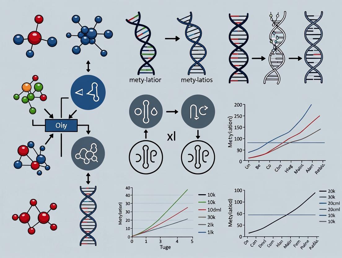
Visualizations
Title: Sources of Variability in the Epigenetic Workflow
Title: Bisulfite Conversion Chemistry and Key Steps
The Scientist's Toolkit: Research Reagent Solutions
| Item | Function & Importance |
|---|---|
| QIAamp DNA FFPE Tissue Kit | Silica-membrane based extraction optimized for cross-linked, fragmented FFPE DNA. Minimizes co-purification of inhibitors. |
| EZ DNA Methylation-Lightning Kit | Rapid bisulfite conversion reagent (90 minutes). Consistent performance with low DNA input and degraded samples. |
| PyroMark PCR Kit (Qiagen) | Includes HotStart Taq and optimized buffer for robust amplification of bisulfite-converted DNA, which is GC-rich and complex. |
| PyroMark Q48 Advanced Reagents | Pre-dispensed, single-use cartridges for pyrosequencing containing enzymes, substrate, and nucleotides. Reduces pipetting variability. |
| Methylated & Unmethylated DNA Controls | Absolute standards for assay validation and calibration. Used to construct standard curves and monitor assay linearity. |
| CUT&Tag Assay Kit | For low-input, high-signal ChIP-like experiments. Uses protein A-Tn5 fusion to tag target regions, reducing background vs. traditional ChIP. |
| SPRIselect Beads | Size-selective magnetic beads for post-bisulfite library cleanup (RRBS, WGBS) and fragment size selection. Critical for reproducible sequencing libraries. |
| Illumina Infinium HD Methylation Assay | Complete microarray kit for EPIC array processing. Includes all reagents for amplification, fragmentation, hybridization, and staining. |
Technical Support Center: Epigenetic Biomarker Protocol Troubleshooting
FAQ: Common Issues & Solutions
Q1: Our bisulfite conversion yields are consistently low (<95%), leading to high background noise in pyrosequencing. What are the primary culprits? A: Low conversion efficiency is often due to suboptimal DNA quality or incomplete bisulfite reaction. Ensure:
- Input DNA Integrity: Use agarose gel electrophoresis or a Fragment Analyzer to confirm DNA is high molecular weight (A260/280 ~1.8-2.0, A260/230 >2.0).
- Bisulfite Reaction Conditions: Verify pH of bisulfite solution is precisely 5.0. Incubation temperature must be tightly controlled (recommended: 95°C for denaturation, then 50-60°C for incubation). Use a thermal cycler with a heated lid, not a water bath.
- Desalting/ Purification: Post-conversion clean-up is critical. Use silica-column based kits designed for bisulfite-treated DNA. Ensure ethanol concentrations in wash buffers are correct.
Q2: We observe high inter-assay variability in DNA methylation levels (%5mC) measured by ELISA-based kits across different lab members. How can we standardize this? A: This variability typically stems from inconsistent sample handling and plate-reader calibration.
- Standardized Protocol: Create a detailed, step-by-step SOP with exact incubation times (use a timer), shaking speeds (use an orbital microplate shaker), and temperature conditions (use a controlled-temperature incubator).
- Reference Controls: Include a calibration curve with known %5mC controls (e.g., 0%, 50%, 100% methylated DNA) in every run. Also include a "positive control" sample of known methylation level.
- Instrument Calibration: Perform regular maintenance and calibration of the microplate reader according to manufacturer specs. Use the same reader for a study series.
Q3: Our ChIP-qPCR results for H3K27ac show poor enrichment and high background. What steps should we check? A: Poor ChIP efficiency can originate from multiple points in the workflow.
- Chromatin Shearing: This is the most critical step. Optimize shearing for each cell type/tissue. Use Covaris or Bioruptor for consistent sonication. Check fragment size (200-500 bp) on a gel after every preparation.
- Antibody Specificity & Quality: Use validated, ChIP-grade antibodies. Include an isotype control IgG and a positive control primer set (e.g., for GAPDH promoter) in every experiment.
- Wash Stringency: Ensure wash buffer compositions are correct and freshly prepared. Increase salt concentration in washes gradually if background is high.
Q4: How can we mitigate batch effects in large-scale methylation array (e.g., Illumina EPIC) studies? A: Batch effects from reagent lots, personnel, and processing days are a major reproducibility threat.
- Experimental Design: Randomize samples across arrays and processing batches. Do not process all cases in one batch and controls in another.
- Technical Replicates: Include at least one replicate sample (split from a large homogeneous DNA sample) on every array or in every batch.
- Reference Samples: Use commercially available, well-characterized reference DNA (e.g., from Coriell Institute) as an inter-batch calibrator.
- Bioinformatic Correction: Apply batch-effect correction algorithms (e.g., ComBat, SVA) in downstream analysis, but document this as a mandatory step in your methods.
Data Presentation: Impact of Standardization Variables
Table 1: Quantitative Impact of Protocol Variables on Experimental Outcomes
| Protocol Variable | Non-Standardized Range | Standardized Practice | Observed Impact on Coefficient of Variation (CV) |
|---|---|---|---|
| Bisulfite Conversion Incubation Time | 4-16 hours | 8 hours ± 15 min | CV reduced from 25% to 8% in %5mC measurement |
| ChIP Sonication Duration | 10-25 min (manual) | 15 min (Covaris, tuned) | H3K4me3 enrichment CV reduced from 40% to 12% |
| Methylation ELISA Development Time | "Until blue" (10-30 min) | 15 min exactly | Inter-operator CV reduced from 35% to 10% |
| DNA Input for Library Prep (NGS) | 50-200 ng | 100 ng ± 10% | Inter-library yield CV reduced from 50% to 15% |
Experimental Protocol: Standardized Bisulfite Conversion & Pyrosequencing
Title: Absolute Quantification of Methylation at a Specific CpG Locus
Methodology:
- DNA Qualification: Quantify 100-500 ng of genomic DNA using Qubit dsDNA HS Assay. Verify integrity via 0.8% agarose gel.
- Bisulfite Conversion: Use the EZ DNA Methylation-Lightning Kit (Zymo Research). Follow manufacturer's protocol with critical modifications:
- Use a thermal cycler: 98°C for 8 min (denaturation), 54°C for 60 min (conversion), hold at 4°C.
- Desalt using the provided spin columns.
- Elute in 20 µL of M-Elution Buffer.
- PCR Amplification: Design primers using PyroMark Assay Design SW. Perform PCR in 25 µL reactions with HotStarTaq Plus DNA Polymerase (Qiagen). Include no-template control.
- Cycling: 95°C 5 min; [95°C 30s, Ta 30s, 72°C 30s] x 45 cycles; 72°C 5 min.
- Pyrosequencing: Prepare single-stranded DNA using the PyroMark Q24 Vacuum Workstation. Sequence on a PyroMark Q24 system using prescribed dispensing order. Analyze results with PyroMark Q24 Software 2.0, which calculates % methylation per CpG site.
Mandatory Visualizations
Title: Targeted DNA Methylation Analysis Workflow
Title: ChIP Failure Mode and Effects Analysis
The Scientist's Toolkit: Research Reagent Solutions
Table 2: Essential Reagents for Standardized Epigenetic Protocols
| Reagent / Kit | Primary Function | Standardization Role |
|---|---|---|
| Qubit dsDNA HS Assay Kit (Thermo Fisher) | Accurate quantification of double-stranded DNA. | Prevents variance from inaccurate DNA input, critical for bisulfite conversion and NGS library prep. |
| EZ DNA Methylation-Lightning Kit (Zymo Research) | Rapid, efficient bisulfite conversion of unmethylated cytosines. | Well-characterized, consistent chemistry reduces conversion variability, a major source of bias. |
| Methylated & Unmethylated Human Control DNA (Zymo or Millipore) | Positive controls for bisulfite-based assays (PCR, arrays). | Provides a calibration standard for conversion efficiency and assay sensitivity across batches. |
| Covaris S220 Ultrasonicator | Reproducible, focused acoustic shearing of chromatin/DNA. | Replaces variable sonication methods, ensuring consistent fragment size for ChIP-seq and NGS. |
| MagMeDIP Kit (Diagenode) | Magnetic bead-based immunoprecipitation of methylated DNA. | Streamlines MeDIP protocol, reducing hands-on time and operator-dependent variability. |
| PyroMark PCR Kit (Qiagen) | Optimized polymerase and buffer for robust amplification of bisulfite-converted DNA. | Minimizes PCR bias and ensures uniform product yield for accurate pyrosequencing. |
| SPRIselect Beads (Beckman Coulter) | Solid-phase reversible immobilization for DNA size selection and clean-up. | Provides a consistent, automatable alternative to column-based clean-ups for NGS library preparation. |
Technical Support Center
Troubleshooting Guides & FAQs
DNA Methylation Analysis
Q: I am getting inconsistent bisulfite conversion efficiency in my samples. What are the primary causes?
- A: Inconsistent conversion is often due to degraded input DNA, suboptimal bisulfite reagent pH/temperature, or incomplete desulfonation. Ensure high-quality, high-molecular-weight DNA input (A260/A280 ~1.8-2.0). Verify the pH of bisulfite solution is between 5.0-5.2. Strictly adhere to recommended thermal cycling conditions (e.g., 95°C for denaturation, 50-60°C for incubation). Include unmethylated and methylated control DNA in every run.
Q: My pyrosequencing or NGS results show low PCR efficiency for bisulfite-converted DNA. How can I improve this?
- A: Design primers specific to the bisulfite-converted sequence, avoiding CpG sites within the primer sequence. Keep amplicons short (<300 bp). Optimize magnesium concentration and use a polymerase specifically optimized for bisulfite-converted DNA (high processivity, uracil tolerance). Increase the number of PCR cycles (e.g., 45-50 cycles).
Histone Modification Analysis
Q: My chromatin immunoprecipitation (ChIP) yields low signal-to-noise ratio or high background. What should I check?
- A: This typically points to antibody specificity or chromatin fragmentation issues.
- Antibody: Always use ChIP-validated antibodies. Perform a titration experiment for each new lot. Include a positive control (known enriched region) and a negative control (non-enriched region).
- Chromatin: Optimize sonication or enzymatic digestion conditions for your cell type to achieve fragments between 200-500 bp. Over-fixation (>10 mins with 1% formaldehyde) can mask epitopes; consider a time course (5-15 mins).
- Wash Stringency: Increase salt concentration in wash buffers step-wise to reduce non-specific binding.
- A: This typically points to antibody specificity or chromatin fragmentation issues.
Q: How do I normalize ChIP-qPCR data effectively?
- A: Use a combination of controls. Standard methods include:
- Percent Input Method: % Input = 2^(Ct[Input] - Ct[IP]) * 100. Accounts for chromatin prep efficiency.
- Normalization to Histone H3 or Total Histone: For histone marks, correct for nucleosome density.
- Internal Reference Locus: Normalize to a constitutively silent region (e.g., gene desert) to account for non-specific background. Data should be presented as fold enrichment over this control.
- A: Use a combination of controls. Standard methods include:
Non-Coding RNA Analysis
Q: I detect high variability in miRNA recovery from biofluids like plasma. How can I standardize my protocol?
- A: Variability stems from pre-analytical factors and isolation methods.
- Sample Collection: Standardize blood collection tubes (e.g., use cell-free RNA tubes), processing time (<2 hours), and centrifugation steps (e.g., 1600 x g, then 16,000 x g).
- Isolation: Use phenol-free, column-based kits optimized for small RNAs. Include a carrier RNA during isolation to improve low-concentration miRNA yields.
- Normalization: Use spike-in synthetic, non-human miRNAs (e.g., C. elegans miR-39, -54, -238) added at the beginning of isolation to correct for technical variability in extraction and reverse transcription.
- A: Variability stems from pre-analytical factors and isolation methods.
Q: My RT-qPCR for lncRNAs shows non-specific amplification. What are the troubleshooting steps?
- A: Design primers spanning exon-exon junctions to avoid genomic DNA amplification. Use a hot-start, high-fidelity polymerase. Optimize annealing temperature with a gradient PCR. Always include a no-reverse-transcriptase (-RT) control. For low-abundance lncRNAs, consider using a locked nucleic acid (LNA)-enhanced qPCR assay for superior specificity.
Table 1: Common Quantitative Benchmarks for Epigenetic Assays
| Biomarker Class | Assay | Key Performance Metric | Optimal/Target Value | Corrective Action if Out of Range |
|---|---|---|---|---|
| DNA Methylation | Bisulfite Conversion | Conversion Efficiency | ≥99% | Optimize bisulfite reaction pH, time, and temperature. |
| DNA Methylation | Pyrosequencing | CpG Site CV (between replicates) | <5% | Re-optimize PCR, ensure homogeneous template. |
| Histone Modifications | ChIP-qPCR | Signal-to-Noise (Enrichment over IgG) | ≥10-fold | Titrate antibody, optimize wash stringency, check chromatin quality. |
| Histone Modifications | CUT&Tag / CUT&RUN | Sequencing Library Size Distribution | Peak ~200-500 bp | Titrate digestion enzyme (pA-Tn5), optimize incubation time. |
| Non-Coding RNAs | miRNA RT-qPCR | Amplification Efficiency (from standard curve) | 90-110% (Slope -3.1 to -3.6) | Redesign primers/probe, optimize reagent concentrations. |
| Non-Coding RNAs | RNA-Seq (lncRNA) | rRNA Depletion Efficiency | >90% rRNA removed | Use more input RNA, ensure riboprobe/rRNA binder is fresh. |
Detailed Methodologies for Key Experiments
Protocol 1: Bisulfite Pyrosequencing for DNA Methylation Quantification
- DNA Bisulfite Conversion: Use 500 ng of genomic DNA with the EZ DNA Methylation-Lightning Kit (Zymo Research). Incubate at 98°C for 8 minutes, then 54°C for 60 minutes. Desulfonate and elute in 20 µL.
- PCR Amplification: Amplify 2 µL of converted DNA with HotStarTaq Plus (Qiagen). Program: 95°C for 5 min; 45 cycles of (95°C 30s, specific Ta 45s, 72°C 45s); final extension 72°C for 5 min. Use biotinylated primer.
- Pyrosequencing: Bind PCR product to Streptavidin Sepharose HP beads, denature, and anneal sequencing primer. Run on a PyroMark Q48 instrument using PyroMark Gold Q48 reagents. Quantify methylation percentage per CpG using PyroMark Q48 Autoprep software.
Protocol 2: Chromatin Immunoprecipitation (ChIP) for Histone H3K27ac
- Crosslinking & Lysis: Crosslink 1x10^6 cells with 1% formaldehyde for 10 min at RT. Quench with 125 mM glycine. Lyse cells in SDS Lysis Buffer.
- Chromatin Shearing: Sonicate lysate to shear DNA to 200-500 bp fragments. Centrifuge to pellet debris.
- Immunoprecipitation: Dilute chromatin in ChIP Dilution Buffer. Pre-clear with Protein A/G beads for 1h. Incubate 10 µg chromatin with 1 µg of anti-H3K27ac antibody (e.g., Abcam ab4729) overnight at 4°C. Add beads and incubate 2h.
- Washes & Elution: Wash sequentially with Low Salt, High Salt, LiCl, and TE buffers. Elute chromatin in Elution Buffer (1% SDS, 0.1M NaHCO3).
- Reverse Crosslinks & Purification: Add NaCl and heat at 65°C overnight. Treat with Proteinase K. Purify DNA with spin columns. Analyze by qPCR.
Protocol 3: Isolation and qPCR Profiling of Circulating miRNA from Plasma
- Plasma Preparation: Collect blood in EDTA or citrate tubes. Centrifuge at 1600 x g for 15 min at 4°C. Transfer plasma to a new tube. Re-centrifuge at 16,000 x g for 10 min to remove cell debris. Aliquot and store at -80°C.
- RNA Isolation: Use the miRNeasy Serum/Plasma Advanced Kit (Qiagen). Add 3.5 µL of a 1.6 nM synthetic C. elegans miRNA spike-in mix (e.g., miR-39, -54) to 200 µL plasma. Add Qiazol, mix, and proceed with chloroform extraction. Bind RNA to column, wash, and elute in 20 µL RNase-free water.
- RT-qPCR: Use the miRCURY LNA RT Kit (Qiagen). Perform reverse transcription. For qPCR, use miRCURY LNA SYBR Green PCR Assays. Run in triplicate on a CFX96 system. Normalize Cq values to the spike-in Cq using the 2^-ΔΔCt method.
Visualizations
Bisulfite Pyrosequencing Workflow
Chromatin Immunoprecipitation (ChIP) Workflow
Interplay Between Epigenetic Biomarker Classes
The Scientist's Toolkit: Research Reagent Solutions
Table 2: Essential Reagents for Standardized Epigenetic Biomarker Analysis
| Reagent/Material | Primary Function | Example Product/Brand |
|---|---|---|
| DNA Bisulfite Conversion Kit | Chemically converts unmethylated cytosine to uracil, leaving 5-methylcytosine unchanged. | EZ DNA Methylation-Lightning Kit (Zymo Research), Epitect Bisulfite Kit (Qiagen). |
| ChIP-Validated Antibody | High-specificity antibody for immunoprecipitation of a specific histone modification or chromatin protein. | Cell Signaling Technology ChIP Validated Antibodies, Abcam histone modification antibodies. |
| Magnetic Protein A/G Beads | Efficient capture of antibody-chromatin complexes for washing and elution. | Dynabeads Protein A/G (Thermo Fisher), ChIP-grade magnetic beads (Millipore). |
| Cell-Free RNA Collection Tube | Stabilizes extracellular RNA profile in blood samples by inhibiting RNases and preventing cellular RNA release. | PAXgene Blood ccfRNA Tube (PreAnalytiX), Cell-Free RNA BCT (Streck). |
| Small RNA Isolation Kit | Optimized silica-membrane columns or magnetic beads for efficient recovery of miRNAs and other small RNAs. | miRNeasy Serum/Plasma Advanced Kit (Qiagen), mirVana miRNA Isolation Kit (Thermo Fisher). |
| Universal cDNA Synthesis Kit | Reverse transcription system with high processivity and uniform efficiency for diverse RNA inputs, including small RNAs. | miRCURY LNA RT Kit (Qiagen), TaqMan Advanced miRNA cDNA Synthesis Kit (Thermo Fisher). |
| Spike-In Control RNA (Artificial) | Synthetic, non-homologous RNAs added to samples for normalization of extraction, RT, and qPCR efficiency. | C. elegans miRNA Spike-In Kit (Thermo Fisher), UniSpike RNA (Exiqon). |
Technical Support Center: Troubleshooting Guides & FAQs
Context: This support center is designed within the framework of a thesis on standardizing epigenetic biomarker protocols. It addresses common experimental pitfalls related to major international consortia and guideline specifications.
Frequently Asked Questions (FAQs)
Q1: During a BLUEPRINT-style ChIP-seq assay for H3K27ac, I obtain low enrichment and high background. What are the primary troubleshooting steps?
A: This is often related to antibody quality or chromatin fragmentation. Follow this protocol:
- Validate Antibody: Perform a dot-blot or western blot against recombinant histone with the modification of interest. Use a spike-in control (e.g., Drosophila chromatin) for quantitative assessment.
- Optimize Sonication: Verify fragment size distribution on a bioanalyzer. Target 200-500 bp fragments. If suboptimal, adjust sonication parameters (duration, pulse settings, sample volume). Over-sonication can cause denaturation.
- Increase Wash Stringency: Add an additional high-salt (500 mM NaCl) wash step after IP to reduce non-specific binding.
- Quantify Input DNA: Ensure you are using the recommended 2% input DNA for library normalization and peak calling.
Q2: When aligning bisulfite-seq data for methylation analysis per IHEC standards, alignment rates are consistently below 70%. How to resolve?
A: Low alignment typically stems from incomplete bisulfite conversion or adapter contamination.
- Bisulfite Conversion Check: Include unmethylated (e.g., Lambda phage DNA) and fully methylated controls in your conversion reaction. Calculate conversion efficiency (>99% is required). Inefficient conversion creates C's not accounted for in the bisulfite-aware aligner.
- Adapter Trimming: Use strict adapter trimming tools (e.g., Trim Galore!, Cutadapt) before alignment. Insperse fastqc reports for adapter content.
- Aligner Selection & Parameters: Use recommended aligners (Bismark, BS-Seeker2). Ensure you are providing the correct strandedness parameter (
--non_directionalfor post-bisulfite adaptor tagging protocols). - Reference Genome: Confirm you are using the same genome build (GRCh38/no-alt) as specified in the IHEC metadata standards.
Q3: For cell-free DNA (cfDNA) methylation sequencing, as discussed in ISO/TC 276 guidelines, how do I mitigate PCR duplicates arising from low input?
A: ISO/TC 276 emphasizes molecular tagging to distinguish technical duplicates from true biological fragments.
- Protocol: Use a library preparation kit that incorporates Unique Molecular Identifiers (UMIs) or randomers during the initial adapter ligation or pre-amplification step.
- Bioinformatics: After alignment, use tools like
picard MarkDuplicates(withBARCODE_TAGoption) orUMI-toolsto group reads by their UMI and genomic start/end coordinates. Deduplicate based on UMI families, not just mapping coordinates. - Quantitative Table: The impact of UMI-based deduplication:
| Deduplication Method | Input Material | Estimated Retained Reads | Advantage |
|---|---|---|---|
| Coordinate-Only | High-input gDNA | ~40-60% | Standard, simple. |
| UMI-Based | Low-input cfDNA (<50 ng) | ~70-85% | Preserves true biological diversity, critical for low allele-frequency biomarker detection. |
Q4: My ATAC-seq data, following current best practices from consortia, shows high mitochondrial read contamination (>50%). How can I reduce this?
A: High mitochondrial reads indicate excessive cell lysis or insufficient nuclei purification.
- Optimize Lysis: Titrate the concentration and incubation time of the lysis buffer (typically NP-40 or digitonin). Use cold lysis buffers and perform steps on ice. Visually check nuclei integrity under a microscope after lysis.
- Nuclei Wash: Add two extra gentle centrifugation washes with cold PBS after lysis, before the transposition reaction.
- Cell Type Consideration: Some cell types (e.g., adipocytes, muscle cells) naturally have high mitochondrial content. You may need to physically isolate nuclei via density gradient centrifugation.
- Bioinformatics Removal: Align to the full genome (including chrM) and filter mitochondrial reads post-alignment. However, this wastes sequencing depth; experimental mitigation is preferred.
Detailed Experimental Protocols
Protocol 1: BLUEPRINT-Compliant ChIP-seq for Histone Modifications
- Cross-linking: Treat 1-5 million cells with 1% formaldehyde for 10 min at RT. Quench with 125 mM glycine.
- Chromatin Prep: Lyse cells (LB1: 50 mM HEPES-KOH pH7.5, 140 mM NaCl, 1 mM EDTA, 10% Glycerol, 0.5% NP-40, 0.25% Triton X-100). Pellet nuclei, then lyse nuclei (LB2: 10 mM Tris-HCl pH8.0, 200 mM NaCl, 1 mM EDTA, 0.5 mM EGTA). Pellet and resuspend in shearing buffer.
- Sonication: Sonicate using a focused ultrasonicator (e.g., Covaris) to achieve 200-500 bp fragments. Centrifuge to remove debris.
- Immunoprecipitation: Incubate 10-50 µg chromatin with 1-5 µg validated antibody overnight at 4°C with rotation. Add protein A/G beads for 2 hours. Wash sequentially with: Low Salt Wash Buffer, High Salt Wash Buffer, LiCl Wash Buffer, and TE Buffer.
- Elution & Decrosslinking: Elute in ChIP Elution Buffer (1% SDS, 0.1M NaHCO3). Add NaCl to 200 mM and incubate at 65°C overnight. Treat with RNase A and Proteinase K.
- DNA Purification: Purify using SPRI beads. Quantify by qPCR at positive and negative control genomic regions before library prep.
Protocol 2: IHEC-Aligned Whole Genome Bisulfite Sequencing (WGBS)
- DNA Qualification: Start with high-quality, high-molecular-weight DNA (RIN > 8.5). Use Qubit for quantification.
- Bisulfite Conversion: Use the EZ DNA Methylation-Lightning Kit (Zymo Research) or equivalent. Follow manufacturer's instructions precisely. Include 0.5 ng of unmethylated lambda DNA to calculate conversion efficiency.
- Library Preparation: Perform post-bisulfite adaptor tagging (PBAT) or a standard library prep with bisulfite-converted DNA. Use KAPA Uracil+ or Pico Methyl libraries for degraded/low-input samples.
- Quality Control: Assess library fragment size on a Bioanalyzer (Agilent) and quantify by qPCR (KAPA Library Quant). Calculate bisulfite conversion efficiency from lambda DNA alignment (>99%).
- Sequencing: Sequence on an Illumina platform to a minimum depth of 30x coverage (CpG sites).
Visualizations
Diagram 1: IHEC Epigenomic Data Generation Workflow
Diagram 2: ISO/TC 276 cfDNA Methylation Analysis with UMIs
The Scientist's Toolkit: Key Research Reagent Solutions
| Reagent/Kit | Primary Function | Key Application & Rationale |
|---|---|---|
| Covaris S2/S220 | Acoustic Shearing | Provides consistent, tunable fragmentation of chromatin for ChIP-seq or DNA for sequencing libraries, critical for reproducible peak definition. |
| Diagenode Bioruptor | Sonication | Alternative for chromatin shearing; uses water bath sonication for multiple samples simultaneously. |
| Zymo EZ DNA Methylation-Lightning Kit | Bisulfite Conversion | Rapid, high-efficiency conversion of unmethylated cytosines to uracil for bisulfite sequencing assays. |
| KAPA HyperPrep Kit (with UDI Indexes) | NGS Library Preparation | Robust, flexible library construction for ChIP-seq, ATAC-seq, etc. UDI indexes prevent index hopping errors in multiplexed sequencing. |
| NEBNext Ultra II FS DNA Library Kit | Library Prep for FFPE/degraded DNA | Incorporates a repair step and is optimized for challenging, fragmented input like cfDNA or archival samples. |
| SPRIselect Beads (Beckman Coulter) | Size Selection & Cleanup | Paramagnetic bead-based purification for precise selection of DNA fragment sizes post-sonication or post-PCR. |
| Anti-H3K27ac antibody (Diagenode, C15410196) | Histone Modification IP | Highly validated antibody for ChIP-seq of active enhancer and promoter marks; cited in BLUEPRINT studies. |
| Drosophila melanogaster S2 Chromatin (Active Motif) | Spike-in Control | Added to human ChIP reactions to normalize for technical variation (antibody efficiency, IP losses) across experiments. |
Technical Support Center: Troubleshooting Epigenetic Biomarker Analysis
FAQs & Troubleshooting Guides
Q1: During multi-center analysis of DNA methylation via bisulfite sequencing, we observe high inter-site variability in methylation percentages for control samples. What are the primary technical sources? A: Variability often stems from pre-analytical and bisulfite conversion steps. Key factors include:
- DNA Input Quality & Quantity: Degraded DNA or inconsistent quantification leads to uneven conversion.
- Bisulfite Conversion Kit/Protocol Differences: Variation in incubation time, temperature, or reagent purity significantly impacts conversion efficiency.
- Post-Bisulfite DNA Handling: Inconsistent desalting or elution can cause fragment loss, biasing PCR amplification.
Q2: Our chromatin immunoprecipitation (ChIP) results for histone marks (e.g., H3K27ac) show poor signal-to-noise ratio and are not reproducible across labs. How can we troubleshoot this? A: This typically indicates issues with antibody specificity or chromatin preparation.
- Antibody Validation: Use validated antibodies (e.g., from the ENCODE consortium) and include both positive and negative control genomic regions in every assay.
- Cross-linking Conditions: Over- or under-crosslinking can mask epitopes or cause non-specific trapping. Optimize formaldehyde concentration and duration for your cell type.
- Sonication Consistency: Fragment size distribution must be consistent and verified via capillary electrophoresis. Differing sonicators or settings create bias.
Q3: When performing RRBS (Reduced Representation Bisulfite Sequencing) across sites, we get inconsistent coverage of CpG islands. What steps should we check? A: Inconsistent coverage usually originates from the restriction digest and size selection steps.
- Restriction Enzyme Activity: Ensure consistent enzyme lots and avoid freeze-thaw cycles. Verify complete digestion by running a QC gel.
- Size Selection Precision: Strict adherence to magnetic bead-to-sample ratios and incubation times is critical. Use a calibrated automated system or highly trained personnel.
- Library Quantification Method: Use fluorometric methods (e.g., Qubit) over spectrophotometry for accurate post-bisulfite library quantification before sequencing.
Q4: How do we address batch effects in epigenetic data when pooling results from multiple centers for regulatory submission? A: Batch correction must be planned prospectively.
- Experimental Design: Distribute samples from all experimental groups across all participating sites and sequencing batches.
- Use of Reference Materials: Include shared, aliquoted reference control samples (e.g., commercial methylated/unmethylated DNA, or a universal cell line) in every processing batch at each site.
- Bioinformatic Correction: Apply established algorithms (e.g., ComBat, RUVm) but document and justify all parameters. Provide raw and corrected data to regulators.
Standardized Experimental Protocols
Protocol 1: Harmonized Bisulfite Conversion for DNA Methylation Analysis
- Principle: Sodium bisulfite converts unmethylated cytosines to uracil, while methylated cytosines remain unchanged.
- Standardized Method:
- DNA QC: Quantify input DNA (50ng) using fluorometry. Accept only samples with 260/280 ratio of 1.8-2.0 and fragment size >1kb on gel.
- Bisulfite Conversion: Use a specified commercial kit. Incubate at 98°C for 10 minutes, then 60°C for 2.5 hours exactly.
- Desalting: Bind DNA to provided magnetic beads at room temperature (25°C) for 15 minutes. Wash twice with 80% ethanol.
- Elution: Elute in 25 µL of low-EDTA TE buffer (pH 8.0). Store at -80°C if not used immediately for PCR.
Protocol 2: Harmonized ChIP-qPCR for Histone Modification Validation
- Principle: Antibodies specific to histone modifications immunoprecipitate bound DNA fragments for quantification.
- Standardized Method:
- Cross-linking & Sonication: Fix 1x10^6 cells with 1% formaldehyde for 10 minutes. Quench with 125mM glycine. Sonicate to achieve 200-500 bp fragments (verified post-sonication).
- Immunoprecipitation: Use 2 µg of validated antibody and 20 µL of protein A/G magnetic beads per reaction. Incubate overnight at 4°C with rotation.
- Washing & Elution: Wash sequentially with Low Salt, High Salt, and LiCl buffers. Elute complexes in 100 µL of elution buffer (1% SDS, 100mM NaHCO3) at 65°C for 30 minutes.
- DNA Recovery: Reverse cross-links at 65°C overnight. Purify DNA using a PCR purification kit. Analyze via qPCR against predefined genomic controls.
Data Presentation
Table 1: Impact of Protocol Harmonization on Multi-Center DNA Methylation Data Variability
| Metric | Pre-Harmonization (6 sites) | Post-Harmonization (6 sites) |
|---|---|---|
| CV* of Control Sample Methylation (%) | 18.5% | 4.2% |
| Inter-site Correlation Coefficient (r) | 0.76 | 0.97 |
| Data Yield Variance (SD) | 15.8 Gb | 3.2 Gb |
| Protocol Adherence Rate | 65% | 95% |
| CV: Coefficient of Variation |
Table 2: Key Reagent Solutions for Standardized Epigenetic Workflows
| Reagent / Material | Function & Rationale for Standardization |
|---|---|
| Universal Human Methylated DNA Standard | Provides a bisulfite conversion and sequencing control across all batches and sites for data normalization. |
| Validated ChIP-Grade Antibody Panel | Pre-qualified antibodies for specific histone marks (H3K4me3, H3K27me3) ensure specificity and reproducibility. |
| CpG Methyltransferase (M.SssI) | Used to generate fully methylated control DNA for assay calibration and efficiency calculations. |
| Magnetic Beads for Size Selection | Standardized bead chemistry and size (e.g., SPRIs) ensures reproducible fragment selection in RRBS/NGS. |
| Cell Line Reference (e.g., GM12878) | A widely characterized, publicly available cell line serves as a shared biological control across centers. |
Visualizations
Title: Harmonized Multi-Center Study Workflow
Title: Standardized DNA Methylation Analysis Protocol
Building Robust Pipelines: Best Practices for Key Epigenetic Techniques
Troubleshooting Guides & FAQs
Q1: During blood plasma collection for cell-free DNA (cfDNA) analysis, my yields are low and highly variable. What are the critical steps I might be missing? A: The pre-analytical phase is paramount for cfDNA, a key epigenetic biomarker source. Ensure:
- Centrifugation Protocol: Use a standardized two-step centrifugation protocol. First, centrifuge whole blood in EDTA tubes at 800-1600 x g for 10 minutes at 4°C to separate plasma from cells. Then, transfer the plasma to a new tube and centrifuge at 16,000 x g for 10 minutes at 4°C to remove residual cells and platelets. This prevents genomic DNA contamination.
- Timing: Process blood samples within 1-2 hours of collection. Prolonged storage of unprocessed blood at room temperature causes leukocyte lysis and contaminates the cfDNA pool.
- Tube Type: Use dedicated cfDNA/ctDNA stabilization tubes (e.g., Streck, Roche) if immediate processing is not possible. These tubes preserve cell integrity for several days.
Q2: My FFPE tissue-derived DNA is fragmented, and subsequent bisulfite conversion for DNA methylation analysis fails. How can I improve sample input quality? A: FFPE introduces cross-linking and fragmentation. Standardize these steps:
- Sectioning & De-crosslinking: Use a fresh microtome blade for each block. For nucleic acid extraction, perform a de-crosslinking step post-deparaffinization: incubate the pellet in a buffer containing proteinase K at 56°C for 3 hours to overnight, followed by a heat step (90°C for 20-30 minutes).
- Extraction & QC: Use a silica-membrane-based kit optimized for FFPE. Quantify DNA using a fluorometric method (e.g., Qubit) and assess fragment size distribution with a Bioanalyzer or TapeStation. Acceptable samples for bisulfite conversion should have a DV200 (percentage of fragments >200bp) >30%. Bisulfite conversion kits with carrier RNA are recommended for low-input/degraded samples.
Q3: After long-term storage of extracted nucleic acids at -80°C, I notice a drop in PCR amplification efficiency. What are the best practices for archiving? A: Degradation can occur even at -80°C due to residual nuclease activity and freeze-thaw cycles.
- Storage Buffer: Elute or resuspend DNA/RNA in 10 mM Tris-HCl (pH 8.0-8.5) or TE buffer, not nuclease-free water, which lacks buffering capacity.
- Aliquoting: Divide the sample into single-use aliquots to avoid repeated freeze-thaw cycles (more than 2-3 cycles is detrimental).
- Storage Vessels: Use low-protein-binding, nuclease-free tubes. For RNA, consider RNAstable or similar products for ambient temperature storage.
Q4: My nucleic acid extraction yields from biofluids (urine, saliva) are inconsistent. How can I standardize this? A: Biofluids have inherent variability. Introduce an internal control.
- Protocol: Add a known quantity of synthetic, non-human spike-in nucleic acid (e.g., from Salmonella, Phage Lambda, or artificially engineered sequences) to the biofluid sample immediately upon receipt or at the start of lysis. This controls for variability in extraction efficiency and allows for normalization of target analyte recovery.
- Standardization: The recovery rate of the spike-in can be used to calculate a correction factor for the target analyte's measured concentration, improving inter-assay comparability.
Table 1: Impact of Blood Processing Delay on cfDNA Yield and Integrity
| Time to Plasma Processing (hrs, RT) | cfDNA Concentration (ng/mL plasma) | Genomic DNA Contamination (ΔCq value)* | % of Samples with DV200 >50% |
|---|---|---|---|
| <2 | 5.2 ± 1.8 | 8.5 ± 0.9 | 98% |
| 4-6 | 8.1 ± 3.5 | 5.2 ± 1.1 | 75% |
| 24 (in Stabilization Tube) | 6.5 ± 2.1 | 8.1 ± 0.8 | 95% |
*ΔCq = Cq[genomic target] - Cq[cfDNA target]. Higher values indicate less contamination.
Table 2: Recommended Storage Conditions for Nucleic Acids in Epigenetic Studies
| Nucleic Acid Type | Recommended Buffer | Optimal Temp. | Max # Freeze-Thaws | Alternative for Long-Term |
|---|---|---|---|---|
| Genomic DNA | TE Buffer (pH 8.0) | -80°C | 5 | 4°C (for stable DNA) |
| Bisulfite-converted DNA | TE Buffer or Kit Elution Buffer | -80°C | 1 (avoid if possible) | Desiccant at -20°C |
| Total RNA | TE Buffer or RNase-free Water | -80°C | 3 | RNA stabilization matrix |
| cell-free RNA | TE Buffer with Carrier RNA | -80°C | 0 (store aliquoted) | Not recommended |
Detailed Experimental Protocols
Protocol 1: Standardized Two-Step Plasma Isolation from Whole Blood for cfDNA Analysis
- Collection: Draw blood into K2EDTA tubes. Invert gently 8-10 times.
- First Spin (Cell Separation): Centrifuge tubes at 1600 x g for 10 minutes at 4°C (brake ON).
- Plasma Transfer: Using a sterile pipette, carefully transfer the upper plasma layer (approximately 1-2 mL, avoiding the buffy coat) to a fresh 2 mL microcentrifuge tube.
- Second Spin (Platelet Removal): Centrifuge the transferred plasma at 16,000 x g for 10 minutes at 4°C (brake ON).
- Final Plasma Harvest: Transfer the supernatant (platelet-poor plasma) to a final, clearly labeled cryovial.
- Storage: Immediately freeze at -80°C. For cfDNA extraction, process from fresh plasma is preferred.
Protocol 2: Internal Control Spike-in for Biofluid Nucleic Acid Extraction
- Spike-in Solution Preparation: Dilute the synthetic, non-homologous nucleic acid (e.g., 1x10^6 copies/µL) in nuclease-free buffer.
- Spiking: Prior to adding lysis buffer, add a fixed volume of the spike-in solution to your biofluid sample (e.g., 5 µL of 1x10^4 copies/µL into 1 mL urine). Vortex gently.
- Extraction: Proceed with your standard extraction protocol (e.g., silica-column or magnetic bead-based).
- QC and Normalization: Quantify both the spike-in (using a dedicated qPCR assay) and your target analyte. Calculate the percent recovery of the spike-in. The target analyte concentration can be adjusted using the formula: Normalized Concentration = (Measured Concentration) / (Percent Recovery/100).
Diagrams
Title: Standardized Plasma Processing Workflow for cfDNA
Title: Biofluid Extraction Standardization with Spike-in Control
The Scientist's Toolkit: Research Reagent Solutions
| Item | Function in Standardization |
|---|---|
| cfDNA BCT Tubes (Streck) | Preserves blood cell integrity, prevents lysis, and stabilizes cfDNA for up to 14 days at room temperature, standardizing pre-processing timelines. |
| Proteinase K (Molecular Grade) | Essential for efficient digestion of proteins and reversal of formaldehyde crosslinks in FFPE tissues, ensuring complete nucleic acid release. |
| RNA/DNA Shield (Zymo Research) | A stabilization buffer that instantly inactivates nucleases in biological samples, allowing for ambient temperature storage and transport. |
| Silica-Membrane Spin Columns | Provide a consistent, automatable method for nucleic acid purification, removing PCR inhibitors and yielding high-purity extracts crucial for downstream epigenetic assays. |
| ERCC RNA Spike-in Mix (Thermo Fisher) | A set of synthetic RNA transcripts at known concentrations used to normalize RNA-seq data, controlling for technical variation in extraction and library prep. |
| Lambda Phage DNA | A common non-human spike-in control for DNA extraction protocols, used to monitor and normalize for extraction efficiency losses. |
| Bisulfite Conversion Kit (e.g., EZ DNA Methylation) | Standardizes the harsh bisulfite conversion process, ensuring complete and reproducible deamination of unmethylated cytosines for methylation analysis. |
| High-Sensitivity DNA/RNA Assays (e.g., Qubit, Bioanalyzer) | Fluorometric and electrophoretic QC tools essential for accurately quantifying and assessing the integrity of precious, low-input epigenetic samples. |
Technical Support Center: Troubleshooting & FAQs
FAQs on Bisulfite Conversion
Q1: My post-conversion DNA yield is extremely low (<10%). What are the primary causes? A: Low recovery is commonly due to DNA degradation. Ensure pH of the bisulfite reaction is precisely between 5.0-5.2. Use a recent, high-quality bisulfite reagent kit. For FFPE samples, optimize de-crosslinking prior to conversion. Always include a high-molecular-weight DNA control to assess process efficiency.
Q2: How can I assess the completeness of bisulfite conversion before proceeding to arrays or sequencing? A: Perform a methylation-specific PCR (MSP) or pyrosequencing for a known, fully unmethylated control locus (e.g., ALU elements). A successful conversion will show >99% C-to-T conversion in the unmethylated cytosines. Dedicated qPCR assays for conversion efficiency are also commercially available.
FAQs on Array-Based Analysis (e.g., Illumina Infinium)
Q3: My sample fails the Infinium array staining intensity threshold. What steps should I take? A: This typically indicates poor bisulfite-converted DNA quality or quantity. Re-quantify converted DNA using a fluorescence-based assay specific for ssDNA. Ensure the restoration step was performed correctly. Verify that the hybridization oven temperature and flow cell conditions are within specification.
Q4: I see high background noise or poor cluster separation in my array data. How can I troubleshoot? A: This can result from suboptimal beadchip washing or blocking. Ensure all washing buffers are at the correct temperature and prepared freshly. Check for expired or contaminated staining reagents. Perform a visual inspection of the beadchip surface for bubbles or debris after assembly.
FAQs on Bisulfite Sequencing (WGBS/RRBS)
Q5: My bisulfite sequencing library shows excessive adapter dimers. How do I mitigate this? A: This is common in WGBS due to the low input and fragmented DNA. Increase the ratio of clean-up bead size selection and perform double-sided size selection. Use adapter-specific depletion beads if available. Optimize PCR cycle number to prevent over-amplification of small fragments.
Q6: My genome alignment rate for WGBS is lower than expected (<70%). What could be wrong? A: Incomplete bisulfite conversion leads to un-converted cytosines that mis-map. Check conversion efficiency first. Also, ensure your aligner (e.g., Bismark, BS-Seeker2) is using the correct genome index (bisulfite-converted in silico). High duplication rates from low input can also reduce apparent alignment; examine duplicate marking metrics.
Table 1: Comparison of Key DNA Methylation Analysis Platforms
| Platform | Typical Input (Converted DNA) | CpG Coverage | Cost per Sample | Best For |
|---|---|---|---|---|
| Infinium EPIC v2.0 | 250 ng | > 935,000 CpG sites | $$ | Targeted, high-throughput biomarker studies |
| Whole-Genome Bisulfite Sequencing (WGBS) | 50-100 ng | ~28 million CpGs | $$$$ | Discovery, non-CpG methylation, comprehensive analysis |
| Reduced Representation Bisulfite Sequencing (RRBS) | 10-100 ng | ~2-3 million CpGs | $$$ | Cost-effective discovery focusing on CpG islands/promoters |
| Pyrosequencing | 10-20 ng | 5-10 CpGs per assay | $ | Validation of specific loci, high quantitative accuracy |
Table 2: Common Bisulfite Conversion Kit Performance Metrics
| Kit | Optimal Input Range | Incubation Time | Average Recovery* | DNA Fragment Size Post-Conversion |
|---|---|---|---|---|
| Kit A (Premium) | 10 pg - 2 µg | 90 min | 50-70% | < 500 bp |
| Kit B (High-Throughput) | 100 ng - 1 µg | 60 min | 40-60% | < 1 kb |
| Kit C (FFPE-Optimized) | 50 ng - 500 ng | Overnight | 30-50% | < 300 bp |
*Recovery is highly sample-dependent. Values are for high-quality genomic DNA.
Experimental Protocols
Protocol 1: Standardized Bisulfite Conversion for FFPE DNA (Based on CAPP-Seq Principles)
Context for Standardization: This protocol aims to minimize variability in de-crosslinking and conversion, a major hurdle in biomarker research.
- De-paraffinization & De-crosslinking: Cut 2-3 x 10 µm FFPE sections. Deparaffinize with xylene, wash with ethanol. Incubate in digestion buffer (Proteinase K, 20mg/ml) at 56°C for 3 hours, then 80°C for 2 hours.
- DNA Clean-up: Purify using magnetic beads sized for 100-300bp fragments to select for compatible FFPE-derived DNA. Elute in 40 µL of low TE buffer.
- Bisulfite Reaction: Use 20 µL of purified DNA with a commercial kit optimized for low-input/degraded DNA. Program thermocycler: Denaturation (95°C, 5 min), Incubation (60°C, 90 min), Hold (20°C).
- Desulfonation & Clean-up: Transfer reaction to a clean tube with desulphonation buffer, incubate (room temp, 15 min). Clean up using the provided columns or beads. Elute in 20 µL of low TE. Store at -80°C.
Protocol 2: Validation of Array-Based Biomarkers by Pyrosequencing
Context for Standardization: Essential for cross-platform validation of discovered epigenetic biomarkers.
- Primer Design: Design PCR primers using PyroMark Assay Design Software. One primer is biotinylated. Amplicon must be <200 bp.
- PCR Amplification: Perform PCR on bisulfite-converted DNA (10-20 ng) with a hot-start Taq polymerase. Verify amplicon on agarose gel.
- Pyrosequencing Preparation: Bind 10-20 µL of biotinylated PCR product to streptavidin-sepharose beads. Wash and denature with NaOH. Anneal sequencing primer (0.3 µM) to the template.
- Run & Analysis: Load samples into a Pyrosequencer. Dispense nucleotides (dATPαS, dCTP, dGTP, dTTP) sequentially. Analyze peaks in the PyroMark software which calculates % methylation at each CpG.
Visualizations
Bisulfite Sequencing Analysis Workflow
Infinium Methylation Array Assay Steps
The Scientist's Toolkit: Research Reagent Solutions
| Item | Function in DNA Methylation Analysis |
|---|---|
| Sodium Bisulfite (Reagent Grade) | The core chemical for deaminating unmethylated cytosines to uracil. Must be fresh for high efficiency. |
| DNA Cleanup Magnetic Beads (SPRI) | Size-selective purification of bisulfite-converted DNA and sequencing libraries. Critical for input normalization and adapter dimer removal. |
| Proteinase K | Essential for digesting proteins and de-crosslinking formalin-fixed tissues prior to bisulfite conversion. |
| 5-mC Spike-in Control DNA | Synthetic DNA with known methylation patterns. Used to quantitatively monitor bisulfite conversion efficiency and sequencing/array performance. |
| Hot-Start Bisulfite-Taq Polymerase | PCR enzyme resistant to inhibitors, crucial for robust amplification of GC-rich, converted templates for RRBS, pyrosequencing, or targeted assays. |
| CpG Methyltransferase (M.SssI) | Enzyme used to generate fully methylated positive control DNA for assay validation and standardization. |
| Bisulfite Conversion-Specific DNA Quantification Dye | Fluorescent dye binding specifically to single-stranded DNA for accurate quantitation of fragmented, converted DNA. |
Technical Support Center
Frequently Asked Questions (FAQs)
Q1: My ChIP-seq samples show high background noise. What could be the cause? A: High background often stems from insufficient antibody specificity or over-fixation. Standardized protocols recommend:
- Antibody Validation: Use antibodies with ENCODE or CUT&RUN validated credentials. Titrate each new lot.
- Fixation Optimization: For histone modifications, use 1% formaldehyde for 10 minutes at room temperature. For transcription factors, crosslinking time may be reduced.
- Wash Stringency: Increase salt concentration in wash buffers incrementally (e.g., 150 mM to 500 mM NaCl) to reduce non-specific binding.
Q2: ATAC-seq library yields are low. How can I improve this? A: Low yields frequently result from suboptimal transposition or inadequate PCR amplification.
- Cell Viability & Count: Use >50,000 live, nuclei. Dead cells degrade DNA.
- Transposition Reaction: Ensure the reaction is not inhibited by residual detergents. Purify nuclei thoroughly. Titrate the Tn5 enzyme amount.
- Library Amplification: Use a qPCR-based method to determine the optimal number of PCR cycles to avoid under- or over-amplification. Standardize to ½ to ¾ of the maximum fluorescence.
Q3: My histone modification ChIP-seq peaks are inconsistent between replicates. A: Inconsistency points to variability in chromatin shearing or immunoprecipitation efficiency.
- Shearing Standardization: Use Covaris or Bioruptor for reproducible sonication. Aim for 200-500 bp fragments. Run an agarose gel to verify size after every experiment.
- Input DNA Normalization: Always include a 1-10% input control and use it for peak calling normalization in bioinformatics pipelines.
- Replicate Concordance: Use metrics like the Irreproducible Discovery Rate (IDR) for peak calling. ENCODE standards require two replicates passing an IDR threshold of 0.05.
Troubleshooting Guides
Issue: Poor Fragment Size Distribution in ATAC-seq Libraries
- Symptoms: Smear on Bioanalyzer instead of a nucleosomal ladder.
- Steps:
- Check Nuclei Integrity: Stain with Trypan Blue or DAPI. Clumped nuclei indicate lysis issues.
- Optimize Transposition Time: Reduce time if over-digested (e.g., from 30 min to 10 min at 37°C).
- Re-purify DNA: Use a double-sided SPRI bead cleanup (e.g., 0.5x and 1.5x ratios) to remove small primers and dimers.
- Preventive Standardization: Establish a fixed number of cells and a calibrated Tn5 enzyme batch for all experiments in a study.
Issue: Low Signal-to-Noise Ratio in Transcription Factor ChIP-seq
- Symptoms: Few peaks called despite high sequencing depth.
- Steps:
- Verify Crosslinking: Test a range of fixation times (3-15 min). Quench with 125 mM glycine.
- Increase Sonication Efficiency: Ensure samples are kept on ice during sonication. Increase cycles in short bursts.
- Perform a QC ChIP-qPCR: Include positive and negative genomic control regions before proceeding to sequencing.
- Preventive Standardization: Implement a standard operating procedure (SOP) that defines exact crosslinking, sonication, and antibody incubation conditions.
Table 1: Standardized QC Metrics for Epigenomic Assays (Based on ENCODE & ATAC-seq Guidelines)
| Assay | Key QC Step | Target Metric | Acceptable Range | Purpose |
|---|---|---|---|---|
| ChIP-seq | Post-shearing Fragment Size | Average Fragment Length | 200-500 bp | Ideal for sequencing library preparation. |
| ChIP-seq | Library Complexity | Non-Redundant Fraction (NRF) | >0.8 | Measures library diversity and potential PCR duplication. |
| ATAC-seq | Post-Transposition Fragment Analysis | Nucleosomal Periodicity | Clear ~200 bp ladder | Indicates successful tagmentation of accessible chromatin. |
| ATAC-seq | Sequencing Alignment | Mitochondrial Read Percentage | <20% (ideally <10%) | Indicates insufficient nuclear purification. |
| All | Replicate Concordance | Irreproducible Discovery Rate (IDR) | ≤ 0.05 | Statistical measure of reproducibility between replicates. |
Table 2: Recommended Sequencing Depth for Standardized Biomarker Discovery
| Assay Type | Minimum Depth (M reads)* | Recommended Depth (M reads)* | Primary Justification |
|---|---|---|---|
| Histone Mark ChIP-seq (Broad domains) | 20 | 40-50 | To robustly cover diffuse genomic regions. |
| Transcription Factor ChIP-seq (Sharp peaks) | 15 | 20-30 | For high-confidence, narrow peak calling. |
| ATAC-seq (Cell lines) | 25 | 50-100 | To capture variation in accessibility and nucleosome positions. |
*M reads = Million mapped reads per replicate.
Detailed Experimental Protocols
Standardized ChIP-seq Protocol for H3K27ac This protocol is framed within the thesis context of standardizing active enhancer biomarker detection.
- Crosslinking: Treat 1x10^6 cells with 1% formaldehyde for 10 min at RT. Quench with 0.125 M glycine.
- Cell Lysis & Shearing: Lyse cells in SDS lysis buffer. Sonicate chromatin using a Covaris S220 to achieve 200-500 bp fragments (Peak Incident Power: 140, Duty Factor: 5%, Cycles/Burst: 200, Time: 8 min). Verify fragment size on a 2% agarose gel.
- Immunoprecipitation: Dilute sheared chromatin 10-fold in ChIP Dilution Buffer. Pre-clear with Protein A/G beads for 1 hour. Incubate 10 µg chromatin with 5 µg of validated anti-H3K27ac antibody (e.g., Abcam ab4729) overnight at 4°C. Add beads and incubate for 2 hours.
- Washes & Elution: Wash beads sequentially with Low Salt, High Salt, LiCl, and TE buffers. Elute DNA in Elution Buffer (1% SDS, 0.1M NaHCO3) at 65°C for 15 min. Reverse crosslinks at 65°C overnight with 200 mM NaCl.
- Library Preparation & Sequencing: Purify DNA with SPRI beads. Construct libraries using a ThruPLEX DNA-seq kit. Perform qPCR for library quantification. Sequence on an Illumina platform to a depth of 50 million paired-end 75bp reads.
Standardized ATAC-seq Protocol for Frozen Tissue This protocol is framed within the thesis context of standardizing chromatin accessibility profiling from biobanked samples.
- Nuclei Isolation from Frozen Tissue: Grind 10-20 mg frozen tissue in a pre-chilled mortar. Homogenize in cold Nuclei EZ Lysis Buffer. Filter through a 40 µm cell strainer. Centrifuge at 500xg for 5 min at 4°C. Resuspend pellet in the same buffer, incubate on ice for 5 min, and centrifuge. Wash once in PBS.
- Tagmentation: Count nuclei. Use 50,000 nuclei per reaction. Centrifuge and resuspend pellet in 50 µL transposition mix (25 µL 2x TD Buffer, 2.5 µL Tn5 Transposase (Illumina), 22.5 µL nuclease-free water). Incubate at 37°C for 30 min in a thermomixer.
- DNA Purification: Immediately purify tagmented DNA using a MinElute PCR Purification Kit. Elute in 21 µL Elution Buffer.
- Library Amplification: Amplify purified DNA in a 50 µL PCR reaction using NEBNext High-Fidelity 2X PCR Master Mix and custom Nextera PCR primers. Determine optimal cycles via qPCR side reaction. Run the main PCR for N cycles. Purify final library with double-sided SPRI bead selection (0.5x and 1.5x ratios).
- QC & Sequencing: Assess library quality on a Bioanalyzer (High Sensitivity DNA chip) for nucleosomal ladder. Quantify by qPCR. Sequence on an Illumina platform to a depth of 100 million paired-end 50bp reads.
Diagrams
Diagram 1: ChIP-seq Experimental Workflow
Diagram 2: ATAC-seq Transposition & Library Concept
Diagram 3: Thesis Workflow for Protocol Standardization
The Scientist's Toolkit
Table 3: Essential Research Reagent Solutions for Standardized Chromatin Assays
| Item | Function | Example & Notes for Standardization |
|---|---|---|
| Validated Antibody | Binds specific protein or histone modification for ChIP. | H3K27ac (Abcam ab4729). Use lot-controlled, ChIP-seq grade antibodies cited by ENCODE. |
| Magnetic Protein A/G Beads | Captures antibody-chromatin complexes. | Pierce Magnetic A/G Beads. Size and binding capacity should be consistent across purchases. |
| Tn5 Transposase | Simultaneously fragments and tags accessible chromatin. | Illumina Tagment DNA TDE1 Enzyme. Critical to standardize enzyme batch and concentration. |
| Covaris SonoLab | Reproducible acoustic shearing of crosslinked chromatin. | Covaris S220. Standardized settings (W/D/C/T) are essential for fragment size control. |
| SPRIselect Beads | Size-selective purification of DNA fragments. | Beckman Coulter SPRIselect. Calibrate bead-to-sample ratios precisely (e.g., 0.5x, 1.0x, 1.5x). |
| High-Fidelity PCR Mix | Amplifies libraries with low error rate. | NEBNext Ultra II Q5 Master Mix. Minimizes PCR bias and duplicates. |
| Cell Strainer (40 µm) | Removes cell clumps and debris during nuclei prep. | Falcon Cell Strainers. Ensure consistent pore size for uniform nuclei isolation. |
| Nuclei Extraction Buffer | Lyses cell membrane while keeping nuclei intact. | 10 mM Tris-HCl, 10 mM NaCl, 3 mM MgCl2, 0.1% NP-40, pH 7.5. Prepare in large, single-use aliquots. |
Frequently Asked Questions (FAQs)
Q1: How many biological replicates are considered sufficient for a robust ChIP-seq experiment in human cell lines? A1: The ENCODE Consortium and recent literature recommend a minimum of 2-3 biological replicates (distinct cell cultures/passages) for high-quality experiments. For differential analysis or clinical samples, 3-5 replicates per condition are strongly advised to achieve adequate statistical power.
Q2: What types of controls are mandatory for a valid ChIP-seq or bisulfite sequencing experiment? A2:
- Input/IGG Control: A no-antibody (input) or isotype control (IGG) is mandatory for peak calling and background subtraction.
- Positive Control Region: A known enriched genomic region to verify antibody efficacy.
- Spike-in Control: For experiments comparing different conditions (e.g., drug-treated vs. untreated), an exogenous spike-in (e.g., Drosophila chromatin, unmethylated lambda phage DNA) is critical for normalization.
- Bisulfite Conversion Control: For methylation sequencing, an unmethylated DNA control (e.g., whole genome amplified DNA) is required to verify >99% conversion efficiency.
Q3: My sequencing depth is low. What is the minimum acceptable depth for identifying differentially methylated regions (DMRs) in WGBS? A3: For Whole Genome Bisulfite Sequencing (WGBS), a minimum of 10-15x coverage per strand is required for initial discovery. For robust DMR detection, especially in complex backgrounds, 20-30x coverage per sample is now considered the standard.
Q4: How do I determine if my ATAC-seq experiment has sufficient sequencing saturation? A5: Sequencing saturation measures the fraction of total unique fragments identified. You should sequence until the library complexity plateaus, typically achieving >80% saturation. This often corresponds to 50-100 million reads for human samples, depending on complexity.
Troubleshooting Guides
Issue: High Background/Noise in ChIP-seq Data
- Potential Cause 1: Inadequate antibody specificity or concentration.
- Solution: Titrate the antibody. Validate with a positive control region via qPCR. Use ChIP-grade antibodies with published validation.
- Potential Cause 2: Insufficient washing during immunoprecipitation.
- Solution: Increase stringency of wash buffers (e.g., include a high-salt wash step). Perform more wash cycles while keeping samples cold.
- Potential Cause 3: Over-fixation causing epitope masking or chromatin fragmentation issues.
- Solution: Optimize cross-linking time (typically 10-15 min for histone marks, longer for transcription factors). Include a sonication optimization step to achieve fragments of 200-500 bp.
Issue: Inconsistent Replicate Data in Methylation Sequencing
- Potential Cause 1: Incomplete or uneven bisulfite conversion.
- Solution: Always include an unmethylated conversion control. Use a commercial bisulfite conversion kit with high-efficiency guarantees and ensure precise incubation times and temperatures.
- Potential Cause 2: Biological variation not adequately captured.
- Solution: Ensure replicates are truly biological (from different cell passages, animals, or patient samples), not technical. Increase the number of biological replicates to 5+ per group for human studies.
- Potential Cause 3: Insufficient sequencing depth leading to stochastic sampling errors.
- Solution: Re-sequence libraries to the recommended depth (see table below). Use power analysis tools (e.g.,
BSpower) prior to the experiment to determine depth.
- Solution: Re-sequence libraries to the recommended depth (see table below). Use power analysis tools (e.g.,
Quantitative Data Standards
Table 1: Minimum Sequencing Depth Guidelines (Human Genome)
| Assay Type | Minimum Recommended Depth (per sample) | Key Rationale | Primary Control Needed |
|---|---|---|---|
| ChIP-seq (Transcription Factor) | 20-50 million reads | To identify narrow, high-specificity peaks. | Input DNA, Spike-in (for differential). |
| ChIP-seq (Histone Mark) | 30-60 million reads | To map broad domains accurately. | Input DNA, Spike-in. |
| ATAC-seq | 50-100 million reads | To fully capture open chromatin landscape and achieve >80% saturation. | Mitochondrial DNA depletion assessment. |
| Whole Genome Bisulfite Seq (WGBS) | 20-30x coverage | For single-CpG resolution and reliable DMR calling. | Bisulfite Conversion Control (>99%). |
| Reduced Representation Bisulfite Seq (RRBS) | 5-10 million reads | Focuses on CpG-rich regions, requiring less depth. | Bisulfite Conversion Control. |
| RNA-seq | 20-40 million reads | For gene-level expression quantification. | External RNA Controls Consortium (ERCC) spike-ins. |
Table 2: Replicate and Control Standards
| Experimental Goal | Minimum Biological Replicates | Essential Control Types | Statistical Note |
|---|---|---|---|
| Discovery/Differential Binding (ChIP-seq) | 3 per condition | Input, Positive Control Region, Spike-in for normalization between conditions. | Use IDR (Irreproducible Discovery Rate) analysis for 2 replicates; DESeq2/edgeR for >2. |
| Differential Methylation (WGBS/RRBS) | 5 per condition (clinical) | Unmethylated Conversion Control, Possibly Methylated Spike-in. | Use DSS or methylSig tools which model biological variance. |
| Accessibility Profiling (ATAC-seq) | 2-3 per condition | Tn5 Enzyme Control (optional), Mitochondrial Read Mapping. | Use peak overlap consistency metrics and tools like DiffBind. |
Detailed Experimental Protocol: Standard ChIP-seq with Spike-in Normalization
Title: Protocol for Quantitative ChIP-seq with Drosophila Spike-in Normalization.
Principle: This protocol incorporates exogenous Drosophila melanogaster chromatin as a spike-in control to normalize for technical variations (e.g., cell count differences, IP efficiency) between samples, enabling accurate quantitative comparisons.
Materials:
- Cultured cells for each experimental condition.
- Drosophila S2 cells (fixed chromatin, commercially available as spike-in).
- ChIP-validated antibody.
- Protein A/G magnetic beads.
- Cell lysis buffers, Nuclei lysis buffer, Immunoprecipitation wash buffers.
- Elution buffer, RNase A, Proteinase K.
- PCR purification kit or phenol-chloroform for DNA cleanup.
- Library preparation kit for next-generation sequencing.
Method:
- Cross-linking & Harvesting: Fix cells with 1% formaldehyde for 10 min at RT. Quench with glycine.
- Spike-in Addition: Lyse human cells. Add a fixed amount (e.g., 5-10%) of pre-fixed Drosophila S2 chromatin to each sample lysate before sonication.
- Chromatin Shearing: Sonicate lysate to shear DNA to 200-500 bp fragments. Confirm size by gel electrophoresis.
- Immunoprecipitation: Incubate sheared chromatin with target antibody overnight at 4°C. Add Protein A/G beads the next day, incubate, and wash extensively with low-salt, high-salt, and LiCl wash buffers.
- Elution & Reverse Cross-link: Elute complexes from beads. Add NaCl and reverse cross-links at 65°C overnight.
- DNA Purification: Treat with RNase A and Proteinase K. Purify DNA using a PCR cleanup kit.
- Library Prep & Sequencing: Prepare sequencing libraries from purified DNA. Use dual-indexed primers. Sequence on an appropriate platform to achieve depth from Table 1.
- Bioinformatics: Map reads to a combined human/Drosophila reference genome. Normalize human signal using the mapped Drosophila spike-in reads (e.g., using tools like
ChIPseqSpikeInFreeorSpikeIn).
Visualizations
Diagram 1: ChIP-seq with Spike-in Control Workflow
Diagram 2: Relationship Between Replicates, Depth, and Statistical Power
The Scientist's Toolkit: Essential Research Reagents
| Item | Function in Epigenetic Protocols | Example/Note |
|---|---|---|
| ChIP-Validated Antibody | Specifically enriches target protein-DNA complexes. Crucial for signal-to-noise ratio. | Use antibodies with published ChIP-seq datasets (e.g., from CUT&Tag or traditional ChIP). |
| Drosophila Chromatin Spike-in | Exogenous control for normalizing between samples in quantitative ChIP-seq/ATAC-seq. | Commercially available from Active Motif or EpiCypher. Ensures comparisons reflect biology, not technical variation. |
| Tn5 Transposase (for ATAC-seq) | Enzyme that simultaneously fragments and tags accessible genomic DNA with sequencing adapters. | Use a pre-loaded, commercial kit for highest efficiency and reproducibility. |
| High-Efficiency Bisulfite Conversion Kit | Chemically converts unmethylated cytosines to uracil while leaving methylated cytosines intact. | Kits from Zymo Research or Qiagen guarantee >99% conversion, which is critical for accuracy. |
| Magnetic Protein A/G Beads | Solid-phase support for antibody capture during immunoprecipitation. | Offer cleaner backgrounds and easier handling than agarose beads. |
| DNA Size Selection Beads | For post-library prep clean-up and precise fragment selection (e.g., for RRBS). | SPRI/AMPure beads are standard for NGS library purification. |
| PCR-Free Library Prep Kit | For WGBS to avoid PCR bias in methylation quantification. | Essential for producing the most unbiased representation of the methylome. |
Technical Support Center
Troubleshooting Guides & FAQs
FAQ Category 1: Nucleic Acid Yield & Purity
Q1: My DNA yield from FFPE tissue is consistently low. What are the main factors to check?
- A: Low yield from FFPE tissue is often due to fixation-induced cross-linking and degradation. Key checks:
- Fixation Time: Over-fixation (>24-48 hours) severely fragments DNA. Review tissue collection protocols.
- Deparaffinization: Ensure xylene/ethanol steps are complete. Residual paraffin inhibits downstream reactions.
- Digestion Optimization: Increase proteinase K incubation time (overnight) and temperature (56°C), and consider adding a cross-link reversal step.
- Post-Extraction Cleanup: Use silica-membrane columns designed for FFPE or bead-based kits with a fragment size selection bias.
- A: Low yield from FFPE tissue is often due to fixation-induced cross-linking and degradation. Key checks:
Q2: I am isolating cfDNA from plasma, but my yields are variable and often contaminated with high-molecular-weight genomic DNA (gDNA). How can I improve consistency?
- A: Contamination with gDNA from lysed leukocytes is a major challenge. Standardize the pre-analytical phase:
- Double-Centrifugation: Process blood within 2 hours. Initial centrifugation at 1,600-2,000 x g for 10 min to isolate plasma, followed by a second high-speed spin at 16,000 x g for 10 min to remove residual cells.
- Cell Stabilization Tubes: Use blood collection tubes with cell-stabilizing agents (e.g., Streck, PAXgene) if processing delays are expected.
- QC Assay: Implement a multiplex qPCR assay that simultaneously targets long (e.g., >300bp, gDNA) and short (e.g., 100bp, cfDNA) genomic amplicons to quantify contamination.
- A: Contamination with gDNA from lysed leukocytes is a major challenge. Standardize the pre-analytical phase:
FAQ Category 2: Bisulfite Conversion & Downstream Analysis
Q3: After bisulfite conversion of FFPE-DNA, my PCR amplification fails. What could be the cause?
- A: FFPE-DNA is already fragmented, and bisulfite treatment causes further degradation (~90-99% DNA loss).
- Input Amount: Start with a higher input DNA (e.g., 200-500 ng) to compensate for loss.
- Conversion Kit Selection: Use kits specifically validated for FFPE or low-input samples. They often include carrier RNA or improved desulfonation buffers.
- Post-Conversion Cleanup: Ensure complete removal of salts and bisulfite, which inhibit Taq polymerase.
- PCR Design: Design primers for short amplicons (80-150 bp). Use "hot-start" polymerase and touchdown PCR cycles.
- A: FFPE-DNA is already fragmented, and bisulfite treatment causes further degradation (~90-99% DNA loss).
Q4: My sequencing data from bisulfite-converted cfDNA shows low complexity and high duplicate rates. How can I mitigate this?
- A: This is common due to the ultra-low input nature of cfDNA.
- Library Prep Kit: Use dedicated low-input bisulfite sequencing kits that incorporate unique molecular identifiers (UMIs) to tag original molecules and enable bioinformatic deduplication.
- PCR Cycles: Minimize the number of amplification cycles during library construction. Use 8-12 cycles if possible.
- Input Scale: If plasma volume allows, scale the cfDNA input. For example, use the yield from 2-4 mL of plasma per library instead of 1 mL.
- A: This is common due to the ultra-low input nature of cfDNA.
Detailed Methodologies for Key Experiments
Protocol 1: Standardized cfDNA Isolation & QC for Bisulfite Sequencing
- Blood Processing: Collect blood in cell-stabilizing tubes. Centrifuge at 1,600 x g for 10 min at 4°C. Transfer supernatant (plasma) to a fresh tube. Re-centrifuge at 16,000 x g for 10 min. Aliquot and store at -80°C.
- cfDNA Extraction: Use a magnetic bead-based cfDNA extraction kit. Thaw plasma on ice. Bind cfDNA to beads in the presence of binding buffer and isopropanol. Wash twice with 80% ethanol. Elute in 20-30 µL of low-EDTA TE buffer or nuclease-free water.
- Quality Control: Quantify using a fluorometer with a high-sensitivity dsDNA assay. Assess fragment size distribution using a Bioanalyzer or TapeStation with a High Sensitivity DNA kit (expected peak ~167 bp).
- Bisulfite Conversion: Use a high-recovery conversion kit. Incubate 10-50 ng of cfDNA with bisulfite reagent (cycling: denaturation 95°C, incubation 50-60°C, 60-90 minutes). Desulphonate and purify using silica columns. Elute in 10-20 µL.
Protocol 2: Robust DNA Extraction from FFPE Tissue for Methylation-Specific PCR (MSP)
- Deparaffinization: Cut 2-3 x 10 µm sections into a microfuge tube. Add 1 mL xylene, vortex, incubate 5 min at RT. Centrifuge 2 min at full speed. Remove supernatant. Repeat with fresh xylene.
- Ethanol Washes: Add 1 mL 100% ethanol, vortex, centrifuge. Repeat with 90% and 70% ethanol. Air-dry pellet for 5-10 min.
- Digestion & Extraction: Resuspend pellet in 200 µL digestion buffer (e.g., with proteinase K, SDS). Incubate at 56°C overnight with agitation. Add 5 µL RNase A, incubate 30 min at 37°C.
- DNA Purification: Add an equal volume of binding buffer from a commercial FFPE DNA kit. Follow kit protocol for column-based binding, washing, and elution (typically in 50-100 µL).
- Bisulfite Conversion & MSP: Convert 200-500 ng of purified DNA using a standard kit. Perform MSP with primers specific to methylated and unmethylated sequences. Include controls (universal methylated/unmethylated DNA, no-template control).
Data Presentation Tables
Table 1: Comparison of Key Parameters for DNA from Different Matrices
| Parameter | FFPE Tissue | Whole Blood (gDNA) | Plasma cfDNA |
|---|---|---|---|
| Typical Yield | 0.5 - 5 µg per section | 20 - 40 µg per mL blood | 5 - 30 ng per mL plasma |
| DNA Integrity | Highly fragmented (100-500 bp) | High molecular weight (>10 kb) | Fragmented, nucleosome-sized (~167 bp peak) |
| Main Contaminant | Proteins, paraffin, formalin | Hemoglobin, heparin, EDTA | High-mol-weight gDNA (if cells lysed) |
| Optimal QC Method | Fluorometry + Fragment Analyzer | Spectrophotometry (A260/280) & Agarose Gel | Fluorometry + Bioanalyzer (size profile) |
| Bisulfite Conversion Input | High (200-1000 ng recommended) | Standard (50-200 ng) | Low (10-50 ng, kit-dependent) |
Table 2: Troubleshooting Common Issues in Epigenetic Analysis
| Issue | Probable Cause | Solution |
|---|---|---|
| Low sequencing library yield (cfDNA) | Insufficient input, suboptimal adapter ligation | Increase plasma volume, use low-input library kits with UMIs, optimize ligation time/temp |
| Inconsistent methylation values (FFPE) | Incomplete bisulfite conversion | Include control DNA with known methylation status; ensure fresh bisulfite reagent; check pH of conversion solution |
| High background in Pyrosequencing | Non-specific PCR product | Re-design primers for bisulfite-converted DNA; optimize Mg2+ concentration; use hot-start polymerase |
| Poor multiplexing (NGS) | Incomplete index primer annealing | Use validated dual-indexed primers; purify final library with size selection beads to remove primer dimers |
Visualizations
Title: Standardized Workflow for Methylation Analysis
Title: Troubleshooting gDNA Contamination in cfDNA
The Scientist's Toolkit: Research Reagent Solutions
| Item | Function in Epigenetic Biomarker Research |
|---|---|
| Cell-Free DNA Blood Collection Tubes (e.g., Streck) | Stabilizes nucleated blood cells to prevent lysis and gDNA release, preserving the native cfDNA profile during transport and storage. |
| Magnetic Beads for Size-Selective Cleanup (e.g., SPRI beads) | Allow ratio-based purification to selectively retain cfDNA-sized fragments while excluding larger gDNA contaminants post-extraction. |
| High-Recovery Bisulfite Conversion Kit | Minimizes DNA degradation during the harsh conversion process, critical for low-input samples like cfDNA and fragmented FFPE-DNA. |
| Unique Molecular Identifiers (UMIs) | Short, random nucleotide tags ligated to DNA molecules pre-amplification, enabling accurate deduplication and quantitative sequencing. |
| DNA Methylation Reference Standards (e.g., SeraCare) | Commercially available controls with known, validated methylation percentages at specific loci for assay calibration and QC. |
| Hot-Start Methylation-Specific Polymerase | Reduces non-specific amplification and primer-dimer formation in MSP and qMSP assays, improving sensitivity and specificity. |
Navigating Technical Hurdles: Solutions for Common Epigenetic Protocol Pitfalls
Technical Support Center: Troubleshooting Guides & FAQs
FAQ: Frequently Asked Questions
Q1: In our multi-site study for DNA methylation biomarker discovery, we observe strong clustering by processing date in our PCA plot. What is the first step we should take? A1: The first step is to audit your experimental metadata. Correlate the principal components (PCs) that separate the batches (e.g., PC1) with all known technical variables: nucleic acid extraction kit lot, bisulfite conversion kit batch, array hybridization date, personnel, and instrument calibration date. This identifies the likely source.
Q2: After applying ComBat to our methylation beta-values, the batch separation is reduced, but now some negative values have appeared. Is this expected and how should we proceed? A2: ComBat can generate negative values when adjusting beta-values (which are theoretically bounded 0-1). This is a known issue. Best practice is to apply ComBat to M-values (logit-transformed beta-values), which have an unbounded range, and then convert back to beta-values for interpretation.
Q3: Our randomized block design was compromised when samples from one clinical group had to be processed a week later due to shipment delay. How can we statistically salvage this confounded study?
A3: Incorporate the batch variable as a covariate in your primary differential analysis model. For example, in a limma model: ~ batch + group. This adjusts for the batch effect while testing for group differences, provided the batch and group are not perfectly confounded.
Q4: We used Reference-Dependent Correction (RSC) on whole-blood methylation data, but our candidate biomarker signal vanished. What might have gone wrong? A4: RSC adjusts for cell type composition shifts. If your epigenetic biomarker is associated with a change in specific immune cell proportions, RSC may incorrectly remove this biologically meaningful signal. Validate using a cell-type-specific method (e.g., sorted cell analysis) to confirm if the signal is intrinsic or compositional.
Troubleshooting Guide: Common Issues and Solutions
| Issue Observed | Potential Cause | Diagnostic Step | Corrective Action |
|---|---|---|---|
| Strong batch drift in control samples over time | Degradation of reagents (e.g., bisulfite) or instrument drift | Plot control probe intensities (e.g., normalization probes) by processing date. | 1. Re-process earliest and latest batches together. 2. Use a batch-correction method that utilizes control probes (e.g., Noob normalization for arrays). |
| High intra-batch correlation but low inter-batch correlation | Over-optimized protocol adjustments or different technicians | Calculate average correlation of samples within-batch vs. between-batches. | Re-train all personnel on the standardized SOP. Use a single, aliquoted master mix for all batches where possible. |
| SVA or RUV residuals still show batch structure | Surrogate variables are correlated with biology of interest | Test association of estimated SVs with primary phenotype. | Use a supervised method like ComBat with known batch variables, or limit SV adjustment to factors unassociated with the phenotype. |
| Failure of positive control samples post-correction | Over-correction by an aggressive algorithm | Check signal retention in spike-in controls or known validated differential loci. | Tune the correction strength parameter (e.g., mean.only=TRUE in ComBat) or switch to a less aggressive method like mean-centering. |
Key Experimental Protocols for Batch Effect Mitigation
Protocol 1: Pre-Hoc Experimental Design for Multi-Center Epigenetic Studies
Objective: To minimize batch effects via sample randomization and blocking. Materials: Pre-characterized reference sample (e.g., commercially available methylated DNA), standardized extraction kit, centralized bisulfite conversion kit.
- Sample Allocation: For each biological group (e.g., Case/Control), divide samples into processing batches. Use a randomized block design where each batch contains an equal proportion of samples from all groups.
- Reference Integration: Include an aliquot of the same reference sample in every processing batch, from extraction through to final sequencing/array processing.
- Replication: Designate 5-10% of samples as technical replicates, distributed across different batches and operators.
- Blinding: Ensure the core processing team is blinded to the sample group identity during all technical steps.
Protocol 2: Post-Hoc Diagnostic Pipeline for Batch Effect Detection
Objective: To quantitatively assess the presence and magnitude of batch effects.
- Data Preparation: Generate final methylation matrix (CpG sites x Samples).
- Exploratory Visualization:
- Perform Principal Component Analysis (PCA).
- Generate a boxplot of the first principal component (PC1) values, colored by documented batch ID.
- Calculate the Percent of Variation Explained by the batch variable using the
pvcaR package.
- Quantification: A PVCA score where the batch factor explains >10% of variance typically warrants formal correction.
Protocol 3: Application of ComBat Adjustment for Methylation Array Data
Objective: To remove batch effects while preserving biological signal.
- Input Data: Use M-values for adjustment. Convert Beta-values:
M = log2(Beta / (1 - Beta)). - Model Specification: Provide the model matrix for biological covariates of interest (e.g.,
model.matrix(~ disease_status)). - Execution in R:
- Back-Conversion: Convert corrected M-values back to Beta-values for reporting:
Beta = 2^corrected_M / (1 + 2^corrected_M).
Table 1: Comparison of Major Batch Effect Correction Methods
| Method | Type | Key Principle | Best For | Software/Package |
|---|---|---|---|---|
| ComBat | Post-hoc, Model-based | Empirical Bayes adjustment of mean and variance. | Known batches, balanced designs. | sva (R) |
| SVA / ISVA | Post-hoc, Model-based | Estimates surrogate variables for unmodeled factors. | Unknown or complex batch sources. | sva, isva (R) |
| RUV | Post-hoc, Model-based | Uses control probes/samples to guide correction. | Studies with negative controls or replicates. | ruv (R) |
| Mean-Centering | Post-hoc, Simple | Centers each feature's measurements to the mean per batch. | Mild batch effects with similar variance. | Custom script |
| Reference-Based | Post-hoc/Design | Aligns batches to a common reference profile. | Multi-site studies with shared reference. | bacon (R) |
Table 2: Impact of Batch Effect Mitigation Strategies on Data Quality Metrics
| Strategy | Median Absolute Deviation (MAD) Before | MAD After | % Variance from Batch (PVCA) Before | % Variance from Batch (PVCA) After |
|---|---|---|---|---|
| No Correction | 0.012 | (Baseline) | 25% | (Baseline) |
| Randomized Block Design | 0.011 | 0.011 | 8% | 8% |
| ComBat Adjustment | 0.012 | 0.011 | 25% | 3% |
| SVA Adjustment | 0.012 | 0.010 | 25% | 5% |
Visualizations
Diagram 1: Batch Effect Mitigation Decision Workflow
Diagram 2: ComBat Statistical Adjustment Mechanism
The Scientist's Toolkit: Key Research Reagent Solutions
| Item | Function in Mitigating Batch Effects | Example Product/Brand |
|---|---|---|
| Universal Methylated DNA Standard | Serves as an inter-batch reference for normalization and quality control across all experiments. | Zymo Research's Universal Methylated Human DNA Standard |
| Bisulfite Conversion Kit (Large Scale) | Enables conversion of all samples in a study using a single, homogeneous reagent lot to minimize conversion variability. | EZ DNA Methylation-Lightning Kit (Zymo) or EpiTect Fast 96 (Qiagen) |
| Methylation-Specific qPCR Assay Controls | Positive and negative controls for target genes to verify technical performance post-correction. | TaqMan Methylation Assays (Thermo Fisher) |
| DNA Extraction Kit with RNA Carrier | Standardizes yield and purity from challenging samples (e.g., FFPE), reducing input-based variability. | QIAamp DNA FFPE Kit (Qiagen) with RNA carrier |
| Methylation Array BeadChip (Same Lot) | Purchasing all arrays from the same manufacturing lot eliminates a major source of probe-specific bias. | Infinium MethylationEPIC v2.0 BeadChip (Illumina) |
Troubleshooting Guides & FAQs
FAQ 1: What are the primary indicators of incomplete bisulfite conversion in downstream analysis?
Answer: The main indicators include persistent, non-physiological cytosine signals at non-CpG sites in sequencing data, high sequencing background in non-converted regions, and failure of methylation-independent control assays (e.g., unconverted lambda DNA spike-in). Quantitative analysis shows >1% residual non-CpG cytosine in converted samples. This compromises the accuracy of CpG methylation calls and invalidates biomarker discovery.
FAQ 2: What are the most common causes of severe DNA degradation during the conversion process?
Answer: The primary causes are:
- Prolonged Incubation: Excessive time in the harsh acidic desulfonation buffer (typically pH 5.0-5.5).
- High Temperature: Desulfonation or conversion steps performed above the recommended 55-60°C range.
- Inadequate Desalting: Residual salts or bisulfite carryover during clean-up cause alkaline hydrolysis in elution buffers.
- Low Input DNA Mass: Sub-nanogram inputs are highly susceptible to fragment loss.
- Physical Shearing: Over-vigorous pipetting or vortexing of the already single-stranded, fragile DNA.
FAQ 3: How can I systematically troubleshoot and identify the root cause of conversion failure?
Answer: Implement a controlled diagnostic experiment using defined controls:
| Control Type | Purpose | Expected Result (Successful Conversion) | Interpretation of Failure |
|---|---|---|---|
| Unmethylated Control DNA(e.g., cloned PCR product, whole genome amplification) | Monitor conversion efficiency. | >99.9% conversion of all C's to U's (reads as T's). | Inefficient chemical reaction. Points to reagent or protocol issue. |
| Methylated Control DNA(e.g., SssI-treated genomic DNA) | Monitor deamination specificity. | <0.1% conversion of 5mC's to T's (remains as C's). | Over-conversion or degradation. |
| Spike-in Control(e.g., unconverted Lambda or pUC19 DNA) | Distinguish incomplete conversion from PCR bias. | Complete conversion of non-CpG C's in spike-in. | Incomplete conversion is a wet-lab issue, not bioinformatics. |
| No-DNA Negative Control | Detect contamination. | No amplification product. | Contamination leads to false positives. |
Diagnostic Protocol: Run the above controls in parallel with your sample using a standardized protocol. Use the same reagent batch. Post-conversion, perform a Methylation-Independent PCR targeting a multi-CG region of your spike-in control. Clone and Sanger sequence 10-20 amplicons. Calculate the non-CpG C-to-T conversion percentage.
FAQ 4: Are there specific reagent or kit modifications to prevent DNA degradation for low-input or FFPE samples?
Answer: Yes. For fragile or precious samples, modify commercial kit protocols as follows:
- Add Carrier RNA: Include 1 µg of purified yeast tRNA or poly-A RNA during conversion to minimize DNA adsorption to tubes.
- Reduce Desulfonation Time: Halve the recommended desulfonation step (e.g., 15 mins instead of 30 mins) at 25°C.
- Elution Buffer: Elute in a slightly acidic TE buffer (pH 7.5-8.0) or nuclease-free water instead of alkaline buffers to reduce hydrolysis.
- Alternative Clean-Up: For non-kit methods, use column-based clean-up over ethanol precipitation to reduce shear stress.
- Incubation Volume: Keep reaction volumes as small as the kit allows to maintain high reagent concentration without increasing volume-related physical loss.
Key Experimental Protocol: Bisulfite Conversion Efficiency Validation
Title: Quantitative Validation of Bisulfite Conversion Efficiency Using Spike-in Controls.
Objective: To precisely measure the non-CpG cytosine conversion efficiency and detect incomplete conversion.
Materials:
- Test genomic DNA sample.
- Unmethylated Lambda phage DNA (e.g., Promega, Cat# D1521).
- Commercial bisulfite conversion kit (e.g., Zymo Research EZ DNA Methylation-Lightning).
- PCR reagents, primers for a Lambda phage region devoid of CpG sites.
- Cloning kit (e.g., TA Cloning) and Sanger sequencing services.
Detailed Methodology:
- Spike-in: Add 50 pg of unmethylated Lambda DNA per 1 µg of test genomic DNA (or proportional for lower inputs).
- Conversion: Perform bisulfite conversion according to the modified kit protocol (see FAQ 4 for low-input samples).
- PCR Amplification: Design primers for a ~200bp region of Lambda DNA with at least 10 non-CpG cytosines. Perform PCR on the converted DNA.
- Cloning & Sequencing: Clone the PCR product. Pick 10-20 random bacterial colonies for plasmid preparation and Sanger sequencing.
- Data Analysis: Align sequences to the in-silico converted Lambda reference. At every non-CpG cytosine position in the original reference, record if it is read as a T (converted) or C (unconverted).
- Calculation:
Conversion Efficiency (%) = [Total non-CpG C positions read as T] / [Total non-CpG C positions analyzed] * 100Acceptable efficiency is ≥99.5%.
Visualizations
Diagram 1: Bisulfite Conversion Troubleshooting Decision Tree
Diagram 2: Bisulfite Conversion Chemical Workflow & Degradation Points
The Scientist's Toolkit: Key Research Reagent Solutions
| Reagent/Material | Function in Bisulfite Conversion | Critical for Standardization |
|---|---|---|
| High-Purity Sodium Bisulfite (NaHSO₃) | Source of sulfonating ions. Must be fresh (<6 months old) and free of oxidizing contaminants. | Lot-to-lot consistency is paramount for reproducible conversion rates. |
| Hydroquinone | Antioxidant. Prevents sulfite oxidation to sulfate, which inhibits the reaction. | Essential for in-house reagent formulations to maintain long-term reaction efficacy. |
| DNA Damage-Protectant Buffer | Contains radical scavengers (e.g., n-propyl gallate) to reduce DNA strand breaks during conversion. | Crucial for standardizing recovery from low-input and degraded samples (FFPE). |
| Unmethylated & Methylated Control DNAs | Process controls to independently monitor deamination efficiency and specificity. | Required for cross-experiment and cross-laboratory calibration in biomarker studies. |
| Inert Carrier (tRNA/poly-A RNA) | Binds to tube surfaces, reducing physical loss of single-stranded converted DNA. | Standardizes yield recovery, especially for sub-nanogram input protocols. |
| Desulfonation Buffer (pH >7) | Alkaline environment removes the sulfonate group from uracil, completing conversion. | Precise pH and incubation time standardization prevents over-degradation. |
| Methylation-Dependent Restriction Enzyme (e.g., McrBC) | Used in QC assays to digest unconverted, methylated DNA post-conversion. | Provides a functional check of conversion completeness complementary to sequencing. |
Optimizing Antibody Performance and Specificity for Chromatin Immunoprecipitation
Technical Support Center
Troubleshooting Guide
Q1: What are the primary causes of high background noise in my ChIP-qPCR results, and how can I address them?
A: High background is frequently linked to antibody non-specificity or inadequate washing. Key troubleshooting steps include:
- Antibody Validation: Use ChIP-validated antibodies, preferably monoclonal. Perform a titration series (1-10 µg per reaction) to determine the optimal signal-to-noise ratio.
- Crosslinking Optimization: Over-crosslinking can mask epitopes. Test formaldehyde concentrations (0.5%-1.5%) and crosslinking times (5-15 min).
- Stringent Washes: Implement a wash buffer with higher salt (e.g., 500 mM NaCl LiCl buffer) or add 0.1% SDS to later wash steps to reduce non-specific binding.
- Control Experiments: Always include an isotype control (IgG) and an input DNA control. A negative genomic locus control is essential for qPCR.
Q2: My antibody works in Western Blot but fails in ChIP. Why does this happen, and what can I do?
A: This is common. Western Blots use denatured proteins, while ChIP requires the antibody to recognize a native, often crosslinked, epitope that may be partially obscured or in a complex.
- Solution: Source antibodies specifically validated for ChIP or ChIP-seq. Check vendor datasheets for application-specific citations.
- Alternative Strategy: Use a tag-based approach (e.g., GFP-Trap, FLAG-Trap) if you can express a tagged version of your target protein.
Q3: How do I determine the optimal amount of chromatin and antibody for my ChIP experiment?
A: This requires an empirical titration. The table below summarizes a standard titration experiment framework:
Table 1: Titration Experiment for Chromatin and Antibody Optimization
| Factor | Test Range | Typical Optimal Starting Point | Goal |
|---|---|---|---|
| Input Chromatin | 0.5 µg - 10 µg DNA equivalents | 1 µg for histone marks; 5-10 µg for transcription factors | Maximize specific signal while conserving sample. |
| Antibody Amount | 0.5 µg - 5 µg per reaction | 1 µg for most commercial antibodies | Find the plateau where signal no longer increases with more antibody. |
| Incubation Time | 2 hours - O/N at 4°C | O/N for low-abundance targets | Balance between binding efficiency and increased background. |
- Protocol: Fix cells with 1% formaldehyde for 10 min. Quench with 125 mM Glycine. Sonicate to achieve 200-500 bp fragments. Pre-clear lysate with protein A/G beads. Split chromatin into aliquots for the titration matrix. Incubate with antibody, wash, reverse crosslinks, purify DNA, and analyze via qPCR at a positive control locus.
Q4: What are the critical controls required for a rigorous ChIP experiment?
A: Proper controls are foundational for protocol standardization. The essential set includes:
- Isotype Control (IgG): Accounts for non-specific antibody and bead binding.
- Input DNA (5%): Represents total chromatin before IP; used for normalization in qPCR (%Input) or sequencing.
- Positive Control Locus: A genomic region known to be enriched for your target.
- Negative Control Locus: A gene desert or inactive promoter region.
Table 2: Essential Controls for ChIP Experiments
| Control Type | Purpose | Recommended Use |
|---|---|---|
| Isotype (IgG) | Baseline for non-specific binding | Include in every experiment. |
| Input DNA | Reference for chromatin shearing & quantity | Reserve 5% of pre-cleared lysate. |
| Positive Locus | Confirms antibody functionality | Test during optimization. |
| Negative Locus | Determines assay background | Use for final data normalization. |
Frequently Asked Questions (FAQs)
Q5: Should I use monoclonal or polyclonal antibodies for ChIP?
A: Monoclonal antibodies offer superior specificity and lot-to-lot consistency, critical for standardized biomarker protocols. Polyclonals may have higher affinity but risk batch variability and non-specificity. For standardization, monoclonal antibodies are preferred.
Q6: How important is chromatin shearing efficiency, and how do I optimize it?
A: Critical. Inconsistent fragment sizes (too large or too small) drastically affect resolution and background.
- Optimization Protocol: After crosslinking, lyse cells. Shear chromatin using a focused ultrasonicator. Test a range of cycles (e.g., 5-15 cycles of 30 sec ON/30 sec OFF). Run 2% of sheared, reverse-crosslinked DNA on a 1.5% agarose gel. The ideal smear should be centered between 200-500 bp. Over-shearing can destroy epitopes.
Q7: How can I improve the specificity of my ChIP for low-abundance transcription factors?
A: Low-abundance targets require enhanced signal-to-noise.
- Methodology: Increase starting cell number (10^7-10^8 cells). Use a dual-crosslinking protocol (e.g., DSG + formaldehyde) for better protein-DNA fixation. Extend antibody incubation to overnight. Use carrier reagents like BSA (0.5 mg/mL) or salmon sperm DNA in the IP buffer. Consider sequential ChIP (ChIP-reChIP) for ultimate specificity.
Q8: What is the best method for normalizing ChIP-qPCR data?
A: The most robust method is the %Input method.
- Calculate the
%Input = 100 * 2^(Ct[Input] - Ct[IP]). - The
Ct[Input]is adjusted for the dilution factor (e.g., if 5% input is used,Ct[Input] = Ct[5% Input] - log2(100/5)orCt[Input] - 4.32). - Finally, subtract the
%Inputvalue obtained from the IgG control from the specific antibody%Inputvalue.
Experimental Protocols
Protocol 1: Standard Crosslinking Chromatin Immunoprecipitation (X-ChIP)
- Crosslink: Treat cells with 1% formaldehyde for 10 min at room temperature. Quench with 125 mM glycine.
- Harvest & Lyse: Wash cells, resuspend in lysis buffer (50 mM HEPES pH 7.5, 140 mM NaCl, 1 mM EDTA, 10% Glycerol, 0.5% NP-40, 0.25% Triton X-100) + protease inhibitors. Incubate 10 min on ice.
- Nuclear Lysis: Pellet nuclei, resuspend in SDS lysis buffer (10 mM EDTA, 50 mM Tris-HCl pH 8.1, 1% SDS) + PI. Incubate 10 min on ice.
- Shear: Sonicate lysate to shear DNA to 200-500 bp fragments. Clear debris by centrifugation.
- Immunoprecipitation: Dilute lysate 10-fold in ChIP Dilution Buffer (0.01% SDS, 1.1% Triton X-100, 1.2 mM EDTA, 16.7 mM Tris-HCl pH 8.1, 167 mM NaCl). Add 1-5 µg of specific antibody or IgG control. Incubate O/N at 4°C with rotation.
- Bead Capture: Add pre-blocked Protein A/G magnetic beads. Incubate 2 hours.
- Wash: Wash beads sequentially for 5 min each: Low Salt Wash Buffer, High Salt Wash Buffer, LiCl Wash Buffer, and twice with TE Buffer.
- Elution & Reverse Crosslink: Elute DNA in Elution Buffer (1% SDS, 100 mM NaHCO3). Add NaCl to 200 mM and reverse crosslinks at 65°C for 4 hours or O/N.
- DNA Purification: Treat with Proteinase K, then purify DNA with a PCR purification kit. Analyze by qPCR or sequencing.
Visualizations
Title: Standard X-ChIP Experimental Workflow
Title: ChIP Troubleshooting Logic for Background/Signal
The Scientist's Toolkit: Research Reagent Solutions
Table 3: Essential Reagents for Optimized ChIP Experiments
| Reagent | Function & Importance | Optimization Tip |
|---|---|---|
| ChIP-Validated Antibody (Monoclonal) | High-specificity binding to the native, crosslinked target epitope. The single most critical reagent. | Prioritize antibodies with peer-reviewed ChIP-seq data. Perform a small-scale titration. |
| Protein A/G Magnetic Beads | Efficient capture of antibody-target complexes. Magnetic beads reduce non-specific background vs. agarose. | Pre-block beads with BSA/salmon sperm DNA. Match bead type to antibody species/isotype. |
| Formaldehyde (37%) | Reversible crosslinker for fixing protein-DNA interactions. | Use fresh aliquots. Titrate concentration (0.5-1.5%) and time (5-15 min) for each target. |
| Protease Inhibitor Cocktail (PIC) | Preserves protein integrity and epitopes during cell lysis and shearing. | Use a broad-spectrum, EDTA-free cocktail. Add fresh to all buffers before use. |
| Ultrasonic Shearing Device | Fragments chromatin to optimal size (200-500 bp) for high resolution. | Calibrate for each cell type. Avoid overheating. Confirm size on agarose gel. |
| ChIP-Grade Dilution/Wash Buffers | Maintain proper ionic strength and detergent conditions to minimize non-specific binding. | Include a final high-salt (500 mM NaCl) or LiCl wash for stringent backgrounds. |
| PCR Purification Kit | Purifies low-abundance ChIP DNA free of proteins, salts, and reagents that inhibit qPCR/NGS. | Use kits designed for low-elution volumes (10-20 µL) to concentrate DNA. |
| Positive Control Primer Set | Validates the entire ChIP procedure by targeting a known enriched region. | Essential for optimization and routine quality control of the assay. |
Addressing Challenges in Low-Input and Single-Cell Epigenomic Protocols
Technical Support Center: Troubleshooting & FAQs
FAQ 1: Why is my single-cell ATAC-seq data so sparse with low unique fragment counts?
- Answer: Low fragment counts are common in low-input protocols. Key causes include:
- Cell Lysis Inefficiency: Imperfect lysis reduces nuclear accessibility.
- Transposition Time/Temperature: Suboptimal tagmentation conditions.
- PCR Amplification Bias: Too few PCR cycles under-amplify; too many increase duplicates.
- Solution: Use a validated lysis buffer (see Toolkit). Optimize tagmentation time using a fixed cell titration. Employ pre-amplification strategies like Linear Amplification via Transposon Insertion (LIANTI) or using polymerases better suited for GC-rich regions.
FAQ 2: How can I mitigate amplification bias and duplicate reads in low-input ChIP-seq (e.g., CUT&Tag)?
- Answer: Amplification bias is the primary challenge. Mitigation strategies include:
- Controlled Amplification: Use a high-fidelity polymerase and determine the minimum necessary PCR cycles via qPCR on a pilot reaction.
- Unique Molecular Identifiers (UMIs): Integrate UMIs during adapter ligation to bioinformatically distinguish PCR duplicates from unique fragments.
- Molecule Balancing: Use commercial kits specifically optimized for low-input epigenomics that include bias-reducing buffers and enzymes.
FAQ 3: What are the main sources of batch effect in single-cell epigenomic workflows, and how can they be minimized?
- Answer: Batch effects arise from reagent lots, personnel, and instrument runs. Standardization is critical for biomarker research.
- Sources: Variability in enzyme activity (Tn5), antibody lot (for CUT&Tag), bead purification efficiency, and sequencer flow cell.
- Minimization: Use large, aliquoted master mixes. Include reference/control cells in every batch. For single-cell, use multiplexed sample pooling (e.g., cell hashing) to process multiple samples in one library. Employ batch correction algorithms (e.g., in Seurat, ArchR) post-sequencing.
FAQ 4: My single-cell methylation data (scBS-seq/scWGBS) has very low coverage. How can I improve it?
- Answer: Low coverage stems from DNA loss during bisulfite conversion and subsequent amplification.
- Improvement: Use post-bisulfite adapter tagging (PBAT) methods to reduce handling loss. Implement enzymatic conversion (EM-seq) as a gentler alternative to sodium bisulfite. Employ library preparation kits with methylated adapter compatibility and efficient post-conversion clean-up systems.
Experimental Protocol: Optimized Low-Input CUT&Tag for Histone Marks This protocol is designed for standardization in biomarker discovery studies.
- Cell Preparation: Wash 10,000-50,000 cells gently in PBS. Adhere to a pre-activated Concanavalin A-coated magnetic bead surface.
- Primary Antibody Incubation: Permeabilize cells with Digitonin buffer (0.01%). Incubate with validated primary antibody (1:50 dilution in Antibody Buffer) overnight at 4°C on a rotator.
- Secondary Antibody & pA-Tn5 Binding: Wash away unbound antibody. Incubate with Guinea Pig anti-Rabbit IgG (1:100) for 1hr at RT. Wash. Incubate with in-house or commercial pre-loaded pA-Tn5 complex (diluted in Digitonin buffer) for 1hr at RT.
- Tagmentation: Wash to remove unbound pA-Tn5. Resuspend beads in Tagmentation Buffer (containing Mg2+). Incubate at 37°C for 1 hour.
- DNA Extraction & PCR: Stop reaction with EDTA/Proteinase K. Incubate at 50°C for 1hr to digest proteins. Extract DNA with SPRI beads. Amplify with indexed i5/i7 primers using a high-fidelity polymerase for 12-14 cycles.
- Clean-up & QC: Perform double-sided SPRI bead size selection (e.g., 0.55x / 1.5x ratios) to remove primer dimers and large fragments. Quantify with a fluorometer and analyze fragment distribution on a Bioanalyzer/TapeStation.
Table 1: Comparison of Low-Input Epigenomic Methods (Typical Yield)
| Method | Recommended Input | Avg. Unique Fragments per Cell (or per 1k cells) | Key Challenge | Success Rate* |
|---|---|---|---|---|
| scATAC-seq | 1,000 - 10,000 cells | 5,000 - 25,000 (per cell) | Data sparsity, nucleus isolation | 70-85% |
| Low-Input CUT&Tag | 500 - 50,000 cells | 2M - 10M (per 1k cells) | Amplification bias, background | >90% |
| scWGBS (PBAT) | Single Cell | 1-5 million reads (per cell) | Coverage uniformity, conversion efficiency | 60-75% |
| Low-Input ChIP-seq | 1,000 - 10,000 cells | 5M - 15M (per sample) | High background, low signal-to-noise | 70-80% |
*Success rate defined as % of samples passing QC thresholds for library complexity and mapping.
Table 2: Troubleshooting Common QC Failures
| Problem | Possible Cause | Diagnostic Check | Solution |
|---|---|---|---|
| High Adapter Dimer Peak | Over-amplification, inefficient bead clean-up | Bioanalyzer trace: peak at ~80-120bp | Optimize SPRI bead ratios; reduce PCR cycles; use bead-based clean-up twice. |
| Low Mapping Rate | Poor quality DNA, contaminating RNA | Check Bioanalyzer for RNA peaks; assess DNA integrity number (DIN). | Use RNase A treatment; ensure proper cell lysis and DNA purification. |
| Low Complexity Libraries | Insufficient input, poor tagmentation | Calculate PCR bottleneck coefficient (PBC) or NRF. | Increase cell input (if possible); titrate and increase tagmentation time. |
| High Background (CUT&Tag) | Non-specific pA-Tn5 binding | Check signal in negative control (IgG). | Increase wash stringency; optimize antibody concentration; include more digitonin. |
Visualizations
Low-Input CUT&Tag Experimental Workflow
Data Analysis Pipeline for Single-Cell Epigenomics
The Scientist's Toolkit: Key Research Reagent Solutions
| Item | Function in Low-Input/Single-Cell Protocols | Key Consideration for Standardization |
|---|---|---|
| High-Activity Tn5 Transposase | Enzyme that simultaneously fragments DNA and adds sequencing adapters. Critical for ATAC-seq and CUT&Tag. | Use a commercial, pre-loaded, QC'd lot or standardize in-house production aliquots to minimize batch variance. |
| Concanavalin A Magnetic Beads | Used in CUT&Tag to immobilize cells, enabling efficient buffer exchanges with minimal loss. | Batch test for cell-binding efficiency. Aliquot and store at -80°C for long-term consistency. |
| Digitonin | A gentle, cholesterol-dependent detergent for cell permeabilization, allowing antibody/enzyme entry while preserving nuclear integrity. | Titrate for each new lot; optimal concentration is critical for signal-to-noise ratio. |
| SPRI (Solid Phase Reversible Immobilization) Beads | Magnetic beads for size-selective DNA clean-up and purification. Used in nearly all library prep steps. | Calibrate bead-to-sample volume ratios precisely for reproducible size selection and yield. |
| PCR Polymerase for GC-Rich DNA | Specialized polymerases that efficiently amplify bisulfite-converted (GC-rich) or otherwise challenging epigenomic libraries. | Essential for scWGBS/EM-seq. Validate performance using a standard control DNA to ensure uniform coverage. |
| Validated Primary Antibodies | For CUT&Tag and low-input ChIP-seq, specificity is non-negotiable. | Use antibodies with published epigenomic data (e.g., from ENCODE). Purchase a large lot for thesis-wide standardization. |
| Unique Molecular Index (UMI) Adapters | Adapters containing random molecular barcodes to tag unique molecules pre-amplification, allowing bioinformatic duplicate removal. | Crucial for quantifying true molecule count and reducing amplification bias in ultra-low-input protocols. |
| Commercial Cell Hashing Antibodies | Antibodies conjugated to sample-specific barcodes that label cells from different samples, allowing multiplexed single-cell sequencing. | Enables pooling of samples, reducing technical batch effects and library preparation costs. |
Welcome to the Technical Support Center for Epigenomic Pipeline Standardization. This resource, framed within a broader thesis on standardizing epigenetic biomarker protocols, provides targeted guidance for researchers, scientists, and drug development professionals.
Troubleshooting Guides & FAQs
Q1: Why do I get vastly different differential methylation results from the same raw FASTQ files when using two different alignment tools (e.g., Bismark vs. BSMAP)? A: This is a classic pitfall stemming from how aligners handle bisulfite-converted reads and ambiguous bases. Key differences include:
- Mismatch Tolerance: The allowed number of mismatches in the seed region.
- Inosine Handling: Treatment of inosine residues in sequencing adapters.
- Bowtie2 vs. SOAP2: Underlying alignment algorithms have different sensitivities.
- Protocol: For reproducible alignment:
- Fix Parameters: Standardize core parameters. Example:
--score_min L,0,-0.6for Bismark (Bowtie2) to ensure consistent scoring. - Use a Control Region: Align a spike-in control sequence (e.g., lambda phage DNA) with known methylation status to benchmark tools.
- Deduplication Method: Consistently use either sequence-based or positional deduplication across all samples.
- Fix Parameters: Standardize core parameters. Example:
Q2: My normalized gene expression counts (RNA-seq) show a batch effect correlating with sequencing date, not with treatment. How do I diagnose and correct this? A: Batch effects are a major normalization challenge. Follow this diagnostic protocol:
Diagnosis:
- Perform Principal Component Analysis (PCA) on your normalized count matrix.
- Color samples by
sequencing_dateand bytreatment_group. - If PCA1/2 separate samples by date, a batch effect is present.
Correction Protocol (if using R/Bioconductor):
Q3: After ChIP-seq peak calling, my negative control sample has an unusually high number of peaks. What went wrong? A: This indicates potential issues in early processing steps.
- Check Alignment & Deduplication Metrics:
- Compare the percentage of uniquely mapped reads and PCR duplicate rates between your ChIP and control (Input/IgG) samples using a table like the one below.
- Check Fragment Size:
- Inspect the cross-correlation plot. A sharp peak at the read length implies poor signal.
- Protocol - Rigorous Filtering:
- Remove low-quality reads before alignment (Trimmomatic, Fastp).
- Use
samtools view -q 10to filter out non-uniquely mapped reads. - Be consistent with deduplication (e.g.,
picard MarkDuplicates).
Table 1: Example QC Metrics for Diagnosing Poor ChIP-seq Controls
| Sample | Total Reads | % Aligned | % Duplicates | FRiP Score | Peaks Called |
|---|---|---|---|---|---|
| H3K27ac_ChIP | 42,105,890 | 95.2% | 18.5% | 0.25 | 15,842 |
| Input_Control | 39,856,221 | 94.8% | 65.7% | 0.002 | 10,245 |
| Expected Input | 30-40M | >90% | 20-30% | <0.01 | <500 |
In this example, the control's high duplication rate and peak count suggest DNA contamination or inadequate library complexity.
Q4: How should I handle zero-inflated count data from single-cell ATAC-seq during normalization? A: Simple scaling methods fail. Use a dedicated normalization method.
- Recommended Protocol (Signac/Seurat):
- Term Frequency-Inverse Document Frequency (TF-IDF): This method up-weights peaks accessible in few cells and down-weights peaks accessible in many cells, reducing technical bias.
- Latent Semantic Indexing (LSI): Apply singular value decomposition on the TF-IDF matrix for dimensionality reduction.
- Avoid: Reads in peaks (RIP) or counts per cell median scaling, as they do not account for the binary nature and sparsity of the data.
The Scientist's Toolkit: Research Reagent Solutions
Table 2: Essential Materials for Standardized Epigenomic Processing
| Item | Function | Example/Note |
|---|---|---|
| Spike-in Control DNA | For bisulfite conversion efficiency (e.g., EpiTect PCR Control DNA Set) or ChIP-seq normalization (e.g., S. cerevisiae chromatin). | Enables cross-sample technical normalization. |
| Unmethylated Lambda Phage DNA | Bisulfite conversion control and alignment benchmarking. | Added to all samples pre-bisulfite treatment. |
| Commercial Methylated & Unmethylated Controls | Standard curves for bisulfite-PCR or pyrosequencing assays. | For absolute quantification of methylation levels. |
| Universal Human Methylated DNA Standard | Whole-genome reference for methylation array or WGBS pipeline calibration. | Used to assess genome-wide technical variation. |
| Indexed Adapter Kits (Unique Dual Indexes) | Enables multiplexing while eliminating index hopping crosstalk. | Critical for high-throughput sequencing runs. |
| Benchmarking Cell Lines | Well-characterized epigenetic profiles (e.g., ENCODE cell lines like K562). | Positive controls for pipeline validation. |
Standardized Experimental Workflow Diagram
Workflow for Standardized Epigenomic Data Processing
ChIP-seq Signal-to-Noise Decision Pathway
ChIP-seq QC Failure Decision Tree
Benchmarking and Validation: Ensuring Reliability and Comparability Across Studies
Technical Support Center: Troubleshooting Epigenetic Assay Validation
This support center is designed to assist researchers in the analytical validation of epigenetic assays, a critical component in the broader thesis on the standardization of epigenetic biomarker protocols for clinical research and drug development.
Frequently Asked Questions (FAQs)
Q1: Our bisulfite sequencing assay shows high variability between technical replicates. How can we improve precision? A: Poor precision in bisulfite sequencing often stems from incomplete or inconsistent bisulfite conversion. Ensure rigorous control of conversion conditions (time, temperature, pH). Use a high-quality commercial kit with a proven buffer system and include fully methylated and unmethylated control DNA in every run to monitor conversion efficiency. Quantify input DNA precisely using a fluorometric method, not spectrophotometry, for accuracy.
Q2: When validating a candidate DNA methylation biomarker via qMSP, how do we definitively determine the assay's sensitivity (LOD) and specificity? A: Sensitivity (Limit of Detection, LOD) must be determined using a dilution series of a synthetic target (e.g., plasmid with the methylated sequence) spiked into a background of unmethylated genomic DNA. Perform at least 20 replicates per dilution near the expected LOD. The LOD is the lowest concentration detected in ≥95% of replicates. Specificity is tested against a panel of non-target DNA, including samples with high sequence homology, unmethylated alleles, and bisulfite-converted DNA from cell types lacking the biomarker. Inclusivity/exclusivity testing is key.
Q3: Our ChIP-qPCR results for a specific histone modification lack reproducibility between experimenters. What are the critical protocol steps? A: Key sources of variability in ChIP include chromatin shearing, antibody specificity, and wash stringency. Standardize sonication to yield 200-1000 bp fragments and check fragment size on an agarose gel every time. Use validated, high-specificity antibodies (preferably monoclonal or from reputable sources like CUT&Tag-validated). Document and strictly adhere to precise wash buffer compositions, incubation times, and temperatures. Normalize results not only to Input but also to a control IgG and a positive control genomic region.
Q4: How do we validate the specificity of an antibody for a chromatin immunoprecipitation (ChIP) assay? A: Employ a multi-faceted approach: 1) Use knockout cell lines (e.g., via CRISPR) for the target histone mark or protein – signal should be abolished. 2) Perform peptide competition assays where the antibody is pre-incubated with its target antigenic peptide before ChIP; this should block immunoprecipitation. 3) Compare to a well-characterized positive control antibody for the same target. 4) Analyze results across known positive and negative genomic regions via qPCR or sequencing.
Q5: In ddPCR-based methylation analysis, what can cause false-positive droplets in the negative control? A: False positives in no-template controls (NTCs) in ddPCR methylation assays are often due to: 1) Carryover contamination: Use dedicated pre- and post-PCR pipettes and workspaces. 2) Probe/primer dimerization: Redesign assays to minimize this and optimize annealing temperatures. 3) Degraded or contaminated reagents: Aliquot all reagents, especially probes. 4) Inadequate bisulfite conversion: Leads to residual amplified signal from unconverted DNA. Always include and pass bisulfite conversion controls.
Table 1: Representative Validation Parameters for Common Epigenetic Assays
| Assay Type | Typical Sensitivity (LOD) | Key Specificity Controls | Acceptable Precision (%CV) |
|---|---|---|---|
| Pyrosequencing | 5% methylation allele frequency | Bisulfite conversion control, primer specificity | Intra-assay: <5%, Inter-assay: <10% |
| Methylation-Specific qPCR (qMSP) | 0.1-1% methylated alleles | Unmethylated control assay, no-template control | Intra-assay: <10%, Inter-assay: <15% |
| Digital Droplet PCR (ddPCR) | 0.01-0.1% methylated alleles | NTC, unmethylated DNA control, copy number variation | Intra-assay: <8% (for rare alleles) |
| ChIP-qPCR | Dependent on antibody & target | Species-matched IgG, input DNA, knockout control | Intra-assay: <15%, Inter-assay: <20% |
| RNA-seq (for expression) | ~0.1-1 transcript per cell | Spike-in RNA controls (e.g., ERCC), ribosomal RNA depletion | Library prep CV < 20% |
Table 2: Essential Controls for Epigenetic Assay Validation
| Control Type | Purpose | Example |
|---|---|---|
| Positive Process Control | Verifies the entire experimental workflow functions. | Commercially available fully methylated human DNA. |
| Negative Process Control | Confirms no contamination or non-specific signal. | Unmethylated human DNA (e.g., from whole genome amplified DNA). |
| Bisulfite Conversion Control | Assesses completeness of conversion (>99%). | PCR for non-CpG cytosines in a converted region. |
| Technical Replicate | Measures precision of the assay protocol. | Same sample processed in triplicate through entire protocol. |
| Biological Replicate | Measures biological variability. | Different samples from the same cohort/condition. |
Detailed Experimental Protocols
Protocol 1: Determining Limit of Detection (LOD) and Limit of Quantification (LOQ) for a DNA Methylation ddPCR Assay
- Material Preparation: Generate a standard curve by serially diluting a synthetic, fully methylated target DNA sequence (e.g., gBlock) into a constant background of unmethylated human genomic DNA (e.g., from peripheral blood leukocytes). The dilution series should span from 10% to 0.01% methylated alleles.
- Bisulfite Conversion: Convert 500 ng of each dilution standard using a trusted bisulfite kit. Elute in a consistent volume.
- ddPCR Setup: Assemble 20μL reactions per sample containing: 1x ddPCR Supermix, optimized primer/probe concentrations for the methylated target, and 2-5μL of bisulfite-converted DNA. Use a separate well with an assay for a reference gene (e.g., ACTB) for copy number normalization.
- Droplet Generation & PCR: Generate droplets per manufacturer's instructions. Perform PCR with optimized cycling conditions.
- Data Analysis: Read the droplet count and concentration (copies/μL) for target and reference. Calculate the fractional abundance (methylation ratio).
- LOD/LOQ Calculation: Perform ≥20 independent replicates of the dilution containing the expected LOD. The LOD is the lowest concentration where ≥19/20 replicates are positive. The LOQ is the lowest concentration where the coefficient of variation (CV) of the measured concentration is ≤25%.
Protocol 2: Validating Antibody Specificity for Histone ChIP-seq
- Knockout Validation: Use a cell line with a CRISPR/Cas9-mediated knockout of the gene encoding the histone methyltransferase responsible for depositing the mark of interest, or a catalytically dead mutant. Perform ChIP-qPCR on known positive target loci and compare signals to wild-type cells. A significant drop (>80%) indicates specificity.
- Peptide Blocking Assay: a. Aliquot the ChIP-grade antibody (typically 1-5 μg). b. Pre-incubate one aliquot with a 5-10x molar excess of the target antigenic peptide overnight at 4°C. Incubate a control aliquot with PBS. c. Use both antibody preparations in parallel ChIP experiments from the same chromatin preparation. d. Analyze by qPCR at positive and negative control loci. The peptide-blocked antibody should show >70-90% reduction in signal.
Visualizations
Title: DNA Methylation Assay Validation Workflow
Title: Sensitivity & Specificity Decision Logic
The Scientist's Toolkit: Research Reagent Solutions
| Reagent/Material | Function in Validation | Key Considerations |
|---|---|---|
| Universal Methylated Human DNA | Positive control for DNA methylation assays; used for standard curves and spike-in recovery experiments. | Ensure it is bisulfite-convertible and covers your target sequence. |
| Unmethylated Human DNA (e.g., from WGA) | Negative control for methylation-specific assays. | Confirm absence of target methylation via deep sequencing. |
| Bisulfite Conversion Control Kits | Quantify conversion efficiency (>99%) via probes for non-CpG cytosine conversion. | Essential for every batch of conversions. |
| CRISPR-modified Cell Lines | Validate antibody specificity (KO for histone marks/chromatin proteins) or create controlled methylated/unmethylated models. | Isogenic wild-type control is mandatory. |
| Spike-in Synthetic Oligonucleotides | For normalization in ChIP-seq/CUT&Tag (e.g., S. cerevisiae spike-in) or as absolute quantitative standards in ddPCR. | Must be biologically inert in your system. |
| ChIP-validated Antibodies | Immunoprecipitation of specific chromatin features. | Look for citations using the exact catalog number in ChIP-seq. Monoclonal preferred. |
| Digital PCR Supermix for Probes | Enables precise, absolute quantification of methylated alleles without a standard curve. | Choose a mix compatible with your bisulfite-converted DNA (often high GC-content). |
| Size-selection Beads | Critical for consistent library preparation in NGS-based assays (WGBS, ChIP-seq). | Maintain strict bead-to-sample ratios for reproducibility. |
Comparative Analysis of Major Platform Technologies (e.g., Microarray vs. NGS, Targeted vs. Genome-wide)
Technical Support Center
Troubleshooting Guides & FAQs
FAQ 1: During post-hybridization washing for our Illumina MethylationEPIC microarray, we observe high background fluorescence. What could be the cause and how can we resolve it?
- Potential Causes: Incomplete blocking of non-specific binding sites; contaminated or improperly prepared wash buffers; insufficient washing stringency; degraded or contaminated sample DNA.
- Resolution Protocol:
- Re-block Slide: Perform an additional blocking step using pre-warmed 55°C 1x Blocking Solution (provided in kit) for 15 minutes.
- Validate Buffers: Prepare fresh Wash A and Wash B buffers using nuclease-free water and molecular biology-grade reagents. Ensure correct pH.
- Adjust Stringency: Increase temperature of stringent wash (Wash B) to 55°C and ensure agitation.
- Check Sample Integrity: Re-quantify input DNA via fluorometry; ensure no carryover of salts or organic contaminants. Re-perform bisulfite conversion if necessary.
FAQ 2: Our whole-genome bisulfite sequencing (WGBS) library yields are consistently low, affecting sequencing depth. What steps should we troubleshoot?
- Potential Causes: Inefficient bisulfite conversion; DNA over-fragmentation; losses during size selection or bead-based cleanups; suboptimal PCR amplification.
- Resolution Protocol:
- Monitor Conversion Efficiency: Include unmethylated (lambda phage) and methylated control DNA in every conversion batch. Calculate efficiency via alignment to control genome; aim for >99%.
- Optimize Fragmentation: For sonication, calibrate time/settings to yield a tight fragment distribution centered at 300bp. Avoid over-fragmentation.
- Modify Cleanup: Increase bead-to-sample ratio during size selection (e.g., from 0.8x to 1.0x) to recover more fragments. Let beads dry completely before elution.
- Optimize PCR: Use a uracil-tolerant, high-fidelity polymerase. Limit PCR cycles (4-8 cycles) to minimize duplicates. Re-quantify after each major step using a high-sensitivity dsDNA assay.
FAQ 3: In our targeted NGS panel for a 10-gene biomarker signature, coverage is highly uneven. Some amplicons have >1000x depth while others are below 50x.
- Potential Causes: Primer design issues (secondary structure, GC content extremes); PCR bias during target enrichment; probe or bait inefficiency in hybrid capture approaches; sequence complexity in genomic regions.
- Resolution Protocol:
- Re-design Primers/Baits: Use dedicated bisulfite-converted DNA-aware design software. Avoid regions with known SNPs, repeats, or high secondary structure. Re-balance primer concentrations in multiplex pools.
- Optimize Hybridization: For capture panels, increase hybridization time to 16-24 hours and use a dedicated thermal cycler with a heated lid. Ensure Cot-1 DNA is included to block repeats.
- Post-Capture PCR Optimization: If using PCR-based enrichment, titrate the number of post-capture amplification cycles. Consider switching to a capture-based approach for more uniform coverage.
Data Presentation
Table 1: Quantitative Comparison of Key Epigenetic Profiling Platforms
| Feature | Methylation Microarray (e.g., EPIC) | Whole-Genome Bisulfite Sequencing (WGBS) | Targeted Bisulfite Sequencing (e.g., Panel) |
|---|---|---|---|
| Genomic Coverage | ~850,000 CpG sites (pre-defined) | >28 million CpG sites (genome-wide) | User-defined (typically 100 - 50,000 CpGs) |
| Typical Input DNA | 250 ng (bisulfite-converted) | 50-100 ng (native) | 10-50 ng (bisulfite-converted) |
| Resolution | Single CpG | Single-base | Single-base |
| Average Cost per Sample | $200 - $500 | $1,000 - $3,000 | $150 - $800 |
| Primary Best Use | Biomarker discovery in large cohorts; standardized clinical assays | Discovery of novel regions; non-CpG methylation; imprinting studies | Validation and deep sequencing of known biomarkers; liquid biopsy |
| Key Limitation | Limited to pre-designed content; cannot discover novel sites | High cost & data complexity; requires high sequencing depth | Discovery limited to targeted regions; panel design is critical |
Experimental Protocols
Protocol 1: Standardized Bisulfite Conversion for Downstream Microarray or Targeted NGS
- Principle: Treatment of DNA with sodium bisulfite deaminates unmethylated cytosine to uracil, while methylated cytosine remains unchanged.
- Detailed Method:
- Input: 250 ng of high-quality genomic DNA in 20 µL of nuclease-free water.
- Denaturation: Add 130 µL of CT Conversion Reagent (from Zymo Research EZ DNA Methylation-Lightning Kit) and incubate at 98°C for 8 minutes in a thermal cycler.
- Conversion: Incubate at 54°C for 60 minutes.
- Binding: Load sample onto a Zymo-Spin IC Column with 600 µL of M-Binding Buffer.
- Washing: Wash with 100 µL of M-Wash Buffer, then 200 µL of M-Desulphonation Buffer (5 min RT incubation), followed by 200 µL of M-Wash Buffer twice.
- Elution: Elute DNA in 10 µL of M-Elution Buffer.
Protocol 2: Library Preparation for Whole-Genome Bisulfite Sequencing (WGBS) using Post-Bisulfite Adapter Tagging (PBAT)
- Principle: Bisulfite conversion is performed first, followed by adapter ligation and limited PCR to preserve strand-specific information and minimize bias.
- Detailed Method:
- Bisulfite Conversion: Convert 50 ng of DNA using a harsh, non-desulphonating method (e.g., Qiagen EpiTect Fast DNA Bisulfite Kit).
- First-Strand Synthesis: Use a random-primed, biotinylated primer and a strand-displacing polymerase to synthesize the first strand. Capture on streptavidin beads.
- Second-Strand Synthesis & Adapter Ligation: Perform second-strand synthesis directly on beads with a primer containing the second adapter sequence.
- PCR Amplification: Elute library and perform 6-10 cycles of PCR with indexed primers compatible with your sequencer.
- Cleanup & QC: Size-select for 300-500 bp fragments using SPRI beads and quantify via qPCR.
Mandatory Visualization
Title: Workflow for DNA Methylation Analysis
Title: Bisulfite Sequencing Chemical Principle
The Scientist's Toolkit: Research Reagent Solutions
Table 2: Essential Reagents for Standardized Epigenetic Biomarker Protocols
| Reagent/Material | Function & Importance in Standardization |
|---|---|
| Commercial Bisulfite Conversion Kit | Ensures consistent, high-efficiency conversion with minimal DNA degradation. Critical for cross-platform reproducibility. |
| Unmethylated & Methylated Control DNA | Provides a baseline for calculating bisulfite conversion efficiency (>99% required) in every batch. |
| Universal Human Methylated DNA Standard | A whole-genome reference for quantifying methylation levels and calibrating assays across labs. |
| High-Sensitivity DNA Fluorometry Kit | Accurately quantifies low-concentration, bisulfite-converted DNA for optimal library input. |
| Uracil-Tolerant High-Fidelity Polymerase | Essential for unbiased amplification of bisulfite-converted DNA (rich in uracil) during NGS library prep. |
| Indexed Adapter Primers (Dual-Indexed) | Enables multiplexing of samples while minimizing index hopping, increasing throughput and reducing cost. |
| Size Selection SPRI Beads | For reproducible clean-up and fragment size selection during NGS library construction. |
Review of Cross-Laboratory Benchmarking Studies (e.g., SEQC2 Epigenomics Project, ABRF Studies)
Within the critical research on the standardization of epigenetic biomarker protocols, cross-laboratory benchmarking studies are indispensable. They assess the reproducibility, accuracy, and technical variability of methodologies across different sites and platforms. This technical support center provides troubleshooting and FAQs derived from lessons learned in major consortia like the SEQC2 (Sequencing Quality Control Phase 2) Epigenomics Project and the Association of Biomolecular Resource Facilities (ABRF) studies, focusing on common epigenetic applications such as DNA methylation and ChIP-seq analysis.
Troubleshooting Guides & FAQs
Q1: We observe high inter-site variability in genome-wide DNA methylation (e.g., WGBS) data. What are the primary sources and solutions? A: Based on SEQC2 findings, key sources are bisulfite conversion efficiency and sequencing depth disparities.
- Troubleshooting: Implement a standardized, rigorous bisulfite conversion control using spike-in controls (e.g., lambda phage DNA). For sequencing, adhere to a pre-agreed minimum depth (e.g., 30x coverage). Use a common bioinformatics pipeline for base calling and methylation scoring to reduce algorithmic variability.
Q2: Our ChIP-seq results show poor concordance in peak calling between laboratories using the same cell line. How can we align our results? A: ABRF studies highlight chromatin shearing efficiency and antibody specificity as major variables.
- Troubleshooting:
- Shearing: Optimize and document sonication parameters (time, intensity, cycles) using agarose gel or bioanalyzer to ensure a consistent fragment size distribution (200-500 bp).
- Antibody: Use consortium-validated antibodies (e.g., from ABRF recommendations) and include a positive control histone mark (e.g., H3K4me3) in each run.
- Normalization: Employ spike-in chromatin (e.g., from Drosophila) for cross-sample normalization.
Q3: How do we handle batch effects introduced by different sequencing platforms (e.g., Illumina vs. Ion Torrent) in a multi-site study? A: Reference materials and balanced study design are crucial.
- Troubleshooting: Distribute common reference samples (e.g., commercially available methylated/unmethylated DNA controls, or defined cell line aliquots) across all sequencing batches and platforms. Apply batch-effect correction algorithms (e.g., ComBat, RUV) during data analysis, but prioritize harmonizing wet-lab protocols first.
Q4: Our qPCR-based methylation assay yields inconsistent results across replicate plates. What steps should we check? A: This often stems from pipetting inaccuracy and assay design.
- Troubleshooting:
- Pipetting: Calibrate pipettes regularly. Use master mixes for reagent consistency.
- Assay Design: Ensure primers/probes are designed to avoid known SNPs and have similar efficiency for both methylated and unmethylated sequences. Validate with standard curves.
- Control: Include inter-plate calibrators and non-template controls on every plate.
Table 1: Key Performance Metrics from SEQC2/ABRF Epigenomic Benchmarking
| Study & Assay | Primary Metric | Inter-Lab CV Range | Key Influencing Factor | Recommended Standard |
|---|---|---|---|---|
| SEQC2 (WGBS) | Methylation Beta Value Concordance | 5-15% | Bisulfite Conversion Rate | >99% conversion efficiency |
| ABRF ChIP-seq | Irreproducible Discovery Rate (IDR) | 10-40% | Antibody Lot & Peak Caller | IDR < 0.05; Use validated antibodies |
| Cross-platform Methylation Array | Differential Methylation P-value Reproducibility | <5% (Top Hits) | Probe Design & Normalization | Use manufacturer's recommended normalization |
| Multi-site qPCR DNA Methylation | Ct Value Variability | 8-20% | Assay Design & Template Input | Minimum 50ng converted DNA input |
Detailed Experimental Protocols
Protocol 1: Consensus WGBS Workflow for Multi-Site Studies (Derived from SEQC2)
Objective: Generate reproducible whole-genome bisulfite sequencing data. Materials: High-quality genomic DNA, Zymo Research EZ DNA Methylation-Lightning Kit, Kapa HiFi HotStart Uracil+ ReadyMix, Illumina sequencing platform. Method:
- DNA QC: Assess integrity via Bioanalyzer (RIN > 8.0).
- Bisulfite Conversion: Convert 100ng DNA using a standardized kit. Include unmethylated (lambda) and methylated (pUC19) spike-in controls (0.1% each) to calculate conversion efficiency.
- Library Prep: Perform dual-indexed library construction per Kapa protocol with 12 PCR cycles. Clean up with AMPure XP beads.
- QC: Quantify libraries via qPCR (Kapa Library Quant Kit) and pool at equimolar ratios.
- Sequencing: Run on Illumina NovaSeq to achieve ≥30x coverage with 150bp paired-end reads.
- Bioinformatics: Process all data through a unified pipeline (e.g.,
bismarkfor alignment,MethylDackelfor extraction). Calculate conversion efficiency from spike-ins.
Protocol 2: Standardized ABRF-style ChIP-seq for Histone Modifications
Objective: Perform reproducible chromatin immunoprecipitation and sequencing. Materials: Cells, validated antibody (e.g., Cell Signaling Technology, Diagenode), Protein A/G magnetic beads, Drosophila S2 chromatin spike-in (Addigen), NEBNext Ultra II DNA Library Prep Kit. Method:
- Crosslinking & Shearing: Fix 1x10^6 cells with 1% formaldehyde for 10 min. Quench with glycine. Sonicate chromatin to 200-500 bp fragments. Verify size on bioanalyzer.
- Immunoprecipitation: Aliquot sheared chromatin. Add 1µg antibody and 5µl Drosophila spike-in per reaction. Incubate overnight at 4°C. Add beads, wash, and elute.
- Decrosslinking & Cleanup: Reverse crosslinks at 65°C overnight. Treat with RNase A and Proteinase K. Purify DNA with SPRI beads.
- Library Prep & Sequencing: Construct libraries using the NEBNext kit. Sequence on an Illumina platform to a target depth of 20-40 million non-duplicate reads.
- Analysis: Align to a combined (human + Drosophila) genome. Use spike-in reads for normalization. Call peaks with SPP or MACS2 with consistent parameters (IDR threshold 0.05).
Diagrams
WGBS Benchmarking Workflow
ChIP-seq Reproducibility Improvement
The Scientist's Toolkit: Key Research Reagent Solutions
Table 2: Essential Materials for Standardized Epigenetic Profiling
| Item | Function in Benchmarking | Example Product/Brand |
|---|---|---|
| Bisulfite Conversion Control | Monitors conversion efficiency; critical for WGBS/methylation array QC. | Lambda Phage DNA (unmethylated); pUC19 (methylated) |
| Chromatin Spike-in | Enables normalization for ChIP-seq variability between samples and labs. | Drosophila melanogaster S2 Chromatin (Active Motif) |
| Validated Antibodies | Ensures specificity and reproducibility in ChIP-seq experiments. | Histone Mod Antibodies (Cell Signaling Tech., Diagenode) |
| Universal Human Methylated Standard | Serves as a positive control for methylation assays across sites. | Fully Methylated Human Genomic DNA (Zymo Research) |
| Library Quantitation Kit | Accurate, PCR-based quantification for consistent sequencing library pooling. | Kapa Library Quantification Kit (Roche) |
| Size Selection Beads | Reproducible cleanup and size selection for NGS libraries. | AMPure XP SPRI Beads (Beckman Coulter) |
| Reference Cell Line | Common biological material for cross-site protocol alignment. | GM12878 (Coriell Institute) or HEK293 |
Technical Support Center: Troubleshooting Epigenetic Biomarker Assay Development
This support center addresses common technical challenges encountered during the development and validation of epigenetic biomarker assays within a standardization research framework.
FAQs & Troubleshooting Guides
Q1: During the discovery phase, our genome-wide methylation sequencing (e.g., WGBS) shows high duplicate read rates and low mapping efficiency. What are the primary causes and solutions?
A: High duplication (>20%) often indicates insufficient input DNA, PCR over-amplification, or library quantification errors. Low mapping (<60% for bisulfite-converted reads) typically stems from excessive DNA degradation or suboptimal bisulfite conversion.
- Protocol Adjustment: Implement a dual-quantification step using both fluorometric (Qubit) and quantitative PCR (qPCR) methods for library quantification to ensure accurate pooling. For input, use a minimum of 100ng of high-quality DNA (DV200 > 70% for FFPE). Include spike-in control DNA (e.g., Lambda phage) to bisulfite conversion reactions.
- Troubleshooting Steps:
- Re-assess DNA quality via Bioanalyzer/Fragment Analyzer.
- Re-optimize bisulfite conversion time/temperature using a control with known methylation status.
- If using FFPE samples, consider an additional repair step or switch to an oxidative bisulfite sequencing (oxBS) protocol if 5hmC is a concern.
- For library prep, increase fragmentation time to achieve the desired insert size and optimize PCR cycle number.
Q2: When transitioning a candidate biomarker panel (e.g., 5-10 CpG sites) from discovery to a targeted assay (pyrosequencing/ddPCR), we observe high technical variability (CV > 15%) between replicates. How can we standardize this?
A: High inter-assay CV at this stage threatens analytical validity. Variability often arises from inconsistent bisulfite conversion, primer/bias, or pipetting inaccuracies.
- Standardized Protocol:
- Bisulfite Conversion: Use a commercial kit with a defined, validated protocol. Include fully methylated and unmethylated controls in every batch. Monitor conversion efficiency—all non-CpG cytosines should show >99% conversion.
- Primer Design: Re-design primers using dedicated software (e.g., MethPrimer, PyroMark Assay Design). Ensure they are bisulfite-specific, avoid CpG sites, and are located in regions with low sequence complexity. Validate primer specificity via melt curve analysis.
- Pipetting Standardization: Use calibrated pipettes and consider switching to liquid handling robots for master mix preparation. Implement a digital PCR (ddPCR) method for absolute quantification, as it is less sensitive to amplification efficiency variations than qPCR.
Q3: Our laboratory-developed test (LDT) shows excellent performance in-house, but during external verification at a CAP-accredited lab, the results are discordant. What pre-verification steps did we miss?
A: Discordance typically points to a lack of rigorous standardization of pre-analytical and analytical variables.
- Pre-Verification Checklist:
- Sample Processing SOP: Document and share exact protocols for blood collection tube type, plasma/serum separation time/temperature, FFPE block sectioning thickness, and nucleic acid extraction kit/lot.
- Reagent Calibration: Provide the exact catalog numbers and lot numbers of all critical reagents (bisulfite kits, polymerases, buffers). Standardize against internationally available reference materials (e.g., from Horizon Discovery or the NIST).
- Data Analysis Pipeline: Lock down the bioinformatics pipeline, including software version, alignment algorithm, and methylation calling threshold. Provide the validation report of the pipeline's accuracy using simulated or reference datasets.
Q4: What are the key analytical performance metrics that must be established for a CLIA/CAP-compliant epigenetic LDT, and what are the typical acceptance criteria?
A: Compliance requires demonstration of assay robustness, accuracy, precision, and reportable range.
Table 1: Key Analytical Validation Metrics for a Methylation-Based LDT
| Performance Metric | Description | Typical Acceptance Criterion |
|---|---|---|
| Accuracy | Agreement with a reference method or material. | Bias < 10% absolute methylation difference. |
| Precision | Repeatability (within-run) and Reproducibility (between-run, day, operator). | CV < 10% for replicates. |
| Limit of Detection (LoD) | Lowest methylation level detectable above blank. | e.g., 1% methylated alleles in background of unmethylated DNA. |
| Limit of Quantification (LoQ) | Lowest level quantifiable with stated precision and accuracy. | CV < 20% at the LoQ. |
| Reportable Range | Methylation values over which the test provides reliable quantitative results. | e.g., 5% to 100% methylation. |
| Analytical Specificity | Resistance to interference from co-occurring variants, homologous sequences, or contaminants. | No significant change in result with interfering substance present. |
| DNA Input Range | Range of DNA input quantities that yield reliable results. | e.g., 10ng - 200ng input DNA. |
The Scientist's Toolkit: Research Reagent Solutions for Standardized Validation
Table 2: Essential Materials for Epigenetic Assay Validation
| Item | Function | Example (for illustration) |
|---|---|---|
| Universal Methylated & Unmethylated Human DNA | Positive controls for bisulfite conversion and assay performance across the dynamic range. | Zymo Research Human Methylated & Non-methylated DNA Set. |
| Cell Line-Derived Reference Materials | Controls for assay variability, made from mixtures of methylated/unmethylated cell lines (e.g., HCT116 DKO). | Horizon Discovery PCR Methylation Reference Standards. |
| FFPE Reference Standards | Controls for performance in degraded sample matrices, critical for oncology biomarkers. | Seraseq FFPE Methylation DNA Reference Material. |
| Bisulfite Conversion Kit with Carrier RNA | Ensures complete, reproducible conversion of cytosine to uracil; carrier improves yield from low-input samples. | Qiagen EpiTect Fast DNA Bisulfite Kit. |
| Digital PCR (ddPCR) Master Mix for Methylation | Enables absolute, bias-resistant quantification of methylation levels without standard curves. | Bio-Rad ddPCR Supermix for Probes (No dUTP). |
| Nucleic Acid Integrity Assessment Reagents | Critical for pre-analytical QC of input material, especially for FFPE samples. | Agilent High Sensitivity DNA Kit for Fragment Analyzer. |
Visualizations
This technical support center is framed within a thesis on the critical need for standardization in epigenetic biomarker research. Successful case studies from oncology, neurology, and geroscience demonstrate that harmonizing protocols for assays like DNA methylation analysis and histone modification profiling is essential for producing reproducible, clinically actionable data. The following guides address common experimental pitfalls.
Troubleshooting Guides & FAQs
Q1: Our Illumina Infinium MethylationEPIC array data shows high background noise and poor probe intensity. What are the primary causes and solutions?
- A: This is often due to suboptimal bisulfite conversion or DNA degradation.
- Troubleshooting Steps:
- Verify Bisulfite Conversion Efficiency: Use control probes on the array or run a droplet digital PCR (ddPCR) assay for converted vs. non-converted sequences. Efficiency should be >99%.
- Assess DNA Quality: Check DNA Integrity Number (DIN) on a Bioanalyzer/TapeStation. DIN should be ≥7 for optimal results.
- Review Hybridization: Ensure the correct hybridization oven temperature (48°C) and rotation speed.
- Protocol Adjustment: Follow the Bisulfite Conversion for FFPE Samples – Standardized Protocol (v.2.1) from the International Cancer Methylome Consortium (ICMC).
- Troubleshooting Steps:
Q2: In our ChIP-seq experiments for histone marks (e.g., H3K27ac) from brain tissue, we get low library complexity and high PCR duplication rates. How can we improve this?
- A: This typically stems from insufficient chromatin input or over-amplification.
- Troubleshooting Steps:
- Quantify Chromatin Accurately: Use a fluorometric assay post-sonication, not spectrophotometry.
- Optimize Sonication: Perform a shearing test to ensure fragment size is 200-500 bp. Over-sonication damages epitopes.
- Limit PCR Cycles: Use the minimum number of cycles needed for library amplification (often 12-14). Incorporate unique dual indexes (UDIs) to mitigate PCR bias.
- Protocol Adjustment: Adopt the BETR (Benchmarking Epigenetic Tissue Resource) Protocol for Neurological Tissues which specifies 5μg starting chromatin mass and fixed sonication energy.
- Troubleshooting Steps:
Q3: When analyzing DNA methylation clocks (e.g., Horvath's pan-tissue clock) across multiple studies, we observe batch effects that confound age predictions. How can we standardize this?
- A: Batch effects arise from technical variation across labs, kits, and array lots.
- Troubleshooting Steps:
- Implement Pre-processing: Use robust normalization (e.g., Noob, SWAN) in R (
minfipackage). - Apply Batch Correction: Use ComBat or ComBat-seq on beta-values prior to clock application.
- Use Reference-Based Calibration: Include standard reference DNA samples (e.g., from Coriell Institute) in every experimental batch.
- Implement Pre-processing: Use robust normalization (e.g., Noob, SWAN) in R (
- Protocol Adjustment: Adhere to the Standardized Protocol for Epigenetic Clock Analysis in Multi-Center Studies as defined by the Aging Biomarker Consortium (ABC).
- Troubleshooting Steps:
Table 1: Impact of Standardization on Biomarker Reproducibility
| Field | Assay | Pre-Standardization CV (%) | Post-Standardization CV (%) | Key Standard Adopted |
|---|---|---|---|---|
| Cancer (Liquid Biopsy) | ctDNA Methylation (ddPCR) | 25-40 | 8-12 | ICTM (International Circulating Tumor Methylation) Guidelines |
| Neurology (Alzheimer's) | p-tau181 in Plasma (Simoa) | 18-30 | 6-10 | ATN Framework (NIA-AA) Biofluid Protocol |
| Aging | DNA Methylation Clock (Array) | 15-25 | 3-7 | ABC Pre-processing & Batch Control Pipeline |
Table 2: Recommended Minimum Sample Quality Metrics
| Material | Metric | Acceptable Threshold | Ideal Threshold | Analytical Method |
|---|---|---|---|---|
| FFPE DNA | DIN (DNA Integrity Number) | ≥5.0 | ≥7.0 | Agilent TapeStation |
| Plasma cfDNA | Concentration | ≥2 ng/μL | ≥5 ng/μL | Qubit dsDNA HS Assay |
| Chromatin for ChIP | Fragment Size Range | 200-1000 bp | 200-500 bp | Agarose Gel Electrophoresis |
Detailed Experimental Protocols
Protocol 1: Standardized Bisulfite Conversion for Degraded FFPE DNA (ICMC v.2.1)
- DNA Extraction: Extract using a silica-membrane kit optimized for FFPE (e.g., QIAamp DNA FFPE Tissue Kit). Elute in 10mM Tris-HCl, pH 8.5.
- Quantification: Use Qubit Fluorometer with dsDNA HS assay. Do not use Nanodrop.
- Bisulfite Conversion: Use 500ng input DNA with the Zymo Research EZ DNA Methylation-Lightning Kit.
- Incubate: 98°C for 8 minutes, 54°C for 60 minutes.
- Desulfonate: Room temperature for 30 minutes.
- Clean-up: Perform column-based clean-up per kit instructions. Elute in 20μL M-Elution Buffer.
- QC: Run ddPCR assay for converted ACTB vs. non-converted ACTB sequences. Accept conversion efficiency >99.5%.
Protocol 2: ChIP-seq for Histone Marks from Frozen Brain Tissue (BETR Protocol)
- Crosslinking & Homogenization: Homogenize 50mg frozen tissue in PBS with 1% formaldehyde for 15 min at RT. Quench with 125mM glycine.
- Chromatin Preparation: Lyse tissue, isolate nuclei, and sonicate using a Covaris S220 (135s, 200 cycles/burst, 140W peak power) to achieve 200-500 bp fragments.
- Immunoprecipitation: Incubate 5μg of chromatin with 2μg of validated antibody (e.g., H3K27ac, Diagenode C15410174) and protein A/G magnetic beads overnight at 4°C.
- Library Prep: Use the NEBNext Ultra II DNA Library Prep Kit with 12 PCR cycles. Clean up with size selection (0.8x / 1.2x SPRIselect ratio).
Visualizations
Title: Standardized FFPE DNA Methylation Workflow
Title: ATN Framework for Alzheimer's Biomarkers
The Scientist's Toolkit: Research Reagent Solutions
Table 3: Essential Reagents for Standardized Epigenetic Workflows
| Item | Function & Rationale | Example Product (Research-Use Only) |
|---|---|---|
| DNA Bisulfite Conversion Kit | Converts unmethylated cytosines to uracil while leaving methylated cytosines intact. Key step for methylation analysis. | Zymo Research EZ DNA Methylation-Lightning Kit |
| Methylation-Specific ddPCR Assay | Absolute quantification of methylation at specific loci. Used for validation and bisulfite conversion QC. | Bio-Rad ddPCR Methylation Assay Probes |
| Validated ChIP-grade Antibody | High-specificity antibody for precise immunoprecipitation of target histone modifications or DNA-binding proteins. | Diagenode Anti-H3K27ac (C15410174) |
| Size Selection Beads | SPRI (Solid Phase Reversible Immobilization) beads for reproducible size selection and clean-up of NGS libraries. | Beckman Coulter SPRIselect |
| Universal Human Methylated/Non-methylated DNA Standard | Provides positive and negative controls for methylation assays across experiments and batches. | Zymo Research Human Methylated & Non-methylated DNA Set |
| Pre-processed Reference DNA | Standardized DNA from characterized cell lines (e.g., GM12878) for batch correction and platform calibration. | Coriell Institute NA12878 DNA |
Conclusion
Standardizing epigenetic biomarker protocols is not a constraint on innovation but a fundamental enabler of robust, reproducible science with tangible clinical impact. By establishing consensus on foundational definitions, adopting methodological best practices, proactively troubleshooting technical variability, and rigorously validating assays against common benchmarks, the research community can transform epigenetic markers from promising discoveries into reliable tools. The future of personalized medicine depends on this collaborative effort. The path forward requires sustained commitment from consortia, journals, funding bodies, and industry to endorse and implement standardized frameworks, ultimately accelerating the development of epigenetic diagnostics and therapeutics for complex diseases.