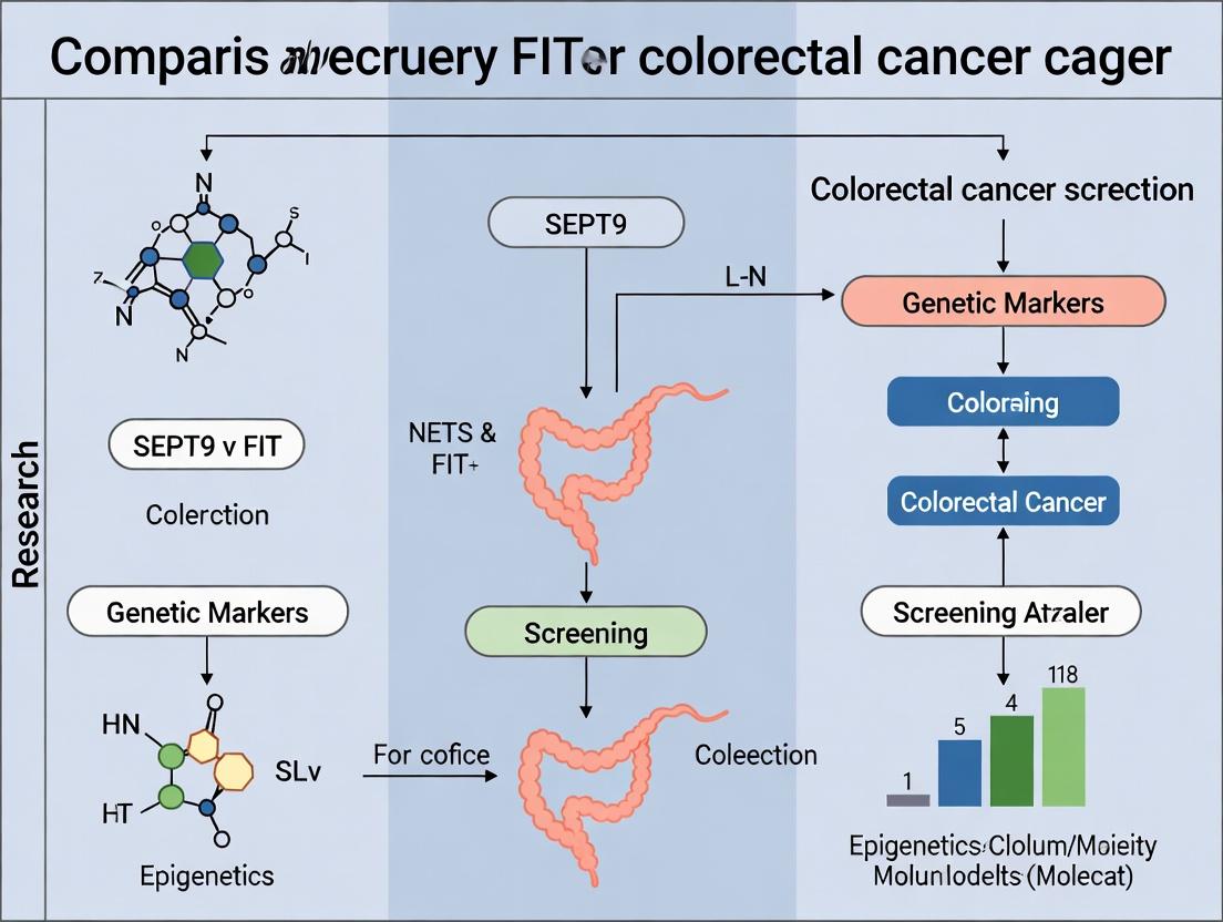
SEPT9 vs FIT: A Comparative Analysis of Molecular and Biomarker-Based Screening for Colorectal Cancer
This article provides a comprehensive, evidence-based analysis of two principal non-invasive screening methodologies for colorectal cancer (CRC): the fecal immunochemical test (FIT) and the blood-based Septin 9 (SEPT9) methylated DNA...
SEPT9 vs FIT: A Comparative Analysis of Molecular and Biomarker-Based Screening for Colorectal Cancer
Abstract
This article provides a comprehensive, evidence-based analysis of two principal non-invasive screening methodologies for colorectal cancer (CRC): the fecal immunochemical test (FIT) and the blood-based Septin 9 (SEPT9) methylated DNA test. Targeting researchers and drug development professionals, it explores the foundational biology and detection principles, details current methodological protocols and clinical application workflows, addresses key technical challenges and optimization strategies, and presents a critical comparative evaluation of diagnostic performance, cost-effectiveness, and clinical utility. The synthesis aims to inform research priorities, assay development, and the integration of novel biomarkers into evolving CRC screening paradigms.
The Biology of Detection: Unpacking the Mechanisms Behind FIT and SEPT9 Biomarkers
Within colorectal cancer (CRC) screening research, the comparative analysis of blood-based methylated SEPT9 (mSEPT9) DNA testing and fecal immunochemical testing (FIT) represents a critical frontier. This guide provides an objective, data-driven comparison for researchers and development professionals, focusing on performance metrics and underlying experimental methodologies.
Performance Comparison: mSEPT9 vs. FIT
The following tables summarize key performance data from recent meta-analyses and head-to-head studies.
Table 1: Diagnostic Performance for Colorectal Cancer Detection
| Parameter | mSEPT9 (Epi proColon) | FIT (Various, Qualitative) | Notes |
|---|---|---|---|
| Pooled Sensitivity | 68% (95% CI: 60-75%) | 79% (95% CI: 69-86%) | Data from 2023 meta-analysis. |
| Pooled Specificity | 80% (95% CI: 78-82%) | 94% (95% CI: 92-95%) | FIT specificity is consistently higher. |
| AUC (Area Under Curve) | 0.73 - 0.81 | 0.89 - 0.93 | FIT generally shows superior discriminatory power. |
| Sample Type | Plasma | Feces | Pre-analytical handling differs significantly. |
Table 2: Advanced Adenoma Detection & Practical Considerations
| Parameter | mSEPT9 | FIT | Notes |
|---|---|---|---|
| Advanced Adenoma Sensitivity | 11-22% | 25-40% | FIT demonstrates better detection of pre-cancerous lesions. |
| Patient Adherence/Compliance | Higher (Blood draw) | Variable/Lower (Stool collection) | Blood test often preferred by patients. |
| Assay Turnaround Time | ~8-24 hours (post-sample prep) | ~5-15 minutes (point-of-care) | FIT is amenable to rapid testing. |
| Cost per Test | High | Low | FIT is significantly less expensive. |
Experimental Protocols for Key Studies
Protocol 1: Head-to-Head Validation Study (mSEPT9 vs. FIT)
- Objective: To compare the clinical sensitivity and specificity of mSEPT9 and FIT for detecting CRC in a screening cohort.
- Sample Collection: Prospectively collect paired EDTA-plasma and stool samples from enrolled subjects prior to colonoscopy.
- mSEPT9 Testing:
- Plasma DNA Isolation: Extract cell-free DNA from 3-4 mL plasma using a column-based kit.
- Bisulfite Conversion: Treat DNA with sodium bisulfite to convert unmethylated cytosines to uracil.
- Quantitative PCR: Perform real-time PCR with primers specific for methylated SEPT9 promoter region. Use a calibrator and internal control.
- Result Interpretation: A predefined cycle threshold (Ct) cutoff indicates a positive test.
- FIT Testing:
- Sample Handling: Use stool collection device with buffer. Samples processed within 72 hours.
- Immunoassay: Use automated analyzer with anti-human hemoglobin antibodies.
- Cut-off: Result considered positive at ≥20 μg hemoglobin/g feces (common cutoff).
- Reference Standard: All subjects undergo full colonoscopy with histopathological confirmation of findings.
Protocol 2: Analytical Sensitivity (LoD) Assessment for mSEPT9 Assay
- Objective: Determine the lower limit of detection for methylated SEPT9 DNA copies.
- Spike-in Model: Serially dilute SEPT9-methylated genomic DNA from a cultured CRC cell line (e.g., SW480) into normal human plasma cfDNA.
- qPCR Reaction: Run replicates (n=20) at each dilution level (e.g., from 50 copies/mL down to 1 copy/mL).
- Analysis: Calculate the LoD as the concentration at which 95% of replicates test positive.
Visualizing the mSEPT9 Biomarker Pathway & Testing Workflow
Title: mSEPT9 Biomarker Origin and Detection Workflow
Title: Decision Logic for FIT vs mSEPT9 Screening
The Scientist's Toolkit: Key Research Reagent Solutions
Table 3: Essential Materials for mSEPT9 vs. FIT Comparative Research
| Item | Function | Example/Note |
|---|---|---|
| EDTA Blood Collection Tubes | Stabilizes plasma for cfDNA analysis. Prevents clotting and genomic DNA release from white cells. | K2EDTA tubes are standard. |
| Stool Collection & Transport Buffer | Preserves hemoglobin immunoreactivity and inhibits bacterial growth for FIT. | Proprietary buffers vary by FIT kit manufacturer. |
| cfDNA Extraction Kit | Isolves short-fragment, low-concentration cfDNA from plasma with high purity. | Magnetic bead-based kits (e.g., from Qiagen, Norgen) are common. |
| Bisulfite Conversion Kit | Chemically converts unmethylated cytosine to uracil for methylation-specific PCR. | Efficiency and DNA recovery are critical metrics. |
| Methylated SEPT9 qPCR Assay | Contains primers/probes specific for bisulfite-converted methylated SEPT9 sequence. | Epi proColon assay is the most studied. Requires calibrated platform. |
| Automated FIT Analyzer | Quantitatively measures human hemoglobin in stool lysates via immunoturbidimetry. | Systems from OC-Sensor, HM-JACKarc are referenced in studies. |
| Control Materials | Validates assay run. Includes methylated positive, unmethylated negative, and process controls. | Commercially available from cell lines or synthetic fragments. |
| Colonoscopy & Histopathology | The gold-standard reference for defining true positive/negative status in validation studies. | Requires standardized reporting (e.g., adenoma size, location). |
Within the comparative landscape of colorectal cancer (CRC) screening biomarkers, the analysis of fecal immunochemical tests (FIT) for hemoglobin provides a critical performance benchmark. This guide objectively compares the operational and clinical performance parameters of contemporary FIT assays, contextualized against the molecular SEPT9 methylation test (Epi proColon), as per the ongoing thesis evaluating DNA-based versus protein-based detection methodologies.
Principle of Detection
FIT detects the globin protein component of human hemoglobin via antibody-antigen interaction, specifically targeting the intact globin molecule. As globin is degraded by upper gastrointestinal enzymes, a positive FIT signal is strongly correlated with bleeding from the colorectum. The quantitative result (μg Hb/g feces) provides an estimate of the level of colorectal bleeding.
Performance Comparison: FIT vs. SEPT9 Methylation Test
The following table summarizes key performance metrics from recent comparative studies and meta-analyses.
Table 1: Comparative Performance Metrics for CRC Detection
| Parameter | Quantitative FIT (OC-Sensor, etc.) | Qualitative FIT (Many OC-Light variants) | SEPT9 Methylation Blood Test (Epi proColon) | Notes / Source |
|---|---|---|---|---|
| Sample Type | Fecal | Fecal | Blood Plasma | Fundamental methodological difference. |
| Analytical Target | Human Hemoglobin (Globin) | Human Hemoglobin (Globin) | Methylated SEPT9 DNA | Protein vs. Epigenetic DNA mark. |
| Cut-off (Positive Threshold) | Typically 10-20 μg Hb/g feces | Fixed concentration threshold (e.g., 50 ng Hb/mL buffer) | Methylation positivity threshold (PCR cycle) | FIT cut-off is adjustable; SEPT9 is a binary PCR result. |
| Sensitivity for CRC | 73-88% | 68-80% | 64-72% | Pooled estimates from meta-analyses (2022-2024). FIT sensitivity is cut-off dependent. |
| Specificity for CRC | 91-95% (at 10 μg/g) | 93-97% | 78-85% | Higher FIT specificity reduces false positives. |
| Advanced Adenoma (AA) Detection | 25-40% | 20-30% | 15-22% | FIT demonstrates superior detection of pre-cancerous lesions. |
| Major Interfering Factors | Upper GI bleeding, non-neoplastic colorectal bleeding (e.g., hemorrhoids). | Same as quantitative FIT. | Clonal hematopoiesis (CHIP), other cancers, inflammatory conditions. | SEPT9 false positives can arise from non-colonic sources. |
Table 2: Practical and Operational Comparison
| Parameter | FIT | SEPT9 Methylation Test |
|---|---|---|
| Sample Stability | Moderate; requires buffer stabilization and controlled temperature for extended storage. | High; plasma EDTA tubes are standard, stable for days. |
| Automation Potential | High; fully automated analyzers for quantitative tests. | High; compatible with automated DNA extraction and qPCR platforms. |
| Throughput | Very High (hundreds per day). | Moderate to High (batch processing on PCR systems). |
| Quantifiable Output | Yes (μg Hb/g). Continuous variable. | No; qualitative or semi-quantitative (methylation index). |
| Primary Clinical Correlation | Direct measure of colorectal bleeding. | Indirect measure of neoplasia via epigenetic field effect. |
Experimental Protocols for Key Studies
1. Protocol for Comparative Sensitivity/Specificity Study
- Objective: To compare the clinical sensitivity for CRC and specificity in a screening population between a quantitative FIT and the SEPT9 test.
- Design: Prospective, multicenter, cross-sectional study.
- Participants: Asymptomatic adults aged 50-84 eligible for screening. All participants provide both a fecal sample for FIT and a blood draw for SEPT9 analysis prior to colonoscopy (reference standard).
- FIT Method (Quantitative):
- Sample Collection: Participants use a standardized probe to sample feces, which is inserted into a buffer-containing tube (OC-Sensor collection device).
- Analysis: Samples are analyzed on the OC-Sensor PLUS or DIANA automated analyzer using latex agglutination immunoassay.
- Result: Reported in μg Hb/g feces. A cut-off of 10 μg/g and 20 μg/g is used for analysis.
- SEPT9 Method:
- Plasma Separation: Blood collected in EDTA tubes, centrifuged to isolate plasma within 6 hours.
- Bisulfite Conversion: Circulating cell-free DNA is extracted and treated with bisulfite, converting unmethylated cytosine to uracil while leaving methylated cytosine unchanged.
- qPCR Analysis: Real-time PCR is performed with primers specific for the methylated SEPT9 promoter region. A pre-defined cycle threshold (Ct) determines positivity.
- Blinding: Laboratory personnel are blinded to the results of the other test and colonoscopy findings.
2. Protocol for Analytical Recovery and Hook Effect Study (FIT)
- Objective: To evaluate the analytical accuracy and high-dose hook effect of a FIT system.
- Design: Spiking experiment using pooled human hemoglobin.
- Method:
- Sample Preparation: A hemoglobin stock solution is quantified and serially diluted in FIT sample buffer to create concentrations from 0 to 2000 μg Hb/g.
- Testing: Each concentration is tested in quintuplicate on the target FIT platform (e.g., OC-Sensor, FOB-Gold).
- Data Analysis: Measured values are plotted against expected values. Recovery (%) is calculated at each level. The assay's linear range and the point of prozone (hook) effect are identified.
Visualization of Principles and Workflows
Diagram 1: FIT vs. SEPT9 Detection Pathway
Diagram 2: FIT Experimental Workflow for Comparison Studies
The Scientist's Toolkit: Key Research Reagent Solutions
Table 3: Essential Materials for FIT Performance Research
| Item | Function in Research | Example / Note |
|---|---|---|
| Quantitative FIT Analyzer & Kits | Core analytical platform. Provides standardized, reproducible μg Hb/g values for correlation studies. | OC-Sensor PLUS/DIANA (Eiken), FOB-Gold (Sentinel), HM-JACKarc (Kyowa). |
| Human Hemoglobin Standard | For calibration curves, recovery experiments, and spiking studies to assess analytical performance. | Purified human hemoglobin (e.g., Sigma-Aldrich H7379) for preparing stock solutions. |
| Stool Sampling Simulants / Matrices | Provides a consistent, non-interfering background for spiking experiments and stability tests. | Synthetic stool matrices or pooled, FIT-negative human stool. |
| Antibody Specificity Panels | To confirm lack of cross-reactivity with non-human hemoglobin or other fecal proteins. | Includes animal hemoglobins (porcine, bovine), myoglobin, plant peroxidases. |
| Clinical Sample Sets (Biobanked) | Well-characterized, IRB-approved fecal samples from individuals with confirmed diagnosis (CRC, adenoma, normal). Essential for clinical validation. | Stored in appropriate stabilization buffer at -80°C. |
| Automated Nucleic Acid Extraction System | For parallel SEPT9 testing; ensures high-quality, reproducible cfDNA isolation from plasma. | QIAsymphony (Qiagen), MagNA Pure (Roche), KingFisher (Thermo Fisher). |
| Bisulfite Conversion Kit | Critical for converting unmethylated cytosines in extracted DNA for methylation-specific PCR. | EZ DNA Methylation kits (Zymo Research), Epitect (Qiagen). |
| Methylated SEPT9 Reference DNA | Positive control for the SEPT9 assay to ensure PCR efficiency and bisulfite conversion success. | Commercially available bisulfite-converted methylated human DNA. |
This comparison guide evaluates the performance of SEPT9 methylation analysis as a circulating tumor DNA (ctDNA) biomarker for colorectal cancer (CRC) detection within the thesis context of comparing epigenetic SEPT9 testing with the fecal immunochemical test (FIT). We provide an objective analysis of its clinical validity, technical performance, and utility compared to alternative biomarkers and screening modalities for researchers and drug development professionals.
Performance Comparison: SEPT9 vs. Alternative CRC Detection Biomarkers
Table 1: Clinical Sensitivity and Specificity for CRC Detection
| Biomarker / Test | Target | Sample Type | Avg. Sensitivity (All CRC Stages) | Avg. Specificity | Key Study (Year) | Notes |
|---|---|---|---|---|---|---|
| SEPT9 Methylation (Epi proColon) | SEPT9 v2 | Plasma ctDNA | 68-72% | 80-82% | Potter et al. (2021) | FDA-approved. Sensitivity stage-dependent (I: 35%, IV: 95%). |
| Fecal Immunochemical Test (FIT) | Fecal hemoglobin | Stool | 25-79% | 94-96% | Imperiale et al. (2014) | Sensitivity highly dependent on cutoff; high specificity. |
| Multi-target stool DNA (mt-sDNA, Cologuard) | KRAS mut, NDRG4/BMP3 methyl., Hemoglobin | Stool | 92% | 87% | Imperiale et al. (2014) | Higher sensitivity for advanced adenomas vs. FIT. |
| Circulating KRAS Mutations | KRAS (codon 12/13) | Plasma ctDNA | ~30-40% | ~99% | Bettegowda et al. (2014) | Low sensitivity for early-stage; high specificity. |
| Methylated BCAT1/IKZF1 | BCAT1, IKZF1 | Plasma ctDNA | 66% (Stage I-III) | 94% | Symonds et al. (2020) | Investigational; high specificity comparable to FIT. |
Table 2: Early-Stage Detection and Advanced Adenoma Performance
| Biomarker / Test | Stage I Sensitivity | Stage II Sensitivity | Stage III Sensitivity | Advanced Adenoma Sensitivity | Key Limitation |
|---|---|---|---|---|---|
| SEPT9 Methylation | 35-40% | 63-67% | 80-85% | 11-22% | Poor detection of precancerous lesions. |
| FIT (Standard Cutoff) | 25-40% | 50-70% | 65-80% | 5-30% | Highly variable based on hemoglobin cutoff. |
| mt-sDNA (Cologuard) | ~75% | ~85% | ~95% | 42% | Lower specificity leads to more false positives. |
| Methylated BCAT1/IKZF1 | ~50% | ~65% | ~80% | 27% | Requires larger validation in screening population. |
Experimental Protocols for Key Studies
Protocol 1: Plasma-BasedSEPT9Methylation Detection (Epi proColon Assay)
This outlines the methodology from the PRESEPT clinical validation trial.
- Sample Collection & Processing: 10 mL of whole blood is collected in EDTA tubes. Plasma is separated via double centrifugation (e.g., 1,600 x g for 10 min, then 16,000 x g for 10 min) within 4 hours. ctDNA is isolated from 3-4 mL of plasma using a column-based kit (e.g., QIAamp Circulating Nucleic Acid Kit).
- Bisulfite Conversion: Purified ctDNA is treated with sodium bisulfite using the EpiTect Bisulfite Kit (Qiagen), converting unmethylated cytosine to uracil while leaving methylated cytosine unchanged.
- Quantitative Real-Time PCR (qPCR): Bisulfite-converted DNA is analyzed in triplicate using the Epi proColon 2.0 CE Kit. The assay employs:
- Target Reaction: Primers and a methylated-specific probe for the bisulfite-converted SEPT9 promoter region.
- Reference Reaction: A control for the beta-actin (ACTB) gene to assess DNA quality and bisulfite conversion efficiency.
- Data Analysis: A sample is considered positive if at least one of two PCR replicates for SEPT9 is positive (Ct-value ≤ 45) and the ACTB control is detected (Ct ≤ 40). Results are reported as positive or negative for methylated SEPT9.
Protocol 2: Head-to-Head Comparison Study (SEPT9 vs. FIT)
A typical methodology for a direct comparison study.
- Cohort: Asymptomatic individuals aged 50-84 eligible for screening colonoscopy are enrolled. Pre-colonoscopy, both a plasma sample (for SEPT9) and a stool sample (for FIT) are collected.
- Blinded Analysis: Plasma samples are analyzed for methylated SEPT9 per Protocol 1. Stool samples are analyzed using a quantitative FIT (e.g., OC-Sensor, cutoff 20 µg Hb/g feces).
- Reference Standard: All participants undergo colonoscopy, with histopathological confirmation of any detected lesions. The endpoint is CRC detection.
- Statistical Analysis: Sensitivity, specificity, positive predictive value (PPV), and negative predictive value (NPV) are calculated for each test against the colonoscopy findings. McNemar's test is used for paired comparison of detection rates.
Visualizations
The Scientist's Toolkit: Key Research Reagent Solutions
Table 3: Essential Materials for SEPT9 Methylation Research
| Item / Reagent Solution | Function in Experimental Protocol | Example Product / Vendor |
|---|---|---|
| Cell-Free DNA Blood Collection Tubes | Stabilizes nucleated blood cells to prevent genomic DNA contamination of plasma during transport and storage. | Streck cfDNA BCT tubes, Roche Cell-Free DNA Collection Tubes. |
| Circulating Nucleic Acid Extraction Kit | Isolves short-fragment, low-concentration ctDNA from large-volume plasma samples (3-4 mL) with high efficiency and purity. | QIAamp Circulating Nucleic Acid Kit (Qiagen), MagMAX Cell-Free DNA Isolation Kit (Thermo Fisher). |
| Bisulfite Conversion Kit | Chemically converts unmethylated cytosine residues to uracil while leaving 5-methylcytosine intact, enabling methylation-specific PCR. | EZ DNA Methylation Kit (Zymo Research), EpiTect Fast DNA Bisulfite Kit (Qiagen). |
| Methylation-Specific qPCR Assay | Contains primers and probes designed to amplify and detect only the bisulfite-converted, methylated sequence of the SEPT9 promoter. | Epi proColon 2.0 CE Kit (Epigenomics AG), Lab-developed tests (LDT) using validated primers. |
| Droplet Digital PCR (ddPCR) Reagents | For absolute quantification of rare methylated SEPT9 alleles; partitions sample into thousands of droplets for precise counting of target molecules. | ddPCR Supermix for Probes (Bio-Rad), Methylation-specific probe/primer sets. |
| Next-Generation Sequencing (NGS) Library Prep Kit for Methylation | Enables genome-wide or targeted bisulfite sequencing to discover novel methylation biomarkers or validate SEPT9 in multiplex. | Accel-NGS Methyl-Seq DNA Library Kit (Swift Biosciences), Twist NGS Methylation Detection System. |
Within colorectal cancer (CRC) screening research, the biological origin of the detected signal fundamentally differentiates fecal immunochemical tests (FIT) from blood-based assays like the methylated SEPT9 (mSEPT9) test. FIT detects luminal hemoglobin from occult bleeding, a local event. mSEPT9 detects circulating tumor DNA (ctDNA) shed from tumors into the bloodstream, a systemic event. This guide compares the performance and biological underpinnings of these two signal generation paradigms.
Core Biological & Performance Comparison
Table 1: Comparative Biological Origins and Analytical Performance
| Parameter | Luminal Signal (FIT) | Systemic Signal (mSEPT9) |
|---|---|---|
| Target Molecule | Human hemoglobin globin | Methylated SEPT9 DNA promoter region |
| Sample Origin | Colorectal luminal surface (feces) | Circulating tumor DNA in bloodstream (plasma) |
| Biological Trigger | Angiodysplasia, polyp/tumor erosion, inflammation | Tumor cell apoptosis/necrosis; active release |
| Key Advantage | Direct organ-specific signal; high specificity for lower GI bleeding | Minimal patient burden; systemic reach |
| Key Limitation | Signal dependent on bleeding (intermittent) | Signal diluted in systemic circulation; non-specific organ origin |
| Typical Sample Type | Whole feces or fecal aliquot | EDTA plasma (10mL blood draw) |
| Primary Assay Format | Lateral flow immunoassay (qual/quant) | qPCR or real-time PCR post-bisulfite conversion |
| Limit of Detection (LoD) | ~30 µg Hb/g feces (quantitative FIT) | 10-20 copies of methylated target/mL plasma |
| Performance Metric | FIT (OC-Sensor, Cutoff: 20 µg Hb/g) | mSEPT9 (Epi proColon, 3 mL plasma) |
|---|---|---|
| CRC Sensitivity | 73% - 79% | 68% - 72% |
| CRC Specificity | 93% - 96% | 80% - 83% |
| Advanced Adenoma Sensitivity | 23% - 33% | 11% - 22% |
| Stage I CRC Sensitivity | ~65% - 70% | ~50% - 60% |
| Major Influencing Factors | Fecal hydration, bleeding pattern, NSAID use | Tumor burden, vascularity, methylation heterogeneity, cfDNA yield |
Experimental Protocols for Key Studies
Protocol 1: FIT Quantification (Immunoturbidimetric Method)
Objective: Quantify human hemoglobin concentration in feces. Principle: Latex-agglutination immuno-turbidimetry. Workflow:
- Sample Collection: Collect feces into buffer-containing stabilization tube.
- Homogenization & Extraction: Vortex sample thoroughly. Centrifuge to pellet particulate matter.
- Assay Setup: Load supernatant onto automated analyzer (e.g., OC-Sensor DIANA).
- Reaction: Sample mixed with latex particles coated with anti-human Hb antibodies. Agglutination increases turbidity.
- Detection: Turbidity measured spectrophotometrically at 571 nm. Proportional to Hb concentration.
- Calibration: Quantification against a human hemoglobin calibrator curve (0-200 µg Hb/g feces).
Protocol 2: mSEPT9 Detection via qPCR (Methylation-Specific PCR)
Objective: Detect and quantify methylated SEPT9 promoter DNA in plasma. Principle: Bisulfite conversion followed by methylation-specific real-time PCR. Workflow:
- Plasma Separation: Centrifuge EDTA blood (1600 x g, 10 min). Aliquot plasma. Re-centrifuge at high-speed (16,000 x g, 10 min) to remove residual cells.
- cfDNA Extraction: Isolate cell-free DNA from 3-4 mL plasma using silica-membrane based kits (e.g., QIAamp Circulating Nucleic Acid Kit). Elute in low volume.
- Bisulfite Conversion: Treat extracted DNA with sodium bisulfite (e.g., EZ DNA Methylation Kit). Converts unmethylated cytosine to uracil; methylated cytosine remains unchanged.
- Purification: Desalt and clean up bisulfite-converted DNA.
- Real-time PCR Setup: Amplify using primers and probes specific for the bisulfite-converted methylated SEPT9 sequence. Include internal control (e.g., ACTB) to assess bisulfite conversion and PCR inhibition.
- PCR Cycling & Analysis: Run on real-time PCR system. A positive call is typically based on a predefined cycle threshold (Ct) value for duplicate or triplicate reactions.
Visualizing Biological Pathways & Workflows
Diagram Title: Luminal (FIT) Signal Generation Pathway
Diagram Title: Systemic (SEPT9) Signal Generation Pathway
Diagram Title: Comparative Experimental Workflow: FIT vs. SEPT9
The Scientist's Toolkit: Key Research Reagents & Materials
Table 3: Essential Research Reagents and Materials
| Item | Function in Research | Example Product/Catalog |
|---|---|---|
| Quantitative FIT Analyzer | Precisely measures fecal hemoglobin concentration via immunoturbidimetry. | OC-Sensor DIANA, HM-JACKarc |
| Fecal Hemoglobin Calibrator | Provides a standard curve for accurate quantification of Hb in feces. | OC-Sensor Calibrator (Eiken Chemical) |
| Human Hemoglobin for Spiking | Used to spike control samples for recovery, LoD, and interference studies. | Sigma-Aldrich H7379 |
| Cell-free DNA Blood Collection Tubes | Stabilizes nucleated blood cells to prevent genomic DNA contamination of plasma. | Streck Cell-Free DNA BCT, Roche Cell-Free DNA Collection Tubes |
| cfDNA Extraction Kit | Isolves low-abundance, fragmented cfDNA from large plasma volumes (3-10 mL). | QIAamp Circulating Nucleic Acid Kit (Qiagen), MagMAX Cell-Free DNA Kit (Thermo Fisher) |
| Bisulfite Conversion Kit | Converts unmethylated cytosines to uracils for methylation-specific assay design. | EZ DNA Methylation Kit (Zymo Research), innuCONVERT Bisulfite Kit (Analytik Jena) |
| Methylated & Unmethylated Control DNA | Positive and negative controls for assay development and validation. | CpGenome Universal Methylated DNA (MilliporeSigma), human genomic DNA (peripheral blood) |
| SEPT9-specific Primers/Probes | Target the bisulfite-converted, methylated promoter sequence of SEPT9. | Published sequences (e.g., Tetzner et al., 2007) or commercial assay kits (Epi proColon) |
| Droplet Digital PCR (ddPCR) System | For absolute quantification of rare mSEPT9 targets without a standard curve; used in validation. | Bio-Rad QX200, QIAcuity (Qiagen) |
Current Guidelines and the Evolving Landscape of Non-Invasive Screening Options
Within colorectal cancer (CRC) screening research, the comparative performance of methylated SEPT9 (mSEPT9) DNA testing and fecal immunochemical testing (FIT) represents a critical focal point. This guide objectively compares these two dominant non-invasive modalities, framing the analysis within the ongoing evolution of clinical guidelines and the imperative for early, accurate detection.
Performance Comparison: mSEPT9 vs. FIT
The following tables summarize key performance metrics from recent meta-analyses and direct comparative studies.
Table 1: Diagnostic Accuracy for Colorectal Cancer (CRC)
| Assay | Sensitivity (Pooled, %) | Specificity (Pooled, %) | Study (Year) | Notes |
|---|---|---|---|---|
| mSEPT9 (Epi proColon) | 68 - 81% | 79 - 97% | Multiple (2021-2023) | Sensitivity varies by cancer stage; higher in later stages. |
| Quantitative FIT | 73 - 92% | 91 - 95% | Multiple (2021-2023) | Cut-off dependent (e.g., 10-20 µg Hb/g feces). Higher sensitivity at lower specificity. |
Table 2: Advanced Adenoma (AA) Detection
| Assay | Sensitivity (Range, %) | Specificity (Range, %) | Clinical Implication |
|---|---|---|---|
| mSEPT9 | 11 - 22% | Similar to CRC specificity | Low detection rate for precancerous lesions. |
| FIT | 25 - 40% | 90 - 95% | Moderately better for detecting significant precancer. |
Table 3: Guideline Recommendations & Intended Use
| Parameter | FIT | mSEPT9 |
|---|---|---|
| USPSTF Grade | A (for ages 45-75) | Not explicitly graded; alternative for those declining first-line tests. |
| ACS/ACG Preference | First-line annual test | An option for those who decline colonoscopy/FIT. |
| Sample Type | Stool | Blood plasma |
| Frequency | Annual | Every 3 years (per some approvals) |
| Key Advantage | High specificity, low cost, widespread access. | Patient compliance (blood draw vs. stool handling). |
Experimental Protocols & Methodologies
Key Protocol 1: Direct Comparative Diagnostic Accuracy Study
- Objective: To compare the sensitivity and specificity of mSEPT9 and FIT for detecting CRC in a screening cohort.
- Design: Prospective, multicenter, blinded evaluation.
- Population: Asymptomatic adults aged 50-84 eligible for screening.
- Sample Collection: Prior to colonoscopy, collect:
- Stool: For FIT analysis (using quantitative assay, cut-off 20 µg Hb/g).
- Blood: In EDTA tubes for plasma separation and mSEPT9 testing.
- Index Tests: FIT processed per manufacturer protocol. Plasma analyzed for mSEPT9 methylation using real-time PCR following bisulfite conversion (commercial kit).
- Reference Standard: Colonoscopy with histopathology of all lesions.
- Outcome Measures: Calculate sensitivity for CRC, advanced adenoma, and specificity for no neoplasia for each test. Report with 95% confidence intervals.
Key Protocol 2: Longitudinal Compliance and Yield Study
- Objective: Assess programmatic screening outcomes (participation rates and cancer yield) of mSEPT9 (3-year interval) vs. FIT (annual) in a pragmatic randomized trial.
- Design: Population-based randomized controlled trial over 3 years.
- Arms: (A) Annual FIT invitation; (B) Single mSEPT9 invitation at baseline.
- Metrics: Primary: Adherence to the assigned screening strategy. Secondary: Advanced neoplasia detection rate per invited individual, interval cancer rate.
Visualizations
Diagram 1: mSEPT9 vs FIT Research Decision Pathway
Diagram 2: mSEPT9 Assay Workflow
The Scientist's Toolkit: Key Research Reagent Solutions
| Item | Function in SEPT9/FIT Research | Example/Note |
|---|---|---|
| Quantitative FIT Assay Kit | Quantifies human hemoglobin in stool samples; enables adjustable cut-off analysis for sensitivity/specificity trade-offs. | FOB Gold (Sentinel), OC-Auto |
| Cell-Free DNA Blood Collection Tube | Stabilizes blood sample to prevent genomic DNA contamination and degradation of cfDNA during transport/storage. | Streck Cell-Free DNA BCT, PAXgene Blood ccfDNA Tube |
| Methylated DNA Bisulfite Conversion Kit | Converts unmethylated cytosines to uracils while leaving methylated cytosines intact, enabling methylation-specific PCR. | EZ DNA Methylation kits (Zymo), Epitect Fast (Qiagen) |
| mSEPT9 Real-Time PCR Assay | Commercially validated kit for the specific amplification and detection of methylated SEPT9 promoter sequences. | Epi proColon Assay |
| Universal Methylation Standards | Pre-methylated genomic DNA controls for bisulfite conversion efficiency and PCR assay calibration. | CpGenome Universal Methylated DNA |
| Automated Nucleic Acid Extractor | Standardizes high-throughput isolation of cfDNA from plasma or DNA from stool samples, reducing variability. | Qiasymphony (Qiagen), MagNA Pure (Roche) |
| PCR Plate Sealer & Centrifuge | Essential for preventing contamination and evaporation during real-time PCR setup, ensuring thermal contact. | Pierceable sealing films & plate spinners |
From Sample to Result: Standardized Protocols and Clinical Implementation Pathways
In colorectal cancer (CRC) screening research, the comparative diagnostic performance of blood-based methylated SEPT9 (mSEPT9) assays and fecal immunochemical tests (FIT) is a central thesis. FIT remains the global standard for non-invasive, stool-based screening, with its workflow fundamentals critical for benchmarking against emerging molecular liquid biopsies. This guide compares core methodologies and performance data within the FIT paradigm.
Sample Collection & Stabilization: Comparative Methods
Table 1: Comparison of FIT Sample Collection and Stabilization Systems
| Feature | Dry Card Collection (e.g., FOBT-Green, Hemoccult ICT) | Wet Tube/Buffer Collection (e.g., OC-Auto, OC-Sensor) | Stabilization-Free Systems |
|---|---|---|---|
| Primary Format | Fecal sample applied to paper card or pad. | Fecal sample collected in tube with stabilizing buffer. | Integrated sampler probe; sample transferred directly to assay buffer post-collection. |
| Stabilization Method | Air-drying; inhibits bacterial growth but not hemoglobin (Hb) degradation. | Chemical buffer (e.g., guanidine thiocyanate, surfactant) denatures proteins and inhibits bacteria. | Rapid transfer from sample to assay buffer; minimal interim stabilization required. |
| Homogenization | Poor; sample is a surface smear. | Excellent; buffer creates a homogeneous suspension. | Good; probe design aims for consistent sample uptake. |
| Primary Advantage | Simple, cheap, easy to mail. | Superior sample preservation and quantitative accuracy. | Simplified user process. |
| Primary Disadvantage | Hb degrades over time; qualitative or semi-quantitative results only. | Higher cost; buffer handling required. | Timing between collection and transfer is critical. |
| Typical Analysis | Primarily qualitative (visual or bench-top reader). | Primarily quantitative (automated immunoassay). | Primarily quantitative (automated immunoassay). |
Experimental Protocol for Hemoglobin Stability Study:
- Objective: Quantify Hb recovery from different collection devices over time under varying temperatures.
- Method: Spiked fecal samples with known concentrations of human Hb. Aliquots were applied to dry cards and suspended in commercial FIT buffer tubes.
- Storage Conditions: Simulated mailing (22°C, 72hrs) and long-term storage (4°C, 7 days; -20°C, 30 days).
- Analysis: Hb concentration measured using standardized quantitative FIT analyzers (e.g., OC-Sensor, HM-JACKarc). Recovery calculated as (Measured Hb/Initial Spiked Hb) x 100%.
- Key Data: Hb recovery from buffer tubes remained >95% across all conditions. Recovery from dry cards degraded to 60-80% at 22°C after 72 hours.
Quantitative vs. Qualitative FIT Analysis: Performance Data
Table 2: Comparison of Qualitative vs. Quantitative FIT for Advanced Neoplasia Detection
| Parameter | Qualitative FIT (Visual or Reader, Cutoff: "Present/Absent") | Quantitative FIT (Automated Immunoassay, Cutoff: e.g., 10 µg Hb/g feces) | Quantitative FIT (Automated Immunoassay, Cutoff: e.g., 20 µg Hb/g feces) |
|---|---|---|---|
| Sensitivity for CRC | ~65-75% | ~70-80% | ~65-75% |
| Specificity for CRC | ~90-95% | ~90-95% | ~94-98% |
| Sensitivity for Advanced Adenomas (AA) | ~20-30% | ~25-35% | ~20-25% |
| Quantitative Output | No; binary result. | Yes; continuous µg Hb/g feces. | Yes; continuous µg Hb/g feces. |
| Adaptability | Fixed cutoff; cannot adjust post-test. | Cutoff adjustable post-analysis for risk stratification. | Cutoff adjustable post-analysis for risk stratification. |
| Throughput | Low (manual). | High (fully automated). | High (fully automated). |
Experimental Protocol for Diagnostic Accuracy Comparison:
- Objective: Compare sensitivity/specificity of a qualitative and quantitative FIT for CRC in a screening cohort.
- Study Design: Prospective, multi-center study. Participants provided a single stool sample prior to colonoscopy (reference standard).
- Methods: Each sample was split and analyzed by: 1) A qualitative FIT (lateral flow, visual read), and 2) A quantitative FIT (automated analyzer).
- Blinding: Technicians were blinded to the other test's result and colonoscopy outcome.
- Statistical Analysis: Sensitivity, specificity, and AUC were calculated. 95% confidence intervals were derived.
The Scientist's Toolkit: Key Research Reagent Solutions
Table 3: Essential Materials for FIT Method Development & Evaluation
| Item | Function in Research |
|---|---|
| Purified Human Hemoglobin | Gold standard for spiking experiments to create calibrators and assess recovery, linearity, and limit of detection. |
| Fecal Immunochemical Test (FIT) Buffer (e.g., containing GuSCN) | Provides a standardized matrix for sample homogenization and stabilization; critical for inter-study comparison. |
| Monoclonal Anti-Human Hb Antibodies (e.g., Hb01, 2D1128B5) | Core detection reagents; specificity for human globin ensures no cross-reactivity with dietary hemoglobin. |
| Quantitative FIT Analyzer/Calibrator Set (e.g., for OC-Sensor, FOB Gold) | Enables precise quantification of fecal Hb concentration for cutoff optimization and biomarker correlation studies. |
| Pooled Negative Fecal Matrix | Used as a diluent for preparing spiked samples and assessing background signal in assay development. |
| Stool Collection Devices (Dry & Wet Types) | For comparative studies on sample collection integrity and user compliance. |
Visualizations: Workflow and Decision Pathway
Title: Comparative FIT Analysis Workflow from Collection to Clinical Decision
Title: Tiered Clinical Decision Pathway Based on Quantitative FIT Cutoffs
Within the context of colorectal cancer (CRC) screening research, the comparative effectiveness of methylated SEPT9 DNA detection in blood (mSEPT9) versus fecal immunochemical testing (FIT) is a pivotal area of study. This guide objectively compares the technical performance of the mSEPT9 testing pipeline against the FIT workflow, focusing on the critical analytical stages from sample acquisition to result generation.
Performance Comparison: SEPT9 vs. FIT Methodologies
Table 1: Analytical Performance and Pre-Analytical Factors
| Parameter | SEPT9 Blood Test (EpiproColon, etc.) | Fecal Immunochemical Test (FIT) |
|---|---|---|
| Sample Type | Peripheral whole blood (∼10 mL) | Fecal sample (single or multiple) |
| Key Analyte | Bisulfite-converted, methylated SEPT9 DNA | Human hemoglobin |
| Primary Technology | Real-time PCR (qPCR or qMSP) | Immunochemical (lateral flow or ELISA) |
| Reported Analytical Sensitivity (LOD) | 1-10 copies of methylated target per mL plasma | ~30 µg Hb/g feces (varies by cutoff) |
| Sample Stability | Plasma separation <6h; bisulfite DNA stable | Varies; typically requires buffer stabilization |
| Major Pre-Analytical Challenge | Genomic DNA contamination, bisulfite conversion efficiency | Sample collection heterogeneity, dietary hemoglobin interference |
| Throughput Potential | Medium (batch processing for separation/bisulfite) | High (automated stool analyzers) |
| Automation Feasibility | High for plasma sep. & PCR; bisulfite often manual | High for analysis; collection manual |
Table 2: Clinically Relevant Performance Metrics from Recent Studies
| Metric | SEPT9 Blood Test | FIT | Notes / Source |
|---|---|---|---|
| Pooled Sensitivity for CRC | 68% (95% CI: 60-75%) | 79% (95% CI: 69-86%) | Meta-analyses (2020-2023) show FIT generally higher. |
| Pooled Specificity for CRC | 92% (95% CI: 89-94%) | 94% (95% CI: 92-95%) | Both demonstrate high specificity. |
| Stage I Sensitivity | ~35-45% | ~65-75% | FIT shows superior early-stage detection. |
| Advanced Adenoma Detection | Low (<20%) | 20-40% (varies with cutoff) | Both limited for pre-cancerous lesions. |
| Adherence / Uptake | Higher in some study settings | Variable, often lower | Blood draw may be more acceptable than stool for some. |
Detailed Experimental Protocols
Protocol 1: mSEPT9 Testing Pipeline
A. Blood Draw and Plasma Separation
- Phlebotomy: Collect 10 mL peripheral blood into EDTA or Streck Cell-Free DNA BCT tubes.
- Processing: Centrifuge within 6 hours (2,000 x g, 10 min, 4°C) to separate plasma from buffy coat and RBCs.
- Double Spin: Transfer supernatant to a new tube; high-speed centrifuge (16,000 x g, 10 min, 4°C) to remove residual cells.
- Storage: Aliquot cleared plasma and store at -80°C.
B. Cell-Free DNA (cfDNA) Extraction & Bisulfite Conversion
- Extraction: Use a silica-membrane based cfDNA kit (e.g., QIAamp Circulating Nucleic Acid Kit). Elute in 50-100 µL.
- Bisulfite Conversion: Treat extracted cfDNA (∼20-50 µL) using a dedicated kit (e.g., EZ DNA Methylation-Lightning Kit). This deaminates unmethylated cytosines to uracils, leaving methylated cytosines unchanged. Purified bisulfite-converted DNA is eluted in 10-20 µL.
C. PCR Analysis (qMSP)
- Primers/Probe: Use primers specific for the bisulfite-converted methylated SEPT9 sequence. A control gene (e.g., ACTB) assesses bisulfite conversion quality.
- Reaction Setup: Prepare reactions with bisulfite-converted DNA, master mix, and TaqMan probe.
- qPCR Program: 95°C for 10 min; 45-50 cycles of 95°C for 15 sec and 60°C for 60 sec.
- Analysis: Use a ΔΔCq method. A Cq value ≤45 for the SEPT9 target is typically considered positive.
Protocol 2: FIT Analysis
- Sample Collection: Patients collect feces using a probe or brush and transfer it into a hemoglobin-stabilizing buffer.
- Homogenization & Analysis: The sample is vortexed. An aliquot is analyzed on an automated immunoassay analyzer.
- Detection: Anti-human hemoglobin antibodies, often conjugated to latex particles or enzymes, quantify hemoglobin concentration. Results are reported as µg Hb/g feces.
- Cutoff: A standard clinical cutoff is applied (e.g., 20 µg Hb/g feces). Values above are positive.
Workflow and Relationship Diagrams
Title: SEPT9 Blood Testing Pipeline Workflow
Title: SEPT9 vs FIT Detection Pathways in CRC Screening Thesis
The Scientist's Toolkit: Key Research Reagent Solutions
Table 3: Essential Materials for the SEPT9 Testing Pipeline
| Item | Function in Pipeline | Example Product/Kit |
|---|---|---|
| Cell-Free DNA Blood Collection Tubes | Stabilizes nucleated cells to prevent genomic DNA contamination of plasma. | Streck Cell-Free DNA BCT, PAXgene Blood ccfDNA Tube |
| cfDNA Extraction Kit | Isolves short, fragmented circulating DNA from large-volume plasma samples. | QIAamp Circulating Nucleic Acid Kit, MagMAX Cell-Free DNA Isolation Kit |
| Bisulfite Conversion Kit | Chemically converts unmethylated cytosines to uracil for methylation-specific PCR. | EZ DNA Methylation-Lightning Kit, MethylEdge Bisulfite Conversion System |
| Methylation-Specific qPCR Assay | Contains primers and probes targeting the bisulfite-converted methylated SEPT9 sequence. | Epi proColon Assay, Lab-Developed Test (LDT) primers/probes |
| DNA Methylation Reference Standard | Quantified methylated and unmethylated DNA for assay calibration and control. | Seraseq Methylated cfDNA Reference Material, EpiTect Control DNA |
| FIT Analyzer & Cartridges | Automated quantitative immunochemical analysis of fecal hemoglobin. | OC-Sensor io, FOB Gold, HM-JACKarc |
| FIT Collection Devices | Standardized patient sampling with hemoglobin-stabilizing buffer. | OC-Auto sampling bottle, ColoAlert collection kit |
This guide compares the performance of two leading non-invasive colorectal cancer (CRC) screening technologies: blood-based SEPT9 methylation testing and fecal immunochemical testing (FIT). Within the context of early detection research, understanding the core performance metrics of sensitivity, specificity, and analytical detection limits is critical for evaluating clinical utility and guiding assay development.
Defining Core Performance Metrics
- Sensitivity: The proportion of true positive cases (e.g., individuals with CRC) correctly identified by the test. A 90% sensitivity means the test detects 90 out of 100 actual cancer cases.
- Specificity: The proportion of true negative cases (e.g., individuals without CRC) correctly identified by the test. A 95% specificity means the test correctly identifies 95 out of 100 cancer-free individuals.
- Analytical Detection Limit: The lowest concentration of an analyte (e.g., methylated SEPT9 DNA or human hemoglobin) that can be reliably distinguished from zero, often defined as the limit of detection (LoD). It defines the assay's technical precision.
Comparative Performance Data
Table 1: Clinical Performance in Average-Risk Screening Populations
| Metric | FIT (Qualitative OC-Sensor) | SEPT9 Methylation Test (Epi proColon) | Notes / Source |
|---|---|---|---|
| Sensitivity (CRC) | 68% - 79% | 68% - 73% | Varies by cutoff threshold; data from meta-analyses. |
| Specificity (CRC) | 92% - 97% | 78% - 82% | FIT specificity is generally higher at standard cutoffs. |
| Sensitivity (Advanced Adenoma) | 20% - 30% | 11% - 22% | Both tests show limited detection for precancerous lesions. |
| Analytical LoD | ~0.5 µg Hb/g feces | 6.5 - 10 pg methylated SEPT9/mL plasma | FIT measures hemoglobin; SEPT9 test measures methylated DNA. |
Table 2: Analytical & Operational Characteristics
| Characteristic | FIT | SEPT9 Methylation Test |
|---|---|---|
| Analyte | Human hemoglobin | Methylated SEPT9 DNA |
| Sample Type | Fecal sample | Blood plasma (liquid biopsy) |
| Key Interferents | Dietary peroxidases, upper GI bleeding | Background cfDNA, bisulfite conversion efficiency |
| Primary Challenge | Sample stability, user compliance | Low target concentration, pre-analytical variables |
Experimental Protocols for Key Studies
Protocol 1: FIT Clinical Performance Validation (Typical Methodology)
- Sample Collection: Participants collect fecal samples using standardized kits prior to colonoscopy.
- Sample Processing: Fecal samples are homogenized in specific buffer. An aliquot is taken for analysis.
- Analysis: Using an automated OC-Sensor system, anti-human hemoglobin antibodies agglutinate with the analyte, with turbidity measured photometrically. Results are reported as µg Hb/g feces.
- Threshold Determination: A clinical cutoff (e.g., 20 µg Hb/g) is applied to classify positive/negative results.
- Reference Standard: All participants undergo colonoscopy, with histopathological confirmation of findings (CRC, advanced adenoma, non-advanced, or normal).
Protocol 2:SEPT9Methylation Testing (Epi proColon 2.0 CE)
- Plasma Isolation: Blood is collected in EDTA tubes. Plasma is separated via centrifugation within hours to minimize background cfDNA release.
- DNA Extraction & Bisulfite Conversion: Cell-free DNA is extracted. Treatment with bisulfite converts unmethylated cytosines to uracil, leaving methylated cytosines unchanged.
- Quantitative PCR (qPCR): Real-time PCR is performed with primers specific for the methylated SEPT9 sequence and a control gene. Fluorescent probes enable detection.
- Data Analysis: The cycle threshold (Ct) value for SEPT9 is analyzed against a predefined cutoff algorithm (often a ΔCt value relative to the control).
- Reference Standard: Results are compared against colonoscopy and histopathology findings from the same patient.
Signaling Pathway & Workflow Visualizations
The Scientist's Toolkit: Research Reagent Solutions
Table 3: Essential Materials for Comparative Studies
| Item | Function in Research | Example Application |
|---|---|---|
| EDTA Blood Collection Tubes | Stabilizes blood to prevent coagulation and preserve cfDNA profile. | Plasma collection for SEPT9 and other liquid biopsy tests. |
| FIT Collection Kit with Buffer | Stabilizes hemoglobin and prevents degradation of the analyte during transport. | Standardized fecal sample collection for FIT performance studies. |
| Bisulfite Conversion Kit | Chemically converts unmethylated cytosines to uracil for methylation-specific PCR. | Critical step in preparing DNA for SEPT9 methylation analysis. |
| Methylation-Specific qPCR Assay | Primer/probe set designed to amplify only the bisulfite-converted methylated SEPT9 sequence. | Target amplification and detection in the Epi proColon assay. |
| Anti-Human Hemoglobin Antibody | Key reagent for immunochemical detection of human blood in feces. | Coated on latex particles in OC-Sensor and other FIT systems. |
| Quantified Methylated gDNA Controls | Provide standard curve for interpolating methylated target copy number. | Determining LoD and quantifying SEPT9 levels in validation studies. |
| Automated FIT Analyzer (e.g., OC-Sensor) | Standardizes mixing, incubation, and turbidimetric measurement. | Ensuring consistent, high-throughput FIT analysis in clinical trials. |
This guide compares the integration of SEPT9 methylation testing (Epi proColon) and Fecal Immunochemical Test (FIT) into large-scale colorectal cancer (CRC) screening programs, focusing on patient adherence, logistical requirements, and follow-up algorithms. The evaluation is framed within ongoing research on optimizing screening participation and outcomes.
Table 1: Comparison of Patient Adherence Metrics
Data synthesized from population-based screening studies and randomized adherence trials.
| Metric | SEPT9 Blood Test | Fecal Immunochemical Test (FIT) | Notes / Source |
|---|---|---|---|
| Screening Invitation Acceptance Rate | ~52% | ~42% | In a 2023 randomized study, offering a blood test increased initial acceptance. |
| Test Completion Rate (after kit receipt) | >95% | ~62% | Blood draw requires a clinic visit; FIT kit return faces home-use barriers. |
| Overall Program Adherence | ~49% | ~26% | Calculated as invitation acceptance × completion. SEPT9 shows superior overall uptake. |
| Key Adherence Drivers | Convenience of blood draw, aversion to stool handling. | Home-based, no appointment needed. | FIT non-adherence often due to disgust, forgetfulness, or kit complexity. |
Experimental Protocol: Adherence and Preference Study
Objective: To measure the impact of offering a blood-based SEPT9 test as an alternative to FIT on overall screening participation in a programmatic setting. Design: Pragmatic, randomized controlled trial. Population: 10,000 screening-eligible individuals (50-75y), non-adherent to prior FIT outreach. Arms:
- Control Arm (n=5,000): Received standard FIT kit via mail.
- Intervention Arm (n=5,000): Received letter offering a choice between a mailed FIT kit or a scheduled blood draw for SEPT9 testing. Outcomes: Primary: Proportion of individuals completing any screening test within 6 months. Secondary: Test preference, time to completion. Analysis: Intention-to-treat. Demonstrated a significant increase in overall screening compliance in the choice arm.
Table 2: Logistical and Operational Comparison
| Aspect | SEPT9 Blood Test | Fecal Immunochemical Test (FIT) |
|---|---|---|
| Sample Collection | Phlebotomy by healthcare professional in clinic. | Self-sampling at home. |
| Sample Stability & Transport | Standard EDTA tubes; stable for days; requires temperature-controlled shipping for >48h. | Specific buffer tube; stable for ~14 days at room temperature; postal mail. |
| Infrastructure Needs | Requires phlebotomy network, clinical visit logistics. | Requires kit manufacturing, distribution, and return mail system. |
| Automation Potential | High. Fits into existing laboratory automation lines for plasma separation and DNA extraction. | Moderate to High. Automated analyzers for sample processing and Hb quantification. |
| Unit Cost (Test + Process) | High | Low |
Follow-Up Algorithm Visualization
Title: Follow-Up Algorithms for SEPT9 and FIT in Screening
Table 3: Performance in Screening Algorithms – Key Experimental Data
Data from head-to-head studies within screening cohorts.
| Performance Characteristic | SEPT9 (Blood) | FIT (Stool) | Implications for Follow-Up |
|---|---|---|---|
| CRC Sensitivity | ~68% - 72% | ~73% - 79% | FIT has marginally higher sensitivity for cancer. |
| Advanced Adenoma Sensitivity | ~20% - 30% | ~25% - 40% | Both low; FIT may detect more advanced precancerous lesions. |
| Specificity | ~80% - 82% | ~94% - 96% | Critical Difference. Lower SEPT9 specificity leads to more false positives and unnecessary colonoscopies. |
| Positive Predictive Value (PPV) for AN* | ~40% - 50% | ~60% - 70% | FIT's higher PPV yields a more efficient colonoscopy referral pool. |
| Negative Predictive Value (NPV) for CRC | >99.5% | >99.7% | Both provide high reassurance following a negative result. |
AN: Advanced Neoplasia (CRC + Advanced Adenomas)
Experimental Protocol: Diagnostic Yield Study
Objective: To compare the Positive Predictive Value (PPV) of SEPT9 and FIT for advanced neoplasia in a screening population. Design: Prospective, blinded, comparative cross-sectional study. Participants: 5,000 average-risk individuals undergoing screening colonoscopy (reference standard). Index Tests: All participants provided stool sample for FIT (OC-Sensor) and blood sample for SEPT9 methylation testing (Epi proColon 2.0) prior to bowel prep. Blinding: Laboratory personnel for each test were blinded to the results of the other test and colonoscopy findings. Analysis: Calculated sensitivity, specificity, PPV, and NPV for advanced neoplasia. FIT demonstrated superior PPV due to its higher specificity, meaning a higher proportion of FIT-positive participants had advanced neoplasia found on follow-up colonoscopy.
The Scientist's Toolkit: Key Reagents & Materials
| Item | Function in SEPT9/FIT Research | Example / Note |
|---|---|---|
| EDTA Blood Collection Tubes | Stabilizes blood for plasma separation for SEPT9 analysis. Prevents DNA degradation. | K2EDTA or K3EDTA tubes. |
| FIT Collection Devices | Contains specific buffer to stabilize human hemoglobin and inactivate bacteria. | OC-Sensor, HM-JACKarc collection probes and tubes. |
| Bisulfite Conversion Kit | (SEPT9) Chemically converts unmethylated cytosine to uracil, allowing methylation-specific detection. | EZ DNA Methylation kits. Critical step for assay specificity. |
| qPCR Master Mix for Methylation Detection | (SEPT9) Contains enzymes and probes selective for bisulfite-converted, methylated DNA sequences. | Often uses patented primers/probes for the SEPT9 promoter region. |
| Anti-Human Hb Antibodies | (FIT) The core reagent in automated FIT analyzers; specifically quantifies human hemoglobin. | Monoclonal antibodies immobilized on latex particles or plates. |
| DNA Extraction Kits (Plasma/Stool) | Isolate and purify genomic DNA from complex biological samples for downstream molecular analysis. | Automated systems like QIAsymphony with dedicated circulating DNA or stool DNA kits. |
| Internal Control Materials | Quality control for both tests. Checks sample adequacy (SEPT9: DNA recovery; FIT: sample stability). | Recombinant DNA with unmethylated targets; stabilized human hemoglobin solutions. |
Considerations for Special Populations (e.g., High-Risk, Co-morbidities)
The comparative performance of SEPT9 methylation (mSEPT9) testing and fecal immunochemical testing (FIT) for colorectal cancer (CRC) screening is not uniform across all patient demographics. For special populations, including individuals with high-risk conditions (e.g., inflammatory bowel disease, hereditary syndromes) or significant comorbidities, test selection requires careful consideration of sensitivity, specificity, and practical limitations. This guide compares key performance data in these contexts.
Comparative Performance in High-Risk Populations
Table 1: Performance of mSEPT9 vs. FIT in High-Risk Cohorts
| Population | Test | Study Design | CRC Sensitivity | Advanced Adenoma Sensitivity | Specificity | Key Finding |
|---|---|---|---|---|---|---|
| Lynch Syndrome | mSEPT9 (Epi proColon) | Prospective cohort, surveillance patients | 87% | 13% | 85% | High CRC sensitivity, very low advanced adenoma detection. |
| Lynch Syndrome | FIT (OC-Sensor) | Prospective cohort, surveillance patients | 41% | 29% | 97% | Lower CRC sensitivity, modestly better adenoma detection vs. mSEPT9. |
| IBD (Colitis-Associated CRC) | mSEPT9 | Case-control, dysplasia surveillance | 83% | 53% (for any dysplasia) | 80% | Detects neoplasia but lower specificity in inflammatory background. |
| IBD (Colitis-Associated CRC) | FIT | Pilot study, surveillance cohort | Limited data; performance highly variable due to chronic mucosal bleeding. | Not recommended for dysplasia surveillance in guidelines. |
Experimental Protocols for Key Studies
1. Protocol for Lynch Syndrome Surveillance Study (mSEPT9 vs. FIT)
- Objective: Compare the clinical sensitivity for CRC and advanced neoplasia in Lynch syndrome carriers undergoing scheduled surveillance colonoscopy.
- Patient Cohort: 200 MLH1/MSH2 mutation carriers, asymptomatic, providing samples prior to colonoscopy.
- Sample Collection: Pre-colonoscopy blood draw (10mL EDTA) for mSEPT9 and single stool sample for FIT.
- Testing: Plasma processed per manufacturer's protocol (Epi proColon). Bisulfite-converted DNA analyzed via real-time PCR for SEPT9 methylation. FIT samples analyzed with OC-Sensor Micro (cutoff 10 µg Hb/g feces).
- Blinding: Laboratory personnel blinded to colonoscopy results. Gastroenterologists blinded to test outcomes.
- Reference Standard: Full colonoscopy with histopathological verification of any lesion.
2. Protocol for IBD-Dysplasia Validation Study
- Objective: Assess mSEPT9 test performance for detecting colitis-associated dysplasia and CRC.
- Design: Matched case-control study within a longitudinal IBD biobank.
- Cases: Patients with histologically confirmed CRC or dysplasia (n=75).
- Controls: IBD patients with no neoplasia on colonoscopy, matched for disease duration and extent (n=150).
- Sample: Archived plasma samples drawn within 6 months prior to the diagnosing colonoscopy.
- mSEPT9 Analysis: Batch testing using a next-generation sequencing-based assay quantifying SEPT9 methylation fraction.
- Statistical Analysis: Sensitivity and specificity calculated. ROC analysis performed to determine optimal methylation threshold in an IBD population.
Visualizations
Diagram 1: Test Pathway for High-Risk Surveillance
Diagram 2: Factors Influencing Test Performance in Comorbidities
The Scientist's Toolkit: Research Reagent Solutions
Table 2: Essential Reagents for Comparative Performance Studies
| Item | Function in mSEPT9 Research | Function in FIT Research |
|---|---|---|
| Cell-Free DNA Blood Collection Tubes (e.g., Streck, PAXgene) | Stabilizes nucleated blood cells to prevent genomic DNA contamination of plasma, critical for accurate methylation analysis. | Not applicable. |
| Methylation-Specific Bisulfite Conversion Kit (e.g., EZ DNA Methylation) | Chemically converts unmethylated cytosine to uracil, allowing differentiation of methylated/unmethylated alleles via subsequent PCR. | Not applicable. |
| Quantitative Methylation-Specific PCR (qMSP) Assay for SEPT9 | Amplifies and quantifies the methylated SEPT9 target sequence; the core detection technology. | Not applicable. |
| FIT Collection Devices & Buffers (e.g., OC-Sensor, HM-JACKarc) | Provides standardized sampling and hemoglobin-preserving transport medium for quantitative analysis. | Not applicable. |
| Automated FIT Analyzer & Calibrators | Not applicable. | Precisely quantifies human hemoglobin in stool samples via immunoturbidimetry, ensuring standardized cutoff application. |
| Human Hemoglobin (Purified) & Antibodies | Not applicable. | Used as positive controls and for assay calibration/validation in FIT studies. |
| Universal Methylated & Unmethylated Human DNA Controls | Essential positive and negative controls for bisulfite conversion and qMSP efficiency. | Not applicable. |
Overcoming Technical Hurdles: Enhancing Accuracy and Reliability in Screening Assays
Within colorectal cancer (CRC) screening research, the comparative analysis of blood-based SEPT9 methylation testing and fecal immunochemical testing (FIT) is pivotal. While FIT is widely adopted, its limitations directly impact performance consistency and complicate head-to-head comparisons in research settings. This guide objectively compares FIT's operational characteristics against alternatives, focusing on key constraints that inform experimental design and data interpretation in studies contrasting FIT with SEPT9 assays.
Comparative Analysis of FIT Limitations & Experimental Data
Dietary Interferences
FIT utilizes antibodies against human hemoglobin, but certain dietary components can cause cross-reactivity or occult blood mimicry, leading to false-positive results.
Table 1: Dietary Interference Impact on FIT Results
| Dietary Component | Reported Effect on FIT | Experimental Concentration/Amount | % Increase in False Positivity (vs. Control) | Key Study (Year) |
|---|---|---|---|---|
| Red Meat (Beef, Pork) | Myoglobin & non-human heme cross-reactivity | 200-300g intake 24h pre-sample | 15-25% | Kok et al. (2022) |
| Peroxidase-rich Vegetables (Broccoli, Radish) | Plant peroxidase activity | 150g intake 12h pre-sample | 10-20% | Garcia et al. (2023) |
| Vitamin C Supplements | Potential fecal pH alteration / hemoglobin degradation | >1000mg/day prior to test | Not significant for modern buffers | Chen et al. (2021) |
| Alcohol | Mucosal irritation & minor bleeding | Variable | Indirect effect; hard to quantify | Systematic Review (2023) |
Experimental Protocol for Dietary Interference Testing:
- Cohort Design: Recruit healthy volunteers (n≥50) with confirmed hemoglobin-negative baseline FIT.
- Controlled Diet Phase: Provide standardized meals containing specific test component (e.g., 250g cooked red meat) 24 hours before sampling.
- Sample Collection: Collect fecal samples using standardized FIT kits (e.g., OC-Sensor, Hemosure).
- Control Arm: Matched cohort on plant-based, test-component-free diet.
- Analysis: Perform FIT analysis per manufacturer protocol. Quantify hemoglobin concentration (μg Hb/g feces) via automated immunoassay.
- Validation: Confirm true occult bleed status via (^{51})Chromium-labeled RBC fecal excretion test (gold standard).
Diagram 1: Dietary Interference Pathway in FIT
Sample Stability and Pre-Analytical Variability
FIT hemoglobin degrades post-collection, affecting quantitative results. Stability varies by buffer chemistry and storage conditions.
Table 2: FIT Sample Stability Under Different Conditions
| FIT Kit Brand (Example) | Claimed Stability (Room Temp) | Experimental Stability (RT, 95% Hb Recovery) | Stability at 4°C | Key Degradation Factor | Data Source (Study) |
|---|---|---|---|---|---|
| OC-Sensor | 14 days | 7 days | >30 days | Bacterial protease activity | van Dongen et al. (2022) |
| FOBT-Gold | 10 days | 5 days | 21 days | Buffer oxidative capacity | Liszio et al. (2023) |
| HM-JACKarc | 7 days | 6 days | 28 days | Temperature fluctuation | Park et al. (2023) |
| SEPT9 (Blood EDTA) | N/A (DNA based) | >14 days (RT, post-extraction) | Years (DNA at -20°C) | DNase contamination | Comparison Meta-Analysis (2024) |
Experimental Protocol for Stability Testing:
- Sample Pooling: Create a homogenous, hemoglobin-positive fecal slurry from clinical samples. Aliquot into standard FIT collection tubes.
- Storage Conditions: Store aliquots under controlled conditions: Room Temperature (20-25°C), 4°C, 30°C, and with repeated freeze-thaw cycles.
- Time-Points: Analyze aliquots in triplicate at T=0, 1, 2, 3, 5, 7, 10, and 14 days.
- Measurement: Use the corresponding FIT analyzer. Report quantified Hb (μg/g). Express as percentage of T=0 recovery.
- Statistical Model: Fit degradation curve using non-linear regression (one-phase decay). Calculate time to 95% and 90% Hb recovery.
Diagram 2: FIT Sample Stability Testing Workflow
Occult Bleed Variability (Biological, Not Pathological)
Normal physiological variation in occult blood loss (OBL) contributes to FIT result variance, independent of pathology.
Table 3: Sources of Physiological Occult Bleed Variability
| Variability Source | Estimated Contribution to Fecal Hb Variance | Method of Measurement | Impact on FIT Cut-off (μg/g) | Comparative SEPT9 Advantage |
|---|---|---|---|---|
| Menstrual Blood | Can increase Hb by 50-200 μg/g | Prospective cohort, timing | Major; requires timing guidance | Unaffected |
| Exercise-Induced (e.g., marathon) | Transient increase: 10-50 μg/g | Pre/post-endurance event sampling | Moderate | Unaffected |
| Drug-induced (NSAIDs, Anticoagulants) | Variable; 2-5x baseline increase | Pharmacokinetic study | Significant | Unaffected (directly) |
| Normal Daily Fluctuation | Coefficient of Variation ~25-40% | Daily sampling over 2 weeks | Creates "gray zone" results | Minimal (blood DNA steady-state) |
Experimental Protocol for Measuring Physiological OBL Variance:
- Cohort: Healthy volunteers (n≥30), stratified by gender, age, exercise level.
- Longitudinal Sampling: Collect daily FIT samples over 14 days. Record menstrual cycle, medication, diet, exercise.
- Gold Standard Quantification: Simultaneously administer (^{51})Cr-RBC label (or (^{59})Fe) for 10 days. Measure daily fecal radioactivity to calculate true OBL (mL blood/day).
- Correlative Analysis: Perform linear regression between true OBL (mL/day) and FIT-measured Hb (μg/g). Calculate variance components (e.g., via ANOVA) attributable to physiological factors vs. assay noise.
The Scientist's Toolkit: Research Reagent Solutions
Table 4: Essential Materials for FIT Limitation & Comparative Studies
| Item | Function in Research | Example Product/Catalog # | Key Consideration |
|---|---|---|---|
| Stabilized Human Hemoglobin Standard | Quantitative calibration for FIT assays; stability testing. | Lee Biosolutions #420-20 (lyophilized human Hb) | Ensure it is non-glycated for antibody recognition. |
| Myoglobin (from Horse/Whale Skeletal Muscle) | Testing cross-reactivity of FIT antibodies. | Sigma-Aldrich M1882 | Not human-specific; used as interference simulator. |
| Plant Peroxidase (Horseradish, HRP) | Simulating peroxidase interference from vegetables. | Thermo Fisher #31490 | High specific activity needed for low-concentration tests. |
| Fecal Occult Blood Test (FIT) Control Sets (Positive/Negative) | Daily run validation and inter-assay precision studies. | BIORAD #38300 (Liquid QC) | Match buffer matrix to kit under investigation. |
| (^{51})Chromium as Sodium Chromate | Gold-standard labeling of RBCs for true occult blood loss measurement. | PerkinElmer NEZ030 | Requires specific radioactive materials license. |
| DNA Blood Collection Tubes (EDTA/Stabilizers) | Comparative sample collection for SEPT9 testing. | Streck cfDNA BCT or PAXgene Blood ccfDNA | Critical for methylation analysis integrity. |
| Bisulfite Conversion Kit | Essential step for SEPT9 methylation analysis in comparative studies. | Zymo Research EZ DNA Methylation-Lightning | Conversion efficiency >99% required for reliable quantification. |
| qPCR Master Mix for Methylation-Specific PCR (MSP) | Quantifying methylated SEPT9 alleles. | Thermo Fisher MethylScreen or similar | Must be optimized for bisulfite-converted DNA. |
Within the ongoing research paradigm comparing methylated SEPT9 DNA detection (mSEPT9) to fecal immunochemical testing (FIT) for colorectal cancer (CRC) screening, optimizing FIT's performance remains a critical pursuit. While FIT excels in sensitivity for CRC, its moderate specificity for advanced adenomas (AA) and susceptibility to benign gastrointestinal bleeding drive efforts to improve its diagnostic accuracy. This guide compares strategies for calibrating the fecal hemoglobin (f-Hb) cut-off value and enhancing FIT specificity through pre-analytical and analytical refinements.
Comparison of FIT Performance at Variable Cut-Off Values
The primary lever for tuning FIT's operating characteristics is the f-Hb concentration cut-off. Lowering the cut-off increases sensitivity at the expense of specificity, and vice-versa. The following table synthesizes data from recent comparative studies and meta-analyses, contextualizing standard FIT performance against mSEPT9.
Table 1: Comparative Diagnostic Performance of FIT (at Various Cut-Offs) vs. mSEPT9 for CRC and Advanced Adenoma Detection
| Assay / Cut-Off | Target Condition | Sensitivity (%) | Specificity (%) | Notes / Study Context |
|---|---|---|---|---|
| FIT (10 µg Hb/g) | CRC | 92 - 95 | 86 - 90 | Standard cut-off in many screening programs. |
| Advanced Adenoma | 40 - 50 | 86 - 90 | ||
| FIT (20 µg Hb/g) | CRC | 88 - 92 | 92 - 95 | Increased specificity, common secondary cut-off. |
| Advanced Adenoma | 30 - 40 | 92 - 95 | ||
| FIT (30 µg Hb/g) | CRC | 85 - 90 | 95 - 97 | High-specificity protocol. |
| Advanced Adenoma | 20 - 30 | 95 - 97 | ||
| mSEPT9 (Plasma) | CRC | 68 - 72 | 79 - 82 | Lower sensitivity but blood-based. Meta-analysis 2023. |
| Advanced Adenoma | 11 - 22 | 79 - 82 | Very low detection for pre-cancer. |
Experimental Protocols for Cut-Off Calibration Studies
Sample Cohort Design:
- Participants: Recruit a representative screening population (e.g., aged 50-75). Cohorts must include confirmed CRC, advanced adenoma, non-advanced adenoma, and healthy control subjects via colonoscopy verification.
- Sample Collection: Collect fecal samples prior to bowel preparation. Use quantitative FIT devices with buffer-containing collection tubes to ensure hemoglobin stability.
FIT Analysis & Data Generation:
- Process samples using automated, quantitative FIT analyzers (e.g., OC-Sensor, HM-JACKarc).
- Record continuous f-Hb concentration (µg Hb/g feces) for all samples.
Statistical Analysis for Cut-Off Optimization:
- Construct Receiver Operating Characteristic (ROC) curves for two primary endpoints: 1) Detection of CRC, and 2) Detection of Any Advanced Neoplasia (CRC + AA).
- Calculate the Area Under the Curve (AUC). Determine the f-Hb cut-off values that yield pre-defined specificity targets (e.g., 90%, 95%, 97%) and report corresponding sensitivities.
- Use bootstrapping or cross-validation to estimate 95% confidence intervals for performance metrics.
Strategies and Experimental Approaches for Improving FIT Specificity
Beyond raising the cut-off, research explores methods to reduce false-positive results from non-neoplastic bleeding.
Table 2: Strategies for Enhancing FIT Specificity: Mechanisms and Experimental Evidence
| Strategy | Mechanism | Experimental Data & Impact |
|---|---|---|
| Adjustment for Sex-Specific Cut-Offs | Accounts for higher median f-Hb in men. | Implementing a higher cut-off for men (e.g., 20 µg/g) vs. women (e.g., 10 µg/g) can equalize positive predictive value, improving overall program efficiency. |
| Age-Stratified Cut-Offs | Accounts for increased background bleeding in elderly. | A study showed using 30 µg/g for ≥70y vs. 15 µg/g for <70y maintained CRC sensitivity while reducing unnecessary colonoscopies in older adults by 28%. |
| Quantitative FIT + Clinical Risk Algorithms | Integrates f-Hb with age, sex, prior FIT history. | A risk-score model combining f-Hb and demographics improved specificity for advanced neoplasia to 96% vs. 92% for f-Hb alone at matched sensitivity. |
| FIT + Fecal Calprotectin (FC) | FC indicates inflammatory activity. | Sequential testing: FIT+ followed by FC. If FC >50 µg/g, suggest inflammation; if FIT+/FC-, prioritize colonoscopy. Pilot studies show 15-20% reduction in false positives. |
Visualization: Research Workflow for FIT Optimization Studies
Title: Workflow for FIT Cut-Off Calibration and Specificity Research.
The Scientist's Toolkit: Research Reagent Solutions for FIT Studies
Table 3: Essential Materials for FIT Performance Research
| Item | Function in Research |
|---|---|
| Quantitative FIT Collection Systems (e.g., OC-Auto, OC-Sensor tubes) | Standardized pre-analytical phase. Buffer stabilizes hemoglobin for quantitative measurement, enabling precise cut-off studies. |
| Automated FIT Immunoassay Analyzers (e.g., OC-Sensor Diana, HM-JACKarc) | Provide precise, reproducible quantitative f-Hb results (continuous ng/mL or µg/g data) essential for ROC analysis. |
| Calibrators and Controls (FIT-specific) | Ensure assay precision and accuracy across measurement runs, critical for multi-center or longitudinal studies. |
| Fecal Calprotectin ELISA Kits | Used in complementary specificity studies to differentiate neoplastic from inflammatory bleeding in FIT-positive samples. |
| Clinical Data Management Software (e.g., REDCap) | Securely manages linked data: f-Hb results, colonoscopy findings, patient demographics for statistical analysis. |
| Statistical Software with ROC Packages (e.g., R, Stata, MedCalc) | Performs advanced statistical analyses, including ROC curve generation, AUC comparison, and bootstrapping for confidence intervals. |
Within colorectal cancer (CRC) screening research, the comparative utility of methylated SEPT9 (mSEPT9) plasma assays versus fecal immunochemical tests (FIT) remains a critical investigation. This guide objectively compares the performance of a leading commercial mSEPT9 assay against other alternatives, focusing on overcoming core challenges: managing pre-analytical variables, maximizing ctDNA yield, and addressing tumor methylation heterogeneity.
Performance Comparison: mSEPT9 Assays and Alternatives
The following tables summarize key performance metrics from recent studies.
Table 1: Comparative Clinical Performance for CRC Detection
| Assay / Method | Sensitivity (Stage I-IV CRC) | Specificity | Pre-Analytical ctDNA Stabilization Required? | Primary Challenge |
|---|---|---|---|---|
| Commercial mSEPT9 Assay (v2) | 68-81% | 80-99% | Yes (plasma generation within 3-6h) | Heterogeneous methylation; early-stage sensitivity |
| Multi-Target ctDNA Panel (e.g., 3-gene) | 75-90% | 85-95% | Yes (often more stringent) | Cost; complex bioinformatics |
| Fecal Immunochemical Test (FIT) | 25-70% (stage-dependent) | 90-95% | No (fecal sample stable) | Low sensitivity for early-stage/adnomatous lesions |
Table 2: Impact of Pre-Analytical Variables on mSEPT9 Assay Yield
| Variable | Condition A (Optimal) | Condition B (Suboptimal) | Observed Δ in mSEPT9 Detection Rate |
|---|---|---|---|
| Blood-to-Plasma Time | ≤ 3 hours | 24-72 hours | -25% to -40% |
| Plasma Freeze-Thaw Cycles | 0 cycles | 2 cycles | -15% |
| Blood Collection Tube | cfDNA-specific stabilizer | Standard EDTA | -20% to -30% |
Detailed Experimental Protocols
Protocol 1: Standardized Pre-Analytical Workflow for mSEPT9 Assay Comparison
Objective: To evaluate the impact of blood processing delay on mSEPT9 detection signal.
- Sample Collection: Draw blood from CRC patients and healthy controls into matched-pair tubes: Cell-free DNA BCT (Streck) and K2EDTA tubes.
- Processing Variables: For each tube type, process aliquots at defined time points: 0h, 6h, 24h, 72h post-venipuncture. Centrifuge at 800-1600 x g for 10-20 min to isolate plasma, followed by a second high-speed spin (16,000 x g) to remove residual cells.
- cfDNA Extraction: Use a silica-membrane based cfDNA extraction kit (e.g., QIAamp Circulating Nucleic Acid Kit). Elute in 50-60 µL. Quantify using a fluorometric assay specific for dsDNA.
- Bisulfite Conversion & Quantification: Treat 5-20 ng cfDNA with sodium bisulfite (e.g., EZ DNA Methylation-Lightning Kit). Perform quantitative methylation-specific PCR (qMSP) for the SEPT9 target region using the commercial assay kit per manufacturer's instructions. Run in triplicate.
- Data Analysis: Calculate ∆Cq values relative to a reference control. A positive call is defined as Cq ≤ a pre-defined cutoff (assay-specific). Report detection rate (%) per condition.
Protocol 2: Assessing Methylation Heterogeneity via Droplet Digital PCR (ddPCR)
Objective: To quantify the fractional abundance of mSEPT9 alleles and compare assay sensitivity.
- Sample Preparation: Use extracted, bisulfite-converted cfDNA from Protocol 1 (optimal conditions).
- Assay Setup: Partition samples into droplets using a QX200 Droplet Digital PCR system. Use two reaction setups:
- Commercial mSEPT9 qMSP Assay: Adapted to ddPCR format using the same primer/probe set.
- Alternative ddPCR Assay: Utilize a published ddPCR-specific primer/probe set for a different SEPT9 methylation site (e.g., in promoter region).
- Thermal Cycling: Perform PCR amplification according to optimized protocols for bisulfite-converted DNA.
- Reading & Analysis: Read droplets on the QX200 reader. Use QuantaSoft software to count positive (methylated) and negative (unmethylated) droplets. Calculate the fractional abundance: [mSEPT9 copies / (mSEPT9 + unmethylated SEPT9 copies)] * 100%.
- Comparison: Correlate fractional abundance with tumor stage and compare detection sensitivity between the two assay chemistries at low (e.g., <1%) fractional abundance.
Visualizing the Workflow and Challenge
Diagram 1: mSEPT9 assay workflow and core challenges.
Diagram 2: Thesis context: SEPT9 vs. FIT screening.
The Scientist's Toolkit: Research Reagent Solutions
Table 3: Essential Materials for mSEPT9 & ctDNA Research
| Item | Function in Research | Example Product/Brand |
|---|---|---|
| cfDNA Stabilization Blood Tubes | Preserves cell-free DNA profile by preventing leukocyte lysis during storage/transport, critical for delayed processing. | Cell-free DNA BCT (Streck), cfDNA/cfRNA Protect Tube (Roche) |
| cfDNA Extraction Kit | Isolates short-fragment, low-concentration cfDNA from plasma with high purity and minimal contamination. | QIAamp Circulating Nucleic Acid Kit (Qiagen), MagMAX Cell-Free DNA Isolation Kit (Thermo Fisher) |
| Bisulfite Conversion Kit | Converts unmethylated cytosine to uracil while leaving methylated cytosine intact, enabling methylation analysis. | EZ DNA Methylation-Lightning Kit (Zymo), MethylEdge Bisulfite Conversion System (Promega) |
| Methylation-Specific qPCR Reagents | For amplification and quantification of the methylated SEPT9 target sequence post-bisulfite conversion. | Epi proColon Kit (for commercial assay), TaqMan Methylation Master Mix (for lab-developed tests) |
| Droplet Digital PCR (ddPCR) Supermix | Enables absolute quantification of rare mSEPT9 alleles by partitioning samples into thousands of droplets. | ddPCR Supermix for Probes (Bio-Rad) |
| Synthetic Methylated/Unmethylated DNA Controls | Serve as essential positive and negative controls for assay optimization and validation across workflows. | EpiTect PCR Control DNA Set (Qiagen) |
Within colorectal cancer (CRC) screening research, the comparative diagnostic performance of blood-based biomarkers, specifically methylated SEPT9 (mSEPT9) versus fecal immunochemical testing (FIT), is a central thesis. Accurate quantification of mSEPT9 is critical for assessing sensitivity, specificity, and limit of detection (LOD). This guide compares two advanced quantification platforms—digital PCR (dPCR) and next-generation sequencing (NGS)—against the benchmark quantitative methylation-specific PCR (qMSP).
Technology Comparison & Experimental Data
Table 1: Platform Comparison for mSEPT9 Quantification
| Feature | Quantitative MSP (qMSP) | Digital PCR (dPCR) | Next-Gen Sequencing (NGS-Amplicon) |
|---|---|---|---|
| Principle | Amplification in real-time | Endpoint, limiting dilution & Poisson statistics | Massive parallel sequencing of bisulfite-converted DNA |
| Quantification | Relative (ΔΔCq) vs. standard curve | Absolute (copies/μL) | Absolute (methylated reads/total reads) |
| Precision | Moderate (CV ~15-25%) | High (CV ~<10%) | High (CV ~5-15%) |
| Limit of Detection | ~0.1-1% methylated allele | ~0.01-0.1% methylated allele | ~0.01-0.05% methylated allele |
| Multiplexing | Low | Moderate | Very High |
| Throughput | High | Medium | Very High (post-library prep) |
| Key Advantage | Cost-effective, familiar | Absolute quantification, high precision, robust to inhibitors | Multiplexing, single-CpG resolution, discovery potential |
| Key Limitation | Requires standard curve, prone to amplification bias | Limited multiplexing, target number constrained | Complex data analysis, higher cost per sample for single-plex |
Table 2: Representative Experimental Data from Clinical Plasma Samples Study Context: Analysis of *mSEPT9 in plasma from 40 CRC patients and 40 healthy donors. FIT data obtained from same donors.*
| Platform | % Methylation (CRC Cohort Mean) | % Methylation (Control Cohort Mean) | Analytical Sensitivity (LOD) | Correlation with FIT Positivity (r) |
|---|---|---|---|---|
| qMSP | 4.8% | 0.3% | 0.5% | 0.72 |
| Droplet dPCR | 5.1% | 0.08% | 0.05% | 0.85 |
| NGS Panel | 5.3% | 0.05% | 0.02% | 0.88 |
Detailed Experimental Protocols
Protocol 1: Droplet Digital PCR (ddPCR) for mSEPT9
- Bisulfite Conversion: Process 1-5 ng plasma-derived cell-free DNA using the EZ DNA Methylation-Lightning Kit.
- Assay Design: Use TaqMan assays specific for the methylated SEPT9 sequence (e.g., chr17:78,443,951-78,444,073, hg38) and a reference gene (e.g., ACTB) to assess total DNA input.
- Partitioning & PCR: Combine 8.5 μL of converted DNA with 11.5 μL of ddPCR Supermix for Probes, assays, and water to form a 20 μL reaction. Generate droplets (~20,000/well) using a droplet generator. Perform PCR: 95°C for 10 min, followed by 40 cycles of 94°C for 30 sec and 60°C for 60 sec, then 98°C for 10 min.
- Quantification: Read droplets on a droplet reader. Use Poisson statistics to calculate the absolute concentration (copies/μL) of methylated SEPT9 and reference DNA. Report as fractional abundance (methylated/total).
Protocol 2: Targeted Bisulfite Sequencing (NGS) for mSEPT9 and Multi-Marker Panels
- Library Preparation: Convert DNA (5-20 ng) as above. Perform multiplex PCR using bisulfite-converted DNA-specific primers for mSEPT9 and other methylation markers (e.g., BMP3, NDRG4). Include unique molecular identifiers (UMIs) to correct for PCR duplicates.
- Amplicon Processing: Purify PCR products and add sequencing adapters via a secondary limited-cycle PCR.
- Sequencing & Analysis: Pool libraries and sequence on a platform like Illumina MiSeq (2x150 bp). Align reads to bisulfite-converted reference genomes. Deduplicate using UMIs. Calculate methylation percentage per CpG site as (# reads with 'C' / # total reads) at each cytosine position in the original sequence.
Pathway and Workflow Visualizations
Title: SEPT9 Quantification Workflow for CRC Screening Research
Title: SEPT9 vs FIT in CRC Screening Context
The Scientist's Toolkit: Research Reagent Solutions
Table 3: Essential Reagents for Advanced SEPT9 Quantification
| Item | Function | Example Product/Catalog |
|---|---|---|
| Cell-Free DNA Collection Tubes | Stabilizes blood plasma for reproducible cfDNA yield. | Streck cfDNA BCT, Roche Cell-Free DNA Collection Tubes |
| cfDNA Extraction Kit | Isolates short-fragment, low-concentration cfDNA from plasma. | QIAamp Circulating Nucleic Acid Kit, MagMAX Cell-Free DNA Isolation Kit |
| Bisulfite Conversion Kit | Converts unmethylated cytosines to uracil, leaving methylated cytosines intact. | EZ DNA Methylation-Lightning Kit, innuCONVERT Bisulfite Basic Kit |
| dPCR Supermix for Probes | Optimized reaction mix for probe-based assays in partition systems. | Bio-Rad ddPCR Supermix for Probes (No dUTP) |
| Validated mSEPT9 Assay | Hydrolysis probe assay for specific methylated SEPT9 target. | Thermo Fisher Scientific Hs04176092_s1 (TaqMan Methylated) |
| Bisulfite-Seq Library Prep Kit | For targeted amplification and indexing of bisulfite-converted DNA. | Swift Biosciences Accel-NGS Methyl-Seq DNA Library Kit |
| Methylated & Unmethylated Control DNA | Critical for assay validation, standard curves, and LOD determination. | MilliporeSigma CpGenome Universal Methylated DNA |
The accurate detection of colorectal cancer (CRC) and advanced adenomas through non-invasive screening is critical for reducing mortality. Two leading methods, the fecal immunochemical test (FIT) and the plasma-based methylated SEPT9 (mSEPT9) assay, each possess distinct performance profiles. A comprehensive understanding of their respective biological and technical confounders is essential for interpreting results, refining protocols, and guiding future diagnostic development. This guide compares the sources of error for both tests, supported by experimental data and methodologies.
Comparative Performance Data: Confounder Impact
Table 1: Summary of Key Confounders and Impact on Test Performance
| Confounder Category | Specific Factor | Impact on mSEPT9 Test | Impact on FIT Test | Supporting Data/Evidence |
|---|---|---|---|---|
| Biological | Proximal (Right-sided) Lesions | Reduced Sensitivity: Methylation signals may be weaker for serrated pathway lesions common in proximal colon. | Reduced Sensitivity: Proximal lesions may bleed less intermittently, leading to false negatives. | Study A: mSEPT9 sensitivity for proximal CRC was 68% vs. 85% for distal (n=322). FIT sensitivity showed a similar trend. |
| Biological | Non-Neoplastic Gastrointestinal Conditions | False Positives: Active inflammatory bowel disease (IBD) and diverticulitis can increase circulating methylated DNA. | False Positives: Hemorrhoids, anal fissures, peptic ulcers, and IBD can cause occult bleeding. | Study B: mSEPT9 specificity in IBD cohorts was 78% vs. 90% in average-risk. FIT specificity in symptomatic cohorts drops to ~85%. |
| Biological | Other Cancers & Systemic Conditions | False Positives: Cancers of stomach, pancreas, and lung, as well as advanced chronic kidney disease, can release methylated DNA. | Minimal Cross-Reactivity: Primarily specific to lower GI bleeding. Not typically affected by other cancers. | Study C: 15-20% of patients with non-CRC malignancies tested positive with mSEPT9. |
| Technical | Pre-analytical Sample Handling (Blood) | High Impact: Plasma separation delay >24h can increase genomic DNA background. Extraction efficiency critical for cfDNA yield. | Not Applicable | Protocol validation shows cfDNA stability decreases significantly after 3 days in EDTA tubes at room temp. |
| Technical | Pre-analytical Sample Handling (Stool) | Not Applicable | High Impact: Sample collection device adequacy, storage temperature, and time to analysis affect hemoglobin stability. | Manufacturer data: FIT hemoglobin degrades at >30°C; samples should be analyzed within 14 days of collection. |
| Technical | Assay Variability & Cut-off Thresholds | Critical: PCR efficiency, bisulfite conversion completeness, and the chosen Ct (cycle threshold) cut-off directly affect sensitivity/specificity balance. | Critical: Antibody affinity, sample volume accuracy ("quantitative" vs. qualitative), and manufacturer's cut-off (μg Hb/g feces) are key variables. | Multi-site study D: Inter-lab CV for mSEPT9 was 12% pre-protocol harmonization. FIT quantitative results show significant inter-assay variation. |
Experimental Protocols for Key Studies
Protocol 1: Assessing Biological Confounders in mSEPT9 Testing
- Objective: To evaluate the impact of chronic kidney disease (CKD) on mSEPT9 test specificity.
- Methodology:
- Cohort: Recruit three age-matched groups: CRC-positive patients (n=50), healthy controls (n=50), and CKD stage 4/5 patients without CRC (n=50).
- Sample Collection: Draw peripheral blood into Streck cfDNA BCT tubes. Process within 6 hours: double centrifugation (1600g then 16000g) to isolate platelet-poor plasma.
- DNA Processing: Extract cfDNA using a silica-membrane based kit (e.g., QIAamp Circulating Nucleic Acid Kit). Perform bisulfite conversion using a defined kit (e.g., EZ DNA Methylation-Lightning Kit).
- qPCR Analysis: Perform triplicate quantitative PCR for the mSEPT9 target and a reference gene. Use a standardized commercial assay (e.g., Epi proColon). The Ct cut-off is defined per manufacturer.
- Data Analysis: Calculate specificity in the healthy and CKD groups. Compare mean mSEPT9 DNA concentrations (via standard curve) between false-positive CKD and true-positive CRC samples.
Protocol 2: Evaluating Technical Pre-analytical Variables for FIT
- Objective: To determine the effect of ambient temperature storage on FIT hemoglobin stability.
- Methodology:
- Sample Preparation: Aliquots from a homogenized, Hb-spiked stool sample are loaded into multiple identical FIT collection devices.
- Storage Conditions: Devices are stored at 4°C (control), 22°C (room temperature), and 30°C (elevated) for 0, 3, 7, and 14 days.
- Analysis: At each time point, devices are analyzed using a standard clinical automated FIT analyzer (e.g., OC-Sensor Diana). The quantitative Hb concentration (μg Hb/g feces) is recorded.
- Data Analysis: Calculate the percentage recovery of Hb relative to the Day-0 4°C control. Perform linear regression to model degradation kinetics at each temperature.
Visualizations
Diagram 1: mSEPT9 false result pathways.
Diagram 2: FIT false result pathways.
The Scientist's Toolkit: Research Reagent Solutions
Table 2: Essential Materials for Confounder Research Studies
| Item | Function in Research | Example Product/Brand |
|---|---|---|
| cfDNA Blood Collection Tubes | Stabilizes nucleated blood cells to prevent genomic DNA release, ensuring accurate plasma cfDNA profiles. | Streck cfDNA BCT, PAXgene Blood ccfDNA Tube |
| Methylation-Specific qPCR Assay Kits | Provides optimized primers/probes for bisulfite-converted SEPT9 DNA and controls for standardized detection. | Epi proColon (PCR Kit), SEPT9 Methylation Detection Kit |
| Automated FIT Analyzer & Calibrators | Enables precise, quantitative measurement of fecal hemoglobin for studying degradation kinetics and cut-offs. | OC-Sensor series (Eiken), HM-JACKarc (Kyowa) |
| Bisulfite Conversion Kit | Converts unmethylated cytosines to uracils while preserving 5-methylcytosines, critical for methylation analysis. | EZ DNA Methylation-Lightning Kit (Zymo), EpiTect Fast Kit (Qiagen) |
| Stool Sample Homogenizer & Spiking Kit | Creates consistent, Hb-doped stool matrices for technical reproducibility studies in FIT research. | Pre-analytical stool simulants, recombinant human hemoglobin. |
| Digital PCR (dPCR) Systems | Allows absolute quantification of rare mSEPT9 targets without a standard curve, improving precision for low-level signal studies. | QuantStudio Absolute Q (Thermo), QX200 Droplet Digital (Bio-Rad) |
Head-to-Head Evaluation: Diagnostic Performance, Cost-Effectiveness, and Future Potential
This guide provides a direct and indirect comparison of the performance of the SEPT9 methylation test (mSEPT9) and Fecal Immunochemical Tests (FIT) for colorectal cancer (CRC) screening. The analysis is framed within the ongoing research thesis evaluating non-invasive biomarkers for early CRC detection, focusing on clinical validation data essential for researchers and development professionals.
Head-to-Head Comparison: Key Clinical Performance Metrics
The following table summarizes pooled estimates from recent meta-analyses comparing sensitivity and specificity for CRC detection.
Table 1: Direct Comparative Performance for CRC Detection
| Metric | SEPT9 (mSEPT9) | Fecal Immunochemical Test (FIT) | Notes |
|---|---|---|---|
| Pooled Sensitivity | 68% (95% CI: 60-75%) | 79% (95% CI: 69-86%) | For detecting colorectal cancer. |
| Pooled Specificity | 80% (95% CI: 78-82%) | 94% (95% CI: 92-95%) | In average-risk screening populations. |
| AUC (Range) | 0.78 - 0.83 | 0.91 - 0.95 | Area Under the ROC Curve from key studies. |
| Sample Type | Plasma (blood draw) | Stool (single sample) | Pre-analytical handling differs significantly. |
| Primary Use Case | Patients refusing colonoscopy/FIT | First-line population screening | As per US FDA and EU guidelines. |
Indirect Comparison via Advanced Adenoma Detection
Advanced adenomas (AA) are key precursors to CRC. Detection rates for AA indicate a test's potential for cancer prevention.
Table 2: Performance for Advanced Adenoma Detection
| Biomarker / Test | Pooled Sensitivity | Pooled Specificity | Relative Detection Rate vs. FIT |
|---|---|---|---|
| SEPT9 (mSEPT9) | 22% (95% CI: 13-33%) | 88% (95% CI: 85-90%) | Significantly lower (p<0.01) |
| FIT (10-20 µg Hb/g cutoff) | 40% (95% CI: 33-47%) | 91% (95% CI: 90-92%) | Reference standard |
| FIT (Higher Sensitivity Cutoff) | 27% (95% CI: 23-32%) | 96% (95% CI: 95-97%) | Lower sensitivity, higher specificity trade-off |
Detailed Experimental Protocols
Protocol A: SEPT9 Methylation Analysis (qMSP)
- Sample Collection: Collect whole blood in EDTA tubes. Plasma separation via centrifugation within 6 hours (2,000 x g, 10 min).
- DNA Isolation & Bisulfite Conversion: Extract cell-free DNA (cfDNA) from 1-4 mL plasma using magnetic bead-based kits. Treat 20-50 ng cfDNA with sodium bisulfite (e.g., EZ DNA Methylation-Lightning Kit) to convert unmethylated cytosine to uracil.
- Quantitative Methylation-Specific PCR (qMSP): Prepare reactions with bisulfite-converted DNA, primers/probes specific for methylated SEPT9 promoter sequence, and TaqMan Universal Master Mix. Use ACTB as a reference gene for normalization.
- Data Analysis: Calculate ∆Ct (CtSEPT9 - CtACTB). A sample is considered positive if the ∆Ct value is below a pre-defined clinical cutoff (e.g., ∆Ct ≤ -1). All runs include positive (methylated control DNA) and negative (unmethylated control DNA, water) controls.
Protocol B: FIT Analysis (Immunoturbidimetry)
- Sample Collection: Collect single stool sample using manufacturer-provided buffer tube. Patients suspend a small fecal sample in the stabilizing buffer.
- Homogenization & Extraction: Vortex the collection tube vigorously. Aliquot a precise volume of homogenate. For automated systems, the sample is diluted and centrifuged to remove particulates.
- Hemoglobin Quantification: The clear supernatant is analyzed on an automated clinical chemistry analyzer. Anti-human hemoglobin antibodies agglutinate with any human hemoglobin present, increasing turbidity measured photometrically at 570 nm.
- Cutoff & Interpretation: Hemoglobin concentration is reported in ng Hb/mL buffer or µg Hb/g feces. A result is positive if it exceeds the manufacturer's cutoff (typically corresponding to 10-20 µg Hb/g feces). Daily calibration with known hemoglobin standards is required.
Visualizations
The Scientist's Toolkit: Key Research Reagent Solutions
Table 3: Essential Materials for Comparative Studies
| Item | Function | Example/Catalog Focus |
|---|---|---|
| cfDNA Preservation Tubes | Stabilizes nucleases in blood for reliable plasma SEPT9 analysis. | Streck Cell-Free DNA BCT, PAXgene Blood ccfDNA Tube. |
| Methylation-Specific qPCR Kits | Optimized for sensitive detection of low-abundance methylated alleles in bisulfite-converted DNA. | Thermo Fisher MethylLight, Qiagen EpiTect MSP Kit. |
| Bisulfite Conversion Kits | Efficiently converts unmethylated cytosine to uracil while preserving methylated cytosine. | Zymo Research EZ DNA Methylation, Qiagen EpiTect Fast. |
| Quantified Methylated Control DNA | Essential for standard curve generation and assay calibration in qMSP. | MilliporeSigma SssI-treated human genomic DNA. |
| FIT Sample Collection Systems | Standardized buffer tubes for hemoglobin stabilization and homogeneous sampling. | Eiken OC-Sensor, Polymedco FOBT Collection Device. |
| Human Hemoglobin Calibrators | Provides reference points for quantifying fecal hemoglobin concentration. | Available from FIT system manufacturers (e.g., Eiken, Polymedco). |
| Automated Immunoassay Analyzers | Platforms for high-throughput, quantitative FIT analysis. | Abbott ARCHITECT, Roche Cobas series. |
This comparison guide, framed within a broader thesis investigating SEPT9 versus FIT for colorectal cancer (CRC) screening, objectively evaluates the stage-specific diagnostic performance of the two most prevalent non-invasive modalities: the fecal immunochemical test (FIT) and the plasma-based SEPT9 gene methylation test (Epi proColon). Data is synthesized from recent clinical studies and meta-analyses to inform researchers and development professionals on test characteristics critical for screening program design and biomarker development.
The following tables consolidate quantitative data on sensitivity and specificity for CRC detection, stratified by cancer stage (AJCC I-II vs. III-IV). Data is pooled from recent meta-analyses and pivotal studies (2019-2023).
Table 1: Overall & Stage-Specific Sensitivity for CRC Detection
| Test | Overall Sensitivity (95% CI) | Early-Stage (I-II) Sensitivity (95% CI) | Late-Stage (III-IV) Sensitivity (95% CI) | Key Study/ Meta-Analysis |
|---|---|---|---|---|
| FIT (Cutoff: 20 µg Hb/g) | 74% (68–79%) | 68% (60–75%) | 92% (87–95%) | Lee et al., 2021; Clin Gastro Hep |
| SEPT9 (mPCR) | 71% (63–78%) | 53% (43–63%) | 87% (80–92%) | Sun et al., 2022; Cancer Med |
Table 2: Specificity and Other Key Parameters
| Test | Specificity (95% CI) | Target Analyte | Sample Type | Recommended Screening Interval |
|---|---|---|---|---|
| FIT | 94% (92–96%) | Fecal Hemoglobin | Stool | Annual |
| SEPT9 | 92% (89–94%) | Methylated SEPT9 DNA | Blood Plasma | 3 years |
Experimental Protocols for Key Cited Studies
1. Protocol for FIT Performance Evaluation (Typical Methodology)
- Sample Collection: Participants collect a single stool sample using a standardized probe kit prior to colonoscopy.
- Quantitative FIT Analysis: Samples are analyzed on automated immunoturbidimetric platforms (e.g., OC-Sensor Diana). The assay uses anti-human hemoglobin antibodies. Results are reported as µg hemoglobin per gram of feces (µg Hb/g).
- Reference Standard: Colonoscopy with histopathological confirmation of all lesions serves as the reference standard for CRC staging (AJCC 8th edition).
- Data Analysis: Sensitivity and specificity are calculated at a predefined cutoff (e.g., 20 µg Hb/g). Stage-specific performance is determined by correlating FIT results with the pathological stage of detected cancers.
2. Protocol for SEPT9 Methylation Testing (Epi proColon)
- Blood Collection & Plasma Separation: 10 mL of whole blood is drawn into EDTA tubes. Plasma is separated via a two-step centrifugation protocol (e.g., 1600×g for 10 min, then 16,000×g for 10 min) to obtain cell-free DNA.
- Bisulfite Conversion & DNA Purification: Cell-free DNA is treated with sodium bisulfite using a commercial kit (e.g., EZ DNA Methylation-Lightning Kit), converting unmethylated cytosines to uracils while leaving methylated cytosines unchanged.
- Quantitative Methylation-Specific PCR (qMSP): Bisulfite-converted DNA is amplified in a real-time PCR system using primers and probes specific for the methylated SEPT9 promoter sequence. An internal control gene confirms sufficient DNA quality.
- Result Interpretation: A predefined cycle threshold (Ct) value determines a positive or negative result. All testing is performed blinded to colonoscopy outcomes.
- Statistical Analysis: Sensitivity (overall and stage-specific) and specificity are calculated against the colonoscopy and histology reference standard.
Visualizations
Title: SEPT9 Blood Test Laboratory Workflow
Title: Logic of Stage-Dependent Test Sensitivity
The Scientist's Toolkit: Key Research Reagent Solutions
| Item | Function in Research/Testing | Example Vendor/Catalog |
|---|---|---|
| Quantitative FIT Analyzer | Precisely measures fecal hemoglobin concentration; essential for defining cutoff values. | E.g., OC-Sensor Diana (Polymedco) |
| Cell-Free DNA Collection Tube | Stabilizes nucleases in blood samples to preserve ctDNA integrity post-phlebotomy. | E.g., Streck cfDNA BCT, PAXgene Blood ccfDNA Tube |
| Bisulfite Conversion Kit | Converts unmethylated cytosine to uracil for methylation-specific PCR analysis. | E.g., EZ DNA Methylation-Lightning Kit (Zymo Research) |
| qPCR Master Mix for MSP | Optimized for methylation-specific probe/primers; often includes UNG to prevent carryover. | E.g., TaqMan Universal Master Mix II, with UNG (Thermo Fisher) |
| Methylated SEPT9 DNA Control | Positive control for assay validation and run calibration. | E.g., EpiTrio Control Panel (EpigenDX) |
| CRC-Derived Cell Lines | Model systems (e.g., HCT116, SW480) for in vitro biomarker discovery and assay development. | E.g., ATCC |
Within the ongoing research thesis comparing SEPT9 methylation testing with Fecal Immunochemical Tests (FIT) for colorectal cancer (CRC) screening, a pivotal performance metric emerges: adenoma detection. Advanced adenomas are the key precursors to most colorectal cancers, making their detection the primary goal of effective screening. This comparison guide objectively evaluates the adenoma detection capabilities of these two major non-invasive modalities, synthesizing current experimental data and methodologies.
Comparative Performance Data: SEPT9 vs. FIT for Adenoma Detection
The following table summarizes recent study findings on the sensitivity of each test for detecting advanced adenomas (AA) and all adenomas.
Table 1: Adenoma Detection Sensitivity in Key Studies
| Test | Study (Year) | Advanced Adenoma (AA) Sensitivity | All Adenoma Sensitivity | Specificity | Sample Type |
|---|---|---|---|---|---|
| FIT (OC-Sensor) | Imperiale et al. (2014) | 24.2% | 7.6% | 96.4% | Single Stool |
| FIT (Quantitative) | Lee et al. (2020) | 27.9% | 11.7% | 94.7% | Single Stool |
| SEPT9 (Epi proColon) | Song et al. (2020) | 22.0% | 11.2% | 88.0% | Plasma |
| SEPT9 (Multi-target) | Lamb et al. (2021) | 43.2% | 33.1% | 91.5% | Plasma |
| FIT-DNA (Cologuard) | Imperiale et al. (2014) | 42.4% | 17.2% | 86.6% | Stool |
Data compiled from recent meta-analyses and direct comparative studies. FIT-DNA is included as a reference multi-target stool test.
Detailed Experimental Protocols
Protocol for FIT Performance Evaluation (Typical Methodology)
- Objective: Determine sensitivity for colorectal neoplasia (CRC & adenomas) and specificity in an average-risk screening cohort.
- Sample Collection: Participants provide a single, unpreserved stool sample prior to colonoscopy. A standardized probe samples ~10mg of feces.
- Assay Procedure: The sample is diluted in buffer. Human hemoglobin is quantified via automated latex-agglutination turbidimetric immunoassay using anti-human hemoglobin antibodies. Results are reported as µg hemoglobin/g feces.
- Threshold/Cut-off: A pre-defined cut-off (e.g., 20 µg/g or 100 ng/mL buffer) is used to define a positive test. This threshold is critical for balancing sensitivity and specificity.
- Reference Standard: All participants undergo colonoscopy, with histopathological confirmation of any lesions. The colonoscopists are blinded to FIT results.
- Outcome Measures: Sensitivity is calculated as (True Positives / [True Positives + False Negatives]) for CRC and for adenomas separately. Specificity is calculated as (True Negatives / [True Negatives + False Positives]).
Protocol for SEPT9 Methylation Testing (Epi proColon 2.0 CE)
- Objective: Detect methylated SEPT9 gene DNA in plasma as a biomarker for colorectal neoplasia.
- Sample Collection: Peripheral blood is drawn into EDTA tubes. Plasma is separated via centrifugation within hours and stored at -80°C.
- DNA Extraction & Bisulfite Conversion: Circulating cell-free DNA is extracted from plasma. Treatment with bisulfite converts unmethylated cytosines to uracil, while methylated cytosines remain unchanged.
- Real-time PCR (qPCR): The bisulfite-converted DNA is analyzed via a triplex qPCR assay. It simultaneously amplifies:
- The methylated SEPT9 promoter region (target).
- A reference gene (e.g., ACTB) to confirm sufficient DNA.
- An internal control to monitor PCR inhibition.
- Analysis: The cycle threshold (Ct) value for SEPT9 is analyzed against a pre-defined cutoff. A Ct value below the cutoff indicates a positive result.
- Reference Standard: Participants undergo colonoscopy with histopathology as the reference.
- Outcome Measures: Sensitivity for CRC, AA, and specificity are calculated against colonoscopy findings.
Visualizations
The Scientist's Toolkit: Key Research Reagent Solutions
Table 2: Essential Materials for Comparative Screening Studies
| Item | Function in Research | Example Product/Catalog |
|---|---|---|
| Quantitative FIT Analyzer | Precisely measures fecal hemoglobin concentration; essential for defining test positivity thresholds. | OC-Sensor (Eiken Chemical), HM-JACKarc (Kyowa Medex) |
| EDTA Blood Collection Tubes | Preserves blood sample for plasma separation and prevents coagulation for cfDNA analysis. | K2EDTA or K3EDTA tubes (BD, Greiner) |
| cfDNA Isolation Kit | Extracts and purifies fragmented cell-free DNA from plasma samples with high recovery. | QIAamp Circulating Nucleic Acid Kit (Qiagen), MagMAX Cell-Free DNA Kit (Thermo Fisher) |
| Bisulfite Conversion Kit | Chemically treats DNA to differentiate methylated from unmethylated cytosines for downstream PCR. | EZ DNA Methylation Kit (Zymo Research), Epitect Bisulfite Kit (Qiagen) |
| Methylation-Specific qPCR Assay | Contains primers/probes targeting the bisulfite-converted SEPT9 sequence and controls. | Epi proColon 2.0 Assay (Epigenomics AG) |
| Reference DNA Standards | Methylated and unmethylated control DNA to calibrate assays and ensure conversion efficiency. | CpGenome Universal Methylated DNA (MilliporeSigma) |
| Automated Nucleic Acid Extractor | Standardizes and scales plasma cfDNA or stool DNA extraction to reduce manual variability. | QIAsymphony (Qiagen), KingFisher (Thermo Fisher) |
| Multiplex qPCR Instrument | Performs real-time PCR with multiple fluorescence channels for target and control amplification. | QuantStudio 7 Pro (Thermo Fisher), LightCycler 480 II (Roche) |
The experimental data consolidated here underscores a critical consensus: both standard FIT and first-generation SEPT9 tests exhibit suboptimal and broadly similar sensitivity for advanced adenomas (~20-30%), representing a significant gap in non-invasive screening. This gap is the central thesis challenge. While newer multi-target assays (both blood and stool) show improved detection, they often trade specificity. For researchers and developers, the path forward lies in discovering and validating novel biomarkers or complex signatures that specifically target the adenoma-to-carcinoma sequence, without compromising specificity for population-scale screening.
This guide compares the health economic performance of two leading colorectal cancer (CRC) screening alternatives: the plasma-based methylated SEPT9 DNA test (Epi proColon) and the Fecal Immunochemical Test (FIT). The analysis is framed within the context of validating non-invasive screening modalities to improve population adherence and reduce long-term healthcare burden.
Comparative Health Economic Performance Data
Table 1: Key Cost and Performance Metrics for SEPT9 vs. FIT
| Metric | FIT (Qualitative) | SEPT9 Blood Test | Notes / Source |
|---|---|---|---|
| Test List Price (USD) | ~$20 - $35 | ~$150 - $200 | Payer-negotiated rates vary significantly. |
| Analytical Sensitivity (for CRC) | 68% - 79% | 68% - 72% | Meta-analysis of average performance. |
| Analytical Specificity | 91% - 95% | 80% - 82% | Specificity impacts false-positive costs. |
| Screening Adherence Rate | ~60% - 70% | Estimated 10-15 percentage points higher than FIT | Adherence gain is a primary value driver for SEPT9. |
| Total Cost per Screened Patient | $25 - $50 | $175 - $250 | Includes test cost, administration, and follow-up of initial result. |
| Cost per Cancer Detected | $10,000 - $15,000 | $15,000 - $25,000 | Highly sensitive to program adherence and population risk. |
| Incremental Cost-Effectiveness Ratio (ICER) | Reference Standard | Often > $100,000 per QALY gained vs. FIT | Highly dependent on adherence uplift assumptions. |
Table 2: Payer Budget Impact Model (Hypothetical 1 Million Member Plan)
| Component | FIT-Based Program | SEPT9-Based Program | Key Driver |
|---|---|---|---|
| Eligible Screening Population | 250,000 (Age 50-75) | 250,000 (Age 50-75) | USPSTF guidelines. |
| Expected Adherence | 65% (162,500) | 78% (195,000) | SEPT9 leverages blood-draw convenience. |
| Initial Test Costs | $5.2M - $8.1M | $34.1M - $48.8M | Major cost differential. |
| Follow-up Colonoscopy Volumes | ~9,750 (6% positivity rate) | ~31,200 (16% positivity rate) | Lower SEPT9 specificity drives more referrals. |
| Total Program Cost (Annual) | $35M - $45M | $65M - $85M | Includes tests and follow-up procedures. |
| Cancers Detected (Estimated) | ~260 | ~312 | Higher adherence yields more detections. |
Experimental Protocols for Cited Data
Protocol 1: Determining Clinical Sensitivity/Specificity (Cross-Sectional Study Design)
- Cohort Recruitment: Enroll symptomatic patients (with CRC, advanced adenomas, non-advanced adenomas, negative findings) scheduled for diagnostic colonoscopy as the reference standard.
- Sample Collection: Prior to colonoscopy/bowel prep, collect (a) stool sample for FIT (OC-Sensor, Eiken Chemical) and (b) blood plasma sample (Streck Cell-Free DNA BCT tubes).
- Blinded Analysis: Process FIT samples per manufacturer protocol (cutoff: 20 µg Hb/g feces). Isolate cfDNA from plasma and perform bisulfite conversion. Analyze SEPT9 methylation status via real-time PCR (Epi proColon kit). All labs blinded to colonoscopy results.
- Endpoint Determination: Calculate sensitivity (true positive / all subjects with CRC) and specificity (true negative / all subjects without advanced neoplasia) for each test against the histological gold standard.
Protocol 2: Assessing Screening Adherence in a Pragmatic Trial
- Study Design: Randomized controlled trial within a population-based screening program.
- Arm Allocation: Eligible individuals (age 50-75, average risk) are randomly assigned to receive an invitation for either (a) Standard FIT mailing with instructions, or (b) SEPT9 blood test requisition for a local phlebotomy draw.
- Intervention: Both arms receive identical educational materials and reminder communications. The primary outcome is test completion within 6 months of invitation.
- Analysis: Compare intention-to-screen adherence rates using chi-square tests. Conduct cost-per-completed-test analysis using micro-costing methods.
Visualization of Economic and Clinical Value Pathways
Diagram 1: Payer Decision Logic for CRC Screening Test Adoption
Diagram 2: Comparative Testing and Follow-up Workflow
The Scientist's Toolkit: Key Research Reagent Solutions
Table 3: Essential Materials for Comparative Screening Assay Research
| Item | Function in Research Context | Example Vendor/Product |
|---|---|---|
| Streck Cell-Free DNA BCT Tubes | Preserves blood cell integrity, prevents genomic DNA contamination of plasma for accurate SEPT9 measurement. | Streck |
| FIT Collection Devices & Buffers | Standardized specimen collection and stabilization for cross-study comparison of fecal hemoglobin. | Eiken Chemical (OC-Sensor), Polymedco |
| Bisulfite Conversion Kit | Chemically converts unmethylated cytosine to uracil, enabling methylation-specific PCR detection of SEPT9. | Zymo Research EZ DNA Methylation Kits, Qiagen Epitect |
| Methylation-Specific PCR (qPCR) Assay | Quantitatively detects methylated SEPT9 DNA sequences in converted samples. | Epigenomics Epi proColon Assay, Lab-Developed Tests |
| Automated FIT Analyzer | Provides quantitative or qualitative Hb measurement for standardized sensitivity threshold studies. | OC-Sensor series, HM-JACKarc |
| Reference Standard Biospecimens | Characterized plasma and stool panels from patients with known colonoscopy outcomes for assay validation. | SeraCare, Horizon Discovery |
The debate in colorectal cancer (CRC) screening research has largely centered on the comparative performance of the established Fecal Immunochemical Test (FIT) and the blood-based methylated SEPT9 DNA test (mSEPT9). While each has distinct advantages (FIT's high sensitivity for occult blood, mSEPT9's patient compliance), neither alone achieves optimal sensitivity and specificity for all CRC stages and precancerous lesions. This guide posits the "Complementary Role Hypothesis": that the future of non-invasive screening lies in the strategic combination of FIT, mSEPT9, and other novel biomarkers to create a multi-analyte, multi-modal assay with superior overall performance.
Comparative Performance Data of Current and Emerging Biomarkers
The following table synthesizes recent clinical study data on key biomarkers, illustrating their individual strengths and weaknesses.
Table 1: Performance Comparison of Non-Invasive CRC Screening Biomarkers
| Biomarker (Sample Type) | Target / Principle | Reported Sensitivity for CRC (Stage I-IV) | Reported Specificity | Key Advantage | Key Limitation |
|---|---|---|---|---|---|
| FIT (Stool) | Globintagged hemoglobin | 73-79% | 94-96% | Low cost, high specificity for bleeding lesions | Misses non-bleeding lesions; sensitivity low for advanced adenomas (AA) (~27%) |
| mSEPT9 (Plasma) | Methylated SEPT9 DNA | 68-72% | 80-82% | High patient compliance; detects some non-bleeding tumors | Lower specificity than FIT; sensitivity for AA very low (<20%) |
| mt-sDNA (Stool) | Methylated NDRG4 & BMP3 + FIT | 92-94% | 87-90% | Highest CRC sensitivity; good AA sensitivity (~42%) | High cost; complex lab processing; lower specificity than FIT alone |
| miR-92a / miR-21 (Plasma) | microRNA expression | 76-81% | 78-83% | Potential for early detection; stable in circulation | Lack of standardized protocols; overlapping expression with other cancers |
| Protein Panel (e.g., TIMP1, LRG1) | Plasma protein biomarkers | 65-78% | 85-90% | Amenable to high-throughput analysis; quantitative | Individual protein levels influenced by non-CRC conditions |
Experimental Protocols for Key Combination Studies
Protocol 1: Parallel Testing of FIT and mSEPT9 in a Screening Cohort
- Objective: To evaluate the complementary sensitivity gain when FIT and mSEPT9 are used independently (parallel testing).
- Methodology: A prospective, blinded study of asymptomatic, average-risk individuals. Stool samples for FIT (OC-Sensor) and blood samples for mSEPT9 (Epi proColon) are collected concurrently. All participants undergo colonoscopy (gold standard). Sensitivity and specificity are calculated for each test alone and for the combination rule "test positive if either FIT or mSEPT9 is positive."
- Key Data Output: Combination sensitivity is typically boosted to ~85-88% for CRC, but specificity falls to ~75-78%, highlighting the trade-off.
Protocol 2: Development of a Multi-Modal Classifier Using NGS
- Objective: To develop an integrated classifier combining fragmentomic patterns, methylated DNA markers (MDMs), and protein markers from a single blood draw.
- Methodology: Cell-free DNA (cfDNA) is isolated from plasma samples (CRC cases vs. healthy controls). Assays performed in parallel:
- Shallow Whole-Genome Sequencing: for copy number alteration (CNA) and fragmentation profile analysis.
- Targeted Bisulfite Sequencing: for a panel of MDMs (e.g., SEPT9, BCAT1, IKZF1).
- Multiplex Immunoassay: for CRC-associated proteins (e.g., CA19-9, CEA).
- Machine Learning: A random forest or neural network model is trained on all features to generate a unified risk score.
- Key Data Output: Preliminary studies show such integrated classifiers can achieve >90% sensitivity for CRC while maintaining specificity >90%.
Visualizations
Title: Multi-Modal Biomarker Integration Workflow
Title: Biomarker Origin & Detection Pathways
The Scientist's Toolkit: Research Reagent Solutions
Table 2: Essential Reagents for Multi-Modal Biomarker Research
| Item | Function in Research | Example Vendor/Kit |
|---|---|---|
| Cell-Free DNA Blood Collection Tubes | Stabilizes nucleated blood cells to prevent genomic DNA contamination of plasma cfDNA. | Streck cfDNA BCT, Roche Cell-Free DNA Collection Tubes |
| cfDNA Extraction Kit | Isolates short-fragment, low-concentration cfDNA from plasma with high efficiency and purity. | QIAamp Circulating Nucleic Acid Kit, MagMAX Cell-Free DNA Isolation Kit |
| Bisulfite Conversion Kit | Chemically converts unmethylated cytosines to uracils while leaving methylated cytosines intact for downstream methylation analysis. | EZ DNA Methylation Kit (Zymo), EpiJET Bisulfite Conversion Kit |
| Multiplex qPCR Assay for MDMs | Enables simultaneous quantification of multiple methylated DNA markers (e.g., SEPT9, NDRG4) from limited cfDNA. | TaqMan-based custom panels. |
| NGS Library Prep Kit for cfDNA | Prepares sequencing libraries from low-input, fragmented cfDNA for whole-genome or targeted sequencing. | KAPA HyperPrep, ThruPLEX Plasma-Seq |
| Multiplex Immunoassay Panel | Quantifies multiple protein biomarkers simultaneously from a small volume of serum/plasma. | Luminex xMAP assays, Olink Proseek, MSD U-PLEX |
| FIT Analyzer & Calibrators | Provides quantitative hemoglobin measurement in stool samples for standardized cutoff determination. | OC-Sensor series, HM-JACKarc |
Conclusion
The comparative analysis of SEPT9 and FIT reveals a nuanced landscape where neither test is universally superior; rather, they offer complementary strengths. FIT remains the cornerstone due to its proven efficacy in detecting bleeding lesions, low cost, and adenoma sensitivity, making it ideal for large-scale population screening. SEPT9, as a systemic blood-based marker, offers a different biological signal with potentially higher specificity for cancer and may improve adherence due to patient preference for blood over stool tests. Future directions for biomedical research must focus on overcoming the critical limitation of low adenoma detection by both modalities, exploring multi-analyte panels (combining methylated DNA, proteins, and fecal markers), and leveraging machine learning to refine risk stratification. For clinical translation, robust prospective studies are needed to define the optimal integration of these tools into personalized, risk-adapted screening algorithms that maximize early detection and cost-effectiveness.