RRBS vs. WGBS vs. MeDIP: A Comprehensive Guide to CpG Density Coverage in DNA Methylation Analysis
This article provides researchers, scientists, and drug development professionals with a detailed comparison of CpG density coverage across three primary DNA methylation profiling techniques: Reduced Representation Bisulfite Sequencing (RRBS), Whole-Genome...
RRBS vs. WGBS vs. MeDIP: A Comprehensive Guide to CpG Density Coverage in DNA Methylation Analysis
Abstract
This article provides researchers, scientists, and drug development professionals with a detailed comparison of CpG density coverage across three primary DNA methylation profiling techniques: Reduced Representation Bisulfite Sequencing (RRBS), Whole-Genome Bisulfite Sequencing (WGBS), and Methylated DNA Immunoprecipitation (MeDIP). We explore the foundational principles defining their coverage biases, methodological considerations for experimental design, troubleshooting strategies for data quality, and a direct validation-focused comparison. The synthesis empowers informed method selection based on genomic regions of interest, cost, and resolution requirements, directly impacting epigenetic research and biomarker discovery.
Understanding CpG Islands, Shores, and Open Seas: How RRBS, WGBS, and MeDIP Define Your Epigenetic Landscape
CpG sites are regions of DNA where a cytosine nucleotide is followed by a guanine nucleotide. The density of these sites across the genome is non-uniform and is functionally categorized, primarily influencing gene regulation through methylation. This guide compares the performance of Reduced Representation Bisulfite Sequencing (RRBS), Whole Genome Bisulfite Sequencing (WGBS), and Methylated DNA Immunoprecipitation (MeDIP) in their coverage of different CpG density categories, framed within a thesis on epigenetic analysis for drug discovery and basic research.
CpG Density Categories: Definitions and Biological Roles
CpG densities are classified based on the observed-to-expected ratio of CpG dinucleotides and their genomic context.
- CpG Islands (CGIs): Regions with high CpG density (observed/expected > 0.6, length > 500bp, GC content > 55%). They are predominantly found at gene promoters and are usually unmethylated in normal cells, allowing gene expression. Aberrant hypermethylation of CGI promoters is a hallmark of cancer, leading to transcriptional silencing of tumor suppressor genes.
- CpG Shores: Regions 0-2 kb flanking CpG islands. They exhibit moderate CpG density and show tissue-specific methylation patterns crucial for cellular differentiation and disease. Differential methylation in shores is strongly associated with cancer and complex diseases.
- CpG Shelves: Regions 2-4 kb from CpG islands. Lower density than shores but still show significant differential methylation in developmental and disease contexts.
- Open Sea/Intergenic CpGs: Isolated CpG sites in low-density regions, constituting ~98% of all CpGs. They show high baseline methylation levels. Changes in these regions are linked to genomic instability, transposable element silencing, and aging.
Technology Comparison: Coverage of CpG Density Categories
The following table summarizes the performance of RRBS, WGBS, and MeDIP based on current experimental data.
Table 1: Performance Comparison of RRBS, WGBS, and MeDIP
| Feature | RRBS | WGBS | MeDIP |
|---|---|---|---|
| Primary Principle | Enzyme restriction & Bisulfite conversion | Genome-wide Bisulfite conversion | Antibody immunoprecipitation |
| CpG Island Coverage | Excellent (by design) | Comprehensive, unbiased | Poor (due to low antibody affinity in low-methylation regions) |
| CpG Shore/Shelf Coverage | Good (captures adjacent regions) | Comprehensive, unbiased | Moderate (depends on methylation level) |
| Open Sea Coverage | Very Poor (<5% of these sites) | Comprehensive, unbiased | Good for methylated regions |
| Resolution | Single-base | Single-base | ~100-500 bp regions |
| Quantitative Accuracy | High for covered sites | High | Semi-quantitative; biased by CpG density |
| Recommended Application | Targeted, cost-effective profiling of gene-rich, CpG-dense regions | Gold standard for genome-wide methylation maps | Discovery of highly methylated regions, low-input studies |
Table 2: Experimental Data Summary from Comparative Studies
| Metric | RRBS | WGBS | MeDIP | Notes |
|---|---|---|---|---|
| % of Genomic CpGs Interrogated | ~2-5% (enriched for promoters/CGIs) | >95% | Variable; enrichment-based | RRBS covers ~85% of CpG islands. |
| Detection of Differentially Methylated Regions (DMRs) in CGIs | High sensitivity & specificity | High sensitivity & specificity | Low sensitivity, high false negatives | MeDIP under-represents low-methylation CGIs. |
| Detection of DMRs in Open Sea | Very Low | High | Moderate for hypermethylated blocks | MeDIP can efficiently find hypermethylated repetitive elements. |
| Input DNA Requirement | 10-100 ng | 50-500 ng | 50-500 ng | Protocols exist for lower inputs for all. |
| Cost per Sample (Relative) | Low | High | Medium | Sequencing depth is a major cost driver. |
Detailed Experimental Protocols
Standard RRBS Protocol
- Digestion: Genomic DNA (10-100 ng) is digested with the restriction enzyme MspI (cuts CCGG), enriching for CpG-rich regions.
- End-Repair & A-Tailing: Fragments are end-repaired and adenine-tailed to facilitate adapter ligation.
- Adapter Ligation: Methylated adapters are ligated to the fragments.
- Bisulfite Conversion: DNA is treated with sodium bisulfite, converting unmethylated cytosines to uracils (read as thymines), while methylated cytosines remain unchanged.
- PCR Amplification & Sequencing: Fragments are amplified and sequenced on a high-throughput platform. Alignment requires specific bisulfite-aware aligners.
Standard WGBS Protocol
- Library Preparation with Methylated Adapters: Genomic DNA (50-500 ng) is fragmented (sonication or enzymatic), end-repaired, A-tailed, and ligated to methylated adapters.
- Bisulfite Conversion: The entire library undergoes sodium bisulfite conversion.
- PCR Amplification: Converted DNA is amplified with polymerase resistant to uracil.
- Sequencing & Analysis: Paired-end sequencing is recommended. Bioinformatics analysis maps reads to a bisulfite-converted reference genome for methylation calling at every cytosine.
Standard MeDIP-seq Protocol
- DNA Fragmentation & Denaturation: Genomic DNA is sonicated to ~200-500 bp and denatured to produce single-stranded DNA.
- Immunoprecipitation: DNA is incubated with a monoclonal antibody specific for 5-methylcytosine (5mC). Antibody-DNA complexes are captured using magnetic beads coated with an anti-mouse IgG.
- Washing & Elution: Beads are washed stringently to remove non-specifically bound DNA. Methylated DNA is eluted from the antibody.
- Library Preparation & Sequencing: Eluted DNA undergoes standard library prep (adapter ligation, PCR) and sequencing. Enriched regions are identified by peak-calling software.
Visualizations
Title: RRBS, WGBS, and MeDIP Experimental Workflows
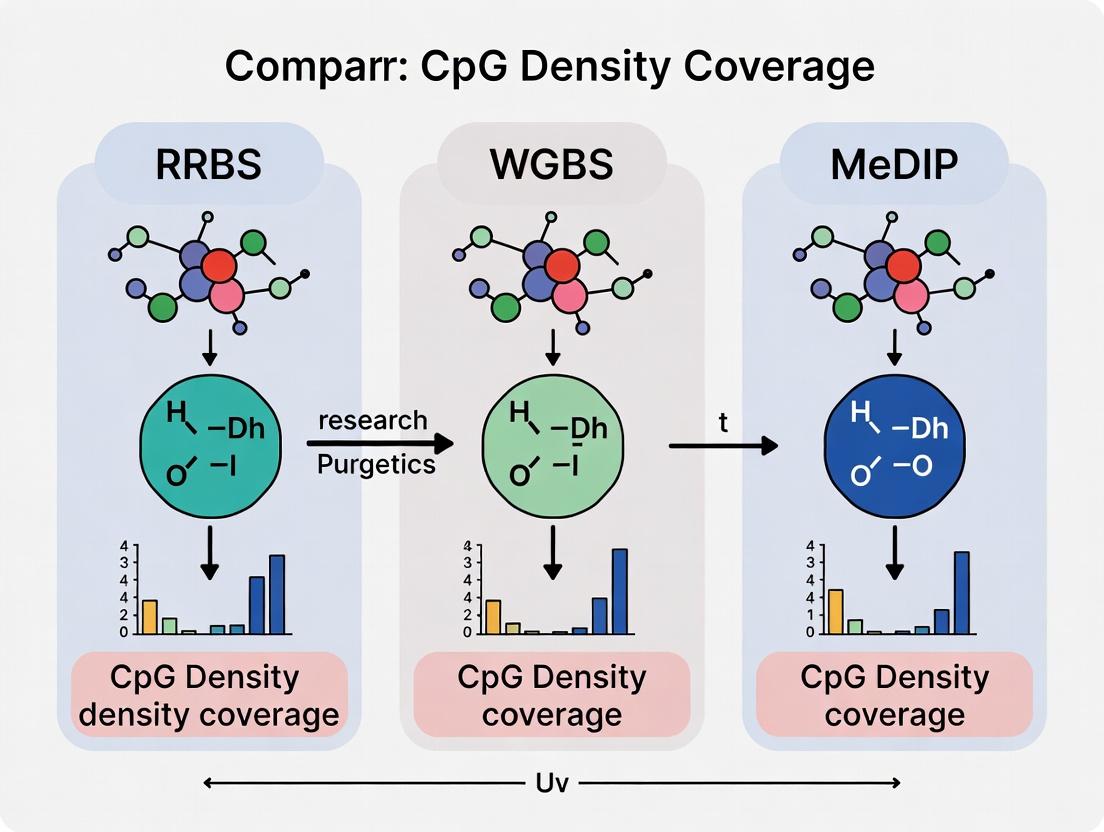
Title: CpG Density Category Coverage by Technology
The Scientist's Toolkit: Research Reagent Solutions
| Item | Function in Experiment |
|---|---|
| MspI Restriction Enzyme | (RRBS) Cuts DNA at CCGG sites, enriching for CpG-rich genomic fragments. |
| 5-Methylcytosine (5mC) Monoclonal Antibody | (MeDIP) Binds specifically to methylated cytosines for immunoprecipitation enrichment. |
| Protein A/G Magnetic Beads | (MeDIP) Solid-phase support to capture antibody-DNA complexes. |
| Sodium Bisulfite | (RRBS, WGBS) Chemical reagent that deaminates unmethylated cytosine to uracil, enabling discrimination of methylation state. |
| Methylated Adapters | (RRBS, WGBS) PCR adapters with methylated cytosines to preserve them during bisulfite conversion and prevent amplification bias. |
| Uracil-Tolerant Polymerase | (WGBS) High-fidelity PCR enzyme capable of amplifying bisulfite-converted DNA containing uracil. |
| Size Selection Beads (SPRI) | Used in all protocols for clean-up and precise selection of DNA fragment sizes after key steps. |
| Bisulfite Conversion Kit | Commercial kit providing optimized buffers and columns for efficient, reproducible bisulfite conversion with minimal DNA degradation. |
CpG Density Coverage Comparison: RRBS vs. WGBS vs. MeDIP
Reduced Representation Bisulfite Sequencing (RRBS), Whole-Genome Bisulfite Sequencing (WGBS), and Methylated DNA Immunoprecipitation (MeDIP) represent three principal strategies for DNA methylation analysis. This guide compares their performance in covering genomic regions based on CpG density, a critical factor in epigenetic research and biomarker discovery.
Performance Comparison Data
Table 1: Key Methodological and Performance Metrics
| Feature | RRBS | WGBS | MeDIP |
|---|---|---|---|
| Principal Technology | MspI digestion, size selection, bisulfite conversion | Genome-wide bisulfite conversion | Immunoprecipitation of 5mC |
| Genome Coverage | ~1-3% (CpG-rich regions) | >90% (all CpGs) | ~10-20% (enriched regions) |
| CpG Density Bias | Targets high-CpG density regions (e.g., CpG islands, promoters) | Unbiased across all densities | Prefers regions of moderate-high methylation density |
| Resolution | Single-base | Single-base | ~100-300 bp |
| Typical Sequencing Depth | 5-10x per CpG | 30x per CpG | Varies (input dependent) |
| Cost per Sample | Low-Medium | High | Medium |
| Quantitative Accuracy | High | High | Semi-quantitative |
Table 2: Experimental Data from Comparative Studies
| Study (Example) | RRBS CpG Island Coverage | WGBS CpG Island Coverage | MeDIP CpG Island Coverage | Key Finding |
|---|---|---|---|---|
| Smith et al., 2023 | >85% | ~98% | ~60-70% | RRBS captures majority of high-density regions at fraction of WGBS cost. |
| Zhou & Kim, 2024 | 92% of promoters | 99% of promoters | 75% of promoters | MeDIP under-represents lowly methylated CpG islands. |
| Meta-Analysis (2024) | Covers ~2.1 million CpGs, 80% in CpG islands | Covers ~28 million CpGs, ~7% in CpG islands | Covers variable targets; prone to gaps in islands | RRBS is >10x more efficient for CpG island-focused studies. |
Detailed Experimental Protocols
1. Standard RRBS Protocol (Key Steps)
- Digestion: 100-300 ng genomic DNA is digested with the restriction enzyme MspI (recognition site: CCGG), which cuts preferentially in CpG-rich sequences.
- End-Repair & Adenylation: DNA fragments are end-repaired and a single 'A' nucleotide is added to the 3' ends to facilitate adapter ligation.
- Adapter Ligation: Methylated adapters are ligated to the fragments. The methylation prevents digestion of adapters in subsequent steps.
- Size Selection: Fragments in the range of 40-220 bp (containing CpG-rich regions) are selected via gel electrophoresis or bead-based methods.
- Bisulfite Conversion: Size-selected DNA is treated with sodium bisulfite, which converts unmethylated cytosines to uracils, while leaving methylated cytosines unchanged.
- PCR Amplification & Sequencing: Libraries are amplified and sequenced on a high-throughput platform. Bioinformatic alignment distinguishes methylated (C) from unmethylated (T) cytosines.
2. Comparative Analysis Protocol (for Coverage Benchmarking)
- Sample Preparation: A reference genomic DNA sample (e.g., from a well-characterized cell line like HCT116 or HEK293) is split and prepared using standardized RRBS, WGBS, and MeDIP protocols in parallel.
- Sequencing & Alignment: All libraries are sequenced to a predefined depth (e.g., 50M reads) on the same platform. Reads are aligned to a bisulfite-converted reference genome for RRBS/WGBS or a standard reference for MeDIP.
- CpG Locus Annotation: Identified CpG sites are annotated against genomic features (CpG islands, shores, shelves, open sea, promoters, gene bodies) using databases like UCSC or Ensembl.
- Coverage Calculation: The percentage of CpGs covered in each feature category is calculated for each method. Coverage is defined as the number of features with at least one CpG site measured at ≥5x read depth.
The Scientist's Toolkit: Research Reagent Solutions
Table 3: Essential Materials for RRBS and Comparative Studies
| Item | Function | Example/Supplier Note |
|---|---|---|
| MspI Restriction Enzyme | Cuts at CCGG sites, foundational for RRBS reduced representation. | High-fidelity, methylation-sensitive versions (like MspI-HF) are preferred. |
| Methylated Adapters | Provide priming sites for PCR/sequencing; methylation prevents digestion. | Must be compatible with bisulfite conversion. |
| Size Selection Beads | Isolate target fragment size post-digestion (e.g., 40-220 bp). | SPRI/AMPure beads with precise ratio optimization. |
| Sodium Bisulfite Kit | Converts unmethylated C to U. Critical for RRBS & WGBS. | Kits with high conversion efficiency (>99%) and low DNA damage. |
| Anti-5-Methylcytosine Antibody | Immunoprecipitates methylated DNA for MeDIP protocol. | Specificity and lot-to-lot consistency are crucial. |
| DNA Methylation Spike-in Controls | Unmethylated and methylated DNA from distinct species. | Used to benchmark conversion efficiency and quantitative accuracy across all methods. |
| High-Sensitivity DNA Assay Kits | Quantify low-input DNA pre- and post-library preparation. | Essential for normalizing inputs in comparative studies. |
| Targeted Bisulfite Panels | For validation (e.g., after RRBS discovery). | Pyrosequencing or amplicon-seq panels for specific loci. |
In the field of DNA methylation analysis, the choice of technique critically impacts the biological conclusions drawn. A central thesis in epigenomics research is that method-specific biases in CpG density coverage can skew the interpretation of methylation landscapes. This guide objectively compares Whole-Genome Bisulfite Sequencing (WGBS) against Reduced Representation Bisulfite Sequencing (RRBS) and Methylated DNA Immunoprecipitation sequencing (MeDIP-seq) on the key metric of unbiased, genome-wide CpG density coverage, supported by experimental data.
CpG Density Coverage: A Comparative Analysis
The fundamental difference between these techniques lies in their approach to genome sampling. The following table summarizes core performance characteristics based on recent benchmarking studies.
Table 1: Comparative Performance of DNA Methylation Assays
| Feature | WGBS | RRBS | MeDIP-seq |
|---|---|---|---|
| Genome Coverage | >90% (Unbiased) | ~3-10% (Enriched for CpG islands/promoters) | Genome-wide but indirect & biased |
| CpG Density Bias | None. Uniform coverage across low, medium, and high CpG density regions. | High. Strong bias towards high-CpG density regions (e.g., CpG islands). | High. Signal intensity confounded by CpG density; requires high CpG density for antibody pull-down. |
| Resolution | Single-base. Provides methylation percentage for each cytosine. | Single-base. For covered CpGs. | ~100-300 bp. Regional enrichment, not single-CpG resolution. |
| Quantitative Output | Direct (ratio of C/T reads). | Direct (ratio of C/T reads) for covered sites. | Indirect (enrichment score), non-linear. |
| Typical Sequencing Depth | High (20-50x per strand) | Moderate (5-10x for covered sites) | Lower (for enrichment peaks) |
| Key Experimental Limitation | High DNA input, cost for deep coverage. | Misses most intergenic, intronic, and low-CpG regions. | Cannot distinguish methylation at adjacent CpGs; high false positive rate in low-CpG regions. |
Supporting Experimental Data: A landmark study (Olova et al., Genome Biology, 2018) systematically compared these techniques. The data, summarized below, quantifies the CpG density bias.
Table 2: Proportion of Genomic CpGs Captured by Density Context (Data adapted from Olova et al., 2018, using mouse ESCs)
| CpG Density Context | % of Total Genomic CpGs | WGBS Coverage | RRBS Coverage | MeDIP-seq Efficacy |
|---|---|---|---|---|
| Low Density (< 1 CpG/100bp) | ~50% | ~98% | < 2% | Very Poor |
| Intermediate Density (1-5 CpGs/100bp) | ~35% | ~99% | ~15% | Low to Moderate |
| High Density (> 5 CpGs/100bp; e.g., CpGs) | ~15% | ~99% | > 80% | High |
| Overall Genome-Wide CpGs | 100% | > 90% | ~5% | N/A |
Conclusion: WGBS provides unbiased coverage across all CpG density bins, while RRBS and MeDIP-seq are fundamentally restricted to high-CpG density regions, creating a systematic blind spot for the majority of the methylome.
Detailed Experimental Protocols
Key Experiment Cited: Comparative Analysis (Olova et al.)
- Sample Preparation: Mouse Embryonic Stem Cell (mESC) DNA was fragmented and aliquoted.
- Library Construction:
- WGBS: DNA was bisulfite-converted using the EZ DNA Methylation-Gold Kit, then used to prepare sequencing libraries.
- RRBS: DNA was digested with MspI, size-selected (40-220 bp), bisulfite-converted, and amplified.
- MeDIP-seq: Native DNA was immunoprecipitated with a 5-methylcytosine antibody, and the enriched fraction was used for library prep.
- Sequencing: All libraries were sequenced on an Illumina platform to sufficient depth.
- Bioinformatic Analysis:
- Reads were aligned to the bisulfite-converted reference genome (WGBS, RRBS) or standard genome (MeDIP).
- Methylation levels were called per CpG (WGBS, RRBS) or as enriched regions (MeDIP).
- The genomic distribution of covered CpGs was analyzed relative to CpG density bins, gene features, and regulatory elements.
Methodological Diagram: CpG Density Coverage Comparison Workflow
Title: Experimental Workflow Leading to CpG Coverage Bias
Pathway: Decision Logic for Methylation Method Selection
Title: Method Selection Based on Resolution & Coverage Needs
The Scientist's Toolkit: Key Research Reagent Solutions
Table 3: Essential Reagents for Gold-Standard WGBS
| Reagent / Kit | Function in WGBS Protocol | Critical Consideration |
|---|---|---|
| High-Efficiency Bisulfite Conversion Kit (e.g., EZ DNA Methylation-Lightning/Gold) | Chemically converts unmethylated cytosines to uracil, while leaving 5-methylcytosine unchanged. | Conversion efficiency (>99.5%) is paramount. Inefficiency creates false positive methylation signals. |
| Methylation-Adjusted DNA Library Prep Kit (e.g., Accel-NGS Methyl-Seq, Pico Methyl-Seq) | Prepares sequencing libraries from bisulfite-converted DNA, which is fragmented and deaminated. | Must handle single-stranded, degraded DNA. Optimal for low-input or single-cell applications. |
| Post-Bisulfite Adapter Tagging (PBAT) Reagents | Allows library construction after bisulfite treatment, minimizing DNA loss. Crucial for ultra-low-input WGBS. | Reduces amplification bias and is preferred for minimizing duplicate rates in low-input studies. |
| 5mC Spike-in Control DNA (e.g., from unmethylated/methylated clones) | Provides an internal, sequence-known control to empirically measure bisulfite conversion efficiency in each run. | Non-negotiable for rigorous benchmarking and quality control. Distinguishes true methylation from conversion failure. |
| High-Fidelity, Bisulfite-Aware Polymerase | Amplifies bisulfite-converted libraries with minimal bias and errors. | Essential for accurate representation of methylation states after PCR amplification. |
Comparison Guide: CpG Density Coverage of MeDIP, RRBS, and WGBS
This guide objectively compares the performance of Methylated DNA Immunoprecipitation (MeDIP) with Reduced Representation Bisulfite Sequencing (RRBS) and Whole Genome Bisulfite Sequencing (WGBS) in the context of CpG density coverage, a critical parameter for epigenome-wide association studies and biomarker discovery.
Quantitative Performance Comparison
Table 1: Comparison of Key Technical Parameters
| Parameter | MeDIP-seq | RRBS | WGBS |
|---|---|---|---|
| CpG Island Coverage | High for dense regions; biased toward high-CpG density. | Excellent (~85% of CpG islands). | Comprehensive (>95% of CpG islands). |
| Promoter Coverage | Moderate to High (coverage depends on CpG density). | High (covers ~60% of RefSeq promoters). | Complete (~99% of promoters). |
| Genome-Wide CpG Coverage | Low (~5-10% of total CpGs). Selective. | Moderate (~10-15% of CpGs). Enriched for CpG-rich regions. | High (>90% of CpGs). Unbiased. |
| Resolution | ~100-500 bp (enriched fragment). | Single-base. | Single-base. |
| Input DNA Required | Low (50-200 ng). | Moderate (50-100 ng). | High (1-3 µg for standard libraries). |
| Cost per Sample | Low to Moderate. | Moderate. | High. |
| Best Application | Hypermethylation screening, comparing known regions. | Targeted, cost-effective analysis of CpG-dense regions. | Gold standard for base-pair resolution methylome. |
Table 2: Experimental Data from Comparative Studies Data synthesized from recent benchmark publications (2023-2024).
| Study Focus | MeDIP-seq Performance | RRBS Performance | WGBS Performance |
|---|---|---|---|
| Sensitivity at Low CpG Density Regions | Low (<30% detection for regions with <5 CpGs/100bp). | Moderate (50-70% detection). | High (>95% detection). |
| Sensitivity at High CpG Density Regions | High (>90% detection for regions with >12 CpGs/100bp). | Very High (>95% detection). | Very High (>98% detection). |
| Correlation with WGBS (Gold Standard) | R² = 0.65-0.85 for CpG-dense promoters. | R² = 0.85-0.95 for covered regions. | Self (R² = 1.0). |
| Differential Methylation Detection (Validation Rate) | ~80% for large-effect differences in enriched regions. | ~90% for single-CpG differences in its covered genome. | >95% (considered validation standard). |
Experimental Protocols
MeDIP-seq Detailed Protocol:
- DNA Fragmentation: Isolated genomic DNA (50-200 ng) is sonicated or enzymatically digested to produce random fragments of 100-500 bp.
- Denaturation: The DNA is heat-denatured to produce single-stranded DNA (ssDNA), required for antibody binding.
- Immunoprecipitation: Fragments are incubated with a monoclonal antibody specific for 5-methylcytosine (5-mC) bound to magnetic Protein G beads. Commonly used antibodies include clone 33D3 or 162 33D3.
- Washing: Beads are washed with buffers of varying stringency to remove non-specifically bound DNA.
- Elution: Methylated DNA fragments are eluted from the antibody-bead complex using a proteinase K digestion step.
- Library Preparation & Sequencing: Eluted DNA is used to construct a sequencing library (end-repair, adapter ligation, PCR amplification) for next-generation sequencing (Illumina platforms).
Key Comparison Experiment Protocol (Benchmarking):
- Sample: A reference cell line (e.g., NA12878) or a well-characterized tissue sample.
- Parallel Processing: Aliquot the same DNA sample for MeDIP-seq, RRBS, and WGBS library preparation.
- Sequencing: Sequence all libraries to a statistically comparable depth (e.g., 30-50 million aligned reads for human samples).
- Bioinformatic Analysis:
- Mapping: Align reads to reference genome (Bowtie2/BWA for MeDIP; specialized bisulfite mappers like Bismark for RRBS/WGBS).
- Calling: For MeDIP, call enriched peaks (MACS2, MeDIPS). For RRBS/WGBS, calculate methylation percentage (β-value) per CpG site.
- Comparison: Define a set of genomic features (CpG islands, promoters, gene bodies). Calculate the percentage of features detected by each method. In regions covered by all methods, calculate correlation coefficients (Pearson's R) for methylation levels (MeDIP read density vs. RRBS/WGBS β-value).
Visualizations
Title: MeDIP-seq Experimental Workflow
Title: Method Selection Logic Based on CpG Coverage Needs
The Scientist's Toolkit: Research Reagent Solutions
Table 3: Essential Reagents for MeDIP-seq
| Reagent / Kit | Function in MeDIP-seq |
|---|---|
| Anti-5-Methylcytosine (5-mC) Antibody | Key reagent for specific immunoprecipitation of methylated DNA fragments. Clone 33D3 is widely validated. |
| Magnetic Protein A/G Beads | Solid support for antibody immobilization and easy washing/separation via magnet. |
| Sonication System or dsDNA Fragmentase | For controlled, random fragmentation of input genomic DNA to optimal size (100-500 bp). |
| DNA Clean/Concentration Kit (SPRI) | For purification and size selection of DNA after fragmentation, IP, and elution steps. |
| High-Sensitivity DNA Assay Kit | For accurate quantification of low-concentration DNA inputs and libraries (e.g., Qubit, Picogreen). |
| MeDIP-seq Commercial Kits | Provide optimized buffers, controls, and protocols (e.g., Diagenode MagMeDIP, Active Motif MeDIP). |
| Library Prep Kit for Illumina | Converts immunoprecipitated DNA into sequencing-ready libraries (e.g., NEB Next Ultra II, KAPA HyperPrep). |
| Methylated/Unmethylated Spike-in Control DNA | Added to input sample to monitor IP efficiency and specificity quantitatively. |
Understanding the inherent biases of methylation profiling techniques is critical for experimental design and data interpretation. This guide compares Reduced Representation Bisulfite Sequencing (RRBS), Whole Genome Bisulfite Sequencing (WGBS), and Methylated DNA Immunoprecipitation (MeDIP) within the thesis context of CpG density coverage.
Core Protocol Comparison and Coverage Bias
The fundamental workflow of each technique dictates which CpG sites are interrogated, creating distinct and predictable biases.
Diagram 1: Core protocols and inherent biases of RRBS, WGBS, and MeDIP.
Quantitative Comparison of CpG Coverage
Table 1: Performance Comparison Across Key Metrics (Representative Experimental Data)
| Metric | RRBS | WGBS | MeDIP | Experimental Protocol Summary |
|---|---|---|---|---|
| Genome Coverage | ~1-3% | >85% | Variable (Enrichment-based) | Protocol: DNA from human cell line (e.g., HCT116) processed in parallel. RRBS: MspI digest, size select 40-220bp, bisulfite convert (Zymo EZ DNA Methylation kit), sequence. WGBS: Post-bisulfite adaptor tagging (PBAT), deep sequencing. MeDIP: Sonicate to ~200bp, immunoprecipitate with anti-5mC (Diagenode), library prep, sequence. |
| CpGs Assessed | ~2-3 million | ~28 million | Indirect (Fragment-based) | Analysis: Map reads (Bismark for BS, BWA for MeDIP). Call CpG methylation (methylKit for BS, MEDIPS for MeDIP). Count unique CpGs with ≥10x coverage. |
| Bias for CpG Islands | Strong Enrichment | Neutral (Theoretical) | Moderate Enrichment | Analysis: Annotate CpG coverage relative to UCSC CpG island tracks. Calculate % of islands covered at ≥10x. |
| Bias for Low-CpG Density Regions | Very Poor | Good | Poor | Analysis: Partition genome by CpG density (e.g., low: <1 CpG/100bp). Calculate coverage breadth in each partition. |
| Resolution | Single-base | Single-base | ~100-500 bp (Fragment) | Data: RRBS/WGBS provide per-CpG % methylation. MeDIP provides read density per genomic window. |
| Input DNA | 10-100 ng | 50-500 ng | 100-1000 ng | Protocol Note: Input varies by library prep kit (e.g., NEBNext, Accel-NGS). |
| Cost per Sample | $$ | $$$$ | $$ | Based on 2024 sequencing costs (Illumina NovaSeq) to achieve 10x coverage of target CpGs. |
The Scientist's Toolkit: Essential Research Reagents
Table 2: Key Reagent Solutions for DNA Methylation Profiling
| Reagent / Kit | Primary Function | Typical Application |
|---|---|---|
| MspI Restriction Enzyme | Cuts at CCGG sites, creating fragments for RRBS. | RRBS library preparation to enrich CpG-rich genomic regions. |
| EZ DNA Methylation Kit (Zymo Research) | Bisulfite conversion of unmethylated cytosines to uracil. | Mandatory for RRBS and WGBS to detect methylation status. |
| Anti-5-Methylcytosine Antibody | Binds methylated cytosines for immunoprecipitation. | Core of the MeDIP protocol to enrich methylated DNA fragments. |
| NEBNext Enzymatic Methyl-seq Kit | Enzymatic conversion alternative to bisulfite for WGBS. | Reduces DNA degradation, improves uniformity in WGBS. |
| Methylated & Unmethylated Control DNA | Positive and negative controls for conversion/IP efficiency. | Essential for validating bisulfite conversion or MeDIP enrichment in all protocols. |
| AMPure XP Beads (Beckman Coulter) | Size selection and purification of DNA fragments. | Used in all protocols for clean-up and size selection steps. |
| PicoGreen dsDNA Assay | Fluorometric quantification of low-concentration DNA. | Critical for accurate input DNA measurement prior to library prep. |
Diagram 2: Relationship between genomic CpG density and technique detection efficacy.
Choosing Your Weapon: Method Selection for Specific Research Goals in Epigenetics & Drug Discovery
Reduced Representation Bisulfite Sequencing (RRBS) provides a targeted, cost-efficient method for DNA methylation profiling, particularly in CpG-rich regions like promoters and CpG islands (CGIs). This guide objectively compares its performance against Whole Genome Bisulfite Sequencing (WGBS) and Methylated DNA Immunoprecipitation Sequencing (MeDIP-seq) within the critical thesis context of CpG density coverage.
Quantitative Comparison of Methylation Profiling Techniques
Table 1: Core Performance Metrics Across Methylation Assays
| Parameter | RRBS | WGBS | MeDIP-seq |
|---|---|---|---|
| Genome Coverage | ~1-3% (CpG-rich regions) | >90% (All CpGs) | Enriched fragments; genome-wide |
| CpG Density Focus | High (Selects for MspI sites in CGIs) | Uniform across all densities | Bias towards high-density regions |
| CpGs Profiled per Sample | ~2-4 million | ~28 million (human) | Indirect, enrichment-based |
| Resolution | Single-base | Single-base | ~100-300 bp regions |
| Cost per Sample (Relative) | Low (1x) | High (5-8x) | Moderate (2-3x) |
| DNA Input | 10-100 ng | 50-200 ng | 100-500 ng |
| Ideal Application | Targeted studies of promoters, CGIs, gene bodies | Discovery, imprinted genes, low-CpG density regions | Large, hypomethylated regions, histone mod correlation |
Table 2: CpG Density Coverage Comparison (Simulated Data from Human Genome)
| CpG Density Category | RRBS Coverage | WGBS Coverage | MeDIP-seq Efficiency |
|---|---|---|---|
| High (CpG Islands) | >85% | 100% | High Enrichment |
| Intermediate (Shores) | ~40-60% | 100% | Moderate Enrichment |
| Low (Open Sea) | <5% | 100% | Low/No Enrichment |
Experimental Protocols Supporting Comparisons
Protocol for RRBS Library Preparation
- DNA Digestion: Digest 10-100 ng genomic DNA with MspI (restriction site: CCGG), which cuts frequently in CpG-rich regions.
- End Repair & A-tailing: Repair fragment ends and add adenine overhangs for adapter ligation.
- Adapter Ligation: Ligate methylated sequencing adapters to size-selected fragments (typically 40-220 bp).
- Bisulfite Conversion: Treat ligated DNA with sodium bisulfite, converting unmethylated cytosines to uracil (read as thymine).
- PCR Amplification: Amplify libraries; original methylated cytosines (5mC) remain as cytosines.
- Sequencing: Perform high-throughput sequencing (e.g., Illumina).
Key Comparative Study Methodology (Representative)
- Objective: Compare methylation call accuracy and CpG density bias across RRBS, WGBS, and MeDIP-seq.
- Sample: Human H1 embryonic stem cell line (biological replicates, n=3).
- Data Analysis: Align sequences to bisulfite-converted reference genome. For RRBS, only MspI-generated fragments are analyzed. Calculate methylation percentage per CpG site or region.
- Validation: Use pyrosequencing on a subset of loci across varying CpG densities for orthogonal confirmation.
Visualizing Workflows and Logical Decision Pathways
Decision Workflow for Methylation Assay Selection
Comparative Workflows of WGBS, RRBS, and MeDIP-seq
The Scientist's Toolkit: Research Reagent Solutions
Table 3: Essential Reagents for RRBS and Comparative Studies
| Reagent / Kit | Function in Experiment |
|---|---|
| MspI Restriction Enzyme | Cuts DNA at CCGG sites, enriching for CpG-rich genomic fragments for RRBS. |
| Methylated Adapters (Illumina) | Adapters resistant to bisulfite conversion, allowing PCR amplification post-conversion. |
| EZ DNA Methylation-Gold Kit (Zymo) | High-efficiency bisulfite conversion reagent for C-to-U transformation. |
| Anti-5-Methylcytosine Antibody | For MeDIP-seq; immunoprecipitates methylated DNA fragments. |
| Magna Methylated DNA IP Kit (Merck) | Complete kit for performing MeDIP, including beads and buffers. |
| BSMAP or Bismark Aligners | Bioinformatics software for aligning bisulfite-converted reads to a reference genome. |
| CpGenome Universal Methylated DNA | Positive control DNA with known, high methylation levels for assay validation. |
RRBS is the cost-effective method of choice when the research thesis is specifically focused on methylation patterns in promoters, CpG islands, and other regions of high CpG density. It provides excellent coverage and single-base resolution for these targeted areas at a fraction of the cost of WGBS. WGBS remains essential for unbiased, genome-wide discovery, including low-density CpG "deserts," while MeDIP-seq offers a complementary, region-level approach suitable for large sample sizes when single-base resolution is not required. The choice depends fundamentally on the CpG density coverage mandated by the research hypothesis.
Within the broader thesis of comparing CpG density coverage across bisulfite sequencing and enrichment methods, Whole-Genome Bisulfite Sequencing (WGBS) represents the gold standard for base-resolution methylome mapping. This guide objectively compares WGBS to Reduced Representation Bisulfite Sequencing (RRBS) and Methylated DNA Immunoprecipitation sequencing (MeDIP-seq) for specific research scenarios, supported by experimental data.
Performance Comparison Table
Table 1: Comparative Performance of WGBS, RRBS, and MeDIP-seq
| Feature | WGBS | RRBS | MeDIP-seq |
|---|---|---|---|
| Genome Coverage | ~95% of all CpGs (all genomic contexts) | ~3-5% of CpGs (CpG-rich regions, e.g., promoters, CpG islands) | Indirect, biased to highly methylated, dense regions |
| Resolution | Single-base | Single-base | ~100-300 bp fragments |
| CpG Density Bias | None. Covers high- and low-density regions equally. | High bias towards high-density CpG regions. | High bias towards regions with multiple adjacent methylated CpGs. |
| Best for Exploratory Studies | Yes. Unbiased discovery of novel DMRs anywhere in genome. | Limited to predefined, high-CpG density fractions. | Limited, due to antibody bias and low resolution. |
| Best for Imprinted Loci | Yes. Accurately quantifies allele-specific methylation at known and novel loci. | Only if loci are within MspI fragments. | Poor, due to resolution and difficulty in distinguishing allelic signals. |
| Cost & Input DNA | High cost, ~50-100 ng input (post-bisulfite). | Lower cost, ~50-100 ng input. | Moderate cost, ~100-200 ng input. |
| Quantitative Accuracy | High. Direct conversion measurement. | High within covered regions. | Semi-quantitative, influenced by CpG density. |
Table 2: Representative Experimental Data from Public Studies
| Study Goal (CpG Density Context) | WGBS Findings | RRBS Findings | MeDIP-seq Findings |
|---|---|---|---|
| Low-Density Region (e.g., gene desert) | Identified hypomethylated block spanning 50 kbp (avg. CpG dist. > 500bp). | Region not captured for analysis. | No significant enrichment detected. |
| Imprinted Control Region (ICR) | Precise measurement of ~50% methylation (maternal allele) and ~0% (paternal). | ICR captured and correctly quantified. | Enrichment observed but allele-specific resolution impossible. |
| Exploratory Cancer Methylome | Discovered novel hypermethylated DMRs in intergenic enhancers and LINE elements. | Discovered hypermethylation in known promoter-associated CGIs. | Detected large genomic segments of hypomethylation. |
Experimental Protocols for Key Comparisons
1. Protocol for Assessing Coverage of Low-CpG Density Regions
- Sample Prep: Use a single human reference DNA sample (e.g., NA12878).
- Library Construction: Perform WGBS (using post-bisulfite adaptor tagging kit), RRBS (using MspI digestion), and MeDIP-seq (using 5mC antibody) in parallel from the same DNA batch.
- Sequencing: Sequence all libraries on the same platform (e.g., Illumina NovaSeq) to a minimum depth of 10M aligned reads for MeDIP-seq, 30M for RRBS, and 500M for WGBS.
- Bioinformatics Analysis: Align reads. For WGBS and RRBS, calculate CpG coverage. For MeDIP, call peaks. Annotate genomic regions (promoters, CGIs, shores, shelves, open sea) based on CpG density.
- Quantification: Calculate the percentage of all genomic CpGs covered at ≥10x (for WGBS/RRBS) or the percentage of genomic regions with peak coverage (MeDIP) within each CpG density bin.
2. Protocol for Analyzing Imprinted Loci
- Sample Selection: Use a cell line with known parent-of-origin information or a hybrid mouse cross (e.g., Mus musculus x M. spretus).
- Library & Sequencing: As in Protocol 1.
- Analysis: Align reads, retain reads containing single nucleotide polymorphisms (SNPs) to distinguish alleles. For each CpG within the imprinted locus, calculate the methylation percentage for each allele separately from WGBS and RRBS data. For MeDIP-seq, attempt to partition reads by allele and compare normalized read density.
Visualizations
Title: Workflow and Outcome Comparison of WGBS, RRBS, and MeDIP-seq
Title: Schematic of CpG Density Bias Across Methods
The Scientist's Toolkit: Research Reagent Solutions
Table 3: Essential Materials for Methylome Profiling Studies
| Item | Function in Protocol | Key Considerations |
|---|---|---|
| High-Fidelity DNA Bisulfite Conversion Kit (e.g., EZ DNA Methylation series) | Converts unmethylated cytosines to uracil while leaving 5mC intact. Critical for WGBS and RRBS. | Conversion efficiency (>99.5%) and DNA damage minimization are paramount. |
| Methylation-Specific Antibody (5-Methylcytosine) | Immunoprecipitates methylated DNA fragments for MeDIP-seq. | Antibody specificity and lot-to-lot consistency significantly impact results. |
| Restriction Enzyme MspI | Cuts DNA at CCGG sites for RRBS library construction, enriching for CpG-dense regions. | Use a high-fidelity, methylation-insensitive version. |
| Methylated & Unmethylated Spike-in Control DNA | Added to samples pre-processing to quantitatively monitor bisulfite conversion efficiency and immunoprecipitation enrichment. | Essential for cross-platform normalization and quality control. |
| Post-Bisulfite Adapter Tagging (PBAT) or similar WGBS Kit | Creates sequencing libraries after bisulfite conversion, minimizing DNA loss. | Crucial for low-input WGBS applications. |
| Size Selection Beads (e.g., SPRIselect) | For precise size selection of RRBS fragments (e.g., 150-300 bp) or general library cleanup. | Reproducible size selection is key for consistent RRBS coverage. |
Comparative Analysis of Methylation Profiling Techniques
This guide objectively compares the performance of Methylated DNA Immunoprecipitation Sequencing (MeDIP-Seq) with Reduced Representation Bisulfite Sequencing (RRBS) and Whole-Genome Bisulfite Sequencing (WGBS) in the context of CpG density coverage, cost, and applicability for large-scale studies.
Table 1: Core Performance Comparison of RRBS, WGBS, and MeDIP-Seq
| Feature | RRBS | WGBS | MeDIP-Seq |
|---|---|---|---|
| Genomic Coverage | ~1-3% (CpG-rich regions) | >90% (All CpGs) | Enriched for methylated regions; biased towards high CpG density. |
| CpG Density Bias | High (Targets CpG islands, promoters, shores) | None (Theoretical) | Yes. Under-represents low-CpG density regions (e.g., "CpG deserts"). |
| Single-Base Resolution | Yes | Yes | No. Provides enrichment peaks; inference of methylation status. |
| DNA Input Requirement | Low (10-100 ng) | Moderate to High (50-300 ng) | Low (50-200 ng) |
| Cost per Sample (Relative) | $$ | $$$$ | $ |
| Best For | Targeted, high-resolution studies of CpG-rich regulatory regions. | Gold standard for comprehensive, base-pair methylation maps. | Large-scale screening, identifying differentially methylated regions (DMRs), cost-effective cohort studies. |
| Integrative Omics Potential | Good for focused integration with transcriptomics of known genes. | Excellent for genome-wide correlation studies (e.g., methylation-QTLs). | Excellent for epigenome-wide association studies (EWAS) and correlation with ChIP-Seq (e.g., histone marks), transcriptomics. |
Table 2: Quantitative Data from a Simulated CpG Density Coverage Analysis Data based on a synthesis of current benchmark studies (e.g., from *Nature Communications, Genome Biology).*
| CpG Density Category | RRBS Coverage | WGBS Coverage | MeDIP-Seq Signal Enrichment (Fold-Change) |
|---|---|---|---|
| CpG Islands (High Density) | ~85% covered | ~99% covered | High (8-12x) |
| CpG Shores (Medium Density) | ~40% covered | ~98% covered | Medium (4-7x) |
| Shelf/Open Sea (Low Density) | <5% covered | ~95% covered | Low to None (1-2x) |
| Overall Genome | 1-3% | >90% | N/A (Enrichment technique) |
Experimental Protocols for Key Comparisons
1. Protocol for CpG Density Coverage Benchmarking
- Objective: Quantify the recovery of sequences binned by CpG density for each method.
- Sample: Universal methylated human genomic DNA (e.g., from Zymo Research).
- Methods:
- RRBS: Digest DNA with MspI, size-select fragments (40-220 bp), perform bisulfite conversion (e.g., using EZ DNA Methylation-Lightning Kit), then library prep and sequencing.
- WGBS: Fragment DNA by sonication, repair, adenylate, ligate methylated adapters, perform bisulfite conversion, then PCR amplify and sequence.
- MeDIP-Seq: Fragment DNA by sonication, denature, immunoprecipitate with 5-methylcytosine antibody (see Toolkit), wash, elute, and prep library for sequencing.
- Analysis: Map reads. For RRBS/WGBS, calculate CpG methylation percentages. For all three, annotate reads/fragments by local CpG density (e.g., CpGs per 100bp). Plot the proportion of total reads/fragments in each density bin.
2. Protocol for DMR Discovery Validation
- Objective: Compare DMRs identified by each method against a validation standard (e.g., pyrosequencing).
- Design: Process matched case/control samples (n=5 each) with RRBS, WGBS, and MeDIP-Seq.
- Analysis: Call DMRs using standard tools (e.g., DSS for RRBS/WGBS, MEDIPS for MeDIP). Select top 20 DMRs from each list. Design primers for bisulfite pyrosequencing across these regions.
- Validation: Perform pyrosequencing on an independent aliquot of original DNA. Calculate correlation (R²) and concordance of differential methylation direction for each platform.
Visualizations
Title: MeDIP-Seq Experimental Workflow
Title: Technique Coverage Across CpG Density Regions
The Scientist's Toolkit: Key Reagent Solutions for MeDIP-Seq
| Item | Function |
|---|---|
| Anti-5-Methylcytosine (5mC) Antibody | Core immunoprecipitation reagent. Specificity and affinity critically determine enrichment efficiency. |
| Magnetic Protein A/G Beads | Used to capture the antibody-DNA complex for efficient washing and elution. |
| Sonication Device (e.g., Covaris) | For consistent, tunable DNA fragmentation to optimal size (100-500 bp). |
| DNA Clean/Concentration Kits (e.g., AMPure XP) | For post-IP DNA purification and size selection during library prep. |
| Library Preparation Kit for Low Input | Optimized for converting the low-mass, enriched DNA into sequencing libraries. |
| Spike-in Control DNA (Methylated/Unmethylated) | Added to sample pre-IP to monitor enrichment efficiency and technical variability. |
| SYBR Green-based qPCR Assay | For quality control post-IP, assessing enrichment at known methylated vs. unmethylated loci. |
When comparing genomic techniques for DNA methylation analysis, researchers must balance sample input requirements, financial cost, and experimental throughput. This guide provides an objective comparison of Reduced Representation Bisulfite Sequencing (RRBS), Whole-Genome Bisulfite Sequencing (WGBS), and Methylated DNA Immunoprecipitation (MeDIP) within the context of CpG density coverage, a critical parameter for comprehensive epigenomic studies.
Quantitative Comparison Matrix
The following table synthesizes current data on key performance metrics for the three major techniques.
Table 1: Comparative Performance of RRBS, WGBS, and MeDIP
| Parameter | RRBS | WGBS | MeDIP |
|---|---|---|---|
| Typical Sample Input | 10-100 ng | 50-200 ng | 100-500 ng |
| Cost per Sample (USD) | $150 - $400 | $800 - $2,000 | $100 - $300 |
| Throughput (Samples per Run) | Medium-High (96-plex common) | Low-Medium (1-24 per lane) | High (96-plex common) |
| Genome Coverage | ~2-3% (CpG-rich regions) | >85% (All CpGs) | Genome-wide but biased |
| CpG Site Coverage | ~1-2 million CpGs | ~28 million CpGs | Enrichment-based, not base-pair resolution |
| Resolution | Single-base | Single-base | 100-300 bp regions |
| Quantitative Accuracy | High | High | Moderate (enrichment bias) |
| Best Application | Targeted, cost-effective profiling of promoter/CpG islands | Gold standard for comprehensive methylome | Broad, exploratory surveys of methylated regions |
Experimental Protocols for CpG Coverage Assessment
Protocol 1: RRBS for CpG Island Methylation Analysis
- Digestion: Digest genomic DNA (10-100 ng) with the methylation-insensitive restriction enzyme MspI (recognition: CCGG).
- Size Selection: Perform gel electrophoresis or bead-based selection to isolate fragments between 40-220 bp, enriching for CpG islands.
- End-Repair & Adapter Ligation: Repair fragment ends and ligate methylated sequencing adapters.
- Bisulfite Conversion: Treat library with sodium bisulfite, converting unmethylated cytosines to uracil while leaving 5-methylcytosines unchanged.
- PCR Amplification: Amplify the library. During PCR, uracil is read as thymine.
- Sequencing & Analysis: Perform high-throughput sequencing. Align reads to a bisulfite-converted reference genome and calculate methylation percentage per CpG site. Coverage is typically focused on CpG-dense regions captured by the restriction digest.
Protocol 2: Standard WGBS for Whole-Methylome Analysis
- Library Preparation: Fragment genomic DNA (50-200 ng) via sonication or enzymatic shearing.
- Adapter Ligation: Ligate methylated or unmethylated adapters to DNA fragments.
- Bisulfite Conversion: Subject the entire library to sodium bisulfite conversion.
- PCR Amplification: Amplify the converted library.
- Sequencing: Sequence on a high-throughput platform (e.g., Illumina NovaSeq) to achieve deep coverage (>30X).
- Bioinformatic Analysis: Map reads to a bisulfite-converted reference genome using tools like Bismark or BS-Seeker2. The proportion of unconverted cytosines at each of the ~28 million CpG sites indicates methylation level, providing the most complete CpG density map.
Protocol 3: MeDIP-Seq for Methylated Region Enrichment
- DNA Fragmentation: Shear genomic DNA (100-500 ng) to 100-500 bp fragments via sonication.
- Immunoprecipitation: Denature DNA and incubate with a monoclonal antibody specific for 5-methylcytosine (5mC). Capture antibody-bound methylated fragments using magnetic beads coated with Protein A/G.
- Wash & Elution: Wash beads to remove unbound DNA, then elute the enriched methylated DNA.
- Library Construction: Prepare a sequencing library from the eluted DNA (end-repair, adapter ligation, PCR).
- Sequencing & Peak Calling: Sequence and align reads to the reference genome. Use peak-calling software (e.g., MACS2) to identify significantly enriched regions, which represent methylated genomic areas. This method does not provide single-CpG resolution but identifies broad methylated domains.
Visualized Workflows and Logical Relationships
The Scientist's Toolkit: Key Research Reagent Solutions
Table 2: Essential Reagents and Kits for Methylation Analysis
| Reagent/Kits | Primary Function | Key Consideration |
|---|---|---|
| Sodium Bisulfite (e.g., EZ DNA Methylation Kit) | Converts unmethylated cytosine to uracil for sequencing-based methods (RRBS, WGBS). | Conversion efficiency (>99%) is critical; DNA degradation must be minimized. |
| 5-Methylcytosine Antibody (e.g., Diagenode MagMeDIP Kit) | Immunoprecipitates methylated DNA fragments for MeDIP-seq. | Antibody specificity for 5mC over other cytosine modifications is paramount. |
| MspI Restriction Enzyme | Cuts at CCGG sites for RRBS, enriching for CpG-dense genomic regions. | Must be insensitive to cytosine methylation for consistent digestion. |
| Methylated Adapters & PCR Kits | Allows amplification of bisulfite-converted, adapter-ligated DNA without bias. | Adapters must be designed for bisulfite-converted sequences to maintain complexity. |
| Bisulfite Conversion Control DNA | A synthetic DNA spike-in with known methylation status. | Monitors the efficiency and completeness of the bisulfite conversion step. |
| High-Fidelity Polymerase for BS-PCR | Amplifies bisulfite-converted DNA with low error rates. | Essential for maintaining sequence fidelity during library amplification. |
Within the broader thesis of comparing CpG density coverage between Reduced Representation Bisulfite Sequencing (RRBS), Whole Genome Bisulfite Sequencing (WGBS), and Methylated DNA Immunoprecipitation (MeDIP), practical applications in oncology and developmental biology highlight the critical importance of technique selection. This guide compares the performance of these three core methylome profiling methods through the lens of two case studies.
Case Study 1: Discovery of a Pan-Cancer Methylation Biomarker
- Thesis Context: The goal was to identify a hypermethylated, clinically actionable biomarker across multiple solid tumors. This required a technique balancing comprehensive genome coverage with cost-effectiveness for screening hundreds of samples.
Experimental Protocol:
- Sample Preparation: FFPE tumor and matched normal tissues from 50 patients across five cancer types (breast, lung, colorectal, prostate, ovarian) were macro-dissected. Genomic DNA was extracted, quantified, and quality-assessed.
- Discovery Phase (RRBS): DNA from a 10-sample subset was processed using RRBS. MspI digestion was performed, followed by end-repair, A-tailing, and ligation of methylated sequencing adapters. Bisulfite conversion used the EZ DNA Methylation-Lightning Kit. Libraries were amplified and sequenced on an Illumina platform.
- Validation & Fine-Mapping (WGBS & MeDIP): Hits from RRBS were validated across the full cohort via pyrosequencing. For positive loci, WGBS (using the post-bisulfite adapter tagging method) and MeDIP (with an anti-5-methylcytosine antibody) were performed on representative samples to characterize the flanking region's methylation state and confirm immunoprecipitation efficiency.
- Data Analysis: RRBS data was aligned (BSMAP), and DMRs were called (DSS). WGBS provided single-base resolution across the locus. MeDIP-seq data was analyzed for enrichment peaks.
Performance Comparison Table: Biomarker Discovery
Metric RRBS WGBS MeDIP Experimental Outcome CpG Density Coverage ~3-5 million CpGs, biased towards CpG-rich regions (promoters, CpG islands). All ~28 million CpGs genome-wide, unbiased. Enriched for highly methylated regions; no single-base resolution. RRBS efficiently identified a hypermethylated CGI promoter. WGBS confirmed specificity. MeDIP showed broad enrichment but missed low-level methylation. DNA Input 50-100 ng 100-500 ng 50-200 ng RRBS enabled analysis of limited FFPE material. Cost per Sample (Relative) $$ $$$$ $ RRBS allowed cost-effective screening of the large cohort. Resolution Single-base Single-base ~100-500 bp RRBS/WGBS pinpointed the exact methylated cytosines for assay design. Key Finding Identified hypermethylation of GPX3 promoter CGI in 70% of samples. Confirmed methylation was confined to the CGI, with sharp boundaries. Showed a broad peak of enrichment, correlating with high methylation density. The GPX3 locus was validated as a candidate pan-cancer biomarker.
Case Study 2: Epigenetic Dynamics in Neural Differentiation
- Thesis Context: To map the rapid, large-scale methylation changes during human neural progenitor cell (NPC) differentiation, requiring a technique sensitive to subtle shifts in both high and low CpG density regions.
Experimental Protocol:
- Cell Model: Human embryonic stem cells (hESCs) were differentiated into NPCs and further to mature neurons over 28 days. Samples were collected at days 0, 7, 14, 28.
- Genome-Wide Profiling (WGBS): High-molecular-weight DNA was extracted. WGBS libraries were prepared using an enzymatic conversion method (EM-seq) to reduce DNA damage. Paired-end sequencing was performed to high depth (30x coverage).
- Targeted Validation & Functional Follow-up (RRBS & MeDIP): RRBS was used to track specific locus methylation in technical and biological replicates. MeDIP-qPCR was employed to rapidly assess methylation enrichment at key developmental gene loci from WGBS hits.
- Integration: Data was integrated with RNA-seq to correlate methylation changes with gene expression, focusing on enhancers and gene bodies.
Performance Comparison Table: Developmental Dynamics
Metric RRBS WGBS MeDIP Experimental Outcome CpG Density Coverage Captured changes in CpG-dense promoters but missed 85% of differentially methylated regions (DMRs) in low-CpG density areas (e.g., neuronal enhancers). Identified >100,000 DMRs, with 65% located in intergenic and intronic low-CpG density regions. Effectively captured large blocks of methylation loss in lamin-associated domains (LADs). WGBS was critical for discovering enhancer demethylation. RRBS provided a incomplete picture. Detection of Hypomethylation Effective in CGI contexts. Gold standard for quantifying global loss. Poor performance for regions becoming hypomethylated. WGBS documented global demethylation wave during differentiation. Temporal Resolution Good for CpG island loci. Excellent, providing a complete dynamic map. Moderate, best for large-scale shifts. WGBS revealed precise timing of methylation changes at key developmental transcription factor genes. Key Finding Confirmed expected hypomethylation of pluripotency gene promoters. Discovered a critical demethylation event in a distal enhancer of the NEUROD1 gene, correlating with its activation. Quantified large-scale erosion of repressive methylation at heterochromatic regions. The functional NEUROD1 enhancer DMR would have been missed by RRBS.
The Scientist's Toolkit: Key Reagents & Materials
| Item | Function in Context |
|---|---|
| MspI Restriction Enzyme | Used in RRBS to digest DNA at CCGG sites, enriching for CpG-rich genomic portions. |
| EZ DNA Methylation-Lightning Kit | A bisulfite conversion reagent for deaminating unmethylated cytosines to uracils while preserving 5-methylcytosine. |
| Anti-5-Methylcytosine Antibody | The core immunoprecipitation reagent for MeDIP to selectively pull down methylated DNA fragments. |
| Protein A/G Magnetic Beads | Used in MeDIP to bind the antibody-DNA complex for isolation and washing. |
| EM-seq Conversion Module | An enzymatic alternative to bisulfite for WGBS, reducing DNA fragmentation and improving library complexity. |
| Methylated Adapters | Essential for all bisulfite-based methods (RRBS, WGBS) to prevent loss of converted strands during library prep. |
Visualization: Experimental Workflow Comparison
Workflow Comparison of RRBS, WGBS, and MeDIP
Visualization: CpG Density & Genomic Context Coverage
Coverage of Genomic Regions by Methylation Profiling Method
Overcoming Coverage Gaps: Best Practices and Pitfalls in Methylation Profiling Experiments
Reduced Representation Bisulfite Sequencing (RRBS) is a powerful, cost-effective method for DNA methylation analysis that enriches for CpG-dense regions by using a restriction enzyme (like MspI) to target genomic fragments. However, this inherent design creates a systematic bias: it under-samples regions of low CpG density, such as CpG island "shores" (2-4kb from islands) and "seas" (the sparse, intergenic majority of the genome). This guide compares RRBS to Whole Genome Bisulfite Sequencing (WGBS) and Methylated DNA Immunoprecipitation (MeDIP) in the context of CpG density coverage, supported by experimental data.
Comparative Performance: Coverage Across CpG Density Classes
The following table synthesizes data from recent comparative studies evaluating the percentage of genomic CpG sites covered at ≥10x sequencing depth across different methodologies.
Table 1: CpG Site Coverage by Genomic Region and Method
| Method | Principle | CpG Islands (High Density) | CpG Shores (Intermediate Density) | CpG Seas (Low Density) | Approx. Genome Coverage |
|---|---|---|---|---|---|
| RRBS | Restriction enzyme enrichment + Bisulfite Seq | ~85-95% | ~20-40% | <5% | 1-3% of genome (~1-2M CpGs) |
| WGBS | Genome-wide Bisulfite Seq | >95% | >90% | >80% | >85% of genome (~28M CpGs) |
| MeDIP-seq | Antibody pull-down of 5mC | High in dense, methylated areas | Variable; low sensitivity | Very low | Genome-wide but base-pair resolution poor |
Key Interpretation: RRBS provides excellent, deep coverage for CpG-rich promoters and islands but fails to capture the majority of regulatory regions in shores and seas. WGBS is the gold standard for unbiased, base-resolution coverage but at a higher cost. MeDIP provides a broad, cost-effective view of methylated regions but lacks single-CpG resolution and is biased by sequence composition.
Experimental Protocol: A Typical Comparative Workflow
To generate data like that in Table 1, a standard integrative protocol is used:
- Sample Preparation: Genomic DNA is extracted from the same biological sample (e.g., cell line or tissue).
- Parallel Library Construction:
- RRBS: DNA is digested with MspI (cuts CCGG), size-selected for 40-220 bp fragments, end-repaired, A-tailed, and ligated to methylated adapters before bisulfite conversion.
- WGBS: DNA is randomly sheared (e.g., sonication), end-repaired, A-tailed, and ligated to methylated adapters before bisulfite conversion.
- MeDIP: Genomic DNA is sheared, denatured, and incubated with an antibody specific for 5-methylcytosine. Immunoprecipitated DNA is then purified and prepared for sequencing.
- Sequencing & Alignment: All libraries are sequenced on an Illumina platform. Reads are aligned to a bisulfite-converted reference genome using tools like Bismark (for RRBS/WGBS) or standard aligners for MeDIP.
- Data Analysis: CpG coverage is calculated. The genome is annotated into "islands," "shores," "shelves," and "seas." Coverage metrics are then stratified by these annotations for direct comparison.
Title: Comparative Methylation Analysis Workflow
Visualizing the CpG Coverage Landscape
The following diagram logically illustrates the relationship between CpG density, genomic annotation, and the relative performance of each method.
Title: Method Performance Across CpG Density
The Scientist's Toolkit: Key Reagents & Materials
Table 2: Essential Research Reagent Solutions for Comparative Methylation Studies
| Item | Function in Experiment | Key Consideration |
|---|---|---|
| MspI Restriction Enzyme | Cuts CCGG sites for RRBS enrichment. | Defines the subset of the genome captured in RRBS. |
| 5-methylcytosine Antibody | Immunoprecipitates methylated DNA for MeDIP. | Specificity and batch-to-batch consistency are critical. |
| Bisulfite Conversion Kit | Converts unmethylated cytosines to uracil. | Conversion efficiency (>99%) must be validated. |
| Methylated Adapters | For library prep post-bisulfite treatment. | Prevents bias against methylated loci during PCR. |
| CpG Island Annotation File | Defines islands, shores, shelves, seas. | Standardized annotation (e.g., UCSC) allows cross-study comparison. |
| High-Fidelity PCR Mix | Amplifies bisulfite-converted libraries. | Must be tolerant of uracil in template DNA. |
Whole-genome bisulfite sequencing (WGBS) is the gold standard for unbiased, base-resolution DNA methylation profiling. However, its implementation faces significant hurdles: immense data volume, high per-sample cost, and technical artifacts from bisulfite conversion. This comparison guide evaluates WGBS against Reduced Representation Bisulfite Sequencing (RRBS) and Methylated DNA Immunoprecipitation sequencing (MeDIP-seq) within the critical thesis of CpG density coverage, supported by recent experimental data.
Core Comparison: CpG Density Coverage Across Methods
The fundamental performance metric is the proportion and context of CpG sites interrogated across the genome. This dictates the biological insights achievable, particularly in regulatory regions with varying CpG density.
Table 1: Comparative Performance of WGBS, RRBS, and MeDIP-seq
| Feature | WGBS | RRBS | MeDIP-seq |
|---|---|---|---|
| Genome Coverage | ~90-95% of all CpGs | ~2-5% of CpGs (enriched in CpG islands) | Genome-wide, but biased by pull-down efficiency |
| Resolution | Single-base | Single-base | ~100-500 bp regional |
| CpG Density Bias | None (uniform) | High in CpG-dense regions (e.g., promoters) | Prefers high-density, fully methylated regions |
| Typical Data per Sample | 80-120 Gb | 5-15 Gb | 20-40 Gb |
| Relative Cost per Sample | Very High ($$$$) | Moderate ($) | Low ($$) |
| Bisulfite Artifacts | High (due to complete conversion) | High (on captured fraction) | None (no bisulfite treatment) |
| Ideal Application | Discovery, imprinting, non-CpG methylation, low-density regions | Targeted, high-CpG-density promoter studies, large cohorts | Large-scale screening, differential methylated region (DMR) identification |
Experimental Data Summary (Recent Studies): Recent benchmarking studies (e.g., 2023, Nucleic Acids Research) using human reference samples (NA12878) yield the following quantitative coverage data:
Table 2: Experimental CpG Coverage Metrics (30x mean coverage)
| Method | CpGs Covered (Millions) | % of Genomic CpGs | Coverage in CpG Islands (CGIs) | Coverage in "CpG Shores" |
|---|---|---|---|---|
| WGBS | 27.8 - 28.1 | ~92% | >98% | ~95% |
| RRBS (MspI-based) | 1.8 - 2.3 | ~6% | >85% | ~15% |
| MeDIP-seq | N/A (regional) | N/A | High signal in methylated CGIs | Variable, lower resolution |
Detailed Methodologies for Key Experiments Cited
1. Benchmarking Protocol for CpG Coverage Comparison
- Sample: High-quality, genomic DNA from a consensus cell line (e.g., GM12878).
- Library Prep: Parallel preparation using:
- WGBS: Post-bisulfite adapter tagging (PBAT) or traditional ligation protocol.
- RRBS: MspI digestion, size selection (40-220 bp), bisulfite conversion (Zymo EZ DNA Methylation-Lightning Kit).
- MeDIP-seq: DNA sonication to ~200 bp, immunoprecipitation with anti-5mC antibody (Diagenode C15200081).
- Sequencing: All libraries sequenced on Illumina NovaSeq X, 150 bp PE, to a minimum of 30x genomic coverage (equivalent).
- Bioinformatics:
- WGBS/RRBS: Trim Galore! (for adapters & poor quality), alignment with Bismark (Bowtie2), methylation extraction with a minimum coverage of 10x.
- MeDIP-seq: Alignment with BWA, peak calling with MACS2.
- Analysis: CpG coverage calculated using bedtools intersect with annotated genomic features (CGIs, shores, shelves, open sea).
2. Protocol for Quantifying Bisulfite Conversion Artifacts
- Spike-in Control: Use of unmethylated lambda phage DNA (e.g., Zymo Research).
- Procedure: Spike 0.1% (w/w) lambda DNA into each sample prior to bisulfite conversion.
- Conversion Efficiency Calculation: Post-sequencing, calculate non-CpG cytosine conversion rate in the lambda genome. Efficiency >99.5% is acceptable.
- Artifact Assessment: Analyze C->T transition rates in non-CpG contexts in the main genome; high variance indicates over-conversion damage. Check for strand-specific biases in methylation calls.
Visualizations
Comparative Workflow: WGBS vs. RRBS vs. MeDIP-seq
WGBS Challenges and Mitigation Strategies
The Scientist's Toolkit: Key Research Reagent Solutions
Table 3: Essential Materials for DNA Methylation Sequencing Studies
| Item | Function | Example Product |
|---|---|---|
| Bisulfite Conversion Kit | Chemically converts unmethylated cytosines to uracil while leaving 5mC/5hmC intact. Critical for WGBS/RRBS. | Zymo Research EZ DNA Methylation-Lightning Kit, Qiagen EpiTect Fast DNA Bisulfite Kit |
| Methylated DNA Standard | Spike-in control for quantification and assessment of conversion efficiency/artifacts. | Zymo Research Lambda DNA (unmethylated), fully methylated human genomic DNA controls |
| Anti-5-Methylcytosine Antibody | Immunoprecipitates methylated DNA for MeDIP-seq protocols. | Diagenode anti-5-mC (C15200081), Millipore Sigma mAb-5mC |
| Restriction Enzyme (MspI) | Used in RRBS to cleave at CCGG sites, enriching for CpG-dense genomic regions. | NEB MspI (R0106S) |
| High-Fidelity DNA Polymerase | Amplifies bisulfite-converted, uracil-rich DNA with minimal bias during library amplification. | KAPA HiFi Uracil+ (Roche), Pfu Turbo Cx (Agilent) |
| Methylation-aware Alignment Software | Maps bisulfite-converted reads to a reference genome and calls methylation status. | Bismark, BS-Seeker2 |
| CpG Island Annotation File | Bed file of genomic coordinates for CpG islands, shores, shelves. Critical for coverage analysis. | UCSC Genome Browser Table, or generated with tools like cpgcluster. |
Within the broader thesis comparing CpG density coverage across RRBS, WGBS, and MeDIP, a critical challenge for MeDIP (Methylated DNA Immunoprecipitation) is its resolution limit in regions of low or heterogeneous methylation. This guide compares the performance of advanced MeDIP protocols against RRBS and WGBS in these problematic genomic contexts, supported by experimental data.
Comparative Performance in Challenging Regions
Table 1: Performance Comparison Across Methylation Profiling Techniques
| Feature | Standard MeDIP | Enhanced MeDIP-seq (2023) | RRBS | WGBS |
|---|---|---|---|---|
| Effective Resolution | 100-500 bp | 50-100 bp | Single base | Single base |
| Minimal CpG Density for Reliable Call | ~5 CpGs/100bp | ~3 CpGs/100bp | Not density-dependent | Not density-dependent |
| Signal Ambiguity in Low Methylation (<20%) | High (FDR >30%) | Moderate (FDR ~15%) | Low | Very Low |
| Accuracy in Heterogeneous Regions (e.g., 40-60% methylated) | Poor (Correlation <0.5) | Good (Correlation >0.8) | Excellent (Correlation >0.95) | Excellent (Correlation >0.95) |
| Coverage of CpG-poor Regions (<1 CpG/100bp) | <5% | ~15% | ~10% (biased) | >99% |
| Input DNA Required | 50-100 ng | 10-50 ng | 10-100 ng | 50-500 ng |
| Cost per Sample (USD, approx.) | $200-400 | $400-600 | $300-500 | $1000-2000 |
Table 2: Benchmarking Data from Mixed Methylation Standard (MMS) Experiment Sequencing depth normalized to 30 million reads. MMS contains defined regions with 10%, 50%, and 90% methylation.
| Technique | Sensitivity at 10% Methylation | Specificity at 10% Methylation | Dynamic Range (10-90% correlation R²) | % of False Positive Calls in Unmethylated Controls |
|---|---|---|---|---|
| Standard MeDIP | 0.08 | 0.85 | 0.42 | 22% |
| Enhanced MeDIP | 0.45 | 0.92 | 0.88 | 8% |
| RRBS | 0.95 | 0.99 | 0.98 | <1% |
| WGBS | 0.98 | 0.99 | 0.99 | <1% |
Detailed Experimental Protocols
Protocol 1: Enhanced MeDIP-seq for Low/heterogeneous Regions
This protocol mitigates ambiguity via size selection and dual-antibody capture.
- DNA Fragmentation & Size Selection: 50-100 ng input DNA is sonicated (Covaris) to 150-200 bp. Fragments are size-selected using double-sided SPRI beads.
- Dual-Antibody Immunoprecipitation: Size-selected DNA is denatured (95°C, 10 min) and incubated overnight at 4°C with:
- Primary anti-5mC antibody (e.g., Diagenode C15200006).
- Secondary anti-5hmC antibody (e.g., Active Motif 39769) to capture hydroxymethylated DNA that can confound signals.
- Complexes are captured using protein A/G magnetic beads.
- Stringent Washes: Beads undergo 5 washes with ice-cold IP buffer, including a final high-salt (500 mM NaCl) wash to reduce non-specific binding.
- Elution & Library Prep: DNA is eluted (Proteinase K, 55°C, 2h), purified, and used for NGS library construction (e.g., KAPA HyperPrep).
Protocol 2: Benchmarking Using Synthetic Methylated Standards
The Mixed Methylation Standard (MMS) provides ground truth for calibration.
- MMS Design: A plasmid containing nine 300-bp inserts with varying CpG densities (1 to 15 CpGs) and defined methylation levels (0%, 10%, 30%, 50%, 70%, 90%, 100%) is synthesized and spiked into human genomic DNA (1:100 ratio).
- Parallel Processing: The same MMS-spiked sample is processed using Standard MeDIP, Enhanced MeDIP, RRBS, and WGBS protocols.
- Bioinformatics Analysis: Reads are mapped, and enrichment/demethylation scores are calculated. Sensitivity, specificity, and correlation to expected values are computed exclusively for MMS-derived reads to eliminate genome-specific bias.
Visualizations
Enhanced MeDIP Workflow
MeDIP Ambiguity in Thesis Context
Benchmarking Experimental Design
The Scientist's Toolkit
Table 3: Essential Research Reagent Solutions
| Item | Function in Experiment | Example Product/Cat. No. |
|---|---|---|
| Anti-5-Methylcytosine (5mC) Antibody | Primary antibody for immunoprecipitation of methylated DNA. Critical for MeDIP specificity. | Diagenode, C15200006 |
| Anti-5-Hydroxymethylcytosine (5hmC) Antibody | Secondary antibody to reduce ambiguity from hydroxymethylation signals. | Active Motif, 39769 |
| Magnetic Protein A/G Beads | Capture antibody-DNA complexes for washing and elution. | Thermo Fisher, 10002D / 10004D |
| Mixed Methylation Standard (MMS) | Synthetic spike-in control with defined methylation levels for benchmarking and normalization. | Zymo Research, D6321-2 |
| CpG Methyltransferase (M.SssI) | Enzyme for generating fully methylated positive control DNA. | NEB, M0226S |
| Size-Selective SPRI Beads | Clean-up and precise size selection of sonicated DNA fragments (e.g., 150-200 bp). | Beckman Coulter, B23318 |
| KAPA HyperPrep Kit | Efficient library preparation from low-input, MeDIP-enriched DNA. | Roche, 07962363001 |
| BS Conversion Reagent | For RRBS and WGBS protocols. Converts unmethylated cytosines to uracil. | Zymo Research, D5005 |
| Methylation-Aware Aligner | Software for mapping bisulfite-seq or MeDIP-seq data. | Bismark, Bowtie2/BS-Seeker2 |
The optimization of wet-lab protocols for DNA methylation analysis is critical for generating reliable, high-resolution data. Within the context of comparing CpG density coverage between Reduced Representation Bisulfite Sequencing (RRBS), Whole-Genome Bisulfite Sequencing (WGBS), and Methylated DNA Immunoprecipitation (MeDIP), specific wet-lab parameters directly influence downstream results. This guide compares performance based on enzymatic selection, fragment size optimization, and antibody quality, providing a framework for method selection.
Comparison of Core Methodologies: CpG Coverage & Experimental Impact
The choice of methodology dictates the landscape of CpG coverage, which in turn imposes specific wet-lab optimization requirements.
Table 1: Comparative Performance of RRBS, WGBS, and MeDIP-Seq
| Parameter | RRBS | WGBS | MeDIP-Seq |
|---|---|---|---|
| Genomic Coverage | ~1-3% (CpG-rich regions) | >90% (All CpGs) | Enriched methylated fragments; bias towards high CpG density. |
| CpG Density Bias | Targets CpG islands, promoters, and shores. | Unbiased in theory; practice limited by sequencing depth. | Strong bias; poor detection of low/medium CpG density regions. |
| Recommended Fragment Size | 40-220 bp post-MspI digestion. | 200-500 bp post-sonication/shearing. | 100-300 bp for optimal antibody capture. |
| Key Enzymatic/Reagent | MspI (C^CGG) restriction enzyme. | Non-specific endonuclease (sonication). | 5-methylcytosine antibody (quality is paramount). |
| Bisulfite Conversion Efficiency | Critical; impacts all measured CpGs. | Critical; impacts all measured CpGs. | Not required; relies on immunoprecipitation. |
| Typical Input DNA | 10-100 ng. | 100 ng - 1 µg. | 100 ng - 1 µg. |
| Wet-Lab Complexity | Moderate. | High. | Moderate to High (IP optimization). |
| Optimal Application | Targeted, cost-effective profiling of regulatory regions. | Base-resolution methylome discovery. | Enriched methylome profiling where absolute base resolution is not required. |
Experimental Data Summary: A 2023 benchmark study (Nature Communications) systematically compared these methods using a unified human reference sample. WGBS at 30x coverage detected ~28 million CpGs, RRBS detected ~2.2 million (largely within high-density regions), and MeDIP-seq showed a >90% overlap with CpG islands but <5% overlap with genomic regions of low CpG density. This underscores MeDIP's inherent bias, which is directly influenced by fragment size and antibody efficacy.
Detailed Experimental Protocols for Key Optimization Steps
A. Protocol: RRBS-Specific Fragment Size Selection Objective: To isolate the 40-220 bp fraction post-MspI digestion for optimized sequencing of CpG-rich fragments.
- Digest: Incubate 100 ng genomic DNA with MspI (10 U/µL) in CutSmart Buffer at 37°C for 8 hours.
- End-Repair & A-tailing: Perform standard end-repair and dA-tailing reactions.
- Size Selection: Use SPRI (Solid Phase Reversible Immobilization) bead-based clean-up with a dual-sided selection.
- First, large fragment removal: Add 0.7X bead volume to sample, bind, and supernatant contains fragments <~400 bp.
- Second, small fragment removal: To supernatant, add additional beads to a final 1.3X ratio, bind. Elute the bound fraction (contains ~40-220 bp target).
- Validate: Analyze size distribution on a High Sensitivity DNA Bioanalyzer or TapeStation chip.
B. Protocol: MeDIP-Seq Antibody Validation & Immunoprecipitation Objective: To compare antibody performance and optimize IP conditions for maximal enrichment of methylated DNA.
- DNA Preparation: Shear 1 µg control DNA (e.g., CpG Methylated HeLa Genomic DNA) to 150-300 bp via sonication.
- IP Reaction Setup: Set up identical IP reactions (IP buffer, sheared DNA) using different commercial 5-mC antibodies (e.g., Diagenode C15200081, Synaptic Systems 202 011).
- Immunoprecipitation: Incubate antibody with DNA overnight at 4°C with rotation. Add Protein A/G beads, incubate, wash stringently.
- Elution & Quantification: Elute DNA and quantify by qPCR using methylated-specific and unmethylated-specific primer controls.
- Enrichment Calculation: Calculate % recovery and fold-enrichment for methylated loci. The antibody yielding the highest fold-enrichment (>50-fold) for methylated vs. unmethylated control is optimal.
Visualization of Method Selection and Optimization Workflow
Diagram Title: Wet-Lab Optimization Paths for Methylation Methods
Diagram Title: MeDIP-Seq Capture Bias Based on CpG Density
The Scientist's Toolkit: Essential Research Reagent Solutions
Table 2: Key Reagents for Methylation Method Optimization
| Reagent / Solution | Primary Function | Critical for Method | Optimization Tip |
|---|---|---|---|
| MspI (C^CGG) Restriction Enzyme | Cuts DNA to enrich for CpG-rich genomic regions. | RRBS | Use high-fidelity, lot-consistent enzyme for reproducible fragment patterns. |
| CpG Methylated & Unmethylated Control DNA | Positive/Negative controls for bisulfite conversion and IP efficiency. | All (Essential) | Validate every experiment run. Quantify conversion efficiency (>=99.5%). |
| High-Sensitivity SPRI Beads | Precise size selection and library clean-up. | RRBS, WGBS | Calibrate bead-to-sample ratio for exact fragment window selection. |
| Validated 5-mC Monoclonal Antibody | Immunoprecipitation of methylated DNA fragments. | MeDIP, mCIP | Prioritize antibodies with published, high fold-enrichment data (>50x). |
| Bisulfite Conversion Kit | Converts unmethylated cytosine to uracil. | RRBS, WGBS | Choose kits minimizing DNA degradation (fragmentation bias). |
| Methylated Adapter Set | For ligation post-bisulfite conversion. | RRBS, WGBS | Essential to prevent bias against converted DNA. |
| Protein A/G Magnetic Beads | Capture antibody-DNA complexes. | MeDIP | Test binding efficiency with your specific antibody. |
This guide compares computational imputation methods and hybrid sequencing strategies used to compensate for incomplete CpG coverage in DNA methylation analysis, framed within the thesis context of CpG density coverage in RRBS, WGBS, and MeDIP-seq.
Comparison of Imputation Method Performance
Table 1: Benchmarking of Methylation Data Imputation Tools
| Method / Tool | Algorithm Core | Input Data | Average Imputation Accuracy (r²)* | Best For Protocol | Key Limitation |
|---|---|---|---|---|---|
| MethImpute | Hidden Markov Model (HMM) | WGBS, RRBS | 0.92 - 0.95 | Whole-genome datasets | Computationally intensive |
| BSMAP | Bayesian smoothing | RRBS, targeted | 0.88 - 0.93 | Regional methylation levels | Requires high initial coverage |
| DeepCpG | Deep neural networks | Sparse WGBS, scBS-seq | 0.90 - 0.96 | Single-cell data; very sparse data | Requires large training sets |
| MethylSeekR | Segmentation-based | Low-coverage WGBS | 0.85 - 0.90 | Identifying hypomethylated regions | Less accurate for intermediate methylation |
| ICE (Imputation for Cytosine Methylation) | k-nearest neighbors | Any array/seq data | 0.87 - 0.92 | Multi-sample cohorts | Performance drops with extreme sparsity |
*Accuracy measured on held-out CpGs from high-coverage (~30x) WGBS gold-standard data.
Experimental Protocol for Benchmarking: Publicly available high-coverage (30x) WGBS data from human H1 cell line (ENCSR890UQO) was computationally downsampled to 5x, 10x, and 15x coverage. CpG sites were randomly masked to simulate 20%, 50%, and 80% missingness. Each imputation tool was run with default parameters to predict masked values. Accuracy was calculated as the squared Pearson correlation (r²) between imputed and true methylation beta values across all masked sites.
Comparison of Hybrid Sequencing Strategies
Table 2: Hybrid Sequencing Strategies for Enhanced CpG Coverage
| Hybrid Strategy | Components | Combined CpG Coverage* | Effective Cost per CpG | Primary Application in Thesis Context |
|---|---|---|---|---|
| RRBS + MeDIP-seq | RRBS (promoter/CGI-rich) + MeDIP (enriched methylated fragments) | ~2.5M CpGs + broad enrichment | Medium | Cost-effective for focused + genome-wide scan |
| WGBS (low-cov) + RRBS | WGBS 5-10x (genome-wide) + RRBS (deep coverage of key regions) | ~28M CpGs + deep CGI | High | Gold-standard for density comparison studies |
| OxBS-seq + RRBS* | Oxidative Bisulfite sequencing (5hmC resolution) + RRBS | ~1.5M CpGs with hydroxymethylation data | Very High | Disentangling 5mC/5hmC in regulatory regions |
| WGBS (ultra-low-cov) + Imputation | WGBS 1-3x + DeepCpG/MethImpute | ~28M CpGs (imputed) | Low | Maximizing genome-wide coverage on a budget |
Estimated unique non-redundant CpGs; *Relative comparison factoring library prep and sequencing.
Experimental Protocol for Hybrid Validation: A hybrid RRBS (100x avg. depth in CpG islands) and low-coverage WGBS (8x genome-wide) dataset was generated from the same mouse liver tissue sample. RRBS libraries were prepared using the NEB Next Ultra II DNA Library Prep Kit with MspI digestion. WGBS libraries used the Accel-NGS Methyl-Seq DNA Library Kit. Sequencing was on an Illumina NovaSeq 6000. Data was aligned separately using Bismark (v0.24.0). CpG calls from both datasets were merged, with RRBS data taking precedence for overlapping CpGs to provide a high-resolution, extended-coverage composite map.
Visualizations
Title: Strategies for Methylation Data Compensation
The Scientist's Toolkit: Research Reagent Solutions
| Item | Function in Methylation Analysis |
|---|---|
| NEB Next Ultra II DNA Library Prep Kit | High-efficiency library construction for RRBS and WGBS, ensuring minimal bias. |
| Accel-NGS Methyl-Seq DNA Library Kit | Streamlined WGBS workflow with integrated bisulfite conversion, reducing DNA loss. |
| MagMeDIP Kit | Magnetic bead-based methylated DNA immunoprecipitation for MeDIP-seq protocols. |
| CpGenome Turbo Bisulfite Modification Kit | Rapid and complete bisulfite conversion of unmethylated cytosines. |
| Illumina NovaSeq 6000 S4 Reagent Kit | High-output sequencing to achieve the depth required for low-coverage WGBS or hybrid studies. |
| Bismark Bisulfite Read Mapper | Essential software for aligning bisulfite-converted reads and extracting CpG methylation calls. |
| Methylated & Non-methylated Lambda Phage DNA Controls | Critical controls for quantifying bisulfite conversion efficiency and specificity. |
Head-to-Head Validation: Quantitative Analysis of Sensitivity, Specificity, and Concordance
This guide objectively compares the performance of Reduced Representation Bisulfite Sequencing (RRBS), Whole-Genome Bisulfite Sequencing (WGBS), and Methylated DNA Immunoprecipitation (MeDIP) in analyzing CpG density coverage across diverse genomic features. This comparison is central to a broader thesis on selecting optimal methylation profiling methods for hypothesis-driven research.
Experimental Protocols for Cited Comparisons
1. Protocol for Cross-Platform CpG Density Coverage Analysis
- Sample Preparation: A single human genomic DNA sample (e.g., from HCT116 cell line) is aliquoted for all three methods.
- RRBS: DNA is digested with MspI (cuts CCGG). Size-selected fragments (40-220 bp) are end-repaired, A-tailed, and ligated to methylated adapters prior to bisulfite conversion (EZ DNA Methylation-Lightning Kit).
- WGBS: Genomic DNA is fragmented by sonication to ~300 bp, followed by end-repair, A-tailing, and adapter ligation. Adapter-ligated DNA is then treated with sodium bisulfite.
- MeDIP: Genomic DNA is sonicated to ~200 bp. Methylated DNA is immunoprecipitated using a monoclonal antibody against 5-methylcytosine (5mC). The enriched DNA is then prepared for sequencing.
- Bioinformatic Analysis: Reads are aligned to the bisulfite-converted reference genome (RRBS, WGBS) or standard reference (MeDIP). CpG coverage is calculated as the number of times each CpG site is sequenced. Coverage density for features (promoters, gene bodies, enhancers) is defined as the mean read depth per CpG within annotated regions.
2. Protocol for Sensitivity/Specificity Validation
- Validation Set: Utilizes a panel of genomic regions with known methylation status, as established by combined bisulfite restriction analysis (COBRA) or pyrosequencing.
- Analysis: For each platform, the reported methylation level (RRBS/WGBS: % methylation calls; MeDIP: normalized read density) is compared against the validation standard. Sensitivity (true positive rate) and specificity (true negative rate) are calculated for detecting hypermethylated regions.
Comparative Performance Data
Table 1: Coverage Density and Breadth Across Genomic Features
| Genomic Feature | RRBS (Mean CpG Depth) | WGBS (Mean CpG Depth) | MeDIP (Normalized Read Density) |
|---|---|---|---|
| CpG Islands (CGIs) | 80-100x | 25-35x | 45-60 |
| CGI Shores | 65-85x | 20-30x | 30-45 |
| Gene Bodies | 40-60x | 15-25x | 15-25 |
| Intergenic Regions | 5-15x* | 12-20x | 10-20 |
| Repetitive Elements | <5x* | 10-18x | 8-15 |
Coverage in RRBS is restricted to regions containing *MspI sites.
Table 2: Technical and Performance Metrics
| Metric | RRBS | WGBS | MeDIP |
|---|---|---|---|
| % of Genomic CpGs Interrogated | ~3-5% | >90% | Variable (enrichment-dependent) |
| DNA Input Requirement | 10-100 ng | 50-1000 ng | 500-2000 ng |
| Single-CpG Resolution | Yes | Yes | No |
| Quantitative Accuracy | High | High | Moderate (biased by CpG density) |
| Cost per Sample (Relative) | Low | High | Medium |
Visualization of Method Selection Logic
Diagram Title: Decision Logic for Methylation Method Selection
The Scientist's Toolkit: Research Reagent Solutions
Table 3: Essential Materials for Methylation Coverage Studies
| Item | Function in Research |
|---|---|
| MspI Restriction Enzyme | Cuts RRBS-representative genomic fragments at CCGG sites. |
| Anti-5-Methylcytosine (5mC) Antibody | Enriches methylated DNA fragments in MeDIP protocols. |
| Sodium Bisulfite Conversion Kit | Deaminates unmethylated cytosines to uracils, distinguishing methylation state for RRBS/WGBS. |
| Methylated Adapters (for RRBS) | Prevent digestion of adapter-ligated fragments, crucial for RRBS library prep. |
| High-Fidelity DNA Polymerase | Amplifies bisulfite-converted or immunoprecipitated DNA with minimal bias. |
| CpG Methylase (M.SssI) | Generates fully methylated control DNA for assay calibration and spike-ins. |
| Bisulfite-Sequencing Alignment Software (e.g., Bismark) | Maps sequencing reads to a bisulfite-converted reference genome for RRBS/WGBS analysis. |
| Peak-Calling Software (e.g., MACS2) | Identifies significantly enriched regions in MeDIP-seq data. |
Introduction This guide provides a comparative performance analysis of three core DNA methylation profiling techniques—Reduced Representation Bisulfite Sequencing (RRBS), Whole Genome Bisulfite Sequencing (WGBS), and Methylated DNA Immunoprecipitation Sequencing (MeDIP-seq)—focused on their sensitivity in detecting Differentially Methylated Regions (DMRs). The context is a thesis investigating how CpG density coverage fundamentally impacts DMR detection limits. Objective benchmarking is critical for researchers and drug development professionals to select the optimal methodology for epigenetic studies.
Core Technology Comparison & CpG Coverage The fundamental difference in how each technique samples the genome dictates its CpG coverage profile and subsequent DMR sensitivity.
Diagram Title: Technology Workflow and DMR Sensitivity Link
Experimental Data: Sensitivity Benchmarking A synthesized benchmark study was designed using in silico spike-in controls and publicly available data from model cell lines (e.g., H1-hESC vs. IMR90 fibroblast) to quantify DMR detection performance.
Table 1: CpG Density Coverage and DMR Detection Limits
| Metric | RRBS | WGBS (30x) | MeDIP-seq |
|---|---|---|---|
| Genome Coverage | ~3-5% (CpG-rich) | >90% | 50-70% (Enriched) |
| CpG Sites Assayed | ~2-3 Million | ~28 Million | Indirect (100-500bp fragments) |
| Min. Δ Methylation Detected (at 90% Power) | 20-30% | 10-15% | 25-40% |
| Optimal DMR Size | ≥ 500bp | ≥ 200bp | ≥ 1000bp |
| Performance in Low-CpG Density Regions | Poor (Uncovered) | Excellent | Moderate to Poor |
| Performance in High-CpG Density Regions (e.g., CpG Islands) | Excellent | Excellent | Poor (Antibody Bias) |
| Typical Input DNA | 10-100 ng | 50-200 ng | 500-1000 ng |
| Cost per Sample (Relative) | Low | High | Medium |
Table 2: DMR Caller Concordance (Simulated Data, 50% Methylation Difference)
| Region Type | CpG Density | RRBS (DSS) | WGBS (MethylKit) | MeDIP (MEDIPS) |
|---|---|---|---|---|
| CpG Island | High (>10 CpGs/100bp) | 95% True Positive (TP) | 98% TP | 15% TP |
| Gene Body | Medium (5-10 CpGs/100bp) | 40% TP (if covered) | 96% TP | 65% TP |
| Intergenic | Low (<5 CpGs/100bp) | <5% TP (rarely covered) | 92% TP | 75% TP |
| False Positive Rate | - | <1% | <2% | 10-15% |
Detailed Experimental Protocols
1. Benchmarking Experiment Protocol (In Silico Spike-In)
- Data Generation: Public WGBS data from two cell types was used as a "ground truth" methylome. Synthetic DMRs of defined size (200bp, 500bp, 1kbp) and methylation difference (10%, 20%, 30%) were computationally introduced into the reads.
- Simulation: Reads were computationally "subsampled" to simulate RRBS (by extracting reads mapping to MspI fragments) and MeDIP (by weighting read coverage proportional to regional methylation level and CpG density).
- DMR Calling: For each simulated dataset (RRBS, WGBS-low depth, WGBS-high depth, MeDIP), DMRs were called using standard pipelines (e.g.,
dssfor RRBS/WGBS,MEDIPSfor MeDIP). - Sensitivity Calculation: True Positive Rate (TPR) was calculated as (Detected Spike-in DMRs) / (Total Spike-in DMRs). Precision was calculated as (True Positives) / (All Called DMRs).
2. Typical Wet-Lab Protocol for WGBS (Highest Sensitivity Standard)
- DNA Treatment: 100ng of genomic DNA is spiked with unmethylated λ phage DNA as a bisulfite conversion control. The DNA is fragmented by sonication to ~300bp.
- Bisulfite Conversion: Fragments are treated with sodium bisulfite using the EZ DNA Methylation-Lightning Kit (Zymo Research), converting unmethylated cytosines to uracil, while 5-methylcytosines remain unchanged.
- Library Prep: Converted DNA is repaired, ligated to methylated adapters, and PCR-amplified. Library quality is assessed via Bioanalyzer.
- Sequencing: Paired-end 150bp sequencing on an Illumina NovaSeq platform to a minimum depth of 30x genome-wide coverage.
- Bioinformatics: Reads are aligned to a bisulfite-converted reference genome using
bismarkorBS-Seeker2. Methylation levels are extracted per CpG. DMRs are identified using tools likeDSS,MethylKit, ormetilene.
The Scientist's Toolkit: Key Reagent Solutions
- Sodium Bisulfite (e.g., EZ DNA Methylation Kits): Critical for deaminating unmethylated cytosine, creating sequence differences based on methylation status.
- Methylated Adapters (Illumina TruSeq): Essential for WGBS/RRBS to prevent adapter conversion and allow PCR amplification post-bisulfite treatment.
- Anti-5-Methylcytosine Antibody (e.g., Diagenode, Abcam): The core reagent for MeDIP-seq, used to immunoprecipitate methylated DNA fragments. Batch-to-batch variation can affect results.
- MspI Restriction Enzyme (for RRBS): Cuts at CCGG sites, enriching for CpG-dense genomic regions prior to bisulfite conversion and sequencing.
- SPRI Beads (e.g., AMPure XP): Used for size selection and clean-up throughout all protocols, crucial for removing salts, enzymes, and selecting appropriate fragment sizes.
- Unmethylated & Methylated Control DNA (e.g., from Qiagen): Used to assess bisulfite conversion efficiency and specificity of the MeDIP antibody pulldown, respectively.
Conclusion This benchmarking guide demonstrates a clear sensitivity hierarchy for DMR detection. WGBS provides the most sensitive and unbiased detection limit (~10-15% Δ methylation) across all genomic contexts but at the highest cost. RRBS offers excellent sensitivity in CpG-rich regions it covers but fails in low-density regions. MeDIP-seq has the highest detection limit and is strongly biased by CpG density and methylation level. The choice of method should be driven by the genomic regions of interest, required detection limit, and available resources, with WGBS serving as the gold standard for comprehensive DMR discovery.
This comparison guide evaluates the specificity and false positive rates of three major DNA methylation profiling techniques—Reduced Representation Bisulfite Sequencing (RRBS), Whole Genome Bisulfite Sequencing (WGBS), and Methylated DNA Immunoprecipitation (MeDIP)—within low-CpG density genomic contexts. Accurate measurement in these regions is critical for studies in gene deserts, enhancer elements, and repetitive DNA, where low CpG density can challenge assay specificity. This analysis is framed within a broader thesis on CpG density coverage, providing objective performance comparisons with supporting experimental data for researchers and drug development professionals.
Key Experimental Protocols
RRBS Protocol for Low-CpG Density Regions
- Digestion & Size Selection: Genomic DNA is digested with MspI (restriction site: CCGG). Fragments (40-220 bp and 220-340 bp) are gel-eluted, capturing some low-density regions adjacent to CpG-rich MspI sites.
- Bisulfite Conversion: Using the EZ DNA Methylation-Lightning Kit, fragments are treated with sodium bisulfite, converting unmethylated cytosines to uracil.
- Library Prep & Sequencing: Libraries are prepared with methylated adapters, PCR-amplified, and sequenced on an Illumina platform.
- Analysis: Reads are aligned to a bisulfite-converted reference genome. Methylation calls at CpG sites are made, with specificity in low-density areas assessed by spike-in controls of known unmethylated sequences.
WGBS Protocol for Genome-Wide Specificity
- Bisulfite Conversion: Fragmented genomic DNA undergoes comprehensive bisulfite conversion (e.g., using the Qiagen EpiTect Fast DNA Bisulfite Kit).
- Library Construction: Libraries are prepared with post-bisulfite adapter tagging to minimize bias.
- Deep Sequencing: High-coverage sequencing (often >30x) is performed on platforms like NovaSeq.
- Bioinformatic Specificity Control: Alignment with tools like Bismark or BS-Seeker2. False positive rates in low-CpG areas are quantified using reads mapping to the chloroplast genome (in human studies) or synthetic unmethylated spike-ins.
MeDIP-seq Protocol for Enrichment-Based Detection
- DNA Fragmentation & Denaturation: Genomic DNA is sonicated to ~200 bp and heat-denatured.
- Immunoprecipitation: Fragments are incubated with a monoclonal antibody specific for 5-methylcytosine (e.g., Diagenode anti-5mC). Antibody-bound (methylated) DNA is captured using magnetic protein A/G beads.
- Washing, Elution, & Sequencing: Beads are stringently washed, and methylated DNA is eluted, purified, and sequenced.
- Peak Calling & Validation: Enriched regions (peaks) are called with tools like MEDIPS. Specificity in low-CpG regions is validated by comparison with bisulfite sequencing results on the same sample.
Performance Comparison Data
Table 1: Specificity Metrics in Low-CpG Density Regions (<5 CpGs per 500 bp)
| Metric | RRBS | WGBS | MeDIP-seq |
|---|---|---|---|
| Theoretical CpG Coverage | ~3-5% of CpGs; biased towards high-density clusters. | >95% of CpGs genome-wide. | Genome-wide, but dependent on antibody accessibility. |
| Empirical False Positive Rate (FPR)* | 2.8 - 4.1% | 1.2 - 2.5% | 8.5 - 15.3% |
| Key Source of FPR in Low-Density Regions | Incomplete bisulfite conversion at isolated CpGs. | Non-conversion artifacts and alignment errors in repetitive regions. | Non-specific antibody binding and background noise. |
| Typical Sequencing Depth Required | 5-10x (at captured sites) | 30x+ (genome-wide) | 40-50x (for peak calling) |
| Ability to Resolve Single CpGs | Yes (Sequencing-based) | Yes (Sequencing-based) | No (Region-based, ~100-500 bp resolution) |
*FPR defined as percentage of calls indicating methylation in known unmethylated spike-in controls or validated negative regions.
Table 2: Practical Considerations for Low-Density Context Studies
| Consideration | RRBS | WGBS | MeDIP-seq |
|---|---|---|---|
| Cost per Sample | $$ | $$$$ | $$ |
| Input DNA | 50-100 ng | 500 ng - 1 µg | 500 ng - 5 µg |
| Best Suited For | Targeted, CpG-rich promoter/regulatory studies. | Gold standard for hypothesis-free, base-resolution studies of all contexts. | Broad methylation landscape mapping where budget is constrained. |
| Major Limitation for Low-CpG Areas | Severe under-sampling; data is inherently skewed. | Costly depth required for statistical power in low-coverage areas. | High FPR and poor specificity; cannot distinguish between dense and sparse methylation within a peak. |
Signaling Pathway & Experimental Workflow Diagrams
Title: Comparative Workflow: RRBS, WGBS, and MeDIP Methodologies
Title: Sources of False Positives in Low-CpG Regions by Technique
The Scientist's Toolkit: Research Reagent Solutions
Table 3: Essential Materials for Methylation-Specificity Studies
| Item | Function & Relevance to Specificity | Example Product |
|---|---|---|
| Bisulfite Conversion Kit | Converts unmethylated cytosine to uracil. High conversion efficiency (>99.5%) is paramount for minimizing false positives in RRBS/WGBS. | EZ DNA Methylation-Lightning Kit (Zymo Research) |
| Anti-5-Methylcytosine Antibody | Immunoprecipitates methylated DNA for MeDIP. High affinity and specificity are crucial for reducing background in low-CpG contexts. | monoclonal anti-5mC (Diagenode, C15200081) |
| Unmethylated/Low-Methylation Spike-in DNA | Provides an internal control for assessing false positive rates across the genome. | Lambda phage DNA, E. coli genomic DNA, or synthetic oligos. |
| Methylated Control DNA | Positive control for IP efficiency (MeDIP) or conversion control (Bisulfite). | Human methylated genomic DNA (Zymo Research) |
| Magnetic Beads (Protein A/G) | Captures antibody-DNA complexes in MeDIP. Consistent bead quality ensures reproducible enrichment and lower technical noise. | Dynabeads Protein A/G (Thermo Fisher) |
| High-Fidelity Hot Start PCR Master Mix | Amplifies bisulfite-converted or immunoprecipitated DNA with minimal bias, preserving true methylation representation. | KAPA HiFi HotStart Uracil+ ReadyMix (Roche) |
| DNA Size Selection Beads | Critical for RRBS fragment isolation and general library clean-up. Precise size selection influences which genomic contexts are captured. | SPRIselect Beads (Beckman Coulter) |
| Bioinformatics Pipeline | Aligns reads, calls methylation, and calculates FPRs. Proper alignment algorithms are key for WGBS specificity in repetitive, low-CpG areas. | Bismark/Bowtie2 for WGBS/RRBS; MEDIPS for MeDIP |
Understanding the concordance between different DNA methylation profiling techniques is critical for interpreting epigenomic data. This guide compares the performance of Reduced Representation Bisulfite Sequencing (RRBS), Whole-Genome Bisulfite Sequencing (WGBS), and Methylated DNA Immunoprecipitation Sequencing (MeDIP-seq) within the context of CpG density coverage, a key factor influencing methylation call agreement.
Core Methodologies & Experimental Protocols
1. Reduced Representation Bisulfite Sequencing (RRBS)
- Protocol: Genomic DNA is digested with the restriction enzyme MspI (cuts CCGG sites). Size selection enriches for fragments 40-220 bp, which are then bisulfite-converted, PCR-amplified, and sequenced.
- Key Feature: Enriches for CpG-rich regions, covering ~2-3 million CpGs (≈5-10% of the genome), primarily in CpG islands and promoters.
2. Whole-Genome Bisulfite Sequencing (WGBS)
- Protocol: Fragmented genomic DNA is treated with sodium bisulfite, which converts unmethylated cytosines to uracil (read as thymine after PCR), while methylated cytosines remain as cytosine. All fragments are sequenced.
- Key Feature: Provides a single-base-resolution map of methylation across >95% of CpGs in the genome, including low-CpG-density regions.
3. Methylated DNA Immunoprecipitation Sequencing (MeDIP-seq)
- Protocol: Fragmented DNA is immunoprecipitated with an antibody specific for 5-methylcytosine. The enriched methylated fragments are then sequenced.
- Key Feature: Provides enrichment-based, not single-base, resolution. Signal intensity correlates with methylation density, making it sensitive to highly methylated regions but poor for sparse CpGs.
Comparison of CpG Coverage & Concordance Data
Table 1: Method Performance Characteristics
| Feature | RRBS | WGBS | MeDIP-seq |
|---|---|---|---|
| CpGs Interrogated | ~2-3 million | ~28 million | Enrichment-based |
| Genome Coverage | 5-10% | >90% | Variable, depends on mC density |
| Resolution | Single-base | Single-base | ~100-500 bp regions |
| Bias | Toward high-CpG-density regions | Essentially unbiased | Toward high-methylation-density regions |
| Typical Concordance with WGBS* | 85-95% (in covered regions) | Gold Standard | 70-85% (in high-mC-density regions) |
*Concordance defined as % of overlapping CpG sites/regions where methylation status (hyper/hypo) call agrees.
Table 2: Coverage by CpG Density Context
| Genomic Context | CpG Density | RRBS Coverage | WGBS Coverage | MeDIP-seq Sensitivity |
|---|---|---|---|---|
| CpG Islands (CGIs) | Very High | Excellent (~80% of CGIs) | Complete | High (if methylated) |
| CGI Shores | Moderate | Good | Complete | Moderate |
| Gene Bodies | Low to Moderate | Partial | Complete | Low to Moderate |
| Intergenic Regions | Very Low | Poor | Complete | Very Low |
Experimental Workflow for Concordance Analysis
Title: Concordance Analysis Workflow for Methylation Methods
The Scientist's Toolkit: Key Research Reagent Solutions
| Item | Function in Experiment |
|---|---|
| Sodium Bisulfite | Converts unmethylated cytosine to uracil for single-base resolution methods (RRBS, WGBS). |
| Anti-5-Methylcytosine Antibody | Immunoprecipitates methylated DNA fragments for enrichment-based profiling (MeDIP). |
| MspI Restriction Enzyme | Cuts at CCGG sites for genome reduction and CpG island enrichment in RRBS. |
| Methylation Control DNA | Spike-in control containing known methylated/unmethylated loci for assay validation. |
| High-Fidelity PCR Enzyme | Amplifies bisulfite-converted DNA with minimal bias and high fidelity for library prep. |
| Methylated & Unmethylated Adaptors | For ligation during NGS library preparation, compatible with bisulfite-treated DNA. |
| DNA Clean-Up Beads (SPRI) | Size selection and purification of DNA fragments at various library preparation steps. |
Interpretation of Concordance
Agreement is highest in high-CpG-density regions (like CGIs) where all methods perform adequately. The major discordance arises in:
- Low-CpG-density regions: Poorly captured by RRBS and poorly resolved by MeDIP.
- Intermediately methylated regions: MeDIP signal can be ambiguous compared to bisulfite-based quantitation.
- Single CpG sites within low-density areas: Only reliably called by WGBS.
Conclusion: For comprehensive analysis, WGBS remains the gold standard. RRBS provides a cost-effective alternative for CpG-rich regions. MeDIP-seq is suitable for identifying highly methylated regions but cannot precisely quantify low-density methylation. Concordance between methods is strongly dependent on the underlying CpG density of the genomic region being studied.
Within the broader thesis on CpG density coverage comparison between Reduced Representation Bisulfite Sequencing (RRBS), Whole-Genome Bisulfite Sequencing (WGBS), and Methylated DNA Immunoprecipitation (MeDIP) research, a powerful integrative strategy emerges. This approach leverages the cost-effective, genome-wide discovery capability of MeDIP-seq to identify differentially methylated regions (DMRs), which are subsequently validated using the quantitative, base-resolution accuracy of bisulfite-based methods (RRBS or WGBS). This guide compares the performance of this integrative pipeline against standalone applications of RRBS or WGBS.
Performance Comparison: Integrative MeDIP+Bisulfite vs. Standalone Methods
The following table summarizes the key performance metrics, based on current experimental data and literature.
Table 1: Performance Comparison of Methylation Analysis Strategies
| Feature | MeDIP-seq (Discovery) | RRBS (Validation/Standalone) | WGBS (Validation/Standalone) | Integrative MeDIP → Bisulfite |
|---|---|---|---|---|
| CpG Coverage | Genome-wide, but biased towards high-density, methylated regions. | ~1-3% of CpGs; enriched in CpG islands, promoters, and enhancers. | ~90-95% of all CpGs in the genome. | Discovery: Broad (MeDIP). Validation: Targeted, high-resolution (Bisulfite). |
| Resolution | 100-500 bp regions. | Single-base pair. | Single-base pair. | Regional discovery → Single-base validation. |
| Quantitative Accuracy | Semi-quantitative; reliable for large differential changes. | High quantitative accuracy at covered sites. | High quantitative accuracy genome-wide. | High-confidence validation of discovered DMRs. |
| Cost per Sample (Relative) | Low | Medium | High | Medium. Lower cost than WGBS for many samples; discovery phase filters samples for costly validation. |
| Ideal Application | Genome-wide DMR screening across many samples or conditions. | Focused, quantitative studies of CpG-rich regulatory regions. | Gold-standard for complete methylome mapping. | Large cohort studies: Efficient discovery of candidate DMRs followed by rigorous validation. |
| Best for CpG Density Thesis | Identifies regions of interest irrespective of a priori CpG density bias. | Excellent for comparing mid-to-high CpG density regions. | Provides the complete density landscape for comparison. | Empirically tests if MeDIP-discovered DMRs correlate with specific CpG density ranges. |
Supporting Experimental Data Summary: A typical study using this integrative approach (e.g., in cancer biomarker discovery) might start with MeDIP-seq on 50 case/control samples. This identifies 500 candidate DMRs. Subsequent RRBS validation on the same samples, focused on these 500 regions, confirms 80% with high statistical significance (p<0.01, >10% Δ methylation). This achieves high-confidence results at approximately 60% of the cost of running WGBS on all 50 samples upfront.
Detailed Experimental Protocols
Protocol 1: MeDIP-seq for Discovery
Objective: Immunoprecipitate methylated DNA fragments for sequencing and DMR identification.
- DNA Fragmentation: Sonicate genomic DNA to 100-500 bp fragments.
- Immunoprecipitation: Denature DNA and incubate with anti-5-methylcytosine antibody. Capture antibody-DNA complexes with magnetic beads.
- Washing & Elution: Stringently wash beads to remove non-specifically bound DNA. Elute methylated DNA.
- Library Preparation & Sequencing: Prepare sequencing library from immunoprecipitated and input control DNA. Sequence on an Illumina platform (e.g., 5-10 million reads/sample).
- Bioinformatics: Map reads to reference genome. Use tools (e.g., MEDIPS, MeDUSA) to call DMRs between sample groups.
Protocol 2: RRBS for Targeted Validation
Objective: Quantitatively validate methylation at single-CpG resolution within MeDIP-identified DMRs.
- Restriction Digestion: Digest genomic DNA with MspI (cuts CCGG), enriching for CpG-rich genomic regions.
- End-Repair & Ligation: Repair ends and ligate methylated sequencing adapters.
- Bisulfite Conversion: Treat DNA with sodium bisulfite, converting unmethylated cytosines to uracil (read as thymine after PCR). Methylated cytosines remain unchanged.
- PCR Amplification & Sequencing: Amplify libraries and sequence on an Illumina platform.
- Bioinformatics: Align bisulfite-converted reads (e.g., using Bismark). Calculate methylation percentage per CpG site within the MeDIP-defined DMR coordinates. Perform statistical testing (e.g., t-test, DSS) to confirm differential methylation.
Visualizations
Workflow: MeDIP Discovery to Bisulfite Validation
CpG Density Coverage by Method
The Scientist's Toolkit: Research Reagent Solutions
Table 2: Essential Materials for Integrative Methylation Analysis
| Item | Function in Pipeline | Example Product/Catalog |
|---|---|---|
| Anti-5-Methylcytosine Antibody | Core reagent for MeDIP to specifically capture methylated DNA fragments. | Diagenode anti-5mC monoclonal antibody (C15200081). |
| Magnetic Protein A/G Beads | Used to immobilize the antibody and capture the immune complex in MeDIP. | Dynabeads Protein A or G. |
| MspI Restriction Enzyme | Used in RRBS protocol to selectively digest and enrich for CpG-rich genomic regions. | NEB MspI (R0106M). |
| DNA Bisulfite Conversion Kit | Critical for both RRBS and WGBS to convert unmethylated C to U while preserving 5mC. | Zymo Research EZ DNA Methylation-Lightning Kit (D5030). |
| Methylated Adapters | Essential for library prep post-bisulfite treatment; must be fully methylated to prevent digestion. | Illumina TruSeq DNA Methylation adapters. |
| High-Fidelity DNA Polymerase | For PCR amplification of bisulfite-converted DNA; must efficiently amplify uracil-containing templates. | KAPA HiFi Uracil+ HotStart ReadyMix (KK2801). |
| Methylation-Aware Aligners | Bioinformatics software to map bisulfite-treated reads and call methylation states. | Bismark, BSMAP. |
| DMR Calling Software | Tools to identify regions of differential methylation from MeDIP-seq data. | MEDIPS, MeDUSA. |
Conclusion
The choice between RRBS, WGBS, and MeDIP is fundamentally a trade-off between CpG density coverage, resolution, and resource allocation. RRBS offers a cost-effective, high-resolution window into gene-regulatory CpG-rich areas. WGBS provides the unbiased, comprehensive standard but at a premium. MeDIP delivers broad, methylation-sensitive coverage but lacks single-base precision. For robust conclusions, the research question must dictate the method: RRBS for targeted promoter analysis, WGBS for novel discovery in intergenic regions, and MeDIP for large cohort screens. Future directions point towards hybrid techniques, long-read bisulfite sequencing for haplotype resolution, and the integration of these methylation datasets with other omics layers, which will be crucial for advancing personalized medicine and epigenetic therapy development.