From Data to Discovery: A Strategic Guide to Hypothesis Generation from Epigenomic Data
This article provides a comprehensive guide for researchers and drug development professionals on generating robust biological and clinical hypotheses from complex epigenomic data.
From Data to Discovery: A Strategic Guide to Hypothesis Generation from Epigenomic Data
Abstract
This article provides a comprehensive guide for researchers and drug development professionals on generating robust biological and clinical hypotheses from complex epigenomic data. It begins by establishing the foundational principles of epigenetic regulation and the major data types, from DNA methylation to chromatin conformation. It then details modern methodological pipelines, including single-cell and multi-omic integration strategies, and explores how machine learning can uncover hidden patterns. The guide addresses common analytical pitfalls, optimization strategies for study design, and methods for rigorous statistical and functional validation. By synthesizing insights across these four core intents, the article aims to equip scientists with a structured framework to translate epigenomic observations into testable hypotheses with significant potential for understanding disease mechanisms and identifying novel therapeutic targets.
Decoding the Epigenetic Landscape: Core Principles and Data Types for Hypothesis Generation
The epigenome comprises a collective set of chemical modifications to DNA and histone proteins that regulate gene expression without altering the underlying DNA sequence. These modifications are heritable through cell division and can be influenced by environmental factors, providing a critical interface between genotype and phenotype. Within the context of hypothesis generation for research and drug development, understanding the epigenome's dynamic nature allows scientists to formulate testable propositions about disease mechanisms, biomarker discovery, and novel therapeutic targets, moving beyond static genomic information.
Core Components of the Epigenome
The mammalian epigenome is built upon three primary, interconnected pillars:
- DNA Methylation: The covalent addition of a methyl group to the 5' carbon of cytosine, primarily in CpG dinucleotides, typically associated with transcriptional repression.
- Histone Modifications: Post-translational modifications (e.g., acetylation, methylation, phosphorylation) to histone tails that alter chromatin structure and recruit effector proteins.
- Chromatin Remodeling: ATP-dependent complexes that slide, evict, or restructure nucleosomes to control DNA accessibility.
- Non-Coding RNAs: Molecules like miRNAs and lncRNAs that can guide epigenetic complexes to specific genomic loci.
These layers interact to establish stable patterns of gene expression, defining cell identity and function.
Quantitative Landscape of the Human Epigenome
Recent large-scale consortia like the International Human Epigenome Consortium (IHEC) and ENCODE have generated comprehensive reference maps.
Table 1: Key Quantitative Features of the Human Epigenome
| Epigenetic Feature | Genomic Prevalence | Primary Functional Association | Detection Method |
|---|---|---|---|
| CpG Methylation | ~70-80% of all CpGs | Gene silencing, X-inactivation, imprinting | Whole-genome bisulfite sequencing (WGBS) |
| Histone H3K4me3 | Promoters of active/poised genes | Transcriptional activation | Chromatin Immunoprecipitation Sequencing (ChIP-seq) |
| Histone H3K27ac | Active enhancers and promoters | Enhancer/promoter activity | ChIP-seq |
| Histone H3K9me3 | Constitutive heterochromatin | Transcriptional repression | ChIP-seq |
| Histone H3K27me3 | Facultative heterochromatin | Developmental gene repression (Polycomb) | ChIP-seq |
| ATAC-seq Peaks | Variable (~50,000-150,000/cell) | Open chromatin regions | Assay for Transposase-Accessible Chromatin (ATAC-seq) |
Table 2: Epigenomic Alterations in Disease States (Examples)
| Disease | Epigenetic Alteration | Observed Change vs. Normal | Potential Functional Impact |
|---|---|---|---|
| Cancer (e.g., AML) | Global DNA hypomethylation | ~20-60% decrease in 5mC | Genomic instability, oncogene activation |
| Cancer | Focal hypermethylation at CpG Island promoters | Methylation increase from <10% to >70% | Silencing of tumor suppressor genes |
| Alzheimer's Disease | H4K16ac loss in brain tissue | Significant reduction in specific regions | Dysregulated learning/memory gene expression |
| Rheumatoid Arthritis | Hypomethylation in synovial fibroblasts | ~30% of differentially methylated regions | Pathogenic fibroblast activation |
Experimental Protocols for Epigenomic Analysis
Whole-Genome Bisulfite Sequencing (WGBS) for DNA Methylation
Principle: Bisulfite treatment converts unmethylated cytosines to uracil (read as thymine in sequencing), while methylated cytosines remain unchanged. Detailed Protocol:
- DNA Extraction & Fragmentation: Isolate high-molecular-weight genomic DNA. Fragment to 200-500bp via sonication or enzymatic digestion.
- Bisulfite Conversion: Treat fragmented DNA with sodium bisulfite (e.g., using EZ DNA Methylation-Gold Kit). Perform cycle: Denaturation (95°C, 30s), Incubation (50°C, 60 min), Desulfonation. Purify.
- Library Construction: Repair ends, add A-tails, and ligate methylated adapters compatible with bisulfite-converted DNA. Amplify with PCR using polymerase resistant to uracil (e.g., KAPA HiFi Uracil+).
- Sequencing & Analysis: Sequence on Illumina platform. Align reads to a bisulfite-converted reference genome using tools like Bismark or BS-Seeker2. Calculate methylation percentage per cytosine.
Chromatin Immunoprecipitation Sequencing (ChIP-seq)
Principle: Antibodies specific to a histone modification or chromatin-associated protein are used to immunoprecipitate bound DNA fragments for sequencing. Detailed Protocol:
- Cross-linking & Cell Lysis: Treat cells with 1% formaldehyde for 10 min at room temperature to cross-link proteins to DNA. Quench with glycine. Lyse cells.
- Chromatin Shearing: Sonicate lysate to shear DNA to 200-600bp fragments. Verify size on agarose gel.
- Immunoprecipitation: Incubate chromatin with validated, specific antibody (e.g., anti-H3K27ac) overnight at 4°C. Capture antibody-chromatin complexes with Protein A/G beads.
- Washing, Elution & Reverse Cross-linking: Wash beads stringently. Elute complexes. Reverse cross-links at 65°C with high salt. Purify DNA.
- Library Prep & Sequencing: Prepare sequencing library from immunoprecipitated DNA. Sequence. Align reads and call peaks using MACS2.
ATAC-seq (Assay for Transposase-Accessible Chromatin)
Principle: Hyperactive Tn5 transposase inserts sequencing adapters into accessible regions of native chromatin. Detailed Protocol:
- Nuclei Isolation: Lyse cells in cold lysis buffer to isolate intact nuclei. Count nuclei.
- Tagmentation: Incubate 50,000-100,000 nuclei with pre-loaded Tn5 transposase (Illumina Nextera) at 37°C for 30 min. Immediately purify DNA using a MinElute column.
- PCR Amplification: Amplify tagmented DNA with 10-12 cycles of PCR using barcoded primers.
- Clean-up & Sequencing: Purify library and sequence on a high-output flow cell. Analyze for insert size periodicity (nucleosome positioning) and call peaks.
Visualizing Epigenetic Pathways and Workflows
Title: Core Epigenetic Regulation Pathway
Title: Hypothesis Generation from Epigenomic Data
The Scientist's Toolkit: Key Research Reagent Solutions
Table 3: Essential Reagents for Epigenomic Research
| Category | Item (Example) | Function & Key Application |
|---|---|---|
| DNA Methylation | EZ DNA Methylation-Gold Kit (Zymo Research) | Reliable bisulfite conversion of DNA for methylation analysis. |
| SssI Methyltransferase (NEB) | Positive control enzyme that fully methylates all CpG sites. | |
| Histone Analysis | Validated Histone Modification Antibodies (e.g., Cell Signaling, Abcam) | Specific immunoprecipitation for ChIP-seq or detection for Western blot. |
| Trichostatin A (TSA) | Pan-histone deacetylase (HDAC) inhibitor; used to test role of acetylation. | |
| Chromatin Accessibility | Nextera DNA Library Prep Kit (Illumina) | Contains the engineered Tn5 transposase for ATAC-seq library generation. |
| Functional Validation | dCas9-p300 / dCas9-KRACRISPR Plasmid Systems | For targeted epigenome editing to activate or repress specific genes. |
| EPZ-6438 (Tazemetostat) | EZH2 (H3K27 methyltransferase) inhibitor; validates Polycomb target dependency. | |
| Sequencing | KAPA HiFi Uracil+ Polymerase (Roche) | High-fidelity PCR for bisulfite-converted or formalin-fixed libraries. |
This technical guide details the core epigenetic mechanisms, providing a foundation for hypothesis generation from epigenomic data. Understanding these layers of regulation is critical for interpreting large-scale sequencing data and formulating testable models in development, disease, and therapeutic discovery.
DNA Methylation
DNA methylation involves the covalent addition of a methyl group to the 5-carbon of cytosine, primarily in CpG dinucleotides. This stable mark is catalyzed by DNA methyltransferases (DNMTs) and is a key regulator of transcriptional silencing, genomic imprinting, and X-chromosome inactivation.
Key Quantitative Data: Table 1: DNA Methylation Patterns and Enzymes
| Feature | Typical Genomic Context | Enzymes (Writer/Eraser) | Functional Outcome |
|---|---|---|---|
| 5mC | CpG Islands (promoters), Gene bodies, Repetitive elements | Writer: DNMT3A/B (de novo), DNMT1 (maintenance) | Transcriptional repression, genomic stability |
| Hydroxymethylation (5hmC) | Enhancers, Gene bodies (high in neurons) | Writer: TET1/2/3 (oxidation of 5mC) | Intermediate in demethylation; potential active role |
| Global Levels | Varies by tissue | N/A | ~60-80% of CpGs methylated in somatic cells; ~4-8% 5hmC in brain |
Experimental Protocol: Bisulfite Sequencing (Gold Standard)
- DNA Treatment: Fragment genomic DNA (200-500bp). Treat with sodium bisulfite, which converts unmethylated cytosines to uracil, while methylated (5mC/5hmC) cytosines remain unchanged.
- Library Prep & Sequencing: Amplify converted DNA (uracil read as thymine) and prepare sequencing library. PCR-amplified products are sequenced.
- Data Analysis: Map sequences to a bisulfite-converted reference genome. Calculate methylation percentage per CpG as (reads with 'C') / (reads with 'C' + reads with 'T').
- Advanced: Oxidative bisulfite sequencing (oxBS-Seq) or Tet-assisted bisulfite sequencing (TAB-Seq) are used to distinguish 5mC from 5hmC.
Histone Modifications
Histone proteins (H2A, H2B, H3, H4) in nucleosomes undergo post-translational modifications (PTMs) on their N-terminal tails. These dynamic marks, deposited by "writer" and removed by "eraser" enzymes, are recognized by "reader" proteins to dictate chromatin state.
Key Quantitative Data: Table 2: Common Histone Modifications and Their Functions
| Modification | Typical Location | Writer/Eraser Examples | Associated Function |
|---|---|---|---|
| H3K4me3 | Active gene promoters | Writer: SET1/COMPASS; Eraser: KDM5 | Transcriptional activation |
| H3K27ac | Active enhancers and promoters | Writer: p300/CBP; Eraser: HDAC1-3 | Active chromatin, enhancer marking |
| H3K36me3 | Gene bodies of actively transcribed genes | Writer: SETD2; Eraser: Unknown | Transcriptional elongation, splicing |
| H3K27me3 | Poised/repressed gene promoters | Writer: EZH2 (PRC2); Eraser: KDM6A/B | Facultative heterochromatin, repression |
| H3K9me3 | Constitutive heterochromatin, repetitive elements | Writer: SUV39H1/2; Eraser: KDM4 | Transcriptional silencing |
Experimental Protocol: Chromatin Immunoprecipitation Sequencing (ChIP-seq)
- Crosslinking & Fragmentation: Fix cells with formaldehyde to crosslink proteins to DNA. Lyse cells and shear chromatin by sonication to ~200-500 bp.
- Immunoprecipitation: Incubate with antibody specific to target histone modification (e.g., anti-H3K27ac). Capture antibody-bound complexes using protein A/G beads.
- Library Prep & Sequencing: Reverse crosslinks, purify DNA. Prepare sequencing library from immunoprecipitated DNA.
- Data Analysis: Map reads to reference genome. Identify enriched regions (peaks) using tools like MACS2. Visualize and integrate with other omics data.
Chromatin Architecture
Higher-order chromatin structure, including nucleosome positioning, looping, and topologically associating domains (TADs), dictates physical interactions between regulatory elements and genes.
Key Quantitative Data: Table 3: Chromatin Architecture Features
| Feature | Scale | Key Proteins | Functional Role |
|---|---|---|---|
| Nucleosome Positioning/Depletion | ~147 bp | ATP-dependent remodelers (SWI/SNF), Histone variants | Regulates transcription factor access |
| Chromatin Looping | Kb - Mb | Cohesin, CTCF, Mediator | Enhancer-promoter communication |
| Topologically Associating Domains (TADs) | ~100 Kb - 1 Mb | Cohesin, CTCF (boundary) | Insulate regulatory neighborhoods |
| Compartments (A/B) | Chromosome-wide | N/A | Active (A) vs. Inactive (B) genomic regions |
Experimental Protocol: Hi-C (Genome-wide Chromatin Conformation Capture)
- Crosslinking & Digestion: Crosslink cells with formaldehyde. Lyse nuclei and digest DNA with a restriction enzyme (e.g., MboI).
- Proximity Ligation: Mark digested ends with biotin, then perform ligation under dilute conditions to favor intra-molecular ligation of crosslinked fragments.
- Library Prep & Sequencing: Reverse crosslinks, purify DNA, and shear. Capture biotinylated ligation junctions with streptavidin beads to create the sequencing library.
- Data Analysis: Process paired-end reads to identify valid ligation products. Generate contact probability matrices. Identify TADs (e.g., using Arrowhead) and chromatin loops (e.g., using HiCCUPS). Assign A/B compartments via principal component analysis.
The Scientist's Toolkit: Key Research Reagent Solutions
Table 4: Essential Reagents for Epigenetic Research
| Reagent/Material | Primary Function |
|---|---|
| Sodium Bisulfite | Chemical conversion of unmethylated cytosine for methylation analysis. |
| Anti-5mC / Anti-Histone PTM Antibodies | Highly specific antibodies for enrichment (ChIP) or detection (immunofluorescence). |
| DNMT Inhibitors (e.g., 5-Azacytidine) | Nucleoside analogs that inhibit DNMT1, used for DNA demethylation studies. |
| HDAC Inhibitors (e.g., Trichostatin A) | Small molecules inhibiting histone deacetylases, used to study acetylation roles. |
| Protein A/G Magnetic Beads | Efficient capture of antibody-protein-DNA complexes in ChIP protocols. |
| Formaldehyde (37%) | Reversible crosslinking agent for fixing protein-DNA interactions in ChIP and Hi-C. |
| Tn5 Transposase (Tagmentase) | Enzyme used for rapid library preparation in assays like ATAC-seq (chromatin accessibility). |
| dCas9-KRAB/gRNA System | CRISPR-based tool for locus-specific recruitment of epigenetic repressors (e.g., for hypothesis testing). |
Visualizations
Title: DNA Methylation Catalytic Mechanism
Title: Histone Modification Dynamics Cycle
Title: Hi-C Experimental Workflow
Title: Hypothesis Generation from Epigenomic Data
Within the framework of hypothesis generation for epigenomic research, the selection of an appropriate assay is paramount. This guide details core epigenomic technologies, enabling researchers to map chromatin architecture, transcription factor binding, and histone modifications, thereby formulating testable hypotheses regarding gene regulation in development, disease, and therapeutic response.
Core Bulk Epigenomic Assays
Chromatin Immunoprecipitation Sequencing (ChIP-seq)
Purpose: Identifies genome-wide binding sites for transcription factors (TFs) or histone modifications.
Detailed Protocol:
- Crosslinking: Treat cells with formaldehyde to covalently link proteins to DNA.
- Cell Lysis & Chromatin Shearing: Lyse cells and fragment chromatin via sonication to 200-600 bp.
- Immunoprecipitation: Incubate with antibody-specific to target protein/modification. Capture antibody-protein-DNA complexes using magnetic beads.
- Reverse Crosslinks & Purify: Heat to reverse formaldehyde links and purify the enriched DNA fragments.
- Library Prep & Sequencing: Prepare sequencing library (end-repair, A-tailing, adapter ligation, PCR amplification) for high-throughput sequencing.
- Data Analysis: Align reads to reference genome; call significant peaks (enriched regions) using tools like MACS2.
Assay for Transposase-Accessible Chromatin with Sequencing (ATAC-seq)
Purpose: Maps regions of open, nucleosome-depleted chromatin, indicative of regulatory activity.
Detailed Protocol:
- Nuclei Isolation: Lyse cells with a mild detergent to isolate intact nuclei.
- Tagmentation: Treat nuclei with hyperactive Tn5 transposase preloaded with sequencing adapters. Tn5 simultaneously cuts open chromatin regions and inserts adapters.
- Purification & Amplification: Purify tagged DNA and amplify via PCR using adapter-specific primers.
- Sequencing & Analysis: Sequence and align reads. Peaks correspond to regions of chromatin accessibility; fragment size distribution can infer nucleosome positioning.
Table 1: Comparison of Core Bulk Epigenomic Assays
| Assay | Target | Key Output | Typical Read Depth | Primary Application in Hypothesis Generation |
|---|---|---|---|---|
| ChIP-seq | Protein-DNA Interaction | Binding site peaks | 20-50 million reads | Identifying direct targets of a TF; mapping regulatory landscapes via histone marks (H3K4me3 for promoters, H3K27ac for enhancers). |
| ATAC-seq | Chromatin Accessibility | Open chromatin peaks | 50-100 million reads | Discovering putative regulatory elements (enhancers, promoters) active in a cell population. |
| Whole-Genome Bisulfite Sequencing (WGBS) | DNA Methylation | Cytosine methylation percentage | 30x genome coverage | Generating genome-wide methylation maps to identify differentially methylated regions (DMRs) in diseases like cancer. |
Advanced & Multi-Omics Assays
Single-Cell Epigenomics
scATAC-seq: Profiles chromatin accessibility in individual cells, enabling cell type discovery and reconstruction of regulatory trajectories. scChIP-seq: Emerging methods for profiling histone modifications at single-cell resolution. Multiome Assays: Commercial solutions (e.g., 10x Multiome) simultaneously profile gene expression (scRNA-seq) and chromatin accessibility (scATAC-seq) in the same single nucleus.
Spatial Epigenomics
Spatial ATAC-seq: Combines in situ tagmentation with barcoded spatial oligos on a tissue slide, mapping open chromatin within the original tissue architecture. Spatial CUT&Tag: Uses antibody-directed tethering of Tn5 to map histone modifications or TF binding in situ on tissue sections.
Table 2: Advanced Single-Cell & Spatial Epigenomic Assays
| Assay | Scale | Key Output | Complexity | Hypothesis Generation Context |
|---|---|---|---|---|
| scATAC-seq | Single Cell | Cell-by-peak matrix | High | Deconvoluting heterogeneous tissues; inferring gene regulatory networks (GRNs) per cell type; tracking regulatory changes during differentiation. |
| Multiome (ATAC + GEX) | Single Nucleus | Paired accessibility & expression per cell | Very High | Directly linking regulatory elements to target genes, validating enhancer-gene predictions. |
| Spatial ATAC-seq | Single Cell / Spatially Resolved | Open chromatin maps with 2D coordinates | Very High | Understanding how tissue microenvironment correlates with chromatin state; identifying spatially variable regulatory programs. |
Visualizing Epigenomic Data & Hypotheses
Title: Hypothesis Generation Cycle in Epigenomics
Title: ATAC-seq Experimental Workflow
The Scientist's Toolkit: Key Research Reagent Solutions
Table 3: Essential Reagents for Epigenomic Assays
| Category | Item | Function & Application |
|---|---|---|
| Antibodies | Validated ChIP-seq Grade Antibodies | High-specificity antibodies for TFs (e.g., CTCF) and histone modifications (e.g., H3K27ac, H3K9me3) are critical for successful ChIP-seq. |
| Enzymes | Hyperactive Tn5 Transposase | The core enzyme for ATAC-seq and derivatives; commercially available pre-loaded with adapters. |
| Library Prep | Dual Indexed UMI Adapter Kits | Enable multiplexing and reduce PCR duplicate bias during NGS library construction for all assays. |
| Magnetic Beads | Protein A/G Magnetic Beads | For immunoprecipitation in ChIP-seq. Streptavidin beads used in other capture-based protocols. |
| Bisulfite Conversion | Sodium Bisulfite Conversion Kits | Essential for WGBS and related methods to convert unmethylated cytosines to uracil. |
| Single-Cell | Partitioning Reagents & Microfluidic Chips | Gel Beads in Emulsion (GEM) for 10x Genomics platforms; chips for Fluidigm C1. Enable single-cell barcoding. |
| Spatial Genomics | Barcoded Spatial Slide & Permeabilization Enzymes | Glass slides with positionally encoded oligos for capturing genomic material; optimized enzymes for in situ reactions. |
The central thesis of modern epigenomic research posits that the genome’s functional state, defined by chemical modifications, is the primary determinant of cellular phenotype and gene expression. The core challenge is moving from descriptive catalogs of epigenomic marks (e.g., histone modifications, DNA methylation, chromatin accessibility) to causal, predictive models that define their quantitative relationship to phenotypic outputs. This technical guide details the methodologies and analytical frameworks essential for testing hypotheses generated from this thesis, bridging observation to mechanistic understanding.
Key Epigenomic Marks and Their Quantitative Associations
The following table summarizes primary epigenomic marks, their canonical associations, and key quantitative metrics relevant for correlation studies.
Table 1: Core Epigenomic Marks, Their Functional Associations, and Measurement Metrics
| Epigenomic Mark | Genomic Context | Canonical Correlation with Gene Expression | Key Quantitative Metrics (Assay) |
|---|---|---|---|
| DNA Methylation (5mC) | CpG Islands, Gene Promoters | Repressive (promoter hypermethylation) | % Methylation per locus (WGBS, RRBS) |
| Histone H3K27ac | Active Enhancers, Promoters | Strongly Activating | Read Density / Signal Enrichment (ChIP-seq, CUT&Tag) |
| Histone H3K4me3 | Transcription Start Sites (TSS) | Activating (poised or active) | Peak Width, Height at TSS (ChIP-seq) |
| Histone H3K9me3 | Heterochromatin, Repressed Regions | Repressive | Broad Domain Size (ChIP-seq) |
| Histone H3K36me3 | Gene Bodies of Actively Transcribed Genes | Activating (elongation) | Read Density across gene body (ChIP-seq) |
| ATAC-seq Signal | Open Chromatin Regions | Permissive/Activating | Insertion Size, Peak Count (ATAC-seq) |
Experimental Protocols for Correlation Studies
Protocol A: Multi-Omic Profiling from a Single Sample
This protocol enables the measurement of chromatin accessibility, DNA methylation, and transcriptome from the same cellular population, critical for direct correlation.
- Cell Nuclei Isolation: Harvest 50,000-100,000 cells. Lyse with ice-cold lysis buffer (10mM Tris-HCl pH7.4, 10mM NaCl, 3mM MgCl2, 0.1% IGEPAL CA-630). Pellet nuclei.
- Split Nuclei Aliquot:
- Aliquot 1 (50%): For ATAC-seq. Proceed with transposition using Tn5 transposase (Illumina Tagmentase) for 30 min at 37°C. Purify DNA for library prep.
- Aliquot 2 (25%): For bisulfite sequencing. Extract genomic DNA and treat with sodium bisulfite using the EZ DNA Methylation-Lightning Kit (Zymo Research) for WGBS or RRBS library prep.
- Aliquot 3 (25%): For RNA-seq. Isolate total RNA with TRIzol LS, perform poly-A selection or rRNA depletion, and construct stranded RNA-seq libraries.
- Sequencing & Analysis: Sequence libraries on an appropriate Illumina platform. Align reads and perform integrated analysis using a tool like
SnapATAC2orArchRfor multi-omic integration.
Protocol B: Causal Validation using Epigenome Editing (CRISPR-dCas9)
To test causal hypotheses generated from correlations, targeted perturbation is required.
- Guide RNA (gRNA) Design: Design 2-3 gRNAs targeting the genomic locus of interest (e.g., a candidate enhancer marked by H3K27ac) for dCas9-effector fusion proteins.
- Effector Delivery: Transfect cells with plasmids or RNP complexes encoding:
- dCas9-p300 Core: To add H3K27ac and activate enhancers.
- dCas9-KRAB: To deposit H3K9me3 and silence enhancers/promoters.
- dCas9-TET1: To demethylate DNA at targeted CpGs.
- Phenotypic & Molecular Readout:
- After 72h: Harvest cells for flow cytometry (phenotype) and RT-qPCR of putative target genes.
- After 96-120h: Perform targeted or genome-wide assays (e.g., RNA-seq, H3K27ac ChIP-seq) to assess transcriptomic and epigenomic changes.
Visualizing the Analytical Workflow and Signaling Pathways
Diagram 1: Multi-omic correlation analysis workflow
Diagram 2: From correlation to causal validation pathway
The Scientist's Toolkit: Key Research Reagent Solutions
Table 2: Essential Reagents and Kits for Epigenomic Correlation Studies
| Reagent / Kit Name | Provider (Example) | Primary Function |
|---|---|---|
| Chromium Next GEM Single Cell Multiome ATAC + Gene Expression | 10x Genomics | Enables simultaneous profiling of chromatin accessibility and gene expression from the same single nucleus. |
| TruSeq DNA Methylation or EZ DNA Methylation-Lightning Kit | Illumina / Zymo Research | Library preparation (TruSeq) or bisulfite conversion (EZ) for whole-genome or targeted DNA methylation sequencing. |
| CUT&Tag Assay Kit | Cell Signaling Technology | A low-input, high-signal-to-noise alternative to ChIP-seq for mapping histone modifications and transcription factors. |
| Hyperactive Tn5 Transposase | Illumina / Diagenode | Enzyme for tagmentation in ATAC-seq and related chromatin accessibility protocols. |
| dCas9-Effector Plasmids (p300, KRAB, TET1) | Addgene | For targeted epigenome editing to test causality of specific marks. |
| Synthego CRISPR gRNA Synthesis | Synthego | For high-quality, modified synthetic gRNAs for efficient epigenome editing with dCas9-effectors. |
| NEBNext Ultra II DNA Library Prep Kit | New England Biolabs | A versatile, high-efficiency kit for constructing sequencing libraries from ChIP, ATAC, or DNA-seq samples. |
Within the paradigm of modern epigenomic research, the transition from observational data to causal hypothesis generation represents a critical methodological pivot. High-throughput assays such as ChIP-seq (histone modifications, transcription factors), ATAC-seq (chromatin accessibility), and whole-genome bisulfite sequencing (DNA methylation) generate vast, correlative datasets. These observations reveal associations between epigenetic states and phenotypic outcomes—be it disease susceptibility, drug response, or developmental processes. However, correlation is not causation. The core scientific challenge is to systematically interrogate these associations to formulate initial hypotheses that posit causal mechanisms, where a specific epigenetic mark or chromatin state is hypothesized to directly influence gene regulation and, consequently, cellular or organismal phenotype. This guide outlines a structured framework for this process, contextualized within a broader thesis on hypothesis generation in epigenomics.
The initial data landscape is derived from population-scale or case-control epigenomic profiling. Key consortia like the International Human Epigenome Consortium (IHEC) and ENCODE provide foundational resources. Core observational metrics are summarized below.
Table 1: Core Observational Epigenomic Data Types and Associated Quantitative Metrics
| Data Type | Primary Assay | Key Quantitative Metrics | Typical Observational Association |
|---|---|---|---|
| DNA Methylation | Whole-Genome Bisulfite Sequencing (WGBS) | Methylation beta-value (0-1), Differentially Methylated Regions (DMRs) | Hypermethylation at gene promoters associated with transcriptional silencing in cancer. |
| Histone Modifications | Chromatin Immunoprecipitation Sequencing (ChIP-seq) | Read density (RPKM/CPM), Peak calls, Histone modification fold-change. | H3K27ac enrichment at enhancers linked to active gene expression. |
| Chromatin Accessibility | Assay for Transposase-Accessible Chromatin (ATAC-seq) | Insertion site density, Peak calls, Nucleosome positioning patterns. | Open chromatin at regulatory elements associated with cell-type specificity. |
| 3D Chromatin Architecture | Hi-C, ChIA-PET | Contact frequency, Topologically Associating Domain (TAD) boundaries. | Disease-associated genetic variants often map to distal chromatin contact regions. |
A Framework for Causal Question Generation
The transition from Table 1 metrics to causal questions follows a multi-step reasoning process.
- Identification of Differential Signals: Statistically identify regions with significant differences in epigenetic state between conditions (e.g., disease vs. healthy).
- Genomic Context Annotation: Annotate these differential regions with respect to genomic features (promoter, enhancer, insulator), nearby gene transcription data (from RNA-seq), and known genetic associations (e.g., GWAS SNPs).
- Inference of Regulatory Potential: Use established rules (e.g., enhancer-promoter contact, specific histone codes) to hypothesize which differentially modified region likely regulates which target gene.
- Formulation of the Causal Question: Pose a testable, mechanistic hypothesis. The generic form is: "Does the alteration of epigenetic mark X at genomic locus Y cause a change in the expression of gene Z, thereby driving phenotypic outcome P?"
Experimental Protocols for Initial Causal Validation
Before large-scale perturbation, initial validation experiments test the core links in the hypothesized causal chain.
Protocol 4.1: CRISPR-based Epigenomic Editing for Causal Testing
- Objective: To directly test if a specific epigenetic state at a candidate cis-regulatory element (cCRE) controls target gene expression.
- Methodology:
- Design: Design guide RNAs (gRNAs) targeting the genomic coordinates of the candidate enhancer or promoter identified from ATAC-seq/ChIP-seq data.
- Effector Choice:
- CRISPR-dCas9-KRAB: For inducing de novo heterochromatin (H3K9me3) and silencing active regions.
- CRISPR-dCas9-p300 Core: For inducing de novo histone acetylation (H3K27ac) and activating silent regions.
- CRISPR-dCas9-DNMT3A/TET1: For targeted DNA methylation or demethylation.
- Delivery: Co-transfect a stable cell line with plasmids expressing dCas9-effector and specific gRNAs. Include non-targeting gRNA and targeting a known functional region as controls.
- Validation:
- 72 hours post-transfection, harvest cells.
- Q1: Perform ChIP-qPCR for the relevant epigenetic mark (e.g., H3K27ac) at the target site to confirm on-target editing.
- Q2: Perform RT-qPCR for expression of the hypothesized target gene(s).
- Interpretation: A significant, specific change in target gene expression concurrent with confirmed epigenetic alteration provides initial causal evidence.
Protocol 4.2: HiChIP for Validating Enhancer-Promoter Connectivity
- Objective: To experimentally confirm physical chromatin looping between a candidate differential region and a gene promoter.
- Methodology:
- Cell Fixation: Crosslink cells with 2% formaldehyde.
- Chromatin Preparation & Immunoprecipitation: Lyse cells, digest chromatin with MboI, and perform ChIP using an antibody against a bridging protein (e.g., cohesin subunit RAD21) or a mark of active enhancers/promoters (H3K27ac).
- Proximity Ligation: Perform in situ proximity ligation of immunoprecipitated chromatin fragments.
- Library Prep & Sequencing: Reverse crosslinks, purify DNA, and prepare a sequencing library.
- Analysis: Use tools like HiC-Pro and FitHiChIP to identify significant chromatin contacts originating from the candidate region.
Visualizing the Hypothesis Generation Workflow and Pathways
Title: From Epigenomic Data to a Causal Hypothesis
Title: Hypothesized Causal Signaling Pathway
The Scientist's Toolkit: Key Research Reagent Solutions
Table 2: Essential Reagents for Epigenomic Causal Hypothesis Testing
| Reagent / Tool Category | Specific Example | Function in Causal Testing |
|---|---|---|
| CRISPR-dCas9 Epigenetic Effectors | dCas9-KRAB, dCas9-p300, dCas9-DNMT3A | Targeted deposition or removal of specific epigenetic marks to test their sufficiency in gene regulation. |
| High-Specificity Antibodies | Anti-H3K27ac (C15410196, Diagenode), Anti-H3K9me3 (C15410093, Diagenode) | Validation of epigenetic mark changes via ChIP-qPCR post-perturbation. |
| Chromatin Conformation Capture Kits | HiChIP Kit (Active Motif, 58009) | Experimental validation of physical enhancer-promoter contacts hypothesized from correlative data. |
| Multi-Omics Integration Software | CistromeGO, GREGOR, LOLA | Bioinformatics tools to annotate differential regions with GWAS hits, TF motifs, and functional annotations to prioritize causal candidates. |
| Epigenome Editing Validation Assays | EpiTAQ DNA Methylation Quantification Kit (BioRad) | Sensitive quantification of locus-specific DNA methylation changes following targeted editing. |
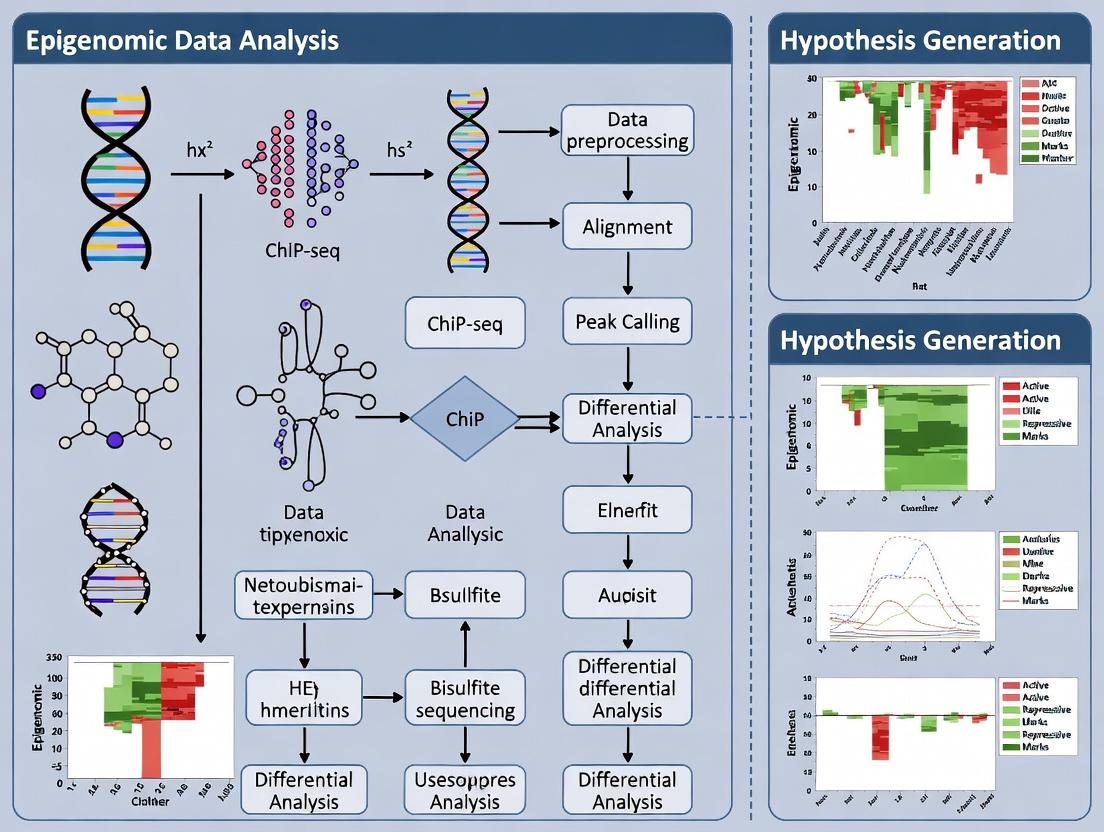
The Analytical Pipeline: Modern Methods for Mining Epigenomic Data and Formulating Hypotheses
This technical guide details a standardized workflow for processing epigenomic data, specifically from ChIP-seq and ATAC-seq experiments. It is framed within a broader thesis on hypothesis generation from epigenomic data research. The systematic conversion of raw sequencing reads into annotated peaks is a foundational step. This process enables researchers to map protein-DNA interactions and chromatin accessibility, forming the basis for generating testable biological hypotheses regarding gene regulation, cellular differentiation, and disease mechanisms—critical insights for drug development professionals.
The Standardized Workflow
The core workflow is a linear pipeline with distinct quality control and branching for different assay types.
Title: Standardized Epigenomic Data Analysis Pipeline
Detailed Experimental Protocols
Protocol 1: Raw Data Quality Control and Preprocessing
Objective: Assess raw read quality and prepare reads for alignment.
- FastQC Analysis: Run FastQC (v0.12.1) on raw FASTQ files to generate HTML reports summarizing per-base sequence quality, adapter contamination, and GC content.
- Adapter Trimming & Filtering: Execute Trim Galore! (v0.6.10), which wraps Cutadapt and FastQC. Standard parameters:
--quality 20 --stringency 3 --length 20 --pairedfor paired-end data. This removes low-quality bases, adapter sequences, and discards short reads. - Post-trimming QC: Run FastQC again on the trimmed FASTQ files to confirm quality improvement.
Protocol 2: Alignment to Reference Genome
Objective: Map filtered reads to a reference genome.
- Index Preparation: Pre-build a genome index for the chosen aligner (e.g.,
bwa indexfor BWA). - Alignment with BWA-MEM: For paired-end ChIP/ATAC-seq, use BWA-MEM (v0.7.17):
bwa mem -t 8 <reference_genome.fa> <trimmed_R1.fq> <trimmed_R2.fq> > output.sam. - SAM to BAM Conversion: Convert SAM to BAM, sort, and index using samtools (v1.15):
samtools view -bS output.sam | samtools sort -o sorted.bam -@ 8 && samtools index sorted.bam.
Protocol 3: Post-Alignment Processing and Filtering
Objective: Obtain a high-quality, PCR-duplicate-free BAM file.
- Mark Duplicates: Use Picard Tools (v2.27) to mark PCR duplicates:
java -jar picard.jar MarkDuplicates I=sorted.bam O=marked_duplicates.bam M=metrics.txt. - Filtering: Use samtools to filter out unmapped, low-quality, and duplicate-marked reads. For ATAC-seq, also filter mitochondrial reads:
samtools idxstats marked_duplicates.bam | cut -f 1 | grep -v chrM > non_chrM.list && samtools view -b -L non_chrM.list marked_duplicates.bam > filtered.bam. - Index Final BAM:
samtools index filtered.bam.
Protocol 4: Peak Calling for ChIP-seq
Objective: Identify enriched regions (peaks) of transcription factor binding or histone modification.
- Input Control: A control/input sample is mandatory for robust peak calling.
- Run MACS2: Use MACS2 (v2.2.7.1) for broad or narrow peaks. For a transcription factor (narrow):
macs2 callpeak -t treatment.bam -c input.bam -f BAMPE -g hs -n TF_output --outdir peaks -B. For histone marks (broad):macs2 callpeak -t treatment.bam -c input.bam -f BAMPE -g hs -n Histone_output --outdir peaks --broad. - Output: Primary outputs are
*_peaks.narrowPeakor*_peaks.broadPeakfiles (BED6+4 format).
Protocol 5: Peak Calling for ATAC-seq
Objective: Identify regions of open chromatin.
- Shift Reads: Account for Tn5 transposase binding which offsets reads. MACS2 can model this shift.
- Run MACS2 for ATAC-seq:
macs2 callpeak -t atac_seq.bam -f BAMPE -g hs -n ATAC_output --outdir atac_peaks --nomodel --shift -100 --extsize 200. - Alternative Tool - Genrich: An effective, dedicated ATAC-seq tool:
Genrich -t atac_seq.bam -o atac_peaks.narrowPeak -j -y -r -v.
Protocol 6: Peak Annotation and Motif Discovery
Objective: Assign biological context to called peaks.
- Genomic Annotation with ChIPseeker: Use the R/Bioconductor package ChIPseeker (v1.34.1). Load peak files and a TxDb object (e.g.,
TxDb.Hsapiens.UCSC.hg38.knownGene). TheannotatePeakfunction annotates peaks to promoter, intron, exon, or intergenic regions. - In-silico Motif Discovery with HOMER: Run
findMotifsGenome.plon a peak BED file:findMotifsGenome.pl peaks.bed hg38 motif_output_dir -size 200 -mask. This identifies de novo and known transcription factor binding motifs enriched in the peaks.
Table 1: Key QC Metrics and Benchmarks for Epigenomic Sequencing Data
| Metric | Tool/Source | Optimal Range / Target | Implication of Deviation |
|---|---|---|---|
| Raw Read Quality (Q20/Q30) | FastQC | Q30 > 80% of bases | High % of low-quality bases can compromise alignment and variant calling. |
| Adapter Content | FastQC/Trim Galore | < 5% (post-trimming: ~0%) | High content indicates inefficient library prep, leads to poor alignment. |
| Alignment Rate | BWA/samtools | > 70-80% (species/genome-dependent) | Low rates suggest contamination, poor library quality, or wrong reference. |
| Duplicate Rate | Picard MarkDuplicates | ChIP-seq: < 20-30%ATAC-seq: < 20% | High rates indicate low library complexity, limiting statistical power. |
| Fraction of Reads in Peaks (FRiP) | MACS2/featureCounts | TF ChIP-seq: > 1-5%Histone ChIP-seq: > 10-30% | Low FRiP signals a failed or noisy experiment with high background. |
| Non-Redundant Fraction (NRF) for ATAC-seq | Derived from alignment | > 0.8 | Measures library complexity; lower values indicate over-amplification. |
| TSS Enrichment Score (ATAC-seq) | pyATAC/picard | > 10 (higher is better) | Quantifies signal-to-noise at transcription start sites; low score indicates poor data quality. |
Table 2: Common Peak Callers and Their Applications
| Tool | Latest Version | Primary Use Case | Key Strength | Typical Command Line Parameters |
|---|---|---|---|---|
| MACS2 | 2.2.7.1 | General ChIP-seq (narrow/broad), ATAC-seq | Robust, widely used, excellent documentation. | -f BAMPE -g hs -q 0.05 --call-summits |
| Genrich | 0.6 | ATAC-seq, DNase-seq | Fast, no input control required, removes PCR duplicates. | -t input.bam -o output.narrowpeak -j -y -r |
| SEACR | 1.3 | CUT&RUN, CUT&Tag | Uses control to set threshold via AUC; good for sparse data. | --norm relaxed (for stringent) or --norm non |
| HOMER findPeaks | 4.11 | ChIP-seq (with style option) | Integrated with HOMER suite for motif analysis. | -style factor or -style histone |
The Scientist's Toolkit: Research Reagent Solutions
Table 3: Essential Materials and Reagents for Epigenomic Workflows
| Item | Function / Purpose | Example Product / Kit |
|---|---|---|
| Chromatin Immunoprecipitation (ChIP) Kit | Provides optimized buffers, beads, and protocols for efficient antibody-based chromatin enrichment. | Cell Signaling Technology SimpleChIP Kit, Diagenode True MicroChIP Kit. |
| ATAC-seq Assay Kit | Contains all reagents for the Tn5 transposase-based tagmentation reaction, purification, and PCR amplification. | Illumina Tagment DNA TDE1 Kit, Nextera DNA Flex Library Prep Kit. |
| High-Specificity Primary Antibody | Binds target protein (TF or histone mark) with high affinity and specificity for ChIP. Critical for success. | Validated antibodies from Abcam, Cell Signaling Technology, Active Motif. |
| Magnetic Protein A/G Beads | Binds antibody-chromatin complexes for separation and washing in ChIP protocols. | Dynabeads Protein A/G, Sera-Mag Magnetic Beads. |
| DNA Clean-up & Size Selection Beads | Purifies and size-selects DNA fragments post-enrichment/tagmentation (e.g., selects 150-600 bp fragments for ATAC-seq). | SPRIselect / AMPure XP Beads. |
| High-Fidelity PCR Mix | Amplifies library fragments with minimal bias and errors for sequencing. | NEBNext Ultra II Q5 Master Mix, KAPA HiFi HotStart ReadyMix. |
| Dual-Indexed Adapters | Unique barcodes for multiplexing samples in a single sequencing run. | Illumina IDT for Illumina UD Indexes. |
| Library Quantification Kit | Accurate quantification of sequencing library concentration (qPCR-based) for proper pooling. | KAPA Library Quantification Kit for Illumina platforms. |
| Control/Input DNA | Genomic DNA (for ChIP-seq) or tagmentation control (for ATAC-seq) used as a background control for peak calling. | Sonicated genomic DNA from same cell type (ChIP). Buffer-only tagmentation reaction (ATAC). |
Advancements in high-throughput sequencing and mass spectrometry have enabled the independent generation of epigenomic, transcriptomic, and proteomic datasets. The core challenge and opportunity lie in moving beyond descriptive cataloging to hypothesis generation. This whitepaper posits that systematic integration of these omic layers is not merely correlative but is essential for constructing causal models of gene regulation. By linking epigenetic states (the hypothesis-generating layer) to transcriptional and translational outputs (the functional validation layers), researchers can formulate testable mechanistic hypotheses about disease etiology, identify novel therapeutic targets, and discover master regulatory nodes. This guide details the technical frameworks for achieving this integration.
Foundational Data Layers & Quantitative Landscape
Each omic layer provides a distinct, quantifiable snapshot of cellular state. Key metrics and technologies are summarized below.
Table 1: Core Omic Layers, Technologies, and Key Quantitative Outputs
| Omic Layer | Primary Technology | Key Measured Features | Typical Output Metrics | Temporal Dynamics |
|---|---|---|---|---|
| Epigenomics | ChIP-seq, ATAC-seq, WGBS | Histone modifications, TF binding, chromatin accessibility, DNA methylation | Peak counts, read density, % methylation, differential accessibility scores | Stable to moderate |
| Transcriptomics | RNA-seq (bulk/single-cell) | Gene expression levels, splice variants, non-coding RNAs | TPM/FPKM, read counts, differential expression (log2FC, p-value) | Rapid |
| Proteomics | LC-MS/MS (TMT, LFQ), Affinity Arrays | Protein abundance, post-translational modifications | Intensity, spectral counts, fold-change, phosphorylation stoichiometry | Moderate |
Table 2: Common Multi-Omic Integration Findings & Data Correlations
| Observed Relationship | Epigenomic Data | Transcriptomic Correlation | Proteomic Correlation | Interpreted Biological Hypothesis |
|---|---|---|---|---|
| Active Enhancer | H3K27ac, H3K4me1, open chromatin | Strong positive | Moderate positive | Enhancer regulates proximal gene(s). |
| Promoter Activation | H3K4me3, open chromatin, low DNA methylation | Strong positive | Strong positive | Canonical gene activation. |
| Repressed State | H3K9me3, H3K27me3, high DNA methylation | Strong negative | Strong negative | Stable long-term silencing. |
| Post-Transcriptional Regulation | Active chromatin/ promoter marks | Strong positive | Weak or negative | Hypothesis for miRNA, translational control, or protein degradation. |
Detailed Experimental Protocols for Integration
Protocol 2.1: Concurrent Multi-Omic Profiling from a Single Sample
Goal: Generate epigenomic, transcriptomic, and proteomic data from an identical cell population to minimize biological noise. Method: SHARE-seq (Simultaneous high-throughput ATAC and RNA expression sequencing) coupled with subsequent proteomics.
- Cell Preparation: Harvest 1x10^6 cells, wash with PBS.
- Tagmentation & Fixation: Resuspend in ATAC-seq tagmentation buffer (Tn5 transposase) for 30 min at 37°C. Immediately fix with 1% formaldehyde for 10 min, quench with 125mM glycine.
- Nuclear Permeabilization & Reverse Transcription: Permeabilize with 0.1% Triton X-100. Perform in-nucleus reverse transcription using barcoded poly(dT) primers to generate cDNA.
- Library Split: Split the processed sample.
- Portion A (Epigenome/Transcriptome): Proceed with SHARE-seq protocol: separate ATAC and cDNA products via size selection, amplify, and sequence.
- Portion B (Proteome): Lyse cells in RIPA buffer with protease/phosphatase inhibitors. Digest proteins with trypsin, desalt, and label with TMTpro 16-plex reagents. Pool and fractionate by high-pH reverse-phase HPLC before LC-MS/MS.
- Data Alignment: Align ATAC-seq reads to reference genome (e.g., hg38). Align RNA-seq reads and quantify gene expression. Match MS/MS spectra to protein sequence databases.
Protocol 2.2: Causal Inference via Epigenetic Perturbation
Goal: Test hypotheses generated from correlative integration. Method: dCas9-based epigenetic editing followed by multi-omic readout.
- Guide RNA Design: Design sgRNAs targeting genomic regions of interest (e.g., an enhancer identified from integrated analysis).
- Cell Transduction: Co-transfect cells with plasmids encoding:
- dCas9-p300 (for activation) OR dCas9-KRAB (for repression).
- sgRNA expression construct.
- Include non-targeting sgRNA control.
- Validation & Harvest: At 72-96 hours post-transfection:
- Validate editing efficiency via ChIP-qPCR for H3K27ac (activation) or H3K27me3 (repression) at the target locus.
- Multi-Omic Readout: Harvest transfected cell pools and perform:
- ATAC-seq/ChIP-seq on target histone mark.
- RNA-seq for transcriptome changes.
- LC-MS/MS (label-free quantification) for proteome changes.
- Analysis: Identify genes/proteins significantly altered only in the targeted perturbation group, establishing a causal link between the epigenetic state and functional outputs.
Visualizing Integration Workflows & Relationships
Title: Multi-Omic Integration to Hypothesis Testing Workflow
Title: Epigenetic to Protein Output Signaling Pathway
The Scientist's Toolkit: Key Research Reagent Solutions
Table 3: Essential Reagents for Integrative Multi-Omic Experiments
| Reagent/Material | Provider Examples | Function in Multi-Omic Integration |
|---|---|---|
| Tn5 Transposase (Loaded) | Illumina (Nextera), Diagenode | For ATAC-seq library prep; fragments DNA and adds sequencing adapters simultaneously. |
| Magnetic Protein A/G Beads | Thermo Fisher, MilliporeSigma | For ChIP-seq; immunoprecipitation of histone- or transcription factor-DNA complexes. |
| TMTpro 16-plex Label Reagents | Thermo Fisher | Isobaric labels for multiplexed quantitative proteomics, allowing comparison of up to 16 samples in one MS run. |
| dCas9-Effector Fusions (p300, KRAB) | Addgene (Plasmids) | For epigenetic perturbation; target-specific activation or repression to test enhancer-gene hypotheses. |
| Triple KO (TKO) Cell Lines | Horizon Discovery | HEK293 cells with knocked-out TP53, RB1, and MYC to reduce confounding genetic heterogeneity. |
| Multi-Omic Reference Standards | Horizon Discovery, SeraCare | Well-characterized cell line mixes (e.g., Methylated, Copy Number, Expression controls) for platform benchmarking. |
| Cross-linking Reagents (e.g., DSG) | Thermo Fisher | For ChIP-seq; stabilizes weak protein-DNA interactions prior to formaldehyde cross-linking. |
| Single-Cell Multi-Omic Kits (ATAC + GEX) | 10x Genomics | Enables simultaneous profiling of chromatin accessibility and gene expression in single cells. |
Within epigenomic research, the generation of robust biological hypotheses is paramount for advancing our understanding of gene regulation, disease mechanisms, and therapeutic targets. This technical guide elucidates a core analytical pipeline—integrating dimensionality reduction, clustering, and predictive modeling—designed to discover latent patterns from high-dimensional epigenomic data (e.g., DNA methylation, histone modification, chromatin accessibility assays). This pipeline transforms raw, complex data into testable hypotheses regarding functional genomic elements and regulatory dynamics.
The Analytical Pipeline: A Technical Workflow
Dimensionality Reduction
High-dimensional epigenomic datasets (often with tens of thousands of genomic bins or peaks across few samples) suffer from the "curse of dimensionality." Dimensionality reduction is the first critical step to capture essential biological variance.
Key Methods & Protocols:
Principal Component Analysis (PCA):
- Protocol: 1) Input a normalized matrix (features x samples). 2) Center the data (subtract mean for each feature). 3) Compute covariance matrix. 4) Perform eigendecomposition to obtain principal components (PCs). 5) Project data onto top K PCs (typically explaining >80% cumulative variance).
- Application: Removes technical noise, visualizes broad sample relationships (e.g., batch effects, major cell type differences).
t-Distributed Stochastic Neighbor Embedding (t-SNE):
- Protocol: 1) Compute pairwise similarities (conditional probabilities) in high-dimensional space. 2) Construct a similar probability distribution in low-dimensional (2D/3D) space. 3) Minimize the Kullback-Leibler divergence between the two distributions using gradient descent (typical perplexity: 30-50, iterations: 1000).
- Application: Visualizes local clusters of similar epigenomic profiles, often used for single-cell ATAC-seq data.
Uniform Manifold Approximation and Projection (UMAP):
- Protocol: 1) Construct a topological framework of the high-dimensional data using nearest neighbors (nneighbors~15). 2) Optimize a low-dimensional layout to preserve this topological structure (mincross~0.1).
- Application: Preserves more global structure than t-SNE, effective for revealing hierarchical patterns in bulk or single-cell epigenomics.
Quantitative Comparison of Dimensionality Reduction Methods: Table 1: Key characteristics of dimensionality reduction techniques for epigenomic data.
| Method | Preserves Global Structure | Preserves Local Structure | Computational Scalability | Primary Use Case in Epigenomics |
|---|---|---|---|---|
| PCA | High | Low | High | Noise reduction, batch assessment, linear feature extraction |
| t-SNE | Low | High | Medium | Cluster visualization for homogeneous cell populations |
| UMAP | Medium-High | High | Medium | Hierarchical structure discovery, single-cell trajectory inference |
Clustering for Unsupervised Pattern Discovery
Following dimensionality reduction, clustering identifies discrete or continuous cell states/regulatory modules without prior labels.
Key Methods & Protocols:
k-Means Clustering:
- Protocol: 1) Specify k (number of clusters). Use elbow method on within-cluster sum of squares (WCSS) to inform choice. 2) Randomly initialize k centroids. 3) Assign each data point to nearest centroid. 4) Recalculate centroids. 5) Iterate until convergence. Requires scaled data.
- Application: Segmenting genomic regions into distinct chromatin state categories (e.g., enhancers, promoters, repressed regions).
Hierarchical Clustering:
- Protocol: 1) Compute pairwise distance matrix (Euclidean, correlation). 2) Employ agglomerative (bottom-up) strategy, merging closest clusters (linkage: Ward's, average). 3) Cut dendrogram at height yielding biologically meaningful clusters.
- Application: Revealing nested relationships between samples (e.g., tumor subtypes) or correlated epigenetic marks.
Density-Based Spatial Clustering (DBSCAN):
- Protocol: 1) For each point, count points within epsilon (ε) radius. 2) Classify as core point if count ≥ min_samples. 3) Expand clusters from core points, connecting density-reachable points. 4) Label points not assigned as noise.
- Application: Identifying rare cell populations from single-cell epigenomic data without pre-specifying cluster number.
Predictive Modeling for Hypothesis Generation
Supervised models leverage discovered patterns to predict functional outcomes, generating causal hypotheses.
Key Methods & Protocols:
Random Forest for Feature Importance:
- Protocol: 1) Train a Random Forest classifier/regressor (e.g., to predict gene expression from chromatin features). 2) Use out-of-bag error or permutation importance (scikit-learn's
feature_importances_). 3) Rank genomic features (e.g., specific histone marks) by their mean decrease in accuracy/Gini impurity. - Application: Identifies epigenetic marks most predictive of transcriptional activity or disease state, suggesting key regulatory elements.
- Protocol: 1) Train a Random Forest classifier/regressor (e.g., to predict gene expression from chromatin features). 2) Use out-of-bag error or permutation importance (scikit-learn's
Regularized Regression (LASSO):
- Protocol: 1) Apply L1 penalty to linear regression, minimizing:
(1/(2*n_samples)) * ||y - Xw||^2_2 + α * ||w||_1. 2) Perform k-fold cross-validation to tune hyperparameter α. 3) Features with non-zero coefficients are selected as predictive. - Application: Selects a sparse set of CpG sites predictive of a phenotypic trait, pinpointing candidate loci for functional validation.
- Protocol: 1) Apply L1 penalty to linear regression, minimizing:
Deep Learning (Convolutional Neural Networks):
- Protocol: 1) Format genomic sequences (e.g., ±5kb around TSS) as one-hot encoded tensors. 2) Architect a CNN with convolutional layers (ReLU activation), pooling, and dense layers. 3) Train to predict transcription factor binding or chromatin accessibility from sequence.
- Application: Discovers de novo sequence motifs and combinatorial rules driving epigenetic states, hypothesizing novel regulatory codes.
Integrated Workflow for Epigenomic Hypothesis Generation
Diagram Title: Integrated ML Pipeline for Epigenomic Discovery
The Scientist's Toolkit: Research Reagent & Computational Solutions
Table 2: Essential tools and resources for implementing the ML pipeline in epigenomics.
| Category | Item/Reagent | Function & Explanation |
|---|---|---|
| Wet-Lab Reagents | Illumina TruSeq / NovaSeq Kits | Generate high-throughput sequencing libraries from ChIP, ATAC, or bisulfite-converted DNA. |
| Cell Signaling Technology Antibodies | Validated antibodies for specific histone modifications (e.g., H3K27ac, H3K9me3) for ChIP-seq. | |
| Tn5 Transposase (Nextera) | Enzyme for tagmentation-based assays like ATAC-seq, simultaneously fragments and tags chromatin. | |
| Computational Tools | Snakemake / Nextflow | Workflow management systems to create reproducible, scalable preprocessing pipelines. |
| scikit-learn (Python) | Core library implementing PCA, k-Means, Random Forest, LASSO with consistent APIs. | |
| Scanpy (Python) | Comprehensive toolkit for single-cell epigenomics analysis, including clustering and UMAP. | |
| TensorFlow / PyTorch | Deep learning frameworks for building custom predictive models on sequence data. | |
| Data Resources | ENCODE / Roadmap Epigenomics | Reference epigenomic maps across cell types for comparative analysis and feature selection. |
| UCSC Genome Browser | Visualization platform to overlay discovered patterns (e.g., clusters) with genomic annotations. |
Case Study: Discovering Disease Subtypes from DNA Methylation Arrays
Experimental Protocol:
- Data: Download Illumina 450K/EPIC array data (beta-values) for a disease cohort and healthy controls from GEO (e.g., GSE123456).
- Preprocessing: Perform quantile normalization (
minfiR package), remove batch effects (ComBat), and filter probes (p-value > 0.01, SNPs, cross-reactive). - Dimensionality Reduction: Apply PCA to top 50,000 most variable CpGs. Plot PC1 vs. PC2 to assess gross outliers.
- Clustering: Use UMAP (nneighbors=15, mindist=0.1) on normalized data, followed by DBSCAN (ε=0.5, min_samples=5) to identify disease subgroups without forcing cluster number.
- Predictive Modeling: Train a Random Forest classifier (500 trees) using all CpGs to differentiate a discovered aggressive subtype from others. Extract top 100 CpGs by feature importance.
- Hypothesis Generation: Annotate top CpGs to genes and pathways (GREAT tool). Hypothesize: "Hypermethylation of CpGs in the WNT signaling pathway promoter regions defines an aggressive disease subtype with poor prognosis."
- Validation Pathway: Design CRISPR-dCas9-TET1 mediated demethylation of candidate CpGs in cell lines, followed by RNA-seq and phenotypic assays.
Diagram Title: Hypothesized Epigenetic Mechanism from ML Discovery
The systematic application of dimensionality reduction, clustering, and predictive modeling forms a powerful, iterative cycle for hypothesis generation in epigenomics. By moving from unsupervised pattern discovery to supervised prediction of functional outcomes, researchers can prioritize key regulatory features and formulate precise, experimentally tractable hypotheses. This data-driven approach accelerates the translation of epigenomic maps into mechanistic insights and therapeutic opportunities.
Within the broader thesis of hypothesis generation from epigenomic data, single-cell and spatial epigenomics represent a paradigm shift. The core thesis posits that cellular heterogeneity, driven by epigenetic variation, is a primary determinant of tissue function, disease progression, and therapeutic response. Traditional bulk epigenomic assays average signals across thousands of cells, obscuring critical minority populations and dynamic states. This technical guide details how advanced single-cell epigenomic profiling, integrated with spatial mapping, transforms raw data into testable biological hypotheses regarding cell fate decisions, regulatory networks, and disease mechanisms.
Key Single-Cell Epigenomic Assays
The following table summarizes the core quantitative outputs, resolution, and applications of leading single-cell epigenomic assays.
Table 1: Comparison of Major Single-Cell Epigenomic Technologies
| Assay Name | Target Epigenomic Layer | Key Output Metric | Typical Cells per Run | Resolution | Primary Hypothesis-Generation Use |
|---|---|---|---|---|---|
| scATAC-seq | Chromatin Accessibility | Insertion site counts per cell (peak matrix) | 5,000 - 100,000+ | ~150 bp (peaks) | Identifying candidate cis-regulatory elements (cCREs) & cell-type-specific TF activity. |
| scCUT&Tag | Histone Modifications (H3K27ac, H3K4me3, etc.) | Tagmentation site counts per cell | 1,000 - 10,000 | ~150 bp (peaks) | Mapping active promoters/enhancers & defining chromatin states at single-cell resolution. |
| snmC-seq / scBS-seq | DNA Methylation (5mC) | Methylation ratio per CpG site per cell | 1,000 - 10,000+ | Single CpG | Tracing lineage relationships & identifying metastable epialleles driving heterogeneity. |
| scChIC-seq | Combined Histone Mods | Multi-modal readouts per cell | Hundreds - Thousands | ~150 bp (peaks) | Testing co-occurrence of histone marks within single cells. |
| CITE-seq / REAP-seq | Surface Proteins + Transcriptome | Antibody-derived tag (ADT) counts | 5,000 - 100,000+ | Protein epitope | Generating hypotheses linking epigenetic state to surface phenotype. |
Spatial Epigenomic and Multiomic Technologies
Table 2: Spatial Technologies for Contextualizing Heterogeneity
| Technology | Spatial Resolution | Epigenomic Readout | Throughput / Multiplexing | Key for Hypotheses on |
|---|---|---|---|---|
| Visium HD (10x Genomics) | 2-8 cells (8x8 µm) | Compatible with ATAC (spatial-ATAC) | Whole Transcriptome / 5000+ spots | Niche effects on chromatin accessibility. |
| MERFISH / seqFISH+ | Subcellular (~0.1 µm) | RNA, indirectly infers regulation | 100s - 10,000s of RNA species | Spatial gene expression patterns hinting at regulatory logic. |
| Paired-Tag | Cell (~10 µm) | H3K27ac + Transcriptome | Multiomic (1-2 epigenomic marks + transcriptome) | Direct spatial coupling of enhancer activity and gene expression. |
| Spatial-CUT&Tag | Single-cell (~10 µm) | Histone modifications (e.g., H3K27me3) | 1-2 histone marks | Mapping repressive/active chromatin domains in tissue architecture. |
| Slide-seqV2 / Sci-Space | ~10 µm (near-cellular) | Transcriptome (epigenomic extensions emerging) | Whole transcriptome | Correlating spatial neighborhood with inferred epigenetic states. |
Experimental Protocols
Protocol: High-Throughput Single-Nucleus ATAC-seq (snATAC-seq)
Objective: To profile chromatin accessibility in tens of thousands of individual nuclei from frozen tissue. Key Hypotheses Generated: Identification of rare regulatory cell types; reconstruction of gene regulatory networks (GRNs); mapping of disease-associated variant activity (e.g., GWAS SNPs) to specific cell populations.
Detailed Methodology:
- Nuclei Isolation: Mechanically dissociate 1-50 mg of frozen tissue in chilled lysis buffer (10mM Tris-HCl pH 7.4, 10mM NaCl, 3mM MgCl2, 0.1% NP-40, 1% BSA, 0.2U/µl RNase inhibitor). Homogenize with a dounee homogenizer. Filter through a 40 µm flow cell strainer. Pellet nuclei at 500 rcf for 5 min at 4°C.
- Tagmentation: Resuspend nuclei in tagmentation buffer from the 10x Genomics Chromium Next GEM Single Cell ATAC Kit. Use the engineered Tn5 transposase loaded with sequencing adapters to simultaneously fragment and tag open chromatin regions. Incubate at 37°C for 60 minutes.
- GEM Generation & Barcoding: Load tagmented nuclei, gel beads, and oil onto a 10x Chromium chip to generate Gel Bead-In-Emulsions (GEMs). Within each GEM, a unique 10x barcode from a gel bead is linked to all DNA fragments from a single nucleus via a primer extension reaction.
- Post GEM-RT Cleanup & Amplification: Break emulsions, pool barcoded DNA, and purify using SPRIselect beads. Perform a PCR amplification (12-14 cycles) to add sample indexes and complete adapter sequences.
- Library Construction & Sequencing: Size-select libraries (~200-600 bp insert) using SPRIselect beads. Quantify by qPCR or Bioanalyzer. Sequence on an Illumina NovaSeq 6000 using paired-end sequencing (e.g., 50 bp x 50 bp) to a target depth of ~25,000 reads per nucleus.
Protocol: Spatial ATAC-seq with Visium HD
Objective: To map chromatin accessibility across a tissue section while retaining spatial context. Key Hypotheses Generated: How tissue microenvironment (e.g., tumor edge vs. core) influences chromatin state; identification of spatially restricted regulatory programs.
Detailed Methodology:
- Fresh-Frozen Tissue Sectioning: Cryosection tissue at 10 µm thickness onto the Visium HD Spatial Tissue Capture slide. Immediately fix in pre-chilled methanol at -20°C for 30 minutes.
- On-Slide Tagmentation: Permeabilize tissue with a detergent buffer. Apply a Tn5 transposase mixture directly onto the slide, allowing tagmentation to occur in situ within intact tissue architecture. Incubate in a humidified chamber at 37°C for 45-60 min.
- Spatial Barcode Transfer: Following tagmentation, a second permeabilization step releases the tagged DNA fragments. These fragments diffuse to the slide surface where they bind to oligonucleotides containing unique spatial barcodes (x,y coordinates) for each 8x8 µm bin.
- Library Preparation: Release spatially barcoded cDNA from the slide via NaOH treatment. Construct sequencing libraries using a standard NGS library protocol with PCR amplification.
- Sequencing & Data Alignment: Sequence on an Illumina platform. Align reads to the reference genome and demultiplex based on spatial barcodes to generate a counts-by-bin-by-genomic-peak matrix.
Visualization of Workflows and Relationships
Title: Integrating Single-Cell and Spatial Epigenomics for Hypothesis Generation
Title: From scATAC-seq Data to a Mechanistic Regulatory Hypothesis
The Scientist's Toolkit: Key Research Reagent Solutions
Table 3: Essential Reagents and Kits for Single-Cell/Spatial Epigenomics
| Item Name | Vendor Examples | Function in Experiment | Critical for Hypothesis Generation Because... |
|---|---|---|---|
| Chromium Next GEM Single Cell ATAC Kit | 10x Genomics | Provides all reagents for nuclei tagmentation, GEM generation, and library prep for snATAC-seq. | Enables robust, high-throughput profiling of chromatin accessibility, the foundation for identifying regulatory elements. |
| CUT&Tag Assay Kit (for Histone Modifications) | Cell Signaling Technology / EpiCypher | Contains concanavalin A beads, antibodies, and pA-Tn5 for targeted profiling of histone marks in single cells or spatially. | Allows mapping of specific activating/repressive chromatin states, refining hypotheses on transcriptional regulation. |
| Visium HD Spatial Tissue Optimization & Gene Expression Kit | 10x Genomics | Used to determine optimal permeabilization conditions and perform spatial transcriptomics on Visium HD slides. | Prerequisite for spatial-ATAC; provides correlative transcriptomic data to link accessibility to expression. |
| ATAC-Seq Buffer Set (TD Buffer, TDE1) | Illumina / Diagenode | Contains the Tn5 transposase and reaction buffers for in-situ or in-vitro tagmentation. | Core enzyme for accessibility assays; quality directly impacts signal-to-noise and hypothesis validity. |
| DAPI (4',6-diamidino-2-phenylindole) | Sigma-Aldrich / Thermo Fisher | Fluorescent nuclear stain used during nuclei isolation for FACS sorting or quality checks. | Ensures high viability of single-nucleus suspensions, reducing ambient RNA/DNA and improving cluster resolution. |
| RNase Inhibitor (e.g., Protector) | Roche / Sigma-Aldrich | Added to lysis and wash buffers during nuclei isolation. | Preserves nascent RNA in multiomic assays (e.g., scATAC-seq + RNA), enabling linked hypotheses on regulation and output. |
| SPRIselect Beads | Beckman Coulter | Used for post-reaction cleanup, size selection, and library normalization. | Critical for removing adapter dimers and selecting properly sized fragments, ensuring high-quality sequencing libraries. |
| Dual Index Kit TT Set A | 10x Genomics / Illumina | Provides unique dual indices for multiplexing samples in a single sequencing run. | Allows cost-effective pooling of multiple conditions/patients, enabling comparative hypotheses about disease states. |
Within a thesis on hypothesis generation from epigenomic data research, the transition from EWAS discovery to testable biological and clinical hypotheses is a critical challenge. This case study exemplifies the process, using a contemporary EWAS on rheumatoid arthritis (RA) as a foundation. We detail the steps from statistical association to mechanistic exploration and therapeutic target nomination.
Case Study Foundation: An EWAS on Rheumatoid Arthritis
A recent large-scale meta-analysis identified differential DNA methylation (DNAm) associated with RA. Key data is summarized below.
Table 1: Top EWAS Hits from RA Meta-Analysis (Illustrative)
| CpG Site | Chr | Gene Context | Δβ (RA vs Control) | P-value | FDR |
|---|---|---|---|---|---|
| cg06690548 | 1 | SLC9A9 (Body) | +0.08 | 3.2e-14 | 0.003 |
| cg07362190 | 6 | HLA-DRB5 (TSS1500) | -0.12 | 1.1e-31 | <0.001 |
| cg15826982 | 16 | IRF8 (Promoter) | +0.15 | 8.7e-19 | 0.001 |
Table 2: Enriched Pathways from Gene Set Analysis (GSEA)
| Pathway Name | Source (e.g., KEGG) | NES | FDR |
|---|---|---|---|
| JAK-STAT signaling pathway | KEGG 2021 | 2.45 | 0.008 |
| Cytokine-cytokine receptor interaction | KEGG 2021 | 2.31 | 0.012 |
| Osteoclast differentiation | KEGG 2021 | 2.18 | 0.018 |
Hypothesis Generation Workflow
The primary hypothesis generated: Hypermethylation of the IRF8 promoter in peripheral blood monocytes leads to its transcriptional silencing, dysregulating the JAK-STAT pathway and contributing to pro-inflammatory cytokine production in RA.
Step 1: Candidate Prioritization & Causal Inference
- Protocol for Mendelian Randomization (MR): To assess if DNAm changes are causal for RA or a consequence (reverse causation).
- Instrument Selection: Extract SNPs strongly associated (P < 5e-08) with methylation at the IRF8 CpG (cis-mQTLs) from a public mQTL database (e.g., GoDMC).
- Outcome Data: Obtain effect estimates for the same SNPs from a large RA GWAS consortium.
- Analysis: Perform Two-Sample MR using the Inverse-Variance Weighted (IVW) method. Sensitivity analyses (MR-Egger, weighted median) assess pleiotropy.
- Interpretation: A significant MR result (P < 0.05) supports a causal role for the methylation change in RA etiology.
Step 2:In VitroFunctional Validation
- Protocol for Targeted Methylation Editing & Transcriptional Assay:
- Cell Culture: Isolate CD14+ monocytes from healthy donor buffy coats using magnetic-activated cell sorting (MACS).
- Targeted Demethylation: Transfect monocytes with a dCas9-TET1 catalytic domain fusion protein complexed with sgRNAs targeting the IRF8 promoter region (cg15826982). A non-targeting sgRNA serves as control.
- Validation of Editing: 72h post-transfection, harvest cells.
- Bisulfite Pyrosequencing: Quantify methylation percentage at the target CpG and flanking sites.
- qRT-PCR: Isolate RNA, synthesize cDNA, and measure IRF8 mRNA levels using TaqMan assays. Normalize to GAPDH.
- Phenotypic Assay: Stimulate edited and control monocytes with IFN-γ (100 ng/mL, 24h). Measure supernatant levels of TNF-α and IL-6 via ELISA.
The Scientist's Toolkit: Research Reagent Solutions
Table 3: Essential Materials for Functional Follow-Up
| Item | Function / Role | Example Product/Catalog |
|---|---|---|
| CD14 MicroBeads, human | Positive selection of monocytes for primary cell culture. | Miltenyi Biotec, 130-050-201 |
| dCas9-TET1 CD Plasmid | Targeted DNA demethylation via CRISPR-dCas9 epigenome editing. | Addgene, #113865 |
| sgRNA in vitro Transcription Kit | Generation of sgRNAs for complex assembly. | NEB, #E3322S |
| Lipofectamine CRISPRMAX | Transfection reagent for delivery of RNP complexes into primary cells. | Thermo Fisher, CMAX00008 |
| Zymo EZ DNA Methylation-Lightning Kit | Bisulfite conversion of genomic DNA for methylation analysis. | Zymo Research, D5030 |
| IRF8 TaqMan Gene Expression Assay | Precise quantification of IRF8 mRNA levels. | Thermo Fisher, Hs00175238_m1 |
| Human TNF-α ELISA Kit | Quantification of secreted cytokine protein levels. | BioLegend, 430204 |
Visualizing the Hypothesis and Workflow
Diagram 1: Hypothesis Generation and Validation Workflow (82 chars)
Diagram 2: Proposed IRF8-JAK-STAT Dysregulation Pathway (80 chars)
From Hypothesis to Drug Development
The validated hypothesis directly informs therapeutic development. IRF8 itself is a challenging direct target, but its downstream effectors in the JAK-STAT pathway are not. This epigenomic insight strengthens the rationale for:
- Patient Stratification: Identifying RA patients with IRF8 hypermethylation as potential "super-responders" to JAK inhibitors (e.g., Tofacitinib).
- Combination Therapy: Exploring DNA methyltransferase inhibitors (e.g., decitabine) in combination with immunomodulators in refractory cases.
- Novel Target Discovery: Upstream regulators responsible for establishing this methylation state become new drug targets.
This case study demonstrates a structured, multi-step framework for generating high-confidence, testable hypotheses from EWAS data. By integrating causal inference, precise functional genomics, and pathway analysis, epigenomic associations transition from statistical observations to actionable biological insights with clear translational potential for drug development.
Navigating Pitfalls and Enhancing Power: Optimization Strategies for Robust Epigenomic Studies
In the context of hypothesis generation from epigenomic data, technical noise represents a fundamental barrier to biological insight. Accurate identification of differentially methylated regions, histone modification shifts, or chromatin accessibility changes hinges on the rigorous separation of technical artifacts from genuine biological signals. This guide provides a comprehensive framework for diagnosing, mitigating, and controlling for batch effects, confounding variables, and quality issues in epigenomic research, thereby ensuring robust and reproducible hypothesis generation.
Quantitative Landscape of Technical Noise
Data derived from recent literature and repositories (e.g., GEO, ENCODE) highlight the pervasive impact of technical variability.
Table 1: Prevalence and Impact of Technical Artifacts in Common Epigenomic Assays
| Assay Type | Typical Batch Effect Contribution (PVE%) | Primary Confounding Variables | Common QC Failure Rate |
|---|---|---|---|
| Whole-Genome Bisulfite Seq (WGBS) | 15-40% | Bisulfite conversion efficiency, read depth, library preparation date | 10-25% |
| ChIP-Seq (Histone Marks) | 10-30% | Antibody lot, fragmentation time, sequencing lane | 5-20% |
| ATAC-Seq | 20-50% | Transposase activity (lot), cell viability, nucleocytoplasmic ratio | 15-30% |
| Methylation Array (EPIC) | 5-25% | Array slide, processing batch, sample position | 3-12% |
| Hi-C/3D Chromatin | 25-60% | Crosslinking efficiency, restriction enzyme, ligation efficiency | 20-40% |
PVE%: Percent Variance Explained. Data synthesized from recent studies (2022-2024).
Table 2: Efficacy of Correction Methods for Batch Effects
| Correction Method | Applicable Data Type | Reduction in Batch PVE (Median %) | Risk of Signal Attenuation |
|---|---|---|---|
| ComBat (Empirical Bayes) | Methylation arrays, normalized counts | 70-85% | Moderate |
| Surrogate Variable Analysis (SVA) | RNA-seq, ChIP-seq, WGBS | 60-80% | Low-Moderate |
| Remove Unwanted Variation (RUV) | ATAC-seq, scEpigenomics | 65-90% | Low |
| Principal Component Correction | All assays | 50-75% | High |
Limma removeBatchEffect |
Linear models, arrays | 70-80% | Moderate |
Experimental Protocols for Diagnosis and Control
Protocol 3.1: Pre-Experimental Design for Confounding Minimization
Objective: To design an epigenomic study that minimizes the confounding of technical variables with biological factors of interest.
- Randomization: Assign samples from all biological groups (e.g., case/control) randomly across all technical batches (library prep days, sequencing lanes, arrays).
- Blocking: If full randomization is impossible, use a balanced block design. Ensure each batch contains proportional representation from all biological groups.
- Replication: Include at least two technical replicates (same biological sample processed independently) for a subset of samples to estimate technical variance.
- Sample Tracking: Log metadata for all potential confounding variables: sample collection date, technician ID, reagent lot numbers, instrument ID, processing time points.
Protocol 3.2: Post-Sequencing QC & Diagnostic Workflow for WGBS/ChIP-seq
Objective: To assess raw data quality and diagnose batch effects prior to advanced analysis.
- Raw Read QC: Run
FastQC(v0.12.0) on all FASTQ files. Aggregate results withMultiQC(v1.15). - Alignment & Deduplication: Align reads with appropriate aligners (
Bismarkfor WGBS,bowtie2for ChIP-seq). Remove PCR duplicates usingpicard MarkDuplicates. - Metrics Calculation:
- WGBS: Calculate global CpG methylation percentage and bisulfite conversion efficiency (from lambda phage or spike-in control). Expect >99% conversion.
- ChIP-seq: Compute
phantompeakqualtools(cross-correlation) for signal-to-noise. FRiP (Fraction of Reads in Peaks) should be >1% for broad marks, >5% for sharp marks.
- Batch Effect Diagnosis: Using normalized count/coverage matrices, perform PCA. Color samples by technical batch (e.g., sequencing run) and biological group. Visual overlap indicates confounding.
Protocol 3.3: Systematic Confounding Variable Audit
Objective: To statistically identify hidden confounding variables.
- Metadata Correlation: For each continuous metadata variable (e.g., RIN, PMI, cell passage), calculate correlation with the first 3 principal components of the epigenomic data.
- Association Testing: For categorical variables (e.g., technician, lot), perform PERMANOVA or Kruskal-Wallis test on sample-wise distance matrices.
- Surrogate Variable Estimation: Use the
svaR package (v3.50.0) to estimate hidden factors of variation (num.svfunction). These can be included as covariates in downstream models.
Visualization of Concepts and Workflows
Title: Workflow for Addressing Technical Noise
Title: How Noise Leads to Flawed Hypotheses
Title: Key Variable Definitions Table
The Scientist's Toolkit: Research Reagent Solutions
Table 3: Essential Reagents and Kits for Controlling Technical Noise
| Item Name | Provider(s) | Primary Function in Noise Control |
|---|---|---|
| ERCC (External RNA Controls Consortium) Spike-Ins | Thermo Fisher | Distinguishes technical from biological variation in assays like scATAC-seq; normalizes for library preparation efficiency. |
| Lambda Phage DNA | e.g., NEB, Roche | Unmethylated control for bisulfite conversion efficiency assessment in WGBS/EPIC. |
| SNAP-Chip | EpiCypher | Defined nucleosome standard for ChIP-seq antibody benchmarking and QC; quantifies enrichment performance. |
| CpG Methylation Spike-Ins (e.g., EpiScope) | Takara Bio | Methylated/unmethylated controls for absolute quantification and inter-batch calibration in methylation studies. |
| Cell Line Controls (e.g., GM12878, K562) | ATCC, Coriell | Reference epigenomes for cross-study batch alignment and protocol performance tracking. |
| Tn5 Transposase (Tagmented) | Illumina, Diagenode | Consistent, lot-controlled enzyme for ATAC-seq to minimize batch variation in chromatin accessibility profiles. |
| Histone Modification Antibody Panels with Validation | Active Motif, Abcam | Antibodies with ChIP-seq grade validation and consistent lots to reduce immunoprecipitation variability. |
| Methylated DNA Standard Panels | Zymo Research | Controls for methylation array and sequencing to assess linearity, sensitivity, and reproducibility. |
Within the broader thesis of hypothesis generation from epigenomic data, the "cell type conundrum" represents a fundamental challenge. Bulk epigenomic assays (e.g., ATAC-seq, ChIP-seq, DNA methylation arrays) generate averaged signals across heterogeneous cell populations, obscuring the distinct regulatory landscapes of constituent cell types. This confounding factor severely limits the accuracy of hypotheses regarding cell-type-specific gene regulation, disease mechanisms, and therapeutic targets. This guide details strategies to overcome this limitation through computational deconvolution and experimental single-cell resolution, thereby enabling precise hypothesis generation from complex epigenomic datasets.
Computational Deconvolution: Inferring Proportions from Bulk Data
Deconvolution algorithms estimate the fractional composition of cell types within a bulk tissue sample using reference profiles.
Core Methodologies & Quantitative Performance
Table 1: Comparison of Major Deconvolution Tools for Epigenomic Data
| Tool Name | Algorithm Type | Input Data Type | Key Assumption | Reported Median RMSE (Prop.) | Reference Required |
|---|---|---|---|---|---|
| MuSiC | Non-negative least squares (NNLS) with cross-subject weighting | RNA-seq | Gene expression is linear mix of cell-type-specific expression | 0.02 - 0.08 (simulated) | scRNA-seq |
| CIBERSORTx | ν-support vector regression (ν-SVR) | RNA-seq / Methylation Array | Signature matrix is sufficient to describe population | 0.05 - 0.15 (validated) | Signature Matrix (bulk or sc) |
| EpiDISH | Robust partial correlations (RPC) / NNLS | DNA Methylation Array | Reference centroids represent pure cell types | 0.04 - 0.10 (blood) | Methylation Centroids |
| deconvATAC | Multivariate linear regression | ATAC-seq (bulk) | Accessibility is additive; uses cell-type-specific peaks | N/A (methodological) | scATAC-seq Peak Matrix |
| Bisque | Transform-both-sides model | RNA-seq | Non-linear transformation allows compatibility | ~0.07 (tissue) | scRNA-seq |
Detailed Protocol: Deconvolution using EpiDISH on DNA Methylation Data
Objective: Estimate proportions of 7 blood cell types from a bulk Illumina EPIC methylation array dataset.
Materials:
- Bulk beta-value matrix (samples x CpG probes).
- Reference centroid matrix for blood (e.g.,
centDHSbloodDMC.mfrom EpiDISH package).
Procedure:
- Data Preprocessing: Normalize bulk data using
preprocessENmixornormalize.quantiles. Ensure probe IDs match the reference. - Subset Probes: Retain only the CpG probes present in the reference centroid matrix (e.g., ~600 DHS-DMCs).
- Run RPC Method: Execute the
EpiDISHfunction withmethod='RPC'. RPC uses robust partial correlations to handle technical noise.
- Quality Control: Check that
out$r(goodness-of-fit metrics) are high (>0.9 suggests good fit). Ensure fractions sum to ~1 per sample. - Downstream Analysis: Correlate estimated cell fractions with phenotypic traits (e.g., disease status) to generate hypotheses about immune cell involvement.
Single-Cell Resolution: Direct Profiling of Heterogeneity
Single-cell epigenomic technologies provide the ground truth for deconvolution and enable direct hypothesis generation at the cellular level.
Experimental Protocol: Single-Nucleus ATAC-seq (snATAC-seq)
Objective: Profile chromatin accessibility in individual nuclei from frozen tissue.
Workflow:
Diagram Title: snATAC-seq Experimental Workflow
Detailed Steps:
- Nuclei Isolation: Mince 10-50 mg frozen tissue on dry ice. Homogenize in 1-2 mL cold lysis buffer (10 mM Tris-HCl pH 7.4, 10 mM NaCl, 3 mM MgCl2, 0.1% NP-40, 0.1% Tween-20, 0.01% Digitonin). Incubate 5 min on ice. Filter through a 40-μm flow-through cell strainer. Pellet nuclei at 500 rcf for 5 min at 4°C. Resuspend in wash buffer (without detergents). Count with trypan blue.
- Tagmentation: Combine ~10,000 nuclei with Tn5 transposase (from Nextera kit) and tagmentation buffer. Incubate at 37°C for 30 min. Immediately quench with SDS (0.2% final concentration).
- 10x Genomics Library Prep: Follow the Chromium Next GEM Single Cell ATAC v2.0 protocol. Load nuclei, transposed DNA, and gel beads onto a Chromium chip. The microfluidics system co-encapsulates single nuclei in droplets with a uniquely barcoded gel bead. Perform emulsion PCR and library indexing.
- Sequencing & Analysis: Sequence on an Illumina platform (recommended: 50,000 read pairs per nucleus). Process data using Cell Ranger ATAC pipeline for alignment, barcode filtering, and peak calling. Import fragments file into Signac (R) or ArchR for dimensionality reduction (LSI), clustering, and identification of differentially accessible peaks.
Integrating Deconvolution and Single-Cell Data for Hypothesis Generation
The synergy between both strategies is critical. Single-cell data provides high-quality reference profiles for deconvolution algorithms. Conversely, results from deconvolution of large bulk cohorts can prioritize cell types for deeper investigation with targeted single-cell assays.
Table 2: Hypothesis Generation Pathway from Integrated Data
| Step | Action | Tool/Approach | Generated Hypothesis Example |
|---|---|---|---|
| 1. Discovery | Deconvolve 500 bulk ATAC-seq profiles from diseased tissue. | CIBERSORTx using a public scATAC-seq reference. | "Regulatory changes in Disease X are primarily driven by CD8+ T cells and macrophages." |
| 2. Validation | Perform targeted snATAC-seq on a subset of samples, enriching for hypothesized cell types. | FACS sorting (CD8+, CD14+) followed by snATAC-seq. | "Confirmed: CD8+ T cells from patients show altered accessibility at the PD-1 locus." |
| 3. Mechanistic Insight | Identify transcription factors (TFs) driving accessibility changes in the specific cell type. | Motif enrichment (HOMER, chromVAR) on differential peaks. | "Nuclear factor NFAT is the candidate upstream regulator of the altered CD8+ T cell program." |
| 4. Functional Test | Perturb the identified TF in the relevant primary cell type and assess phenotype. | CRISPRi in primary T cells + functional assay. | "NFAT knockdown reverses the hyperactivation phenotype observed in Disease X-derived T cells." |
Diagram Title: Hypothesis Generation from Bulk to Single-Cell
The Scientist's Toolkit: Essential Research Reagents & Materials
Table 3: Key Research Reagent Solutions for Deconvolution and Single-Cell Epigenomics
| Item | Function & Application | Example Product/Kit |
|---|---|---|
| 10x Chromium Controller | Microfluidic platform for partitioning single cells/nuclei into nanoliter droplets with unique barcodes. | 10x Genomics Chromium Controller (Single Cell ATAC v2.0) |
| Tn5 Transposase | Engineered transposase that simultaneously fragments DNA and adds sequencing adapters (tagmentation). | Illumina Nextera Tn5 / 10x Genomics Tagment Enzyme |
| Nuclei Isolation Kit | Optimized buffers for extracting intact nuclei from complex, especially frozen or hard-to-digest, tissues. | 10x Genomics Nuclei Isolation Kit / Covaris truChIP Tissue Kit |
| Methylation Reference Atlas | Curated DNA methylation profiles of purified cell types for deconvolution of blood/tissues. | EpiDISH centDHSbloodDMC.m / FlowSorted.Blood.EPIC (R package) |
| Cell Sorting Reagents | Fluorophore-conjugated antibodies for fluorescence-activated cell sorting (FACS) to enrich specific cell populations prior to single-cell analysis. | BioLegend TotalSeq Antibodies for CITE-seq / Standard FACS Antibodies |
| Chromatin Analysis Software | Specialized pipelines for processing single-cell epigenomic data, including clustering and motif analysis. | 10x Cell Ranger ATAC / Signac (R) / ArchR |
| Deconvolution Software | Specialized packages implementing algorithms to estimate cell-type proportions from bulk omics data. | EpiDISH (R), CIBERSORTx (web/R), MuSiC (R) |
Within the broader thesis on hypothesis generation from epigenome-wide association studies (EWAS), robust study design is paramount. This guide details the technical considerations for optimizing power, determining sample size, and planning for replication in DNA methylation studies.
Statistical Power & Effect Size in EWAS
Power in EWAS is the probability of detecting a true epigenetic association given a specific effect size, sample size, and significance threshold. Key parameters include the desired power (typically 80% or 90%), the Type I error rate (alpha), the expected effect size (e.g., mean methylation difference), and the underlying variance.
Primary Factors Influencing EWAS Power:
- Multiple Testing Burden: The epigenome-wide significance threshold after Bonferroni correction for ~850,000 CpG sites (Illumina EPIC array) is ~6E-08.
- Effect Size: DNA methylation differences are often subtle, with biologically relevant changes as small as 2-5%.
- Biological & Technical Variation: Includes cell-type heterogeneity, batch effects, and array probe design properties.
- Sample Type: Tissue-specific vs. peripheral blood.
Table 1: Example Sample Size Requirements for a Two-Group EWAS Comparison
| Desired Power | Effect Size (Δβ) | Alpha Threshold | Required N per Group (Estimated) |
|---|---|---|---|
| 80% | 0.05 (5%) | 1E-07 | ~150 |
| 80% | 0.03 (3%) | 1E-07 | ~400 |
| 90% | 0.05 (5%) | 1E-07 | ~200 |
| 90% | 0.03 (3%) | 1E-07 | ~550 |
Note: Estimates assume a two-sided t-test, standard deviation of β-value ~0.1-0.15, and homogeneous cell composition. Real requirements vary with study-specific factors.
Sample Size Calculation Protocols
A Priori Calculation UsingpwrPackage in R
This is the standard method for designing a new study.
Simulation-Based Power Estimation
For complex designs (e.g., accounting for cell composition, covariates), simulation is recommended.
- Model Specification: Define a statistical model (e.g., linear regression: β ~ Exposure + Covariates).
- Parameter Setting: Set coefficients for the exposure (effect size) and covariates based on prior knowledge.
- Data Simulation: Simulate methylation data for a range of sample sizes, incorporating expected variance structure.
- Analysis & Replication: Run the EWAS model across many iterations (e.g., 1000) for each sample size.
- Power Calculation: Power = (Number of iterations where p < threshold) / (Total iterations).
Replication Strategy Framework
A multi-stage replication framework is essential to confirm true-positive findings and control the false discovery rate (FDR).
Table 2: Phases of EWAS Replication
| Phase | Purpose | Design | Significance Threshold |
|---|---|---|---|
| Discovery | Identify novel associations | Well-powered, often heterogeneous | Strict (e.g., Bonferroni: 6E-08) |
| Technical Replication | Verify array signal | Subset of discovery samples on alternate platform (e.g., pyrosequencing) | Nominal (p < 0.05) |
| Biological Replication | Confirm in independent cohort | New sample, same tissue/population | Adjusted for number of CpGs tested in this stage |
| Meta-analysis | Maximize power and generalizability | Combine discovery and replication cohorts | Study-wide threshold (e.g., 5E-08) |
| Functional Replication | Establish biological plausibility | In vitro/in vivo experiments | Nominal |
Diagram 1: EWAS replication and validation workflow
Key Experimental Protocol: EPIC Array Processing for EWAS
Objective: Generate high-quality, normalized DNA methylation β-values for analysis.
Protocol Steps:
- DNA Extraction & Bisulfite Conversion: Use a validated kit (e.g., Zymo EZ DNA Methylation Kit) to convert unmethylated cytosines to uracil.
- Infinium MethylationEPIC BeadChip Array: Hybridize bisulfite-converted DNA to the Illumina EPIC array following manufacturer's protocol. This interrogates >850,000 CpG sites.
- Scanning & Intensity Data Export: Scan array with iScan system and export IDAT files.
- Preprocessing & Quality Control (R/Bioconductor):
- Use
minfiorsesamepackages. - Detection P-value Filtering: Remove probes with p > 0.01 in >1% of samples.
- Normalization: Apply functional normalization (
preprocessFunnorm) to remove technical variation using control probes. - Probe Filtering: Exclude cross-reactive probes, polymorphic probes, and probes on sex chromosomes (for autosomal-only analysis).
- Batch Effect Correction: Use
ComBatorremoveBatchEffectif needed.
- Use
- β-value Calculation: β = M / (M + U + 100). M = methylated signal, U = unmethylated signal. Export matrix for statistical analysis.
The Scientist's Toolkit: EWAS Research Reagent Solutions
Table 3: Essential Materials for EWAS Discovery Phase
| Item | Function | Example Product |
|---|---|---|
| DNA Bisulfite Conversion Kit | Converts unmethylated cytosine to uracil for downstream detection. | Zymo Research EZ DNA Methylation-Lightning Kit |
| Infinium MethylationEPIC BeadChip | Array platform for genome-wide methylation profiling at >850K CpG sites. | Illumina Infinium MethylationEPIC Kit |
| Whole Genome Amplification & Hybridization Reagents | Amplifies bisulfite-converted DNA and prepares it for array hybridization. | Included in Illumina EPIC Kit |
| iScan System Consumables | Required for scanning the hybridized BeadChip. | Illumina iScan Flow Cell |
| Cell-Type Deconvolution Reference | Estimates cell-type proportions in heterogeneous tissue (e.g., blood). | FlowSorted.Blood.EPIC (R package) |
| Pyrosequencing Assay Design Software | Designs primers for technical replication of hits via bisulfite pyrosequencing. | Qiagen PyroMark Assay Design SW 2.0 |
| High-Throughput Bisulfite Sequencing Kit | For validation or deep replication in targeted regions. | Swift Biosciences Accel-NGS Methyl-Seq DNA Library Kit |
Diagram 2: Inputs and output for EWAS sample size calculation
1. Introduction and Thesis Context
In epigenomic data research, the central thesis for hypothesis generation posits that disease states and therapeutic responses can be predicted from patterns of non-genetic modifications, such as DNA methylation, histone marks, and chromatin accessibility. However, the dimensionality of this data is immense, often comprising hundreds of thousands to millions of features (e.g., CpG sites, chromatin regions) across limited sample sizes (n << p problem). This landscape creates a profound risk of overfitting, where models learn noise or idiosyncrasies of the training cohort, failing to generalize. This guide details rigorous methodologies for feature selection and validation to derive robust, biologically interpretable hypotheses from high-dimensional epigenomic datasets.
2. Core Feature Selection Methodologies: A Comparative Analysis
Feature selection techniques are categorized into filters, wrappers, and embedded methods. Their performance characteristics are summarized below.
Table 1: Comparison of Feature Selection Methods for Epigenomic Data
| Method Type | Example Algorithm | Key Principle | Advantages | Disadvantages | Overfitting Risk |
|---|---|---|---|---|---|
| Filter | Mutual Information, Variance Threshold | Selects features based on statistical scores independent of the model. | Fast, scalable, model-agnostic. | Ignores feature interactions, may select redundant features. | Low |
| Wrapper | Recursive Feature Elimination (RFE) | Uses a model's performance to iteratively select/add features. | Considers feature interactions, often finds high-performance subsets. | Computationally intensive, prone to overfitting without strict validation. | High |
| Embedded | LASSO (L1 Regularization), Elastic Net | Performs feature selection as part of the model training process. | Balances performance and computation, built-in regularization. | Model-specific, tuning complexity. | Medium |
3. Experimental Protocols for Validated Hypothesis Generation
Protocol 1: Nested Cross-Validation with Elastic Net for Methylation Data Objective: Identify a sparse set of predictive CpG sites for a binary phenotype (e.g., responder vs. non-responder).
- Preprocessing: Normalize beta-values from array or counts from sequencing. Apply variance filter (remove lowest 10%).
- Outer Loop (Performance Estimation): Split data into k1 folds (e.g., 5). For each fold: a. Hold out one fold as the test set. b. Inner Loop (Model Selection): On the remaining k1-1 folds, perform a second k2-fold (e.g., 5) cross-validation to tune Elastic Net hyperparameters (α [mixing parameter], λ [penalty strength]) via grid search. c. Train the best Elastic Net model on the entire inner-loop training set. Extract the non-zero coefficient features. d. Apply the model and selected features to the held-out outer test fold. Record performance metric (e.g., AUC).
- Final Model & Hypothesis: After all outer folds, average performance metrics. Train a final Elastic Net on the entire dataset using the optimal α and λ. The stable non-zero features across multiple resampling runs constitute the hypothesis-driven feature set for biological validation.
Protocol 2: Stability Selection with Random Forest for ATAC-Seq Peak Data Objective: Identify robust chromatin-accessible regions associated with a continuous trait.
- Subsampling: Perform 100 random subsamples of the data (e.g., 80% of samples each).
- Feature Ranking: On each subsample, fit a Random Forest regressor. Record feature importance (e.g., Gini importance or permutation importance).
- Stability Calculation: For each feature, compute the proportion of subsamples where it ranks in the top q (e.g., top 20%) of important features.
- Thresholding: Select features with a stability score above a pre-defined cutoff (e.g., 0.8). This set is considered stable and less likely to be noise-driven.
4. Visualization of Experimental Workflows
Title: Nested Cross-Validation Workflow for Feature Selection
Title: Stability Selection with Random Forest
5. The Scientist's Toolkit: Research Reagent Solutions
Table 2: Essential Tools for Epigenomic Feature Selection & Validation
| Reagent/Tool | Provider/Example | Primary Function in Workflow |
|---|---|---|
| Methylation Profiling Array | Illumina EPIC v2.0 Array | Genome-wide quantification of DNA methylation at > 900,000 CpG sites. Primary data source. |
| Chromatin Accessibility Kit | 10x Genomics Chromium Single Cell ATAC | High-throughput profiling of open chromatin regions for feature generation at single-cell resolution. |
| Bisulfite Conversion Reagent | Zymo Research EZ DNA Methylation Kit | Converts unmethylated cytosine to uracil for downstream methylation-specific sequencing or array analysis. |
| Feature Selection Library (Python) | scikit-learn (SelectKBest, RFE, LassoCV) | Implements filter, wrapper, and embedded methods with a unified API for computational analysis. |
| Regularized Regression Tool | glmnet (R) / sklearn.linear_model (Python) | Efficiently fits LASSO and Elastic Net models with cross-validation, crucial for embedded selection. |
| Stability Selection Package | stabs (R) / custom implementation | Provides frameworks for stability selection to assess feature selection robustness. |
| Hyperspectral Imaging Dye | Akoya Biosciences PhenoImager HT | For spatial validation of selected protein biomarkers in tissue context (post-feature selection). |
| CRISPR Epigenetic Modulator | Sage Laboratories dCas9-DNMT3A/3L | Enables functional validation of selected methylated loci by targeted editing for hypothesis testing. |
1. Introduction
In hypothesis generation from epigenomic data research, computational tools identify patterns of DNA methylation, histone modifications, or chromatin accessibility linked to phenotypes. However, these findings are prone to false positives from technical noise, batch effects, or overfitting. Validation through orthogonal assays and independent cohorts transforms a computationally observed correlation into a biologically and clinically credible insight. This guide details the methodological framework for rigorous validation, ensuring robustness for downstream applications in target discovery and drug development.
2. The Validation Pyramid: A Tiered Approach
A systematic, multi-layered strategy is essential. The validation pyramid ascends from technical confirmation to biological and clinical relevance.
Table 1: The Validation Pyramid for Epigenomic Findings
| Tier | Objective | Typical Methods | Outcome |
|---|---|---|---|
| Tier 1: Technical Replication | Confirm the original signal within the same cohort. | Re-analysis of raw data with different pipelines, re-extraction/sequencing from same samples. | Rules out computational or sample-handling errors. |
| Tier 2: Orthogonal Validation | Confirm the finding using a different methodological principle. | Bisulfite pyrosequencing for WGBS, ChIP-qPCR for ChIP-seq, targeted ATAC-seq for open chromatin. | Confirms the molecular event exists independently of the discovery platform. |
| Tier 3: Independent Cohort Validation | Assess generalizability in a separate population. | Apply the same or orthogonal assay in a new, well-characterized cohort. | Establishes reproducibility and mitigates cohort-specific biases. |
| Tier 4: Functional Validation | Establish causal or mechanistic role. | CRISPR-based epigenetic editing (e.g., dCas9-DNMT3A, dCas9-TET1), inhibitor studies, phenotypic assays. | Links the epigenetic mark to gene regulation and cellular phenotype. |
3. Detailed Methodologies for Key Orthogonal Assays
3.1. Validating DNA Methylation from WGBS or Arrays
- Assay: Bisulfite Pyrosequencing.
- Protocol:
- Design: Design PCR primers flanking the CpG site(s) of interest, ensuring they are bisulfite-converted specific (avoiding CpGs in primer sequence).
- Bisulfite Conversion: Treat 500 ng of genomic DNA with sodium bisulfite (e.g., EZ DNA Methylation-Lightning Kit) to convert unmethylated cytosines to uracil.
- PCR Amplification: Amplify the target region using hot-start Taq polymerase. Purify the PCR product.
- Pyrosequencing: Use the sequencing primer on the Pyrosequencing instrument. The ratio of T (unmethylated) to C (methylated) incorporation at each CpG is quantified as percentage methylation.
- Advantage: Quantitative, high accuracy, and suitable for low-quantity DNA.
3.2. Validating Histone Marks or Transcription Factor Binding from ChIP-seq
- Assay: Chromatin Immunoprecipitation Quantitative PCR (ChIP-qPCR).
- Protocol:
- Crosslinking & Shearing: Crosslink cells with 1% formaldehyde for 10 min. Quench with glycine. Sonicate chromatin to ~200-500 bp fragments.
- Immunoprecipitation: Incubate chromatin with 1-5 µg of validated, target-specific antibody (e.g., H3K27ac) or IgG control. Use Protein A/G beads to capture complexes.
- Wash & Elution: Wash beads stringently. Reverse crosslinks at 65°C with high salt.
- DNA Purification & qPCR: Purify DNA. Perform qPCR using primers for the target region and a negative control region. Enrichment is calculated as % Input or fold-change over IgG.
- Advantage: Direct, antibody-based confirmation of protein-DNA interactions.
4. The Scientist's Toolkit: Key Research Reagent Solutions
Table 2: Essential Reagents for Epigenetic Validation
| Reagent / Kit | Provider Examples | Primary Function in Validation |
|---|---|---|
| EZ DNA Methylation-Lightning Kit | Zymo Research | Rapid, complete bisulfite conversion of DNA for downstream methylation analysis. |
| Magna ChIP Kit | MilliporeSigma | Streamlined protocol for chromatin immunoprecipitation, including beads and buffers. |
| CRISPR/dCas9 Epigenetic Effector Systems (e.g., dCas9-DNMT3A, dCas9-p300) | Addgene, Sigma-Aldrich | Targeted deposition or removal of epigenetic marks for functional causality testing. |
| TRIzol Reagent | Thermo Fisher Scientific | Simultaneous isolation of high-quality RNA, DNA, and proteins from a single sample for multi-omic correlation. |
| KAPA HyperPrep Kit | Roche | Library preparation for targeted next-generation sequencing validation of regions from discovery screens. |
| Validated ChIP-Qualified Antibodies (e.g., for H3K4me3, H3K27me3) | Cell Signaling Technology, Abcam | High-specificity antibodies critical for reliable ChIP-qPCR or sequential ChIP validation. |
5. Experimental Workflow and Pathway Diagrams
Diagram 1: Multi-Tier Validation Workflow
Diagram 2: Logic of Orthogonal Verification
Diagram 3: Functional Validation Pathway
6. Statistical Considerations for Independent Cohorts
Validation requires a priori power calculation for the independent cohort. Key parameters include effect size from the discovery cohort, desired statistical power (typically ≥80%), and significance threshold. Cohort matching for age, sex, and technical variables is critical. Analysis must adjust for potential confounders using multivariate regression.
Table 3: Statistical Framework for Independent Validation
| Parameter | Description | Example Value |
|---|---|---|
| Primary Endpoint | The specific epigenomic metric to test. | Mean methylation difference at CpG cg123456. |
| Effect Size (δ) | Difference from discovery (e.g., Δβ). | δ = 0.15 (15% Δ methylation) |
| Significance Level (α) | False positive rate (adjusted for multiple testing if needed). | α = 0.01 |
| Power (1-β) | Probability of detecting the effect if real. | 0.85 |
| Required Sample Size (per group) | Calculated based on δ, α, and 1-β. | n ≈ 65 (with SD=0.2) |
7. Conclusion
Rigorous validation is the cornerstone of translating epigenomic hypotheses into credible biology. The sequential application of technical replication, orthogonal assays, independent cohort studies, and functional tests creates an irrefutable chain of evidence. This disciplined approach de-risks downstream investment in mechanistic studies and drug development programs, ensuring that resources are focused on the most robust and reproducible epigenetic targets.
From Correlation to Causation: Rigorous Validation and Translational Potential of Epigenomic Hypotheses
Within the broader thesis of hypothesis generation from epigenomic data, achieving statistical rigor is paramount. This guide addresses two interconnected pillars: correcting for the false discovery inflation inherent in high-throughput epigenomic testing and leveraging these corrections to robustly define the fundamental unit of epigenomic organization—the block structure.
The Multiple Testing Problem in Epigenomics
High-resolution assays like ChIP-seq, ATAC-seq, and bisulfite sequencing generate millions of simultaneous hypothesis tests (e.g., for differential binding, accessibility, or methylation). Without correction, this leads to an untenable number of false positives.
Correction Methods: Theory and Application
The table below compares prevalent multiple testing correction methods, detailing their approach, control metric, and suitability for epigenomic contexts.
Table 1: Multiple Testing Correction Methods for Epigenomic Data
| Method | Control Type | Core Principle | Epigenomic Use Case | Key Assumption/Note |
|---|---|---|---|---|
| Bonferroni | Family-Wise Error Rate (FWER) | P-value threshold = α / m (m=total tests) | Small, pre-defined candidate regions; highly conservative. | Independent tests; overly strict for genome-wide assays. |
| Holm-Bonferroni | FWER | Step-down procedure: sort p-values, apply threshold α/(m−i+1). | Similar to Bonferroni but slightly more powerful. | Less conservative than Bonferroni while maintaining FWER control. |
| Benjamini-Hochberg (BH) | False Discovery Rate (FDR) | Step-up procedure to find largest k where p₍ₖ₎ ≤ (k/m)*α. | Default for most differential analyses (e.g., DiffBind, DESeq2). | Independent or positively correlated tests. |
| Benjamini-Yekutieli (BY) | FDR | Modifies BH threshold by harmonic sum: (k/(m∑ᵐᵢ₌₁ 1/i))α. | Any dependency structure; more conservative than BH. | Controls FDR under arbitrary dependence. |
| q-value / Storey's Method | FDR | Estimates π₀ (proportion of true nulls) from p-value distribution. | Large-scale epigenomic screens; often yields more power than BH. | Relies on accurate estimation of the null distribution. |
| Permutation-Based FDR | FDR | Uses label shuffling to generate empirical null distribution of test statistics. | Complex designs, non-parametric data; tools like ChIPComp. |
Computationally intensive; requires careful permutation design. |
Practical Protocol: Implementing FDR Control in a Differential Peak Analysis
A standard workflow for a differential ChIP-seq analysis using the BH method is as follows:
- Peak Calling & Count Matrix Generation: Call peaks per sample (e.g., with MACS2). Create a consensus peak set. Count reads in each peak for all samples (e.g., using
featureCounts). - Statistical Testing: Using an R/Bioconductor package like
DESeq2oredgeR:- Normalize count data (e.g., using the median of ratios method in
DESeq2). - Fit a generalized linear model (e.g., ~ condition + batch).
- Perform Wald test or likelihood ratio test for the coefficient of interest.
- Output a p-value for each consensus peak.
- Normalize count data (e.g., using the median of ratios method in
- FDR Correction: Apply the BH procedure to the resulting p-values. In R:
From Corrected Loci to Epigenomic Block Structures
Statistically significant loci are rarely independent. Epigenomic block structures (EBS)—large genomic domains with coordinated epigenetic states—are critical for biological interpretation and hypothesis generation.
Defining Blocks: Methods and Protocols
Blocks can be established using segmentation or clustering of corrected epigenomic signals.
Protocol: Establishing Blocks via Chromatin State Segmentation (ChromHMM/Segway)
- Input Data Preparation: Convert multiple, FDR-filtered epigenomic marks (e.g., H3K4me3, H3K27me3, H3K9me3, H3K27ac) into genome-wide binary presence/absence tracks (BED format) at a defined resolution (e.g., 200bp).
- Model Training: Execute a hidden Markov model (HMM) tool.
- ChromHMM Command:
- ChromHMM Command:
- State Interpretation & Block Calling: The HMM emits a segmentation file. Neighboring bins sharing the same chromatin state are merged to form initial blocks.
- Block Filtering & Annotation: Filter blocks by minimum size (e.g., >1kb). Annotate blocks using overlapping genes and regulatory elements.
Table 2: Key Algorithms for Epigenomic Block Structure Definition
| Algorithm | Core Methodology | Input | Primary Output | Strengths for Hypothesis Generation |
|---|---|---|---|---|
| ChromHMM | Multivariate Hidden Markov Model (HMM) | Multiple binary epigenetic mark tracks. | Chromatin state segmentation. | Interpretable states; models mark co-occurrence. |
| Segway | Dynamic Bayesian Network (DBN) | Continuous-valued epigenomic signals (e.g., bigWig). | Labeled genome segmentation. | Handles continuous data; more flexible model. |
| RSEG | Hierarchical Bayesian Model | ChIP-seq data for a specific mark vs. control. | Domain calls for broad marks (e.g., H3K9me3). | Specialized for broad domains; accounts for control. |
| Enhancer Clustering | Density-based clustering (e.g., DBSCAN) | Genomic coordinates of significant enhancer peaks (e.g., H3K27ac). | Enhancer clusters ("super-enhancers"). | Identifies key regulatory hubs with high transcriptional output. |
Visualization: The Workflow from Testing to Blocks
The logical and analytical pathway from raw data to biologically interpretable block structures is depicted below.
Diagram Title: From Testing to Blocks and Hypothesis Workflow
Table 3: Research Reagent Solutions for Epigenomic Block Analysis
| Item / Resource | Function / Purpose | Example Product / Tool |
|---|---|---|
| Chromatin Immunoprecipitation (ChIP) Grade Antibodies | Specific enrichment of histone modifications or chromatin-associated proteins for generating input tracks. | Anti-H3K27ac (Diagenode C15410174), Anti-H3K4me3 (Cell Signaling 9751S). |
| Tagmentation Enzyme (Tn5) | For ATAC-seq libraries to map open chromatin regions, a key input for block definition. | Illumina Tagment DNA TDE1 Enzyme. |
| Bisulfite Conversion Kit | For DNA methylation analysis (e.g., WGBS, RRBS) to define hypo/hypermethylated blocks. | Zymo Research EZ DNA Methylation-Lightning Kit. |
| High-Throughput Sequencing Platform | Generation of raw sequencing reads for all epigenomic assays. | Illumina NovaSeq X, PacBio Revio (for long-read epigenomics). |
| Peak Caller Software | Converts aligned reads into initial genomic intervals for statistical testing. | MACS2, HOMER, F-Seq2. |
| Statistical Analysis Suite | Performs differential analysis and implements multiple testing corrections. | R/Bioconductor (DESeq2, edgeR), DiffBind. |
| Segmentation Algorithm Software | Integrates multiple tracks to define chromatin state blocks. | ChromHMM, Segway, IDEAS. |
| Genome Browser | Critical for visualization and validation of called blocks and underlying signals. | IGV, UCSC Genome Browser, WashU Epigenome Browser. |
Statistical rigor, enforced by appropriate multiple testing corrections, transforms noisy epigenomic data into reliable loci. The systematic aggregation of these loci into epigenomic block structures provides a stable, high-level framework for the genome. This framework is the essential cartography upon which robust biological hypotheses—about disease mechanisms, regulatory dysfunction, and therapeutic targets—can be built and tested, fulfilling the core objective of hypothesis-driven epigenomic research.
Within the broader thesis that epigenomic association studies are powerful generators of mechanistic hypotheses, functional validation emerges as the critical, definitive step. High-throughput sequencing reveals correlations between epigenetic marks—DNA methylation, histone modifications, chromatin accessibility—and phenotypic outcomes in development and disease. However, correlation does not equal causation. CRISPR-based epigenome editing provides the essential toolkit to transition from observing associations to experimentally testing causal links. This guide details the technical application of these tools to validate hypotheses derived from epigenomic data, thereby transforming observational research into mechanistic discovery with direct implications for therapeutic development.
Core Technologies and Reagents
CRISPR-based epigenome editing systems repurpose a catalytically "dead" Cas9 (dCas9) fused to epigenetic effector domains. This complex is guided by a single guide RNA (sgRNA) to specific genomic loci to deposit or remove epigenetic marks without altering the underlying DNA sequence.
The Scientist's Toolkit: Key Research Reagent Solutions
Table 1: Essential Reagents for CRISPR-based Epigenome Editing Experiments
| Reagent / Material | Function and Key Characteristics |
|---|---|
| dCas9 Core Variants | Catalytically inactive S. pyogenes Cas9 (D10A, H840A). Serves as a programmable DNA-binding scaffold. Engineered variants (e.g., dCas9-p300, dCas9-DNMT3A) are fused to effector domains. |
| Epigenetic Effector Domains | Enzymatic domains for writing or erasing marks. Common writers: p300 core (H3K27ac), TET1 CD (DNA demethylation), DNMT3A (DNA methylation). Erasers: LSD1 (H3K4me1/2 demethylase). |
| Single Guide RNA (sgRNA) | 20-nt sequence confers genomic targeting specificity. Must be designed for open chromatin regions (e.g., via ATAC-seq data) for optimal efficiency. Chemical modifications enhance stability. |
| Delivery Vectors | Plasmid, lentivirus, or AAV systems expressing the dCas9-effector and sgRNA(s). Lentivirus allows stable integration in dividing cells; AAV is preferred for in vivo and primary cells. |
| Positive Control sgRNAs | Guides targeting known regulatory elements (e.g., enhancers of housekeeping genes) to validate system activity. Essential for troubleshooting. |
| Negative Control sgRNAs | Non-targeting guides or guides targeting inert genomic regions (e.g., intergenic desert). Critical for establishing baseline measurement. |
| Readout Assay Kits | qPCR kits for gene expression (e.g., SYBR Green), antibody-based kits for ChIP-qPCR (H3K27ac, H3K4me3), and bisulfite conversion kits for DNA methylation analysis. |
Quantitative Framework: Expected Effects and Validation Metrics
Table 2: Quantitative Benchmarks for Successful Epigenome Editing
| Parameter | Typical Target Range / Expected Outcome | Measurement Method |
|---|---|---|
| Editing Efficiency (Mark Addition/Removal) | 2- to 10-fold change in mark level at target site vs. control. | ChIP-qPCR (fold enrichment), CUT&RUN, bisulfite sequencing (for CpG methylation). |
| Transcriptional Change | 2- to 20-fold mRNA change for strong enhancers; often 1.5- to 5-fold for typical targets. | RT-qPCR, RNA-seq. |
| Temporal Onset | Epigenetic mark changes detectable within 24-48 hrs; mRNA changes follow within 48-72 hrs post-delivery. | Time-course ChIP & RT-qPCR. |
| Spatial Specificity | Mark changes should be localized to within ~1 kb of sgRNA target site. Broad spreading indicates poor specificity. | ChIP-seq spanning target locus. |
| Phenotypic Penetrance (e.g., Cell Differentiation) | Varies widely; 10-40% population-level shift in marker expression is common for successful reprogramming. | Flow cytometry, immunofluorescence. |
Detailed Experimental Protocols
Protocol A: Validating an Enhancer-Gene Hypothesis Using dCas9-p300
Hypothesis: A region of open chromatin with H3K27ac enrichment, identified via ChIP-seq/ATAC-seq in disease-state cells, is a functional enhancer for Gene X.
Objective: Recruit acetyltransferase activity to the putative enhancer to test if it can causally upregulate Gene X.
Methodology:
- Design & Cloning: Design 3-5 sgRNAs within the accessible chromatin peak. Clone sgRNAs into a lentiviral vector co-expressing dCas9-p300 (e.g., Addgene #61425).
- Delivery: Transduce target cell line (disease-relevant) with lentiviral particles at an MOI of 5-10. Include negative control (non-targeting sgRNA) and positive control (sgRNA to a known super-enhancer).
- Harvest: Harvest cells at 72 hours post-transduction for RNA and at 48 hours for chromatin analysis.
- Validation – Epigenetic: Perform ChIP-qPCR for H3K27ac at the target site and control regions (e.g., a promoter, a neutral region). Calculate fold enrichment over IgG control and negative sgRNA.
- Validation – Transcriptional: Perform RT-qPCR for Gene X and a housekeeping control. Calculate ΔΔCt relative to negative control.
- Specificity Check: Perform RT-qPCR for several neighboring genes (>100 kb away) to assess off-target transcriptional effects.
Diagram Title: Causal Validation of an Enhancer Hypothesis
Protocol B: Testing Causal Role of DNA Methylation Using dCas9-TET1/dCas9-DNMT3A
Hypothesis: Hypermethylation of a promoter CpG island, identified via whole-genome bisulfite sequencing, causally silences Tumor Suppressor Gene Y.
Objective: Demethylate the promoter to test if it is sufficient to reactivate gene expression.
Methodology:
- Design & Cloning: Design sgRNAs flanking the hypermethylated CpG island. Clone into a vector expressing dCas9-TET1 (demethylase) or dCas9-DNMT3A (methylase) for loss- or gain-of-function tests.
- Delivery & Selection: Transfect/transduce cells. Use FACS or antibiotic selection to isolate cells expressing the editor.
- Validation – Epigenetic: Perform targeted bisulfite sequencing (e.g., using PCR amplicons of the promoter). Report percentage methylation at individual CpG sites.
- Validation – Transcriptional & Functional: Perform RT-qPCR for Gene Y. For tumor suppressor genes, conduct functional assays (e.g., proliferation assay, colony formation) to link epigenetic editing to phenotypic rescue.
Diagram Title: Testing Causality of DNA Methylation
Comprehensive Experimental Workflow
Diagram Title: End-to-End Functional Validation Workflow
CRISPR-based epigenome editing provides a direct, programmable method to test the causal hypotheses that naturally arise from correlative epigenomic studies. By following the structured experimental frameworks and benchmarks outlined here, researchers can rigorously move from associating an epigenetic mark with a phenotype to demonstrating it as a functional driver. This validation step is indispensable for de-risking epigenetic targets in drug discovery, ultimately illuminating which regulatory nodes are worthy of therapeutic intervention.
This whitepaper provides a technical guide for performing cross-species and cross-tissue comparisons of epigenomic data, a critical step for hypothesis generation in modern genomic research. Within the broader thesis that meaningful biological insights arise from the integrative analysis of conserved and divergent regulatory elements, this document details the methodologies to identify these patterns. The ability to distinguish evolutionarily conserved epigenetic marks from species- or tissue-specific ones directly fuels hypotheses regarding gene regulatory mechanisms, functional non-coding elements, and potential therapeutic targets in drug development.
Core Concepts and Quantitative Frameworks
Metrics for Assessing Conservation
Conservation is quantified using metrics that compare epigenomic signal or state across pre-defined genomic intervals (e.g., promoters, enhancers).
Table 1: Quantitative Metrics for Epigenomic Conservation Analysis
| Metric | Formula/Description | Application | Interpretation |
|---|---|---|---|
| Phylogenetic Conservation Score | Computed via tools like phyloP or phastCons using multiple sequence alignments. | Assessing evolutionary constraint on genomic sequence underlying epigenetic feature. | High score indicates sequence is evolving more slowly than neutral expectation, suggesting functional importance. |
| Cross-Species Signal Correlation (e.g., ChIP-seq) | Pearson/Spearman correlation of read density or peak intensity scores across orthologous regions. | Comparing histone modification or transcription factor binding signals. | High correlation suggests conserved regulatory function. |
| Jaccard Index for Peak Overlap | J = ∣A ∩ B∣ / ∣A ∪ B∣, where A and B are peak sets from two species/tissues. | Binary assessment of epigenetic feature presence/absence. | Ranges from 0 (no overlap) to 1 (complete overlap). |
| State Consistency via ChromHMM/segway | Proportion of orthologous base pairs assigned the same chromatin state label. | Comparing genome segmentations from paired epigenomic assays. | High consistency indicates conserved functional genomic architecture. |
Metrics for Assessing Specificity
Specificity identifies features unique to a particular lineage, species, or tissue.
Table 2: Quantitative Metrics for Epigenomic Specificity Analysis
| Metric | Formula/Description | Application | Interpretation |
|---|---|---|---|
| Tissue/Species Specificity Index (τ) | τ = (∑[1 - (xi / xmax)]) / (n - 1), where xi is signal in species/tissue i, xmax is max signal. | Ranking regulatory elements by their restricted activity. | Ranges from 0 (ubiquitous) to 1 (perfectly specific). |
| Fold-Change (FC) & Log2(FC) | FC = Signal in Condition A / Signal in Condition B. | Direct comparison of signal strength between two species or tissues. | High absolute Log2FC indicates divergence. Often used with statistical tests. |
| Specificity via Shannon Entropy | H = -∑ pi log2(pi), where p_i is normalized signal proportion for species/tissue i. | Measuring the dispersion of an epigenetic feature's signal across multiple conditions. | Low entropy indicates high specificity; high entropy indicates broad conservation. |
Experimental Protocols for Key Analyses
Protocol: Aligning Epigenomic Data Across Species
Objective: To compare histone modification profiles (H3K27ac) between mouse liver and human hepatocytes.
- Data Acquisition: Download H3K27ac ChIP-seq BAM files and input controls from public repositories (e.g., ENCODE, Roadmap Epigenomics) for both species.
- Orthologous Region Mapping:
- Download chain files for liftOver (e.g., hg38 to mm10).
- Convert human peaks (BED format) to mouse coordinates:
liftOver humanPeaks.bed hg38ToMm10.over.chain.gz mouseMapped.bed unmapped.bed - Retain only reciprocally unique, one-to-one orthologous regions.
- Signal Quantification: Using deepTools, compute the read density (RPKM or CPM) in fixed-width windows (e.g., 5 kb) centered on orthologous peak summits.
bamCoverage -b sample.bam -o sample.bw --binSize 50 --normalizeUsing CPMcomputeMatrix scale-regions -S human.bw mouse.bw -R orthologousRegions.bed ...
- Conservation Analysis: Calculate pairwise Spearman correlation of the signal profiles across all orthologous regions. Visualize via heatmap.
- Specificity Calling: Calculate the τ specificity index for each orthologous region based on the H3K27ac signal in human hepatocytes vs. mouse liver. Regions with τ > 0.8 are considered species-specific.
Protocol: Cross-Tissue Comparison Within a Species
Objective: To identify brain-specific enhancers using H3K4me1 and H3K27ac data from 5 human tissues.
- Data Processing: Process ChIP-seq data uniformly: adapter trimming, alignment (to hg38), duplicate marking, peak calling (MACS2).
- Enhancer Definition: Define candidate enhancers as non-promoter ( > 2.5 kb from TSS) regions with a H3K4me1 peak.
- Activity Scoring: Score each candidate enhancer with an "activity signal" (e.g., normalized H3K27ac read count within the region).
- Specificity Calculation: For each enhancer, compute the τ index across the 5 tissues' activity signals.
- Validation: Intersect brain-specific enhancers (τ > 0.9) with brain-eQTLs from GTEx and assess enrichment via hypergeometric test.
Visualizing Analytical Workflows and Relationships
Title: Workflow for Comparative Epigenomic Analysis
Title: Hypothesis Generation Cycle from Comparative Data
The Scientist's Toolkit: Key Research Reagent Solutions
Table 3: Essential Materials for Cross-Species/Tissue Epigenomic Studies
| Item / Reagent | Function in Comparative Analysis | Example Product/Catalog |
|---|---|---|
| Species-Matched Antibodies | Critical for ChIP-seq. Antibodies against histone modifications (e.g., H3K27ac) must be validated for cross-reactivity in each model species used. | Active Motif Anti-H3K27ac (Cat# 39133), Diagenode Anti-H3K4me1 (pAb-037-050). |
| Cross-Linking Reagents | For ChIP-seq. Formaldehyde is standard. For tissues, may require optimization of concentration and incubation time for proper fixation. | Thermo Fisher Scientific, 16% Formaldehyde (w/v), Methanol-free (Cat# 28908). |
| Nucleic Acid Extraction Kits (Tissue-Specific) | High-quality DNA/RNA extraction from diverse tissues (e.g., fibrous, fatty) is essential for ATAC-seq or RNA-seq comparisons. | Qiagen AllPrep DNA/RNA/miRNA Universal Kit (Cat# 80224). |
| Chromatin Shearing Enzymes | Enzymatic shearing (e.g., MNase, tagmentation enzyme) can offer more consistent fragment sizes across different tissue types compared to sonication. | Illumina Tagment DNA TDE1 Enzyme (Cat# 20034197). |
| Indexed Adapters & PCR Kits | For multiplexed sequencing of libraries from multiple species/tissues in a single run, reducing batch effects. | Illumina IDT for Illumina UD Indexes (Cat# 20027213), KAPA HiFi HotStart ReadyMix (Cat# KK2602). |
| UltraPure BSA & Protease Inhibitors | Essential for stabilizing enzymatic reactions and preventing proteolysis during chromatin prep from diverse tissue lysates. | Invitrogen UltraPure BSA (Cat# AM2618), Roche cOmplete Protease Inhibitor Cocktail (Cat# 4693132001). |
| Orthologous Genome Annotation Files | Reference genomes, gene annotations (GTF), and liftOver chain files for all species under study. | UCSC Genome Browser downloads, ENSEMBL BioMart. |
| Positive Control Cell/Tissue Lysates | For assay calibration. Lysates from well-characterized cell lines (e.g., K562) with known epigenomic profiles. | Active Motif HeLa Nuclear Lysate (Cat# 36201). |
This whitepresents a technical framework for validating epigenomic hypotheses through translational benchmarking, a process that systematically links in vitro and in vivo epigenetic findings to clinical outcomes. In the context of hypothesis generation from epigenomic data, benchmarking serves as the critical bridge, transforming correlative observations into actionable biological insights and viable biomarkers for drug development.
Epigenomic research generates vast hypotheses regarding gene regulation in disease. However, the high dimensionality of data—from DNA methylation arrays, ChIP-seq for histone modifications, and ATAC-seq for chromatin accessibility—creates a risk of false discovery. Translational benchmarking imposes a rigorous, multi-stage validation pipeline to prioritize hypotheses with genuine clinical relevance, thereby de-risking therapeutic and biomarker development.
Core Framework: A Three-Pillar Benchmarking Approach
Effective translational benchmarking rests on three interconnected pillars:
Pillar 1: Technical Validation: Replication of the initial epigenomic association (e.g., differential methylation region) using orthogonal assays and independent cohorts. Pillar 2: Functional Causality: Establishing that the epigenetic mark has a causal role in regulating gene expression and phenotype, using perturbation studies. Pillar 3: Clinical Correlation: Demonstrating a robust, stage-dependent association between the epigenetic alteration and patient outcomes (e.g., survival, treatment response).
Quantitative Landscape of Epigenomic Data in Translation
The following table summarizes key quantitative metrics and success rates from recent translational epigenomics studies.
Table 1: Benchmarking Metrics from Recent Epigenomic Biomarker Studies
| Study Focus | Initial Hit Rate (Discovery Cohort) | Technical Validation Rate (Orthogonal Assay) | Independent Cohort Replication Rate | Clinical Outcome Correlation (AUC/HR) |
|---|---|---|---|---|
| ctDNA Methylation in Early Cancer Detection | 100-500 DMRs per cancer type | 70-85% (bisulfite-seq vs. array) | 60-75% | AUC: 0.85-0.95 |
| Histone H3K27ac in Autoimmune Disease Stratification | 50-200 hyperacetylated enhancers | ~80% (ChIP-seq vs. CUT&Tag) | 65-70% | HR for progression: 2.5-4.0 |
| PBMC ATAC-seq for Immunotherapy Response | 1000+ differential accessibility peaks | 75-90% (ATAC-seq vs. DNase-seq) | 50-60% | AUC for response: 0.76-0.82 |
| Multi-omic Integration for Neurodegenerative Disease | 10,000+ epigenetic features integrated | 60-70% (multi-platform consensus) | 40-50% | Correlation with cognitive decline (r): 0.6-0.7 |
Abbreviations: DMR: Differentially Methylated Region; ctDNA: circulating tumor DNA; AUC: Area Under Curve; HR: Hazard Ratio; PBMC: Peripheral Blood Mononuclear Cell.
Experimental Protocols for Key Benchmarking Stages
Protocol: Orthogonal Validation of DNA Methylation Signatures
Purpose: To confirm array-based methylation findings using a sequencing-based method. Steps:
- Sample: 50-100 ng of genomic DNA from discovery cohort subset.
- Bisulfite Conversion: Use the EZ DNA Methylation-Lightning Kit (Zymo Research). Incubate DNA in bisulfite reagent at 98°C for 8 minutes, then 54°C for 60 minutes.
- Library Preparation: Employ the Swift Accel-NGS Methyl-Seq Kit for targeted bisulfite sequencing. Use custom probes designed for DMRs from the discovery phase.
- Sequencing: Run on Illumina NovaSeq, 2x150 bp, aiming for >500x coverage per CpG site.
- Analysis: Align reads using Bismark. Calculate methylation beta-values. Confirm DMR if >80% of CpG sites replicate direction and effect size (delta beta > 0.1) from array data.
Protocol: Functional Validation via CRISPR-dCas9 Epigenetic Editing
Purpose: To establish causality between a specific histone modification and gene expression. Steps:
- Design: Design sgRNAs targeting dCas9-p300 (for acetylation) or dCas9-KRAB (for deacetylation/silencing) to a putative enhancer region identified by H3K27ac ChIP-seq.
- Cell Transfection: Co-transfect HEK293T or relevant cell line with plasmids encoding dCas9-effector and sgRNAs using Lipofectamine 3000.
- Validation of Epigenetic Change: 72 hours post-transfection, perform ChIP-qPCR for H3K27ac at the target site vs. control sites.
- Phenotypic Readout: Measure mRNA expression of putative target gene(s) via RT-qPCR 96-120 hours post-transfection. Assess downstream cellular phenotypes (e.g., proliferation, migration).
Visualizing the Translational Benchmarking Workflow
Title: Translational Benchmarking Validation Pipeline
Pathway from Epigenetic Alteration to Clinical Phenotype
Title: Epigenetic Driver to Clinical Outcome Pathway
The Scientist's Toolkit: Key Research Reagent Solutions
Table 2: Essential Reagents for Translational Epigenomics Benchmarking
| Reagent / Kit | Provider Examples | Primary Function in Benchmarking |
|---|---|---|
| Bisulfite Conversion Kits (e.g., EZ DNA Methylation-Lightning) | Zymo Research, Qiagen | Converts unmethylated cytosines to uracil, enabling methylation detection at single-base resolution. Critical for orthogonal validation. |
| Methylated DNA Immunoprecipitation (MeDIP) Kit | Diagenode, Abcam | Enriches methylated DNA fragments using anti-5mC antibodies. Used for low-cost, broad validation of hypermethylated regions. |
| CUT&Tag Assay Kits (e.g., CUTANA) | EpiCypher, Active Motif | Maps histone modifications or transcription factor binding with low cell input and high signal-to-noise. Ideal for patient sample validation. |
| dCas9-Effector Plasmid Systems (dCas9-p300, dCas9-KRAB) | Addgene (various labs) | Targeted epigenetic editing to establish causality between a specific mark and gene expression. |
| Targeted Bisulfite Sequencing Panels (e.g., Accel-NGS Methyl-Seq) | Swift Biosciences, Illumina | High-coverage, cost-effective sequencing of predefined DMRs for validation in large clinical cohorts. |
| Cell-Free DNA Methylation Capture Kits | Roche Sequencing, Twist Bioscience | Enrichment of methylated cfDNA regions for liquid biopsy biomarker development and validation. |
Translational benchmarking is not a final step but an iterative framework that must be integrated from the initial hypothesis generation phase. By demanding orthogonal validation, functional causality, and clinical correlation, researchers can prioritize the most promising epigenomic leads, thereby accelerating the development of robust diagnostics and epigenome-targeted therapeutics. The future lies in automating these benchmarking pipelines, allowing for real-time validation of hypotheses derived from high-throughput epigenomic discovery.
The systematic mapping of epigenetic modifications—DNA methylation, histone marks, chromatin accessibility, and 3D conformation—provides a dynamic readout of cellular state in health and disease. Within a thesis on hypothesis generation from epigenomic data, the transition from observational correlation to causal, druggable targets is the critical translational step. This guide outlines the rigorous, multi-phase pathway for evaluating candidate targets emerging from epigenomic analyses, moving from computational prediction to in vitro and in vivo validation.
From Epigenomic Loci to Candidate Target Lists
Initial hypotheses are generated by integrating differential epigenomic signals with orthogonal datasets.
Table 1: Core Epigenomic Datasets for Target Hypothesis Generation
| Data Type | Measurement Method | Biological Insight | Example Druggable Implication |
|---|---|---|---|
| DNA Methylation | Whole-genome bisulfite sequencing (WGBS) | Promoter/enhancer silencing; genomic instability | DNMT inhibitors; hypermethylated gene reactivation. |
| Histone Modifications | ChIP-seq (H3K27ac, H3K4me3, H3K27me3) | Active/poised/repressed transcriptional states | BET, EZH2, HDAC inhibitors. |
| Chromatin Accessibility | ATAC-seq | Regulatory element activity; TF binding sites | Targeting transcription factors or co-activators. |
| Chromatin Conformation | Hi-C/ChIA-PET | Enhancer-promoter looping; structural variants | Disrupting pathogenic long-range interactions. |
Hypotheses are prioritized by intersecting differentially regulated epigenetic regions with:
- Expression Quantitative Trait Loci (eQTLs): Linking genetic risk variants to target gene regulation.
- Cancer Dependency Maps (e.g., DepMap): Identifying genes essential for survival in specific cell lineages.
- Known Drug-Target Databases: Assessing chemical tractability early.
Phase 1 Evaluation: Functional Validation of Target Mechanism
Candidate targets require causal validation using precise genetic and epigenetic perturbations.
Protocol 3.1: CRISPR-based Functional Screens for Epigenetic Regulators
- Objective: Systematically identify epigenetic modifiers essential in a specific disease context.
- Workflow:
- Library Design: Use a focused sgRNA library targeting ~500-1000 epigenetic readers, writers, erasers, and chromatin remodelers.
- Cell Model: Transduce disease-relevant cell lines (e.g., cancer, iPSC-derived neurons) at low MOI for 500x coverage.
- Selection: Conduct positive selection (cell proliferation/survival) or negative selection (drug resistance) over 14-21 population doublings.
- Analysis: Harvest genomic DNA at baseline and endpoint. Amplify sgRNA regions for NGS. Analyze dropout or enrichment using MAGeCK or CERES algorithms.
- Key Output: Ranked list of epigenetic dependencies; candidates are those whose loss severely impairs viability or modulates a disease phenotype.
Protocol 3.2: CRISPR/dCas9 Epigenetic Editing for Causal Linkage
- Objective: Establish direct causality between a specific epigenetic state at a locus and target gene expression/disease phenotype.
- Workflow:
- Construct Design: Fuse dCas9 to catalytic domains: DNMT3A for methylation, TET1 for demethylation, p300 for H3K27 acetylation, or KRAB for repression.
- sgRNA Design: Target multiple sgRNAs to the candidate cis-regulatory element (enhancer/promoter) identified by ATAC-seq/ChIP-seq.
- Delivery: Co-transfect dCas9-effector and sgRNA plasmids into target cells.
- Validation: After 72-96 hrs, assess:
- Epigenetic State: Pyrosequencing (methylation) or CUT&Tag (histone marks).
- Gene Expression: RT-qPCR of the putative target gene.
- Phenotype: Relevant assays (proliferation, apoptosis, migration).
- Key Output: Direct proof that manipulating the epigenome at a specific site alters gene expression and cellular phenotype.
Title: Causal Validation via Epigenetic Editing
Phase 2 Evaluation: Druggability & Lead Compound Assessment
Once a target is causally validated, its pharmacological potential must be evaluated.
Table 2: Druggability Assessment Criteria for Epigenetic Targets
| Criterion | High Druggability Indicators | Evaluation Methods |
|---|---|---|
| Protein Class | Enzyme with deep catalytic pocket (kinase, methyltransferase), bromodomain. | Structural bioinformatics (PDB analysis), sequence homology. |
| Biochemical Activity | Robust, reproducible in vitro enzymatic/binding assay available. | HTRF, ALPHAScreen, SPR. |
| Known Chemotypes | Active site inhibitors, allosteric modulators, PROTACs described in literature. | Patent/compound database mining. |
| Cellular Potency | IC50 < 100 nM in mechanistic cell-based assay (e.g., target engagement). | CETSA, NanoBRET, or reporter assays. |
| Selectivity | Minimal off-target effects against related family members. | Profiling against panel of recombinant enzymes or cellular models. |
Protocol 4.1: Cellular Target Engagement Assay (NanoBRET)
- Objective: Quantify intracellular binding of a candidate drug to its epigenetic target.
- Materials: Target protein-NanoLuc fusion construct, cell-permeable fluorescent tracer compound, test inhibitors.
- Workflow:
- Transiently transfert cells with the NanoLuc-tagged target construct.
- Incubate cells with a saturating concentration of the tracer.
- Co-treat with a titration of the unlabeled test compound (inhibitor).
- Measure both BRET (NanoLuc emission -> tracer emission) and total luminescence.
- Calculate % displacement and determine cellular IC50.
- Key Output: Direct evidence of compound-target interaction in live cells, a critical milestone for lead optimization.
Phase 3 Evaluation:In Vivo& Translational Proof-of-Concept
Protocol 5.1: Pharmacodynamic (PD) Biomarker Assessment in Preclinical Models
- Objective: Demonstrate on-target activity of a lead compound in vivo and link it to efficacy.
- Workflow:
- Model Establishment: Implant tumor xenografts or use genetically engineered mouse models of disease.
- Dosing: Administer lead compound at pharmacologically relevant doses and schedules.
- Tissue Sampling: Collect tumor/target tissue at specified time points post-dose.
- PD Analysis:
- Direct: Measure reduction in intended histone mark (H3K27me3 for EZH2i) via LC-MS/MS or immunohistochemistry.
- Indirect: Measure transcriptional changes of validated target genes via RNA-seq or Nanostring.
- Correlation: Integrate PD biomarker modulation with pharmacokinetic (PK) data and anti-tumor efficacy (tumor volume regression).
- Key Output: Establishes the PK/PD/efficacy relationship, informing clinical trial biomarker strategy.
Title: PK/PD/Efficacy Relationship In Vivo
The Scientist's Toolkit: Key Research Reagent Solutions
Table 3: Essential Reagents for Target Evaluation
| Reagent / Material | Function in Evaluation Pathway | Example Vendor(s) |
|---|---|---|
| Focused CRISPR Epigenetic sgRNA Library | Enables pooled screens for epigenetic regulator dependencies. | Synthego, Horizon Discovery |
| dCas9-Epigenetic Effector Fusion Plasmids | For precise locus-specific epigenetic rewriting (activation/repression). | Addgene, Thermo Fisher |
| NanoBRET Target Engagement Kits | Live-cell, quantitative measurement of intracellular compound-target binding. | Promega |
| CETSA Kits | Cellular thermal shift assay to monitor target engagement and stabilization. | Thermo Fisher |
| HTRF Epigenetic Assay Kits | Homogeneous, high-throughput biochemical assays for histone methyltransferases/demethylases. | Cisbio |
| Validated Antibodies for Histone PTMs | Essential for ChIP-seq, CUT&Tag, and IHC-based PD biomarker analysis. | Cell Signaling Tech., Active Motif |
| Patient-Derived Organoids / Xenografts | Physiologically relevant models for testing target essentiality and drug efficacy. | ATCC, The Jackson Laboratory, CHOP |
| Bulk & Single-Cell Multiome Kits | Simultaneous profiling of chromatin accessibility (ATAC) and gene expression (RNA) in the same cell. | 10x Genomics |
Conclusion
Effective hypothesis generation from epigenomic data requires a disciplined integration of foundational biology, cutting-edge computational methodology, careful study design, and rigorous validation. The transition from observing correlations—such as differential methylation in a disease cohort—to formulating a causal, testable hypothesis about gene regulation is the critical step that unlocks the translational value of epigenomics. Future directions will be driven by the widespread adoption of single-cell multi-omic technologies, which will refine hypotheses to the cellular level, and by the development of more sophisticated causal inference models and epigenetic editing tools. For biomedical and clinical research, mastering this process promises to illuminate the molecular etiology of complex diseases, reveal the impact of environmental exposures across generations, and identify novel, mechanism-based therapeutic avenues that target the dynamic epigenome.