From Association to Function: Advanced Strategies for Validating EWAS Hits in Disease Research
Epigenome-Wide Association Studies (EWAS) have become a cornerstone for identifying epigenetic variants, primarily DNA methylation marks, associated with complex diseases.
From Association to Function: Advanced Strategies for Validating EWAS Hits in Disease Research
Abstract
Epigenome-Wide Association Studies (EWAS) have become a cornerstone for identifying epigenetic variants, primarily DNA methylation marks, associated with complex diseases. However, moving from statistically significant 'hits' to understanding their functional biological role presents a major challenge. This article provides a comprehensive, step-by-step framework for the functional follow-up of EWAS findings. Tailored for researchers and drug development professionals, it covers foundational concepts, advanced methodological pipelines, strategies to overcome common analytical challenges, and robust validation techniques. By integrating current bioinformatic tools and experimental approaches, this guide aims to bridge the gap between epigenetic discovery and mechanistic insight, ultimately accelerating the translation of EWAS findings into biomarkers and therapeutic targets.
Laying the Groundwork: Understanding Your EWAS Hits and Their Biological Context
Epigenome-wide association studies (EWAS) investigate genome-wide epigenetic variants, most commonly DNA methylation, to identify statistically significant associations with phenotypes of interest [1]. The primary outputs of these analyses are Differentially Methylated Positions (DMPs), single CpG dinucleotides that show statistically significant differences between comparison groups, and Differentially Methylated Regions (DMRs), genomic regions containing multiple adjacent DMPs that collectively demonstrate association [1]. Successfully navigating the path from these initial statistical hits to understanding their functional biological significance represents a critical challenge in epigenomic research. This guide provides a structured framework for interpreting EWAS output and designing appropriate functional validation strategies within the context of a broader thesis on functional follow-up of EWAS hits.
Frequently Asked Questions (FAQs)
Analysis and Interpretation
Q1: What is the fundamental difference between a DMP and a DMR, and which should I prioritize for follow-up?
A: A DMP is a single CpG site that passes statistical significance thresholds for differential methylation, while a DMR is a genomic region containing multiple adjacent DMPs that collectively show association [1]. DMRs are often more biologically relevant and stable than individual DMPs because coordinated methylation changes across multiple CpGs are less likely to represent technical artifacts and more likely to significantly impact gene regulation [2]. Prioritize DMRs or DMPs located in functional genomic elements (promoters, enhancers) that are associated with genes having plausible biological connections to your phenotype.
Q2: What statistical thresholds should I use to define significant DMPs in my EWAS?
A: For EPIC array data measuring ~850,000 CpG sites, the Bonferroni-corrected significance threshold is approximately p < 6 × 10⁻⁸ [3]. However, many studies also consider a suggestive threshold of p < 1 × 10⁻⁵ for initial discovery [3] [4]. The Benjamini-Hochberg false discovery rate (FDR) correction is a less stringent alternative that balances discovery with type I error control [2]. The choice depends on your study goals—Bonferroni for high-confidence hits, FDR for exploratory discovery.
Q3: How can I determine if my DMPs or DMRs are functionally relevant to gene regulation?
A: Several analytical approaches can help assess functional potential:
- Genomic annotation: Map significant CpGs to functional genomic elements using databases like ENCODE and FANTOM5. Promoter and enhancer regions are particularly important [1].
- Methylation quantitative trait loci (methQTL) analysis: Identify genetic variants that influence methylation levels at your significant sites, connecting genetic and epigenetic regulation [1].
- Expression quantitative trait methylation (eQTM) analysis: Test for correlations between methylation levels of your significant CpGs and gene expression levels in relevant tissues [5].
- Pathway enrichment analysis: Tools like Gene Ontology and KEGG can identify biological pathways overrepresented among genes near your significant DMPs/DMRs [5] [4].
Q4: My EWAS identified significant DMPs, but they are in genes with no obvious connection to my phenotype. How should I proceed?
A: This common scenario requires careful consideration:
- Evaluate genomic context: Intergenic DMPs may regulate distant genes through chromatin looping. Use chromatin interaction data (Hi-C) from relevant cell types.
- Assess co-regulation patterns: MethQTL and eQTM analyses may reveal connections to biologically relevant genes [5].
- Consider novel biology: EWAS can reveal entirely new biological mechanisms. Use functional validation experiments to test whether these apparently unrelated genes truly influence your phenotype.
Technical and Experimental Design
Q5: What are the main advantages of longitudinal EWAS designs compared to case-control studies?
A: Case-control EWAS are more common due to practicality and cost, but they cannot determine whether methylation differences cause disease or result from it [1]. Longitudinal studies measuring methylation at multiple timepoints can track intra-individual changes in relation to phenotype development, potentially establishing temporal relationships and causal inference [1]. For disease progression studies, consider linear mixed-effects models in tools like easyEWAS to analyze longitudinal methylation data [2].
Q6: How can I address cell type heterogeneity in blood-based EWAS?
A: Blood contains multiple cell types with distinct methylation profiles. Always use statistical deconvolution methods to estimate and adjust for cell type composition [1] [5]. For cord blood studies, specifically use the FlowSorted.CordBloodCombined.450k package with methods like estimateCellCounts2 to account for nucleated red blood cells unique to cord blood [5]. Failure to adjust for cell type composition is a major source of false positives in EWAS.
Q7: What validation approaches should I consider for my top EWAS hits?
A: Robust validation requires multiple approaches:
- Technical replication: Repeat methylation measurement of top hits using alternative methods (pyrosequencing, targeted bisulfite sequencing).
- Biological replication: Confirm findings in an independent cohort.
- Internal validation: Use bootstrap resampling to assess the stability of effect size estimates [2].
- Functional validation: Implement experimental approaches described in the Functional Follow-up Framework section below.
Troubleshooting Common EWAS Workflow Issues
Low DNA Methylation Signal
Problem: During methylated DNA enrichment, you observe very little or no methylated DNA, with MBD protein binding non-methylated DNA.
Solution: Follow the appropriate protocol for your DNA input amount. The product manual typically specifies different protocols for different input ranges. For low DNA inputs, use specialized low-input protocols to maintain specificity [6].
Poor Bisulfite Conversion Efficiency
Problem: Incomplete bisulfite conversion leads to inaccurate methylation measurements.
Solution: Ensure high DNA purity before conversion. If particulate matter is present after adding conversion reagent, centrifuge at high speed and use only the clear supernatant. Verify all liquid is at the bottom of the tube before conversion [6].
Amplification Challenges with Bisulfite-Converted DNA
Problem: Difficulty amplifying bisulfite-converted DNA templates.
Solution:
- Primer design: Design primers 24-32 nts in length with no more than 2-3 mixed bases. Avoid 3' ends with mixed bases or residues of unknown conversion state.
- Polymerase selection: Use hot-start Taq polymerase (Platinum Taq, AccuPrime Taq). Avoid proof-reading polymerases as they cannot read through uracil.
- Amplicon size: Target ~200 bp fragments. Larger amplicons require optimized protocols due to bisulfite-induced strand breaks.
- Template DNA: Use 2-4 µl of eluted DNA per PCR reaction, keeping total template under 500 ng [6].
Functional Follow-up Framework: From Statistical Hit to Biological Mechanism
Step 1: Prioritize EWAS Hits for Functional Validation
Systematically evaluate and rank your significant DMPs/DMRs using this multi-criteria approach:
Table: DMP/DMR Prioritization Criteria
| Priority Tier | Statistical Evidence | Genomic Context | Biological Plausibility | Technical Considerations |
|---|---|---|---|---|
| High | Bonferroni-significant DMR; Consistent across cohorts | Gene promoter; Enhancer; Known regulatory region | Gene function directly related to phenotype; eQTM support | Probes without known SNPs; Good detection p-values |
| Medium | Suggestive DMR or Bonferroni DMP | Gene body; 3' UTR; Conserved non-coding | Gene in relevant pathway; Limited prior evidence | Passes quality control after preprocessing |
| Low | Suggestive DMP only | Intergenic without annotation | No known connection to phenotype | Located in problematic genomic region |
Step 2: Select Appropriate Functional Validation Experiments
Based on your prioritized hits, choose experimental approaches that match your research questions and available resources:
Table: Functional Validation Experimental Approaches
| Experimental Approach | Primary Research Question | Key Output Measures | Technical Considerations |
|---|---|---|---|
| In vitro methylation editing | Does targeted methylation alteration directly affect gene expression? | Gene expression changes; Phenotypic readouts | CRISPR-dCas9 systems with DNMT3A/ TET1 domains; Control for off-target effects |
| Methylation quantitative trait loci (methQTL) analysis [1] | Do genetic variants influence methylation at this locus? | Genetic variant-methylation associations | Requires genotype data; Large sample sizes increase power |
| Expression quantitative trait methylation (eQTM) analysis [5] | Does methylation correlate with gene expression in relevant tissue? | Correlation between methylation beta values and RNA expression | Need matched methylation and expression data; Tissue-specific effects |
| Pathway enrichment analysis [4] | Are significant genes enriched in specific biological pathways? | Enriched GO terms; KEGG pathways | Use specialized packages (methylglm); Correct for multiple testing |
| 3D chromatin interaction mapping | Do intergenic DMPs physically interact with candidate genes? | Chromatin looping; Enhancer-promoter contacts | Hi-C; ChIA-PET; Requires specific expertise and resources |
Step 3: Implement an Integrated Analysis Workflow
The following workflow diagram illustrates the comprehensive path from raw EWAS data to biological insight:
Table: Key Research Reagents and Computational Tools for EWAS Follow-up
| Resource Category | Specific Tools/Reagents | Primary Function | Application Context |
|---|---|---|---|
| Bioinformatic Pipelines | ChAMP [1], Minfi [1], easyEWAS [2] | Raw data processing, normalization, DMP/DMR detection | Initial EWAS analysis; Accessible tools for non-bioinformaticians |
| Methylation Arrays | Infinium MethylationEPIC v2.0 [3], HumanMethylation450 [5] | Genome-wide methylation profiling | Primary discovery phase; Large cohort studies |
| Functional Annotation | Gene Ontology [5], KEGG [5], ENCODE, FANTOM5 [1] | Genomic context and pathway analysis | Interpreting significant hits; Generating biological hypotheses |
| Validation Technologies | Pyrosequencing, Targeted bisulfite sequencing | Technical validation of array findings | Confirming top hits; Accurate quantification |
| Methylation Editors | CRISPR-dCas9-DNMT3A, CRISPR-dCas9-TET1 | Targeted alteration of methylation | Functional causality testing; Mechanism investigation |
| Cell Type Deconvolution | EstimateCellCounts2 [5], FlowSorted.CordBloodCombined.450k [5] | Blood cell composition estimation | Adjusting for cellular heterogeneity; Cord blood-specific applications |
Advanced Applications and Integration with Other Omics Data
Integrating EWAS with Genomic and Transcriptomic Data
Multi-omics integration significantly enhances the interpretation of EWAS results:
- Triangulate evidence by overlapping significant DMRs with GWAS risk loci for your phenotype [7]
- Incorporate methQTL analysis to identify genetic variants that influence methylation levels at your significant sites [1]
- Perform eQTM analysis to connect methylation changes with gene expression alterations [5]
- Apply Mendelian randomization approaches to infer causal relationships between methylation and phenotype [7]
Translating EWAS Findings to Clinical and Biomarker Applications
EWAS results have promising translational applications:
- Biomarker development: Methylation signatures can serve as diagnostic, prognostic, or treatment response biomarkers [3]
- Drug target identification: Genes with altered methylation in disease may represent novel therapeutic targets
- Elucidating disease mechanisms: Methylation patterns can reveal previously unknown biological pathways involved in disease pathogenesis [4]
Successfully navigating from initial EWAS associations to functional biological insight requires a systematic, multi-step approach that combines rigorous statistical analysis with thoughtful experimental design. By implementing the prioritization frameworks, troubleshooting guides, and validation strategies outlined in this technical support center, researchers can maximize the biological impact of their EWAS findings and make meaningful contributions to our understanding of epigenetic regulation in health and disease.
Frequently Asked Questions (FAQs)
Q1: My EWAS identified hundreds of significant CpG sites. What is the first step to make this data biologically interpretable?
The crucial first step is genomic annotation. This involves mapping each differentially methylated position (DMP) to its genomic context to generate hypotheses about its function. You need to determine the location of each CpG relative to gene features like promoters, enhancers, and gene bodies [8]. This process helps prioritize hits that are more likely to regulate gene expression. Following annotation, you can use pathway analysis tools to see if the genes associated with your significant DMPs converge on specific biological processes [9] [8].
Q2: How does functional follow-up for EWAS hits differ from GWAS follow-up?
While both aim to understand the biology behind statistical hits, their starting points and key challenges differ. GWAS identifies causal genetic variants, whereas EWAS identifies epigenetic associations that can arise from forward causation, reverse causation, or confounding [10]. Therefore, an EWAS follow-up must carefully consider this causal uncertainty. Furthermore, EWAS hits are highly tissue-specific, so validation in a disease-relevant tissue is often more critical than for GWAS [8]. Finally, a key step in EWAS follow-up is integrating methylation quantitative trait loci (meQTL) analysis to determine if the methylation change is under genetic control or is driven by non-genetic factors [11] [12].
Q3: I am getting a low overlap between genes flagged by my EWAS and known GWAS genes for the same trait. Does this mean my results are invalid?
Not at all. Empirical and simulated data show that GWAS and EWAS often capture distinct genes and biological aspects of a complex trait [10]. A lack of substantial overlap can be expected because DNA methylation can mediate non-genetic effects (e.g., environmental exposures) and reflect downstream consequences of the disease state (reverse causation). Your EWAS may be uncovering a unique, non-genetic component of the disease biology.
Q4: What are the best methods for prioritizing annotated genes for functional validation?
There is no single "best" method, and a multi-faceted prioritization strategy is recommended. The table below summarizes key criteria and the data sources you can use to score and rank your candidate genes.
Table: A Multi-Factor Framework for Prioritizing Genes from EWAS Hits
| Prioritization Criterion | Description | Data Sources & Tools |
|---|---|---|
| Genomic Context | Prioritize hits in regulatory regions like promoters or enhancers, especially those with known chromatin marks. | ENSEMBL, UCSC Genome Browser, FANTOM5 Enhancer Atlas [8] |
| meQTL Overlap | Check if the CpG is a known meQTL. Colocalization with a GWAS signal can suggest a shared causal variant. | Public meQTL databases (e.g., MeQTL EPIC Database [12]), colocalization analysis |
| Gene Function & Pathway Enrichment | Prioritize genes involved in biological pathways relevant to your trait of interest. | GO, KEGG, WikiPathways, DAVID, g:Profiler [9] [8] |
| Evidence from Other Omics | Cross-reference with gene expression (eQTL) data or protein-protein interaction networks. | GTEx, expression databases, tools like MetaCore [13] |
| Previous Literature | Check for known associations of the gene with your trait or related phenotypes in biomedical literature. | PubMed, GWAS Catalog |
Q5: What are the common pitfalls when mapping EWAS hits to pathways, and how can I avoid them?
Common pitfalls and their solutions include:
- Pitfall: Incorrect gene mapping. Assigning a CpG to the nearest gene by base-pair distance alone can be misleading.
- Solution: Use a distance-to-TSS (Transcription Start Site) threshold (e.g., within 5-10 kb) and integrate functional genomic data like chromatin interaction maps (Hi-C) to find the true target gene [8].
- Pitfall: Ignoring cell type heterogeneity. Methylation patterns are cell-specific. EWAS on bulk tissue (like whole blood) can detect changes due to shifts in cell composition rather than intrinsic methylation.
- Solution: Perform statistical deconvolution to estimate and adjust for cell type proportions in your analysis [11].
- Pitfall: Using an outdated or biased pathway database.
- Solution: Select a well-curated, current pathway collection and consider using multiple pathway analysis tools to identify robust results [9].
Troubleshooting Guides
Problem: Inconsistent Gene Mappings from Different Annotation Tools
- Issue: You have annotated your list of significant CpGs using two different pipelines (e.g., ChAMP and a custom script) and get slightly different gene lists.
- Explanation: This discrepancy arises from differences in the tools' underlying gene annotation databases (e.g., RefSeq vs. GENCODE) and the rules for assigning a CpG to a gene (e.g., distance to TSS, presence in a defined regulatory region).
- Solution:
- Standardize your input: Use a consistent gene annotation file (e.g., from GENCODE) across your analyses.
- Define a clear mapping rule: Decide on a specific strategy, for example: "Map a CpG to a gene if it is within the gene body or up to 5kb upstream of the TSS."
- Take the union and flag conflicts: For downstream analysis, consider taking the union of genes mapped by different reliable methods, but keep a record of which genes were identified by which tool. Genes mapped by multiple methods are higher confidence.
Problem: Low Statistical Power in Pathway Enrichment Analysis
- Issue: Your pathway analysis returns no significant results, even though your EWAS identified many DMPs.
- Explanation: Standard over-representation analysis (ORA) often fails when the per-gene evidence is moderate and spread across many pathways. It also relies on an arbitrary significance threshold for including genes.
- Solution:
- Use a competitive, rank-based method: Switch from ORA to a method like Gene Set Enrichment Analysis (GSEA). GSEA uses the full ranked list of genes (e.g., ranked by EWAS p-value) and does not require a strict significance cutoff, making it more powerful for detecting subtle but coordinated shifts in pathway activity [9].
- Use pathway topology-aware methods: Some advanced methods incorporate the structure of pathways (e.g., if your hits are concentrated on key pathway nodes), which can increase sensitivity [9].
- Check gene set size limits: Ensure you have not set overly restrictive limits on the size of gene sets to test. Very small sets are underpowered, while very large sets are non-specific [9].
Experimental Protocols
Protocol 1: Functional Enrichment Analysis for an EWAS Hit List
This protocol details the steps for a standard over-representation analysis (ORA) to identify biological pathways enriched among genes associated with your significant DMPs.
Methodology:
Input Preparation:
- Generate a gene list: From your EWAS results, extract all CpG sites that pass your significance threshold (e.g., FDR < 0.05). Map these CpGs to genes using a defined rule (see Troubleshooting Guide above). This is your "foreground" or "test" gene list.
- Define the background: This should be the set of all genes that were theoretically testable in your study, typically all genes that have at least one CpG site on the methylation array used. This is your "background" gene list.
Tool Selection & Execution:
Result Interpretation:
- The tool will output a table of enriched pathways with associated p-values and False Discovery Rate (FDR) corrections. Focus on terms with FDR < 0.05 or 0.01.
- Critically evaluate the results for biological plausibility in the context of your trait.
Protocol 2: Integration of EWAS Hits with meQTL Data
This protocol describes how to determine if the methylation level at your significant CpG site is influenced by genetic variation.
Methodology:
Data Requirements:
- Your EWAS results (CpG list and p-values).
- Genotype data (e.g., SNP array or whole-genome sequencing) for the same individuals used in the EWAS.
- Methylation beta or M-values for the same individuals.
Analysis Pipeline (using R/Bioconductor):
- Quality Control: Ensure both genotype and methylation data have undergone standard QC procedures.
- meQTL Analysis: Use a package like
MatrixEQTLormeQTLto perform a genome-wide scan. For each significant CpG from your EWAS, test for association between all SNPs within a 1 Mb window (cis-meQTL) and the methylation level of that CpG. - Multiple Testing Correction: Apply a multiple testing correction (e.g., Bonferroni or FDR) to identify significant SNP-CpG pairs.
Interpretation and Prioritization:
- A significant meQTL indicates that the methylation level is under partial genetic control.
- Colocalization Analysis: If a GWAS for your trait exists, perform a colocalization analysis (e.g., with
colocR package) to assess if the meQTL and the GWAS signal share the same causal variant [12]. This provides strong evidence for prioritization.
Signaling Pathway and Workflow Visualizations
EWAS Functional Follow-up Workflow
The Scientist's Toolkit: Research Reagent Solutions
Table: Essential Resources for Annotation and Prioritization of EWAS Hits
| Item / Resource | Function / Application |
|---|---|
| Illumina Methylation Arrays (450K, EPIC) | The most widely used platform for generating epigenome-wide methylation data. The EPIC array covers over 850,000 CpG sites [11]. |
| Bioinformatic Pipelines (ChAMP, Minfi) | R-based packages for comprehensive quality control, normalization, and analysis of methylation array data, including DMP and DMR identification [11]. |
| ENSEMBL / UCSC Genome Browser | Public genomic databases used for annotating CpG sites with genomic features (e.g., gene name, distance to TSS, chromatin states). |
| meQTL Databases (e.g., MeQTL EPIC Database [12]) | Online repositories to check if your significant CpG sites are known to be regulated by genetic variants (meQTLs). |
| Pathway Analysis Tools (DAVID, g:Profiler, GSEA) | Software and web services for performing over-representation analysis (ORA) or gene set enrichment analysis (GSEA) on your gene list [9]. |
| Pathway Visualization Tools (PathVisio, Cytoscape) | Software that allows for the visualization of genetic variants and other omics data on pathway diagrams to aid biological interpretation [9]. |
FAQs: Troubleshooting mQTL and Epigenomic Data Integration
FAQ 1: Why should I correct for mQTLs in my EWAS, and how much difference does it make? Genetic variants can significantly influence DNA methylation levels at specific CpG sites. Failing to account for mQTLs can introduce confounding or add noise to your association results. One study found that approximately 15-23% of CpGs on common methylation arrays are affected by mQTLs. Correcting for them can improve EWAS model fit and increase significance for true positive hits. For CpGs in genes related to specific traits like birthweight, accounting for mQTLs changed the regression coefficients by more than 20% compared to models that ignored genetic effects [14].
FAQ 2: I've found EWAS hits in blood. Are they relevant to brain-related traits? There is evidence of some cross-tissue conservation. Analyses have shown that for some CpG sites, DNA methylation variation in blood mirrors variation in the brain [15]. Furthermore, effects of genetic variants on nearby DNA methylation (cis-mQTLs) often correlate strongly between blood and brain cells [15]. However, this correlation is not universal, and tissue-specific effects are common. For brain-related traits, always consult databases that provide mQTL and methylation data from brain tissues when available.
FAQ 3: A large proportion of my EWAS top-hits are known smoking-associated CpGs. How should I proceed? This is a common issue, as exposure to cigarette smoke has a profound effect on the methylome. It is crucial to evaluate whether the smoking-associated methylation signal is a confounder or part of the biological pathway of your trait of interest. In a meta-analysis of aggression, for example, current and former smoking and BMI explained an average of 44% (range 3–82%) of the aggression-methylation association at specific top CpG sites [15]. You should:
- Carefully control for smoking status in your EWAS model where the data is available [15].
- Annotate your top hits against known databases of smoking-associated CpGs to assess the potential for confounding.
- Use sensitivity analyses to determine how your results change with the inclusion of smoking as a covariate.
FAQ 4: What is the best way to identify mQTLs for my dataset? The standard protocol involves performing a linear regression between each genotyped SNP and each CpG site's methylation level (typically within a cis-window, e.g., 1 Mb upstream and downstream of the CpG). Key steps include:
- Cohort-specific analysis: Run mQTL analysis in each cohort/ethnicity separately to account for population structure.
- Quality Control: Apply stringent QC to both genotype and methylation data. Remove CpG probes known to be affected by SNPs [14] [15].
- Meta-analysis: Combine results from multiple datasets using tools like METAL to increase power [14] [15].
- Multiple Testing Correction: Apply appropriate multiple testing corrections (e.g., Bonferroni or FDR) to identify significant CpG-SNP pairs [14].
FAQ 5: My EWAS and mQTL data are from different cohorts. How can I integrate them? You can use publicly available mQTL databases as a resource. For Illumina array data, you can annotate your significant CpG sites against these databases to check if they are known to be under genetic control. Large studies often publish their mQTL databases, which can be used as a look-up table. If you have genotype data for your cohort, you can directly test for mQTL effects. If not, using external mQTL databases still provides valuable biological context about the potential genetic influence on your EWAS findings [14].
Experimental Protocols
Protocol 1: Conducting an EWAS with mQTL Correction
This protocol outlines a standard workflow for an epigenome-wide association study that accounts for genetic confounding.
1. Pre-processing and Quality Control (QC) of Methylation Data
- Platform: Use Illumina Infinium Methylation arrays (450K or EPIC).
- QC Steps:
- Probe Filtering: Remove probes with a high detection p-value (>0.01), probes on sex chromosomes, cross-reactive probes, and probes containing SNPs at the CpG site or single base extension site [15] [1].
- Normalization: Use R packages like minfi or ChAMP to perform normalization (e.g., BMIQ, SWAN) to correct for technical variation between probe types [1].
- Cell Type Composition: Estimate white blood cell proportions (e.g., using the Houseman method) and include them as covariates in the model if using whole blood [1] [16].
2. Association Analysis
- Model: Use linear regression (for continuous traits) or logistic regression (for case-control studies). The basic model (Model 1) is:
Methylation β-value ~ Phenotype + Sex + Age + Technical Covariates + Cell Type Proportions
- Enhanced Model (Model 2): To account for key environmental confounders, extend the model:
Methylation β-value ~ Phenotype + Sex + Age + Technical Covariates + Cell Type Proportions + Smoking Status + BMI[15]
3. Integration of mQTL Information
- Covariate Approach: If you have genotype data, identify significant mQTLs (CpG-SNP pairs) and include the SNP genotypes as additional covariates in the EWAS model for those specific CpGs [14].
- Post-hoc Annotation: If you lack genotype data, annotate your top EWAS hits against published mQTL databases to assess whether they might be driven by genetic variation [14].
4. Multiple Testing Correction
- Apply a multiple testing correction to the results of the EWAS model. A common threshold for epigenome-wide significance is P < 1.2 × 10⁻⁷ (Bonferroni correction for ~450,000 tests) or a False Discovery Rate (FDR) of 5% [15] [1].
Protocol 2: Building a Cohort-Specific mQTL Database
1. Data Preparation
- Obtain matched genotype and methylation data for your cohort.
- Perform standard QC on both datasets independently.
2. Association Testing
- For each CpG site, test for association with all SNPs within a defined cis-window (e.g., 1 Mb on either side).
- Use a linear model:
Methylation β-value ~ SNP Genotype + Principal Components + Sex + Age + Cell Type Proportions.
3. Meta-Analysis
- If you have multiple cohorts, meta-analyze the cohort-level summary statistics using software like METAL to increase power and create a unified mQTL database [14] [15].
4. Database Generation
- For each CpG, record all significantly associated SNPs (after multiple testing correction), their effect sizes, and p-values. This collection forms your mQTL database [14].
Data Presentation
Table 1: mQTL Characteristics from Neonatal Blood Studies
This table summarizes key findings from a study investigating mQTLs in a multiethnic population of newborns, illustrating the scope of genetic influence on the methylome [14].
| Metric | 450K Array | EPIC Array | Notes / Implications |
|---|---|---|---|
| CpGs with mQTLs | 15.4% | 23.0% | A substantial portion of the epigenome is under genetic influence; the EPIC array's higher yield is likely due to a larger sample size [14]. |
| Ethnicity Difference | Lower in NLW | Lower in NLW | Latino (LAT) cohorts had a higher proportion of mQTLs, attributed to their larger sample sizes within the study [14]. |
| Genomic Enrichment | CpG island shores | CpG island shores | mQTL-matched CpGs are significantly enriched in CpG island shore regions, which are key regulatory areas [14]. |
| Enrichment in TF Binding Sites | Less enriched | Less enriched | Transcription Factor (TF) binding sites are less likely to harbor mQTLs, suggesting core regulatory regions are buffered against genetic variation [14]. |
Table 2: Essential Research Reagents and Tools for EWAS/mQTL Analysis
A list of key software tools and databases crucial for conducting and interpreting EWAS and mQTL studies.
| Item Name | Function / Application | Brief Explanation |
|---|---|---|
| ChAMP(R Package) | EWAS Data Analysis | A comprehensive pipeline for importing, quality controlling, normalizing, and analyzing Illumina Methylation array data; can identify DMPs and DMRs [17] [1]. |
| minfi(R Package) | EWAS Data Analysis | Another widely used R package for the analysis of Illumina methylation arrays, offering robust pre-processing and normalization methods [1]. |
| METAL(Software) | Meta-analysis | A tool for performing meta-analysis of genome-wide or epigenome-wide association summary statistics from multiple cohorts [14] [15]. |
| RnBeads(R Package) | EWAS Analysis | A tool for comprehensive analysis of DNA methylation data from various platforms, supporting advanced analyses like cell type composition and differential methylation [17]. |
| DMRcate(R Package) | DMR Identification | Identifies differentially methylated regions (DMRs) from Illumina array or whole-genome bisulfite sequencing data [17]. |
| eFORGE(Web Tool) | Functional Enrichment | An EWAS analysis tool that identifies tissue- or cell type-specific signals by analyzing overlaps with DNase I hypersensitive sites and transcription factor binding [17]. |
| Illumina 450K/EPIC Array(Platform) | Methylation Profiling | The standard microarrays for epigenome-wide association studies, measuring methylation at >450,000 or >850,000 CpG sites, respectively [1]. |
Workflow Visualization
Diagram 1: EWAS with mQTL Integration Workflow
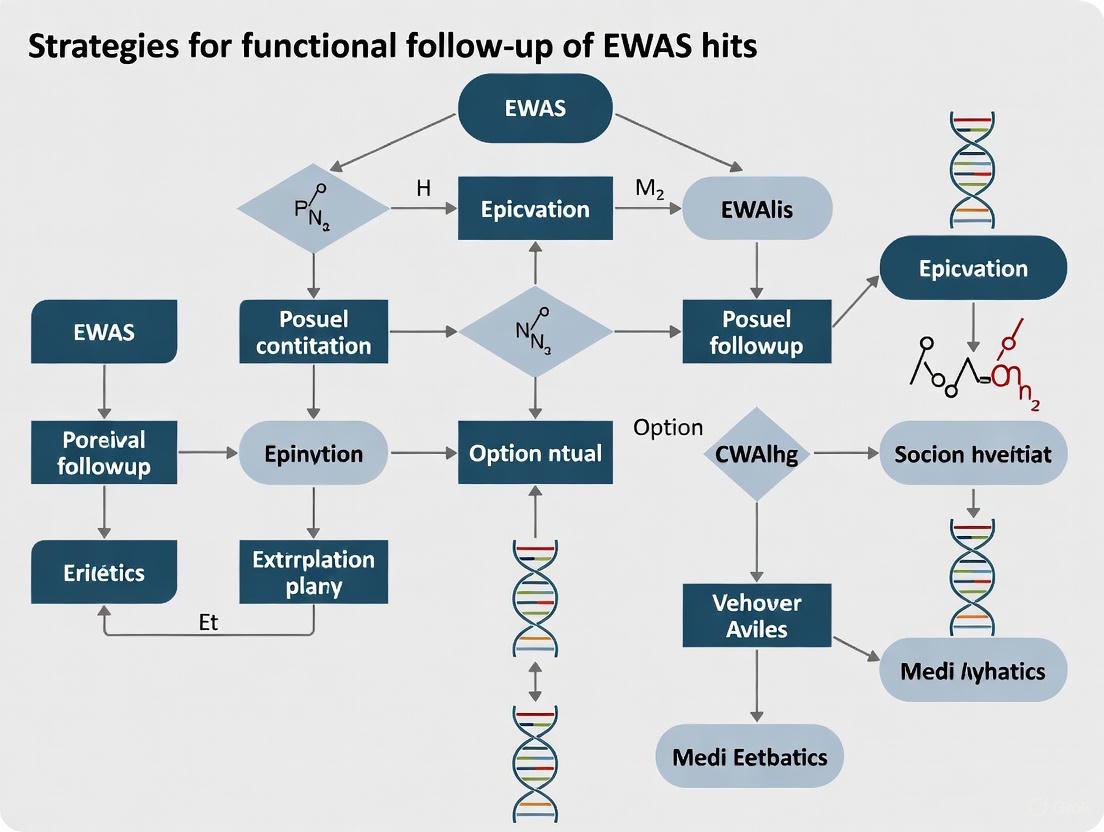
Diagram 2: Functional Follow-up Strategy for EWAS Hits
A fundamental challenge in biomedical research, particularly in the functional follow-up of Epigenome-Wide Association Study (EWAS) hits, is distinguishing causal relationships from mere correlations. Observational studies often identify associations between molecular traits and disease, but these can be misleading due to confounding factors and reverse causation. Mendelian Randomization (MR) has emerged as a powerful methodological framework that addresses these limitations by leveraging genetic variants as instrumental variables to test causal hypotheses. This approach serves as a critical bridge between initial correlation findings from EWAS and establishing actionable causal evidence for downstream drug development.
MR is based on the principle that genetic variants are randomly assigned during gamete formation and conception, much like the randomization in a clinical trial [18] [19]. This natural randomization creates a study design that can provide unconfounded estimates of causal effect, helping researchers prioritize molecular targets with genuine causal evidence for disease outcomes. For researchers investigating EWAS hits, MR offers a methodological pathway to determine whether epigenetic changes likely influence disease, are consequences of disease processes, or simply share common causes.
Core Principles and Assumptions of Mendelian Randomization
The Genetic Instrument: Nature's Randomized Trial
Mendelian randomization uses genetic variants, typically single nucleotide polymorphisms (SNPs), as instrumental variables for modifiable exposures [20]. These genetic instruments must satisfy three core assumptions:
- Relevance: The genetic variant must be robustly associated with the exposure of interest [20]
- Independence: The genetic variant must not be associated with confounders of the exposure-outcome relationship [20]
- Exclusion Restriction: The genetic variant must affect the outcome only through the exposure, not via alternative pathways (no horizontal pleiotropy) [20]
The following diagram illustrates the core MR design and its key assumptions:
Figure 1: Core Mendelian Randomization Design. The genetic instrument (G) must be associated with the exposure (E) but not share common causes with the outcome (O), nor affect the outcome through pathways other than the exposure.
Comparison with Other Study Designs
MR occupies a unique space in the evidence hierarchy between observational studies and randomized controlled trials (RCTs). The table below compares key characteristics:
| Study Design | Randomization Principle | Key Strengths | Key Limitations |
|---|---|---|---|
| Observational Study | No randomization | Efficient for hypothesis generation; suitable for rare outcomes | Susceptible to confounding and reverse causation |
| Mendelian Randomization | Random allocation of genetic variants at conception | Reduces confounding and reverse causation; can assess lifelong exposure effects | Requires specific genetic assumptions; limited by pleiotropy |
| Randomized Controlled Trial | Active randomization of participants | Gold standard for causal inference; minimizes confounding | Expensive; time-consuming; may raise ethical concerns |
Table 1: Comparison of Study Designs for Causal Inference. MR bridges the gap between observational studies and RCTs, leveraging genetic variation as a natural experiment [18].
Implementing Mendelian Randomization: A Technical Guide
Experimental Workflow for MR Analysis
A typical MR analysis follows a structured workflow from study design through interpretation. The following diagram outlines key stages:
Figure 2: Mendelian Randomization Analysis Workflow. The process involves clearly defining the causal question, selecting appropriate genetic instruments, obtaining association estimates, performing statistical analyses, and carefully interpreting results.
MR analyses can be conducted using either individual-level or summary-level genetic data [19]. Summary-data MR has become increasingly popular with the availability of large-scale GWAS consortia data:
- Individual-level data: Contains genotype, exposure, and outcome data for each participant
- Summary-level data: Uses pre-calculated genetic association estimates from different studies (two-sample MR)
Selecting valid genetic instruments is a critical step. Instruments are typically chosen from genome-wide association studies (GWAS) of the exposure, selecting variants that reach genome-wide significance (p < 5×10⁻⁸) [21]. For polygenic traits, multiple genetic variants can be combined into allele scores that collectively instrument the exposure [20].
Statistical Methods for MR Analysis
Several statistical approaches are available for MR analysis, each with different assumptions and applications:
| Method | Description | Key Assumptions | When to Use |
|---|---|---|---|
| Wald Ratio | Single variant estimate: βYX/βGX | Standard IV assumptions | Single instrument available |
| Inverse Variance Weighted (IVW) | Weighted average of ratio estimates | All variants are valid instruments | Multiple independent instruments |
| MR-Egger | Allows for pleiotropy via intercept term | Instrument Strength Independent of Direct Effect (InSIDE) | Suspected directional pleiotropy |
| Weighted Median | Provides consistent estimate if ≥50% valid | Majority of weight from valid instruments | Heterogeneous instrument effects |
Table 2: Common Statistical Methods for Mendelian Randomization Analysis. Method selection depends on the number of available instruments and assumptions about pleiotropy [20] [19].
The IVW method, the most common approach for multiple instruments, can be implemented using the formula:
βMR = Σ(βGYβGXσY⁻²) / Σ(βGX²σY⁻²)
Where βGX represents the genetic variant-exposure association, βGY represents the genetic variant-outcome association, and σY⁻² represents the inverse variance of the genetic variant-outcome association [20].
Troubleshooting Common Issues in Mendelian Randomization
FAQ: Addressing Methodological Challenges
Q1: How can I assess whether my genetic instruments are valid?
- Test for instrument strength using F-statistics (F > 10 indicates adequate strength)
- Assess balance of covariates across genetic instrument groups
- Use MR-Egger regression to test for directional pleiotropy
- Perform leave-one-out analyses to identify influential variants
Q2: What should I do when I suspect horizontal pleiotropy?
- Apply robust MR methods (MR-Egger, weighted median, MR-PRESSO)
- Use tissue-specific instruments where possible
- Perform sensitivity analyses excluding pleiotropic variants
- Consider multivariable MR to account for measured pleiotropic pathways
Q3: How can I address weak instrument bias?
- Use many weak instruments with methods robust to weak instrument bias
- Consider using summarized data from larger GWAS for exposure associations
- Apply methods specifically designed for many weak instruments
- Use family-based designs to eliminate confounding [19]
Q4: What are the options when working with limited sample sizes?
- Utilize two-sample MR with publicly available summary statistics
- Collaborate with consortia to access larger datasets
- Consider polygenic risk scores as instruments
- Use Bayesian methods that can provide more precise estimates with regularization
Q5: How can I distinguish causality from reverse causation?
- Perform bidirectional MR analyses
- Assess temporal relationships using prospective studies
- Use epigenetic age acceleration measures as exposures
- Consider family-based designs to reduce confounding [19]
Research Reagent Solutions for MR Studies
| Resource Type | Examples | Primary Function | Access Information |
|---|---|---|---|
| Software Packages | TwoSampleMR, MR-Base, MendelianRandomization | Statistical analysis of MR data | CRAN, GitHub |
| GWAS Catalog | NHGRI-EBI GWAS Catalog | Source of genetic associations | https://www.ebi.ac.uk/gwas/ |
| Summary Data Platforms | MR-Base, UK Biobank, GWAS ATLAS | Access to summary statistics | Platform-specific access |
| Quality Control Tools | PLINK, PRSice | Genotype data QC and analysis | https://www.cog-genomics.org/plink/ |
Table 3: Essential Research Resources for Mendelian Randomization Studies. These tools and databases support various stages of MR analysis from data acquisition to statistical implementation [21] [22].
Advanced Applications in Functional Follow-up of EWAS Hits
Integrating MR with EWAS for Causal Inference
MR provides a powerful framework for prioritizing EWAS findings by testing whether epigenetic markers likely influence disease (causal), are consequences of disease (reverse causation), or are confounded by other factors [10] [8]. The following diagram illustrates how MR can be applied in the context of EWAS follow-up:
Figure 3: Integrating Mendelian Randomization in EWAS Follow-up. MR uses methylation quantitative trait loci (mQTLs) as instruments to test causal relationships between epigenetic markers and disease outcomes, helping distinguish causal effects from reverse causation and confounding [10].
Research comparing GWAS and EWAS findings has shown that these approaches often capture distinct biological aspects of complex traits [10]. For instance, a systematic comparison of 15 complex traits found substantial overlap between GWAS and EWAS only for diastolic blood pressure, suggesting that in most cases, these study designs identify different genes and biological pathways [10].
Multivariable and Bidirectional MR Applications
Advanced MR extensions can address more complex research questions:
- Multivariable MR: Assesses the direct effect of multiple related exposures
- Bidirectional MR: Tests for directional causality between two traits
- Network MR: Maps causal pathways between multiple interconnected traits
- Mediation MR: Decomposes total effects into direct and indirect effects
For drug development professionals, MR can provide critical evidence for target prioritization by establishing whether proteins or epigenetic markers likely play causal roles in disease pathogenesis [19]. This application has gained traction in pharmaceutical research, with MR analyses increasingly informing target validation decisions.
Mendelian randomization represents a powerful methodological approach for strengthening causal inference in the functional follow-up of EWAS hits. By leveraging the natural randomization of genetic variation, MR helps address fundamental challenges of confounding and reverse causation that plague observational studies. While methodological challenges remain, particularly regarding instrument validity and pleiotropy, ongoing methodological developments continue to enhance the robustness of MR applications.
For researchers and drug development professionals, MR provides a valuable tool for prioritizing molecular targets with genuine causal evidence for disease outcomes. When carefully applied and interpreted with appropriate sensitivity analyses, MR can significantly strengthen the evidence base translating correlational findings from EWAS into actionable insights for therapeutic development.
A Practical Pipeline: From Data Analysis to Functional Hypothesis Generation
Troubleshooting Common Pipeline Errors
This section addresses frequent issues encountered when running the ChAMP and Minfi pipelines for epigenome-wide association studies (EWAS).
ChAMP-Specific Issues and Solutions
Q1: What does the error "Error Match between pd file and Green Channel IDAT file" mean and how do I resolve it?
This error occurs when ChAMP cannot properly match your sample sheet (phenotypic data or "pd" file) with the IDAT files in your directory [23].
Solution: Follow this systematic checklist:
- Verify Column Headers: Ensure your sample sheet contains the exact column headers required by ChAMP. Critical columns include
Slide,Array, andBasename[23]. - Check Sentrix ID Format: Confirm that Sentrix IDs in your sample sheet match exactly with those in your IDAT filenames. Even minor formatting discrepancies can cause this error [23].
- Validate File Paths: If using explicit paths in your
Basenamecolumn, ensure they are correct and accessible from your R working directory. - Confirm Array Type: Explicitly specify your array type (
arraytype = "EPIC"orarraytype = "450K") in thechamp.import()function call.
Q2: Why does my normalization fail with "internet routines cannot be loaded" or similar errors?
This error often occurs during the champ.norm() step, particularly when using parallel processing functions [24].
Solution:
- Check Parallel Processing Configuration: Reduce the number of cores specified in the
coresparameter. Start withcores = 1to test if the error persists. - Verify R Installation: Ensure your R installation can create socket connections for parallel processing. Reinstall R or relevant packages (
parallel,snow) if necessary. - Alternative Normalization: Try a different normalization method. If
BMIQfails, attemptSWANorPBCmethods instead [24].
Q3: Why does FunctionalNormalization fail with "subscript out of bounds" on EPIC data?
This error may occur due to annotation package mismatches when running champ.norm() with method = "FunctionalNormalization" on EPIC data [25].
Solution:
- Annotation Consistency: ChAMP typically installs
IlluminaHumanMethylationEPICanno.ilm10b2.hg19automatically, but FunctionalNormalization may requireIlluminaHumanMethylationEPICanno.ilm10b3.hg19[25]. - Manual Installation: Manually install the required annotation package:
- Alternative Method: Consider using
ssNoobnormalization instead, as FunctionalNormalization may not significantly enhance results overssNoobfor EPIC arrays [25].
Minfi-Specific Issues and Solutions
Q4: Why are my annotation and manifest listed as "unknown" after using read.metharray.exp?
This issue occurs when Minfi cannot properly load the required annotation packages, despite them being installed [26].
Solution:
- Explicitly Load Annotations: Manually load the required annotation packages before running your analysis:
- Check Package Versions: Ensure compatibility between Minfi, annotation packages, and your R version. Consider updating all packages to the latest versions.
- Verify Installation: Confirm annotation packages are installed correctly for your specific array type (EPIC vs. 450K).
Q5: Why does preprocessRaw fail with "there is no package called 'Unknownmanifest'"?
This error directly relates to the previous issue where annotations are not properly loaded [26].
Solution:
- Reinstall Packages: Reinstall both Minfi and relevant annotation packages using BiocManager to ensure compatibility.
- Session Restart: Restart your R session and reload all required packages before reprocessing your data.
- Explicit Manifest Specification: When creating RGChannelSet objects, explicitly specify the manifest when possible.
General Pipeline Issues
Q6: How do I choose between ChAMP and Minfi for my EWAS?
Both pipelines offer comprehensive analysis capabilities, but have different strengths:
Table: Comparison of ChAMP and Minfi Pipelines
| Feature | ChAMP | Minfi |
|---|---|---|
| Primary Use Case | All-in-one solution for EPIC data [11] | Most cited for 450K data [11] |
| Data Import | Direct from IDAT files [11] | Direct from IDAT files [11] |
| Quality Control | Integrated in pipeline [11] | Integrated in pipeline [11] |
| Normalization | Multiple methods (BMIQ, SWAN, FunctionalNormalization) [24] [25] | Multiple methods [11] |
| DMP Detection | Yes [11] | Yes [11] |
| DMR Detection | Yes [11] | Yes [11] |
| Downstream Analyses | Variety available [11] | Variety available [11] |
Q7: What alternative normalization methods exist when standard approaches fail?
If standard normalization methods consistently fail, consider:
- Method Rotation: Try different normalization techniques. ChAMP supports BMIQ, SWAN, PBC, and FunctionalNormalization [24] [25].
- Quality Assessment: Check if normalization fails due to poor sample quality. BMIQ may fail for samples "that did not even show beta distribution" [24].
- Alternative Pipeline: Explore the
sesamepackage, which shows excellent performance on diverse datasets including TARGET and TCGA, though not yet integrated directly into ChAMP [25].
Methodological Framework for EWAS Follow-up
This section provides experimental protocols and methodologies for functional follow-up of EWAS hits within the context of broader research strategies.
EWAS Experimental Design Considerations
Proper experimental design is critical for meaningful EWAS results and downstream functional validation.
Table: EWAS Study Designs for Functional Follow-up
| Design Type | Key Features | Advantages | Limitations | Best Use Cases |
|---|---|---|---|---|
| Case-Control | Compares unrelated cases vs. controls [11] [8] | Large sample sizes possible; utilizes existing biobanks [11] | Cannot determine causality; timing of methylation changes unknown [11] | Initial association discovery; leveraging existing DNA banks [11] |
| Longitudinal | Tracks same individuals over time [11] [8] | Establishes temporal relationships; tracks methylome dynamics [11] | Time-consuming; expensive; pre-disease samples difficult to obtain [11] | Establishing causality; natural history studies; intervention studies [11] |
| Monozygotic Twin Studies | Compares genetically identical twins discordant for disease [8] | Controls for genetic variation; powerful for epigenetic studies [8] | Difficult to recruit large cohorts; cannot determine timing without longitudinal data [8] | Isolating non-genetic components of disease [8] |
| Family Studies | Examines transgenerational inheritance patterns [8] | Can rule out genomic variation effects [8] | Few large cohorts available [8] | Studying transgenerational epigenetic inheritance [8] |
The following diagram illustrates the strategic decision process for selecting appropriate EWAS designs:
Advanced Analytical Approaches for EWAS Follow-up
Methylation Quantitative Trait Loci (methQTL) Analysis: This analysis identifies genetic variants that influence methylation states, helping to determine whether observed methylation changes are driven by genetic variation or environmental factors [11]. MethQTL analysis integrates SNP and methylation data to discover loci where genotype correlates with methylation phenotype, distinguishing primary epigenetic changes from those secondary to genetic variation [27].
Cell-Type Deconvolution: For blood-based EWAS, statistical deconvolution methods estimate cell-type specific methylation signals from mixed cell population data [11]. This is crucial as cellular heterogeneity can confound methylation associations, particularly in whole blood samples where multiple cell types contribute to the methylation signal.
Methylation Age Analysis: The epigenetic clock provides a biological age estimate based on methylation patterns at specific CpG sites [11]. Discrepancies between epigenetic age and chronological age can indicate accelerated aging or disease states, providing functional context for EWAS hits.
Research Reagent Solutions for EWAS
Table: Essential Research Materials for EWAS and Functional Follow-up
| Reagent/Resource | Function | Application in EWAS |
|---|---|---|
| Illumina Methylation BeadChips | Genome-wide methylation profiling | Primary data generation for EWAS (27K, 450K, EPIC arrays) [11] [8] |
| Bisulfite Conversion Kits | Chemical treatment of DNA for methylation detection | Distinguishes methylated vs unmethylated cytosines prior to array analysis [11] |
| Annotation Packages (e.g., IlluminaHumanMethylationEPICanno.ilm10b4.hg19) | Provide genomic context for CpG probes | Essential for mapping CpG sites to genes and regulatory regions during analysis [26] |
| Cell-Type Specific Reference Methylomes | Reference datasets for deconvolution algorithms | Enable estimation of cell-type proportions in mixed samples (e.g., whole blood) [11] |
| Functional Normalization Components | Remove technical variation using control probes | Normalization to improve data quality and reduce false positives [25] |
Workflow Integration and Best Practices
The following diagram illustrates a comprehensive EWAS workflow incorporating both ChAMP/Minfi analysis and functional follow-up strategies:
Integration with Genomic Data
For comprehensive functional follow-up, integrate EWAS results with complementary genomic datasets:
- GWAS Integration: Combine significant EWAS hits with existing GWAS data to dissect complex haplotypes and identify potentially functional variants [28] [27].
- Expression QTL (eQTL) Mapping: Correlate methylation quantitative trait loci with expression quantitative trait loci to understand the functional consequences of methylation changes.
- Multi-omics Approaches: Incorporate proteomic, metabolomic, or other epigenomic data (e.g., histone modifications) to build comprehensive models of disease pathophysiology.
By addressing both technical pipeline challenges and methodological framework for functional follow-up, this guide provides researchers with comprehensive tools for conducting robust EWAS and deriving biologically meaningful insights from methylation data.
Fundamental Concepts FAQ
What is a methylation quantitative trait locus (methQTL) and why is it important for functional follow-up in EWAS?
A methylation quantitative trait locus (methQTL) is a region of the genome where genetic variants (such as single nucleotide polymorphisms or SNPs) are associated with variation in DNA methylation states at specific CpG sites [29]. These associations can range from a few bases to several megabases, potentially resulting in long-range interactions [29]. MethQTLs are crucial for annotating the functional effects of genetic variants discovered in EWAS and help distinguish variants that are merely correlated with disease from those that are functionally involved [30]. They provide a direct link between genetic predisposition and epigenomic regulation, illuminating the mechanistic pathway from genotype to phenotype.
What is the difference between cis- and trans-meQTLs?
- cis-meQTLs are genetic variants that influence DNA methylation at loci located relatively close to the variant, typically within 500 kb to 2 Mb [29] [30].
- trans-meQTLs are genetic variants that affect DNA methylation at loci located far away on the same chromosome or on a different chromosome [30]. These can form complex regulatory networks, such as the ERG-mediated network of 233 trans-methylated CpGs involved in hematopoietic cell differentiation [31].
What is Methylation Age (Epigenetic Clock) and how does it relate to biological aging?
The Methylation Age or Epigenetic Clock is a highly accurate age predictor based on the systematic changes in DNA methylation patterns at specific CpG sites throughout an individual's life [32]. It is calibrated using machine learning models on large-scale methylation datasets. Epigenetic Age Acceleration (EAA) refers to the deviation between DNA methylation-predicted age and chronological age. Positive EAA (where the epigenetic age is older than chronological age) reflects accelerated biological aging and is a significant predictor of health outcomes, including a 16% increased risk of stroke per unit increase in EAA (OR = 1.16, 95% CI 1.13–1.19) [33]. This acceleration is thought to reflect pathophysiological processes like organ functional decline and inflammatory activation [33].
What are the different types of QTLs relevant for multi-omics integration?
QTL analysis has expanded to cover various molecular layers, providing a network view of how variants influence phenotype [30].
- eQTL: Affects gene expression levels.
- meQTL/methQTL: Affects DNA methylation patterns.
- caQTL: Affects chromatin accessibility.
- bQTL: Affects transcription factor binding.
- pQTL: Affects protein abundance.
Troubleshooting Guides
Table 1: Common MethQTL Analysis Issues and Solutions
| Problem | Potential Causes | Solutions & Verification Steps |
|---|---|---|
| No significant meQTLs detected | Insufficient statistical power (small sample size), overly stringent multiple-testing correction, poor-quality methylation/genotype data, cell-type heterogeneity masking effects. | Increase sample size (100s of samples recommended). Use permutation-based FDR control (e.g., via fastQTL). Check data quality: ensure high call rates for SNPs/CpGs, inspect beta value distributions. Account for cell type composition in statistical models [29] [31]. |
| Inability to replicate meQTLs in an independent cohort | Differences in ancestry (presence of ancestry-specific meQTLs), differences in tissue/cell type composition, technical batch effects from different methylation platforms. | Use genetically similar cohorts for replication. Prefer profiling purified cell types over heterogeneous tissues. Harmonize processing pipelines and correct for batch effects. Check for EA-specific mQTLs if working with East Asian populations [31]. |
| Confounding by cell type heterogeneity | Methylation levels are highly cell-type-specific. If cell type proportions vary between samples and are not accounted for, they can create false positives or mask true meQTL signals. | Estimate cell type proportions using reference-based (e.g., Houseman method) or reference-free algorithms. Include these proportions as covariates in the meQTL mapping model. Use pipelines like MAGAR that are designed for multi-tissue/cell-type analysis [29]. |
| Computational challenges in processing WGBS data | WGBS data is computationally intensive to align and analyze, requiring specialized tools and significant memory/CPU resources. | Use established, efficient pipelines like msPIPE or Methy-Pipe [34] [35]. These pipelines integrate all steps from alignment to DMR calling and visualization, and can be run via Docker for easier implementation. |
Table 2: Common Methylation Age Analysis Issues and Solutions
| Problem | Potential Causes | Solutions & Verification Steps |
|---|---|---|
| Poor age prediction accuracy (high MAE) | Using an inappropriate epigenetic clock model (e.g., a blood clock on brain tissue), poor-quality methylation data, data from an unsupported species or tissue, incorrect model calibration. | Use a tissue-appropriate clock (e.g., Horvath's multi-tissue clock, Hannum's blood clock, PhenoAge). Ensure high data quality and that your data preprocessing matches the clock's requirements. For high accuracy in blood, consider non-linear, cohort-based models like GP-age [36]. |
| Inconsistent age acceleration values | Different clocks capture different aspects of biological aging. An individual might show acceleration on one clock but not another. | Clearly define the chosen clock and its biological interpretation (e.g., PhenoAge for morbidity/mortality). Report results consistently with the same clock across a study. Use multiple clocks only if hypothesizing different aging aspects. |
| Interpreting the biological meaning of EAA | EAA is a composite measure and does not point to a specific biological pathway or mechanism. | Correlate EAA with specific health phenotypes (e.g., stroke risk [33]). Perform downstream analyses like GO and KEGG enrichment on the CpG sites most weighted in the clock or those that deviate most from expected methylation levels [32]. |
| Feature selection for custom clock development | Using all CpG sites from a platform (e.g., 450K) is computationally intensive and can lead to overfitting. | Apply machine learning-based feature selection. For example, use gradient-boosting models (XGBoost, LightGBM, CatBoost) to identify a compact set of highly predictive CpG sites. As few as 30 CpG sites can achieve high accuracy in blood [32] [36]. |
Experimental Protocols
Protocol 1: Identifying methQTLs using the MAGAR Pipeline
Objective: To identify genetic variants that influence DNA methylation levels, distinguishing common from cell type-specific effects.
Materials:
- Matched genotyping (e.g., SNP array or WGS) and DNA methylation data (e.g., array or WGBS) from the same individuals.
- Software: MAGAR R package (available via Bioconductor) [29].
Method:
- Data Preprocessing: Process raw genotype and methylation data (IDAT files) using MAGAR's integrated modules, which leverage established tools like RnBeads, PLINK, and CRLMM for quality control and filtering [29].
- CpG Clustering: Group neighboring, highly correlated CpGs into "correlation blocks" to reduce redundancy and account for the correlation structure of methylation haplotypes [29].
- Tag-CpG Selection: For each correlation block, select a single representative "tag-CpG" for association testing [29].
- methQTL Calling: Test for associations between each tag-CpG and all SNPs within a defined genomic window (e.g., 500 kb upstream and downstream). This can be performed using:
- Linear Modeling: A standard linear least-squares regression.
- fastQTL: A permutation-based approach to control for multiple testing [29].
- Colocalization Analysis: Use the output from MAGAR to perform colocalization analysis across different tissues or cell types to define tissue-specific and tissue-independent (common) methQTLs [29].
Protocol 2: Constructing a Methylation Age Prediction Model
Objective: To build a machine learning model that predicts biological age from DNA methylation data.
Materials:
- A large dataset of DNA methylation samples (e.g., from a public repository like GEO) with known chronological ages. The example below uses 8,233 samples with 50,000 methylation sites each [32].
- Software: Python/R with machine learning libraries (e.g., XGBoost, LightGBM, CatBoost, SHAP).
Method:
- Data Preprocessing: Handle missing data (e.g., imputation or filling with 0). Encode categorical variables (e.g., sex). Perform quality control to remove low-quality probes [32].
- Feature Selection: To avoid overfitting and identify the most age-predictive CpGs, use a wrapper method with multiple machine learning models.
- Model Training: Train a final predictive model (e.g., Gradient Boosting) using the selected subset of features. Use k-fold cross-validation to optimize hyperparameters.
- Model Interpretation: Use interpretable ML frameworks like SHAP (SHapley Additive exPlanations) to determine the contribution of each CpG site to the final prediction. This helps identify key loci like cg23995914, which has been highlighted as a major contributor [32].
- Biological Validation: Perform functional enrichment analysis (GO, KEGG) on the genes associated with the top predictive CpG sites to understand the biological pathways underlying the aging signal [32].
Table 3: Key Computational Tools and Pipelines
| Tool Name | Function | Key Feature |
|---|---|---|
| MAGAR [29] | methQTL identification | Discriminates cell type-specific effects by clustering correlated CpGs. |
| msPIPE [34] | WGBS Data Analysis | End-to-end pipeline from pre-processing to DMR calling and publication-quality visualization. Supports Docker. |
| Methy-Pipe [35] | WGBS Data Analysis | Integrated pipeline for bisulfite read alignment (BSAligner) and differential methylation analysis (BSAnalyzer). |
| fastQTL / MatrixEQTL [29] [31] | QTL Mapping | Fast, permutation-based tools for cis-QTL mapping. |
| GP-age [36] | Methylation Age Prediction | Non-linear, cohort-based clock using only 30 CpG sites for accurate blood age prediction. |
| SHAP [32] | Model Interpretation | Explains the output of any machine learning model, identifying key predictive CpG sites. |
Table 4: Key Methylation Clocks and Their Applications
| Clock Name | Tissues | Application Context |
|---|---|---|
| Horvath's Clock [33] | Multi-tissue | The first pan-tissue clock; useful for comparing aging rates across different tissues. |
| Hannum's Clock [33] | Blood | Predictive of age-related phenotypes and mortality risk in blood-based studies. |
| PhenoAge [33] | Blood | Captures physiological dysregulation and is a strong predictor of morbidity and mortality. |
| GP-age [36] | Blood | Compact, accurate clock for blood; ideal for longitudinal studies and forensic profiling. |
Epigenome-wide association studies (EWAS) investigating DNA methylation in easily accessible tissues like whole blood face a fundamental challenge: cellular heterogeneity. Blood is a complex mixture of different cell types, each with a distinct DNA methylation profile. When analyzing a bulk tissue sample, an observed DNA methylation difference could represent either a genuine change within a specific cell type or a mere shift in the cell type proportions between compared groups. Failing to account for this cellular composition is a major source of confounding and misinterpretation in EWAS [37] [38].
Statistical cell mixture deconvolution (CMD) has emerged as a powerful bioinformatic solution to this problem. These methods leverage pre-defined libraries of cell-type-specific DNA methylation markers to computationally estimate the proportional composition of cell types within a heterogeneous sample. These estimates can then be included as covariates in statistical models to control for confounding, allowing researchers to identify cell-type-specific epigenetic changes [38]. This guide addresses frequent questions and troubleshooting issues encountered when implementing these critical methods.
Frequently Asked Questions (FAQs) and Troubleshooting
FAQ 1: What is the core problem that cellular deconvolution solves?
Answer: The core problem is confounding due to cellular heterogeneity. In a whole blood EWAS, if a disease state is associated with an increase in the proportion of neutrophils, and neutrophils have a characteristically low methylation at a particular CpG site, that site will appear to be hypomethylated in the disease group. This association is not driven by the disease process within a cell, but by a change in the underlying cell population. Deconvolution methods control for this by estimating and adjusting for these cell proportion shifts [37] [38].
FAQ 2: My deconvolution results show poor discrimination between certain cell types. What could be wrong?
Answer: Poor discrimination often stems from a suboptimal library of cell-specific methylation markers. The accuracy of deconvolution is entirely dependent on the quality of the reference library used. Libraries assembled using different criteria (e.g., top ANOVA hits vs. an equal number of markers per cell type) can have vastly different performances [38].
- Troubleshooting Steps:
- Evaluate Your Library: Investigate the library's composition. Some older libraries may over-represent markers that discriminate major lineages (e.g., myeloid vs. lymphoid) but perform poorly on lineage-specific subtypes (e.g., CD4+ T-cells vs. CD8+ T-cells) [38].
- Use an Optimized Library: Consider using a dynamically identified, optimized library. For example, the IDOL algorithm identified a 300-CpG library that demonstrated superior overall discrimination of the leukocyte landscape compared to earlier libraries, leading to more accurate cell fraction estimates and improved control of false positives in EWAS [38].
- Check Data Preprocessing: Ensure your DNA methylation data has been properly normalized, especially for the technical differences between Infinium I and Infinium II probe designs on the 450K/EPIC arrays. Methods like BMIQ (Beta Mixture Quantile dilation) are specifically designed to correct this probe type bias [39] [40].
FAQ 3: After deconvolution and adjustment, how do I interpret a significant EWAS hit?
Answer: A significant association after adjusting for estimated cell proportions suggests the DNA methylation change is independent of shifts in the major cell populations. However, precise interpretation requires further investigation:
- Cell-Type-Specific Effects: The association could be present in one, several, or all cell types. To determine this, you need to apply methods designed to identify cell-type-specific differential methylation (DMCTs), such as CellDMC [41]. For instance, a meta-analysis of smoking EWAS found that most smoking-associated hypomethylation was highly specific to cells of the myeloid lineage, a finding that would have been masked in a standard whole-blood analysis [41].
- Biological Context: The gene or region's known biology should be considered alongside the cell-type-specificity of the signal to generate meaningful hypotheses.
FAQ 4: How does cellular deconvolution impact the overlap between GWAS and EWAS findings?
Answer: GWAS and EWAS often identify distinct genomic regions and biological pathways for the same complex trait. Proper cellular deconvolution is critical for ensuring that EWAS signals reflect true intra-cellular epigenetic states rather than confounding by cell composition. When this confounding is controlled for, the remaining EWAS hits are less likely to be tagged by GWAS variants, as the two study designs are capturing different biological mechanisms—genetic predisposition versus environmentally responsive or consequential epigenetic regulation [42].
Essential Reagents and Computational Tools
Table 1: Key Research Reagent Solutions for Cellular Deconvolution Experiments
| Tool/Reagent Name | Type | Primary Function | Key Considerations |
|---|---|---|---|
| IDOL Algorithm [38] | Algorithm & Library | Identifies Optimal L-DMR libraries for deconvolution; provides an optimized 300-CpG library for whole blood. | Designed to maximize accuracy of cell fraction estimates; improves discrimination of lineage-specific cell subtypes. |
| CellDMC Algorithm [41] | Software Algorithm | Identifies cell-type-specific differential methylation from bulk tissue data. | Requires a pre-estimated cell composition as input; uses interaction terms in a linear model. |
| EpiDISH Algorithm [41] | Software Algorithm / Reference | A reference-based method for estimating cell composition from bulk DNA methylation data. | Provides a DNAm reference matrix for seven blood cell subtypes; often used in conjunction with CellDMC. |
| BMIQ [39] [40] | Normalization Method | Corrects for the technical bias between Infinium I and Infinium II probe designs. | Critical preprocessing step; improves data quality and comparability before deconvolution. |
| minfi / waterRmelon [39] [40] | R Packages | Comprehensive toolkits for importing, quality controlling, and normalizing Illumina methylation array data. | Include functions for performing cell mixture deconvolution (minfi) and various normalization schemes (waterRmelon). |
Experimental Workflow for Cell-Type Deconvolution
The following diagram illustrates the standard analytical workflow for conducting an EWAS that properly accounts for cellular heterogeneity, from raw data to biological interpretation.
Advanced Analysis: Protocol for Cell-Type-Specific Inference
For researchers who have identified significant EWAS hits after adjusting for cell proportions, the next logical step is to determine in which specific cell type the effect resides. The following protocol outlines this process using the CellDMC algorithm.
Objective: To identify cell-type-specific differential methylation (DMCTs) from bulk whole blood DNA methylation data.
Primary Software: R statistical environment.
Key R Packages: CellDMC, EpiDISH (or other packages that provide the necessary reference data and functions).
Step-by-Step Procedure:
Data Preparation and Preprocessing:
- Import your beta-value matrix (samples as columns, CpGs as rows) after it has undergone rigorous quality control and normalization [40]. Ensure you have applied a probe bias correction method like BMIQ.
- Prepare your phenotype file, encoding your exposure or trait of interest appropriately (e.g., 0=control, 1=case).
Estimate Cell Type Proportions:
- Use a reference-based method like
EpiDISHto estimate the proportions of major leukocyte subtypes (e.g., neutrophils, monocytes, B-cells, T-cells, NK-cells) in each sample. This will produce a matrix of estimated cell fractions. - Code Example:
- Use a reference-based method like
Run CellDMC Analysis:
- Execute the
CellDMCfunction, providing your normalized beta-value matrix, the phenotype vector, and the matrix of estimated cell fractions. - CellDMC fits a linear model that includes an interaction term between the phenotype and each cell type's fraction. A significant interaction term for a specific CpG and cell type indicates a DMCT.
- Code Example:
- Execute the
Interpretation of Results:
- The output will include, for each CpG site, p-values for the main effect of the phenotype and for the interaction with each cell type.
- Focus on CpGs with a significant interaction p-value (after multiple testing correction) for a specific cell type. This indicates that the association between the phenotype and DNA methylation is different in that cell type. The meta-analysis on smoking, for example, used this approach to pinpoint a hypomethylation signature predominantly in the myeloid lineage [41].
- Follow up on these cell-type-specific hits with functional annotation and pathway analysis to understand their potential biological relevance.
Epigenome-wide association studies (EWAS) systematically identify epigenetic marks, such as DNA methylation, associated with specific phenotypes or diseases [8]. The core challenge in functional genomics lies in moving from merely identifying these statistical associations to understanding their biological significance and mechanistic role in disease etiology [43]. Multi-omics integration provides a powerful framework for this functional follow-up by correlating methylation changes with downstream molecular consequences captured by transcriptomic (gene expression) and proteomic (protein abundance) data [44] [45].
This approach is pivotal because DNA methylation in promoter or regulatory regions can directly influence gene activity, potentially silencing or activating genes without changing the underlying DNA sequence [46] [47]. However, not all methylation changes are functionally consequential. By integrating data across omics layers, researchers can prioritize methylation hits that are associated with changes in gene expression or protein function, thereby distinguishing passenger events from driver events in disease processes [44] [48]. This technical support center is designed to guide you through the experimental and computational methodologies for successful multi-omics integration, directly addressing common pitfalls and providing actionable troubleshooting advice.
Key Concepts and Workflow
Fundamental Principles
- DNA Methylation: An epigenetic mark involving the addition of a methyl group to a cytosine base, typically within a CpG dinucleotide context. Hypermethylation in gene promoter regions is often associated with gene silencing, while hypomethylation can permit gene expression [46] [8].
- Quantitative Trait Loci (QTL) Mapping: A foundational analytical framework for multi-omics integration. This approach treats epigenetic marks as potential regulators and tests their association with molecular phenotypes:
- methylation QTL (mQTL): Genetic loci that influence methylation levels.
- expression QTL (eQTL): Loci that influence gene expression levels.
- protein QTL (pQTL): Loci that influence protein abundance [44].
- Multi-Omics Integration Strategy: The process can be vertical (integrating different omics data from the same samples) or horizontal (integrating the same type of omics data from different studies) [49]. For EWAS follow-up, vertical integration is most common, aiming to trace the flow of information from the epigenome to the transcriptome and proteome within a biological system [45].
The following diagram outlines the core workflow for integrating methylation data with transcriptomic and proteomic data for functional validation of EWAS hits.
Frequently Asked Questions (FAQs)
FAQ 1: How do I prioritize EWAS hits for multi-omics follow-up studies?
Not all significant EWAS hits are equally likely to be functionally important. Prioritization should be based on multiple criteria to maximize the return on investment for costly multi-omics assays.
- Genomic Context: Prioritize methylation changes occurring in promoter regions, enhancers, or other known regulatory elements, as these are more likely to directly influence gene expression [8].
- Association Strength: Hits with larger effect sizes and high statistical significance (epigenome-wide significance, e.g., P < 1×10⁻⁷) are primary candidates [8].
- Gene Relevance: Consider whether the gene linked to the methylation mark is biologically plausible in the context of your disease of interest, for example, known involvement in relevant pathways [48].
- Replication: Prioritize hits that have been replicated in independent cohorts, as this increases confidence in the initial association [43].
FAQ 2: What are the primary statistical methods for correlating methylation with transcriptomic/proteomic data?
The choice of method depends on your study design and the nature of your data.
- Multi-staged Integration: This approach assumes a unidirectional flow of information. It often uses mediation analysis to test if the effect of a methylation mark on a clinical phenotype is mediated by changes in gene expression or protein abundance [44] [49].
- Meta-dimensional Integration: This method models variation from all omics layers simultaneously, without pre-specifying a direction of effect. It is useful for discovering novel, complex interactions and can be performed using multi-block or multivariate methods like MOFA or DIABLO [49].
- Regional Analysis: Instead of analyzing single CpG sites, methods that aggregate methylation across genomic regions (e.g., Differentially Methylated Regions - DMRs) can provide more robust associations with expression/protein data by reducing technical noise and capturing biologically coherent signals [8].
FAQ 3: Which publicly available multi-omics repositories can I use for my research?
Leveraging existing data can validate findings or generate hypotheses. Key repositories include:
- The Cancer Genome Atlas (TCGA): A comprehensive resource with matched DNA methylation, RNA-Seq, and for some cancers, RPPA proteomic data for over 33 cancer types [45] [48].
- Clinical Proteomic Tumor Analysis Consortium (CPTAC): Provides proteomics and phosphoproteomics data corresponding to TCGA tumor samples, offering a direct link to protein-level quantification [45].
- International Cancer Genomics Consortium (ICGC): Coordinates large-scale genomic studies, including whole-genome sequencing data, which can be integrated with other omics layers [45].
- Omics Discovery Index (OmicsDI): A consolidated resource that allows you to search for multi-omics datasets across 11 different repositories [45].
Troubleshooting Guides
Problem 1: No correlation found between significant methylation changes and gene expression
This is a common scenario, as not all methylation changes are functional at the transcript level.
- Potential Cause 1: Incorrect genomic annotation. The methylation change might not be in a regulatory region that directly influences the transcription of the gene you are testing.
- Solution: Perform a more nuanced annotation of the CpG site. Consider its location relative to the gene's transcription start site, its presence in a known enhancer element (using data from resources like ENCODE), and the chromatin state of the region.
- Potential Cause 2: Biological lag or buffering. The effect of methylation on expression might be context-dependent, small, or buffered by other regulatory mechanisms. The protein level, not the RNA level, might be the primary readout.
- Solution: If data is available, check for correlations with protein abundance. Consider analyzing pathways by grouping genes rather than looking at single gene effects.
- Potential Cause 3: Technical issues. Differences in sample processing, batch effects, or low statistical power can obscure real correlations.
- Solution: Ensure rigorous preprocessing, including batch effect correction and normalization specific to each data type [50]. Increase your sample size if possible.
Problem 2: Observed correlation is weak or inconsistent across datasets
A weak correlation can indicate an indirect relationship or technical artifacts.
- Potential Cause 1: Confounding by cell type composition. Methylation patterns are highly cell-type-specific. If your tissue sample is a mixture of cell types, the observed correlation could be driven by shifts in cell population proportions between cases and controls rather than a direct regulatory relationship.
- Solution: Use computational deconvolution methods (e.g., CIBERSORT, EpiDISH) to estimate cell-type proportions and include them as covariates in your models [8].
- Potential Cause 2: Inadequate data preprocessing. Improper normalization or failure to account for batch effects can introduce noise that dilutes the true signal.
- Solution: Revisit your preprocessing pipelines. For methylation arrays, ensure background correction and normalization (e.g., with
minfiin R). For RNA-Seq, use appropriate normalization methods (e.g., TPM, DESeq2's median-of-ratios) [50].
- Solution: Revisit your preprocessing pipelines. For methylation arrays, ensure background correction and normalization (e.g., with
- Potential Cause 3: The relationship is non-linear. The assumption of a linear relationship between methylation and expression may not always hold.
- Solution: Explore non-linear models or categorize methylation levels into quantiles (e.g., low, medium, high) and test for differences in expression between these groups.
Problem 3: Poor bisulfite conversion efficiency or amplification of converted DNA
This is a wet-lab specific issue that compromises the quality of methylation data from bisulfite sequencing methods.
- Potential Cause 1: Impure DNA input. Contaminants can inhibit the bisulfite conversion reaction.
- Solution: Ensure the DNA used for conversion is pure. If particulate matter is present after adding the conversion reagent, centrifuge at high speed and use only the clear supernatant [51].
- Potential Cause 2: Suboptimal primer design or polymerase for bisulfite-converted DNA.
- Solution:
- Primers: Design primers that are 24-32 nucleotides in length, contain no more than 2-3 mixed bases (to account for C/T conversion), and ensure the 3' end does not end in a base whose conversion state is unknown [51].
- Polymerase: Use a hot-start Taq polymerase (e.g., Platinum Taq). Avoid proof-reading polymerases as they cannot read through uracil (which bisulfite conversion creates from unmethylated cytosines) [51].
- Solution:
- Potential Cause 3: Large amplicon size. Bisulfite treatment fragments DNA, making amplification of large regions difficult.
- Solution: Aim for amplicons around 200 bp. Larger amplicons require optimized protocols [51].
The Scientist's Toolkit
Research Reagent Solutions
Table 1: Essential reagents and resources for multi-omics integration studies.
| Item | Function & Application | Notes |
|---|---|---|
| Illumina MethylationEPIC Kit | Genome-wide DNA methylation profiling. Covers over 850,000 CpG sites, including enhancer regions. | The standard platform for EWAS; preferred over older arrays due to broader genomic coverage [8]. |
| RNA-Seq Library Prep Kits (e.g., Illumina TruSeq) | Preparation of sequencing libraries for transcriptome analysis. Provides quantitative data on gene expression. | Allows for the discovery of novel transcripts and isoforms alongside quantitative expression analysis. |
| Platinum Taq DNA Polymerase | PCR amplification of bisulfite-converted DNA. | Hot-start enzyme is recommended for its ability to efficiently amplify uracil-containing templates without proof-reading activity [51]. |
| Cell Type Deconvolution Tools (e.g., CIBERSORT, EpiDISH) | Computational estimation of cell type proportions from bulk tissue data. | Critical for adjusting analyses for cellular heterogeneity, a major confounder in EWAS [8]. |
Experimental & Analytical Pathways for Functional Validation
After identifying high-confidence methylation-expression/protein links, the next step is experimental validation. The following diagram illustrates a standard workflow for moving from a computational finding to validated biological mechanism.
Detailed Methodologies:
- CRISPR/dCas9 Methylation Editing: This is the gold standard for functional validation. Fuse a catalytically dead Cas9 (dCas9) to a DNA methyltransferase (e.g., DNMT3A) or a demethylase (e.g., TET1). Use sgRNAs to target this complex to the specific CpG site identified in your EWAS.
- Protocol Outline:
- Design and clone sgRNAs specific to the genomic region of interest.
- Co-transfect dCas9-effector and sgRNA constructs into a relevant cell line.
- Confirm targeted methylation changes using pyrosequencing or targeted bisulfite sequencing.
- Measure the downstream effect on gene expression (e.g., by qRT-PCR) and/or protein levels (e.g., by Western blot) [48].
- Protocol Outline:
- Luciferase Reporter Assay: Used to test if a specific methylated DNA fragment can regulate transcription.
- Protocol Outline:
- Clone the genomic region encompassing the CpG site (in both methylated and unmethylated states) upstream of a minimal promoter driving a luciferase gene.
- Introduce the reporter construct into a cell model.
- Measure luciferase activity. A significant reduction in activity for the methylated construct indicates a direct repressive role of the methylation mark.
- Protocol Outline:
- Chromatin Immunoprecipitation (ChIP): Determines if methylation affects the binding of transcription factors or other chromatin-associated proteins.
- Protocol Outline:
- Cross-link proteins to DNA in your cells.
- Shear the chromatin and immunoprecipitate it with an antibody against your transcription factor of interest.
- Reverse the cross-links and purify the DNA.
- Measure the enrichment of your target region in the immunoprecipitated DNA compared to a control (e.g., using qPCR). If methylation is inhibitory, you will see reduced enrichment when the site is methylated.
- Protocol Outline:
Data Presentation and Visualization
When reporting results, it is helpful to categorize your integrated findings based on the strength of evidence, as demonstrated in recent studies [44].
Table 2: A tiered system for classifying confidence in multi-omics findings.
| Tier | Required Evidence | Example |
|---|---|---|
| Tier 1 (Highest) | Association supported by two independent expression QTLs (eQTLs) plus evidence from both pQTL (protein) and mQTL (methylation) data. | The gene DCXR was identified as Tier 1 evidence for Benign Prostatic Hyperplasia (BPH) [44]. |
| Tier 2 | Association supported by two independent eQTLs plus evidence from either pQTL or mQTL data. | Genes NOA1 and ELAC2 were Tier 2 evidence for BPH [44]. |
| Tier 3 | Association supported by eQTL evidence plus evidence from either pQTL or mQTL data. | Genes ACAT1 (BPH), TRMU and SFXN5 (prostatitis) were Tier 3 evidence [44]. |
Key Statistical Outputs
- Manhattan Plots: Standard for displaying EWAS results, with each dot representing a CpG site, its x-axis position showing its genomic location, and its y-axis showing the -log10(P-value). CpGs passing the epigenome-wide significance threshold are easily visualized [8].
- Circos Plots: Useful for displaying large-scale interactions across the genome, such as connecting methylation marks on one chromosome with the expression of genes on another.
- Integrated Network Diagrams: Visualize the final, validated relationships between key methylated loci, their target genes/proteins, and the broader biological pathways they influence, providing a systems-level view of the discovered mechanism.
Navigating Analytical Pitfalls: Solutions for Robust and Reproducible EWAS Follow-Up
FAQs: Navigating Critical Confounders in EWAS
1. Why is cell-type composition a critical confounder in blood-based EWAS, and how can I adjust for it?
Cell-type composition is a major confounder because DNA methylation profiles are highly cell-type-specific [52]. In a heterogeneous tissue like whole blood, the measured methylation level is a weighted average of the methylation levels from all the cell types present [52] [53]. If the proportions of these cell types differ systematically between your case and control groups (e.g., in a disease state), any observed methylation difference could be falsely attributed to the disease rather than the underlying difference in cell composition [52] [1]. Several statistical methods have been developed to adjust for this.
Table 1: Overview of Cell-Type Adjustment Methods in EWAS
| Method Name | Type | Key Principle | Reported Performance Notes |
|---|---|---|---|
| Reference-based (Houseman) [53] | Reference-based | Uses an external reference of cell-type-specific methylation profiles to estimate proportions in mixed samples. | Can lead to technology-specific biases if reference and study use different platforms [53]. |
| ReFACTor [52] | Reference-free | An unsupervised method that uses a sparse principal component analysis on the most informative markers for cell-type composition. | Shows comparable statistical power and good control of false positives [52]. |
| SVA/SmartSVA [52] | Reference-free | A supervised method that constructs "surrogate variables" from the data to capture unmeasured sources of variation, including cell-type effects. | Efficiently controls for heterogeneity in studies up to ~200 cases/controls; SmartSVA offers improved convergence [52]. |
| FaST-LMM-EWASher [52] | Reference-free | Uses a linear mixed model with a genetic similarity matrix estimated from methylation data to account for relatedness. | Results in the lowest false-positive rate but can have low statistical power [52]. |
| methylCC [53] | Technology-independent | Estimates cell proportions using biologically driven, technology-independent latent states from differentially methylated regions. | Designed to provide accurate estimates across different technology platforms (e.g., microarray vs. sequencing) [53]. |
| CONFINED [54] | Reference-free, Multi-dataset | Uses sparse canonical correlation analysis across multiple datasets to separate shared biological variation (e.g., cell-type) from dataset-specific technical noise. | More accurate and robust than single-dataset methods for capturing biological variability like cell-type composition [54]. |
2. My samples were processed in multiple batches. How can I identify and correct for batch effects?
Batch effects are technical artifacts introduced when samples are processed at different times, by different technicians, or in different groups [54]. To address them:
- Identification: Use unsupervised methods like Principal Component Analysis (PCA) to see if samples cluster by batch rather than by the biological variable of interest.
- Correction: Include the "batch" as a covariate in your linear model if the effect is known and measured. For unmeasured or unknown technical effects, reference-free methods like SVA or CONFINED can be effective [54]. CONFINED is particularly powerful as it leverages multiple datasets to distinguish technical variability (which is often dataset-specific) from true biological variability (which is shared across datasets on the same tissue) [54].
3. How does chronological age confound EWAS, and what tools can I use to account for it?
DNA methylation patterns change dynamically with age [1] [55]. These age-related changes can be profound, especially in early life, and if not accounted for, can be misinterpreted as being associated with your phenotype of interest [1]. Furthermore, age is strongly correlated with changes in blood cell composition, creating a layered confounding structure [55].
- Accounting for Age: The most straightforward method is to include chronological age as a covariate in your statistical model.
- Epigenetic Clocks: Alternatively, you can use one of several "epigenetic clocks"—predictive models based on the methylation levels of specific CpG sites [55] [56]. These can be used as a covariate to capture biological age or aging rate, which may be more relevant than chronological age in some contexts. Note that different clocks exist for different purposes (e.g., predicting chronological vs. biological age) and age ranges (e.g., the PAYA clock is specialized for adolescents and young adults) [56].
Troubleshooting Common Experimental Issues
Problem: Inflated false-positive results in my EWAS.
- Potential Cause: Unaccounted cell-type heterogeneity or other major biological/technical confounders like age or batch effects are driving spurious associations [52] [1].
- Solution:
- Rerun your analysis including statistical adjustment for cell-type composition using one of the methods listed in Table 1.
- Ensure your model includes all key covariates, especially chronological age, sex, and known batch information.
- Check for genomic inflation (e.g., lambda value). A lambda >> 1.0 suggests pervasive confounding. Using methods like FaST-LMM-EWASher or SmartSVA has been shown to better control this inflation [52].
Problem: My cell-type proportion estimates seem inaccurate when using a reference-based method on data from a new technology platform.
- Potential Cause: Reference-based methods like the Houseman method are sensitive to technology-specific biases. Using a reference profile generated on one platform (e.g., Illumina 450K) to estimate proportions in data from another (e.g., RRBS or EPIC) can lead to inaccurate estimates [53].
- Solution: Use a technology-independent estimation method like methylCC [53]. This method identifies and uses differentially methylated regions (DMRs) that represent fundamental, platform-agnostic biological signatures of cell types.
Problem: I want to find cell-type-specific methylation signals from my whole blood data.
- Potential Cause: Standard EWAS on mixed tissue like whole blood measures an average signal, masking cell-type-specific effects.
- Solution: Employ statistical deconvolution methods. Some reference-based and reference-free approaches can help identify cell-specific differential methylation. This is an advanced analysis that moves beyond simple adjustment to uncover the cell types driving the observed association [1].
Experimental Protocols for Confounder Management
Protocol: A Standardized EWAS Preprocessing and Analysis Pipeline
This protocol outlines a robust workflow for managing confounders, leveraging the ChAMP (Chip Analysis Methylation Pipeline) R package, which is a widely cited tool for EPIC array data [1].
- Data Import and Quality Control (QC): Import raw IDAT files into R using
minfiorChAMP[1]. Perform rigorous QC to detect and remove low-quality samples and probes. Filter out probes associated with single nucleotide polymorphisms (SNPs), those with known cross-reactivity, and probes not present in the latest array versions for consistency [56]. - Normalization: Apply a normalization method to remove technical variation within and between arrays. A common approach is the Normal-exponential out-of-band (Noob) method, which performs background correction and dye-bias correction [56].
- Batch Correction: If batch information is known (e.g., processing date, plate), use a method like ComBat to adjust for these technical effects [56]. Caution: Ensure batch is not confounded with your primary phenotype before application.
- Covariate Adjustment and Statistical Modelling: Fit a linear model for each CpG site to test for association with your phenotype. The model must include:
- The primary variable of interest (e.g., case/control status).
- Estimated cell-type proportions (from Houseman, methylCC, etc.) OR surrogate variables (from SVA, SmartSVA, etc.).
- Chronological age and sex.
- Any other known relevant covariates (e.g., BMI, smoking status) and technical factors (batch if not already corrected).
- Multiple Testing Correction: Apply a stringent multiple testing correction, such as the Bonferroni method or False Discovery Rate (FDR), to identify CpG sites that are significantly associated with the phenotype at an epigenome-wide significance level (typically
P < 1x10⁻⁷) [8].
Protocol: Implementing the CONFINED Method for Multi-Dataset Analysis
CONFINED is ideal when you have access to multiple datasets for the same tissue type and wish to extract robust biological components [54].
- Input Preparation: Create two methylation beta-value matrices (Dataset A and Dataset B). Both matrices must have the same CpG sites (rows) but can have different individuals (columns).
- Run CONFINED: Execute the CONFINED algorithm in R, specifying the number of components (k) and the sparsity parameter (t), which determines the number of CpG sites used.
- Output Integration: CONFINED returns k components for each input dataset. These components represent shared biological variation.
- Association Analysis: In your EWAS model for each dataset, include the CONFINED components as covariates alongside your primary variable of interest. This will adjust for the captured biological variability, leading to more reliable associations.
The diagram below visualizes how CONFINED distinguishes biological from technical variation by leveraging multiple datasets.
The Scientist's Toolkit: Research Reagent Solutions
Table 2: Essential Materials and Tools for Confounder-Adjusted EWAS
| Item / Reagent | Function / Application | Technical Notes |
|---|---|---|
| Illumina Methylation BeadChips | Platform for epigenome-wide methylation profiling. | HumanMethylation450K (450K) and HumanMethylationEPIC (EPIC) are the most common. EPIC covers >850,000 CpG sites [1]. |
| Minfi R/Bioconductor Package | A comprehensive pipeline for importing, preprocessing, and analyzing methylation array data. | Used for importing IDAT files, quality control, normalization (e.g., Noob), and estimating cell counts [1] [56]. |
| ChAMP R/Bioconductor Package | An all-in-one analysis pipeline specifically for methylation array data. | Popular for EPIC data; integrates normalization, QC, cell-type estimation, and DMP/DMR identification [1]. |
| Houseman Reference Dataset | A set of cell-type-specific methylation profiles for blood. | Used as a reference for estimating cell proportions in whole blood samples via the Houseman method [53]. Limitations include platform dependency [53]. |
| methylCC R Package | Technology-independent estimation of cell-type composition. | Use when your data is generated on a different platform than the available reference to avoid technical bias [53]. |
| CONFINED R Package | A reference-free method to separate biological from technical variation using multiple datasets. | Ideal for capturing robust, replicable biological signals while filtering out dataset-specific noise [54]. |
| Elastic Net Regression | A machine learning algorithm used for building epigenetic clocks. | Commonly used in models like the PAYA age predictor to select informative CpG sites and shrink coefficients to prevent overfitting [56]. |
Ensuring Statistical Power and Rigor in Validation Studies
Why is statistical power particularly important for the validation of EWAS hits?
Statistical power is critical in EWAS validation because it directly determines your study's ability to detect true positive associations between epigenetic marks and traits. Underpowered studies risk missing genuine findings (Type II errors), while overpowered studies waste resources. In functional follow-up studies, adequate power ensures that the epigenetic signals you prioritize for costly laboratory experiments are biologically relevant and not false discoveries. Power in EWAS depends on several factors: sample size, the technology used to profile DNA methylation, tissue type, the proportion of truly differentially methylated CpGs, and the effect size (magnitude of methylation difference, Δβ) of those CpGs [57].
How can I estimate the required sample size for my EWAS validation study?
You can estimate sample size using a power calculation tool designed for EWAS, such as pwrEWAS [57]. It uses a semi-parametric, simulation-based approach to provide realistic power estimates.
pwrEWAS Power Estimation Workflow:
Key Parameters for pwrEWAS Sample Size Calculation [57]:
| Parameter | Description | Impact on Power |
|---|---|---|
| Tissue Type | Source of DNAm data (e.g., whole blood, PBMCs, buccal cells). | Different tissues have distinct inter-individual variability [58]. |
| Sample Size | Total number of samples (N). | Increasing N increases power. |
| Effect Size (Δβ) | Difference in mean methylation (e.g., 0.05 for a 5% difference). | Larger effects are easier to detect. |
| Target FDR | Acceptable False Discovery Rate (e.g., 0.05). | A stricter (lower) FDR reduces power. |
| Array Technology | Illumina Methylation array type (EPIC vs. 450K). | More CpGs (EPIC) may require stricter multiple testing correction. |
How does tissue type affect the design and interpretation of my validation study?
The choice of tissue is a fundamental design consideration, as epigenetic marks are highly tissue-specific [58]. Using a disease-relevant tissue is ideal, but easily accessible peripheral tissues (like blood or buccal cells) are often used as proxies.
Considerations for Tissue Selection in EWAS [58]:
| Tissue | Considerations for Validation Studies |
|---|---|
| Peripheral Blood | Ideal for immune-related phenotypes. Cell composition is a major confounder and must be accounted for statistically. |
| Buccal Epithelial Cells | Shows greater inter-individual DNAm variability than blood. Highly variable sites may be more correlated between tissues, suggesting shared genetic influence [58]. |
| Pediatric vs. Adult | DNAm patterns change rapidly during development. Findings from adult tissues may not translate to pediatric studies [58]. |
| Target vs. Proxy Tissue | A strong association in a proxy tissue can still be biologically informative, but functional validation in the target tissue (e.g., brain) is ultimately required. |
A multi-tissue validation strategy strengthens the biological relevance of your findings. The following diagram outlines a rigorous approach for validating EWAS hits from discovery to functional follow-up.
Multi-Tissue Validation Strategy for EWAS Hits:
The Scientist's Toolkit: Research Reagent Solutions
Essential Resources for EWAS Validation [46] [57] [58]:
| Tool / Resource | Function | Example / Note |
|---|---|---|
| pwrEWAS | User-friendly tool for power estimation in two-group EWAS. | Available as an R package or via a Shiny web interface [57]. |
| Illumina Methylation BeadChip | Array-based technology for genome-wide DNAm profiling. | Infinium MethylationEPIC v2.0 covers over 935,000 CpG sites. |
| Bisulfite Pyrosequencing | Gold-standard method for technical validation of DNAm at specific loci. | Provides quantitative, high-resolution data for top hits. |
| Reference Methylomes | Public datasets used for in-silico correction of cell type heterogeneity. | Resources like the Epigenetic Clock Project provide reference profiles. |
| mQTL Databases | Catalogues of genetic variants associated with DNA methylation levels. | Helps determine if a methylation association is driven by genetics [58]. |
Frequently Asked Questions (FAQs)
Q1: My EWAS hit has a small effect size (Δβ < 0.02). How can I validate it given limited resources?
A1: For small effect sizes, sample size becomes critical. Use pwrEWAS to determine the feasible sample size for your available resources. Consider collaborating to form a larger consortium for validation. Technically, use the most precise method available, like bisulfite pyrosequencing, to reliably detect small methylation differences.
Q2: How can I distinguish whether a DNA methylation association is a cause or a consequence of a disease? A2: EWAS alone typically cannot establish causality. To address this in functional follow-up, integrate your data with genetic information (mQTLs) to perform Mendelian Randomization analyses. Furthermore, use in vitro or in vivo models to experimentally manipulate the methylation state at the specific site and observe the resulting phenotypic effect [46] [58].
Q3: What is the most common pitfall in EWAS validation, and how can I avoid it? A3: The most common pitfall is inadequate control for cell type heterogeneity. This can create false associations or mask true ones. To avoid this, always use established bioinformatic methods (e.g., RefFreeEWAS, Houseman) to estimate and adjust for cell composition in your tissue sample, even in validation stages [46] [58].
Probe Evaluation and Filtering for Consistency and Reproducibility
Within the broader thesis on strategies for the functional follow-up of Epigenome-Wide Association Study (EWAS) hits, the step of probe evaluation and filtering is a critical foundation. EWAS investigates the relationship between epigenetic marks, such as DNA methylation, and traits or diseases, primarily using microarray technologies [46]. The reproducibility of these findings, however, is contingent on the quality and reliability of the individual probes on the array. Research demonstrates that measurements from DNA methylation BeadChips are not uniformly reliable; a significant proportion of probes yield inconsistent values when the same DNA sample is measured twice [59]. Failure to address this issue can generate an unknown volume of false positives and false negatives, thereby undermining subsequent functional validation experiments [59]. This guide provides troubleshooting advice and best practices to help researchers ensure that their downstream analyses and follow-up studies are built upon a robust and reproducible epigenetic foundation.
FAQs and Troubleshooting Guides
FAQ 1: Why should I be concerned about individual probe performance? Aren't all probes on the array equally reliable?
Answer: No, probe reliability is highly variable. Treating all ~450,000 or ~850,000 probes as equally reliable is a common misconception that can compromise data integrity.
- Underlying Issue: Probe measurements exhibit differential reliability due to various technical factors, including sequence specificity and propensity for cross-hybridization.
- Evidence: A test-retest study correlating measurements from the same DNA samples across two array platforms (450K and EPIC) found that probe reliabilities, measured by Intraclass Correlation Coefficients (ICCs), ranged from -0.28 to 1.00. The distribution was skewed toward low reliability, with a mean ICC of only 0.21 (median = 0.09) [59].
- Impact: Analyses incorporating unreliable probes are less likely to replicate and are more prone to both false discoveries and missed true associations. Unreliable probes have been shown to be less heritable and show weaker correlations with gene expression and across tissues [59].
FAQ 2: What are the primary causes of unreliable probe measurements?
Answer: Spurious probe signals typically arise from several key issues, which can be systematically identified and filtered.
- Low Signal-to-Noise Ratio: Probes targeting loci with very low fluorescence intensity cannot distinguish true signal from background noise. This is quantified using detection p-values [60] [61].
- Underlying Genetic Variation: Probes containing single-nucleotide polymorphisms (SNPs) within their target sequence can hybridize poorly or not at all, leading to inaccurate methylation calls. A substantial proportion (up to ~25%) of probes on the 450K array may be affected by nearby SNPs [60].
- Cross-Hybridization: Some probes are not perfectly specific and can bind to multiple genomic locations, producing a confounded signal. These are sometimes referred to as "Chen probes" [60].
- Multi-Mapping Probes: Probes designed for a specific autosomal location may inadvertently also match sequences on the sex chromosomes, which can lead to spurious gender effects in the data [60].
FAQ 3: My data has passed sample-level quality control. Why do I need to filter at the probe level?
Answer: Sample-level quality control (QC) identifies failed samples but does not address the performance of individual probes within a passing sample. A sample that passes QC can still contain numerous problematic probes whose measurements do not represent the underlying biology.
- Analogy: Sample-level QC is like checking if a book is physically intact. Probe-level filtering is like checking each sentence in the book for clarity and accuracy. A book with a solid binding can still contain garbled text.
- Consequence: Including these spurious values in downstream analyses, such as an EWAS for your phenotype of interest, can produce strong but irreproducible associations that obfuscate true biological signals [61].
FAQ 4: How can I check if my probe filtering strategy is effective?
Answer: Two straightforward benchmarking methods can assess the performance of your detection p-value filtering.
- Benchmark A: Y-Chromosome Probes in Females. A reliable filtering method should mark most Y-chromosome probes in female samples as "undetected," as they should not be present. A conventional filter (using negative control probes with a p-value cut-off of 0.01) leaves a median of 172 Y-probes (41%) erroneously called in females, whereas an improved method (using non-specific fluorescence) reduces this to a median of 55 probes (13%) [61]. The goal is to minimize this number.
- Benchmark B: Technical Replicate Outliers. Compare your data to technical replicates. An effective filter should identify and remove probes with large differences (e.g., >20 percentage points) between replicate measurements. The improved non-specific fluorescence method catches 30% of such outliers, compared to only 6% caught by a conventional method [61].
Experimental Protocols for Probe Filtering
Protocol 1: A Standardized Workflow for Probe Quality Control
This step-by-step protocol is designed to be integrated into your EWAS pipeline after initial data import but before any downstream association testing or functional follow-up analysis.
Step 1: Filter by Detection P-value
- Objective: Remove probes where the signal cannot be distinguished from background noise.
- Action: For each probe in each sample, calculate a detection p-value. Filter out probes where a significant proportion of samples (e.g., >25%) have a detection p-value above a pre-specified threshold. While a p-value of 0.01 or 0.05 is commonly used, evidence suggests that methods based on non-specific binding (NSP) background fluorescence are more accurate than those using only negative control (NEG) probes [61]. Using an NSP/0.01 filter provides a good balance between data retention and quality.
- Tools: Implemented in
Rpackages such asminfi,ewastools, andIMA[60] [61].
Step 2: Filter SNP-Affected Probes
- Objective: Remove probes whose hybridization is confounded by genetic variation.
- Action: Remove probes that contain SNPs within the probe binding site or at the single-base extension site. Special attention should be paid to CpGs that are themselves SNPs.
- Tools: Options for this filtering are available in several
Rpackages (e.g.,DMRcate) and probe lists are often available from package annotations [60].
Step 3: Filter Cross-Hybridizing and Multi-Mapping Probes
- Objective: Remove probes that are not specific to their intended genomic target.
- Action: Remove probes identified in published studies as having a high potential for cross-hybridization, such as the "Chen probes" [60].
- Tools: Curated lists of these probes are typically available from the supplementary materials of the respective publications or are incorporated into bioinformatics pipelines.
Step 4: Filter Probes on Sex Chromosomes (if applicable)
- Objective: Avoid spurious associations driven by sex differences if not the focus of the study.
- Action: Remove all probes located on the X and Y chromosomes.
- Tools: This can be done easily using the genomic coordinate annotation provided with the array platform.
The following workflow diagram illustrates the sequential filtering steps:
Protocol 2: Utilizing Probe Reliability Metrics
For studies where maximizing reproducibility is paramount, especially in longitudinal designs or when pooling data from different array versions, directly incorporating probe reliability metrics is recommended.
- Objective: Prioritize or filter probes based on their empirically measured test-retest reliability.
- Action:
- Obtain a dataset of probe reliability scores (e.g., Intraclass Correlation Coefficients (ICCs)) derived from repeated measurements of the same samples. Published data from studies like [59] can serve as a reference.
- Integrate this information into your analysis. This can be done by:
- Filtering: Setting a strict ICC threshold (e.g., ICC > 0.8) and excluding all probes below it.
- Informing Interpretation: Flagging low-reliability probes in your results to interpret association findings with appropriate caution.
- Rationale: Probes with high ICCs are more heritable, more replicable, and show stronger functional relevance in terms of gene expression and cross-tissue concordance [59].
The following tables consolidate key quantitative data and recommendations to guide your probe filtering strategy.
Table 1: Summary of Key Probe Filtering Criteria and Recommendations
| Filtering Criteria | Objective | Common Threshold(s) | Recommended Tools / Resources |
|---|---|---|---|
| Detection P-value | Remove low signal-to-noise probes | p < 0.01 or 0.05 (NSP method preferred over NEG) [61] | ewastools, minfi (R packages) |
| SNP Influence | Remove probes confounded by genetic variation | Remove probes with SNPs in probe body/CpG site [60] | Annotation files from Illumina or R packages (e.g., DMRcate) |
| Cross-Hybridization | Remove non-specific probes | Remove published list of "Chen probes" [60] | Curated lists from literature |
| Probe Reliability (ICC) | Retain highly reproducible probes | ICC > 0.5 (moderate) or > 0.8 (high) [59] | Published reliability datasets |
Table 2: Impact of Probe Reliability on Downstream Analyses
| Analysis Type | Impact of Using Unreliable Probes | Benefit of Using Reliable Probes |
|---|---|---|
| Heritability / mQTL Analysis | Lower observed heritability; weaker genetic associations [59] | Higher heritability estimates; stronger, more replicable mQTLs [59] |
| EWAS of Exposure (e.g., Smoking) | Reduced replicability; unknown volume of false negatives [59] | Increased replicability of findings across studies [59] |
| Correlation with Gene Expression | Weaker correlation with expression of proximal genes [59] | Stronger functional correlation with gene expression [59] |
| Cross-Tissue Concordance | Lower correlation of methylation across tissues [59] | Higher cross-tissue concordance, aiding interpretation of blood-based biomarkers [59] [58] |
The Scientist's Toolkit: Research Reagent Solutions
| Item | Function in Probe Evaluation | Example / Note |
|---|---|---|
| R Programming Environment | Platform for executing most quality control and filtering pipelines. | The primary computational environment for analysis [60]. |
| Bioconductor Packages | Curated collections of software for genomic data analysis. | minfi, missMethyl, wateRmelon for data preprocessing and QC [60]. |
| ewastools R Package | A comprehensive package for quality control, specifically implementing improved detection p-value calculations. | Recommended for its enhanced filtering based on non-specific fluorescence [61]. |
| Annotated Probe Manifest Files | Provide genomic context and known issues for each probe on the array. | Essential for identifying SNP-related probes, cross-hybridizing probes, and genomic locations [60]. |
| Probe Reliability Dataset | A reference list of pre-calculated reliability metrics (e.g., ICCs) for probes. | Can be generated in-house with replicate data or sourced from publications like [59]. |
| eFORGE Web Tool | Analyzes EWAS hit lists for enrichment of cell-type-specific regulatory signals, helping to interpret filtered results. | Useful for functional interpretation post-filtering [62]. |
Visualization: Logical Workflow for EWAS Follow-Up
Rigorous probe evaluation and filtering establishes a reliable foundation for all subsequent functional follow-up experiments. The following diagram outlines this critical role in the broader EWAS strategy:
Choosing the Right Functional Assays Based on Genomic Context
Epigenome-wide association studies (EWAS) systematically identify epigenetic marks, such as DNA methylation patterns, associated with specific diseases or traits [46] [8]. A significant challenge researchers face is selecting appropriate functional assays to validate these computationally identified "hits" and understand their biological mechanisms. The genomic context—including the location of the epigenetic mark relative to genes, the tissue type, and the underlying disease biology—heavily influences which functional assay will yield the most meaningful results. This guide provides a structured, troubleshooting-oriented approach to this critical selection process, framed within the broader strategy for functional follow-up of EWAS research.
Assay Selection Framework: Matching Genomic Context to Functional Tools
The choice of functional assay should be guided by the genomic features of your EWAS hit and the specific biological question you are asking. The following table outlines the recommended assay types based on genomic context.
Table 1: Functional Assay Selection Based on Genomic Context
| Genomic Context of EWAS Hit | Recommended Functional Assay Category | Specific Assay Examples | Primary Biological Question Addressed |
|---|---|---|---|
| Promoter/Enhancer Region | Gene Expression Analysis | qPCR, RNA-Seq, Luciferase Reporter Assay [8] | Does the methylation change alter gene transcription? |
| Region with Suspected TF Binding | DNA-Protein Interaction Analysis | Electrophoretic Mobility Shift Assay (EMSA), Chromatin Immunoprecipitation (ChIP) [8] | Does the methylation affect transcription factor binding? |
| Gene Body/Imprinted Region | Allele-Specific Expression | Pyrosequencing, CRISPR-based editing followed by RNA-Seq [8] | Is the methylation mark linked to monoallelic expression? |
| Multi-CpG Region (DMR) | High-Throughput Epigenetic Editing | dCas9-DNMT3A/dCas9-TET1 pools with phenotypic screens [46] | Does targeted methylation/demethylation of the region recapitulate the phenotype? |
| Intergenic Region of Unknown Function | 3D Chromatin Structure Analysis | Hi-C, ChIA-PET, 4C [63] | Does the methylation change alter long-range chromatin interactions? |
The decision-making process for selecting and validating an assay can be visualized as a workflow that ensures your choice is both technically sound and biologically relevant.
Experimental Protocols for Key Functional Assays
Luciferase Reporter Assay for Enhancer/Promoter Validation
This assay tests whether a specific genomic region, identified in your EWAS, has transcriptional regulatory activity and how DNA methylation influences this activity.
Detailed Protocol:
- Cloning: Amplify the genomic region of interest (typically 200-1000 bp) from a sample DNA. Clone this fragment into a luciferase reporter vector (e.g., pGL4-based) both upstream and downstream of a minimal promoter.
- In Vitro Methylation: Treat the constructed plasmid in vitro with a DNA methyltransferase (e.g., M.SssI) to mimic the hypermethylation state observed in your EWAS. Use an untreated plasmid as a control.
- Cell Transfection: Co-transfect the reporter construct along with a Renilla luciferase control plasmid (e.g., pRL-SV40) for normalization into a relevant cell line. Include empty vector and promoter-only controls.
- Measurement: After 24-48 hours, lyse the cells and measure both Firefly and Renilla luciferase activities using a dual-luciferase assay kit.
- Analysis: Normalize Firefly luciferase activity to Renilla activity. Compare the activity of the methylated construct to the unmethylated one. A significant reduction in activity upon methylation suggests the region is a functional regulatory element.
Chromatin Immunoprecipitation (ChIP) for Transcription Factor Binding
ChIP determines if a specific protein (like a transcription factor) binds to the methylated or unmethylated DNA sequence from your EWAS region.
Detailed Protocol:
- Cross-linking: Formaldehyde is used to cross-link proteins to DNA in living cells.
- Cell Lysis and Sonication: Lyse the cells and fragment the chromatin by sonication to an average size of 200-500 bp.
- Immunoprecipitation: Incubate the chromatin with an antibody specific to your protein of interest. Use a non-specific IgG antibody as a negative control. Antibody-bound complexes are then pulled down using protein A/G beads.
- Reversal of Cross-linking and DNA Clean-up: Heat the samples to reverse the cross-links and purify the DNA.
- Analysis: Quantify the enrichment of your target DNA sequence in the immunoprecipitated sample compared to the input control (saved before IP) using qPCR. To test methylation-specific binding, perform the assay in isogenic cell lines where the target CpG site has been specifically edited to be methylated or unmethylated.
In Vitro Co-culture Assay for Microenvironment Interactions
For EWAS hits in immunology or cancer, this assay tests how epigenetic changes in tumor cells affect their interaction with immune cells.
Detailed Protocol:
- Cell Preparation: Culture your target cells (e.g., a cancer cell line) where the EWAS hit gene has been knocked down or overexpressed. Differentiate and culture macrophages (e.g., THP-1-derived or primary).
- Co-culture: Seed the two cell types together in a transwell system (for soluble factor-mediated effects) or in direct contact. Maintain control cultures of each cell type alone.
- Phenotypic Assessment: After 24-72 hours of co-culture, assess the functional outcome.
- Analysis: Compare the phenotypic changes in the co-culture system to the control monocultures to deduce the functional crosstalk.
Frequently Asked Questions (FAQs) and Troubleshooting
Table 2: Common Troubleshooting Guide for Functional Assays
| Problem | Possible Cause | Solution |
|---|---|---|
| High background in reporter assay. | Non-specific signal or impure plasmid prep. | Include promoter-only and empty vector controls. Re-purify plasmid DNA and re-check cloning. |
| Low signal-to-noise in ChIP-qPCR. | Inefficient immunoprecipitation or poor antibody quality. | Titrate the antibody. Use a validated positive control antibody and genomic region. Increase cross-linking time or sonication efficiency. |
| Inconsistent results between replicates. | Technical batch effects or cell line instability [65] [66]. | Standardize cell culture and passage number. Process all samples for an experiment simultaneously. Include technical replicates and use automated liquid handlers where possible. |
| Assay result does not match EWAS prediction. | The epigenetic mark is a consequence, not a cause, or there is a complex regulatory context. | Investigate causality using epigenetic editors (e.g., CRISPR-dCas9). Integrate with other omics data (e.g., ATAC-Seq, Hi-C) to understand the broader regulatory landscape [46] [63]. |
| Poor translation from cell line to primary cells. | The cell line model does not recapitulate the native tissue physiology. | Move to a more physiologically relevant model, such as primary cells or 3D organoids, as soon as validation in cell lines is complete. |
FAQ 1: How many positive and negative controls are needed to consider a functional assay "well-established" for clinical interpretation? According to ClinGen recommendations, a minimum of 11 total pathogenic and benign variant controls are required to achieve moderate-level evidence in the absence of rigorous statistical analysis [67]. The assay must also be validated for its ability to accurately reflect the specific disease mechanism under investigation.
FAQ 2: My EWAS was performed in blood, but the disease affects the brain. How do I choose a model system for functional assays? This is a common challenge due to tissue specificity of epigenetic marks [8]. The recommended strategy is:
- Prioritize Disease-Relevant Tissues: If accessible, use patient-derived tissue or cell lines from the target organ.
- Use Surrogate Tissues with Caution: If using blood or other surrogates, first establish that interindividual differences correlate between the surrogate and the disease tissue for your specific locus, or that the exposure induces similar changes in both [8].
- Leverage Public Data: Consult resources like the GTEx Portal to check if your gene of interest is expressed in your available cell type and if its expression is correlated across tissues.
FAQ 3: How can I distinguish if the DNA methylation change is a cause or a consequence of the disease state?
- Longitudinal Studies: If available, samples taken before disease onset can establish causality [8].
- Epigenetic Editing: Use CRISPR-dCas9 tools to directly methylate or demethylate the specific CpG site in situ and observe if this single change is sufficient to alter gene expression and phenotype [46]. This is the most direct way to test causality in a model system.
The Scientist's Toolkit: Essential Research Reagents
Table 3: Key Reagent Solutions for Functional Follow-Up
| Reagent / Material | Function in Functional Genomics | Example Use Case |
|---|---|---|
| dCas9-Epigenetic Editors (dCas9-DNMT3A, dCas9-TET1) | Targeted methylation or demethylation of specific genomic loci without cutting DNA. | Causally linking a specific methylation change at an EWAS hit to a change in gene expression [46]. |
| Luciferase Reporter Vectors (e.g., pGL4) | Measuring the transcriptional activity of a DNA sequence. | Testing if a genomic region has enhancer/promoter activity and if methylation suppresses it [8]. |
| Validated ChIP-Grade Antibodies | Specific immunoprecipitation of DNA-bound proteins or histone modifications. | Determining if methylation at a site prevents transcription factor binding or is associated with a repressive histone mark. |
| Bisulfite Conversion Kit | Converting unmethylated cytosines to uracils, allowing for quantification of methylation. | Validating the methylation status of a site after epigenetic editing or in different cell models. |
| Transwell Co-culture Systems | Studying cell-cell interactions via soluble factors without direct contact. | Modeling how epigenetic changes in tumor cells influence macrophage behavior through secreted factors [64]. |
Confirming Biological Significance: Experimental and In Silico Validation Strategies
In the context of functional follow-up for Epigenome-Wide Association Studies (EWAS), orthogonal validation serves as a critical step to confirm the reliability and biological relevance of discovered DNA methylation associations. This process involves using two or more methodologically independent techniques to measure the same epigenetic phenomena, thereby increasing confidence in the findings. For DNA methylation analysis, bisulfite pyrosequencing (PSQ) and various forms of targeted bisulfite sequencing (TBS) represent complementary approaches that, when used together, provide a powerful framework for validating EWAS hits before proceeding to more extensive functional experiments.
Orthogonal validation is fundamentally defined as "an additional method that provides very different selectivity to the primary method" [68]. In practice, this means employing techniques with different underlying biochemical principles, technical limitations, and potential sources of bias to cross-verify results. The defining criterion of success is consistency between the known or predicted biological role and the experimental findings [69]. This approach is particularly valuable in epigenetics research, where technical artifacts can easily masquerade as biological signals.
Technical Comparison of Pyrosequencing and Targeted Bisulfite Sequencing
Fundamental Principles and Performance Characteristics
Both pyrosequencing and targeted bisulfite sequencing rely on bisulfite conversion as their initial step, where unmethylated cytosines are converted to uracils while methylated cytosines remain protected [70] [71]. However, the downstream analysis and technological implementations differ significantly, making them excellent candidates for orthogonal validation.
Table 1: Core Technical Characteristics of Pyrosequencing and Targeted Bisulfite Sequencing
| Characteristic | Bisulfite Pyrosequencing (PSQ) | Targeted Bisulfite Sequencing (TBS) |
|---|---|---|
| Principle | Sequencing-by-synthesis with enzymatic light detection | High-throughput sequencing of barcoded libraries |
| Read Length | ~150 bp [72] | Up to ~1.5 kb [72] |
| Throughput | Low to medium [70] | High (can analyze thousands of CpG sites concurrently) [70] |
| Multiplexing Capacity | Limited [70] | High (excellent for biomarker panels) [70] |
| Quantitation | Highly accurate (can distinguish 0.5% differences) [70] | Quantitative with strong correlation to PSQ (r = 0.933) [72] |
| Cost & Time | Expensive and time-consuming for large-scale studies [70] | More cost-effective for analyzing multiple regions [70] |
| Best Applications | Validation of specific CpG sites, small-scale studies | Large-scale methylation analysis, biomarker discovery |
Correlation Between Methods
Multiple studies have demonstrated a strong correlation between pyrosequencing and targeted bisulfite sequencing approaches. One systematic comparison analyzing four CpG sites within neurodevelopmental genes (MAGI2, NRXN3, GRIK4, and GABBR2) found a strong and statistically significant correlation between the percent methylation obtained via bisulfite pyrosequencing and targeted sequencing technologies [70]. Although absolute methylation levels showed an average 5.6% difference between methods, the consistent correlation across a wide range of methylation values supports their utility in orthogonal validation frameworks.
Orthogonal Validation Workflow for EWAS Follow-Up
The following diagram illustrates the strategic integration of both methods in a typical EWAS validation workflow:
Troubleshooting Guides and FAQs
Common Experimental Challenges and Solutions
Q1: We observed inconsistent methylation values between pyrosequencing and targeted bisulfite sequencing for the same CpG sites. What could explain this discrepancy?
A: Discrepancies of 5-10% in absolute methylation levels are commonly reported between these technologies, even when strong correlations exist [70]. Key factors to investigate include:
- Bisulfite conversion efficiency: Ensure conversion rates exceed 95% for both methods. Inefficient conversion artificially inflates methylation estimates [72] [71].
- PCR bias in TBS: Bisulfite-treated DNA is AT-rich and prone to non-specific amplification. Use high-fidelity "hot start" polymerases and optimize annealing temperatures (55-60°C) to minimize this bias [71].
- Primer design: TBS requires longer primers (26-30 bases) that ideally avoid CpG sites. Primers overlapping CpGs can introduce quantification bias [71].
- Sequencing depth: For TBS, ensure sufficient coverage (>100X), particularly for intermediately methylated regions where variability is highest [72].
Q2: Our targeted bisulfite sequencing results show unexpected methylation patterns across adjacent CpGs. How can we determine if this is a technical artifact?
A: This scenario is ideal for orthogonal validation with pyrosequencing:
- Design pyrosequencing assays for specific CpGs within the region of interest, following established protocols [70]. Use 800 ng of genomic DNA for bisulfite conversion with commercial kits (e.g., Zymo EZ DNA Methylation Kit), design assays with PyroMark CpG Assay Design Software, and perform PCR amplification with 50 cycles using optimized annealing temperatures (58-62°C) [70].
- Compare methylation trends rather than absolute values. Consistent patterns (e.g., hypermethylated vs. hypomethylated regions) across methods validate the biological signal.
- Check amplicon length: Longer TBS amplicons (>1.0 kb) show reduced but acceptable correlation with orthogonal methods (r = 0.836-0.897) compared to shorter amplicons (r = 0.940-0.951) [72].
Q3: How should we handle sample-specific effects when validating EWAS hits across multiple individuals?
A: Consider these strategies:
- Include control samples with known methylation levels (0%, 50%, 100% methylation) in both PSQ and TBS experiments to monitor technical variability [70] [72].
- Account for tissue-specific effects: DNA methylation shows tissue-specific patterns [58]. Ensure matched tissue types are used across validation experiments.
- Assess inter-individual variability: Some CpGs naturally show higher variability between individuals. Focus validation efforts on CpGs with consistent differential methylation across multiple samples [58].
Q4: What quality control metrics are essential for both methods in a validation framework?
A: Implement these QC measures for robust orthogonal validation:
Table 2: Essential Quality Control Metrics for Orthogonal Validation
| Quality Aspect | Pyrosequencing Requirements | Targeted Bisulfite Sequencing Requirements |
|---|---|---|
| Bisulfite Conversion | Include non-conversion controls in assay design | Achieve >95% conversion efficiency; filter reads with <95% conversion [72] |
| Amplification | Single robust band from 20 ng bisulfite-modified DNA [70] | Monitor for clonal PCR artifacts (<0.3% of reads) [72] |
| Quantitation | Use defined methylation mixtures (0%, 25%, 50%, 75%, 100%) for calibration [70] | Include spiked-in methylated/unmethylated controls [71] |
| Reproducibility | Technical replicates with <5% variability | Independent PCR replicates with correlation r ≥ 0.972 [72] |
| Coverage | N/A (inherently quantitative) | Minimum 100X read depth, higher for intermediately methylated regions [72] |
Method-Specific Optimization
Q5: How can we improve the success rate of long amplicons in targeted bisulfite sequencing?
A: The maximum stable bisulfite PCR amplicon length is approximately 1.5 kb [72]. To optimize long amplicon performance:
- Select appropriate bisulfite kits: Epigentek Methylamp and Qiagen EpiTect kits show superior performance for longer amplicons [72].
- Lower PCR extension temperature: Use 65°C instead of standard 72°C to better amplify AT-rich bisulfite-converted DNA [72].
- Balance read length and accuracy: While SMRT-BS can sequence ~1.5 kb fragments, consider that amplicons >1.0 kb show reduced correlation with orthogonal methods compared to shorter amplicons [72].
Q6: Our pyrosequencing assays show inconsistent results across multiple CpGs within the same assay. How can we troubleshoot this?
A: This may indicate issues with:
- Primer design: Verify that forward (F), reverse (R), and sequencing (S) primers are designed following established sequences and include biotin labels on reverse primers [70].
- PCR optimization: Ensure PCR produces a single robust band using 20 ng of bisulfite-modified DNA. Adjust cycle number (typically 50 cycles) and annealing temperature (locus-specific, 58-62°C) as needed [70].
- Sample quality: Assess DNA degradation, particularly when working with formalin-fixed, paraffin-embedded (FFPE) samples, which may yield 10% lower sequence libraries than fresh-frozen tissue [71].
Experimental Protocols
Pyrosequencing Protocol for DNA Methylation Analysis
Sample Preparation and Bisulfite Conversion
- Isolate genomic DNA from target tissue (e.g., umbilical cord blood, buccal epithelial cells, or PBMCs) [70] [58].
- Convert 800 ng of genomic DNA using the Zymo EZ DNA Methylation Kit or equivalent [70].
- Purify converted DNA and elute in appropriate buffer for downstream applications.
PCR Amplification
- Design pyrosequencing assays using PyroMark CpG Assay Design Software [70].
- Perform PCR in 25 µL reaction volumes using Pyromark amplification kit with the following thermocycler conditions [70]:
- 95°C for 5 minutes
- 50 cycles of: 94°C for 30s, locus-specific Tm for 30s (58-62°C), 72°C for 30s
- Final extension: 72°C for 10 minutes
- Verify amplification by gel electrophoresis - a single robust band should be visible.
Pyrosequencing and Analysis
- Process amplified products according to pyrosequencing instrument specifications.
- Include defined mixtures of fully methylated and unmethylated human DNA (0%, 25%, 50%, 75%, 100% methylation) in triplicate to generate standard curves [70].
- Analyze methylation percentage using instrument software with quality thresholds established.
Targeted Bisulfite Sequencing Protocol (SMRT-BS Method)
Bisulfite Conversion and Library Preparation
- Convert DNA using optimized bisulfite conversion kits (Epigentek Methylamp or Qiagen EpiTect recommended for long amplicons) [72].
- Amplify bisulfite-treated DNA using region-specific primers coupled with universal oligonucleotide tags.
- Re-amplify amplicon templates using anti-tag universal primers coupled with sample-specific multiplexing barcodes [72].
Sequencing and Data Processing
- Purify amplicons, pool equimolar amounts, and sequence using appropriate platform (SMRT sequencing for long reads, Illumina for high throughput).
- Process sequencing data with the following quality filters [72]:
- Remove reads with conversion rates <95%
- Filter clonal PCR artifacts (identical CpG and non-CpG cytosine patterns)
- Require minimum coverage of 100X per CpG site
- Quantitate methylation levels using bioinformatics tools designed for bisulfite-treated DNA.
The Scientist's Toolkit: Research Reagent Solutions
Table 3: Essential Research Reagents for Orthogonal Validation of DNA Methylation
| Reagent/Category | Specific Examples | Function in Orthogonal Validation |
|---|---|---|
| Bisulfite Conversion Kits | Zymo EZ DNA Methylation Kit, Epigentek Methylamp, Qiagen EpiTect | Convert unmethylated cytosines to uracils while protecting methylated cytosines - critical first step for both methods [70] [72] |
| Pyrosequencing Systems | PyroMark PCR Kit, PyroMark Q96 Instrument | Provide optimized reagents and instrumentation for quantitative methylation analysis by pyrosequencing [70] |
| Targeted Sequencing Kits | QIAseq Targeted Methyl Panel | Enable targeted enrichment and sequencing of specific genomic regions with unique molecular identifiers for accurate quantification [70] |
| PCR Components | High-fidelity "hot start" polymerases, bisulfite-specific primers | Ensure specific amplification of bisulfite-converted DNA while minimizing errors and biases [71] |
| Methylation Standards | Defined mixtures of methylated/unmethylated DNA (0%, 25%, 50%, 75%, 100%) | Calibrate assays and monitor technical performance across both platforms [70] |
| Quality Control Tools | FastQC, spiked-in controls, reference DNA samples | Assess conversion efficiency, read quality, and coverage to ensure data reliability [72] [71] |
Orthogonal validation using pyrosequencing and targeted bisulfite sequencing provides a robust framework for verifying EWAS-derived DNA methylation associations before proceeding to costly functional experiments. While each method has distinct advantages—pyrosequencing offers exceptional quantification accuracy for specific CpGs, while targeted sequencing enables comprehensive regional analysis—their combined application leverages the strengths of both approaches. By implementing the troubleshooting guidelines, experimental protocols, and quality control measures outlined in this technical support document, researchers can significantly enhance the reliability of their epigenetic findings and build a solid foundation for subsequent mechanistic studies in functional genomics.
Epigenome-wide association studies (EWAS) are a powerful, hypothesis-free approach for identifying genome-wide epigenetic marks, such as DNA methylation, associated with specific phenotypes or diseases [8]. In the context of neurodegenerative diseases, EWAS can uncover epigenetic perturbations that contribute to pathogenesis. Progressive supranuclear palsy (PSP) is a fatal neurodegenerative disorder characterized by the intracellular aggregation of Tau protein, encoded by the MAPT gene [73] [74]. As a complex disorder, PSP involves genetic, epigenetic, and environmental factors. While a genetic variant of MAPT is a major risk factor, epigenetic modifications, including aberrant DNA methylation, are also implicated [74].
A key EWAS discovery in PSP revealed that the most pronounced epigenetic alteration is the hypermethylation of the DLX1 gene [73] [74]. DLX1 (Distal-Less Homeobox 1) is a homeobox transcription factor critical for neuronal development and differentiation. This case study details the functional validation of DLX1 hypermethylation, providing a roadmap for the systematic follow-up of EWAS hits.
Key Experimental Findings and Quantitative Data
The initial EWAS compared the genome-wide DNA methylation patterns from the prefrontal lobe tissue of 94 PSP patients and 71 controls without neurological diseases. The following table summarizes the core quantitative findings from this study.
Table 1: Summary of Key EWAS and Functional Validation Data for DLX1 in PSP
| Experimental Metric | Finding in PSP vs. Controls | Technical Method Used | Citation |
|---|---|---|---|
| Differentially Methylated CpG Sites | 717 significant sites (627 hyper-, 90 hypomethylated) | Illumina 450K BeadChip | [73] [74] |
| DLX1 Methylation Change | Hypermethylation at multiple sites (≥5% mean difference) | Illumina 450K BeadChip; Pyrosequencing validation | [73] [74] |
| DLX1 Sense Transcript Level | No significant change | Reverse transcription quantitative PCR (RT-qPCR) | [73] [74] |
| DLX1 Antisense Transcript (DLX1AS) Level | Significantly reduced (0.64-fold expression) | Reverse transcription quantitative PCR (RT-qPCR) | [73] [74] |
| DLX1 Protein Level | Increased in gray matter | Immunohistochemistry/Protein analysis | [73] [74] |
| MAPT (Tau) Expression after DLX1 Overexpression | Downregulated | Cell system overexpression | [73] [74] |
| MAPT (Tau) Expression after DLX1AS Overexpression | Upregulated | Cell system overexpression | [73] [74] |
Detailed Experimental Protocols
This section provides detailed methodologies for the key experiments used to validate DLX1 hypermethylation.
Genome-Wide Methylation Profiling and Validation
Primary Screening with 450K BeadChip
- Objective: To identify differentially methylated CpG sites across the genome in PSP.
- Protocol:
- DNA Extraction: Isolate genomic DNA from postmortem prefrontal lobe tissue.
- Bisulfite Conversion: Treat DNA with bisulfite using a kit (e.g., EZ DNA Methylation Kit) to convert unmethylated cytosines to uracils, while methylated cytosines remain unchanged.
- Array Hybridization: Process the bisulfite-converted DNA on the Illumina Infinium HumanMethylation450 BeadChip, which interrogates over 485,000 methylation sites [75].
- Data Analysis: Normalize data and use a linear regression model with covariates (e.g., age, sex, estimated non-neuronal cell content) to identify significantly differentially methylated sites (FDR-corrected P-value < 0.05) [73] [74].
Technical Validation by Pyrosequencing
- Objective: To confirm methylation differences identified by the array at specific loci.
- Protocol:
- PCR Amplification: Design PCR primers to amplify the target region of interest (e.g., the CpG island in the 3'UTR of DLX1). One PCR primer is biotin-labeled.
- Sample Preparation: Bind the biotinylated PCR product to streptavidin-sepharose beads. Denature the double-stranded DNA and wash.
- Pyrosequencing: Anneal the sequencing primer to the single-stranded template and run the reaction on a pyrosequencer. The machine sequentially dispenses nucleotides, and the incorporation of a nucleotide releases light, quantified in a pyrogram.
- Analysis: The percentage of methylation at each CpG is calculated from the ratio of C to T incorporation signals. This validated the significant hypermethylation at nine CpGs within the DLX1 3'UTR [73] [74].
Functional Analysis of Transcript Expression
Analysis of Sense and Antisense Transcripts by RT-qPCR
- Objective: To determine if DLX1 hypermethylation affects the expression of its sense and antisense transcripts.
- Protocol:
- RNA Extraction & cDNA Synthesis: Isolate total RNA from brain tissue and synthesize complementary DNA (cDNA) using a reverse transcriptase kit.
- Primer Design:
- Quantitative PCR: Perform qPCR reactions using a SYBR Green or TaqMan system. Include reference genes (e.g., GAPDH, ACTB) for normalization.
- Data Analysis: Calculate relative expression using the ΔΔCt method. This protocol revealed that DLX1AS expression was significantly reduced and inversely correlated with methylation degree, while the sense transcript was unchanged [73] [74].
Establishing a Functional Link to Tau Pathology
Cell-Based Overexpression Assay
- Objective: To test the causal relationship between DLX1/DLX1AS and MAPT expression.
- Protocol:
- Vector Construction: Clone the full-length cDNA of DLX1 and DLX1AS into mammalian expression vectors.
- Cell Transfection: Transfect an appropriate neural-derived cell line with the DLX1 expression vector, the DLX1AS expression vector, or an empty vector control.
- Downstream Analysis:
- qPCR: Measure MAPT mRNA levels 24-48 hours post-transfection.
- Western Blot: Analyze Tau protein levels using a Tau-specific antibody.
- Result Interpretation: Overexpression of DLX1 led to MAPT downregulation, while DLX1AS overexpression caused MAPT upregulation, demonstrating their opposing roles in regulating the key Tau protein in PSP [73] [74].
The Scientist's Toolkit: Research Reagent Solutions
Table 2: Essential Research Reagents for Functional Follow-up of Methylation Hits
| Reagent / Tool | Specific Example | Function in Validation Pipeline |
|---|---|---|
| Methylation Profiling Array | Illumina Infinium HumanMethylation450K or EPIC BeadChip | Hypothesis-free discovery of differentially methylated CpG sites [73] [75]. |
| Targeted Methylation Validation | Pyrosequencing (e.g., Qiagen PyroMark system) | Accurate, quantitative validation of methylation levels at specific CpG sites identified by arrays [73] [74]. |
| Bisulfite Conversion Kit | EZ DNA Methylation Kit (Zymo Research) | Converts unmethylated cytosine to uracil for downstream methylation-specific analysis [73]. |
| dCas9 Epigenetic Editing System | dCas9-SunTag-DNMT3A / dCas9-TET1 | Causally links methylation changes to gene expression by enabling locus-specific hyper- or hypomethylation [76]. |
| Gene Expression Vector | pcDNA3.1, pCMV, or lentiviral vectors | For overexpression (as in the DLX1 study) or knockdown of target genes to assess functional consequences [73] [74]. |
Technical Support Center: Troubleshooting Guides and FAQs
Frequently Asked Questions (FAQs)
Q1: My EWAS identified a significant hypermethylated site in a gene's promoter, but RT-qPCR shows no change in the gene's mRNA expression. Does this mean the finding is a false positive? A: Not necessarily. Consider these possibilities:
- Antisense Transcript Regulation: The methylation might regulate an antisense non-coding RNA for the locus, as was the case with DLX1 and DLX1AS [73] [74]. Investigate the presence of such transcripts using stranded RNA-seq.
- Context-Dependent Effects: The change in expression might be cell-type-specific or condition-specific and masked in bulk tissue analysis. Single-cell RNA-seq can help resolve this.
- Alternative Functional Impacts: Methylation might affect other gene features, such as alternative splicing or the activity of a distal enhancer.
Q2: When should I use a pooling strategy for my EWAS, and what are the key considerations? A: DNA pooling is an affordable alternative when large sample sizes are needed or DNA is limited [75].
- When to Use: Ideal for initial discovery screens in large cohorts or when DNA quantity is low.
- Key Considerations:
- Precise Pool Construction: Accurate DNA quantification is critical. Each sample must contribute equally to the pool to minimize technical error.
- Homogeneous Pools: Pools should be constructed from biologically similar samples, as covariate adjustment is impossible post-pooling.
- Follow-up Validation: Findings from pooled analyses must be validated in individual samples. A study showed that pooling provides highly correlated (rho > 0.99) methylation levels compared to individual analysis [75].
Q3: How can I prove that a DNA methylation change is causally driving the functional effect on gene expression, rather than just being correlated? A: Correlation from EWAS does not equal causation. For functional proof, use epigenetic editing:
- CRISPR-based Tools: Employ a nuclease-dead Cas9 (dCas9) fused to epigenetic effectors.
- For hypermethylation: Use dCas9-DNMT3A to target DNA methyltransferases to your locus of interest.
- For hypomethylation: Use dCas9-TET1 to target demethylases.
- This approach was used to confirm that gene-body hypermethylation of DLX1 directly increases its expression [76]. Measure gene expression after targeted editing to establish causality.
Troubleshooting Guide
Problem: Poor correlation between technical replicates in pyrosequencing.
- Potential Cause 1: Incomplete bisulfite conversion. This is the most common issue.
- Solution: Use a commercial bisulfite conversion kit with a built-in conversion efficiency control. Always include fully methylated and unmethylated control DNA in your conversion reaction.
- Potential Cause 2: Impure PCR product.
- Solution: Ensure your PCR is specific by optimizing annealing temperature and using clean, high-quality primers. Visualize the PCR product on an agarose gel to confirm a single band of the expected size.
- Potential Cause 3: Suboptimal primer design.
- Solution: Use specialized software (e.g., PyroMark Assay Design, Qiagen) to design primers that avoid SNPs and secondary structures.
Problem: High background noise in the pyrogram.
- Potential Cause: Non-specific binding during the sequencing reaction.
- Solution: Optimize the concentration of the sequencing primer. Ensure the PCR template is pure and free of contaminants. Re-design the assay if the problem persists.
Problem: Overexpression of my gene of interest in a cell model shows no effect on the putative downstream pathway.
- Potential Cause 1: The cell model lacks the necessary cellular context.
- Solution: Switch to a more biologically relevant cell line (e.g., a neuronal cell line for neurodegenerative disease research) or use primary cells. Consider that the effect might require a specific stimulus or co-factors not present in your system.
- Potential Cause 2: Inefficient transfection and overexpression.
- Solution: Always include a positive control (e.g., GFP expression vector) to monitor transfection efficiency. Use a Western blot to confirm protein overexpression, not just mRNA. Consider using a lentiviral system for more stable and efficient gene delivery.
Signaling Pathways and Workflows
DLX1-DNA Methylation Signaling Pathway
Diagram Title: Proposed DLX1 Hypermethylation Pathway in PSP Pathogenesis
Functional Validation Workflow
Diagram Title: Functional Validation Workflow for EWAS Hits
In vitro and In Vivo Models for Testing Causal Relationships
Frequently Asked Questions (FAQs)
Q1: What is the primary goal of using experimental models in EWAS follow-up research? The primary goal is to move beyond correlational findings from epigenome-wide association studies and establish cause-and-effect relationships. While EWAS can identify DNA methylation sites associated with traits or diseases, experimental models are required to systematically manipulate variables to test whether these epigenetic changes are drivers of the phenotype or consequences of it [77].
Q2: When should I choose an in vitro model over an in vivo model for functional follow-up? In vitro models are ideal for high-throughput first-pass experiments to prove initial cause-and-effect relationships prior to testing in more complex systems. They are relatively cheap, efficient, and produce robust results for studying specific molecular mechanisms in isolation [77] [78]. However, they cannot model how a compound interacts with all the molecules and cell types within a complex organ [78].
Q3: What are the key advantages of in vivo models for establishing causality? In vivo models, particularly germ-free (GF) animals colonized with specific microbiota (gnotobiotic models), allow researchers to analyze the systemic impact of specific microorganisms or epigenetic changes on the whole host. They can demonstrate the compound's effect on the entire body, providing better predictions of safety, toxicity, and efficacy within a dynamic, living environment [77] [78].
Q4: How can I address the challenge of cell-type specificity when interpreting EWAS results from heterogeneous tissues? Tools like eFORGE can help identify cell type-specific signals in EWAS data performed on heterogeneous tissues like whole blood. eFORGE analyzes your set of differentially methylated positions (DMPs) for enrichment of overlap with DNase I hypersensitive sites (DHSs) from hundreds of reference cell types and tissues. This can pinpoint disease-relevant cell types and help determine if your observed signal is driven by a specific cell type within the mixture [79].
Q5: My EWAS and GWAS results for the same trait show little overlap. Does this mean my EWAS findings are not biologically relevant? Not necessarily. GWAS and EWAS often capture different aspects of biology. GWAS identifies causal genetic variants, while EWAS associations can be due to causation (the methylation change drives the trait), reverse causation (the trait alters methylation), or confounding. A lack of overlap suggests the studies may be highlighting distinct genes and pathways, both of which could be relevant to the trait's etiology [42].
Troubleshooting Guides
Issue: Poor Translatability Between In Vitro and In Vivo Findings
Potential Causes and Solutions:
Cause 1: Oversimplified In Vitro System. The in vitro model may not recapitulate the complex tissue architecture and cell-to-cell communication of the in vivo environment [78].
- Solution: Move from 2-dimensional (2D) to 3-dimensional (3D) cell culture systems. 3D models better capture the physiologic environment, including tissue architecture and cellular heterogeneity, providing a more accurate bridge to in vivo conditions [78].
Cause 2: Investigating a Mechanism Driven by Multi-Organ Interaction. The phenotype or toxicity might not result from a direct effect on the target cell but from secondary effects mediated by other organ systems [80].
- Solution: Use computational tools to infer upstream causality from transcriptomic data. Tools like the Causal Reasoning Engine (CRE) can analyze gene expression changes from both in vitro and in vivo studies to predict upstream molecular events (e.g., transcription factor activity). Focus on mechanisms that show convergence between both systems to increase confidence in translatability [80].
Issue: Confounding in EWAS Due to Cell Composition
Problem: An EWAS performed on a heterogeneous tissue like whole blood identifies significant DMPs, but you suspect the result is due to differences in the proportions of underlying cell types between cases and controls, rather than a true epigenetic signal.
Solution Strategy:
- Statistical Correction: During the EWAS analysis, use reference-based or reference-free methods to infer and adjust for cell-type proportions in your statistical model [81]. This is a standard practice to mitigate this confounding.
- Post-Hoc Analysis with eFORGE: After identifying your DMPs, input them into the eFORGE web tool. eFORGE will test if your DMP set is enriched for DHSs from specific blood cell types. A significant enrichment for immune cell DHSs, for example, can confirm that cell composition is a major driver of your EWAS signal, helping you interpret your results accurately [79].
Issue: Handling High-Dimensionality and Non-Linearity in Methylation Data
Problem: Traditional statistical models for associating DNA methylation with phenotypes struggle with multiple hypothesis testing and capturing complex, non-linear interactions in the data.
Solution: Leverage deep learning frameworks like MethylNet. This approach can handle the high-dimensional, continuous nature of DNA methylation data (e.g., from Illumina arrays) by creating meaningful lower-dimensional embeddings (latent representations). MethylNet can be used for:
- Embedding and Clustering: Uncover unknown disease or cellular heterogeneity.
- Prediction Tasks: Accurately estimate age, deconvolute cell types, or classify disease status in a way that captures non-linear interactions missed by other methods [81].
Experimental Protocols for Key Methodologies
Protocol: DNA Methylation Analysis via Illumina Infinium BeadChip
Objective: To generate genome-scale DNA methylation data from human biospecimens for EWAS.
Workflow Summary Table:
| Step | Description | Key Considerations |
|---|---|---|
| 1. DNA Extraction | Isolate genomic DNA from target tissue (e.g., whole blood, FFPE tissue, frozen tissue). | DNA quality and quantity can be assessed via Nanodrop and Agilent Bioanalyzer. FFPE-derived DNA may be of lower quality [82]. |
| 2. Bisulfite Conversion | Treat DNA with sodium bisulfite, which converts unmethylated cytosines to uracils, while methylated cytosines remain unchanged. | This is a critical step. Incomplete conversion leads to false positives. Use commercial kits for reliability [82]. |
| 3. Microarray Processing | Hybridize converted DNA to an Illumina array (e.g., EPIC 850k, 450k). The probe design exploits the sequence difference post-conversion. | Strictly follow manufacturer protocols. Include control samples to monitor technical variability. |
| 4. Data Preprocessing | Process raw data (.idat files) using a pipeline. Steps include background correction, normalization, and quality control. |
Use established packages (e.g., minfi in R). Check for outliers, poor-performing probes, and batch effects [83] [81]. |
| 5. Differential Analysis | Identify differentially methylated positions (DMPs) or regions (DMRs) between experimental groups. | Use linear models accounting for covariates (e.g., age, sex, cell composition). Correct for multiple testing (FDR) [83]. |
Protocol: Functional Validation Using a Gnotobiotic Mouse Model
Objective: To test the causal effect of a specific microbial community or a single bacterial species on a host phenotype of interest.
Workflow Summary Table:
| Step | Description | Key Considerations |
|---|---|---|
| 1. Generate Germ-Free (GF) Mice | Rear mice in sterile isolators to control their exposure to all microorganisms [77]. | Requires specialized facilities. GF animals have physiological differences from conventional mice (e.g., altered immune system) that must be considered [77]. |
| 2. Colonization | Introduce a defined microbial community (human or mouse-derived) or a single bacterial species (mono-association) to the GF mice [77]. | The choice of community is hypothesis-driven. Mono-association allows direct linking of function to a single species but lacks community context [77]. |
| 3. Phenotypic Monitoring | Assess the systemic impact on the host. Measures can include immune profiling, metabolomics, metabolic panels, and behavior tests. | Compare against GF controls and conventionally raised controls to understand the specific effect of the introduced microbiota. |
| 4. Tissue Collection & Analysis | At endpoint, collect relevant tissues (e.g., colon, blood, liver) for downstream molecular analyses (e.g., transcriptomics, histology, methylation analysis). | This step connects the microbial intervention to changes in host biology at the molecular level. |
Signaling Pathways and Experimental Workflows
EWAS Functional Follow-up Workflow
This diagram outlines a strategic pathway from initial EWAS discovery to functional validation of causal mechanisms.
DNA Methylation to Gene Expression Pathway
This diagram illustrates the core mechanism of gene regulation through DNA methylation, a fundamental pathway in EWAS.
The Scientist's Toolkit: Research Reagent Solutions
Table: Essential Materials for EWAS Functional Follow-up
| Item | Function/Benefit | Example Application |
|---|---|---|
| Illumina Methylation Arrays (450K, EPIC 850K) | Genome-scale profiling of DNA methylation at known CpG sites. Robust and widely used technology. | Initial EWAS discovery phase in human cohorts [81]. |
| Bisulfite Conversion Kits | Chemically modifies DNA for methylation detection. Essential step for most downstream assays. | Preparing DNA for sequencing or pyrosequencing validation after array-based discovery [82]. |
| Cell Culture Models (Immortalized lines, Primary cells, iPSC-derived cells) | Provide a controlled system for perturbing methylation and testing gene function. | In vitro functional validation of candidate genes/CpG sites identified in EWAS [78]. |
| Gnotobiotic Animal Models | Germ-free animals that can be colonized with defined microbiota. Crucial for testing microbiome-host interactions. | Establishing causality for EWAS hits linked to gut microbiota in diseases like IBD or metabolic syndrome [77]. |
| Deep Learning Frameworks (e.g., MethylNet) | Handles high-dimensional, non-linear methylation data for embedding, prediction, and data generation. | Extracting features for disease classification, age estimation, and uncovering novel heterogeneity [81]. |
| Computational Tools (e.g., eFORGE, CRE) | eFORGE: Identifies cell type-specific signal from DMPs.CRE: Infers upstream causal mechanisms from gene expression. | Interpreting EWAS results in context of cell composition; generating testable molecular hypotheses from transcriptomic data [79] [80]. |
Cross-Tissue and Cross-Platform Replication of Findings
Successfully identifying significant epigenetic associations in an Epigenome-Wide Association Study (EWAS) is a crucial first step. However, for these findings to gain biological and clinical relevance, they must be robustly replicated. Cross-tissue replication (validating a finding in a different tissue type) and cross-platform replication (confirming a result using a different technological platform) are fundamental to establishing the validity and generalizability of an EWAS hit. This technical support center provides targeted guidance to help researchers navigate the specific methodological challenges associated with this critical replication phase.
FAQs & Troubleshooting Guides
Q1: My top EWAS hit from a blood sample is statistically significant. Why did it fail to replicate in brain tissue?
A: This is a common challenge, primarily due to the tissue specificity of epigenetic marks [8]. A failure to replicate often does not invalidate your initial finding but indicates it may be specific to the original tissue context.
- Troubleshooting Steps:
- Confirm Biological Relevance: First, assess whether you would expect the finding to replicate in the new tissue. Is the gene expressed in the target tissue? Is the epigenetic mark likely to play a regulatory role in that biological context? Use public databases (e.g., GTEx) to check gene expression patterns.
- Check Probe/Region Mapping: Ensure the CpG site or genomic region is measurable and comparable across platforms. Probes on older arrays can map to ambiguous or non-unique regions of the genome.
- Address Cell Type Heterogeneity: This is a critical confounder. The cellular composition of blood is vastly different from that of brain tissue. A signal observed in blood might originate from a specific immune cell population absent in the brain.
- Consider Effect Size: The effect of an environmental exposure on DNA methylation may be stronger in the primary tissue of action. A smaller effect size in the replication tissue may require a much larger sample size to achieve statistical significance.
Q2: I am moving my significant DMPs from the Illumina 450K array to the EPIC array for replication. What are the key technical considerations?
A: Cross-platform replication between different versions of Illumina methylation arrays requires careful handling of probe content and technical performance.
- Troubleshooting Steps:
- Verify Probe Overlap: Not all probes from the 450K array are present on the EPIC array. First, create a list of CpG probes that are common to both platforms. The Illumina manifest files provide the necessary mapping information.
- Check Probe Performance: Some probes are known to be unreliable due to single nucleotide polymorphisms (SNPs) in the probe sequence, cross-hybridization, or poor performance. Consult resources like the " problematic probes" lists from published papers or bioinformatics packages to filter out potentially unreliable CpGs before replication analysis [11].
- Harmonize Data Processing: Apply the same bioinformatic pipelines (e.g., using the
ChAMPorminfipackages in R) for both datasets. This includes identical steps for normalization (e.g., Noob, SWAN), background correction, and probe filtering. Inconsistent pre-processing is a major source of failure in cross-platform replication [11].
Q3: What is the best strategy when the target tissue for replication (e.g., liver, pancreas) is inaccessible?
A: When the ideal tissue is unavailable, researchers must employ surrogate strategies.
- Troubleshooting Steps:
- Leverage Public Data Resources: Explore databases like the Gene Expression Omnibus (GEO) to find methylation datasets from your tissue of interest. While the specific phenotype may not match, you can often assess the baseline methylation state of your candidate CpGs.
- Focus on Mechanistic Follow-up: If direct replication is impossible, shift the focus to functional validation. Use epigenetic editing techniques (e.g., CRISPR/dCas9) to modulate the methylation state of your candidate region in a cell line model and investigate the downstream transcriptional and phenotypic consequences.
- Investigate Correlated Methylation: Some evidence suggests that inter-individual differences in methylation can correlate across certain tissues. While not a perfect replacement, validating a finding in a more accessible tissue like blood can still provide supporting evidence, especially if the association is strong and has a known systemic basis [8].
Experimental Protocols for Replication
Protocol: Cross-Tissue Replication Analysis with Cell-Type Adjustment
Objective: To validate a significant blood-based EWAS hit in a solid tissue (e.g., adipose or buccal tissue) while controlling for differences in cellular composition.
Materials:
- Discovery Dataset: IDAT files or beta/matrix values from the blood-based EWAS.
- Replication Dataset: IDAT files or beta/matrix from the target tissue.
- Software: R statistical environment with packages
minfi,ChAMP,EpiDISHorFlowSorted.Blood.EPIC, and a reference dataset for the target tissue deconvolution.
Methodology:
- Data Pre-processing Harmonization: Process both datasets through the same pipeline. Import raw IDAT files into R using
minfi. Perform identical quality control, filtering for detection p-values, and normalization (e.g., Functional Normalization). - Cell-Type Deconvolution:
- For the blood dataset, use a reference-based method like the Houseman algorithm implemented in
minfiorEpiDISHto estimate proportions of NK cells, B cells, T cells, monocytes, and granulocytes. - For the solid tissue dataset, if a validated reference is available (e.g., for brain or buccal tissue), apply the corresponding deconvolution algorithm. If not, proceed with caution, as this remains a key limitation.
- For the blood dataset, use a reference-based method like the Houseman algorithm implemented in
- Statistical Model Fitting:
- In both datasets, re-run the association analysis for your top CpG sites, but include the estimated cell-type proportions as covariates in the linear regression model.
- Model:
Methylation ~ Phenotype + CellType1 + CellType2 + ... + CellTypeN + Other_Covariates (e.g., age, sex)
- Replication Criteria: Define success as a nominally significant p-value (p < 0.05) in the replication cohort with a consistent direction of effect (i.e., the same sign for the beta coefficient).
Protocol: Cross-Platform Validation Using Pyrosequencing
Objective: To technically validate a significant DMP identified from an Illumina microarray using bisulfite pyrosequencing, a quantitative and targeted method.
Materials:
- DNA Samples: A subset of the same DNA samples used in the microarray study.
- Platform: Pyrosequencer (e.g., Qiagen PyroMark Q96).
- Reagents: Bisulfite conversion kit (e.g., EZ DNA Methylation-Lightning Kit, Zymo Research), PCR master mix, pyrosequencing primers.
Methodology:
- Assay Design: Design pyrosequencing assays targeting your candidate DMP and its immediate surrounding CpG sites (typically within a 100-200bp amplicon). This allows you to assess the methylation state of a small region.
- Bisulfite Conversion: Convert the DNA samples following the manufacturer's protocol. This step converts unmethylated cytosines to uracils, while methylated cytosines remain as cytosines.
- PCR Amplification: Amplify the target region using bisulfite-specific primers. One primer is biotinylated to enable immobilization of the PCR product onto streptavidin-coated beads.
- Pyrosequencing: Perform sequencing by synthesis according to the pyrosequencing system's protocol. The instrument dispenses nucleotides sequentially, and light is emitted when a nucleotide is incorporated, providing a quantitative measure of the proportion of methylated vs. unmethylated alleles at each CpG site.
- Correlation Analysis: Calculate the correlation (Pearson's r) between the methylation beta-values from the microarray and the percentage methylation from pyrosequencing for the same samples. A high correlation (r > 0.8) is considered a successful technical validation.
Visualization of Workflows
Cross-Tissue Replication Workflow
Cross-Platform Validation Strategy
The Scientist's Toolkit: Research Reagent Solutions
Table 1: Essential Materials and Tools for EWAS Replication Studies
| Item | Function in Replication | Example Product/Resource |
|---|---|---|
| Illumina MethylationEPIC Kit | The most common platform for discovery and replication at the epigenome-wide scale. Provides coverage of over 850,000 CpG sites. | Illumina Infinium MethylationEPIC |
| Bisulfite Conversion Kit | Prepares DNA for methylation analysis by converting unmethylated cytosines to uracils. Critical for both microarrays and targeted methods. | Zymo Research EZ DNA Methylation-Lightning Kit |
| Pyrosequencing System | A gold-standard, quantitative method for targeted validation of DNA methylation at specific loci identified from microarrays. | Qiagen PyroMark Q96 Series |
| Cell Type Deconvolution Tool | A bioinformatic package that estimates cell-type proportions from bulk tissue methylation data, crucial for adjusting analyses in cross-tissue studies. | R Package EpiDISH |
| Reference Methylation Atlas | A dataset containing methylation profiles of purified cell types. Serves as a reference for deconvolution algorithms. | FlowSorted.Blood.EPIC (for blood) |
| Functional Annotation Tool | Helps interpret the biological context of replicated hits by annotating CpGs with genomic features (e.g., enhancers, promoters). | Ensembl VEP, MethAnnot [85] |
Conclusion
The functional follow-up of EWAS hits is a multi-stage process that requires a careful blend of sophisticated bioinformatics and rigorous experimental validation. Success hinges on a foundational understanding of the epigenetic landscape, the application of robust methodological pipelines, proactive troubleshooting of analytical challenges, and conclusive validation of biological function. As the field evolves, future directions will be shaped by single-cell EWAS, advanced epigenetic editing tools, and the deeper integration of multi-omics data. By adhering to this structured approach, researchers can confidently translate epigenetic associations into meaningful insights on disease mechanisms, paving the way for novel diagnostic biomarkers and targeted epigenetic therapies.
References
- 1. Epigenome-wide association studies - Clinical Epigenetics [https://clinicalepigeneticsjournal.biomedcentral.com/articles/10.1186/s13148-021-01200-8]
- 2. a flexible and user-friendly R package for epigenome-wide ... [https://pmc.ncbi.nlm.nih.gov/articles/PMC11878637/]
- 3. Correlating DNA Methylation with Bone Biomarkers [https://www.mdpi.com/1422-0067/26/22/11208]
- 4. Meta-analysis of epigenome-wide association studies ... [https://www.nature.com/articles/s41598-022-22744-6]
- 5. Differential methylation patterns in cord blood associated with ... [https://pmc.ncbi.nlm.nih.gov/articles/PMC12153387/]
- 6. DNA Methylation Analysis Support—Troubleshooting [https://www.thermofisher.com/us/en/home/technical-resources/technical-reference-library/epigenetics-support-center/dna-methylation-analysis-support/dna-methylation-analysis-support-troubleshooting.html]
- 7. Editorial: Advancements and prospects of genome-wide ... [https://www.frontiersin.org/journals/genetics/articles/10.3389/fgene.2025.1564006/full]
- 8. - Epigenome - Wikipedia wide association study [https://en.wikipedia.org/wiki/Epigenome-wide_association_study]
- 9. Frontiers | A Review of Pathway -Based Analysis Tools That Visualize... [https://www.frontiersin.org/journals/genetics/articles/10.3389/fgene.2017.00174/full]
- 10. A comparison of the genes and genesets identified by GWAS and... [https://pmc.ncbi.nlm.nih.gov/articles/PMC9763500/]
- 11. - Epigenome : current knowledge, strategies... wide association studies [https://pmc.ncbi.nlm.nih.gov/articles/PMC8645110/]
- 12. Genetic impacts on DNA methylation help elucidate regulatory genomic processes - PubMed [https://pubmed.ncbi.nlm.nih.gov/37525248/]
- 13. for analysis of high content data Pathway mapping tools [https://pubmed.ncbi.nlm.nih.gov/16988414/]
- 14. Incorporation of DNA methylation quantitative trait loci ( mQTLs ) in... [https://clinicalepigeneticsjournal.biomedcentral.com/articles/10.1186/s13148-022-01385-6]
- 15. DNA methylation signatures of aggression and closely related... [https://www.nature.com/articles/s41380-020-00987-x?error=cookies_not_supported&code=78ea8329-a230-4de4-869d-f23cf6812d6a]
- 16. Epigenome-wide Association Studies and the Interpretation of Disease -Omics - PMC [https://pmc.ncbi.nlm.nih.gov/articles/PMC4919098/]
- 17. Epigenetic Tools and Databases for Bioinformatic Analyses [https://epigenie.com/epigenetic-tools-and-databases/]
- 18. Using Mendelian to Improve the Randomization of... Design [https://pmc.ncbi.nlm.nih.gov/articles/PMC8247560/]
- 19. Methods for Causal Inference: Estimands... Mendelian Randomization [https://arxiv.org/html/2509.11519v1]
- 20. - Wikipedia Mendelian randomization [https://en.wikipedia.org/wiki/Mendelian_randomization]
- 21. A tutorial on conducting genome‐wide association studies: Quality control and statistical analysis - PMC [https://pmc.ncbi.nlm.nih.gov/articles/PMC6001694/]
- 22. A tutorial on conducting genome-wide association studies: Quality control and statistical analysis - PubMed [https://pubmed.ncbi.nlm.nih.gov/29484742/]
- 23. pipeline ChAMP ( error .import) champ [https://support.bioconductor.org/p/120043/]
- 24. while using Error ChAMP [https://www.biostars.org/p/222876/]
- 25. Normalization on EPIC Methylation Data ChAMP [https://support.bioconductor.org/p/113708/]
- 26. data load problem · Issue #97 · hansenlab/ minfi · GitHub minfi [https://github.com/hansenlab/minfi/issues/97]
- 27. Epigenome-Wide Association Studies for common human diseases - PMC [https://pmc.ncbi.nlm.nih.gov/articles/PMC3508712/]
- 28. Epigenome-wide association studies for common human diseases | Nature Reviews Genetics [https://www.nature.com/articles/nrg3000]
- 29. Identification of tissue-specific and common methylation ... quantitative [https://epigeneticsandchromatin.biomedcentral.com/articles/10.1186/s13072-021-00415-6]
- 30. Analysis and Mapping QTL [https://emea.illumina.com/techniques/popular-applications/qtl-analysis.html]
- 31. Analysis of blood methylation ... | Nature Genetics quantitative trait loci [https://www.nature.com/articles/s41588-023-01494-9?error=cookies_not_supported]
- 32. Exploring the correlation between DNA methylation and biological age using an interpretable machine learning framework | Scientific Reports [https://www.nature.com/articles/s41598-024-75586-9]
- 33. Frontiers | Accelerated biological aging based on DNA methylation ... [https://www.frontiersin.org/journals/neurology/articles/10.3389/fneur.2025.1640853/full]
- 34. msPIPE: a pipeline for the analysis and visualization of whole-genome bisulfite sequencing data | BMC Bioinformatics | Full Text [https://bmcbioinformatics.biomedcentral.com/articles/10.1186/s12859-022-04925-2]
- 35. Methy-Pipe: An Integrated Bioinformatics Pipeline for Whole Genome Bisulfite Sequencing Data Analysis | PLOS One [https://journals.plos.org/plosone/article?id=10.1371/journal.pone.0100360]
- 36. Accurate age prediction from blood using a small set of DNA methylation sites and a cohort-based machine learning algorithm - PubMed [https://pubmed.ncbi.nlm.nih.gov/37751697/]
- 37. for Accounting is critical in epigenome-wide... cellular heterogeneity [https://pubmed.ncbi.nlm.nih.gov/24495553/]
- 38. Improving cell mixture deconvolution by identifying optimal DNA ... [https://bmcbioinformatics.biomedcentral.com/articles/10.1186/s12859-016-0943-7]
- 39. A data-driven approach to preprocessing Illumina 450K methylation array data - PMC [https://pmc.ncbi.nlm.nih.gov/articles/PMC3769145/]
- 40. A framework for analyzing DNA methylation data from Illumina Infinium HumanMethylation450 BeadChip | BMC Bioinformatics | Full Text [https://bmcbioinformatics.biomedcentral.com/articles/10.1186/s12859-018-2096-3]
- 41. A cell-type deconvolution meta- analysis of whole blood EWAS ... [https://pmc.ncbi.nlm.nih.gov/articles/PMC7508850/]
- 42. A comparison of the genes and genesets identified by GWAS and EWAS of fifteen complex traits | Nature Communications [https://www.nature.com/articles/s41467-022-35037-3]
- 43. Epigenome-wide association studies ( EWAS ) | PET [https://www.progress.org.uk/epigenome-wide-association-studies-ewas/]
- 44. - Multi insights into mitochondrial dysfunction and prostatic... omic [https://pubmed.ncbi.nlm.nih.gov/40919435/]
- 45. Multi-omics Data Integration, Interpretation, and Its Application - PMC [https://pmc.ncbi.nlm.nih.gov/articles/PMC7003173/]
- 46. Cracking the Epigenetic Code: EWAS Essentials [https://www.numberanalytics.com/blog/ewas-essentials-functional-genomics]
- 47. Understanding and Interpreting Methylation Tests: A Beginner's Guide [https://www.rupahealth.com/post/understanding-and-interpreting-methylation-tests-a-beginners-guide]
- 48. - Multi of methyltransferase-like protein family... omics integration [https://www.nature.com/articles/s41598-021-94019-5?error=cookies_not_supported&code=a2eac744-5bee-4ce4-891e-6df8e887820c]
- 49. Approaches to Integrating Metabolomics and Multi-Omics Data: A Primer - PMC [https://pmc.ncbi.nlm.nih.gov/articles/PMC8003953/]
- 50. Ten quick tips for avoiding pitfalls in multi-omics data integration analyses | PLOS Computational Biology [https://journals.plos.org/ploscompbiol/article?id=10.1371/journal.pcbi.1011224]
- 51. Support— DNA Methylation Analysis Troubleshooting [https://www.thermofisher.com/hk/en/home/technical-resources/technical-reference-library/epigenetics-support-center/dna-methylation-analysis-support/dna-methylation-analysis-support-troubleshooting.html]
- 52. A comparative analysis of cell - type adjustment methods for... [https://pmc.ncbi.nlm.nih.gov/articles/PMC6954449/]
- 53. methylCC: technology-independent estimation of cell type composition using differentially methylated regions | Genome Biology | Full Text [https://genomebiology.biomedcentral.com/articles/10.1186/s13059-019-1827-8]
- 54. CONFINED: distinguishing biological from technical sources of... [https://genomebiology.biomedcentral.com/articles/10.1186/s13059-019-1743-y]
- 55. Biological aging of different blood cell types | GeroScience [https://link.springer.com/article/10.1007/s11357-024-01287-w]
- 56. A new blood based epigenetic age predictor for adolescents and young adults | Scientific Reports [https://www.nature.com/articles/s41598-023-29381-7]
- 57. pwrEWAS: a user-friendly tool for comprehensive power estimation for... [https://bmcbioinformatics.biomedcentral.com/articles/10.1186/s12859-019-2804-7]
- 58. Integration of DNA methylation patterns and genetic variation in human... [https://epigeneticsandchromatin.biomedcentral.com/articles/10.1186/s13072-018-0245-6]
- 59. Patterns of Reliability: Assessing the Reproducibility and Integrity of DNA Methylation Measurement - PMC [https://pmc.ncbi.nlm.nih.gov/articles/PMC7467214/]
- 60. Review of processing and analysis methods for DNA methylation array data | British Journal of Cancer [https://www.nature.com/articles/bjc2013496]
- 61. Improved filtering of DNA methylation microarray data by detection p values and its impact on downstream analyses | Clinical Epigenetics | Full Text [https://clinicalepigeneticsjournal.biomedcentral.com/articles/10.1186/s13148-019-0615-3]
- 62. eFORGE: A Tool for Identifying Cell Type-Specific Signal in... [https://pmc.ncbi.nlm.nih.gov/articles/PMC5120369/]
- 63. coMET: visualisation of regional epigenome-wide association scan... [https://bmcbioinformatics.biomedcentral.com/articles/10.1186/s12859-015-0568-2]
- 64. In vitro functional to assess the reciprocal interplay between... assays [https://pubmed.ncbi.nlm.nih.gov/38900063/]
- 65. Overcoming bias and systematic errors in next generation sequencing data - PMC [https://pmc.ncbi.nlm.nih.gov/articles/PMC3025429/]
- 66. Garbage In, Garbage Out: Dealing with Data Errors Bioinformatics [https://blog.omni-inc.com/blog/garbage-in-garbage-out-dealing-with-data-errors-bioinformatics]
- 67. Recommendations for application of the functional evidence... [https://genomemedicine.biomedcentral.com/articles/10.1186/s13073-019-0690-2]
- 68. What is the meaning of orthogonal in validation testing? [https://stats.stackexchange.com/questions/30592/what-is-the-meaning-of-orthogonal-in-validation-testing]
- 69. Hallmarks of Antibody Validation: Orthogonal Strategy | Cell Signaling Technology [https://www.cellsignal.com/about-us/approach-validation-principles/orthogonal-data]
- 70. Direct comparisons of bisulfite versus pyrosequencing ... targeted [https://pmc.ncbi.nlm.nih.gov/articles/PMC8377520/]
- 71. in DNA methylation analysis | Abcam Bisulfite sequencing [https://www.abcam.com/en-us/knowledge-center/dna-and-rna/bisulfite-sequencing]
- 72. Quantitative and multiplexed DNA methylation analysis using long-read... [https://bmcgenomics.biomedcentral.com/articles/10.1186/s12864-015-1572-7]
- 73. Epigenome-wide DNA methylation profiling in Progressive... [https://pmc.ncbi.nlm.nih.gov/articles/PMC6062504/]
- 74. Epigenome-wide DNA methylation profiling in Progressive Supranuclear Palsy reveals major changes at DLX1 | Nature Communications [https://www.nature.com/articles/s41467-018-05325-y]
- 75. levels are highly correlated between pooled samples... DNA methylation [https://pmc.ncbi.nlm.nih.gov/articles/PMC4521379/]
- 76. Homeobox oncogene activation by pan-cancer DNA hypermethylation [https://genomebiology.biomedcentral.com/articles/10.1186/s13059-018-1492-3]
- 77. From meta-omics to causality : experimental models human... for [https://microbiomejournal.biomedcentral.com/articles/10.1186/2049-2618-1-14]
- 78. vs In Preclinical Studies Vitro In Vivo [https://www.news-medical.net/life-sciences/In-Vitro-vs-In-Vivo-Preclinical-Studies.aspx]
- 79. eFORGE: A Tool for Identifying Cell Type-Specific Signal in Epigenomic Data - PMC [https://www.ncbi.nlm.nih.gov/pmc/articles/PMC5120369/]
- 80. Assessing the translatability of In cardiotoxicity mechanisms to In... vivo [https://pmc.ncbi.nlm.nih.gov/articles/PMC3846863/]
- 81. MethylNet: an automated and modular deep learning approach for DNA methylation analysis | BMC Bioinformatics | Full Text [https://bmcbioinformatics.biomedcentral.com/articles/10.1186/s12859-020-3443-8]
- 82. DNA Methylation Analysis: Choosing the Right Method - PMC [https://pmc.ncbi.nlm.nih.gov/articles/PMC4810160/]
- 83. Methods in DNA methylation array dataset analysis: A review - ScienceDirect [https://www.sciencedirect.com/science/article/pii/S2001037024001624]
- 84. Unlocking Epigenetic Secrets: EWAS Guide [https://www.numberanalytics.com/blog/ewas-ultimate-guide-functional-genomics]
- 85. GitHub - nicolasloucheu/MethAnnot: DNA ... methylation annotation [https://github.com/nicolasloucheu/MethAnnot]