EWAS Design and Analysis: A Comprehensive Guide for Biomedical Researchers
This article provides a comprehensive guide to Epigenome-Wide Association Study (EWAS) design and analysis, tailored for researchers, scientists, and drug development professionals.
EWAS Design and Analysis: A Comprehensive Guide for Biomedical Researchers
Abstract
This article provides a comprehensive guide to Epigenome-Wide Association Study (EWAS) design and analysis, tailored for researchers, scientists, and drug development professionals. It covers foundational epigenetic principles and the role of DNA methylation in complex disease etiology. The guide details methodological workflows from sample preparation to data analysis using pipelines like ChAMP and Minfi, alongside practical applications across various disease contexts. It addresses common challenges including confounding factors, cell-type heterogeneity, and statistical power, offering proven optimization strategies. Finally, it explores validation techniques, comparative analyses with GWAS, and the critical issue of diversity in epigenomic research, synthesizing key takeaways and future directions for clinical translation.
The Foundations of EWAS: Unraveling the Epigenome's Role in Complex Disease
Epigenome-wide association studies (EWAS) represent a powerful methodological approach in functional genomics, designed to systematically investigate the association between epigenetic variants and phenotypic traits across the genome [1]. Similar in concept to genome-wide association studies (GWAS), EWAS specifically aims to identify epigenetic markers, most commonly DNA methylation variations, that are associated with diseases, environmental exposures, or other complex traits [2]. The primary significance of EWAS lies in its ability to explore the biological interface where genetic predisposition and environmental factors interact, providing mechanistic insights into disease pathophysiology that cannot be fully explained by genetic variation alone [1] [2]. Over the past decade, EWAS has evolved into a mature field with established protocols and has contributed substantially to our understanding of complex diseases, including cardiovascular disorders, cancer, and metabolic conditions [1] [3].
The fundamental rationale for EWAS stems from the dynamic nature of the epigenome, which serves as a molecular record of both genetic influences and environmental exposures [4]. DNA methylation, the most extensively studied epigenetic mark in EWAS, involves the covalent addition of a methyl group to cytosine bases in CpG dinucleotides, which can regulate gene expression without altering the underlying DNA sequence [1] [2]. This epigenetic mark exhibits chemical and temporal stability while remaining responsive to environmental influences, making it an ideal biomarker for investigating gene-environment interactions in complex diseases [1].
Key Technological Platforms for EWAS
The advancement of EWAS has been propelled by developments in high-throughput technologies for epigenome profiling. The following table summarizes the primary platforms used in contemporary EWAS research:
Table 1: Primary Technological Platforms for EWAS
| Platform Type | Specific Examples | CpG Coverage | Key Features and Applications |
|---|---|---|---|
| Microarray-Based | Illumina Infinium HumanMethylation27 (27k) | 27,578 CpG sites | Early EWAS applications; covers 14,495 genes [1] [2] |
| Illumina Infinium HumanMethylation450 (450k) | 485,000 CpG sites | Most widely used platform; covers CpG islands, promoters, gene bodies [1] [2] | |
| Illumina Infinium MethylationEPIC (EPIC) | 850,000+ CpG sites | Expanded coverage including enhancer regions; current standard [1] [2] | |
| Sequencing-Based | Whole Genome Bisulfite Sequencing (WGBS) | ~28 million CpG sites | Comprehensive methylation mapping; gold standard but cost-prohibitive for large studies [1] |
| Third-Generation Sequencing (SMRT) | Genome-wide | Direct detection without bisulfite conversion; uses polymerase kinetics [1] |
The measurement of methylation levels in microarray-based methods typically employs the beta value (β), calculated as β = M / (M + U + α), where M represents methylated intensity, U represents unmethylated intensity, and α is a constant offset (usually 100 for Illumina platforms) [1]. Beta values range from 0 (completely unmethylated) to 1 (completely methylated), with values ≥0.75 considered fully methylated and values ≤0.25 considered fully unmethylated [1].
Analytical Frameworks and Bioinformatics Tools
Robust bioinformatics pipelines are essential for EWAS data analysis, which involves multiple processing and normalization steps to account for technical variability and confounding factors. The following workflow outlines the core analytical process in a typical EWAS:
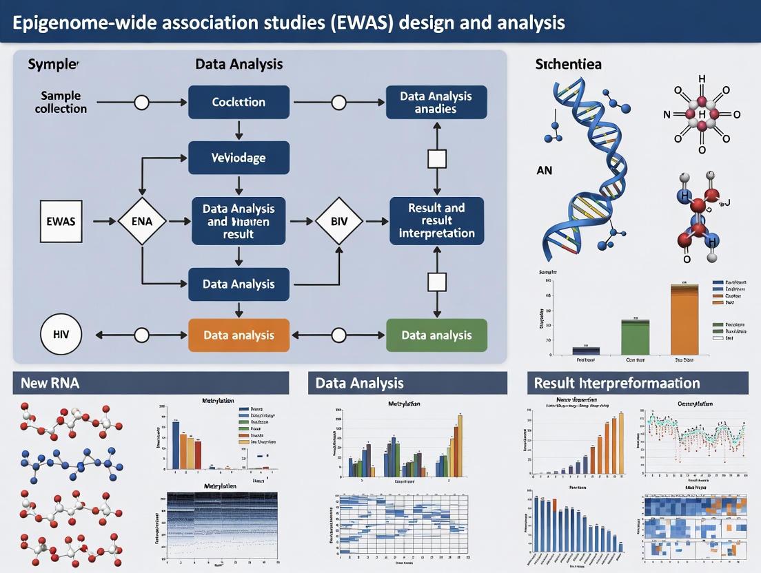
Figure 1: Core Workflow for EWAS Data Analysis
Two primary bioinformatics packages have emerged as standards for EWAS analysis: Minfi and Chip Analysis Methylation Pipeline (ChAMP) [2]. Both packages support the entire analytical workflow from raw data import to identification of differentially methylated positions (DMPs) and regions (DMRs), with ChAMP becoming increasingly prominent for EPIC array data analysis [2]. Additional specialized analyses often integrated into EWAS include:
- Methylation Quantitative Trait Loci (methQTL) analysis: Identifies genetic variants that influence methylation patterns [2]
- Statistical deconvolution methods: Estimates cell-type specific methylation from heterogeneous tissue samples [2]
- Methylation age analysis: Evaluates epigenetic clocks as biomarkers of biological aging [2]
- Mendelian Randomization: Provides causal inference between methylation and disease outcomes [3]
Table 2: Key Bioinformatics Tools for EWAS Analysis
| Tool/Package | Primary Function | Compatible Platforms | Key Features |
|---|---|---|---|
| Minfi | Data preprocessing and analysis | 450K, EPIC | Most cited for 450K data; comprehensive quality control and normalization [2] |
| ChAMP | Integrated analysis pipeline | 450K, EPIC | Growing popularity for EPIC data; combines multiple analysis steps [2] |
| MEFFIL | Quality control and normalization | 450K, EPIC | Functional normalization; cell type composition estimation [5] |
| WaterRmelon | Preprocessing and analysis | 450K, EPIC | BMIQ normalization for probe-type bias correction [4] [5] |
Research Reagent Solutions for EWAS
Successful execution of EWAS requires specific research reagents and materials throughout the experimental workflow. The following table outlines essential solutions and their applications:
Table 3: Essential Research Reagents for EWAS Experiments
| Reagent/Material | Function/Application | Technical Considerations |
|---|---|---|
| Bisulfite Conversion Kits (e.g., EZ-96 DNA Methylation Kit) | Chemical treatment that converts unmethylated cytosines to uracil while methylated cytosines remain unchanged [4] [5] | Conversion efficiency must be verified; over-treatment can degrade DNA [1] |
| Infinium Methylation BeadChips (27K, 450K, EPIC) | Genome-wide methylation profiling using probe hybridization [1] [2] | Platform selection depends on coverage needs and budget; EPIC recommended for enhancer regions [1] |
| DNA Extraction Kits | Isolation of high-quality genomic DNA from biological samples | Yield and purity critical; salting-out protocols commonly used [5] |
| Cell Type Composition Reference Panels | Reference-based estimation of cellular heterogeneity in blood samples [2] [4] | Essential for blood-based EWAS; implemented in Houseman's method [4] [5] |
| Normalization Controls | Technical variation adjustment during data processing | Included in platforms or added during analysis (e.g., NOOB, BMIQ) [4] [5] |
Experimental Design Considerations
EWAS can be implemented through various study designs, each with distinct advantages and limitations. The most common approaches include:
Case-Control Design
The case-control design is the most frequently employed approach in EWAS, comparing methylation patterns between individuals with a specific phenotype (cases) and those without (controls) [2]. This design is logistically feasible and cost-effective, allowing researchers to leverage existing DNA biobanks from previous studies [2]. The primary limitation is the inability to establish temporal relationships, making it difficult to determine whether methylation differences precede or result from the disease state [2].
Longitudinal Design
Longitudinal studies measure methylation at multiple timepoints within the same individuals, enabling the assessment of intra-individual changes over time [2]. This design is particularly valuable for understanding dynamic epigenetic processes throughout the lifespan, such as the extensive methylome remodeling that occurs during early childhood [2]. While logistically challenging and costly, longitudinal designs provide stronger evidence for causal inferences and can track methylation trajectories in relation to disease progression [2].
Specialized Design Considerations
Additional design considerations include family-based studies to estimate heritable components of methylation, twin studies to distinguish genetic and environmental influences, and integrated omics designs that combine EWAS with GWAS, transcriptomics, or proteomics data [2]. Each design requires specific analytical approaches to address potential confounding factors, particularly cell type composition in heterogeneous tissues like blood [2] [4].
Advanced Applications and Case Studies
EWAS of Clonal Hematopoiesis (CHIP)
A recent large-scale EWAS of clonal hematopoiesis of indeterminate potential (CHIP) illustrates the power of this approach in elucidating disease mechanisms [3]. This multiracial meta-analysis included 8,196 participants from four cohorts and identified distinct methylation signatures associated with different CHIP driver genes:
Figure 2: Integrated Workflow for CHIP EWAS Case Study
The study revealed that DNMT3A CHIP mutations were associated with widespread hypomethylation (5,987 of 5,990 CpGs), consistent with DNMT3A's role as a de novo methyltransferase [3]. In contrast, TET2 CHIP mutations showed predominantly hypermethylation (5,079 of 5,633 CpGs), aligning with TET2's function as a demethylase [3]. These findings were functionally validated using CRISPR-Cas9 engineered human hematopoietic stem cell models, demonstrating the mechanistic insights achievable through integrated EWAS approaches [3].
EWAS of Physical Activity
An EWAS of objectively measured physical activity demonstrated the application of this methodology to environmental exposures and lifestyle factors [5]. This study analyzed associations between sedentary behavior, moderate physical activity, and methylation patterns in pregnant women, identifying 122 CpG sites associated with moderate physical activity after adjusting for steps per day [5]. The study highlights challenges in EWAS of complex behaviors, including the need for precise exposure measurement and consideration of potential confounding factors [5].
Methodological Challenges and Solutions
EWAS faces several methodological challenges that require careful consideration in both study design and analysis:
Addressing Population Stratification
Similar to GWAS, population stratification can cause spurious associations in EWAS if not properly accounted for [4]. Traditional approaches use genetic principal components as covariates, but when genetic data are unavailable, methylation-based alternatives have been developed. Recent methodologies include methylation population scores (MPS), which use supervised learning to predict genetic ancestry from methylation data while adjusting for technical and environmental covariates [4]. These scores effectively capture population structure and can reduce test statistic inflation in EWAS of diverse populations [4].
Cell Type Heterogeneity
Cell type composition represents a major confounding factor in tissue-based EWAS, particularly in blood where methylation patterns vary substantially between leukocyte subsets [2] [4]. Reference-based estimation methods, such as Houseman's algorithm, use cell-type specific methylation signatures to deconvolute heterogeneous samples and estimate proportional composition [4] [5]. These estimates should be included as covariates in association analyses to avoid false positives arising from cellular heterogeneity rather than the phenotype of interest [2].
Reverse Causation and Causal Inference
A fundamental limitation of observational EWAS is the challenge of distinguishing cause from effect—whether methylation differences contribute to disease or result from disease processes [2]. Several approaches address this limitation:
- Longitudinal designs: Measure methylation before disease onset to establish temporal sequence [2]
- Mendelian randomization: Uses genetic variants as instrumental variables to infer causal relationships [3]
- Family-based designs: Control for shared genetic and environmental backgrounds [2]
- Integration with functional genomics: Combines EWAS with gene expression and mechanistic studies [3]
EWAS has matured into an essential component of functional genomics, providing unique insights into the molecular mechanisms through which genetic and environmental factors jointly influence complex traits and diseases. The continuing evolution of technologies—from microarrays to comprehensive sequencing approaches—promises enhanced coverage of regulatory elements and more precise mapping of methylation patterns [1]. Future directions include the integration of multi-omics data, development of single-cell epigenetic protocols, and application of machine learning approaches to identify complex epigenetic signatures of disease [1] [2].
The translation of EWAS findings into clinical applications continues to advance, with epigenetic biomarkers showing promise for disease risk prediction, diagnosis, and monitoring of therapeutic responses [1] [3]. As the field progresses, standardization of methodologies, improved reference datasets, and collaborative meta-analyses will further strengthen the robustness and reproducibility of EWAS discoveries across diverse populations and disease contexts [2] [4].
DNA Methylation as the Primary Epigenetic Marker in EWAS
DNA methylation (DNAm), characterized by the addition of a methyl group to a cytosine base in a CpG dinucleotide context, serves as a fundamental epigenetic mark that regulates gene expression without altering the underlying DNA sequence [6] [7]. This modification represents a crucial molecular interface that mediates the interaction between genetic predisposition and environmental exposures, providing critical insights into the pathophysiology of complex diseases [6] [2]. Epigenome-wide association studies (EWAS) systematically investigate genome-wide epigenetic variation to identify associations between DNA methylation patterns and phenotypes, environmental exposures, or disease states [8]. The viability of EWAS has been propelled by rapid advancements in high-throughput measurement technologies, particularly the Illumina Infinium DNA methylation BeadChip microarrays, which enable feasible methylation profiling at a near-genome-wide scale [6] [9].
The selection of DNA methylation as the primary epigenetic marker in EWAS is grounded in its stability, quantifiable nature, and well-characterized functional consequences. DNA methylation patterns are dynamic throughout the lifespan and exhibit tissue-specific signatures, yet remain sufficiently stable to yield reproducible associations in large-scale studies [7] [2]. As the most extensively studied epigenetic mechanism, DNA methylation provides a measurable molecular footprint of both genetic influences and environmental exposures, making it an ideal biomarker for investigating complex disease etiology [2].
Technological Platforms for Methylation Assessment
The evolution of microarray technologies has dramatically expanded the scope and precision of EWAS. The progression from the HumanMethylation27 (27K) to the HumanMethylation450 (450K) and subsequently to the MethylationEPIC (850K) arrays has substantially improved genomic coverage, particularly in regulatory regions beyond promoter-associated CpG islands [2]. The most recent innovation, the Methylation Screening Array (MSA), represents a strategic advance by concentrating coverage on trait-associated methylation signatures and cell-identity-associated methylation variations, achieving approximately 5.6 trait associations per site compared to approximately 2.2 in EPICv2 [9]. This targeted design enhances efficiency for large-scale population studies while maintaining critical biological information.
Table 1: Comparison of Illumina Methylation BeadChip Platforms
| Platform | CpG Coverage | Key Features | Primary Applications |
|---|---|---|---|
| 27K | ~27,000 CpGs | Focus on promoter regions | Early EWAS, candidate gene validation |
| 450K | ~450,000 CpGs | Expanded coverage to gene bodies, intergenic regions | Mainstream EWAS, meQTL studies |
| EPIC/EPICv2 | ~850,000 CpGs | Enhanced coverage of enhancer regions (58% of FANTOM enhancers) | Comprehensive EWAS, regulatory element mapping |
| MSA | ~284,000 CpGs | Enriched for trait-associated loci (~5.6 traits/site); high-throughput 48-sample format | Population-scale screening, epigenetic clock applications |
For comprehensive methylation analysis, whole-genome bisulfite sequencing (WGBS) remains the gold standard, providing base-resolution data across the entire methylome [7]. However, this method remains cost-prohibitive for large cohort studies. Reduced representation bisulfite sequencing (RRBS) offers a cost-effective alternative by targeting CpG-rich regions, while emerging technologies like single-cell whole-genome methylation sequencing (scWGMS) are unlocking cellular heterogeneity but with limitations in sample throughput [9].
Experimental Workflow and Protocols
Standardized EWAS Workflow
A robust EWAS requires meticulous attention to experimental design, sample processing, and computational analysis. The following workflow diagram outlines the critical stages in a comprehensive EWAS investigation:
Sample Preparation and Bisulfite Conversion Protocol
Principle: Bisulfite conversion deaminates unmethylated cytosines to uracils, while methylated cytosines remain unchanged, allowing for discrimination based on methylation status [7] [2].
Procedure:
- DNA Quantification and Quality Assessment: Quantify DNA using fluorometric methods and assess purity (A260/280 ratio ~1.8-2.0). Ensure DNA integrity (DNA Integrity Number >7) for reliable results.
- Bisulfite Conversion: Use commercial bisulfite conversion kits with optimized protocols. Typical reaction conditions: 95°C for 30-60 seconds (denaturation), 50-60°C for 45-60 minutes (conversion), followed by clean-up.
- Conversion Efficiency Check: Include control DNA with known methylation patterns. Assess conversion efficiency through PCR amplification of non-CpG cytosines, which should be fully converted.
- Microarray Processing: Process bisulfite-converted DNA on selected Illumina BeadChip according to manufacturer's specifications, including amplification, fragmentation, hybridization, and scanning.
Technical Notes: Incomplete bisulfite conversion represents a major source of technical artifacts. Incorporate both unmethylated and fully methylated control DNA in each processing batch to monitor conversion efficiency [7].
Data Preprocessing and Quality Control Protocol
Software Implementation: Utilize established R packages such as minfi, ChAMP, or MethylCallR for standardized processing [10] [2].
Quality Control Steps:
- Signal Intensity Review: Remove samples with low intensity (detection p-value > 0.01 in >5% of probes).
- Probe Filtering: Exclude probes with:
- Detection p-value > 0.01 in >5% of samples
- Cross-reactive probes (mapping to multiple genomic locations)
- Probes overlapping SNPs at the CpG site or single-base extension
- Sex chromosome probes for autosomal-only analyses
- Normalization: Apply appropriate normalization methods (e.g., BMIQ, SWAN, Noob) to correct for technical variation between probe types and array positions.
- Batch Effect Correction: Implement ComBat or other empirical Bayesian methods to adjust for technical covariates (array, row, processing batch) [6].
- Outlier Detection: Use multidimensional scaling (MDS) and hierarchical clustering to identify sample outliers. Implement Mahalanobis distance methods to detect potential outlier samples within groups [10].
Cell Type Composition Estimation
Background: Tissue heterogeneity represents a major confounding factor in EWAS, particularly in blood-based studies where cellular composition varies substantially between individuals [2] [11].
Implementation:
- Reference-Based Deconvolution: Utilize established reference methylomes for purified cell types (e.g.,
Flowsorted.Blood.EPICfor blood samples) to estimate proportional composition [10]. - Reference-Free Methods: Apply methods such as MeDeCom, RefFreeCellMix, or EDec when appropriate reference datasets are unavailable [11].
- Statistical Adjustment: Include estimated cell type proportions as covariates in differential methylation analyses to account for heterogeneity effects.
Analytical Frameworks for Differential Methylation
Differential Methylation Position (DMP) Analysis
DMP analysis identifies individual CpG sites with statistically significant differences in methylation levels associated with the phenotype of interest. The easyEWAS package provides a battery of statistical methods tailored to different study designs [6]:
Table 2: Statistical Models for DMP Analysis in EWAS
| Model Type | Formula | Application Context | Output Metrics | |
|---|---|---|---|---|
| General Linear Model (GLM) | CpG = β₀ + β₁X₁ + β₂X₂ + ... + ε |
Case-control studies, continuous exposures | Regression coefficient (β), Standard Error, P-value | |
| Linear Mixed-Effects Model (LMM) | CpG = β₀ + β₁X₁ + ... + u + ε |
Longitudinal studies, repeated measures | β, SE, P-value with random effects (u) | |
| Cox Proportional Hazards (CoxPH) | `h(t | X) = h₀(t)exp(β₁CpG + ...)` | Time-to-event analysis, survival outcomes | Hazard Ratio (HR), 95% CI, P-value |
Implementation Protocol:
- Model Specification: Select appropriate statistical model based on study design. Adjust for relevant covariates including age, sex, batch effects, and estimated cell type proportions.
- Genome-Wide Analysis: Perform site-by-site analysis across all qualified CpG sites.
- Multiple Testing Correction: Apply Benjamini-Hochberg False Discovery Rate (FDR) or Bonferroni correction to account for multiple comparisons. A standard epigenome-wide significance threshold is P < 1×10⁻⁷ [3] [8].
- Effect Size Calculation: Report methylation differences as Δβ values (for β-values) or M-value coefficients, with typical biologically relevant effect sizes considered as |Δβ| ≥ 0.05 [10].
Differential Methylation Region (DMR) Analysis
DMR analysis identifies genomic regions containing multiple adjacent DMPs, often providing more biologically meaningful and robust findings than single CpG associations [6].
DMRcate Protocol:
- Initial Screening: Perform limma-based regression at each CpG site to generate moderated t-statistics and p-values.
- Gaussian Smoothing: Apply kernel smoothing to average effects across neighboring CpGs within a specified window (default: 1000 base pairs).
- Region Definition: Group adjacent CpG sites exceeding significance and effect size thresholds.
- Annotation: Annotate significant DMRs with genomic context (promoter, gene body, intergenic) and proximity to genes.
Bootstrap Internal Validation
To ensure robustness of EWAS findings, implement bootstrap resampling validation:
Procedure:
- Generate multiple resampled datasets (typically 1000+ iterations) through random sampling with replacement.
- Recalculate association statistics for each resampled dataset.
- Derive confidence intervals for regression coefficients using preferred method (percentile, studentized, or bias-corrected).
- Assess stability of significant DMPs across bootstrap iterations [6].
Advanced Analytical Concepts
Ternary-Code DNA Methylation Dynamics
Emerging research recognizes the importance of distinguishing between different cytosine modifications in the "ternary-code" - 5-methylcytosine (5mC), 5-hydroxymethylcytosine (5hmC), and unmodified cytosine [9] [12]. This distinction is crucial as 5hmC represents an intermediate in active demethylation pathways and has distinct genomic distributions and functional consequences.
Profiling Protocol:
- bACE-seq: Apply bisulfite APOBEC-coupled epigenetic sequencing to discriminate 5mC from 5hmC.
- OxBS-seq: Utilize oxidative bisulfite sequencing for precise quantification of 5hmC.
- MSA with Modified Chemistry: Implement the methylation screening array with enhanced chemistry to capture 5hmC signatures [9].
The following diagram illustrates the ternary-code methylation concept and its functional implications:
Integration with Multi-Omics Data
Methylation Quantitative Trait Loci (meQTL) Analysis:
- Identify genetic variants associated with methylation variation.
- Assess cis-meQTLs (within 1Mb of CpG) and trans-meQTLs (distant associations).
- Integrate with GWAS findings to identify potential epigenetic mechanisms underlying genetic associations [2].
Expression Quantitative Trait Methylation (eQTM) Analysis:
- Correlate methylation levels with gene expression data from the same samples.
- Identify potentially regulatory relationships between methylation and transcription [3].
Mendelian Randomization:
- Utilize genetic instruments to infer causal relationships between methylation and disease outcomes.
- Apply two-sample MR approaches with summary statistics from large-scale EWAS and disease GWAS [3].
Research Reagent Solutions
Table 3: Essential Research Reagents and Computational Tools for EWAS
| Category | Specific Tool/Reagent | Function/Application | Implementation Notes |
|---|---|---|---|
| Microarray Platforms | Illumina EPICv2 BeadChip | Genome-wide methylation profiling (∼850,000 CpGs) | Balanced coverage of promoters, enhancers, gene bodies |
| Methylation Screening Array (MSA) | High-throughput trait association screening | 48-sample format; enriched for EWAS associations | |
| Bisulfite Conversion Kits | EZ DNA Methylation kits (Zymo Research) | Convert unmethylated C to U while preserving 5mC | Critical for accurate methylation quantification |
| Computational Packages | Minfi | Preprocessing, normalization, QC of array data | Most cited for 450K data analysis |
| ChAMP | Comprehensive analysis pipeline | Increasingly cited for EPIC data analysis | |
| easyEWAS | User-friendly DMP and DMR analysis | Supports GLM, LMM, CoxPH models; bootstrap validation | |
| MethylCallR | EPICv2-compatible analysis framework | Handles duplicated probes; version conversion | |
| DMRcate | Differentially methylated region identification | Gaussian kernel smoothing approach | |
| Reference Datasets | FlowSorted.Blood.EPIC | Blood cell composition estimation | Reference-based deconvolution for blood samples |
| MeDeCom | Reference-free deconvolution | Identifies latent methylation components | |
| Functional Annotation | missMethyl | Gene set enrichment analysis | Accounts for probe number bias in array design |
Interpretation and Validation Guidelines
Functional Validation Strategies
Experimental Validation:
- Targeted Bisulfite Sequencing: Confirm top DMPs/DMRs using pyrosequencing or deep amplicon sequencing.
- In Vitro Models: Utilize CRISPR-Cas9 to introduce specific methylation changes in cell lines and assess functional consequences [3].
- Functional Assays: Perform luciferase reporter assays to assess regulatory potential of methylated regions.
Biological Interpretation:
- Genomic Context Analysis: Annotate significant CpGs with genomic features (promoters, enhancers, CpG islands, shores, shelves).
- Pathway Enrichment: Conduct gene set enrichment analysis using tools like
gomethto identify overrepresented biological pathways. - Integration with Public Resources: Compare findings with databases such as ENCODE, Roadmap Epigenomics, and GWAS catalog to prioritize functionally relevant hits.
Reporting Standards
Comprehensive EWAS reporting should include:
- Detailed sample characteristics and inclusion/exclusion criteria
- Complete description of preprocessing and normalization methods
- Cell type composition estimates and adjustment approach
- Multiple testing correction method and significance thresholds
- Effect sizes with confidence intervals for top associations
- Validation approaches and results
- Functional annotation of significant findings
DNA methylation profiling remains the cornerstone of epigenome-wide association studies, providing powerful insights into the molecular mechanisms linking genetic predisposition, environmental exposures, and disease phenotypes. The continued refinement of measurement technologies, analytical frameworks, and interpretation tools has established EWAS as an essential component of comprehensive biomedical research. By adhering to standardized protocols, implementing appropriate statistical methods, and applying rigorous validation strategies, researchers can leverage DNA methylation as a robust epigenetic marker to advance understanding of complex disease etiology and identify potential therapeutic targets.
Differentially Methylated Positions (DMPs) and Regions (DMRs)
Core Concepts and Biological Significance
Differentially Methylated Positions (DMPs) are individual cytosine-guanine dinucleotide (CpG) sites that exhibit statistically significant differences in methylation status between biological samples from distinct conditions (e.g., diseased versus normal, treated versus untreated) [13]. The methylation level at a single CpG site is typically quantified as a beta value (β), calculated as β = M/(M + U + α), where M represents the methylated allele intensity, U the unmethylated allele intensity, and α a constant offset (usually 100) to prevent division by zero [14]. DMP analysis provides high-resolution data but may miss broader, coordinated epigenetic patterns.
Differentially Methylated Regions (DMRs) are genomic segments, often spanning hundreds of base pairs, that contain multiple CpG sites showing consistent, statistically significant methylation differences between sample groups [15]. DMRs are regarded as possible functional regions involved in gene transcriptional regulation and provide a more biologically stable signature than single CpG sites, as they are less susceptible to technical noise [15] [13]. They are critical hallmarks of genomic imprinting, where they confer parent-of-origin-specific transcription, and are involved in normal human growth and neurodevelopment [16].
The following table summarizes the core characteristics and identification criteria for DMPs and DMRs.
Table 1: Defining Characteristics and Analysis Criteria for DMPs and DMRs
| Feature | Differentially Methylated Position (DMP) | Differentially Methylated Region (DMR) |
|---|---|---|
| Definition | A single CpG site with significant methylation difference between conditions [13]. | A genomic region with multiple CpGs showing consistent differential methylation [15]. |
| Typical Scope | Single nucleotide. | 50 bp to several kilobases. |
| Biological Significance | Point-specific epigenetic alteration; potential as a biomarker. | Stronger functional implication; often associated with regulatory elements like promoters and enhancers [15]. |
| Common Identification Criteria | Statistical test (e.g., t-test) with FDR correction; minimum methylation difference (e.g., Δβ ≥ 0.1) [17] [18]. | Multiple adjacent significant CpGs; minimum region length (e.g., 50 bp); statistical significance of the entire region [17] [13]. |
| Example Thresholds | FDR < 0.05, Δβ ≥ 0.1 [17]. | ≥ 3-5 CpGs, distance between CpGs ≤ 300 bp, MWU-test p-value < 0.05 [17] [13]. |
Analytical Workflows and Methodologies
The process of identifying DMPs and DMRs involves a multi-step workflow, from experimental profiling to computational analysis, with the specific approach varying based on the technology used.
Profiling Technologies and Data Acquisition
The choice of profiling technology dictates the scope and resolution of the methylation data.
Table 2: Key Technologies for Genome-Wide DNA Methylation Profiling
| Technology | Principle | Throughput | Resolution & Coverage | Primary Use Case |
|---|---|---|---|---|
| Infinium Methylation BeadChip (e.g., EPIC, MSA) [19] [9] | Hybridization of bisulfite-converted DNA to array probes. | High | Base-specific; ~850,000 to ~280,000 pre-selected CpG sites. | Large-scale EWAS, biomarker discovery. |
| Whole-Genome Bisulfite Sequencing (WGBS) [19] | Sequencing following bisulfite conversion, which turns unmethylated cytosines to uracils. | Low | Base-specific; genome-wide. | Comprehensive discovery, novel DMR identification. |
| Reduced Representation Bisulfite Sequencing (RRBS) [19] | Restriction enzyme digestion followed by bisulfite sequencing. | Medium | Base-specific; covers ~85% of CpG islands, primarily in promoters. | Cost-effective targeted analysis. |
The workflow for analyzing data from these technologies, particularly from sequencing-based methods like WGBS and RRBS, follows a structured pipeline to ensure robust results, as illustrated below.
Detailed Protocol: DMP and DMR Analysis from WGBS/RRBS Data
This protocol provides a step-by-step guide for analyzing Bismark-generated coverage files in R to identify DMPs and DMRs [17].
1. Prerequisite: Set Up the R Environment
- Install and load the required Bioconductor packages.
2. Load and Organize Methylation Data
- Read Bismark coverage files and create a BSseq object for analysis.
3. Perform Differential Analysis
- DMP Detection using DSS: The DSS package uses a beta-binomial model to account for biological variation and over-dispersion in count data.
- DMR Detection using dmrseq: This package identifies DMRs by assessing the spatial autocorrelation of methylation differences across the genome.
Detailed Protocol: Analysis of Illumina Infinium BeadChip Data
The analysis of array-based methylation data requires specific steps to handle platform-specific biases, such as those arising from the two different probe types (Infinium I and II) [14].
1. Data Import and Quality Control (QC)
- Import raw IDAT files or preprocessed TXT files using the
minfipackage. - Perform rigorous QC:
- Filter probes with a high detection p-value (e.g., > 0.01).
- Remove probes on sex chromosomes (X, Y) to avoid gender bias.
- Exclude probes known to contain single nucleotide polymorphisms (SNPs) or those that are cross-reactive [14].
2. Normalization and Type Bias Correction
- Apply within-array normalization for background correction and dye bias adjustment.
- Correct for the technical bias between Infinium I and II probes. The Beta Mixture Quantile normalization (BMIQ) method is a robust choice, as it calibrates the distribution of Infinium II probes to match that of the more stable Infinium I probes [14].
3. Differential Methylation Calling
- DMPs can be identified using linear models with the
limmapackage, which employs moderated t-statistics to enhance power in studies with small sample sizes [15] [14]. - DMRs can be called by aggregating nearby DMPs using packages like
BumphunterorDMRcate.
The Scientist's Toolkit: Research Reagent Solutions
Table 3: Essential Reagents and Resources for DMP/DMR Analysis
| Category | Item | Function and Application Notes |
|---|---|---|
| Commercial Kits | Gentra Puregene Kit (Qiagen) [18] | For DNA isolation from whole blood samples, ensuring high-quality input material. |
| PAXgene Blood RNA Kit (Qiagen) [18] | For RNA isolation, enabling integrated methylation and gene expression analysis. | |
| Illumina TotalPrep RNA Amplification Kit [18] | For synthesizing cRNA for gene expression beadchips. | |
| Bisulfite Conversion | Zymo Research EZ DNA Methylation Kits | Converts unmethylated cytosines to uracils while leaving methylated cytosines intact, a critical step for most profiling methods. |
| Microarray Platforms | Infinium MethylationEPIC BeadChip [18] [9] | Interrogates over 850,000 CpG sites; the EPIC version offers extensive coverage of regulatory regions. |
| Methylation Screening Array (MSA) [9] | The latest array design, highly enriched for trait-associated loci from EWAS, enabling ultra-high sample throughput. | |
| Critical Software & Databases | R/Bioconductor [17] [14] | The primary environment for statistical analysis and visualization (e.g., with packages like minfi, DSS, dmrseq). |
| Reference Genomes (UCSC, ENSEMBL) [19] | Essential for the alignment of sequencing reads and annotation of identified DMPs/DMRs. | |
| Public Repositories (GEO, TCGA) [19] | Sources for validation and comparison with public methylation datasets. |
Advanced Applications in Drug Development and Clinical Research
The identification of DMPs and DMRs has moved beyond basic research into applied clinical and pharmaceutical contexts.
Biomarker Discovery for Disease Diagnosis and Prognosis: Aberrant DNA methylation sites can function as powerful biomarkers for disease. For example, specific DMRs between cancer and normal samples demonstrate the aberrant methylation that is a hallmark feature of many cancers [19] [15]. These biomarkers can be used for early detection, molecular subtyping of diseases, and developing liquid biopsy-based diagnostics [19] [9].
Elucidating Mechanisms of CHIP and Cardiovascular Disease: Clonal hematopoiesis of indeterminate potential (CHIP) is an age-related condition driven by mutations in genes like
DNMT3AandTET2, which are epigenetic regulators. A large EWAS revealed thatDNMT3AandTET2CHIP mutations have directionally opposing DNA methylation signatures, consistent with their canonical functions, and these changes are associated with increased cardiovascular disease risk [3]. This provides critical insight into the molecular mechanisms linking CHIP to age-related diseases.Understanding Environmental and Lifestyle Exposures: EWAS investigates how factors like alcohol consumption influence the epigenome. A 2025 study identified 19,255 CpG sites associated with alcohol consumption, with over-representation of genes involved in cancer, the nervous system, and aging [20]. This helps in understanding the molecular mechanisms underlying the harmful effects of environmental exposures.
The process from foundational analysis to clinical application involves integrating multiple data types to build a compelling case for a biomarker or drug target, as shown in the following workflow.
The Dynamic Interplay Between Genetics, Environment, and the Epigenome
Epigenome-wide association studies (EWAS) represent a powerful methodological framework for investigating the interface at which genetic predisposition and environmental exposures interact to influence complex disease risk and outcomes [2] [21]. Unlike genetic variants, which remain static throughout life, epigenetic modifications are dynamic and reversible, reflecting both inherited factors and lifetime environmental experiences [22]. The primary aim of an EWAS is to examine genome-wide epigenetic variants, predominantly DNA methylation at cytosine-phosphate-guanine (CpG) dinucleotides, to detect statistically significant differences associated with phenotypes of interest [2]. These studies have emerged as a complementary approach to genome-wide association studies (GWAS), providing insights into the molecular mechanisms through which both genetic and environmental factors converge to influence health and disease [21].
The most extensively studied epigenetic marker in EWAS is DNA methylation, which involves the covalent addition of a methyl group to the 5-carbon position of cytosine residues, primarily within CpG dinucleotides [22]. This modification can regulate gene expression by altering transcription factor binding or recruiting methyl-binding proteins that remodel chromatin structure [22]. Modern EWAS primarily utilizes array-based technologies such as the Illumina Infinium HumanMethylation450 BeadChip (450K) and the more recent MethylationEPIC BeadChip (EPIC), which Interrogate approximately 450,000 and 850,000 CpG sites respectively [2] [22]. The measurement output is typically represented as beta-values ranging from 0 (completely unmethylated) to 1 (fully methylated), quantifying the methylation fraction at each CpG site [5] [22].
Current Research Landscape in EWAS
Key Application Areas
EWAS approaches have been successfully applied to diverse research areas, illuminating how various exposures and biological processes epigenetically regulate gene expression. The table below summarizes prominent EWAS application areas, their specific focuses, and key findings from recent studies.
Table 1: Key Application Areas of Epigenome-Wide Association Studies
| Application Area | Specific Focus | Key Findings | Representative Studies |
|---|---|---|---|
| Clonal Hematopoiesis | CHIP (Clonal Hematopoiesis of Indeterminate Potential) | Identification of 9615 CpGs associated with any CHIP; DNMT3A and TET2 mutations show opposing methylation patterns [3] | Multiracial meta-analysis (N=8196) [3] |
| Bone Diseases | Osteoporosis and osteoarthritis | Identification of differentially methylated regions in osteoporosis and osteoarthritis [23] | Delgado-Calle et al. (2013) [23] |
| Nutritional Exposure | Dietary patterns, specific foods, micronutrients | Consistent associations at 9 CpG sites (AHRR, CPT1A, FADS2) with fatty acid consumption [22] | Scoping review of 30 studies [22] |
| Physical Activity | Objectively measured sedentary behavior and moderate activity | Association of 122 CpG sites with moderate physical activity after adjustment for steps/day [5] | EPIPREG cohort (n=353) [5] |
| Substance Exposure | Smoking and vaping | Identification of differentially methylated regions using Bonferroni-significance threshold of p < 5.91 × 10⁻⁸ [24] | EWAS protocol for vaping vs. non-smokers [24] |
Analytical Approaches in EWAS
The analytical workflow in EWAS encompasses multiple stages, from quality control to advanced statistical analyses. Two main bioinformatics packages—Minfi and ChAMP—have emerged as open-source tools for processing and analyzing methylation array data [2]. These packages allow researchers to import raw data files, perform quality control, normalization, and detect both differentially methylated positions (DMPs) and regions (DMRs) [2]. Downstream analyses may include methylation quantitative trait loci (methQTL) analysis to identify genetic variants influencing methylation patterns, expression quantitative trait methylation (eQTM) analysis to link methylation changes with gene expression, and causal inference methods like Mendelian randomization to infer potential causal relationships between methylation and disease [3] [2].
Table 2: Common Analytical Approaches in EWAS
| Analytical Method | Purpose | Key Features | Tools/Packages |
|---|---|---|---|
| Quality Control | Identify poor-quality samples and probes | Filtering based on detection p-values, bead count, removal of cross-reactive and SNP-containing probes [5] | Meffil [5], Minfi [2] |
| Normalization | Remove technical variation while preserving biological signals | Functional normalization using control probes or reference datasets [5] | Meffil [5], ChAMP [2] |
| DMP Identification | Find individual CpGs associated with traits | Linear regression with multiple testing correction (Bonferroni, FDR) [2] [24] | Minfi, ChAMP, standard statistical software |
| DMR Identification | Identify genomic regions with coordinated methylation changes | Regions containing ≥2 CpGs within 500bp with consistent effects [24] | dmrff R package [24] |
| Cell Type Deconvolution | Estimate cell-type proportions in mixed samples | Reference-based estimation using cell-type specific methylation markers [2] | Houseman's method [5] |
| Causal Inference | Infer potential causal relationships | Mendelian randomization using genetic instruments [3] [2] | Two-sample MR methods |
Experimental Protocols for EWAS
Multi-Cohort EWAS on Clonal Hematopoiesis
Study Design and Participant Recruitment
This protocol outlines the methods for a recent large-scale EWAS investigating the epigenetic signatures of clonal hematopoiesis of indeterminate potential (CHIP) [3]. The study employed a multiracial meta-analysis design, pooling data from four independent cohort studies: the Framingham Heart Study (FHS), Jackson Heart Study (JHS), Cardiovascular Health Study (CHS), and Atherosclerosis Risk in Communities (ARIC) study, with a total sample size of N = 8,196 participants (462 with any CHIP, 261 with DNMT3A CHIP, 84 with TET2 CHIP, and 21 with ASXL1 CHIP) [3]. Participant characteristics included mean ages ranging from 56-74 years, with a higher proportion of women (54-63%) across all cohorts. CHIP mutations with a variant allele frequency (VAF) ≥ 2% were present in 4-15% of participants across cohorts, with the three most frequently mutated CHIP driver genes being DNMT3A, TET2, and ASXL1 [3].
Laboratory Methods
DNA Methylation Processing: DNA methylation was quantified using the Infinium MethylationEPIC BeadChip (Illumina, San Diego, California, USA), which measures the proportion of methylation at approximately 850,000 CpG sites, generating beta-values ranging from 0 to 1 [5]. Quality control procedures included:
- Removal of sample outliers based on methylated/unmethylated ratio (> 3SD)
- Exclusion of outliers in bisulfite control probes (> 5 SD)
- Filtering of probes with detection p-value < 0.01 and bead count < 3
- Omission of probes on sex chromosomes, cross-reactive probes, and probes containing single nucleotide polymorphisms (SNPs) [5]
Functional Validation: EWAS findings were validated using human hematopoietic stem cell (HSC) models of CHIP. Loss-of-function mutations in DNMT3A, TET2, and ASXL1 were introduced into mobilized peripheral blood CD34+ hematopoietic cells using CRISPR-Cas9 [3]. After seven days in culture, CD34+CD38-Lin- cells were isolated using fluorescence-activated cell sorting, genomic DNA was extracted, and methylation was assayed using biomodal duet evoC [3].
Statistical Analysis
The analysis employed race-stratified epigenome-wide association analyses followed by multiracial meta-analysis [3]. Key analytical steps included:
- Association Testing: Multivariable linear regression at each CpG site, adjusting for age, sex, genetic ancestry, and estimated blood cell composition [3]
- Multiple Testing Correction: Bonferroni-corrected significance threshold of P < 1×10^-7 [3]
- Meta-Analysis: Fixed-effects meta-analysis of race-stratified results using inverse variance weighting
- Sensitivity Analyses: Exclusion of CHIP cases with VAF < 10% to assess robustness of findings
- eQTM Analysis: Expression quantitative trait methylation analysis to identify transcriptomic changes associated with CHIP-associated CpGs
- Causal Inference: Two-sample Mendelian randomization to investigate potential causal relationships between CHIP-associated CpGs and cardiovascular traits [3]
EWAS of Objectively Measured Physical Activity
Study Design and Physical Activity Measurement
This protocol describes methods for an EWAS investigating associations between objectively measured physical activity and DNA methylation in peripheral blood leukocytes [5]. The discovery analysis was conducted in pregnant women from the Epigenetics in Pregnancy (EPIPREG) cohort, including 244 European and 109 South Asian women with both DNA methylation and objectively measured physical activity data [5].
Physical Activity Assessment: Physical activity was measured using the SenseWear Pro3 armband (BodyMedia Inc, Pittsburgh, PA, USA) at approximately gestational week 28. Participants wore the device continuously for 4-7 days, excluding water activities. Data were analyzed using manufacturer software (SenseWear Professional Research Software Version 6.1), with valid day defined as ≥ 19.2 hours of wear time [5]. The analysis extracted:
- Number of steps per day
- Mean hours/day of moderate-intensity physical activity (MPA) (3.0-6.0 METs)
- Sedentary behavior (SB) (< 1.5 METs) [5]
Laboratory Methods
DNA Methylation Quantification: DNA methylation was assessed in peripheral blood leukocytes using the Infinium MethylationEPIC BeadChip (Illumina) [5]. Quality control procedures implemented in the Meffil R package included:
- Removal of 6 sample outliers based on methylated/unmethylated ratio (> 3SD)
- Exclusion of 1 outlier in bisulfite control probes (> 5 SD)
- Removal of 1 sample with sex mismatch
- Filtering of probes with detection p-value < 0.01 and bead count < 3
- Functional normalization standardized for 10 principal components and batch effects [5]
Genotyping: Performed using the CoreExome chip (Illumina), interrogating approximately 250,000 single nucleotides across the genome. Quality control included filtering genetic variants that deviated from Hardy-Weinberg equilibrium (p = 1.0 × 10^-4), with low call rate (< 95%), and with minor allele frequency (MAF) < 1% [5].
Statistical Analysis
EWAS Models: Two primary models were employed:
- Model 1: Linear mixed model adjusted for age, smoking, blood cell composition, with ancestry as random intercept
- Model 2: Model 1 with additional adjustment for total number of steps per day [5]
Multiple Testing Correction: False discovery rate (FDR) < 0.05 was applied to identify significant associations [5].
Downstream Analyses:
- Association of significant CpG sites with cardiometabolic phenotypes
- Methylation quantitative trait loci (methQTL) analysis to identify genetic variants influencing methylation
- Expression quantitative trait methylation (eQTM) analysis to link methylation with gene expression [5]
Visualization of EWAS Workflows and Biological Relationships
Integrated EWAS Workflow from Sample to Discovery
Genetic and Environmental Influences on the Epigenome
The Scientist's Toolkit: Essential Research Reagents and Materials
Table 3: Essential Research Reagents and Materials for EWAS
| Category | Item/Reagent | Specification | Primary Function |
|---|---|---|---|
| Methylation Arrays | Infinium MethylationEPIC BeadChip | ~850,000 CpG sites | Genome-wide methylation profiling [2] [5] |
| Methylation Arrays | Infinium HumanMethylation450 BeadChip | ~450,000 CpG sites | Genome-wide methylation profiling [2] [22] |
| Bioinformatics Tools | ChAMP (Chip Analysis Methylation Pipeline) | R/Bioconductor package | Quality control, normalization, DMP/DMR detection [2] |
| Bioinformatics Tools | Minfi | R/Bioconductor package | Quality control, normalization, DMP/DMR detection [2] |
| Bioinformatics Tools | Meffil | R package | Quality control, normalization, cell composition estimation [5] |
| Bioinformatics Tools | dmrff | R package | Differentially methylated region identification [24] |
| Functional Validation | CRISPR-Cas9 | Gene editing system | Introduction of specific mutations in cell models [3] |
| Functional Validation | CD34+ hematopoietic cells | Primary human cells | Model system for hematopoietic studies [3] |
| Functional Validation | Biomodal duet evoC | Methylation assay platform | Targeted methylation validation [3] |
| Cell Composition | Houseman's Reference-based Algorithm | Computational method | Blood cell type proportion estimation [5] |
EWAS provides a powerful framework for elucidating the dynamic interplay between genetic susceptibility and environmental exposures in shaping disease risk. The protocols and methodologies outlined in this application note highlight the rigorous approaches required for conducting robust epigenome-wide association studies, from careful study design and appropriate sample selection through sophisticated bioinformatic analyses and functional validation. As the field continues to evolve, emerging technologies including long-read sequencing for more comprehensive methylation profiling and multi-omics integration approaches will further enhance our ability to decipher the complex relationships between the genome, environment, and epigenome in human health and disease.
Genome-wide association studies (GWAS) and epigenome-wide association studies (EWAS) represent two powerful hypothesis-free approaches for identifying molecular associations with complex traits and diseases. While both methodologies conduct genome-wide searches for associations, they interrogate fundamentally distinct molecular layers and biological mechanisms. GWAS identifies associations between trait variation and genetic variation, primarily single nucleotide polymorphisms (SNPs), which are largely static throughout an individual's lifetime [25]. In contrast, EWAS assesses associations between traits and DNA methylation (DNAm) at cytosine-guanine dinucleotides (CpG sites), an epigenetic modification that can dynamically respond to environmental exposures, developmental stages, and disease processes [3] [26].
The biological distinction between these approaches has profound implications for the interpretation of results. GWAS associations typically reflect the influence of inherited or acquired genetic variants on disease risk, either directly or through linkage disequilibrium with causal variants [25]. EWAS associations, however, can arise through multiple causal pathways: forward causation (where DNAm influences the trait), reverse causation (where the trait influences DNAm), or confounding (where a separate factor influences both DNAm and the trait) [25]. Recent evidence suggests that DNAm associations with complex traits are frequently attributable to confounding or reverse causation rather than DNAm itself being causal [25].
Key Mechanistic Distinctions Between EWAS and GWAS
Fundamental Biological Principles
The core distinction between GWAS and EWAS lies in their respective biological substrates. GWAS investigates variations in the DNA sequence itself, which remains essentially unchanged throughout an individual's lifetime (except for somatic mutations). EWAS investigates epigenetic modifications, specifically DNA methylation, which represents a dynamic layer of molecular regulation that can change in response to various internal and external factors without altering the underlying DNA sequence [27] [26].
This fundamental difference translates to divergent temporal dynamics in what each method captures. Genetic variants identified by GWAS are fixed (with exceptions for somatic mutations) and present from conception, potentially predisposing individuals to diseases decades before onset. DNA methylation patterns measured in EWAS can reflect current environmental exposures, disease processes, or the cumulative effects of past experiences, making them potentially valuable as biomarkers of disease progression or recent environmental interactions [28] [26].
Causal Inference and Interpretative Challenges
The interpretation of GWAS and EWAS results requires careful consideration of fundamentally different causal frameworks:
GWAS Interpretation: A genetic variant associated with a trait may be causal itself or in linkage disequilibrium with a causal variant. While confounding factors like population stratification exist, statistical adjustments routinely address these issues, and the identified associations with genetic variants are unlikely to be consequences of the disease itself [25] [29].
EWAS Interpretation: DNAm associations can arise from multiple pathways, creating significant interpretative challenges. As illustrated in the causal diagram below, EWAS signals can represent: (1) Forward Causation: DNAm differences causally influencing disease risk; (2) Reverse Causation: Disease processes altering DNAm patterns; or (3) Confounding: Unmeasured environmental or genetic factors influencing both DNAm and disease risk independently [25].
Causal Pathways in EWAS: DNA methylation can be influenced by genetics and environment, and can both influence and be influenced by disease, creating complex causal relationships.
Mendelian randomization analyses have provided evidence that for many complex traits, such as BMI, EWAS signals predominantly reflect reverse causation (the trait causing changes in DNAm) rather than DNAm causing the trait [25]. This contrasts sharply with GWAS, where the direction of effect is typically from genetic variant to trait.
Study Design and Technical Considerations
GWAS and EWAS differ significantly in their technical implementation and analytical challenges:
Cell Type Specificity: DNA methylation patterns are highly cell-type-specific, making EWAS results particularly sensitive to cellular heterogeneity. Failure to properly account for differences in cell type composition between cases and controls can create spurious associations [3] [4]. GWAS is generally less affected by this issue.
Population Stratification: Both methods are susceptible to confounding by population structure, but the approaches for correction differ. GWAS typically uses genetic principal components (GPCs) derived from genome-wide SNP data [4]. EWAS can leverage methylation population scores (MPSs) that predict genetic ancestry using carefully selected CpG sites, which is particularly valuable when genetic data are unavailable [4].
Temporal Dynamics: GWAS requires only a single DNA sample per individual as genotypes are stable. EWAS may benefit from longitudinal sampling to capture dynamic epigenetic changes, giving rise to the concept of Longitudinal Epigenome-Wide Association Studies (LEWAS) that track how somatic epitypes change over time in response to environmental exposures [26].
Table 1: Fundamental Distinctions Between GWAS and EWAS Approaches
| Feature | GWAS | EWAS |
|---|---|---|
| Molecular Target | Genetic variants (SNPs) | DNA methylation (CpG sites) |
| Temporal Stability | Largely static throughout life | Dynamic, responsive to environment |
| Primary Biological Sample | DNA from any tissue (germline) | Tissue-specific DNA recommended |
| Key Confounders | Population stratification, kinship | Cell type heterogeneity, environmental exposures |
| Causal Interpretation | Generally unidirectional (variant to trait) | Multidirectional (forward, reverse, confounding) |
| Typical Sample Sizes | Often very large (N > 50,000) | Smaller (N > 4,500) but increasing [25] |
Complementary Biological Insights from GWAS and EWAS
Empirical Evidence of Overlap and Divergence
Systematic comparisons of GWAS and EWAS results for 15 complex traits reveal that these approaches typically capture distinct biological aspects. One comprehensive analysis found that for most traits, GWAS and EWAS identified substantially different genomic regions, with the number of regions identified by one method but not the other far exceeding the number of overlapping regions [25].
Notable exceptions exist, such as diastolic blood pressure, which showed significant overlap in both identified genes (P = 5.2 × 10⁻⁶) and gene ontology terms (P = 0.001) between GWAS and EWAS [25]. However, for most traits, the magnitude of GWAS effect estimates in a genomic region had limited ability to predict whether DNAm sites in the same region would be associated with the trait (AUC range = 0.43–0.61) [25].
Simulation studies suggest that the degree of overlap between GWAS and EWAS findings depends on the underlying genetic and epigenetic architecture. The overlap increases with both study sample sizes and the proportion of DMPs that are causal for the trait rather than consequences of the trait or confounding [25].
Biological Context: CHIP as a Case Study
Clonal hematopoiesis of indeterminate potential (CHIP) provides an illustrative example of how GWAS and EWAS offer complementary insights. CHIP involves age-related expansion of blood stem cells with leukemogenic mutations and increases risk for cardiovascular disease and other age-related conditions [3].
EWAS of CHIP has revealed thousands of CpG sites associated with CHIP status, with characteristic signatures for different driver genes. DNMT3A and ASXL1 CHIP mutations are predominantly associated with DNA hypomethylation, while TET2 CHIP shows primarily hypermethylation, consistent with the known functions of these genes as epigenetic regulators [3]. These EWAS findings were functionally validated using human hematopoietic stem cell models of CHIP [3].
Notably, the vast majority of CHIP-associated CpGs (>99%) were located remotely (>1 Mb) from the driver genes themselves [3], demonstrating how EWAS can identify downstream epigenetic consequences of genetic mutations that would not be detected through GWAS alone.
Table 2: Comparison of GWAS and EWAS Findings for Selected Complex Traits
| Trait | GWAS Insights | EWAS Insights | Degree of Overlap |
|---|---|---|---|
| Diastolic Blood Pressure | 97 independent loci identified in N ~330,000 [25] | 187 independent loci identified in N ~10,000 [25] | Substantial (Gene overlap P = 5.2×10⁻⁶) [25] |
| CHIP | Identifies genetic variants in driver genes (DNMT3A, TET2, ASXL1) [3] | Reveals downstream epigenetic consequences & remote regulatory effects [3] | Minimal (EWAS captures downstream effects) |
| Severe Obesity | 3 novel signals in known BMI loci (TENM2, PLCL2, ZNF184) [30] | Limited current data | Not assessed |
| Biological Aging | Limited identification of genetic variants associated with aging pace [28] | Multiple epigenetic clocks (Horvath, GrimAge, DunedinPACE) track chronological and biological aging [28] | Not directly comparable |
Integrated Protocols for GWAS and EWAS
Standard EWAS Protocol
The following workflow outlines a comprehensive protocol for conducting an epigenome-wide association study:
EWAS Workflow: Steps from sample collection to functional validation in an epigenome-wide association study.
Step 1: Sample Collection and Processing
- Collect appropriate tissue samples (considering tissue specificity of DNA methylation)
- Extract high-quality genomic DNA
- Treat DNA with bisulfite using kits such as EZ-96 DNA Methylation Kit (Zymo Research) to convert unmethylated cytosines to uracils while leaving methylated cytosines unchanged [4]
Step 2: Methylation Profiling
- Perform genome-wide methylation analysis using Illumina Infinium MethylationEPIC BeadChip or similar platforms covering >850,000 CpG sites
- Process arrays using Illumina GenomeStudio or equivalent software with genotyping call rate threshold ≥0.98 [4]
Step 3: Quality Control and Normalization
- Apply normal-exponential deconvolution using out-of-band probes (Noob) background subtraction for normalization [4]
- Implement BMIQ method for probe-type bias correction [4]
- Perform ComBat or similar batch correction methods to address technical variability [4]
Step 4: Cell Type Composition Estimation
- Estimate cell type subpopulations using reference-based Houseman's method [4]
- Include estimated cell type proportions as covariates in association analyses
Step 5: Association Analysis
- Test associations between methylation β-values (ranging from 0 to 1, representing proportion methylated) and traits of interest using linear or logistic regression
- Adjust for key covariates: age, sex, smoking status, body mass index, technical factors, and cell type proportions [3] [4]
- Address population stratification using methylation population scores (MPSs) when genetic data are unavailable [4]
Step 6: Functional Follow-up
- Annotate significant CpG sites to nearby genes and regulatory regions
- Conduct expression quantitative trait methylation (eQTM) analysis to link methylation changes with gene expression [3]
- Perform pathway enrichment analysis to identify biological processes affected
Causal Inference Protocol for EWAS Findings
Establishing causal relationships in EWAS requires specialized methodological approaches:
Mendelian Randomization Analysis
- Identify genetic instruments (methylation quantitative trait loci, meQTLs) for significant CpG sites
- Apply two-sample Mendelian randomization to test causal relationships between DNAm and traits [3]
- Sensitivity analyses (e.g., MR-Egger, weighted median) to assess pleiotropy and strengthen causal inference
Longitudinal EWAS (LEWAS) Design
- Collect serial samples from participants over time [26]
- Measure DNA methylation at multiple timepoints along with detailed environmental exposure histories
- Model temporal relationships between exposure, methylation changes, and disease onset
Experimental Validation
- Utilize in vitro models (e.g., CRISPR-Cas9 in human hematopoietic stem cells) to functionally validate EWAS findings [3]
- Assess the functional impact of methylation changes on gene expression and cellular phenotypes
Essential Research Reagents and Tools
Table 3: Essential Research Reagents for EWAS and Integrated Studies
| Reagent/Tool | Function | Example/Specification |
|---|---|---|
| DNA Methylation Kits | Bisulfite conversion of DNA for methylation analysis | EZ-96 DNA Methylation Kit (Zymo Research) [4] |
| Methylation Arrays | Genome-wide methylation profiling | Illumina Infinium MethylationEPIC BeadChip (~850,000 CpGs) [4] |
| Cell Sorting Technology | Isolation of specific cell populations for cell-type-specific analysis | Fluorescence-activated cell sorting (FACS) for CD34+CD38-Lin- cells [3] |
| CRISPR-Cas9 Systems | Genetic engineering for functional validation | CRISPR-Cas9 for introducing loss-of-function mutations in candidate genes [3] |
| Methylation Analysis Software | Quality control, normalization, and statistical analysis | R packages: Minfi (normalization), SeSAMe (processing) [4] |
| Reference Methylation Databases | Cell type deconvolution and comparison | Reference methylation signatures for estimating cell type proportions [4] |
Integrated Analysis and Interpretation Framework
The most powerful insights into complex traits emerge from integrating GWAS and EWAS findings within a unified analytical framework. This integration acknowledges that genetic and epigenetic factors work in concert to influence disease risk and progression.
Genetic-Epigenetic Integration Approaches:
- Overlap Analysis: Systematically test whether genes and gene sets identified by GWAS and EWAS show significant overlap, as performed for 15 complex traits [25]
- Mediation Analysis: Assess whether DNA methylation mediates the effects of genetic variants on complex traits
- Multi-omic Pathway Integration: Combine GWAS-identified genetic risk factors with EWAS-identified epigenetic changes to map comprehensive molecular pathways
Interpretative Guidelines:
- Significant overlap between GWAS and EWAS findings may indicate that DNA methylation changes are either tagging molecular features relevant to trait etiology or are on the causal pathway from genetic variant to disease [25]
- Divergent findings suggest that EWAS may capture environmental influences, disease consequences, or age-related changes not reflected in genetic risk factors
- The strong environmental sensitivity of DNA methylation means EWAS can provide insights into modifiable risk factors, even when findings reflect reverse causation or confounding
GWAS and EWAS offer distinct yet complementary windows into the biology of complex traits. While GWAS identifies largely static genetic risk factors, EWAS captures dynamic epigenetic modifications that reflect both genetic influences and environmental exposures. The mechanistic distinctions between these approaches mean they often highlight different genes and biological pathways, together providing a more comprehensive understanding of disease etiology than either method alone.
Future research should prioritize integrated analyses that leverage the complementary strengths of both approaches, along with longitudinal designs and causal inference methods to disentangle the complex relationships between genetics, epigenetics, environment, and disease. The development of increasingly sophisticated functional validation protocols will be essential for translating GWAS and EWAS findings into mechanistic insights and therapeutic opportunities.
EWAS in Practice: Methodological Workflows and Translational Applications
Within the framework of epigenome-wide association studies (EWAS) design and analysis, the selection of an appropriate study design is a critical determinant of scientific validity and translational impact. EWAS investigates genome-wide epigenetic variants, most commonly DNA methylation, to identify associations with phenotypes of interest [2] [8]. The epigenome serves as a biological interface where genetic predispositions and environmental exposures interact, driving the etiology and pathophysiology of complex diseases [2]. This application note provides a structured comparison of case-control, longitudinal, and family-based designs specifically tailored for EWAS investigations, equipping researchers with practical protocols for implementation in drug development and basic research.
The table below summarizes the fundamental characteristics, applications, and methodological considerations of the three primary study designs in EWAS research.
Table 1: Key Characteristics of EWAS Study Designs
| Design Aspect | Case-Control | Longitudinal | Family-Based |
|---|---|---|---|
| Temporal Framework | Retrospective, cross-sectional | Prospective, repeated measures | Cross-sectional or prospective with kinship |
| Primary Application | Hypothesis generation; association screening | Tracking intra-individual change; establishing temporal sequence | Controlling for genetic/environmental confounding |
| Key Strength | Logistically feasible; efficient for rare outcomes | Captures dynamic methylation processes; reduces reverse causation | Controls for population stratification; assesses transgenerational inheritance |
| Major Limitation | Susceptible to reverse causation; confounding | Time-consuming; expensive; participant attrition | Limited availability of large family cohorts |
| Optimal Phenotypes | Prevalent diseases with stable epigenetic signatures | Developmental trajectories; progressive disorders | Heritable conditions with potential epigenetic transmission |
| Sample Size Efficiency | High | Moderate to low | Low to moderate |
| Cost Efficiency | High | Low | Moderate |
Case-Control Study Design
Conceptual Framework and Applications
Case-control studies represent the most frequently employed design in EWAS [2]. This design compares unrelated participants with a specific phenotype (cases) to those without the phenotype (controls) in a cross-sectional manner [2] [8]. Cases and controls are typically matched for potential confounding factors such as age, sex, ethnicity, or genotype at loci previously associated with the phenotype [2]. The primary advantage of this approach is logistical feasibility, particularly when utilizing existing DNA biobanks from previous genome-wide association studies [2].
A significant methodological limitation is the inability to determine temporal relationships—specifically, whether differential methylation precedes disease onset (potentially causal) or results from the disease process (reverse causation) [2] [31]. Case-control EWAS are therefore typically restricted to claims of association rather than causation, though auxiliary approaches like Mendelian randomization can sometimes help infer causal relationships [2].
Implementation Protocol
Step 1: Case Definition and Ascertainment
- Define cases using specific, multi-component diagnostic criteria [32]
- Establish explicit inclusion/exclusion criteria addressing disease heterogeneity
- Source cases from clinical populations, disease registries, or existing cohorts
Step 2: Control Selection
- Select controls from the same 'study base' as cases to minimize selection bias [32]
- Consider control sources: general population, relatives/friends, or hospital patients [32]
- Implement matching for key confounders (age, sex, technical variables)
- Avoid control groups with diseases known to share epigenetic risk factors with the case condition [32]
Step 3: Sample Size Calculation
- Conduct power analysis based on expected methylation differences
- Account for multiple testing (typically P < 1×10⁻⁷ for epigenome-wide significance) [8]
- Consider expected effect sizes (typically small in EWAS: 2-10% methylation differences)
Step 4: Laboratory Processing
- Utilize Illumina MethylationEPIC array or similar platform [2]
- Process samples in randomized batches to avoid technical confounding
- Include replicate samples for quality control
Step 5: Data Analysis
- Conduct site-by-site analysis using linear or logistic regression [8]
- Adjust for cell-type composition using reference-based or reference-free methods [31]
- Correct for multiple testing using false discovery rate or Bonferroni methods
- Validate significant hits in independent cohorts when possible
Longitudinal Study Design
Conceptual Framework and Applications
Longitudinal EWAS tracks the same individuals over time, measuring methylation and phenotype at multiple timepoints [2] [8]. This design is particularly valuable for capturing the dynamic nature of DNA methylation across the lifespan, especially during early years when the methylome undergoes significant remodeling [2]. The major advantage is the ability to establish temporal relationships between methylation changes and phenotypic outcomes, potentially distinguishing causal epigenetic events from consequences of disease processes [2].
Natural history studies that track methylation trajectories from birth in healthy individuals represent the most common form of longitudinal EWAS [2]. However, establishing longitudinal studies for disease states is challenging due to the difficulty in obtaining pre-disease onset samples [2]. The significant time and financial investments required for longitudinal designs remain prohibitive for many research groups [2].
Implementation Protocol
Step 1: Study Type Selection
- Choose between accelerated longitudinal design (multiple cohorts at different starting ages) or single cohort design [33]
- Determine measurement frequency based on expected rate of epigenetic change
- Establish follow-up duration sufficient to capture relevant transitions
Step 2: Participant Recruitment and Retention
- Implement strategies to minimize attrition (regular contact, incentives)
- Obtain consent for long-term participation and potential re-contact
- Plan for ethical challenges in vulnerable populations (e.g., cancer patients) [34]
Step 3: Data Collection Timepoints
- Schedule assessments to capture critical developmental or disease transitions
- Collect comprehensive environmental exposure data at each timepoint
- Standardize biospecimen collection, processing, and storage protocols
Step 4: Laboratory Considerations
- Maintain consistent laboratory methods across timepoints
- Include technical replicates and reference materials to account for batch effects
- Consider using the same laboratory personnel for all measurements when possible
Step 5: Statistical Analysis
- Employ linear mixed effects models to account for within-subject correlations [33]
- Model change over time with age as the time metric [33]
- Test for both cross-sectional and longitudinal effects [33]
- Account for potential cohort effects in accelerated longitudinal designs [33]
Family-Based Study Design
Conceptual Framework and Applications
Family-based designs in EWAS utilize kinship structures to control for genetic and environmental confounding [8]. These designs are particularly valuable for studying transgenerational inheritance patterns of epigenetic marks and distinguishing between genetic and epigenetic effects [8]. By comparing related individuals, these designs can control for population stratification—a significant concern in epigenetic studies where methylation patterns can be influenced by genetic variation [31].
Monozygotic twin studies represent a powerful variant of family-based designs, as twins share identical genomic information [8]. When monozygotic twins are discordant for a particular disease or phenotype, observed epigenetic differences are likely associated with the phenotype rather than genetic variation [8]. A limitation of this approach is the challenge of recruiting sufficiently large cohorts of discordant monozygotic twins with the disease of interest [8].
Implementation Protocol
Step 1: Pedigree Selection and Ascertainment
- Define inclusion criteria based on family structure (sibling pairs, parent-offspring trios, extended pedigrees)
- Recruit through clinical genetics services, population registries, or previous family studies
- Obtain detailed family history to confirm biological relationships
Step 2: Biospecimen Collection
- Collect samples from multiple family members across generations
- Standardize collection methods across all participants
- Consider tissue-specific effects when selecting biospecimen type
Step 3: Genotyping and Methylation Profiling
- Conduct parallel genotyping to confirm biological relationships and identify genetic influences on methylation
- Perform methylation profiling using consistent platforms across all family members
- Account for batch effects by processing related individuals together
Step 4: Data Analysis
- Implement methods to control for familial correlations in methylation patterns
- Conduct methylation quantitative trait loci (methQTL) analysis to identify genetic influences on methylation [2]
- Use discordant sibling pair approaches to identify methylation differences independent of shared genetics and environment
- Apply unified estimators that include both related and unrelated individuals when appropriate [35]
Step 5: Interpretation
- Distinguish between genetic and non-genetic influences on epigenetic variation
- Consider potential mechanisms of epigenetic inheritance
- Account for shared environmental exposures within families
Table 2: Family-Based Design Variations and Applications
| Design Type | Kinship Structure | Primary Application | Key Analytical Approach |
|---|---|---|---|
| Classical Twin | Monozygotic and dizygotic twin pairs | Partitioning genetic vs. environmental variance | Comparison of within-pair concordance |
| Discordant Sibling | Sibling pairs discordant for phenotype | Identifying non-shared environmental effects | Direct comparison of epigenetic profiles |
| Parent-Offspring Trio | Both biological parents and offspring | Assessing transgenerational transmission | Analysis of methylation inheritance patterns |
| Multigenerational Pedigree | Extended families across ≥2 generations | Identifying familial aggregation | Segregation analysis of epigenetic patterns |
The Scientist's Toolkit: Essential Research Reagents and Platforms
Table 3: Essential Research Reagents and Platforms for EWAS
| Reagent/Platform | Function | Application Notes |
|---|---|---|
| Illumina MethylationEPIC BeadChip | Genome-wide DNA methylation analysis covering >850,000 CpG sites | Covers 90% of CpGs from 450K plus regulatory enhancer regions; most cited platform for EPIC data analysis [2] |
| Bisulfite Conversion Reagents | Chemical treatment that converts unmethylated cytosines to uracils | Critical step for distinguishing methylated vs. unmethylated cytosines; requires optimized conversion efficiency [2] |
| Cell Separation Kits | Isolation of specific cell populations from heterogeneous tissues | Essential for addressing cellular heterogeneity; magnetic-activated cell sorting (MACS) or fluorescence-activated cell sorting (FACS) |
| DNA Extraction Kits | High-quality, high-molecular-weight DNA isolation | Quality and purity critical for bisulfite conversion efficiency; assess integrity via spectrophotometry/electrophoresis |
| Bioinformatic Pipelines (ChAMP, Minfi) | Processing, normalization, and analysis of methylation array data | ChAMP becoming most cited for EPIC array analysis; includes quality control, normalization, and DMP/DMR identification [2] |
| Reference Methylomes | Cell-type-specific methylation signatures for deconvolution | Enables estimation of cell-type proportions in mixed samples; publicly available for common blood and tissue cell types [31] |
Integrated Decision Framework
Selecting the optimal study design requires careful consideration of research questions, practical constraints, and interpretive goals. The following framework provides guidance for this selection process:
Research Question Considerations:
- For establishing causality and temporal precedence: Longitudinal designs are optimal but require substantial resources [2]
- For controlling genetic confounding and transgenerational effects: Family-based designs are preferred [8]
- For initial screening and hypothesis generation: Case-control designs offer practical efficiency [2]
Practical Considerations:
- Timeline and funding: Case-control designs are most efficient for short timelines and limited budgets [2]
- Sample availability: Existing biobanks favor case-control designs; prospective collection enables longitudinal approaches [2]
- Population characteristics: Isolated populations with extended pedigrees facilitate family-based designs
Analytical Considerations:
- Cellular heterogeneity: All designs require careful attention to cell-type composition; reference-based adjustment methods are essential [31]
- Batch effects: Technical variability must be controlled through randomization and statistical adjustment
- Multiple testing: All EWAS designs require stringent significance thresholds (typically P < 1×10⁻⁷) [8]
No single design is universally optimal—the research question, practical constraints, and interpretive goals should drive design selection. When resources permit, hybrid designs that combine elements of multiple approaches may offer the most comprehensive insights into epigenetic contributions to complex diseases.
Epigenome-wide association studies (EWAS) have emerged as a powerful approach for investigating the role of epigenetic modifications, particularly DNA methylation, in complex diseases and biological processes. The design and execution of a robust EWAS require careful selection of appropriate technology platforms, with the choice between microarray-based systems and next-generation sequencing (NGS) representing a fundamental decision that impacts all subsequent analytical phases. DNA methylation, the covalent addition of a methyl group to cytosine bases primarily at cytosine-phosphate-guanine (CpG) dinucleotides, serves as a key epigenetic regulator of gene expression that can be influenced by environmental exposures, lifestyle factors, and disease states [22]. The reversibility of DNA methylation and its sensitivity to both genetic and environmental influences make it particularly valuable for understanding gene-environment interactions in complex diseases [22].
Over the past decade, the technological landscape for profiling DNA methylation has evolved significantly, with researchers increasingly transitioning from established microarray platforms to more comprehensive sequencing-based approaches. This evolution reflects a broader trend in genomics toward methods that provide greater coverage, higher resolution, and more discovery power. Within EWAS specifically, this technological transition enables researchers to move beyond pre-selected genomic regions to explore the entire methylome, capturing novel methylation patterns and providing a more complete understanding of epigenetic regulation [36]. The choice between these platforms involves careful consideration of multiple factors, including genomic coverage, resolution, sample throughput, cost efficiency, and analytical requirements—all within the specific context of EWAS experimental design and research objectives.
Technology Platform Comparison
Microarray Technologies: Targeted Interrogation
Microarray technology has served as the workhorse for large-scale EWAS due to its cost-effectiveness, standardized workflows, and compatibility with high-throughput study designs. The core principle involves the hybridization of bisulfite-converted DNA to predefined probes immobilized on a chip surface, allowing for simultaneous quantification of methylation levels at hundreds of thousands of specific CpG sites [37]. The Illumina Infinium MethylationEPIC BeadChip and its predecessor, the HumanMethylation450K BeadChip, represent the most widely adopted platforms, with the EPIC array interrogating over 850,000 CpG sites covering promoter regions, gene bodies, enhancers, and other regulatory elements [36] [22]. This targeted approach provides extensive coverage of known regulatory regions while maintaining relatively low per-sample costs and simplified data analysis pipelines.
The microarray workflow begins with bisulfite conversion of genomic DNA, which converts unmethylated cytosines to uracils while leaving methylated cytosines unchanged. The converted DNA is then amplified, fragmented, and hybridized to the array, where methylation status is determined by single-base extension using fluorescently labeled nucleotides [37]. The resulting fluorescence intensities are used to calculate beta-values, which represent the ratio of methylated probe intensity to the sum of methylated and unmethylated probe intensities, ranging from 0 (completely unmethylated) to 1 (fully methylated) [36] [22]. This quantitative measurement enables population-level analyses of methylation differences associated with disease states, environmental exposures, or other phenotypic variables of interest.
Next-Generation Sequencing: Comprehensive Methylome Profiling
Next-generation sequencing technologies have transformed methylome analysis by providing base-resolution methylation measurements across the entire genome, unrestricted by predefined probe locations. Several NGS methods are currently employed in EWAS, each with distinct advantages and considerations. Whole-genome bisulfite sequencing (WGBS) represents the gold standard for comprehensive methylation profiling, using bisulfite treatment followed by high-throughput sequencing to assess nearly every CpG site in the genome [36] [37]. This approach provides single-base resolution and can detect methylation in non-CpG contexts (CHG and CHH, where H is A, C, or T), which is particularly relevant for studies of brain tissue and plant epigenetics [37].
Reduced representation bisulfite sequencing (RRBS) offers a more targeted sequencing approach that uses restriction enzymes to enrich for CpG-rich regions prior to bisulfite treatment and sequencing, thereby reducing sequencing costs while maintaining coverage of functionally relevant genomic regions such as promoters and CpG islands [37]. More recently, enzymatic methyl-sequencing (EM-seq) has emerged as an alternative to bisulfite-based methods, employing enzymatic conversion rather than chemical bisulfite treatment to distinguish methylated from unmethylated cytosines. This approach reduces DNA damage and improves library complexity and coverage uniformity, particularly for GC-rich regions [36] [37]. Additionally, third-generation sequencing technologies such as Oxford Nanopore Technologies (ONT) enable direct detection of DNA methylation without prior conversion, leveraging changes in electrical signals as DNA passes through protein nanopores to identify modified bases [36].
Comparative Analysis of Platform Capabilities
Table 1: Technical Comparison of Major DNA Methylation Profiling Technologies
| Parameter | Methylation Microarrays | Whole-Genome Bisulfite Sequencing | Reduced Representation Bisulfite Sequencing | Enzymatic Methyl-seq |
|---|---|---|---|---|
| Genomic Coverage | Targeted (~850,000-935,000 CpGs, ~3-4% of genome) [37] | Genome-wide (~80% of CpGs, ~28 million sites) [36] [37] | CpG-rich regions (~10-15% of genome) [37] | Genome-wide (similar to WGBS) [36] |
| Resolution | Single CpG site | Single-base | Single-base | Single-base |
| DNA Input | 0.5-1 μg [37] | 1-5 μg [37] | 1-5 μg [37] | >200 ng [37] |
| Species Compatibility | Human only [37] | Any species with reference genome [37] | Mammals (optimized) [37] | Any species with reference genome [37] |
| Cost per Sample | Low | High | Medium | Medium-High |
| Throughput | High (96+ samples simultaneously) | Low to medium | Medium | Medium |
| Discovery Power | Limited to predefined sites | Unlimited | Limited to restriction fragments | Unlimited |
| Best Applications | Large cohort studies, clinical screening | Discovery research, novel biomarker identification | Targeted analysis of regulatory regions | Studies requiring high data quality, low-input samples |
The choice between microarray and NGS platforms involves balancing multiple factors, with microarrays offering cost efficiency and analytical simplicity for targeted studies of known CpG sites, while NGS methods provide comprehensive genome-wide coverage and superior discovery power for identifying novel methylation patterns. Microarrays are particularly well-suited for large-scale epidemiological studies requiring high sample throughput, such as those investigating population-level associations between DNA methylation and environmental exposures or disease risk [22] [4]. The standardized nature of microarray data also facilitates meta-analyses across multiple cohorts and comparison with previously published datasets.
In contrast, NGS approaches are indispensable for discovery-oriented research aiming to identify novel methylation biomarkers or characterize complete methylome patterns in previously unstudied conditions. The broader dynamic range of sequencing-based quantification provides more accurate measurement of methylation levels, particularly at extremes of high or low methylation [38]. Additionally, NGS methods can detect genetic variants simultaneously with methylation status, enabling integrated analysis of genetic and epigenetic variation [36]. However, these advantages come with substantially higher per-sample costs, more complex data management requirements, and greater computational demands for data processing and analysis.
Experimental Protocols
Microarray-Based EWAS Workflow
The standard protocol for conducting an EWAS using Illumina methylation microarrays involves a series of carefully optimized steps to ensure data quality and reproducibility. The process begins with DNA extraction from the biological source of interest, typically whole blood, tissue, or cell lines, with recommended input of 500 ng to 1 μg of high-quality genomic DNA [37]. The DNA is then subjected to bisulfite conversion using the EZ DNA Methylation Kit (Zymo Research) or similar reagents, following manufacturer protocols to ensure complete conversion while minimizing DNA degradation. This conversion step is critical, as incomplete conversion can lead to false-positive methylation calls [36].
The bisulfite-converted DNA is then processed for analysis on the Illumina Infinium MethylationEPIC BeadChip according to the manufacturer's specifications. The protocol includes whole-genome amplification of converted DNA, followed by fragmentation, precipitation, and resuspension before hybridization to the array. After hybridization, the array undergoes single-base extension with fluorescently labeled nucleotides, followed by imaging using the Illumina iScan system [37]. The resulting image data is processed through quality control steps to assess sample performance, followed by extraction of intensity values and calculation of beta-values and M-values for statistical analysis. Throughout this process, inclusion of control samples and technical replicates is essential for monitoring technical variability and ensuring data quality.
Figure 1: Microarray EWAS workflow diagram illustrating key experimental steps.
Next-Generation Sequencing EWAS Workflow
The protocol for whole-genome bisulfite sequencing begins with quality assessment of genomic DNA, with optimal input of 1-5 μg to ensure sufficient coverage across the genome [37]. The DNA is sheared to an appropriate fragment size (typically 300-500 bp) using acoustic shearing or enzymatic fragmentation, followed by end-repair, A-tailing, and adapter ligation to prepare sequencing libraries. The ligated libraries then undergo bisulfite conversion using optimized protocols that maximize conversion efficiency while minimizing DNA degradation, such as the EZ DNA Methylation-Gold Kit (Zymo Research). After conversion, the libraries are amplified using PCR with methylation-aware polymerase enzymes, with careful optimization of cycle number to prevent overamplification and bias.
The prepared libraries are then sequenced on an Illumina platform (e.g., NovaSeq or HiSeq) with paired-end reads of sufficient length (150 bp) to enable accurate alignment. Sequencing depth is a critical consideration, with recommended coverage of 30x or higher for human genomes to ensure statistical power to detect methylation differences [37]. For large cohort studies, sample multiplexing with unique barcodes enables efficient processing of hundreds of samples in a single sequencing run. The resulting sequencing data undergoes a comprehensive bioinformatic pipeline including quality control, adapter trimming, alignment to a bisulfite-converted reference genome, and methylation calling at individual CpG sites. Specialized tools such as Bismark, BS-Seeker, or MethylDackel are commonly used for these steps, generating methylation reports that can be used for downstream differential methylation analysis.
Figure 2: NGS EWAS workflow showing comprehensive methylome profiling steps.
Protocol Variations for Emerging Technologies
Enzymatic methyl-sequencing (EM-seq) offers an alternative to bisulfite-based methods that reduces DNA damage and improves library complexity. The EM-seq protocol begins with input DNA (>200 ng) that undergoes enzymatic conversion using TET2 and T4-BGT enzymes to protect 5-methylcytosine (5mC) and 5-hydroxymethylcytosine (5hmC) from deamination, followed by APOBEC3A-mediated deamination of unmodified cytosines to uracils [36] [37]. The converted DNA then proceeds through standard library preparation steps including adapter ligation and amplification before sequencing. This method is particularly advantageous for low-input samples and applications requiring high mapping efficiency in GC-rich regions.
For Oxford Nanopore sequencing, the protocol involves native DNA extraction without bisulfite conversion, followed by library preparation using the Ligation Sequencing Kit. The prepared libraries are loaded onto Nanopore flowcells, where DNA strands pass through protein nanopores, with modifications detected through changes in electrical current signals [36]. Basecalling and methylation detection are performed using specialized tools such as Megalodon or Dorado, which can distinguish 5mC, 5hmC, and other modifications based on their characteristic signal deviations. This approach enables real-time methylation analysis and detection of long-range epigenetic patterns through long-read sequencing.
Research Reagent Solutions
Table 2: Essential Research Reagents for DNA Methylation Analysis
| Reagent Category | Specific Products | Application & Function |
|---|---|---|
| Bisulfite Conversion Kits | EZ DNA Methylation Kit (Zymo Research) [36] | Converts unmethylated cytosine to uracil while preserving methylated cytosine for downstream detection |
| Enzymatic Conversion Kits | EM-seq Kit (New England Biolabs) [36] | Enzymatic alternative to bisulfite conversion that minimizes DNA damage |
| DNA Methylation Arrays | Infinium MethylationEPIC v2.0 (Illumina) [37] | High-density microarray for targeted CpG site analysis across >935,000 sites |
| Library Preparation Kits | KAPA HyperPrep Kit (Roche), NEBNext Ultra II DNA Library Prep Kit (NEB) | Preparation of sequencing libraries from input DNA with compatibility for bisulfite-converted DNA |
| Bisulfite-Seq Kits | Accel-NGS Methyl-Seq DNA Library Kit (Swift Biosciences) | Integrated solutions for bisulfite sequencing library preparation |
| Methylation-Specific PCR Reagents | EpiTect MSP Kit (Qiagen) | Targeted validation of methylation status at specific loci |
| DNA Quantitation Tools | Qubit dsDNA HS Assay Kit (Thermo Fisher) | Accurate quantification of input DNA for methylation assays |
| Bisulfite Conversion Controls | EpiTect PCR Control DNA Set (Qiagen) | Verification of bisulfite conversion efficiency |
Applications in EWAS Research
Large-Scale Epidemiological Studies
Microarray technology has enabled landmark EWAS investigating associations between DNA methylation and a wide range of environmental exposures, disease states, and demographic factors. The technology's high throughput and cost efficiency make it particularly suitable for studies involving thousands of participants, such as those investigating the epigenetic signatures of dietary patterns [22], aging, or cardiovascular disease risk. For example, multi-cohort EWAS meta-analyses have identified consistent methylation changes associated with smoking, air pollution, and other environmental exposures, providing insights into potential mechanisms linking these exposures to health outcomes [22]. The standardized nature of microarray data has facilitated the creation of large consortia and repositories, enabling epigenome-wide meta-analyses with sample sizes exceeding 10,000 participants and enhancing statistical power to detect modest methylation changes.
In these large-scale applications, careful consideration of technical variability, batch effects, and population stratification is essential for robust inference. The use of methylation data to estimate cell type proportions has become standard practice in blood-based EWAS, addressing potential confounding due to differences in cellular composition between samples [4]. Additionally, methods for correcting for population stratification using methylation-based principal components or genetic ancestry indicators have been developed to reduce false positives [4]. These methodological advances, combined with the scalability of microarray platforms, have positioned EWAS as a powerful approach for identifying epigenetic biomarkers of exposure and disease risk in population studies.
Discovery-Oriented Mechanistic Studies
Next-generation sequencing approaches have opened new avenues for discovery in EWAS by enabling comprehensive methylome profiling without the constraints of predefined genomic regions. This capability is particularly valuable for studies of rare diseases, cancer epigenetics, and developmental biology, where novel methylation patterns outside traditionally interrogated regions may provide important biological insights. In cancer research, WGBS has revealed widespread methylation changes beyond promoter CpG islands, including hypomethylation of intergenic regions and hypermethylation of gene bodies, with potential functional consequences for genomic stability and transcriptome regulation [36] [37]. The ability to detect methylation in non-CpG contexts has also proven valuable for neurological research, as non-CpG methylation is abundant in neuronal cells and may influence brain-specific gene regulation.
The integration of methylation data with other omics layers represents another powerful application of NGS-based EWAS. Studies combining WGBS with transcriptome sequencing (RNA-Seq) or chromatin immunoprecipitation sequencing (ChIP-Seq) have provided insights into the functional consequences of methylation changes and their relationship with other epigenetic marks. For example, research on clonal hematopoiesis of indeterminate potential (CHIP) has integrated EWAS with genetic data to elucidate how mutations in epigenetic regulators like DNMT3A, TET2, and ASXL1 result in methylation changes that influence cardiovascular disease risk [3]. These integrative approaches are advancing our understanding of the complex interplay between genetic variation, epigenetic regulation, and gene expression in health and disease.
The evolution from microarray technology to next-generation sequencing has significantly expanded the scope and resolution of epigenome-wide association studies, providing researchers with an powerful set of tools for investigating the role of DNA methylation in health and disease. Microarray platforms continue to offer advantages for large-scale epidemiological studies requiring cost-effective analysis of thousands of samples at known genomic regions, while NGS methods provide unparalleled discovery power for comprehensive methylome characterization. The choice between these platforms depends on multiple factors, including research objectives, sample size, budget constraints, and analytical capabilities.
Looking forward, methodological advances in both microarray and sequencing technologies are likely to further enhance their applications in EWAS. Improvements in array design are increasing coverage of regulatory elements, while emerging sequencing approaches such as EM-seq and nanopore sequencing are addressing limitations of traditional bisulfite-based methods. The growing emphasis on multi-omics integration is also driving development of analytical frameworks that combine methylation data with genetic, transcriptomic, and proteomic information to provide more comprehensive insights into biological mechanisms. As these technologies continue to evolve, they will undoubtedly advance our understanding of epigenetic regulation and its role in complex diseases, ultimately supporting the development of novel biomarkers and targeted interventions.
Within the framework of epigenome-wide association studies (EWAS), the identification of genome-wide DNA methylation patterns is fundamental for elucidating the epigenetic mechanisms of disease. The Illumina Infinium Methylation BeadChip has established itself as a platform of choice for EWAS, offering an attractive balance of throughput, coverage, and cost [39]. However, the complexity of the data generated, which combines two different assay types (Infinium I and II), presents a significant analytical challenge [39]. This application note details a robust bioinformatic pipeline utilizing the ChAMP (Chip Analysis Methylation Pipeline) and minfi packages in R to transform raw data from this platform into biologically meaningful insights, focusing on quality control, normalization, and the detection of differentially methylated positions and regions (DMPs/DMRs).
Essential Research Reagents and Computational Tools
The following table catalogues the key software and resources required to execute the analysis pipeline described in this protocol.
Table 1: Key Research Reagent Solutions for Methylation Array Analysis
| Item Name | Function/Description | Specific Application in Pipeline |
|---|---|---|
| Illumina IDAT Files | Raw data files output by the Illumina scanner containing probe intensity data. | The primary input for the minfi and ChAMP pipelines [39]. |
| R and Bioconductor | Open-source programming language and repository for bioinformatics software. | The computational environment for running minfi, ChAMP, and related packages. |
| minfi Package | A comprehensive Bioconductor package for the analysis of Infinium methylation arrays. | Data import, initial quality control, and creation of data objects for downstream analysis [39]. |
| ChAMP Package | An integrated analysis pipeline that incorporates multiple tools for 450k/EPIC array data. | Normalization, batch effect correction, DMP/DMR calling, and copy number variation analysis [39]. |
| BMIQ Normalization | Beta-mixture quantile normalization method. | An algorithm within ChAMP to correct for the technical bias between Infinium I and II probe designs [39]. |
| limma Package | An R package for the analysis of microarray data using linear models. | Statistically rigorous identification of differentially methylated positions (DMPs) [39]. |
Methodological Protocols
Data Import and Quality Control
Protocol Objective: To import raw IDAT files and perform initial quality control to identify problematic samples or probes.
Detailed Procedure:
- Data Import: Begin by loading the raw IDAT files into R using the
read.metharray.expfunction from theminfipackage. This function creates anRGChannelSetobject containing the red and green fluorescence intensities for each probe and sample [39]. - Sample Quality Check: Calculate detection p-values for every probe in each sample using the
detectionPfunction. Filter out probes that fail a detection p-value threshold (e.g., p > 0.01) in one or more samples. This removes probes with unreliable signal [39]. - Probe Filtering: Remove technically problematic probes from the dataset. This includes:
- Cross-reactive probes: Probes that align to multiple locations in the genome.
- SNP-affected probes: Probes containing single nucleotide polymorphisms (SNPs) at the CpG site or at the single-base extension step, which can be flagged based on population databases like the 1000 Genomes Project [39].
- Sex Chromosome Probes: Probes on the X and Y chromosomes if the analysis is to be focused on autosomes.
- Visualization: Generate quality control plots, such as density plots of beta values, to assess the overall distribution of methylation levels across samples and identify any obvious outliers.
The following diagram illustrates the logical workflow from data import through the initial quality control and filtering steps:
Normalization and Batch Effect Correction
Protocol Objective: To correct for technical biases inherent to the platform and account for non-biological experimental variation.
Detailed Procedure:
- Intra-array Normalization: A critical step is to normalize the data for the bias introduced by the two different Infinium assay types. The ChAMP pipeline offers a choice of methods, including:
- PBC (Peak-Based Correction): One of the earliest methods developed for this purpose.
- SWAN (Subset-quantile Within Array Normalization): A method that uses a subset of probes common to both assay types.
- BMIQ (Beta-Mixture Quantile Normalization): A method identified as particularly effective, which is the default in ChAMP. It adjusts the type II probe distribution to align with the type I distribution [39].
- Batch Effect Analysis: Technical artifacts from processing samples in different batches can be a major confounder. ChAMP applies singular value decomposition (SVD) to the data matrix to identify the most significant components of variation. A heatmap is then generated to visualize the association between these components and technical factors (e.g., processing date, array row) [39].
- Batch Effect Correction: If significant batch effects are identified, use the
ComBatfunction, integrated within ChAMP, to adjust for these unwanted sources of variation using empirical Bayes methods [39].
Table 2: Comparison of Normalization Methods in ChAMP
| Method | Underlying Principle | Key Advantage | Consideration |
|---|---|---|---|
| BMIQ | Models the beta-value distribution as a mixture of three beta distributions and adjusts type II probes to match the type I distribution. | High performance in correcting the technical gap between probe types; ChAMP default [39]. | Can be computationally intensive for very large sample sizes. |
| SWAN | Uses a subset of Infinium I and II probes that are matched in terms of CpG density to perform within-array normalization. | Does not require a reference array; based on the internal composition of each sample. | May be less effective than BMIQ in some comparisons. |
| PBC | Utilises the peaks in the density distribution of the methylation data for adjustment. | One of the first methods available for 450k data. | Largely superseded by more recent algorithms. |
DMP and DMR Detection
Protocol Objective: To identify individual CpG sites (DMPs) and genomic regions (DMRs) that exhibit statistically significant differences in methylation between experimental conditions.
Detailed Procedure:
- Differential Methylation Position (DMP) Calling:
- Extract normalized methylation values, typically as M-values (logit-transformed) for statistical testing or beta-values for interpretation.
- Use the
limmapackage, integrated within ChAMP, to fit a linear model to each CpG site. The model should be designed to compare the groups of interest (e.g., case vs. control) while adjusting for relevant covariates (e.g., age, sex, cell type composition) [39]. - Apply multiple testing correction (e.g., Benjamini-Hochberg) to the resulting p-values to control the false discovery rate (FDR). CpG sites with an FDR below a predetermined threshold (e.g., 5%) are declared as DMPs.
- Differential Methylation Region (DMR) Calling:
- Since DNA methylation at nearby CpG sites is highly correlated, grouping significant DMPs into DMRs provides more biologically relevant units.
- ChAMP incorporates a "probe lasso" DMR hunting algorithm. This method considers annotated genomic features and local probe density. It centers a dynamically sized "lasso" on each significant CpG and retains the region if it captures a user-specified minimum number of other significant probes [39].
- DMRs are typically defined by meeting thresholds for statistical significance (e.g., p-value < 0.05) and a minimum absolute change in average methylation (e.g., Δβ > 0.1 or 10%).
The following workflow summarizes the core analytical steps from normalized data to biological interpretation:
Integrated Analysis Workflow
The complete pipeline, from raw data to validated results, integrates all the aforementioned protocols into a seamless workflow. ChAMP is capable of processing studies with up to 200 samples on a standard computer with 8 GB of memory, though larger studies require increased computational resources [39]. The final output of DMPs and DMRs feeds directly into downstream biological interpretation, including annotation of DMRs to gene promoters or bodies, and functional enrichment analysis using resources like Gene Ontology (GO) and the Kyoto Encyclopedia of Genes and Genomes (KEGG) to uncover overrepresented biological pathways among the differentially methylated genes [13]. This integrated approach ensures that the analysis is not only statistically sound but also biologically meaningful, providing crucial insights for EWAS research in disease mechanism and biomarker discovery.
Epigenome-wide association studies (EWAS) have evolved beyond identifying simple associations between DNA methylation (DNAm) and phenotypes. The field now leverages advanced analytical techniques to decipher the complex interplay between genetics, epigenetics, cellular heterogeneity, and aging processes. Three particularly powerful approaches—methylation quantitative trait loci (methQTL) analysis, methylation age estimation, and cell-type deconvolution—have become essential for extracting meaningful biological insights from epigenetic data. These methods address fundamental challenges in EWAS, including the influence of genetic architecture on epigenetic variation, the relationship between epigenetic aging and health outcomes, and the confounding effects of cellular heterogeneity in bulk tissue samples. When integrated into a comprehensive analytical framework, these techniques provide a more nuanced understanding of disease mechanisms and enable the development of predictive biomarkers for complex traits.
Methylation Quantitative Trait Loci (methQTL) Analysis
Conceptual Framework and Biological Significance
Methylation quantitative trait loci (methQTLs) represent genomic regions where genetic variants are associated with DNA methylation levels at specific CpG sites. These associations can range from cis-effects (within 500 kb to 2 Mb of the CpG site) to trans-effects (distant chromosomal locations or different chromosomes) [40]. MethQTLs co-localize with genetic variants associated with diseases and donor phenotypes from genome-wide association studies (GWAS), including obstructive pulmonary disease, prostate cancer risk, osteoarthritis, immune-mediated diseases, asthma, and smoking behavior [40]. The functional interpretation of methQTLs provides mechanistic insights into how non-coding genetic variants might influence disease risk through epigenetic regulation.
Understanding methQTLs is fundamental for interpreting epigenomic data in the context of disease, as they represent a primary interface between genetic predisposition and epigenetic regulation [40]. These analyses help discriminate general from cell type-specific genetic effects on methylation, which is crucial for understanding tissue-specific disease mechanisms. A key challenge in methQTL analysis involves distinguishing between pleiotropy (where a single genetic variant influences both methylation and disease risk) and linkage (where distinct but co-inherited variants independently affect methylation and disease) [41]. Sophisticated statistical approaches like the heterogeneity in dependent instruments (HEIDI) test can differentiate these scenarios, with pleiotropy being of greater biological interest for mechanistic insights [41].
Experimental Protocol for methQTL Mapping
Sample Preparation and Data Requirements
Successful methQTL mapping requires carefully matched genotyping and DNA methylation data from the same individuals. The following protocol outlines the key steps:
Sample Collection and Processing: Collect primary tissues or cell populations of interest. For studies aiming to detect cell type-specific effects, consider fluorescence-activated nuclei sorting (FANS) or other purification methods to isolate homogeneous cell populations [42]. Extract high-quality genomic DNA using standardized protocols.
Genotype Data Generation: Perform genome-wide SNP genotyping using microarray or sequencing technologies. Process raw genotype data through standard quality control pipelines (e.g., PLINK [40]) to remove SNPs with high missingness, deviation from Hardy-Weinberg equilibrium, or low minor allele frequency.
DNA Methylation Profiling: Profile genome-wide methylation using Illumina Infinium microarrays (EPIC or 450K) or bisulfite sequencing. Process raw intensity data (IDAT files) through established pipelines (e.g., RnBeads [40]) to perform quality control, normalization, and beta-value calculation.
Data Integration and Preprocessing: Implement stringent quality control to ensure sample matching between genotype and methylation datasets. Exclude CpG sites with detection p-values > 0.01, low bead counts, or missing data across many samples. Filter out SNPs with call rates <95% and Hardy-Weinberg p-value < 1×10^-6.
Analytical Workflow Using MAGAR
The Methylation-Aware Genotype Association in R (MAGAR) pipeline provides a specialized framework for methQTL discovery that accounts for specific properties of DNA methylation data [40]:
CpG Correlation Block Identification: Group neighboring, highly correlated CpGs into correlation blocks based on their shared behavior across samples. This step reduces redundancy and multiple testing burden by leveraging the observation that DNA methylation states of neighboring CpGs in the same functional units are typically highly correlated [40].
Tag-CpG Selection: For each correlation block, select a representative tag-CpG that captures the methylation pattern of the entire block. This tag-CpG serves as the unit for association testing.
Association Testing: Test for associations between each tag-CpG and all SNPs within a specified genomic distance (typically 500 kb upstream and downstream). This can be performed using:
- Linear models: Standard linear regression assessing the relationship between SNP genotypes (coded as 0,1,2) and methylation beta-values.
- FastQTL approach: Permutation-based method that computes correlations between DNA methylation states and SNP genotypes while addressing multiple testing [40].
Significance Thresholding: Apply multiple testing correction (e.g., Bonferroni or false discovery rate) to account for the large number of tests performed. In a study analyzing ileum, rectum, T cells, and B cells, researchers used a Bonferroni threshold to determine significant methQTLs [40].
Cell Type-Specificity Assessment: Perform colocalization analysis across multiple tissues or cell types to distinguish common from cell type-specific methQTLs. Cell type-specific methQTLs are preferentially located in enhancer elements, highlighting their potential regulatory significance [40].
Table 1: Key Software Tools for methQTL Analysis
| Tool | Primary Function | Key Features | Applicable Data Types |
|---|---|---|---|
| MAGAR [40] | methQTL discovery | CpG correlation blocks, cell type-specificity assessment | Microarray, bisulfite sequencing |
| FastQTL [40] | QTL mapping | Permutation-based significance testing | Various methylation platforms |
| Matrix-eQTL [40] | QTL mapping | Linear model-based approach | Various methylation platforms |
| SMR [41] | Integrative analysis | Mendelian randomization integrating GWAS and methQTL | Summary-level data |
Integration with Other Omics Data
MethQTL analysis becomes particularly powerful when integrated with other molecular data types. Summary data-based Mendelian Randomization (SMR) enables the integration of GWAS and methQTL data to test whether the effect of a genetic variant on a complex trait is mediated by DNA methylation [41]. This approach uses top cis-methQTLs as instrumental variables to test causal relationships between methylation and disease. Additionally, combining methQTLs with expression QTLs (eQTLs) enables the investigation of associations between DNA methylation and gene expression changes, providing a more complete picture of the flow of genetic information from sequence variation to epigenetic regulation to transcriptional output [40].
Methylation Age Estimation
Theoretical Foundations and Epigenetic Clocks
Methylation age refers to the estimation of biological age based on predictable changes in DNA methylation patterns that occur throughout the lifespan. The discrepancy between methylation age and chronological age, termed age acceleration, serves as a biomarker of aging and age-related disease risk. The underlying principle is that epigenetic information is gradually lost with aging, a concept sometimes referred to as the "epigenetic noise" theory of aging [43].
Several established epigenetic clocks have been developed, each with distinct characteristics and applications:
- Horvath Clock: The first multi-tissue epigenetic clock, developed using 353 CpG sites derived from 51 healthy human tissues and cell types. It accurately estimates chronological age but has limited direct disease specificity [44].
- Hannum Clock: Utilizes 71 CpG sites and was developed specifically for blood-based aging analysis, providing improved accuracy in blood samples compared to the Horvath clock [45].
- PhenoAge: Incorporates DNA methylation data with clinical biomarkers to provide a measure of biological age that more closely correlates with physiological decline and mortality risk [45].
- GrimAge: Focuses on predicting mortality and healthspan by incorporating DNA methylation-based surrogates of plasma proteins and smoking history, showing superior performance for predicting time-to-death and onset of age-related diseases [45].
Table 2: Comparison of Major Epigenetic Clocks
| Epigenetic Clock | Number of CpGs | Tissue Specificity | Primary Application | Strengths |
|---|---|---|---|---|
| Horvath | 353 | Pan-tissue | Chronological age estimation | Works across multiple tissue types |
| Hannum | 71 | Blood-specific | Chronological age in blood | High accuracy in blood samples |
| PhenoAge | 513 | Pan-tissue | Biological age, healthspan | Incorporates clinical biomarkers |
| GrimAge | 1030 | Primarily blood | Mortality risk prediction | Best for longevity-related outcomes |
Emerging Approaches: Methylation Entropy
Beyond established clocks based on methylation levels at specific CpG sites, methylation entropy represents a novel approach to measuring epigenetic aging. This method quantifies the randomness or disorder of methylation patterns at specific genomic regions rather than focusing on average methylation levels [43]. Research shows that as people age, entropy at many genomic locations changes reproducibly—sometimes increasing (reflecting more random patterns) and sometimes decreasing (showing more uniformity)—independently of whether overall methylation is increasing or decreasing [43].
Methylation entropy predicts chronological age with accuracy comparable to traditional epigenetic clocks, and combined models incorporating entropy with other measurements like average methylation can estimate age with an average error of just five years [43]. This approach supports the theory that aging is partly caused by a gradual loss of epigenetic information and provides complementary insights to conventional epigenetic clocks.
Protocol for Methylation Age Analysis
Data Collection and Preprocessing
Sample Collection: Collect DNA from appropriate biological samples. Blood and saliva are most common for clinical applications, but other tissues can be used depending on the research question.
DNA Methylation Profiling: Generate genome-wide methylation data using Illumina EPIC or 450K arrays. Process raw IDAT files through standard quality control and normalization pipelines (e.g., using minfi or ChAMP packages [2]).
Beta-value Matrix Preparation: Extract beta-values for all CpG sites included in the chosen epigenetic clock(s). Ensure proper annotation of CpG identifiers to match the reference clock.
Calculation and Interpretation
Age Estimation: Apply the pre-trained algorithm for the selected epigenetic clock(s) to the beta-value matrix. Most established clocks have implemented functions in R packages such as
DNAmAgeor online calculators.Age Acceleration Calculation: Compute the difference between methylation age and chronological age (Δage) or use regression residuals after adjusting for chronological age.
Result Interpretation: Interpret age acceleration values in the context of known associations:
- Positive acceleration (older biological age) associates with increased risk for age-related conditions including cardiovascular disease, neurodegenerative disorders, cancer, and all-cause mortality [44].
- Negative acceleration (younger biological age) may reflect protective factors or healthy aging.
Clinical Translation: For translational applications, compare individual results to reference populations and consider integrating with other biomarkers of aging for a comprehensive assessment.
Cell-Type Deconvolution in Epigenetic Studies
Principles and Applications
Cell-type deconvolution refers to computational methods that estimate the cellular composition of mixed tissue samples from bulk DNA methylation data. This approach is essential in EWAS because DNA methylation profiles are highly cell-type-specific, and variations in cellular proportions between samples can create spurious associations if not properly accounted for [42]. Beyond correcting for cellular heterogeneity, deconvolution enables the identification of which specific cell types are affected by disease-associated methylation changes, providing crucial insights into disease mechanisms.
The need for deconvolution is particularly acute in the analysis of complex tissues like blood and brain, where bulk samples represent mixtures of multiple cell types, each with distinct epigenetic signatures. Without accounting for cellular composition, differences in methylation between case and control groups could simply reflect differences in cell-type abundances rather than genuine epigenetic alterations within cells [42]. Deconvolution addresses this limitation by statistically separating the contributions of different cell types to the bulk methylation profile.
Experimental Design and Quality Control
Study Design Considerations
Reference Selection: Decide between using reference-based deconvolution (requiring external purified cell-type methylation data) or reference-free approaches (discovering cell types directly from the data). Reference-based methods generally provide more biologically interpretable results when high-quality reference data are available.
Sample Size Planning: Conduct power calculations specific to cell-type-resolved analyses. While purified cell populations require more processing, they can yield substantial gains in statistical power for detecting cell-type-specific effects compared to bulk tissue analyses [42].
Cell Type Selection: Include multiple relevant cell types based on the tissue and disease context. For brain studies, this might include neurons, astrocytes, microglia, and oligodendrocytes; for blood, include major leukocyte subsets.
Quality Control for Cell-Specific Studies
Implement extended quality control procedures to verify successful cell isolation in studies using purified populations [42]:
Stage 1 - Data Quality: Confirm standard data quality metrics including detection p-values, bead counts, and signal intensities.
Stage 2 - Sample Identity: Verify sample matching between methylation and phenotypic data.
Stage 3 - Cell-Type Validation: Confirm that isolated cell populations cluster appropriately in principal component analysis based on their known cell-type identities, identifying potential mislabelling or unsuccessful isolation.
Deconvolution Methodologies and Protocols
Reference-Based Deconvolution Workflow
Reference Data Preparation: Obtain methylation profiles from purified cell types relevant to the tissue of interest. Publicly available references exist for blood cell types and increasingly for brain and other tissues.
Model Selection: Choose an appropriate deconvolution algorithm based on the research question and data characteristics. Popular methods include:
- CIBERSORT: Supports deconvolution using support vector regression.
- EPIC: Utilizes constrained least squares regression with reference component correction.
- MethylResolver: Specifically designed for DNA methylation data with blood cell references.
Proportion Estimation: Apply the selected algorithm to bulk methylation data to estimate proportions of constituent cell types in each sample.
Confounder Adjustment: Include estimated cell-type proportions as covariates in EWAS analyses to adjust for cellular heterogeneity.
Advanced Spatial Deconvolution
For spatial transcriptomics and methylation data, specialized deconvolution methods have been developed to resolve cellular heterogeneity while preserving spatial context:
TACIT: An unsupervised algorithm for cell annotation using predefined signatures that operates without training data. TACIT uses unbiased thresholding to distinguish positive cells from background and focuses on relevant markers to identify ambiguous cells in multiomic assays [46].
Cell2location: A probabilistic method that provides high-resolution mapping of cell types via shared-location modeling, estimating both relative and absolute abundances [47].
RCTD: Employs a probabilistic cell mixture model with platform effect normalization and gene-level overdispersion handling [47].
Table 3: Selected Deconvolution Algorithms for Spatial Omics
| Algorithm | Language | Model | Key Features | Reference Required |
|---|---|---|---|---|
| TACIT [46] | Not specified | Unsupervised thresholding | Multi-omics capability, no training data needed | Optional |
| Cell2location [47] | Python | Probabilistic | High-resolution mapping, absolute abundance estimates | Yes |
| RCTD [47] | R | Probabilistic | Platform effect normalization, overdispersion handling | Yes |
| CARD [47] | R | Probabilistic | Spatially aware deconvolution, reference-free capability | Optional |
| STdeconvolve [47] | R | Probabilistic (LDA) | Reference-free deconvolution, data-driven cell type discovery | No |
Integrated Workflow and Data Visualization
Comprehensive Analytical Pipeline
A robust integrated workflow for advanced EWAS analysis combines methQTL mapping, methylation age estimation, and cell-type deconvolution:
Quality Control and Preprocessing: Process raw methylation data (IDAT files) through established pipelines (minfi or ChAMP), implementing stringent quality control metrics.
Cell-Type Deconvolution: Estimate cellular proportions from bulk data using reference-based methods or analyze purified cell populations with appropriate quality control.
Methylation Age Calculation: Compute epigenetic age using one or more established clocks and derive age acceleration metrics.
MethQTL Mapping: Identify genetic variants influencing methylation patterns, assessing both cis and trans effects and testing for cell-type specificity.
Integrative Statistical Modeling: Build comprehensive models that simultaneously consider genetic effects, epigenetic aging, cellular heterogeneity, and phenotypic outcomes.
Functional Validation: Experimentally validate key findings using cellular models (e.g., CRISPR-Cas9 in hematopoietic stem cells for CHIP-related methylation changes [3]) or orthogonal methodologies.
Visual Representation of Analytical Workflows
The following diagram illustrates the integrated analytical pipeline for advanced epigenetic analyses:
Diagram 1: Integrated Workflow for Advanced Epigenetic Analysis. This workflow illustrates the sequential relationship between key analytical steps and how outputs from earlier stages inform subsequent analyses.
Visualization of methQTL Analysis Process
The specialized process for methQTL mapping using the MAGAR pipeline involves these key steps:
Diagram 2: methQTL Discovery Pipeline. This specialized workflow shows the MAGAR approach for identifying methylation quantitative trait loci, highlighting the unique CpG correlation block strategy.
Research Reagent Solutions
Table 4: Essential Research Reagents and Platforms for Advanced Epigenetic Analysis
| Category | Specific Product/Platform | Key Application | Performance Notes |
|---|---|---|---|
| Methylation Arrays | Illumina EPIC (850K) | Genome-wide methylation profiling | Covers 58% of FANTOM enhancers, 27% of proximal regulatory elements [2] |
| Methylation Arrays | Illumina 450K | Genome-wide methylation profiling | Established platform with extensive reference datasets [2] |
| Bisulfite Conversion | Zymo EZ-96 DNA Methylation-Gold Kit | Bisulfite treatment of genomic DNA | Standard for pre-array processing; enables discrimination of methylated cytosines [42] |
| Data Processing | RnBeads [40] | Quality control and normalization of methylation data | Comprehensive pipeline for IDAT file processing and analysis |
| Data Processing | ChAMP [2] | Quality control, normalization, DMP/DMR detection | Increasingly cited for EPIC array data analysis |
| Data Processing | Minfi [2] | Quality control, normalization, DMP/DMR detection | Most cited tool for 450K data analysis |
| Cell Sorting | Fluorescence Activated Nuclei Sorting (FANS) | Isolation of purified cell populations from tissues | Essential for cell-type-specific studies and reference generation [42] |
| Spatial Omics | Akoya Phenocycler-Fusion (CODEX) | Multiplexed spatial proteomics | Enables cell typing in spatial context with 56-antibody panels [46] |
| Spatial Omics | 10x Genomics Visium | Spatial transcriptomics | Standard platform for NGS-based spatial gene expression [47] |
The integration of methQTL analysis, methylation age estimation, and cell-type deconvolution represents the current state-of-the-art in epigenome-wide association studies. These advanced techniques address fundamental challenges in epigenetic research by accounting for genetic architecture, biological aging, and cellular heterogeneity. When implemented through standardized protocols and integrated workflows, these methods transform EWAS from purely correlative analyses to powerful approaches for uncovering mechanistic insights into disease pathophysiology. As these methodologies continue to mature and new technologies like methylation entropy and spatial multiomics emerge, they promise to further enhance our understanding of epigenetic regulation in health and disease, ultimately supporting the development of epigenetic diagnostics and targeted therapies.
Epigenome-wide association studies (EWAS) have emerged as a powerful methodology for investigating the role of epigenetic modifications, particularly DNA methylation, in complex diseases. By systematically analyzing epigenetic variation across the genome, EWAS enables researchers to identify methylation patterns associated with disease states, environmental exposures, and therapeutic responses [1] [48]. Unlike genetic variants, epigenetic marks are dynamic and potentially reversible, making them particularly valuable for understanding how environmental factors interact with the genome to influence disease risk and progression [49]. This application note examines the implementation of EWAS in three major disease categories—cancer, neurological disorders, and metabolic diseases—providing detailed protocols, analytical frameworks, and resource guidance for researchers and drug development professionals working within the broader context of EWAS design and analysis research.
The fundamental premise of EWAS is the identification of differentially methylated positions (DMPs) or regions (DMRs) associated with specific phenotypes or disease states [2]. DNA methylation, the most extensively studied epigenetic mark in EWAS, involves the addition of a methyl group to cytosine bases primarily in cytosine-guanine (CpG) dinucleotides, which can regulate gene expression without altering the underlying DNA sequence [49]. The stability of DNA methylation patterns and the development of high-throughput technologies have positioned EWAS as a complementary approach to genome-wide association studies (GWAS), offering insights into the molecular mechanisms that mediate the effects of both genetic and environmental risk factors [1] [48].
EWAS Technological Platforms and Analytical Frameworks
Evolution of Methylation Array Technologies
The development of EWAS has been propelled by advances in microarray technologies that enable cost-effective, genome-wide methylation profiling. The progression of Illumina BeadChip platforms has significantly expanded coverage of the methylome, enhancing the discovery potential of EWAS:
Table 1: Evolution of Illumina Methylation BeadChip Platforms
| Platform | CpG Coverage | Key Genomic Coverage | Primary Applications |
|---|---|---|---|
| Infinium HumanMethylation27 (27k) | 27,578 CpG sites | 14,495 gene promoters | Early EWAS on complex diseases, drug exposure effects, cancer risk prediction [1] [50] [2] |
| Infinium HumanMethylation450 (450k) | >485,000 CpG sites | CpG islands, shores, promoters, 5'UTR, 3'UTR, first exon | Most widely used platform; identified thousands of disease-associated CpGs including smoking-related sites [1] [51] [2] |
| Infinium MethylationEPIC (EPIC) | ~850,000 CpG sites | >90% of 450k content plus 413,745 novel sites, enhanced enhancer coverage | Current standard with improved regulatory element coverage; enables more comprehensive DMR identification [1] [2] |
The selection of an appropriate platform depends on research objectives, sample size, and budgetary considerations. While microarrays dominate current EWAS due to their cost-effectiveness and standardized analytical pipelines, next-generation sequencing approaches like whole-genome bisulfite sequencing (WGBS) offer comprehensive methylome coverage without the limitations of predefined probe sets [1]. Third-generation sequencing technologies, such as single molecule real time (SMRT) sequencing, enable direct detection of DNA methylation without bisulfite conversion, providing additional information on epigenetic modifications [1].
Standardized Analytical Workflows
Robust analysis of EWAS data requires specialized bioinformatic pipelines that address the unique characteristics of methylation data. Two primary software packages have emerged as standards for processing Illumina methylation array data:
- Minfi: The most cited tool for 450k data analysis, providing comprehensive functions for quality control, normalization, and DMP identification [2].
- ChAMP (Chip Analysis Methylation Pipeline): Increasingly used for EPIC array data, offering integrated quality control, normalization, DMP detection, and DMR identification [2].
These pipelines facilitate critical preprocessing steps including background correction, probe-type normalization, and batch effect correction, which are essential for reducing technical variability and enhancing data reproducibility [2]. The standard EWAS workflow progresses from raw data preprocessing through quality control, normalization, and statistical analysis to functional interpretation, with specific considerations for study design and confounding factors at each stage.
Significance Thresholds and Multiple Testing Correction
Establishing appropriate significance thresholds is crucial for robust EWAS findings. Due to the high dimensionality of methylation data and correlation between proximal CpG sites (co-methylation), standard multiple testing corrections like Bonferroni can be overly conservative [51]. Permutation-based approaches estimate that a significance threshold of α = 2.4×10⁻⁷ is appropriate for the 450k array, while a genome-wide threshold of α = 3.6×10⁻⁸ accounts for all potential CpG sites in the human genome [51]. These thresholds help control false positive rates while maintaining power to detect genuine associations.
Cancer Research Applications
Clonal Hematopoiesis of Indeterminate Potential (CHIP)
EWAS has provided remarkable insights into the epigenetic consequences of somatic mutations in premalignant conditions. A recent large-scale EWAS of clonal hematopoiesis of indeterminate potential (CHIP) revealed extensive, driver gene-specific methylation patterns that illuminate the path from somatic mutation to increased cancer risk [3]. This multiracial meta-analysis (N=8,196) identified thousands of CpG sites associated with CHIP status, with distinct epigenetic signatures for different driver gene mutations:
Table 2: CHIP Driver Gene-Specific Methylation Patterns
| CHIP Driver Gene | Epigenetic Function | Methylation Direction | Key Findings |
|---|---|---|---|
| DNMT3A | DNA methyltransferase (adds methyl groups) | Hypomethylation (99.6% of associated CpGs) | Consistent with loss of methylation function; 99.6% of associated CpGs located >1Mb from driver gene [3] |
| TET2 | DNA demethylase (removes methyl groups) | Hypermethylation (90% of associated CpGs) | Reflects gain of methylation due to impaired removal; minimal overlap with DNMT3A-associated sites [3] |
| ASXL1 | Histone modification regulator | Hypomethylation (76% of associated CpGs) | Suggests cross-talk between histone and DNA modification; specific pattern distinct from other drivers [3] |
The study employed expression quantitative trait methylation (eQTM) analysis to connect CHIP-associated methylation changes to transcriptomic alterations and used Mendelian randomization to infer causal relationships between specific CpGs and cardiovascular outcomes, providing a comprehensive molecular bridge between CHIP mutations and increased disease risk [3].
Functional Validation of Cancer EWAS Findings
The CHIP EWAS implemented a rigorous functional validation protocol using human hematopoietic stem cell (HSC) models:
- CRISPR-Cas9 Engineering: Introduced loss-of-function mutations in DNMT3A, TET2, and ASXL1 into mobilized peripheral blood CD34+ hematopoietic cells [3].
- Cell Sorting: After 7 days in culture, CD34+CD38-Lin- cells were isolated using fluorescence-activated cell sorting to enrich for primitive hematopoietic progenitors [3].
- Methylation Profiling: Genomic DNA was extracted and methylation was assessed using the biomodal duet evoC platform [3].
- Concordance Analysis: Overlap between differentially methylated positions from the HSC models and the human EWAS was evaluated to validate disease-relevant epigenetic changes [3].
This approach confirmed that mutations in CHIP driver genes directly cause reproducible methylation changes, strengthening the causal interpretation of observational EWAS findings.
Metabolic Disease Applications
Metabolic Syndrome and Its Components
EWAS has advanced our understanding of the epigenetic underpinnings of metabolic disorders by identifying methylation markers associated with metabolic syndrome (MetS) and its individual components. A comprehensive EWAS of MetS (N=1,187) revealed specific CpG sites associated with glucose metabolism, lipid regulation, and central obesity [52]:
Table 3: Key EWAS Findings for Metabolic Syndrome Components
| CpG Site | Gene/Region | MetS Component | P-value | Known Associations |
|---|---|---|---|---|
| cg19693031 | TXNIP | Fasting Glucose | 1.80×10⁻⁸ | Type 2 diabetes, glucose and lipid metabolism [52] |
| cg06500161 | ABCG1 | Serum Triglycerides, Waist Circumference | 5.36×10⁻⁹, 5.21×10⁻⁹ | Cholesterol transport, atherosclerosis [52] |
| cg08309687 | Chromosome 21 | Waist Circumference | 2.24×10⁻⁷ | Previously associated with type 2 diabetes [52] |
| cg17901584 | Chromosome 1 | HDL Cholesterol | 7.81×10⁻⁸ | Novel HDL association [52] |
These findings highlight the central role of lipid metabolism in MetS pathophysiology while demonstrating connections between glucose regulation and broader metabolic dysfunction. The association of previously established type 2 diabetes loci with additional MetS components suggests shared epigenetic mechanisms across related metabolic conditions [52].
Integrated Omics Approaches in Metabolic Research
Advanced EWAS designs incorporate multi-omics integration to elucidate functional mechanisms. The first EWAS of metabolic traits in human blood (N=1,814) identified two distinct types of methylome-metabotype associations [53]:
- Genetically Confounded Associations: Eight CpG loci where genetic variants influenced both methylation and metabolic traits (P = 3.9×10⁻²⁰ to 2.0×10⁻¹⁰⁸).
- Epigenetically Driven Associations: Seven CpG sites where methylation associated with metabotypes independent of genetic variation (P = 9.2×10⁻¹⁴ to 2.7×10⁻²⁷), potentially mediated by environmental factors like smoking [53].
This study established analytical frameworks for distinguishing direct epigenetic effects from genetically correlated signals, including iterative covariate adjustment for proximal genetic variants and mass spectrometry-based validation of array findings [53].
Neurological and Psychiatric Disorder Applications
Methodological Considerations for Brain-Based Disorders
EWAS of neurological and psychiatric disorders face unique methodological challenges, particularly regarding tissue accessibility. While brain tissue is the most biologically relevant for neuropsychiatric conditions, practical and ethical constraints limit its availability [54]. Consequently, researchers often utilize peripheral tissues like blood or saliva as proxies, necessitating careful interpretation of findings [54].
Key considerations for neuro-focused EWAS include:
- Cross-Tissue Concordance: Assessment of whether methylation differences in peripheral tissues reflect those in the brain, which varies by genomic context and developmental timing [54].
- Developmental Dynamics: Recognition that the methylome undergoes significant reorganization during early brain development, with potentially critical windows for disease-related epigenetic programming [2].
- Cell-Type Specificity: Statistical deconvolution to account for heterogeneous cellular composition in blood samples, which may confound brain-relevant signals [2].
Social epigenetics research has demonstrated that early life adversity and chronic stress associate with durable methylation changes that may increase vulnerability to psychiatric disorders, highlighting the potential of EWAS to uncover mechanisms linking social exposures to brain health [54].
Longitudinal Designs for Neurodevelopmental Trajectories
Prospective cohort studies with repeated measurements provide particularly valuable insights for neurological and psychiatric EWAS. These designs enable researchers to:
- Track intra-individual methylation changes in relation to developmental milestones
- Identify epigenetic precursors that precede clinical diagnosis
- Distinguish cause from consequence in disease-associated methylation differences [2]
Natural history studies demonstrate that the most dramatic methylation changes occur during early childhood, with hypermethylation predominantly affecting genes involved in neural development, immune function, and cellular signaling [2]. These dynamic periods may represent critical windows during which environmental exposures exert maximal effects on neurodevelopmental trajectories.
Table 4: Key Research Reagent Solutions for EWAS Implementation
| Resource Category | Specific Products/Platforms | Primary Applications | Technical Considerations |
|---|---|---|---|
| Methylation Arrays | Illumina Infinium MethylationEPIC BeadChip (850k), Infinium HD Methylation Protocol | Genome-wide methylation profiling, DMP discovery | Covers 58% of FANTOM enhancers, 27% of proximal regulatory elements; optimal balance of coverage and cost [2] |
| Bisulfite Conversion Kits | EZ DNA Methylation Kit (Zymo Research) | Bisulfite treatment of genomic DNA prior to array analysis | Critical for distinguishing methylated vs unmethylated cytosines; conversion efficiency must be monitored [52] |
| DNA Extraction Kits | NucleoSpin Tissue Kit (Macherey-Nagel) | High-quality DNA isolation from blood or tissue | Salting-out method with isopropanol precipitation; DNA purity assessed via spectrophotometry [52] |
| Validation Platforms | Sequenom EpiTYPER System, Pyrosequencing | Technical validation of significant CpG associations | Mass spectrometry-based method; array-independent confirmation; detects SNPs that may interfere with methylation measurement [53] |
| Bioinformatic Tools | Minfi, ChAMP, Bioconductor packages | Quality control, normalization, DMP/DMR identification | ChAMP preferred for EPIC data; Minfi most cited for 450k; enable comprehensive analysis pipelines [2] |
| Functional Validation | CRISPR-Cas9, Human hematopoietic stem cell models | Experimental validation of causal relationships | CD34+ cell models for hematopoietic traits; establishes mechanism beyond correlation [3] |
EWAS has established itself as an indispensable approach for unraveling the epigenetic components of complex diseases across oncology, metabolism, and neurology. The continued evolution of methylation profiling technologies, analytical methods, and functional validation approaches will further enhance the resolution and translational impact of EWAS findings. Future directions include the integration of multi-omics data, development of single-cell methylation protocols, application of long-read sequencing to resolve epigenetic haplotypes, and implementation of advanced causal inference methods like Mendelian randomization [1] [3] [2].
For researchers designing EWAS in disease contexts, key recommendations include: (1) selecting array platforms based on regulatory element coverage relevant to the disease of interest; (2) implementing robust normalization and batch correction procedures; (3) employing tissue-appropriate significance thresholds; (4) integrating genetic data to distinguish causal epigenetic effects; and (5) including functional validation in disease-relevant cellular models. By adhering to these principles and leveraging the protocols and resources outlined in this application note, researchers can maximize the biological insights and clinical potential of EWAS across the spectrum of human disease.
Navigating EWAS Challenges: Confounding Factors and Optimization Strategies
Epigenome-wide association studies (EWAS) investigate genome-wide epigenetic variants, primarily DNA methylation (DNAm), to identify statistical associations with phenotypes of interest [2]. Unlike genetic studies, epigenetic analyses are highly susceptible to non-genetic factors that can create spurious associations or mask true biological signals if not properly addressed. Three confounding factors pose particularly significant challenges: age, due to dynamic methylation changes across the lifespan; cell-type heterogeneity, because methylation patterns are cell-specific; and batch effects, technical artifacts introduced during sample processing. This application note provides detailed protocols for identifying, assessing, and controlling for these critical confounders to ensure robust and reproducible EWAS findings.
Age as a Confounder: Assessment and Correction Methodologies
Biological Significance of Age in EWAS
DNA methylation undergoes systematic changes throughout an organism's lifespan, serving as both a biomarker and potential mediator of biological aging [2]. Longitudinal EWAS in natural history cohorts have demonstrated that the most drastic methylation remodeling occurs during early life, with a tendency toward global hypermethylation during the first five years [2]. These age-related changes predominantly affect autosomal chromosomes, with hypermethylation occurring in CpG-dense regions including gene promoters, intragenic regions, and transcription start sites [2]. Age-associated epigenetic changes have been implicated in diverse physiological processes, including immune system development, neuronal function, and cell-cell signaling, establishing age as a fundamental confounding variable in epigenetic studies of complex diseases [2].
Table 1: Age-Related Methylation Patterns Across the Lifespan
| Life Stage | Global Trend | Key Genomic Regions Affected | Biological Processes |
|---|---|---|---|
| Early Life (0-5 years) | Global hypermethylation | CpG-dense regions, gene promoters, transcription start sites | Tissue morphogenesis, hematological system development, immune response [2] |
| Adulthood | Relative stability with specific alterations | Tissue-specific regulatory regions | Maintenance of cellular identity, response to environmental exposures |
| Advanced Age | Accelerated epigenetic drift | CpG islands, polycomb group protein target genes | Cellular senescence, chronic inflammation, stem cell exhaustion [55] |
Protocol: Assessing and Correcting for Age Effects
Experimental Principle: Chronological age must be included as a covariate in EWAS statistical models to distinguish true disease-associated methylation changes from age-related epigenetic drift. For enhanced precision, epigenetic age estimators can be derived and included as additional covariates.
Required Reagents and Materials:
- Illumina DNA methylation array data (450K or EPIC)
- Chronological age for all participants
- Statistical software (R recommended)
- Epigenetic age calculation algorithm (Horvath's clock or similar)
Step-by-Step Procedure:
Data Preprocessing: Import raw IDAT files into R using the
minfiorChAMPpackage. Perform quality control and normalization using standard procedures.Chronological Age Adjustment: Include chronological age as a continuous covariate in your linear model when testing for methylation-phenotype associations:
methylation ~ phenotype + age + sex + cell_type_proportions + ...Epigenetic Age Estimation (Optional but Recommended):
- Calculate epigenetic age using established algorithms such as Horvath's pan-tissue clock [56].
- Compute age acceleration residuals by regressing epigenetic age on chronological age.
- Include these residuals as covariates in association analyses to account for deviations from expected epigenetic aging.
Sensitivity Analysis: Conduct stratified analyses by age group where sample sizes permit, to verify that associations are consistent across age strata.
Validation: In studies of age-related conditions, validate that identified age-associated CpGs do not simply reflect chronological age by comparing with established epigenetic age signatures [3].
Cell-Type Heterogeneity: Deconvolution and Adjustment Strategies
The Challenge of Cellular Heterogeneity
Methylation patterns are highly cell-type-specific, making studies of heterogeneous tissues (like whole blood) vulnerable to confounding when cell-type proportions differ between case and control groups [2]. Failure to account for these differences can lead to false positive associations where methylation changes simply reflect underlying differences in cellular composition rather than true epigenetic regulation. This is particularly problematic in immunophenotyping studies, aging research, and investigations of conditions with known immune components.
Protocol: Reference-Based Cell-Type Deconvolution
Experimental Principle: Computational methods can estimate cell-type proportions from bulk methylation data using reference methylation profiles of purified cell types, allowing statistical adjustment for cellular heterogeneity.
Required Reagents and Materials:
- Bulk tissue DNA methylation data (Illumina array)
- Reference methylation profiles for constituent cell types
- Statistical deconvolution software (e.g.,
minfi,EpiDISH,FlowSorted.Blood.EPIC)
Step-by-Step Procedure:
Reference Selection: Select appropriate reference methylation profiles for your tissue type. For blood-based studies, the most common references include:
- CD4+ and CD8+ T-cells
- B-cells
- Natural Killer (NK) cells
- Monocytes
- Granulocytes
Deconvolution Analysis: Use established algorithms to estimate cell-type proportions:
- Houseman Method: Implemented in
minfiand based on robust partial correlations. - CBS: Constrained projection method implemented in
EpiDISH. - FlowSorted.Blood.EPIC: Pre-built reference package specifically for EPIC array data.
- Houseman Method: Implemented in
Quality Assessment: Evaluate deconvolution quality by:
- Checking that estimated proportions sum to approximately 1 (100%) across cell types.
- Verifying that proportions align with expected biological ranges.
- Comparing with complete blood count (CBC) data when available.
Statistical Adjustment: Include estimated cell-type proportions as covariates in EWAS models:
methylation ~ phenotype + age + sex + CD8T + CD4T + NK + Bcell + Mono + GranSensitivity Analysis: Compare results with and without cell-type adjustment to assess the impact of cellular heterogeneity on your findings.
Diagram 1: Cell-type deconvolution workflow for addressing cellular heterogeneity in EWAS. The process estimates constituent cell proportions from bulk tissue data using reference methylation profiles.
Batch Effects: Identification and Correction Protocols
Batch effects are technical artifacts introduced when samples are processed in different groups (batches) due to factors such as processing date, experimenter, reagent lot, or array position. These non-biological variations can create spurious associations or mask true signals if not properly addressed. In EWAS, common sources of batch effects include bisulfite conversion efficiency, array processing date, and position on the methylation array chip.
Protocol: Batch Effect Identification and Correction
Experimental Principle: Proactive experimental design combined with computational correction methods can identify and remove technical artifacts while preserving biological signals of interest.
Required Reagents and Materials:
- Randomized sample plating scheme
- Comprehensive sample metadata tracking
- Quality control metrics from array processing
- Bioinformatics tools for batch correction (
ComBat,SVA,RUVm)
Step-by-Step Procedure:
Preventive Experimental Design:
- Randomize cases and controls across processing batches
- Balance known covariates (age, sex) across batches
- Include technical replicates across batches where feasible
Batch Effect Detection:
- Perform principal component analysis (PCA) on methylation beta values
- Color PCA plots by processing batch to visualize batch clustering
- Use correlation heatmaps to identify batch-driven sample clustering
- Test for association between principal components and processing variables
Batch Effect Correction:
- ComBat: Empirical Bayes method implemented in the
svapackage, effective for known batch variables - SVA: Surrogate variable analysis for unknown batch effects
- RUVm: Remove unwanted variation specifically designed for methylation data
- ComBat: Empirical Bayes method implemented in the
Post-Correction Validation:
- Repeat PCA to confirm batch effect removal
- Verify that biological signals of interest are preserved
- Check that positive control associations remain significant
Reporting: Document all batch variables and correction methods in publications to ensure reproducibility.
Table 2: Common Batch Effect Sources and Correction Methods in EWAS
| Batch Effect Source | Detection Method | Recommended Correction | Considerations |
|---|---|---|---|
| Processing Date | PCA colored by date | ComBat with date as known batch variable | May correlate with seasonal effects |
| Array Position | Correlation heatmaps by row/column | ComBat with position as covariate | Edge effects are common on arrays |
| Bisulfite Conversion Lot | QC metric analysis | Include as covariate in model | Conversion efficiency affects global methylation |
| Sample Plate | PCA by plate | ComBat or include as random effect | Particularly important in multi-center studies |
Integrated Analysis: Managing Multiple Confounders
Protocol: Comprehensive Confounder Adjustment
Experimental Principle: A sequential approach to confounder adjustment ensures that biological signals are accurately distinguished from technical and demographic artifacts.
Step-by-Step Procedure:
Data Preprocessing:
- Load raw IDAT files using
minfiorChAMP - Perform quality control and normalization
- Annotate probes with genomic context
- Load raw IDAT files using
Batch Effect Correction:
- Apply ComBat or similar method to address technical artifacts
- Validate correction effectiveness via PCA
Cell-Type Composition Adjustment:
- Estimate cell-type proportions using reference-based deconvolution
- Include proportions as covariates in statistical models
Age and Other Covariate Adjustment:
- Include chronological age, sex, and other relevant demographic variables
- Consider epigenetic age acceleration for enhanced precision
Association Testing:
- Perform epigenome-wide analysis with comprehensive covariate adjustment
- Use false discovery rate (FDR) correction for multiple testing
Sensitivity Analyses:
- Compare results with different covariate sets
- Stratify by potential effect modifiers where sample size permits
- Validate findings in independent cohorts when available
Diagram 2: Comprehensive EWAS workflow integrating multiple confounder adjustment steps to ensure robust identification of true biological signals.
Research Reagent Solutions for EWAS Confounder Management
Table 3: Essential Research Reagents and Computational Tools for EWAS Confounder Management
| Reagent/Tool | Specific Function | Application in Confounder Management |
|---|---|---|
| Illumina MethylationEPIC BeadChip [2] | Genome-wide methylation profiling at >850,000 CpG sites | Primary data generation for EWAS |
| minfi R Package [2] | Quality control, normalization, and analysis of methylation data | Data preprocessing and batch effect detection |
| ChAMP Pipeline [2] | Comprehensive analysis pipeline for methylation data | Integrated quality control, normalization, and DMP identification |
| FlowSorted.Blood.EPIC Reference [2] | Pre-built reference methylation database for blood cell types | Cell-type deconvolution in blood-based studies |
| EpiDISH Package [2] | Epigenetic dissection of intra-sample-heterogeneity | Reference-based cell-type deconvolution |
| ComBat/SVA Packages [2] | Batch effect correction using empirical Bayes methods | Removal of technical artifacts while preserving biological signals |
| Horvath Epigenetic Clock [56] | Multi-tissue age estimator based on DNA methylation | Assessment of biological age acceleration |
Effective management of age, cell-type heterogeneity, and batch effects is not merely a statistical exercise but a fundamental requirement for biologically meaningful EWAS. The protocols outlined in this application note provide a comprehensive framework for addressing these confounders through integrated experimental design and analytical strategies. As EWAS continues to evolve with larger sample sizes and more diverse tissue types, rigorous confounder adjustment will remain essential for distinguishing true epigenetic regulation from biological and technical artifacts. Implementation of these standardized approaches will enhance the reproducibility, validity, and translational potential of epigenome-wide association studies across diverse research contexts.
Strategies for Power and Sample Size Determination in Study Design
In the design of epigenome-wide association studies (EWAS), determining adequate statistical power and sample size is a critical prerequisite for generating scientifically valid and reproducible findings. Statistical power, defined as the probability that a study will detect an effect when one truly exists, is directly influenced by the sample size, effect size, and the stringency of the statistical threshold employed [57]. Underpowered studies risk failing to identify true biological signals (Type II errors), while overpowered studies inefficiently deplete resources [58]. In the context of EWAS, which involves testing hundreds of thousands of CpG sites, the multiple testing burden is substantial, necessitating stringent significance thresholds that in turn demand larger sample sizes to maintain adequate power [59]. This protocol outlines the key strategies, calculations, and practical tools for robust power and sample size determination in EWAS, providing a framework for researchers to optimize their experimental designs.
Key Factors Influencing EWAS Power
The power of an EWAS is not determined by a single factor, but by the interplay of several key parameters. Understanding and accurately estimating these parameters is essential for a realistic power calculation.
- Sample Size (N): The total number of biological samples (e.g., individuals) in the study. Power increases with sample size, but the relationship is not linear and depends on other factors [59] [57].
- Effect Size (Δβ or % Methylation Difference): The magnitude of the difference in DNA methylation between comparison groups (e.g., cases vs. controls). Detecting smaller effect sizes requires larger sample sizes. In EWAS, effects are often represented as the difference in mean beta-values (Δβ), which range from 0 (unmethylated) to 1 (fully methylated) [59] [58].
- Methylation Variance (σ²): The variability of DNA methylation levels at a specific CpG site across samples. Loci with higher biological or technical variance are harder to distinguish from background noise and require more samples to detect a given effect [59].
- Significance Threshold (α): The p-value threshold for declaring statistical significance. To account for testing ~450,000 to ~850,000 CpG sites, a Bonferroni-corrected genome-wide threshold of P < 1 × 10⁻⁷ is commonly applied. This stringent threshold reduces false positives but demands larger sample sizes [59] [60].
- Statistical Power (1-β): The probability of rejecting the null hypothesis when it is false. A power of 80% (with β=0.20) is a conventional target, meaning an 80% chance of detecting a true effect [57].
- Study Design: The choice of design, such as case-control versus disease-discordant monozygotic (MZ) twin pairs, impacts power. MZ twin designs control for genetic and shared environmental factors, which can increase power for a given sample size by reducing background variance [59].
Quantitative Power Estimates for EWAS
Based on simulation studies, researchers have established quantitative estimates of the sample sizes required to achieve 80% power in EWAS under various conditions. The following table summarizes the required sample sizes for a standard case-control design to detect given mean methylation differences at a genome-wide significance level (P < 1 × 10⁻⁶) [59].
Table 1: Sample Size Requirements for Case-Control EWAS (Power = 80%, α = 1x10⁻⁶)
| Mean Methylation Difference | Required Sample Size per Group |
|---|---|
| 1% | >10,000* |
| 5% | ~400* |
| 10% | 112 |
| 25-30% | ~30* |
Note: Values marked with an asterisk () are extrapolated from the available data in the search results, which explicitly stated the requirement of 112 per group for a 10% difference [59].*
For studies employing a disease-discordant MZ twin design, the required sample sizes are slightly lower due to the increased power from matching. For instance, to detect a 10% mean methylation difference, 98 MZ twin pairs are required to reach 80% power, compared to 112 case-control pairs [59].
Experimental Protocol for Power Estimation in EWAS
This protocol describes a semi-parametric, simulation-based approach to power estimation, mirroring methodologies used by established tools like pwrEWAS [58].
Pre-Calculation Steps: Parameter Definition
- Define the Primary Hypothesis: Clearly state the biological question and the comparison to be made (e.g., case vs. control, exposed vs. unexposed).
- Specify Technology and Tissue Type: Identify the DNA methylation profiling platform (e.g., Illumina EPIC v2) and the biological tissue (e.g., whole blood, PBMCs). The choice of tissue influences the baseline methylation distribution of CpG sites and must be accounted for [58].
- Establish Key Parameters:
- Significance Threshold (α): Set based on the multiple testing correction method (e.g., α = 1 × 10⁻⁷ for Bonferroni correction, or a False Discovery Rate (FDR) like 0.05).
- Target Power (1-β): Set the desired statistical power, typically 80% or 90%.
- Effect Size (Δβ): Define the minimum effect size of biological interest. This can be a single value (e.g., Δβ = 0.05) or a distribution of effect sizes.
- Fraction of Differential Methylation: Estimate the proportion of CpG sites expected to be truly differentially methylated.
- Sample Size Range: Specify a range of potential sample sizes (N) to evaluate, with a defined group allocation ratio (e.g., 1:1 for balanced designs).
Power Calculation Workflow
The following diagram illustrates the core computational workflow for a simulation-based power estimation.
Procedure:
- Input Reference Data: Utilize large, representative DNA methylation datasets (e.g., from public repositories) that match the planned tissue type. These datasets provide empirical, CpG-specific estimates of mean methylation and variance, which serve as the basis for realistic data simulation [58].
- Simulate Methylation Data: For a given sample size (N) in the test range:
- Randomly generate beta-values for each CpG site and each simulated sample. Data for non-differentially methylated CpGs is drawn from a beta distribution defined by the reference mean and variance.
- For CpGs designated as differentially methylated, simulate data for the two groups from beta distributions whose means differ by the specified effect size (Δβ).
- Perform Differential Methylation Analysis: Apply a statistical test (e.g., t-test, linear regression) to the simulated dataset to identify differentially methylated CpGs. Adjust for multiple testing using the specified method (e.g., Bonferroni, FDR).
- Calculate Power Metrics: Repeat Steps 2 and 3 a large number of times (e.g., 1,000 iterations) to obtain stable estimates.
- Marginal Power: Calculate the proportion of simulated true positive CpGs that are correctly identified as significant across all iterations.
- Marginal Type I Error Rate: Calculate the proportion of simulated true negative CpGs that are incorrectly identified as significant (false positives).
- False Discovery Rate (FDR): Calculate the average proportion of significant CpGs that are false discoveries.
Post-Calculation Analysis
- Plot Power Curves: Generate a plot showing the estimated statistical power across the range of specified sample sizes. This visual aid helps researchers select a sample size that balances power with practical constraints.
- Sensitivity Analysis: Explore how power changes with variations in the effect size or the fraction of differentially methylated CpGs. This assesses the robustness of the study design to deviations from initial assumptions.
The Scientist's Toolkit: Research Reagent Solutions
Table 2: Essential Tools and Resources for EWAS Power and Sample Size Determination
| Tool/Resource Name | Type | Primary Function in Power Analysis |
|---|---|---|
pwrEWAS [58] |
R Package / Web Tool | A user-friendly, semi-parametric tool that simulates tissue-specific DNAm data from beta distributions to estimate power for Illumina BeadChip studies. |
| G*Power [61] | Downloadable Software | A general-purpose power analysis tool useful for calculating power for basic statistical tests (e.g., t-tests, correlations) which can inform simpler EWAS models. |
| Illumina Methylation Arrays (450K/EPIC) [59] [58] | Laboratory Platform | The technology for which power is being estimated. The specific number and characteristics of CpG sites on the array define the multiple testing burden. |
| Reference DNAm Datasets (e.g., from GEO, dbGaP) [58] | Data Resource | Empirical data used to inform realistic simulation parameters, such as CpG-specific means and variances for a given tissue type. |
| Sealed Envelope [61] | Web Tool | An online calculator for power estimation in clinical trials with binary, continuous, and time-to-event outcomes. |
Advanced Considerations in EWAS Design
Beyond the basic parameters, several advanced factors can significantly impact power and should be considered during study design.
- Population Stratification: Genetic ancestry and population structure can confound EWAS results if not properly accounted for. Including genetic principal components (PCs) or methylation-based proxies (Methylation Population Scores, MPS) as covariates in the model is essential to control for false positives, but may slightly reduce power [4].
- Cell Type Heterogeneity: In heterogeneous tissues like blood, differences in cell type composition between cases and controls can create spurious methylation signals. Including estimates of cell type proportions as covariates in the analysis model is a standard practice to mitigate this confounding [58] [60].
- Cohort and Sample Selection: The choice of cohort can influence power. For example, studying disease-discordant MZ twins provides excellent matching for genetics and early environment. Furthermore, ensuring that CHIP (Clonal Hematopoiesis of Indeterminate Potential) or other large somatic clonal expansions do not confound the EWAS in older cohorts is an emerging consideration [3].
Data Normalization and Correction Techniques for Robust Results
In epigenome-wide association studies (EWAS), data normalization is not merely a preprocessing step but a foundational process that ensures the accuracy, reproducibility, and biological validity of research findings. EWAS investigates genome-wide patterns of epigenetic modifications, predominantly DNA methylation, to identify associations with diseases, environmental exposures, and physiological states. The complex nature of epigenetic data, characterized by high dimensionality, technical artifacts, and biological heterogeneity, necessitates rigorous normalization approaches to distinguish true biological signals from experimental noise. Normalization in this context refers to the application of statistical and computational techniques to minimize non-biological variation while preserving biologically relevant information [3].
The critical importance of normalization in EWAS stems from the sensitivity of epigenetic measurements to numerous technical confounders. Batch effects, platform differences, sample quality variations, and probe design biases can introduce systematic errors that obscure true biological signals if not properly addressed. For example, in a multi-cohort EWAS investigating clonal hematopoiesis of indeterminate potential (CHIP), researchers implemented sophisticated normalization frameworks to enable valid cross-study comparisons and meta-analyses, ultimately identifying thousands of CpG sites associated with CHIP driver genes after appropriate normalization [3]. Such large-scale investigations rely on robust normalization to ensure that detected epigenetic associations reflect genuine biological phenomena rather than technical artifacts.
Core Normalization Techniques for EWAS
Normalization Methods for DNA Methylation Data
DNA methylation data from array-based platforms (such as Illumina's EPIC array) or sequencing-based approaches (including whole-genome bisulfite sequencing) require specialized normalization techniques to address technology-specific biases while maintaining biological integrity. The selection of appropriate normalization strategies depends on the data generation platform, sample characteristics, and specific research questions.
Table 1: Normalization Methods for DNA Methylation Analysis
| Method Category | Specific Techniques | Primary Applications | Key Considerations |
|---|---|---|---|
| Background Correction | Beta-mixture quantile normalization (BMIQ) | Illumina BeadChip data | Corrects for type I/II probe design biases; essential for array data |
| Intra-Sample Normalization | Subset quantile normalization (SWAN) | Array-based methylation data | Accounts for technical variation between probes while preserving biological signals |
| Inter-Sample Normalization | Quantile normalization | Both array and sequencing data | Standardizes distribution across samples; risk of removing biological variance |
| Model-Based Approaches | Functional normalization (FunNorm) | Large-scale EWAS | Utilizes control probes to remove unwanted variation; preserves biological heterogeneity |
| Sequence-Based Methods | MethylSuite, BSmooth | Bisulfite sequencing data | Handles coverage depth variability and mapping biases in sequencing approaches |
The functional normalization approach has demonstrated particular utility in large-scale EWAS, as it effectively removes unwanted technical variation while preserving biological heterogeneity. This method employs control probes to model and subtract technical noise, making it especially valuable for studies involving diverse sample collections or multiple processing batches [3]. For sequencing-based DNA methylation data, normalization must additionally account for coverage depth variations, sequence context biases, and bisulfite conversion efficiency, often requiring specialized packages such as MethylSuite or BSmooth that implement appropriate normalization strategies for these specific technical challenges.
Advanced Multi-Omics Normalization Strategies
Integrating DNA methylation data with other omics layers (transcriptomics, proteomics, metabolomics) introduces additional normalization complexities, as techniques must address both platform-specific technical artifacts and cross-platform integration challenges. Recent methodological advances have identified optimal normalization approaches for multi-omics temporal studies.
Table 2: Normalization Methods for Multi-Omics Integration in Temporal Studies
| Omics Type | Recommended Methods | Performance Characteristics | Implementation Considerations |
|---|---|---|---|
| Metabolomics | Probabilistic Quotient Normalization (PQN), LOESS QC | Optimal for preserving biological variance while removing technical artifacts | Particularly effective for time-course data; maintains temporal dynamics |
| Lipidomics | PQN, LOESS QC | Enhances QC feature consistency without masking treatment effects | Robust to analytical drift in MS-based measurements |
| Proteomics | PQN, Median Normalization, LOESS | Preserves both treatment-related and time-related variance | Effective for label-free quantification data |
| Machine Learning Approaches | SERRF (Systematical Error Removal using Random Forest) | Can outperform statistical methods in some datasets | Risk of masking biological variance in certain experimental designs |
A comprehensive evaluation of normalization strategies for mass spectrometry-based multi-omics datasets determined that Probabilistic Quotient Normalization (PQN) and LOESS-based approaches consistently outperformed other methods across metabolomics and lipidomics data, while PQN, Median, and LOESS normalization excelled for proteomics applications [62]. These methods demonstrated superior performance in preserving biological variance while effectively removing technical noise, making them particularly suitable for integrated analyses that incorporate DNA methylation data with other molecular profiling approaches in EWAS frameworks.
Experimental Protocols for EWAS Normalization
Protocol 1: Preprocessing and Normalization of Array-Based DNA Methylation Data
Purpose: To systematically normalize DNA methylation data from Illumina Infinium MethylationEPIC arrays for robust EWAS analysis, minimizing technical variation while preserving biological signals.
Materials and Reagents:
- Raw intensity data files (IDAT format) from Illumina array scanning
- High-performance computing environment with R (v4.0+) and Bioconductor
- Essential R packages: minfi, wateRmelon, missMethyl, DMRcate
- Sample metadata including batch information, processing dates, and quality metrics
Procedure:
- Data Import and Quality Control
- Import IDAT files using the
minfipackage, creating a RGChannelSet object - Calculate and evaluate quality control metrics: detection p-values, bisulfite conversion efficiency, sample-independent controls
- Exclude samples with >5% of probes with detection p-value > 0.01
- Generate quality control report with plot densities of methylated and unmethylated signals
- Import IDAT files using the
Background Correction and Normalization
- Apply functional normalization (FunNorm) using control probes to remove unwanted variation:
- Alternatively, for large batch effects, implement subset-quantile within array normalization (SWAN):
Probe Filtering and Annotation
- Remove probes targeting sex chromosomes if not relevant to study design
- Exclude probes containing single nucleotide polymorphisms (SNPs) at CpG sites or extension bases
- Filter cross-reactive probes that map to multiple genomic locations
- Annotate remaining probes to genomic coordinates and CpG island contexts
Batch Effect Correction
- Identify batch effects using principal component analysis (PCA) colored by processing batch
- Implement ComBat or removeBatchEffect from limma package if significant batch effects are detected:
- Validate correction effectiveness through PCA visualization post-adjustment
Beta-value Calculation and Final Quality Assessment
- Calculate beta-values using the formula: β = M/(M + U + α) where M and U represent methylated and unmethylated signal intensities, and α=100 to stabilize variances
- Perform final principal component analysis to confirm preservation of biological signal
- Generate normalized data matrix for downstream statistical analysis
Validation Metrics: Successful normalization should demonstrate: (1) minimal association between principal components and technical factors; (2) clear separation of biological groups of interest in PCA plots; (3) distribution of control probes centered around expected values; and (4) improved replication of known biological relationships in the data [3].
Protocol 2: Normalization Framework for Multi-Omics Integration in EWAS
Purpose: To establish a coordinated normalization pipeline for integrating DNA methylation data with transcriptomic and proteomic datasets, enabling robust cross-omics correlation analysis in EWAS.
Materials and Reagents:
- Normalized DNA methylation data (from Protocol 1)
- Raw or preprocessed transcriptomic data (RNA-seq or microarray)
- Mass spectrometry-based proteomics, lipidomics, or metabolomics data
- Multi-omics integration software environment: R packages MOFA2, mixOmics, or Similarity Network Fusion
Procedure:
- Data Structure Harmonization
- Ensure consistent sample labeling across all omics datasets
- Align missing data patterns and implement appropriate imputation strategies for each data type
- Standardize feature annotation using official gene symbols, UniProt IDs, or HMDB identifiers
Platform-Specific Normalization
- For DNA methylation data: Apply appropriate normalization from Protocol 1
- For transcriptomic data: Implement TMM normalization for RNA-seq or quantile normalization for microarrays
- For mass spectrometry-based proteomics/metabolomics:
- Apply Probabilistic Quotient Normalization (PQN) to account for sample dilution variations:
- For lipidomics data: Implement LOESS normalization based on quality control samples:
Variance Stabilization and Scaling
- Apply variance-stabilizing transformations appropriate for each data type
- Implement mean-centering and unit variance scaling for features within each omics dataset
- Address outlier samples through robust statistical approaches (minimum covariance determinant)
Cross-Platform Batch Effect Adjustment
- Identify inter-omics batch effects using dimensionality reduction methods
- Apply mutual nearest neighbors correction or other cross-platform normalization if required
- Validate integration quality through known biological relationships across platforms
Multi-Omics Data Integration and Validation
- Employ established integration frameworks (MOFA, DIABLO) for simultaneous analysis
- Assess integration success through cross-omics correlation structures
- Validate findings using orthogonal methods or independent cohorts
Validation Metrics: Effective multi-omics normalization should demonstrate: (1) improved correlation between biologically related features across platforms; (2) preservation of known biological relationships; (3) enhanced ability to identify novel cross-omics associations; and (4) consistency with established biological pathways [62].
Visualization of EWAS Normalization Workflows
DNA Methylation Data Normalization Workflow
DNA Methylation Normalization Workflow: This diagram outlines the sequential steps for normalizing array-based DNA methylation data, from raw data import through final quality assessment.
Multi-Omics Integration Normalization Framework
Multi-Omics Normalization Framework: This visualization depicts the parallel normalization of multiple omics data types followed by integrated analysis, highlighting platform-specific methods.
Research Reagent Solutions for EWAS Normalization
Table 3: Essential Research Reagents and Computational Tools for EWAS Normalization
| Category | Specific Tool/Reagent | Primary Function | Implementation Considerations |
|---|---|---|---|
| Quality Control | minfi R/Bioconductor package | Quality assessment of raw methylation data | Provides comprehensive QC metrics and visualization capabilities |
| Array Normalization | wateRmelon package | Multiple normalization methods for Illumina arrays | Implements BMIQ, SWAN, and Dasen methods in coordinated framework |
| Sequencing Normalization | BSmooth, MethylSuite | Normalization for bisulfite sequencing data | Handles coverage biases and spatial effects in sequencing data |
| Batch Correction | ComBat (sva package) | Removal of technical batch effects | Can preserve biological signal while removing technical variation |
| Multi-Omics Integration | MOFA2, mixOmics | Integration of normalized multi-omics datasets | Provides frameworks for factor analysis of cross-omics data |
| Mass Spectrometry Normalization | PQN, LOESS algorithms | Normalization of proteomics/metabolomics data | Optimized for MS-based quantitative data with technical variance |
| Visualization | ggplot2, complexHeatmap | Quality assessment and results visualization | Essential for evaluating normalization effectiveness |
Robust normalization strategies form the methodological foundation of reliable epigenome-wide association studies, directly impacting the validity of biological conclusions and clinical translations. The normalization protocols and frameworks presented here address the unique challenges of DNA methylation data and multi-omics integration, providing researchers with standardized approaches for minimizing technical variance while preserving biological signals. As EWAS methodologies continue to evolve, incorporating emerging technologies like long-read sequencing and single-cell epigenomics, normalization approaches must similarly advance to address new computational and statistical challenges. The implementation of rigorous, transparent normalization practices, as detailed in these application notes and protocols, remains essential for generating biologically meaningful and reproducible insights into the epigenetic basis of human health and disease.
Tissue Selection and Surrogate Tissue Considerations
In epigenome-wide association studies (EWAS), the choice of tissue for DNA methylation profiling is a fundamental design consideration with profound implications for the interpretation and biological relevance of findings. Epigenetic marks, including DNA methylation, are well-established as tissue-specific phenomena, meaning that methylation patterns can vary dramatically between different cell and tissue types within the same individual [63] [8]. This specificity poses a significant challenge for EWAS, as the disease-relevant tissues (the "target tissues") are often impossible or ethically prohibitive to collect from living human subjects, particularly for studies investigating brain or organ-specific pathologies [64]. Consequently, researchers must frequently rely on surrogate tissues—readily accessible biological samples such as blood, buccal cells, or umbilical cord tissue—as proxies for the target tissue of interest [63] [65].
The central challenge lies in the fact that the ideal surrogate tissue should not only be accessible but also exhibit interindividual differences in methylation that correlate with those in the target tissue, and ideally respond similarly to environmental exposures [8]. This document, framed within a broader thesis on EWAS design and analysis, provides detailed application notes and protocols to guide researchers in making informed decisions about tissue selection and in rigorously analyzing data derived from surrogate tissues.
Comparative Analysis of Common Surrogate Tissues
A critical step in EWAS design is understanding the performance characteristics of commonly used surrogate tissues. The table below summarizes key findings from comparative studies, highlighting the trade-offs between different tissue types.
Table 1: Characteristics and Performance of Common Surrogate Tissues in EWAS
| Tissue | Key Characteristics | Best Suited For | Limitations | Key Evidence |
|---|---|---|---|---|
| Peripheral Blood | - High accessibility and availability [2].- Well-established protocols for cell-type composition adjustment [2] [66].- Good surrogate for target tissues of mesodermal origin [63]. | - Large-scale population studies [2].- Biomarker discovery for immune-related and systemic conditions [65].- Studies leveraging existing biobanks [2]. | - Methylation associations can reflect inflammatory states rather than the primary condition [65].- Poor surrogate for some target tissues (e.g., specific brain regions) [64]. | - EWAS meta-analyses have successfully identified blood-based methylation signatures associated with subcortical brain volumes [64]. |
| Buccal Epithelium | - Ectodermal origin, potentially closer to some disease-relevant tissues [67].- Non-invasive collection. | - Neurodevelopmental and psychiatric disorders [67].- Studies where blood draw is not feasible. | - Cellular heterogeneity requires careful deconvolution [66].- Less commonly used, so reference datasets may be smaller. | - EWAS on episodic memory performance successfully performed using buccal swabs, identifying candidate loci [67]. |
| Cord Blood & Tissue | - Critical for investigating developmental origins of health and disease [63].- Captures the in-utero and neonatal environment. | - Prenatal exposure studies (e.g., maternal smoking, nutrition) [63].- Early-life biomarker discovery. | - Interpretation is tissue-specific; cord blood and cord tissue show distinct epigenetic associations [63]. | - Comparative study showed cord tissue had higher inter-individual variability and lower genetic influence on methylation compared to cord blood [63]. |
| Distant Field Defect Indicators | - Epigenetic signatures in one tissue (e.g., cervix) can predict cancer risk in another (e.g., mammary gland) [68].- Enables monitoring of cancer preventive interventions. | - Primary cancer prevention studies [68].- Assessing efficacy of risk-reducing drugs. | - Directionality of methylation changes may be tissue-specific [68].- Emerging field requiring further validation. | - In mouse models, mifepristone reduced mammary cancer risk, an effect mirrored in cervical DNA methylation changes [68]. |
Methodological Protocols for Surrogate Tissue Analysis
Protocol: Designing a Surrogate Tissue EWAS
Objective: To outline a systematic workflow for designing and executing an EWAS using surrogate tissues, from sample collection to data interpretation.
Workflow Diagram: The following diagram illustrates the key decision points and steps in the surrogate tissue EWAS workflow.
Procedure:
Define Target and Surrogate:
- Clearly identify the biological hypothesis and the target tissue most relevant to the disease or phenotype under investigation.
- If the target tissue is inaccessible, select a surrogate tissue based on the criteria in Table 1. Justify the choice with evidence from the literature regarding its suitability as a proxy for your specific research question [63] [69].
Sample Collection and Phenotyping:
- Collect the surrogate tissue using standardized, reproducible protocols to minimize technical batch effects.
- In parallel, collect comprehensive data on potential confounders, including age, sex, lifestyle factors (e.g., smoking, alcohol), and technical variables (e.g., batch, sample storage time) [2] [70].
DNA Methylation Profiling:
- Extract high-quality genomic DNA from the surrogate tissue.
- Perform bisulfite conversion on the DNA. This critical step deaminates unmethylated cytosines to uracils, while methylated cytosines remain unchanged, allowing for quantification [2].
- Profile genome-wide DNA methylation using a high-density platform, such as the Illumina Infinium MethylationEPIC (850k) array, which provides coverage of over 850,000 CpG sites across gene promoters, enhancers, and intergenic regions [2] [67].
Bioinformatic Preprocessing & Quality Control (QC):
- Use established analysis packages like Minfi or ChAMP in R to process raw data files (
idatfiles) [2]. - Perform stringent QC to exclude poor-quality samples and probes. Steps include:
- Detection of low-quality signals and outliers.
- Normalization to remove technical variation (e.g., using quantile normalization) [64].
- Probe filtering (e.g., removal of probes containing single nucleotide polymorphisms or cross-reactive probes).
- Use established analysis packages like Minfi or ChAMP in R to process raw data files (
Cell-type Composition Adjustment:
- Surrogate tissues, especially blood and buccal swabs, are cellularly heterogeneous. Failure to account for this is a major source of confounding [66].
- Estimate cell-type proportions using reference-based algorithms (e.g., EpiDISH or HEpiDISH) [66].
- Include the estimated cell-type proportions as covariates in the statistical model to identify methylation changes independent of shifts in cellular composition [64].
Protocol: Identifying Cell-Type-Specific Differential Methylation
Objective: To detect not only differentially methylated positions (DMPs) in a mixed-tissue sample but to pinpoint the specific cell-type(s) driving the differential methylation.
Rationale: Standard EWAS analysis identifies DMPs in the tissue mixture but cannot determine if the signal originates from all cell types or a specific subset. The CellDMC algorithm overcomes this by testing for interactions between the phenotype and cell-type proportions, allowing for the identification of differentially methylated cell-types (DMCTs) [66].
Workflow Diagram: The following diagram contrasts the standard DMP analysis with the advanced CellDMC approach for identifying cell-type-specific signals.
Procedure:
Input Data Preparation:
- Obtain a matrix of methylation M-values or Beta-values for all samples.
- Have a vector representing the phenotype of interest (case/control or continuous).
- Generate a matrix of estimated cell-type proportions for each sample using a reference-based algorithm like HEpiDISH [66].
Run CellDMC Algorithm:
- Execute the CellDMC function (available in R) using the phenotype, methylation matrix, and cell-type proportion matrix as inputs.
- The algorithm fits a linear model that includes interaction terms between the phenotype and each cell-type fraction.
Interpretation of Results:
- CellDMC outputs a list of CpG sites that are differentially methylated in specific cell-types (DMCTs).
- For each significant DMCT, the results indicate:
- The specific cell-type where the differential methylation occurs.
- The direction of change (hyper- or hypomethylation) in that cell-type.
- The p-value and effect size (methylation difference) for the change.
Validation:
- Findings from CellDMC analysis of surrogate tissues, especially for exposure-related changes, should be validated where possible. For example, smoking-associated DMCTs identified in buccal epithelium were validated in smoking-related lung cancer tissue, confirming the biological relevance of the surrogate tissue findings [66].
Table 2: Key Research Reagent Solutions for Surrogate Tissue EWAS
| Category | Item / Tool | Specification / Function |
|---|---|---|
| Wet-Lab Reagents | DNA Extraction Kits (e.g., for blood, buccal cells) | High-yield genomic DNA isolation from specific surrogate tissues. |
| Bisulfite Conversion Kits | Efficient and complete conversion of unmethylated cytosines for downstream methylation analysis. | |
| Infinium MethylationEPIC BeadChip (Illumina) | Genome-wide interrogation of >850,000 CpG sites, covering enhancer regions identified by the FANTOM5 project. | |
| Bioinformatic Tools | Minfi / ChAMP R Packages | Comprehensive analysis pipelines for importing, quality controlling, normalizing, and analyzing methylation array data [2]. |
| EpiDISH / HEpiDISH | Reference-based algorithms for estimating cell-type proportions in complex tissues, a critical step for confounding adjustment [66]. | |
| CellDMC | Statistical algorithm to identify not just DMPs, but the specific cell-type(s) driving the differential methylation in a mixed-tissue sample [66]. | |
| Reference Data | Epigenome Roadmap Project | Provides DNA methylation maps for a wide range of primary tissues and cell types, useful for comparing surrogate and target tissue profiles [63]. |
| Flow-sorted blood methylomes | Reference datasets required for accurate estimation of immune cell subsets in blood samples [66]. |
Epigenome-wide association studies (EWAS) are powerful approaches designed to characterize population-level epigenetic differences across the genome and link them to disease or phenotypic traits [71]. These investigations most commonly assess DNA methylation status at cytosine-guanine dinucleotide (CpG) sites using high-throughput platforms such as the Illumina Infinium HumanMethylation450K BeadChip or the newer EPIC array, which interrogate approximately 450,000 and 850,000 CpG sites, respectively [4] [72]. The fundamental statistical challenge in EWAS arises from the simultaneous testing of hundreds of thousands of hypotheses, which dramatically increases the probability of false positives unless appropriate multiple testing correction strategies are implemented.
Family-wise error rate (FWER) and false discovery rate (FDR) represent the two primary statistical frameworks for addressing this multiple testing problem [72]. FWER methods, such as Bonferroni correction, control the probability of making one or more false discoveries, offering stringent type I error control but often at the cost of reduced statistical power. In contrast, FDR methods control the expected proportion of false discoveries among all significant findings, typically providing greater power for detecting true associations—a critical consideration in EWAS where sample sizes and effect sizes are often moderate [72]. The selection of an appropriate significance threshold and multiple testing correction approach has profound implications for both discovery and validation in epigenetic research.
Established Significance Thresholds and Statistical Frameworks
Genome-Wide and Platform-Specific Significance Thresholds
Determining an appropriate significance threshold for declaring CpG sites as differentially methylated represents a fundamental consideration in EWAS design. Through permutation methods and simulation extrapolation approaches applied across diverse datasets, researchers have established benchmark thresholds that account for the specific characteristics of epigenetic arrays [71].
Table 1: Established EWAS Significance Thresholds
| Threshold Type | Significance Level | Basis | Application Context |
|---|---|---|---|
| Genome-wide | α = 3.6 × 10⁻⁸ | Simulation extrapolation | Theoretical complete methylome coverage |
| Illumina 450k array | α = 2.4 × 10⁻⁷ | Permutation method | Direct application to 450k data |
| Bonferroni correction | α = 1.0 × 10⁻⁷ | Simple Bonferroni (0.05/450,000) | Conservative threshold for 450k |
| EPIC array | α = 5.0 × 10⁻⁸ ~ 6.0 × 10⁻⁸ | Bonferroni (0.05/850,000-1,000,000) | Applied in recent studies [3] |
These thresholds reflect the need to maintain rigorous type I error control while acknowledging the correlation structure between proximal CpG sites and platform-specific coverage. The Illumina 450k array-specific threshold of α = 2.4 × 10⁻⁷ has been empirically derived and demonstrates that previously recommended sample sizes for EWAS should be adjusted upward, requiring samples between approximately 10% and 20% larger to maintain type I errors at the desired level [71].
Covariate-Adaptive FDR Control Methods
Traditional FDR control methods, including the Benjamini-Hochberg (BH) procedure and Storey's q-value (ST) procedure, do not differentiate between hypotheses and base rejection decisions solely on p-values [72]. However, recent methodological advances have introduced covariate-adaptive FDR control methods that leverage auxiliary information to improve detection power while maintaining the target FDR level.
Table 2: Covariate-Adaptive FDR Control Methods for EWAS
| Method | Underlying Approach | Performance Characteristics | Optimal Use Case |
|---|---|---|---|
| Independent Hypothesis Weighting (IHW) | Uses covariates to weight hypotheses; employs data splitting to control FDR | 25% median power improvement over ST; robust to dependencies | Sparse signal scenarios |
| Covariate Adaptive Multiple Testing (CAMT) | Models p-value distribution as a mixture; covariates inform null probability | 68% median power improvement over ST; handles complex dependencies | Sparse to moderate signals |
| Adaptive Shrinkage (ASH) | Empirical Bayes method that shrinks effect sizes | Moderate power improvement; provides effect size estimates | When effect size estimation is prioritized |
| FDR Regression (FDRreg) | Bayesian method modeling local FDR as function of covariates | Performance varies with covariate informativeness | With highly informative covariates |
| AdaPT | Iteratively learns relationship between p-values and covariates | Strong performance with appropriate covariate selection | When covariate-screen relationship is complex |
These methods operate by relaxing the rejection criterion for more promising hypotheses based on covariate information while tightening the criterion for others, achieving substantial power improvements without affecting the target FDR level [72]. For EWAS applications, IHW and CAMT have demonstrated particularly strong performance, especially in scenarios with sparse signals.
Informative Covariates for EWAS Multiple Testing Correction
The effectiveness of covariate-adaptive FDR methods depends critically on selecting covariates that are both independent of p-values under the null hypothesis and informative about the prior null probability or statistical power of the underlying hypotheses [72]. Through systematic evaluation of 14 potential covariates across 61 EWAS datasets, researchers have identified consistently informative covariates that can significantly enhance detection power.
The evaluation of covariate informativeness can be performed using an omnibus test that assesses the dependency between p-values and covariates by testing associations after dichotomizing p-values at the lower end and splitting continuous covariates into disjoint sets [72]. This approach efficiently detects subtle and complex relationships that might be missed by visual diagnostic tools alone.
Statistical Covariates
Statistical covariates are derived from the intrinsic properties of the methylation data itself and have demonstrated remarkable consistency in their informativeness across diverse EWAS contexts:
- Methylation Mean: The mean beta-value across samples for each CpG site represents one of the most universally informative covariates, as differentially methylated positions often show enrichment in specific methylation intensity ranges [72].
- Methylation Variance: Measures of dispersion, including standard deviation, median absolute deviation (MAD), or inverse precision parameter, consistently improve power as CpG sites with higher variability are more likely to show true associations [72].
- Intraclass Correlation Coefficient (ICC): When technical replicates are available, ICC serves as a valuable covariate by quantifying measurement precision, with lower reliability sites appropriately down-weighted in the analysis [72].
Biological and Technical Covariates
Biological covariates describe genomic context and functional annotations, while technical covariates capture platform-specific characteristics:
- CpG Island Relation: Genomic location relative to CpG islands (island, shore, shelf, open sea) is frequently informative as differentially methylated positions often cluster in specific genomic regions [72].
- Gene Region Location: Annotation relative to gene features (promoter, 5'UTR, first exon, gene body, 3'UTR) can inform association probability, with promoter regions often enriched for differential methylation [72].
- DNase I Hypersensitive Sites: Accessibility information from epigenomic annotations can indicate regulatory regions where methylation changes are more likely to be functional [72].
- Infinium Probe Type: The Illumina platform uses two probe designs (Infinium I and II) with different technical characteristics that can influence statistical power [72].
Integrated Protocol for Multiple Testing Correction in EWAS
Preprocessing and Quality Control Workflow
Rigorous quality control and preprocessing are essential prerequisites for valid multiple testing correction in EWAS. The protocol should include:
Probe Filtering: Remove CpG sites with detection p-values > 0.01, those containing SNPs at the CpG site or single base extension, known cross-reactive probes, and probes on sex chromosomes if not relevant to the analysis [73] [60].
Normalization: Apply appropriate normalization methods such as stratified quantile normalization or normal-exponential deconvolution using out-of-band probes (Noob) to address technical variation while preserving biological signals [4] [73].
Surrogate Variable Analysis: Implement SmartSVA or similar approaches to capture significant sources of methylation variability, including cellular heterogeneity and batch effects, which should be included as covariates in association models to reduce genomic inflation [72].
Association Testing and Covariate-Adaptive FDR Implementation
The core analytical phase involves performing association testing followed by application of multiple testing corrections:
This protocol emphasizes the importance of comparing results across multiple correction approaches to assess robustness. The convergence of findings from covariate-adaptive methods and traditional approaches strengthens confidence in identified associations.
Validation and Interpretation Framework
Following multiple testing correction, a rigorous validation protocol ensures biological relevance:
Independent Replication: Seek validation in independent cohorts when possible, assessing consistency of effect directions and magnitudes [70] [60].
Cross-Tissue Consistency: Evaluate whether blood-based findings show correlation with brain methylation patterns for neuropsychiatric traits or disease-relevant tissues when available [60].
Functional Validation: Employ expression quantitative trait methylation (eQTM) analysis to connect significant CpGs with gene expression changes [74] [3] [73].
Biological Contextualization: Perform gene set enrichment analysis, pathway analysis, and integration with existing GWAS findings to interpret prioritized CpGs in functional contexts [74] [75].
Table 3: Essential Research Reagents and Computational Tools for EWAS
| Category | Specific Resource | Application Purpose | Key Features |
|---|---|---|---|
| Methylation Arrays | Illumina Infinium HumanMethylation450K | Genome-wide methylation profiling | ~450,000 CpG sites, cost-effective |
| Illumina Infinium MethylationEPIC | Comprehensive methylation profiling | ~850,000 CpG sites, enhanced coverage | |
| Laboratory Kits | Zymo Research EZ DNA Methylation Kit | Bisulfite conversion | High conversion efficiency, DNA protection |
| QIAamp DNA Mini Kit | DNA extraction from various sources | High yield and purity, multiple sample types | |
| PAXgene Blood DNA System | Blood collection and stabilization | Standardized blood DNA collection | |
| Bioinformatics Tools | Minfi R Package | Data preprocessing and normalization | Comprehensive QC, Noob normalization, DMR detection |
| SVA R Package | Surrogate variable analysis | Batch effect correction, confounding adjustment | |
| IHW & CAMT R Packages | Covariate-adaptive FDR control | Increased detection power, FDR control | |
| MatrixEQTL | eQTM analysis | Cis/trans methylation-expression associations | |
| Reference Databases | UCSC Genome Browser | Genomic context interpretation | Integration of multiple annotation tracks |
| GO and KEGG Databases | Functional enrichment analysis | Pathway analysis, biological process annotation | |
| EWAS Atlas | EWAS result comparison | Database of published EWAS findings |
The implementation of rigorous multiple testing corrections represents a critical component of statistically sound EWAS. While traditional Bonferroni and FDR methods provide fundamental error rate control, emerging covariate-adaptive approaches offer substantial improvements in detection power without compromising false discovery control. The integration of biological and statistical covariates—particularly methylation mean and variance—can enhance sensitivity for identifying true epigenetic associations.
Future methodological developments will likely focus on leveraging additional informative covariates, including three-dimensional genomic architecture, chromatin states, and single-cell methylation patterns. As EWAS sample sizes continue to grow through international consortia and biobank-scale resources, the refinement of multiple testing frameworks will remain essential for maximizing discovery while maintaining statistical rigor. The protocols and guidelines presented here provide a foundation for robust EWAS design, analysis, and interpretation in diverse research contexts.
Validation, Interpretation, and the Future Landscape of EWAS
Functional Validation and Interpretation of EWAS Loci
Epigenome-wide association studies (EWAS) systematically identify cytosine-guanine dinucleotide (CpG) sites where DNA methylation is associated with a trait or exposure. However, the discovery of significant CpG-trait associations represents only the initial step. Functional validation is crucial to move beyond statistical correlation and establish the biological mechanisms and potential causal roles of these epigenetic markers. This process is essential for transforming EWAS findings into insights applicable for drug discovery and therapeutic development. The following sections provide a detailed framework and protocols for the functional validation and interpretation of EWAS loci, encompassing computational, in vitro, and in vivo approaches.
Recent large-scale studies provide a benchmark for the scale and nature of findings requiring functional validation. The table below summarizes results from a multiracial meta-analysis of clonal hematopoiesis of indeterminate potential (CHIP), illustrating the volume of loci identified and their gene-specific patterns [3].
Table 1: Summary of EPIC Array-based EWAS Meta-Analysis on CHIP (N=8,196)
| CHIP Driver Gene | Number of Associated CpGs (p < 1x10⁻⁷) | Predominant Methylation Direction | Example Top CpG site |
|---|---|---|---|
| Any CHIP | 9,615 | Mixed | cg07865091 (PDE4B) |
| DNMT3A CHIP | 5,990 | Hypomethylation (99.8% of CpGs) | cg13683992 (RPS6KA2) |
| TET2 CHIP | 5,633 | Hypermethylation (90.2% of CpGs) | cg15846855 (LPCAT1) |
| ASXL1 CHIP | 6,078 | Hypomethylation (75.8% of CpGs) | cg13683992 (RPS6KA2) |
This study highlights that mutations in different epigenetic regulator genes (e.g., DNMT3A, TET2) produce distinct and often opposing genome-wide methylation signatures, consistent with their canonical functions [3]. Furthermore, the vast majority (>99%) of these significant CpG sites were located remotely (>1 Mb) from the driver gene itself, underscoring the genome-wide disruptive potential of CHIP mutations and the necessity of functional follow-up to understand their mechanism of action [3].
Experimental Protocols for Validation and Interpretation
A multi-faceted approach is required to dissect the functional impact of EWAS loci. The following protocols outline a pipeline from initial computational follow-up to experimental validation.
Protocol: Computational Follow-up Analysis of EWAS Hits
Objective: To prioritize EWAS loci and generate hypotheses about their functional role using bioinformatic tools and databases. Applications: Triaging CpG sites for downstream experimental validation; identifying potential mechanisms linking methylation to gene regulation. Materials: List of significant CpG-trait associations; access to high-performance computing cluster; R or Python statistical environment; relevant genomic databases (e.g., EWAS Catalog, UCSC Genome Browser, GTEx).
Annotation and Prioritization:
- Input: A list of significant CpG sites (e.g., p < 1x10⁻⁵) with effect sizes (β).
- Annotate each CpG site with its genomic context (e.g., promoter, enhancer, gene body, intergenic) using platforms like the Illumina EPIC array manifest or Bioconductor packages (e.g.,
minfi). - Cross-reference findings with The EWAS Catalog (http://www.ewascatalog.org), a manually curated database of CpG-trait associations from published studies, to assess novelty and replication in other traits [76].
- Prioritize CpGs based on effect size, statistical significance, and functional genomic context (e.g., presence in regulatory enhancer regions).
Integration with Transcriptomic Data (eQTM Analysis):
- Objective: Identify associations between methylation levels at significant CpGs and expression of nearby genes (cis-eQTM) or distant genes (trans-eQTM).
- Using matched methylation and RNA-seq data from the same cohort (N ≥ 100), perform a linear regression for each prioritized CpG (exposure) and each gene's expression level (outcome), adjusting for key covariates (age, sex, cell counts, genetic principal components).
- Apply multiple testing correction (e.g., False Discovery Rate, FDR) to identify significant eQTM pairs. A significant cis-eQTM suggests a plausible mechanism by which a methylation change regulates a specific target gene.
Causal Inference Analysis (Mendelian Randomization):
- Objective: Investigate the causal direction between the trait and DNA methylation.
- Two-Sample MR: Use genetic variants (single nucleotide polymorphisms, SNPs) associated with the trait (from large GWAS) as instrumental variables and assess their effect on methylation levels (from a separate methylation QTL study), or vice versa.
- Apply MR methods (e.g., inverse-variance weighted, MR-Egger) to test for a causal effect. A significant result suggests that genetic predisposition to the trait causally influences methylation levels at the CpG site.
Protocol: Functional Validation using Human Hematopoietic Stem Cell (HSC) Models
Objective: To experimentally validate the functional impact of EWAS loci in vitro using a physiologically relevant cell system. Applications: Directly testing whether perturbation of a gene or CpG site recapitulates the molecular phenotypes observed in human EWAS. Materials: Mobilized peripheral blood CD34+ hematopoietic cells; CRISPR-Cas9 reagents (RNP complexes for DNMT3A, TET2, or ASXL1); culture media (StemSpan with cytokines SCF, TPO, FLT3L); FACS sorter; biomodal duet evoC or Illumina EPIC array for DNA methylation analysis [3].
In Vitro Modeling of CHIP:
- Isonate human CD34+ cells from healthy donors.
- Using CRISPR-Cas9 ribonucleoprotein (RNP) complexes, introduce loss-of-function mutations in key CHIP driver genes (e.g., DNMT3A, TET2, ASXL1) into the CD34+ cells [3]. Include a non-targeting guide RNA (sgNT) as a control.
- Culture the transfected cells for 7 days in serum-free media supplemented with cytokines (SCF, TPO, FLT3L) to maintain stemness and allow clonal expansion.
Cell Sorting and DNA Methylation Analysis:
- After 7 days, harvest cells and stain for surface markers (CD34, CD38, Lineage).
- Use fluorescence-activated cell sorting (FACS) to isolate a pure population of primitive hematopoietic stem and progenitor cells (HSPCs) (CD34+CD38-Lin-).
- Extract genomic DNA from the sorted cell populations.
- Methylation Profiling: Process the DNA using a genome-wide methylation platform (e.g., biomodal duet evoC or Illumina EPIC array) [3].
Data Analysis and Validation:
- Preprocess the methylation data (background correction, normalization).
- Perform a differential methylation analysis between the CHIP-mutant (e.g., DNMT3A-KO) and control (sgNT) cells.
- Validation Criterion: Overlap the differentially methylated CpGs from the in vitro model with the original human EWAS signature. A successful validation is demonstrated by a significant overlap (e.g., Fisher's exact test p < 0.05) and concordant direction of effect for shared CpGs [3].
Research Reagent Solutions
Table 2: Essential Materials for EWAS Functional Validation
| Item/Category | Function in Validation Pipeline | Specific Examples / Properties |
|---|---|---|
| DNA Methylation Array | Genome-wide quantification of methylation levels at single-CpG-site resolution. | Illumina EPIC BeadChip (≈850,000 CpGs); biomodal duet evoC [3]. |
| Primary Human Cells | Physiologically relevant in vitro model for functional studies. | Mobilized peripheral blood CD34+ hematopoietic stem cells [3]. |
| CRISPR-Cas9 System | Precise genome editing to introduce or correct mutations in candidate genes. | CRISPR-Cas9 ribonucleoprotein (RNP) complexes for DNMT3A, TET2, ASXL1 [3]. |
| Cell Culture Media | Supports the growth and maintenance of stemness in primary HSCs in vitro. | Serum-free media (e.g., StemSpan) with cytokine cocktails (SCF, TPO, FLT3L) [3]. |
| Flow Cytometry & FACS | Isolation of pure cell populations based on surface marker expression. | Antibodies for CD34, CD38, Lineage; FACS sorter for CD34+CD38-Lin- population [3]. |
| Bioinformatic Databases | Annotation, prioritization, and contextualization of EWAS hits. | The EWAS Catalog (CpG-trait associations) [76]; UCSC Genome Browser (genomic context). |
Visualization of Experimental Workflows
The following diagrams illustrate the key experimental and analytical workflows described in the protocols.
Multi-Cohort EWAS Design and Analysis
Functional Validation Workflow
Causal Inference Analysis Framework
Integrating EWAS with GWAS and Other Omics Data for Holistic Biology
Epigenome-wide association studies (EWAS) have emerged as a powerful approach for investigating the molecular interface at which genetic predispositions and environmental exposures interact to influence complex diseases [2]. The primary focus of EWAS is to examine genome-wide epigenetic variants, with DNA methylation at cytosine-phosphate-guanine (CpG) dinucleotides being the most extensively studied epigenetic mark [2]. While EWAS alone can identify differential methylation patterns associated with phenotypes, its true potential is realized when integrated with other omics data layers, including genomics, transcriptomics, proteomics, and metabolomics. This multi-omics integration provides a powerful framework for elucidating the flow of biological information from genetic variation to functional consequences, thereby enabling a more holistic understanding of disease mechanisms [77] [78].
The fundamental premise for integrating EWAS with other omics data lies in the ability to bridge the gap between genetic predisposition, regulatory mechanisms, and functional outcomes. While genome-wide association studies (GWAS) successfully identify genetic variants associated with diseases, the biological mechanisms underlying these associations often remain unexplored [79]. DNA methylation can serve as a mediator between genetic variation and phenotypic expression, providing mechanistic insights that complement GWAS findings [3]. Similarly, integrating EWAS with transcriptomic and proteomic data can help determine how methylation changes influence gene expression and protein function, ultimately contributing to disease pathogenesis [80]. This multi-omics approach is particularly valuable for unraveling the complex etiology of common diseases, where both genetic and environmental factors play significant roles.
Key Methodological Approaches for Multi-Omics Integration
Horizontal, Vertical, and Diagonal Integration Strategies
Multi-omics data integration strategies can be conceptually classified into three main categories based on the relationship between the samples and omics layers being integrated [81]:
Horizontal integration involves merging the same omics data type across multiple datasets or studies. This approach is particularly useful for increasing statistical power through meta-analysis but does not constitute true multi-omics integration. For example, a horizontal integration of EWAS from multiple cohorts can identify more robust methylation signatures associated with a phenotype [3].
Vertical integration combines different omics data types (e.g., genome, epigenome, transcriptome, proteome) from the same set of samples. This approach leverages the cell or sample as an anchor to bring these omics layers together, enabling the study of information flow across biological layers within the same biological unit [81] [78].
Diagonal integration represents the most technically challenging form, where different omics data from different cells or different studies are integrated. In this case, the cell cannot be used as an anchor, and instead, integration relies on finding commonality through co-embedded spaces or other computational approaches [81].
Table 1: Multi-Omics Integration Strategies and Their Applications
| Integration Type | Data Relationship | Common Methods | Primary Applications |
|---|---|---|---|
| Horizontal | Same omics type across multiple datasets | Meta-analysis, Batch correction | Increasing statistical power, Validating findings across populations |
| Vertical | Different omics types from same samples | MOFA+, WNN, Canonical Correlation Analysis | Studying information flow, Identifying molecular networks, Causal inference |
| Diagonal | Different omics types from different samples | Manifold alignment, Variational autoencoders | Leveraging disparate datasets, Knowledge transfer between studies |
Computational Tools and Platforms for Multi-Omics Integration
The computational challenge of integrating diverse omics datasets has led to the development of numerous specialized tools and platforms. These tools employ various statistical and machine learning approaches to extract meaningful biological insights from multi-omics data [81] [77].
For vertically integrated data where multiple omics modalities are profiled from the same cells or samples, popular tools include MOFA+ (Multi-Omics Factor Analysis), which uses factor analysis to decompose variation across omics layers; Seurat v4 and v5, which employ weighted nearest neighbor methods for integrated analysis; and various variational autoencoder-based approaches such as scMVAE and totalVI [81]. These tools are particularly valuable for identifying coordinated patterns across different molecular layers and for constructing multi-omics signatures of disease states.
For diagonally integrated data where different omics types come from different samples, tools such as GLUE (Graph-Linked Unified Embedding), BindSC, and UnionCom use manifold alignment and other techniques to project cells into a co-embedded space where commonality can be identified despite the lack of direct sample matching [81]. More recently, mosaic integration approaches have been developed for situations where each experiment has various combinations of omics that create sufficient overlap, with tools such as StabMap and COBOLT enabling integration even when no single sample has all omics layers profiled [81].
Table 2: Selected Computational Tools for Multi-Omics Data Integration
| Tool | Year | Methodology | Compatible Omics Types | Integration Capacity |
|---|---|---|---|---|
| MOFA+ | 2020 | Factor analysis | mRNA, DNA methylation, Chromatin accessibility | Matched |
| Seurat v5 | 2022 | Bridge integration | mRNA, Chromatin accessibility, DNA methylation, Protein | Matched & Unmatched |
| GLUE | 2022 | Graph variational autoencoder | Chromatin accessibility, DNA methylation, mRNA | Unmatched |
| LIGER | 2019 | Integrative non-negative matrix factorization | mRNA, DNA methylation | Unmatched |
| MultiVI | 2021 | Probabilistic modeling | mRNA, Chromatin accessibility | Mosaic |
| StabMap | 2022 | Mosaic data integration | mRNA, Chromatin accessibility | Mosaic |
Experimental Protocols for Multi-Omics Studies
Protocol 1: Integrated EWAS-GWAS Analysis for Functional Validation
This protocol outlines a comprehensive approach for integrating EWAS and GWAS data to elucidate functional mechanisms underlying genetic associations, based on methodologies successfully applied in recent studies [79] [3].
Step 1: Data Generation and Quality Control
- Perform EWAS using DNA from peripheral blood or relevant tissues with Illumina MethylationEPIC (850K) or similar arrays [2]
- Conduct GWAS using standard genotyping arrays with imputation to reference panels
- Apply stringent quality control: for EWAS, exclude probes with detection p-value > 0.01, bead count < 3, or containing SNPs; for GWAS, exclude samples with call rate < 95%, heterozygosity outliers, and mismatched sex information [79]
- Normalize methylation data using appropriate methods (e.g., BMIQ, SWAN, or functional normalization)
Step 2: Methylation Quantitative Trait Loci (methQTL) Analysis
- Identify genetic variants associated with DNA methylation levels using linear regression, adjusting for appropriate covariates (age, sex, cellular composition, genetic ancestry) [2] [79]
- Distinguish cis-meQTLs (within 1 Mb of CpG site) from trans-meQTLs (further than 1 Mb)
- Apply multiple testing correction (e.g., Bonferroni or FDR) to identify significant associations
Step 3: Colocalization and Mendelian Randomization
- Perform colocalization analysis to determine if GWAS and meQTL signals share the same causal variant using methods such as COLOC [79]
- Apply Mendelian randomization to assess potential causal relationships between methylation and complex traits using genetic variants as instrumental variables [2] [3]
Step 4: Functional Validation and Pathway Analysis
- Annotate significant CpG sites to genes based on genomic proximity and chromatin interaction data
- Perform pathway enrichment analysis using databases such as GO, KEGG, and Reactome
- Validate findings experimentally using techniques such as CRISPR-based epigenetic editing in relevant cell models [3]
Protocol 2: Multi-Omics Integration for Causal Inference
This protocol describes an advanced framework for integrating EWAS with other omics layers to establish causal pathways in disease pathogenesis, drawing from large-scale studies such as the Quartet Project [78] and recent methodological advances [79] [3].
Step 1: Study Design and Sample Preparation
- Collect matched samples for multiple omics analyses (genomics, epigenomics, transcriptomics, proteomics) from the same individuals
- Implement the Quartet reference material system or similar standardized references to enable ratio-based quantitative profiling across batches and platforms [78]
- For longitudinal analyses, collect samples at multiple time points to capture temporal dynamics
Step 2: Multi-Omics Data Generation
- Generate data across multiple layers: whole genome sequencing, epigenome-wide methylation (EPIC array), transcriptome (RNA-seq), proteome (LC-MS/MS), and metabolome (LC-MS/MS) [80] [78]
- Process all samples with the same reference materials to enable cross-platform comparisons
- Implement randomized block designs to control for batch effects
Step 3: Data Preprocessing and Normalization
- Apply platform-specific preprocessing and quality control for each omics type
- Use ratio-based profiling by scaling absolute feature values of study samples relative to those of concurrently measured reference samples [78]
- Apply cross-omics batch effect correction using methods such as ComBat or ARSyN
Step 4: Causal Inference Analysis
- Perform multi-omics quantitative trait loci (xQTL) analysis to identify genetic variants influencing each molecular phenotype [79]
- Implement multi-step Mendelian randomization to test causal pathways from genetic variants to methylation to gene expression to protein abundance to complex traits [3]
- Use Bayesian networks or structural equation modeling to infer directed relationships among omics layers
Step 5: Network-Based Integration and Validation
- Construct multi-omics networks connecting genetic variants, methylation sites, transcripts, and proteins
- Identify key regulatory hubs and subnetworks enriched for disease associations
- Validate predictions using experimental approaches such as perturbation experiments in cellular models [3] [80]
Figure 1: Multi-Omics Integration Workflow for Causal Inference. This diagram illustrates the sequential integration of different omics layers to establish causal pathways from genetic variation to complex phenotypes.
Successful multi-omics studies require careful selection of reference materials, computational tools, and experimental resources. The following table catalogs essential reagents and platforms that facilitate robust integration of EWAS with other omics data.
Table 3: Essential Research Reagents and Resources for Multi-Omics Studies
| Resource Category | Specific Product/Platform | Key Features | Application in Multi-Omics |
|---|---|---|---|
| Reference Materials | Quartet Reference Materials [78] | Matched DNA, RNA, protein from family quartet | Cross-platform normalization, Batch effect correction |
| Methylation Arrays | Illumina MethylationEPIC (850K) [2] | >850,000 CpG sites, Enhanced coverage of regulatory regions | EWAS discovery phase |
| Sequencing Platforms | Illumina NovaSeq, PacBio Revio | High-throughput sequencing, Long-read capabilities | WGS, RNA-seq, Epigenomic profiling |
| Proteomics Platforms | LC-MS/MS systems (Thermo Fisher, Bruker) | High-sensitivity protein quantification | Proteogenomic integration |
| Bioinformatics Pipelines | ChAMP, Minfi [2] | Comprehensive quality control, normalization, and DMP/DMR detection | EWAS preprocessing and analysis |
| Multi-Omics Databases | TCGA, ICGC, METABRIC [77] | Curated multi-omics data across multiple cancer types | Validation, Meta-analysis |
| Integration Tools | MOFA+, Seurat v5 [81] | Factor analysis, Weighted nearest neighbors | Vertical integration of matched multi-omics data |
Case Studies in Multi-Omics Integration
Case Study 1: Multi-Omics Elucidation of Clonal Hematopoiesis
A recent landmark study demonstrated the power of integrated EWAS and GWAS to unravel the molecular mechanisms linking clonal hematopoiesis of indeterminate potential (CHIP) with cardiovascular disease risk [3]. CHIP is an age-related condition wherein hematopoietic stem cells acquire mutations in leukemia-associated genes, increasing risk for both hematologic cancers and cardiovascular disease.
Researchers conducted a multiracial meta-EWAS of CHIP in 8,196 participants from four cohort studies, identifying 9,615 CpG sites associated with any CHIP, and 5,990, 5,633, and 6,078 CpGs associated with DNMT3A, TET2, and ASXL1 CHIP subtypes, respectively [3]. The study revealed opposing methylation signatures: DNMT3A mutations were associated with global hypomethylation, while TET2 mutations displayed hypermethylation patterns, consistent with their known enzymatic functions. Through expression quantitative trait methylation (eQTM) analysis, the team connected CHIP-associated methylation changes to transcriptomic alterations. Finally, Mendelian randomization causally linked 261 CHIP-associated CpGs to cardiovascular traits and all-cause mortality, providing a mechanistic bridge between somatic mutations and age-related disease risk.
Case Study 2: Multi-Omics Analysis of Fatty Acid Metabolism
A comprehensive multi-omics study of circulating fatty acid levels illustrates the power of integrating GWAS with diverse molecular phenotypes to elucidate biological mechanisms [79]. Researchers performed GWAS for 19 fatty acid traits in 239,268 UK Biobank participants of European ancestry, identifying 215 genome-wide significant loci for polyunsaturated fatty acids, 163 for monounsaturated fatty acids, and 119 for saturated fatty acids.
The innovative aspect of this study was the integration of GWAS signals with six different molecular QTLs (xQTLs): gene expression, protein abundance, DNA methylation, splicing, histone modification, and chromatin accessibility [79]. This approach revealed that 35% of GWAS loci colocalized with QTL signals for at least one molecular phenotype, providing intermediate molecular mechanisms for the genetic associations. Notably, a novel locus near GSTT1/2/2B for total fatty acids colocalized with QTL signals across all six molecular phenotypes, highlighting a key regulatory hub in fatty acid metabolism.
Figure 2: Multi-Layered xQTL Integration Framework. This diagram shows how genetic variants influence multiple molecular phenotypes that collectively contribute to complex traits such as fatty acid levels.
The integration of EWAS with GWAS and other omics data represents a paradigm shift in biological research, moving from isolated analyses of individual molecular layers to holistic, system-level approaches. The field is rapidly advancing through several key developments that will further enhance the power and applicability of multi-omics integration.
Reference material systems, such as the Quartet family materials, are revolutionizing quality control and cross-platform normalization in multi-omics studies [78]. The ratio-based profiling approach championed by the Quartet Project, which scales absolute feature values of study samples relative to concurrently measured reference samples, addresses fundamental challenges in reproducibility and data integration across batches, labs, and platforms. This approach is particularly valuable for large-scale consortia studies where data generation occurs across multiple centers.
The clinical translation of multi-omics research is accelerating, with epigenetic biomarkers and therapies showing particular promise. The epigenetics market is projected to grow from USD 3.42 billion in 2025 to USD 8.79 billion by 2032, reflecting increased investment in epigenetic therapeutics and diagnostics [82]. Several epigenetic drugs, including DNMT inhibitors and HDAC inhibitors, have already received regulatory approval, primarily for hematologic malignancies [83]. Ongoing clinical trials are exploring epigenetic therapies for solid tumors, neurological disorders, and other conditions, with multi-omics approaches playing an increasingly important role in patient stratification and treatment response monitoring.
Technological advances in single-cell multi-omics and spatial transcriptomics are opening new frontiers for EWAS integration. These technologies enable the profiling of multiple molecular layers simultaneously within individual cells, providing unprecedented resolution to study cellular heterogeneity and tissue microenvironment effects [81]. Additionally, artificial intelligence and machine learning approaches are being increasingly deployed to extract complex patterns from high-dimensional multi-omics data, enabling the identification of novel biomarkers and therapeutic targets.
In conclusion, the integration of EWAS with GWAS and other omics data provides a powerful framework for advancing our understanding of complex biological systems and disease mechanisms. By following standardized protocols, leveraging appropriate computational tools, and utilizing quality reference materials, researchers can overcome the technical challenges associated with multi-omics integration and extract meaningful biological insights that would not be apparent from any single omics approach alone. As these methodologies continue to mature and become more accessible, they hold tremendous promise for advancing precision medicine and developing novel therapeutic strategies for complex diseases.
Within the framework of epigenome-wide association studies (EWAS) design and analysis research, establishing causality between molecular exposures and complex diseases remains a significant challenge. Traditional observational studies are often confounded by environmental factors and reverse causation. This Application Note details the integration of Mendelian Randomization (MR) and longitudinal analyses to strengthen causal inference in epigenetic research. MR uses genetic variants as instrumental variables to mimic a randomized controlled trial, reducing confounding by leveraging the random assortment of alleles at conception [84]. When combined with longitudinal measures of exposures such as DNA methylation, this approach allows for the dissection of time-varying causal effects, providing deeper insights into disease mechanisms over the lifespan [85] [2]. This protocol provides a comprehensive guide for researchers and drug development professionals to implement these methods, complete with workflows, reagent solutions, and analytical frameworks.
Methodological Foundations
Mendelian Randomization (MR) in Causal Inference
Mendelian Randomization is a form of instrumental variable analysis that uses genetic variants—typically single nucleotide polymorphisms (SNPs)—as proxies for modifiable exposures. The core principle rests on Mendel's laws of inheritance, which ensure that genetic assignment is largely independent of confounding environmental factors [84]. A valid MR analysis depends on three key assumptions for the genetic instruments:
- Relevance: The genetic variant must be robustly associated with the exposure of interest.
- Independence: The variant must not be associated with any confounders of the exposure-outcome relationship.
- Exclusion Restriction: The variant must affect the outcome only through the exposure, and not via other pathways [86] [84].
MR has been successfully applied in drug target validation and drug repurposing, as genetically-proxied targets show higher success rates in clinical development pipelines [86]. For example, MR analysis has identified GFPT1 in CD4+ memory T cells as a causal gene contributing to primary open-angle glaucoma (POAG) pathogenesis through immunometabolic dysregulation, nominating existing drugs for therapeutic repurposing [86].
Longitudinal Analyses in EWAS
Longitudinal EWAS designs track intra-individual changes in epigenetic marks, such as DNA methylation, over time. Unlike cross-sectional case-control studies, which can only identify associations, longitudinal studies can help establish the temporal sequence of events, a crucial component for causal inference [2]. These studies are particularly valuable for understanding dynamic biological processes, such as early-life development and disease progression, where the epigenome undergoes significant remodeling [2].
A key advancement is the move beyond analyzing a single, cross-sectional exposure measure. Recent methodologies now incorporate longitudinal exposure data into an MR framework, enabling the estimation of causal effects for an exposure's mean level, rate of change (slope), and within-individual variability over time [85].
Integration for Strengthened Causal Inference
The integration of MR with longitudinal data creates a powerful synergy. MR provides the causal framework to mitigate confounding, while longitudinal analysis captures the temporal dimension, allowing researchers to determine not just if an exposure causes an outcome, but how changes in the exposure over time influence the outcome risk. This is especially relevant for epigenetic marks like DNA methylation, which can be both a cause and a consequence of disease [2] [49]. This integrated approach can be applied to multi-omics datasets, including transcriptomics and proteomics, to map out causal pathways and identify key regulatory nodes for therapeutic intervention [3] [87].
Experimental Protocols
Protocol 1: Two-Sample Mendelian Randomization
This protocol uses summary-level data from genome-wide association studies (GWAS) to infer causality between an exposure and an outcome.
Step 1: Instrumental Variable Selection
- Identify genetic instruments (SNPs) strongly associated (e.g., (p < 5 \times 10^{-8})) with your exposure of interest (e.g., DNA methylation at a specific CpG site) from a relevant eQTL or mQTL database [86] [87].
- Clump SNPs to ensure independence (LD (r^2 < 0.01), window size = 10 Mb) [86].
- Calculate the F-statistic for each SNP to assess instrument strength; an F-statistic > 10 indicates a low risk of weak instrument bias [86].
Step 2: Data Harmonization
- Obtain the effect estimates (beta coefficients and standard errors) for the selected instruments on the outcome from a separate GWAS summary statistics file.
- Harmonize the exposure and outcome datasets to ensure the effect of each SNP on the exposure and the outcome correspond to the same allele.
Step 3: Causal Effect Estimation
Step 4: Validation and Sensitivity Analysis
- Test for directional pleiotropy using the MR-Egger intercept.
- Use the MR-PRESSO method to identify and remove outlier SNPs.
- Perform Steiger filtering to verify the correct causal direction (exposure -> outcome) [86].
- Conduct Bayesian colocalization analysis (e.g., using
colocR package) to evaluate if the exposure and outcome share a common causal variant (posterior probability for H4 > 80%) [86].
Protocol 2: Longitudinal MR with Repeated Exposure Measures
This protocol extends MR to model the causal effects of a time-varying exposure.
Step 1: Define Longitudinal Exposure Traits
- For a repeatedly measured exposure (e.g., DNA methylation at multiple time points), fit a mixed-effects model for each individual to derive three key traits:
- Intercept (Mean): The individual's average exposure level.
- Slope: The rate of change in the exposure over time.
- Variability: The within-individual variance around their personal trajectory [85].
- For a repeatedly measured exposure (e.g., DNA methylation at multiple time points), fit a mixed-effects model for each individual to derive three key traits:
Step 2: Generate Genetic Instruments for Each Trait
- Conduct GWASs on the derived intercept, slope, and variability traits to obtain sets of genetic instruments specific to each longitudinal component.
- Note: High genetic correlation between the instruments for the mean and variability can reduce power for the variability effect and requires careful interpretation [85].
Step 3: Perform Multivariable MR
- Use a multivariable MR framework to model the causal effects of the mean, slope, and variability on the outcome simultaneously. This estimates the direct effect of each component, conditional on the others [85].
- The analysis can be performed using individual-level data or summary statistics for the traits.
Step 4: Model Specification and Power Assessment
- Power to detect effects is high for the mean and slope but can be low for variability, particularly with shared SNPs. Ensure the model is correctly specified to avoid increased type I error [85].
Protocol 3: Integrative SMR Analysis for Epigenetic Regulation
This protocol uses Summary-data-based Mendelian Randomization (SMR) to integrate DNA methylation (EWAS) with gene expression (eQTL) data to infer putative causal chains.
Step 1: Data Integration
Step 2: SMR and HEIDI Testing
- Apply the SMR method to test for association between the methylome/transcriptome and the disease outcome.
- Follow with the HEIDI test to distinguish whether a significant SMR signal is due to a single pleiotropic effect (causality) or linkage (two separate but nearby causal variants). A HEIDI test p-value > 0.05 supports the pleiotropy hypothesis [87].
Step 3: Functional Validation
- Validate key findings experimentally. For example, as done in a recent CHIP EWAS, use human hematopoietic stem cell (HSC) models where CHIP-associated mutations (e.g., in
DNMT3A,TET2) are introduced via CRISPR-Cas9, followed by DNA methylation profiling to confirm observed associations [3].
- Validate key findings experimentally. For example, as done in a recent CHIP EWAS, use human hematopoietic stem cell (HSC) models where CHIP-associated mutations (e.g., in
The Scientist's Toolkit
Key Research Reagent Solutions
Table 1: Essential reagents and tools for MR and longitudinal EWAS research.
| Item | Function/Application | Example/Note |
|---|---|---|
| Illumina Methylation EPIC Array | Genome-wide DNA methylation profiling. Interrogates >850,000 CpG sites. Covers enhancer regions better than its predecessors. | Standard for EWAS; used in large-scale biobanking [2]. |
| Bi-modal duet evoC | A bisulfite-free technology for simultaneous detection of genetic and epigenetic bases from a single DNA sample. | Used for functional validation in stem cell models [3]. |
| CRISPR-Cas9 System | For gene editing in cellular models (e.g., CD34+ HSCs) to introduce or correct disease-associated mutations. | Validates causal role of mutations (e.g., DNMT3A, TET2) on epigenetic marks [3]. |
| ChAMP R Package | Comprehensive analysis pipeline for quality control, normalization, and detection of DMPs/DMRs from methylation array data. | Increasingly cited for EPIC array analysis [2]. |
| TwoSampleMR R Package | A widely used tool for performing MR analysis with summary-level GWAS data. | Harmonizes data, performs multiple MR methods, and sensitivity analyses [86]. |
| coloc R Package | Bayesian test for colocalization to determine if two traits share a common causal genetic variant. | Essential for validating shared genetic architecture (H4 > 80%) [86]. |
Table 2: Key data sources for exposure and outcome genetics.
| Dataset/Resource | Data Type | Utility |
|---|---|---|
| eQTLGen Consortium | Blood cis-eQTLs from 31,684 individuals. | Primary source for gene expression instruments in MR [86]. |
| OneK1K | Single-cell eQTLs from 1.27 million PBMCs. | Enables cell-type-specific causal inference in immune cells [86]. |
| FinnGen | GWAS summary statistics for numerous diseases. | Key source for outcome data in MR (e.g., POAG) [86]. |
| UK Biobank | Deep longitudinal phenotypic and genetic data. | Source for longitudinal exposure traits and outcome data [85] [84]. |
| Pregnancy Outcome Prediction Study (POPS) | Longitudinal pregnancy cohort with genetic data. | Example of application for longitudinal MR [85]. |
Workflow Visualization
Integrated Causal Inference Workflow
Integrated Causal Inference Workflow. This diagram outlines the core steps for integrating Mendelian Randomization with longitudinal data, highlighting the parallel process of deriving time-varying exposure traits for analysis.
MR Instrument Validity and Analysis Flow
MR Instrument Validity and Analysis Flow. This diagram illustrates the three core assumptions for valid Mendelian Randomization, highlighting the paths that must be avoided (dashed red lines) for a valid causal estimate.
Data Presentation and Analysis
Table 3: Example results from a druggable genome MR study on Primary Open-Angle Glaucoma (POAG).
| Causal Gene | MR Method | Odds Ratio (OR) | 95% Confidence Interval | P-value | Interpretation |
|---|---|---|---|---|---|
| YWHAG | Inverse-variance weighted | 1.207 | 1.131 - 1.288 | < 0.001 | Risk Gene |
| GFPT1 | Inverse-variance weighted | 0.874 | 0.840 - 0.910 | < 0.001 | Protective Gene |
| GFPT1 (in CD4+ T cells) | ScMR (OneK1K) | 1.448 | 1.241 - 1.690 | 2.545 x 10⁻⁶ | Cell-type specific effect |
Power and Error in Longitudinal MR Simulations
Table 4: Performance of longitudinal MR across different scenarios based on simulation studies [85].
| Scenario | Causal Effect of Mean | Causal Effect of Slope | Causal Effect of Variability | Key Challenge |
|---|---|---|---|---|
| Strong, unique IVs | High power | High power | Moderate power | Gold standard, but rare in practice |
| Shared SNPs for mean & variability | High power | High power | Low power | Difficult to isolate independent variability effect |
| Model mis-specification | Reduced power | Reduced power | Reduced power | Increased type I error |
The integration of Mendelian Randomization with longitudinal epigenetic analyses provides a robust framework for dissecting causality in complex disease. By leveraging genetic instruments and repeated measures, researchers can move beyond static associations to model dynamic, time-varying causal effects. The protocols and tools outlined in this Application Note offer a practical roadmap for implementing these advanced methods. As with all methods, careful attention to underlying assumptions—particularly regarding instrument validity, pleiotropy, and model specification—is paramount. When rigorously applied, this integrated approach holds great promise for identifying novel therapeutic targets and advancing personalized medicine.
Epigenome-wide association studies (EWAS) investigate the relationship between epigenetic modifications, such as DNA methylation (DNAm), and traits or diseases across the genome. As the most studied epigenetic mark, DNA methylation represents a critical interface between environmental exposures, genetic makeup, and health outcomes [88]. However, the field currently faces a significant challenge: a substantial diversity gap in the populations included in research. This gap limits the generalizability of findings, potentially exacerbates health disparities, and restricts our understanding of the epigenetic mechanisms of disease across different human populations. This Application Note examines the current state of diversity in EWAS, analyzes the biases introduced by limited representation, and provides detailed protocols and solutions for conducting more inclusive and robust epigenomic research.
Current State of Diversity in EWAS
Quantitative Assessment of Population Representation
An analysis of major publicly available EWAS resources reveals a striking lack of population diversity. The following table summarizes the racial and ethnic composition of studies in the EWAS Atlas and individual-level data in the EWAS Data Hub, based on data accessed in late 2021 [88].
Table 1: Population Diversity in EWAS Atlas (Study-Level Data)
| Race/Ethnicity | Number of Studies | Percentage of Total |
|---|---|---|
| European | 620 | 61.38% |
| East Asian | 104 | 10.29% |
| African American/Afro-Caribbean | 74 | 7.32% |
| All Other Groups (individually) | <5% each | - |
Table 2: Population Diversity in EWAS Data Hub (Individual-Level Data)
| Race/Ethnicity | Number of Individuals | Percentage of Total |
|---|---|---|
| European | 14,630 | 66.18% |
| African American/Black | 3,994 | 18.06% |
| Chinese | 735 | 3.32% |
| Asian (non-Chinese) | 560 | 2.53% |
| Hispanic | 472 | 2.13% |
| Indian | 214 | 0.96% |
| Malawian | 200 | 0.90% |
The data demonstrates a pronounced over-representation of individuals of European descent, who constitute approximately two-thirds of available samples. All other populations are significantly underrepresented, limiting the utility of these resources for understanding epigenetic variation across global populations [88].
Diversity Across Assays and Biospecimens
The diversity gap extends beyond just participant demographics to encompass methodological and biological dimensions:
- Array Technologies: The Illumina 450K DNAm array includes data from 9 ethnic groups, while the newer Illumina 850K array contains data from only 4 populations (European, East Asian, African American/Afro-Caribbean, and unspecified African), reflecting a concerning trend of reduced diversity in newer technologies [88].
- Biospecimen Diversity: Studies involving European participants analyze a much wider range of tissues and cell types (39 different types) compared to East Asians (18 types) and African Americans/Afro-Caribbeans (11 types). This tissue disparity further compounds the representation problem [88].
Biases and Consequences of Limited Diversity
Impact on Functional Interpretation
The lack of diversity in EWAS directly impacts the functional interpretation of findings. Regulatory elements identified through chromatin mapping data—which are themselves predominantly generated from European populations—may not adequately facilitate the interpretation of EWAS loci in diverse populations [88].
A compelling example comes from an integrative epigenomic analysis of estimated glomerular filtration rate (eGFR): despite similar numbers of epigenome-wide significant loci in European Americans and African Americans, enrichments in kidney regulatory elements were only detected for top European American CpG sites, with much weaker signals for other analyses [88]. This suggests that functional interpretation gaps exist due to insufficient epigenetic data from non-European populations, particularly problematic for conditions like low eGFR that disproportionately affect minority populations.
Prevalent User and Selection Biases
Beyond diversity limitations, EWAS research is vulnerable to methodological biases that can distort findings:
- Prevalent User Bias: This form of selection bias occurs when a study recruits "prevalent users"——participants who have already been exposed to a treatment or condition for some time—rather than new users [89]. The sample becomes attenuated because individuals who experienced early effects (e.g., adverse drug reactions) may have discontinued exposure, leaving a potentially resistant subgroup for analysis. This results in a risk-depleted sample that no longer represents the original population [89].
- Work-up/Verification Bias: In diagnostic or longitudinal studies, this bias occurs when verification of disease status is not applied uniformly to all participants, often based on initial test results [90]. This can lead to inflated sensitivity estimates and reduced specificity in diagnostic accuracy studies [90].
The following diagram illustrates how prevalent user bias distorts research sampling:
Solutions and Methodological Frameworks
Strategies for Enhancing Diversity
Addressing the diversity gap in EWAS requires coordinated, multi-level interventions:
Table 3: Framework for Enhancing EWAS Diversity
| Approach | Implementation Strategy | Key Stakeholders |
|---|---|---|
| Community Engagement | Foster inclusive research partnerships; ensure culturally appropriate consent processes; develop community advisory boards | Academic institutions, Funding agencies, Community organizations |
| Data Generation | Prioritize funding for diverse cohort studies; support inclusion of underrepresented populations in new studies; establish biobanks specifically for diverse samples | Governmental organizations, Academia, Industry, International consortia (e.g., IHEC, GA4GH) |
| Cost-Effective Methods | Implement locus-specific analysis of ancestry-specific regions; employ targeted bisulfite sequencing; focus on regions surrounding population-specific genetic risk variants | Research laboratories, Method developers, Core facilities |
| Policy & Incentives | Include diversity requirements in grant review checklists; ensure fair peer review of diverse population studies; develop standards for reporting ancestry metrics | Journal editors, Funding agencies, Peer reviewers |
Protocol for Ancestry-Informed EWAS Analysis
Objective: To conduct an EWAS that appropriately accounts for genetic ancestry and population diversity, minimizing confounding and improving discovery across populations.
Materials and Reagents:
- DNA Samples: High-quality DNA from diverse participant cohorts
- Methylation Array: Illumina Infinium EPIC array or targeted bisulfite sequencing platform
- Bioinformatics Tools: DNAm-based ancestry prediction tools [88], EWAS statistical packages (e.g., limma, minfi), functional annotation resources
Procedure:
Study Design and Sample Collection
- Recruit participants from multiple ancestral backgrounds using targeted sampling strategies
- Collect detailed self-reported race/ethnicity information alongside relevant demographic and environmental exposure data
- Ensure appropriate institutional review board approval and informed consent processes
Laboratory Processing
- Extract high-quality DNA from blood, tissue, or other relevant biospecimens
- Process samples using Illumina Infinium MethylationEPIC array or perform whole-genome bisulfite sequencing
- Include technical replicates and randomized processing order to control for batch effects
Bioinformatic Processing and Quality Control
- Process raw intensity data using appropriate normalization methods (e.g., ssNoob, functional normalization)
- Apply quality control filters to remove poor-performing probes and samples
- Impute missing methylation values using appropriate methods
- Predict genetic ancestry from DNAm data using established tools when genetic data unavailable [88]
Statistical Analysis
- Conduct epigenome-wide association testing with appropriate inclusion of ancestry terms as covariates
- Perform stratified analyses by ancestral group to identify population-specific effects
- Apply multiple testing correction appropriate for diverse backgrounds (e.g., Bonferroni, FDR)
- Meta-analyze results across populations to identify trans-ethnic associations
Functional Interpretation and Validation
- Annotate significant CpG sites to genes and regulatory elements
- Utilize population-specific chromatin mapping data where available
- Validate findings in independent diverse cohorts
- Perform functional validation of population-specific findings using experimental approaches
Troubleshooting:
- If ancestry prediction is unclear, consider using genetic markers for more precise ancestry estimation
- If population-specific differences are detected, ensure they are not driven by technical artifacts or confounding variables
- For underpowered analyses in minority groups, consider trans-ethnic meta-analysis approaches or focus on ancestry-specific regions of variation
Research Reagent Solutions
Table 4: Essential Research Reagents and Tools for Diverse EWAS
| Reagent/Tool | Function | Application in Diverse EWAS |
|---|---|---|
| Illumina Infinium MethylationEPIC Array | Genome-wide methylation profiling of ~850,000 CpG sites | Broad coverage of methylation sites; enables comparison across studies; limited diversity in original design |
| Targeted Bisulfite Sequencing Panels | Focused methylation analysis of specific genomic regions | Cost-effective for analyzing ancestry-specific regions; customizable for populations of interest |
| DNAm Ancestry Prediction Tools | Estimate genetic ancestry directly from methylation data | Ancestry assessment without additional genotyping; useful for existing datasets [88] |
| eFORGE | Functional interpretation of EWAS results in tissue context | Identifies enrichment in regulatory elements; limited by European-centric reference data [88] |
| EWAS Atlas & Data Hub | Public repositories of EWAS metadata and individual-level data | Resources for assessing current diversity; identification of gaps [88] |
Bridging the diversity gap in EWAS requires sustained, coordinated effort across the scientific community. The protocols and frameworks outlined here provide a roadmap for generating more inclusive epigenomic data, addressing existing biases, and advancing our understanding of epigenetic mechanisms across all human populations. Future efforts should prioritize the generation of diverse reference epigenomes, development of methods optimized for multi-ethnic analyses, and establishment of standards and incentives that reward inclusive research practices. Only through these comprehensive approaches can EWAS research fulfill its potential to illuminate epigenetic contributions to health and disease across global populations.
Application Notes: Therapeutic Discovery through Single-Cell Epigenomic Technologies
Advancing Target Identification in Oncology
Single-cell technologies have significantly enhanced the identification of novel therapeutic targets, particularly for addressing tumor heterogeneity and drug resistance. By analyzing tumor biological systems at single-cell resolution, these technologies reveal specific cell subpopulations and states that drive cancer progression and therapeutic failure, which are often obscured in bulk analyses [91]. The application of single-cell transcriptomic (scRNA-seq) and single-cell ATAC-seq (scATAC-seq) profiling has successfully identified potential therapeutic targets across various cancer types, as summarized in Table 1 [91].
Table 1: Novel Therapeutic Targets Identified via Single-Cell Technologies
| Tumor Type | Sample Source | Detecting Technologies | Identified Target | Therapeutic Significance |
|---|---|---|---|---|
| Multiple Myeloma | Clinical tumor sample | scRNA-seq | PPIA | Potential novel target for overcoming resistance to Dara-KRd treatment [91] |
| Pediatric Acute Myeloid Leukemia | Clinical tumor sample | scRNA-seq, scATAC-seq | MEF2C | Enhanced transcriptional activation in resistant/relapsed samples [91] |
| Lung Tumor | Mouse model | scRNA-seq | TIGIT | Highly expressed in stem cells [91] |
| Gastric Adenocarcinoma | Primary cell | scRNA-seq | SOX9 | Associated with maintenance of stemness in CSCs [91] |
| Glioblastoma | Clinical tumor sample | scRNA-seq | Wnt signaling | Targeting could eliminate refractory cells and block CTC-mediated recolonization [91] |
| Hepatocellular Carcinoma | Clinical tumor sample | scRNA-seq | CCL5 | Modulated through p38-MAX signaling axis to enable immune escape [91] |
Epigenetic Editing as a Novel Therapeutic Modality
Epigenetic editing represents a transformative approach that expands the reach of gene therapy by regulating gene expression without permanently altering the DNA sequence. This technology leverages catalytically inactive CRISPR/Cas systems fused with epigenetic modulators to introduce stable, heritable changes in gene expression [92] [93]. Unlike traditional gene editing that creates double-strand breaks, epigenetic editing modifies chemical tags on DNA and histones to achieve long-term transcriptional regulation while avoiding the safety risks associated with permanent genomic alterations [92].
The GEMS (Gene Expression Modulation System) platform exemplifies this technology, utilizing disabled Cas proteins (including the compact CasMINI) as targeting modules that deliver epigenetic effectors to specific genomic loci. This system enables both gene silencing and activation with high specificity [92]. Clinical applications are advancing, with EPI-321, an epigenetic editing therapy for facioscapulohumeral muscular dystrophy (FSHD), demonstrating promising preclinical results by silencing the misexpressed DUX4 gene and planned for clinical trials in 2025 [92].
Integration of Single-Cell Multi-Omics for Functional Genomic Screening
The recently developed Single-cell DNA–RNA sequencing (SDR-seq) technology enables simultaneous profiling of up to 480 genomic DNA loci and transcriptomes in thousands of single cells [94]. This integrated approach allows confident linkage of genotypes to gene expression patterns at single-cell resolution, overcoming limitations of previous methods that suffered from high allelic dropout rates (>96%) [94]. SDR-seq combines in situ reverse transcription of fixed cells with multiplexed PCR in droplets, enabling accurate determination of coding and noncoding variant zygosity alongside associated gene expression changes in their endogenous genomic context [94].
This technology is particularly valuable for dissecting the functional impact of noncoding variants, which constitute over 90% of disease-associated variants identified in genome-wide association studies but whose regulatory effects have been challenging to assess [94]. Applications include associating coding and noncoding variants with distinct gene expression patterns in human induced pluripotent stem cells and identifying elevated tumorigenic gene expression in primary B cell lymphoma samples with higher mutational burden [94].
Machine Learning Approaches for Personalized Therapy Prediction
The scTherapy machine learning approach leverages single-cell transcriptomic profiles to prioritize multi-targeting treatment options for individual cancer patients [95]. This method addresses the challenge of intratumoral heterogeneity by predicting therapies that selectively co-inhibit multiple cancer subclones while minimizing toxicity to normal cells. The model uses a pre-trained gradient boosting algorithm (LightGBM) that learns drug response differences from large-scale reference databases containing transcriptomic and viability profiles from drug-treated cancer cell lines [95].
Experimental validations in primary acute myeloid leukemia (AML) patient samples demonstrated that 96% of the predicted multi-targeting treatments exhibited selective efficacy or synergy, while 83% showed low toxicity to normal cells [95]. A pan-cancer analysis across five cancer types revealed that 25% of predicted treatments were shared among patients with the same tumor type, while 19% were patient-specific, highlighting the balance between common therapeutic strategies and personalized approaches [95].
Experimental Protocols
Protocol: Single-Cell DNA–RNA Sequencing (SDR-seq)
Principle: Simultaneous detection of targeted genomic DNA loci and transcriptomes in thousands of single cells to link genotypes with gene expression patterns.
Workflow Diagram:
Step-by-Step Procedure:
Cell Preparation and Fixation
- Prepare single-cell suspension from tissue or culture
- Fix cells using either paraformaldehyde (PFA) or glyoxal
- Permeabilize cells to enable reagent access
In Situ Reverse Transcription
- Perform reverse transcription using custom poly(dT) primers containing:
- Unique Molecular Identifiers (UMIs)
- Sample barcode sequences
- Capture sequences for downstream amplification
- Incubate at appropriate temperature and duration based on fixation method
- Perform reverse transcription using custom poly(dT) primers containing:
Droplet-Based Partitioning
- Load fixed cells onto Tapestri platform (Mission Bio)
- Generate first droplet emulsion containing individual cells
- Lyse cells within droplets using appropriate buffers
- Treat with proteinase K to remove proteins bound to nucleic acids
Targeted Amplification
- Mix cells with reverse primers for intended gDNA and RNA targets
- Generate second droplet containing:
- Forward primers with capture sequence overhangs
- PCR reagents
- Barcoding beads with cell barcode oligonucleotides
- Perform multiplexed PCR to amplify both gDNA and RNA targets
- Achieve cell barcoding through complementary capture sequences
Library Preparation and Sequencing
- Break emulsions and recover amplified products
- Separate gDNA and RNA libraries using distinct overhangs on reverse primers
- Prepare sequencing libraries optimized for each modality:
- Full-length coverage for gDNA variant detection
- Transcript and barcode information for RNA targets
- Sequence using appropriate NGS platforms
Critical Considerations:
- Glyoxal fixation provides superior RNA detection compared to PFA
- Panel size (120-480 targets) affects detection sensitivity
- Species-mixing controls recommended to assess cross-contamination
- Sample barcoding enables multiplexing and doublet removal [94]
Protocol: Epigenetic Editing with CRISPRoff System
Principle: Programmable gene silencing using catalytically dead Cas9 (dCas9) fused to DNMT3A/3L and KRAB domains to introduce DNA methylation and repressive histone modifications.
Epigenetic Editing Pathway Diagram:
Step-by-Step Procedure for KDM4 Targeting [93]:
Vector Design and Construction
- Clone dCas9-DNMT3A/3L-KRAB fusion construct into expression vector
- Design and clone sgRNAs targeting KDM4A/B/C gene promoters
- Select targets with high on-target and low off-target potential
- For in vivo delivery, ensure components fit AAV packaging limits (<4.7kb)
Cell Transfection/Transduction
- For in vitro studies: transfect cells using appropriate methods (lipofection, electroporation)
- For primary T cells: use clinical-grade electroporation protocols
- For in vivo delivery: package system into AAV vectors of appropriate serotype
- Determine optimal vector doses through titration experiments
Epigenetic Editing and Validation
- Culture cells for 3-7 days to establish epigenetic marks
- Assess editing efficiency via:
- DNA methylation analysis (bisulfite sequencing) at target loci
- Histone modification analysis (ChIP-seq for H3K9me3)
- Target gene expression quantification (qPCR, RNA-seq)
- Evaluate persistence through multiple cell divisions
Functional Assessment
- Measure impact on cancer cell growth (proliferation assays)
- Assess combination effects with KDM4 inhibitors (QC6352, JIB-04)
- Evaluate phenotypic consequences in relevant disease models
- Monitor for potential transdifferentiation or cellular stress
Critical Considerations:
- CRISPRoff maintains silencing through ~50-80 cell divisions
- Multiplexing possible for up to 5 genes simultaneously with high cell viability
- No significant DNA damage response compared to conventional CRISPR-Cas9
- Antigenicity concerns minimized due to transient expression requirement [93] [96]
Protocol: scTherapy for Personalized Combination Therapy Prediction
Principle: Machine learning approach that predicts patient-specific multi-targeting therapies by integrating single-cell transcriptomics with large-scale drug response databases.
Workflow Diagram:
Step-by-Step Procedure [95]:
Single-Cell Data Processing
- Process raw scRNA-seq count matrix using standard pipelines (Seurat)
- Perform quality control, normalization, and batch correction
- Identify cell subpopulations using clustering algorithms
- Annotate malignant vs. non-malignant cells using copy number inference
Model Application and Prediction
- Input normalized expression matrix into pre-trained scTherapy model
- Generate differential expression signatures between subclones and normal cells
- Predict drug responses using LightGBM model trained on LINCS/PharmacoDB data
- Calculate Beyondcell Scores for drug perturbation and sensitivity signatures
Therapy Prioritization
- Rank drugs by selective efficacy against cancer subclones
- Identify combinations that co-inhibit multiple resistant subpopulations
- Filter predictions by clinical relevance and toxicity profiles
- Apply switch point analysis to assess response homogeneity
Experimental Validation
- Test top predictions in patient-derived primary cells
- Use dose-response matrices (4×4 combinations) for synergy assessment
- Calculate zero interaction potency (ZIP) scores for combination efficacy
- Validate selective inhibition using high-throughput flow cytometry
Critical Considerations:
- Model requires minimum of 100 cells per subpopulation for robust predictions
- Dose-specific predictions enable clinical translation at tolerable concentrations
- Combination prioritization considers both efficacy and cancer selectivity
- Experimental validation essential due to patient-specific variability [95] [97]
Research Reagent Solutions
Table 2: Essential Research Reagents for Single-Cell EWAS and Epigenetic Editing
| Reagent Category | Specific Products/Systems | Function and Applications |
|---|---|---|
| Single-Cell Multi-Omic Platforms | Tapestri (Mission Bio), 10x Genomics | Simultaneous DNA and RNA profiling, variant detection, transcriptome analysis [94] |
| Epigenetic Editing Systems | CRISPRoff/CRISPRon, dCas9-DNMT3A/3L-KRAB, GEMS Platform | Targeted gene silencing/activation without DNA cutting, long-term epigenetic modification [92] [93] [96] |
| Compact Cas Proteins | CasMINI (<1,500 nucleotides) | Enables AAV packaging for in vivo delivery, target recognition in compact spaces [92] |
| Single-Cell Analysis Tools | Beyondcell, scTherapy, Seurat | Drug sensitivity prediction, therapeutic cluster identification, single-cell data analysis [95] [97] |
| Epi-Drug Compounds | QC6352, JIB-04 (KDM4 inhibitors) | Inhibition of demethylase activity, combination approaches with epigenetic editing [93] |
| Reference Databases | LINCS, CCLE, CTRP, GDSC | Drug response signatures, expression profiles, pharmacogenomic data for prediction models [95] [97] |
The integration of single-cell epigenomic technologies with epigenetic editing platforms represents a paradigm shift in therapeutic development. These approaches enable unprecedented resolution in understanding disease mechanisms while providing precise tools for intervention. The protocols and applications described herein provide a framework for advancing personalized medicine through targeted epigenetic interventions informed by comprehensive single-cell analyses. As these technologies continue to evolve, they hold significant promise for developing more effective, safer therapies for cancer and other complex diseases.
Conclusion
EWAS has emerged as a powerful framework for deciphering the epigenetic underpinnings of complex diseases, offering insights that complement genetic findings from GWAS. Successful study design hinges on careful consideration of tissue specificity, confounding factors, and appropriate analytical pipelines. While challenges such as establishing causality and a current lack of population diversity persist, the field is rapidly advancing through improved methodologies, integrative multi-omics approaches, and larger consortium-based studies. Future directions point toward single-cell resolution, targeted epigenetic therapies, and a crucial expansion of diverse epigenomic resources. For researchers and drug developers, mastering EWAS design and analysis is no longer optional but essential for unlocking novel biomarkers and pioneering the next generation of precision medicine interventions.
References
- 1. Ten Years of EWAS - PMC [https://pmc.ncbi.nlm.nih.gov/articles/PMC8529436/]
- 2. Epigenome-wide association studies - Clinical Epigenetics [https://clinicalepigeneticsjournal.biomedcentral.com/articles/10.1186/s13148-021-01200-8]
- 3. Epigenome-wide DNA methylation association study of ... [https://www.nature.com/articles/s41467-025-59333-w]
- 4. Estimating population structure using epigenome-wide ... [https://pmc.ncbi.nlm.nih.gov/articles/PMC12425055/]
- 5. Epigenome-wide association study of objectively measured ... [https://bmcgenomics.biomedcentral.com/articles/10.1186/s12864-025-11262-0]
- 6. a flexible and user-friendly R package for epigenome-wide ... [https://pmc.ncbi.nlm.nih.gov/articles/PMC11878637/]
- 7. A Practical Guide to the Measurement and Analysis of DNA Methylation [https://pubmed.ncbi.nlm.nih.gov/31264905/]
- 8. Epigenome-wide association study [https://en.wikipedia.org/wiki/Epigenome-wide_association_study]
- 9. Scalable screening of ternary-code DNA methylation ... [https://pmc.ncbi.nlm.nih.gov/articles/PMC12534708/]
- 10. MethylCallR : a comprehensive analysis framework for ... [https://www.nature.com/articles/s41598-024-77914-5]
- 11. Reference-free deconvolution, visualization and... | Nature Protocols [https://www.nature.com/articles/s41596-020-0369-6?error=cookies_not_supported&code=2d5fb1bf-2554-4853-8cab-afea2675eae6]
- 12. Ternary-code DNA methylation dynamics: A new era for ... [https://www.sciencedirect.com/science/article/pii/S2666979X25002629]
- 13. Identification of DMC, DMR, and DMG in DNA Methylation ... [https://www.cd-genomics.com/resource-dmc-dmr-dmg-in-dna-methylation-analysis.html]
- 14. A framework for analyzing DNA methylation data from Illumina ... [https://bmcbioinformatics.biomedcentral.com/articles/10.1186/s12859-018-2096-3]
- 15. Differentially Methylated Regions - an overview [https://www.sciencedirect.com/topics/biochemistry-genetics-and-molecular-biology/differentially-methylated-regions]
- 16. A novel approach identifies new differentially methylated ... [https://pmc.ncbi.nlm.nih.gov/articles/PMC3044860/]
- 17. From WGBS and RRBS Methylation Calls to Biological Insights [https://ngs101.com/how-to-analyze-dna-methylation-data-for-absolute-beginners-part-3-from-wgbs-and-rrbs-methylation-calls-to-biological-insights/]
- 18. Detecting multiple differentially methylated CpG sites and ... [https://clinicalepigeneticsjournal.biomedcentral.com/articles/10.1186/s13148-019-0740-z]
- 19. Methods in DNA methylation array dataset analysis: A review [https://pmc.ncbi.nlm.nih.gov/articles/PMC11153885/]
- 20. Alcohol consumption and DNA methylation: an epigenome ... [https://pubmed.ncbi.nlm.nih.gov/40624694/]
- 21. Epigenome-wide association study (EWAS): Methods and ... [https://www.sciencedirect.com/science/chapter/edited-volume/abs/pii/B978012819414000029X]
- 22. A Scoping Review of Epigenetic Signatures of Diet and ... [https://pmc.ncbi.nlm.nih.gov/articles/PMC12204033/]
- 23. Analysis of Epigenome-Wide Association Studies [https://pubmed.ncbi.nlm.nih.gov/40448787/]
- 24. Epigenome-wide association study (EWAS) [https://bio-protocol.org/exchange/minidetail?id=10792699&type=30]
- 25. A comparison of the genes and genesets identified by ... [https://www.nature.com/articles/s41467-022-35037-3]
- 26. Gene × environment interaction by a longitudinal ... [https://pmc.ncbi.nlm.nih.gov/articles/PMC4195244/]
- 27. Cabernet Sauvignon's Long Memory Revealed [https://www.ucdavis.edu/food/news/cabernet-sauvignons-long-memory-revealed]
- 28. Genetic and environmental contributions to epigenetic ... [https://pubmed.ncbi.nlm.nih.gov/40336042/]
- 29. From variants to mechanisms: Neurogenomics in the post- ... [https://www.sciencedirect.com/science/article/pii/S0896627325007950]
- 30. Genome-wide association study provides novel insight into ... [https://pmc.ncbi.nlm.nih.gov/articles/PMC12443252/]
- 31. Epigenome-wide Association Studies and the ... [https://pmc.ncbi.nlm.nih.gov/articles/PMC4919098/]
- 32. Methodology Series Module 2: Case-control Studies - PMC [https://pmc.ncbi.nlm.nih.gov/articles/PMC4817437/]
- 33. Accelerated longitudinal designs: An overview of modelling ... [https://pmc.ncbi.nlm.nih.gov/articles/PMC5302089/]
- 34. Developing longitudinal qualitative designs: lessons learned ... [https://bmcmedresmethodol.biomedcentral.com/articles/10.1186/1471-2288-13-14]
- 35. Family-based genome-wide association study designs for ... [https://www.nature.com/articles/s41588-025-02118-0]
- 36. Comparison of current methods for genome-wide DNA ... [https://pmc.ncbi.nlm.nih.gov/articles/PMC12376410/]
- 37. Choosing the Appropriate DNA Methylation Sequencing ... [https://www.cd-genomics.com/resource-how-choose-dna-methylation-sequencing-technology.html]
- 38. RNA-Seq vs Microarrays | Compare technologies [https://www.illumina.com/science/technology/next-generation-sequencing/beginners/advantages/rna-seq-vs-arrays.html]
- 39. ChAMP: 450k Chip Analysis Methylation Pipeline - PMC [https://pmc.ncbi.nlm.nih.gov/articles/PMC3904520/]
- 40. Identification of tissue-specific and common methylation ... [https://epigeneticsandchromatin.biomedcentral.com/articles/10.1186/s13072-021-00415-6]
- 41. Integration analysis of methylation quantitative trait loci and ... [https://pmc.ncbi.nlm.nih.gov/articles/PMC7235211/]
- 42. Guidance for the design and analysis of cell-type-specific ... [https://pmc.ncbi.nlm.nih.gov/articles/PMC12368846/]
- 43. DNA Methylation Entropy: A New Way to Track and Predict ... [https://www.aging-us.com/news-room/dna-methylation-entropy-a-new-way-to-track-and-predict-aging]
- 44. Investigating Aging and DNA Methylation: A Path to Improving ... [https://pmc.ncbi.nlm.nih.gov/articles/PMC12204039/]
- 45. DNA Methylation Age Calculator in 2025 [https://www.fountainlife.com/blog/epigenetic-age-calculator]
- 46. Deconvolution of cell types and states in spatial multiomics ... [https://www.nature.com/articles/s41467-025-58874-4]
- 47. From pixels to cell types - PubMed Central - NIH [https://pmc.ncbi.nlm.nih.gov/articles/PMC12577344/]
- 48. Epigenome-Wide Association Studies for common human ... [https://pmc.ncbi.nlm.nih.gov/articles/PMC3508712/]
- 49. Unpacking what an Epigenome-Wide Association Study ... [https://biomodal.com/blog/unpacking-what-an-epigenome-wide-association-study-ewas-is-and-why-it-matters/]
- 50. - Epigenome ( Wide ): Past... | SpringerLink Association Studies EWAS [https://link.springer.com/protocol/10.1007/978-1-4939-1804-1_3]
- 51. Estimation of a significance threshold for epigenome ‐ wide ... [https://pmc.ncbi.nlm.nih.gov/articles/PMC5813244/]
- 52. An epigenome-wide association study of metabolic ... [https://www.nature.com/articles/s41598-020-77506-z]
- 53. an epigenome-wide association study with blood serum ... [https://pmc.ncbi.nlm.nih.gov/articles/PMC3869358/]
- 54. Strengthening Rigor and Reproducibility in Epigenome-Wide ... [https://pmc.ncbi.nlm.nih.gov/articles/PMC12009779/]
- 55. Developing a novel aging assessment model to uncover ... [https://genomemedicine.biomedcentral.com/articles/10.1186/s13073-025-01501-0]
- 56. Epigenome-wide Analysis Identifies Genes and Pathways ... [https://www.nature.com/articles/s41598-019-42654-4]
- 57. Sample size, power and effect size revisited: simplified and ... [https://pmc.ncbi.nlm.nih.gov/articles/PMC7745163/]
- 58. pwrEWAS: a user-friendly tool for comprehensive power ... [https://bmcbioinformatics.biomedcentral.com/articles/10.1186/s12859-019-2804-7]
- 59. Power and sample size estimation for epigenome-wide ... [https://pmc.ncbi.nlm.nih.gov/articles/PMC4588864/]
- 60. Epigenome-wide association studies identify novel DNA ... [https://genomemedicine.biomedcentral.com/articles/10.1186/s13073-024-01417-1]
- 61. Biostatistics Guide: Power Analysis & Sample Size [https://beckerguides.wustl.edu/c.php?g=1345263&p=9922888]
- 62. Evaluation of normalization strategies for mass ... [https://pubmed.ncbi.nlm.nih.gov/40593232/]
- 63. Choice of surrogate tissue influences neonatal EWAS ... [https://pmc.ncbi.nlm.nih.gov/articles/PMC5715509/]
- 64. Epigenome-wide meta-analysis of blood DNA methylation ... [https://www.nature.com/articles/s41380-019-0605-z]
- 65. Epigenome-wide association studies for breast cancer risk ... [https://pmc.ncbi.nlm.nih.gov/articles/PMC5612397/]
- 66. Identification of differentially methylated cell-types in ... [https://pmc.ncbi.nlm.nih.gov/articles/PMC6277016/]
- 67. Epigenome-Wide Association Study in Peripheral Tissues ... [https://pmc.ncbi.nlm.nih.gov/articles/PMC9687249/]
- 68. Epigenetic signatures in surrogate tissues are able to assess ... [https://pmc.ncbi.nlm.nih.gov/articles/PMC11965489/]
- 69. New insights and updated guidelines for epigenome-wide ... [https://www.sciencedirect.com/science/article/pii/S221478451400005X]
- 70. Recommendations for the design and analysis of epigenome ... [https://pubmed.ncbi.nlm.nih.gov/24076989/]
- 71. Estimation of a significance threshold for epigenome - wide ... [https://pubmed.ncbi.nlm.nih.gov/29034560/]
- 72. Leveraging biological and statistical covariates improves the detection... [https://genomebiology.biomedcentral.com/articles/10.1186/s13059-020-02001-7]
- 73. Transcriptome- and Epigenome - Wide Association... | Preprints.org [https://www.preprints.org/manuscript/202511.1141]
- 74. Multi-ancestry epigenome - wide analyses identify methylated sites... [https://www.nature.com/articles/s41598-023-44806-z?error=cookies_not_supported&code=c9bdba06-fe49-45b3-aa53-48d89cf1f02a]
- 75. Multi-epigenome-wide analyses and meta-analysis of child ... [https://www.nature.com/articles/s41380-025-03236-1]
- 76. The EWAS Catalog: a database of epigenome-wide ... [https://pmc.ncbi.nlm.nih.gov/articles/PMC9096146/]
- 77. Multi-omics Data Integration, Interpretation, and Its Application [https://pmc.ncbi.nlm.nih.gov/articles/PMC7003173/]
- 78. Multi-omics data integration using ratio-based quantitative ... [https://www.nature.com/articles/s41587-023-01934-1]
- 79. GWAS and multi-omics integrative analysis reveal novel loci ... [https://pmc.ncbi.nlm.nih.gov/articles/PMC12272887/]
- 80. Integration of multi-omics data accelerates molecular ... [https://www.nature.com/articles/s41467-025-57550-x]
- 81. A Guide to Multi-omics Integration Strategies [https://frontlinegenomics.com/a-guide-to-multi-omics-integration-strategies/]
- 82. Epigenetics Market Report, Trends & Insights 2025-2032 [https://www.coherentmarketinsights.com/market-insight/epigenetics-market-1772]
- 83. Current trends in clinical trials and the development of ... [https://pmc.ncbi.nlm.nih.gov/articles/PMC11233149/]
- 84. Mendelian Randomization Methods for Causal Inference [https://arxiv.org/html/2509.11519v1]
- 85. Mendelian Randomization with longitudinal exposure data [https://www.medrxiv.org/content/10.1101/2025.04.25.25326352v2]
- 86. Druggable Genome Mendelian Randomization and GWAS ... [https://pmc.ncbi.nlm.nih.gov/articles/PMC12584030/]
- 87. Integration of Mendelian randomisation and systems ... [https://www.sciencedirect.com/science/article/pii/S1532046423000667]
- 88. Diversity in EWAS: current state, challenges, and solutions [https://genomemedicine.biomedcentral.com/articles/10.1186/s13073-022-01065-3]
- 89. Prevalent User Bias in Cross-sectional and Longitudinal ... [https://pmc.ncbi.nlm.nih.gov/articles/PMC12336164/]
- 90. Why study design matters: evaluating bias and generalizability ... [https://insightsimaging.springeropen.com/articles/10.1186/s13244-025-02139-7]
- 91. Applications of single-cell technologies in drug discovery for ... [https://pmc.ncbi.nlm.nih.gov/articles/PMC11338156/]
- 92. Epigenetic editing expands the reach of gene therapy [https://www.drugdiscoverynews.com/epigenetic-editing-expands-the-reach-of-gene-therapy-16421]
- 93. Epigenetic editing and epi-drugs: a combination strategy to ... [https://pubmed.ncbi.nlm.nih.gov/40537846/]
- 94. Functional phenotyping of genomic variants using joint ... [https://www.nature.com/articles/s41592-025-02805-0]
- 95. Single-cell transcriptomes identify patient-tailored ... [https://www.nature.com/articles/s41467-024-52980-5]
- 96. Epigenetic editing enables safer and more effective T cell ... [https://www.news-medical.net/news/20251021/Epigenetic-editing-enables-safer-and-more-effective-T-cell-therapies.aspx]
- 97. targeting cancer therapeutic heterogeneity in single-cell RNA ... [https://genomemedicine.biomedcentral.com/articles/10.1186/s13073-021-01001-x]