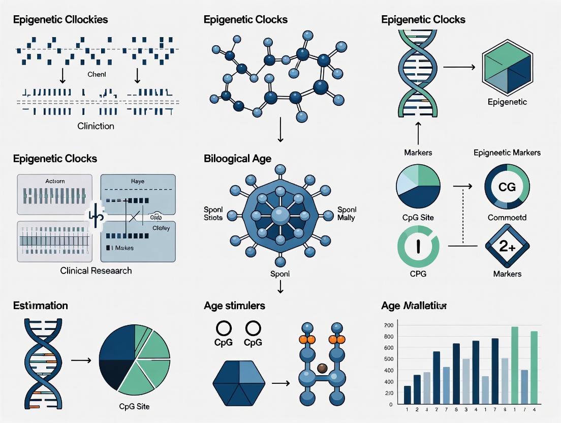
Epigenetic Clocks in Clinical Research: A Guide to Biomarker Selection, Application, and Validation
This article provides a comprehensive overview of epigenetic clocks for researchers, scientists, and drug development professionals.
Epigenetic Clocks in Clinical Research: A Guide to Biomarker Selection, Application, and Validation
Abstract
This article provides a comprehensive overview of epigenetic clocks for researchers, scientists, and drug development professionals. It covers the foundational evolution of DNA methylation-based biomarkers from first-generation chronological age estimators to fourth-generation functional and pathway-level clocks. The scope extends to methodological applications in clinical trials and disease-specific risk stratification, critical troubleshooting of technical noise and sample type validity, and a comparative validation of clock performances for different research intents. The integration of novel approaches like EpiScores and multi-omics data is also explored, offering a roadmap for the reliable use of biological age estimation in translational aging research and therapeutic development.
The Evolution of Epigenetic Clocks: From Chronological Age to Biological Pathways
Defining Epigenetic Clocks and DNA Methylation Biomarkers
Epigenetic clocks are powerful biomarkers based on DNA methylation (DNAm) patterns that estimate the biological age of cells, tissues, or individuals [1]. These clocks have emerged as a transformative tool in aging research, capable of predicting age-related morbidity, mortality, and overall health trajectories with remarkable precision [1] [2]. Unlike chronological age, which simply measures the passage of time, biological age reflects an individual's physiological state and functional decline, providing a more nuanced understanding of the aging process [1].
The fundamental premise of epigenetic clocks lies in the predictable changes that occur in DNA methylation landscapes over time. DNA methylation involves the addition of a methyl group to a cytosine nucleotide, typically at cytosine-phosphate-guanine (CpG) sites, which can influence gene expression without altering the underlying DNA sequence [1]. Age-related methylation changes occur in approximately 28% of the human genome, with specific CpG sites showing progressive hypermethylation or hypomethylation in a clock-like manner [1]. By analyzing these patterns using supervised machine learning techniques, researchers have developed computational models that can accurately estimate biological age across diverse tissues and populations [1] [3].
Table 1: Evolution of Epigenetic Clock Generations
| Generation | Training Basis | Key Examples | Primary Applications |
|---|---|---|---|
| First Generation | Chronological age | Horvath's Clock, Hannum's Clock | Cross-tissue age estimation, basic aging rate assessment [1] [4] |
| Second Generation | Mortality risk, health phenotypes, clinical biomarkers | PhenoAge, GrimAge | Disease risk prediction, mortality assessment, intervention studies [1] [4] |
| Third Generation | Pace of aging, multi-organ system decline | DunedinPACE, DunedinPoAm | Measuring rate of aging, longitudinal tracking of aging trajectories [5] [4] |
| Fourth Generation | Putatively causal sites via Mendelian randomization | Causal Clocks | Identifying causal mechanisms in aging, potential therapeutic targets [5] |
Key Epigenetic Clocks and Their Applications
First-Generation Clocks
First-generation epigenetic clocks were primarily trained to predict chronological age using elastic net regression on large DNA methylation datasets [1] [4]. These clocks established the foundational framework for biological age estimation and demonstrated that DNA methylation patterns could accurately reflect the aging process.
Horvath's Clock, a landmark model in epigenetic aging research, was the first to achieve cross-tissue age prediction by analyzing DNA methylation data from 7,844 samples across 51 tissue and cell types [1]. Utilizing 353 CpG sites (193 positively and 160 negatively correlated with age), this clock shows minimal age-related variance across almost all tissues and organs, including whole blood, brain, kidney, and liver [1]. Its versatility extends to aging research in other mammals and in vitro aging analyses, making it an invaluable tool for studying aging mechanisms [1]. However, limitations include variable predictive accuracy across tissues, particularly in hormonally sensitive tissues and blood samples, along with reduced sensitivity to certain diseases and underestimation of biological age in individuals over 60 [1].
Hannum's Clock was developed specifically for blood samples using the Illumina 450K methylation array from 656 adults aged 19-101 [1]. This model employs 71 age-related CpG sites selected through the Elastic Net algorithm and demonstrates a high correlation of 0.96 between biological and chronological age, with an average absolute error of 3.9 years [1]. Optimized for blood-based studies, Hannum's clock shows strong associations with clinical markers such as body mass index, cardiovascular health, and immune function [1]. It has proven valuable for evaluating clinical interventions like weight loss programs or exercise therapy by tracking changes in biological age before and after interventions [1]. Limitations include restricted applicability to non-blood tissues and lower sensitivity to external factors compared to other clocks [1].
Second and Third-Generation Clocks
Second and third-generation clocks represent significant advancements by incorporating phenotypic data, mortality risk, and pace of aging metrics, thereby enhancing their clinical relevance and predictive power for health outcomes [1] [4].
PhenoAge was developed by incorporating clinical biomarkers to capture aspects of phenotypic aging beyond chronological age [4]. This clock demonstrates stronger associations with mortality risk, age-related functional decline, and disease susceptibility compared to first-generation clocks [4]. In large-scale comparisons, PhenoAge has shown particular utility in predicting conditions such as Crohn's disease and Parkinson's disease [4].
GrimAge represents a further refinement through a two-step process that incorporates DNA methylation surrogates for health-related biomarkers such as smoking exposure and plasma proteins [4]. This clock outperforms most other epigenetic clocks in predicting all-cause mortality and has demonstrated particularly strong associations with respiratory and liver-related conditions, including primary lung cancer and cirrhosis [4]. In a comprehensive analysis of 174 disease outcomes across 18,859 individuals, GrimAge showed the strongest association with all-cause mortality (Hazard Ratio per standard deviation = 1.54) and significantly improved disease classification accuracy for multiple conditions [4].
DunedinPACE and related third-generation clocks focus on measuring the pace of aging rather than a static biological age [4]. These clocks are trained on longitudinal data tracking multi-organ system decline and have shown significant associations with diverse conditions including diabetes (Hazard Ratio = 1.44) [4]. Their development represents a shift toward dynamic measures of aging trajectories rather than cross-sectional assessments.
Table 2: Performance Comparison of Selected Epigenetic Clocks
| Clock Name | CpG Sites | Tissue Specificity | Key Clinical Associations | Median Absolute Error (Years) |
|---|---|---|---|---|
| Horvath | 353 | Pan-tissue | Cancer, mortality, lifestyle impacts [1] | 3.6 [1] |
| Hannum | 71 | Blood | BMI, cardiovascular health, immune function [1] | 3.9 [1] |
| PhenoAge | 513 | Primarily blood | Mortality, frailty, Crohn's disease [6] [4] | Not specified |
| GrimAge | Not specified | Primarily blood | All-cause mortality, lung cancer, cirrhosis [6] [4] | Not specified |
| DunedinPACE | Not specified | Blood | Diabetes, pace of aging [4] | Not specified |
| IC Clock | 91 | Blood and saliva | Intrinsic capacity, mortality, immune function [7] | Not specified |
Technical Protocols and Methodologies
Standard Experimental Workflow
Implementing epigenetic clocks requires a standardized workflow from sample collection to data analysis. The following protocol outlines the key steps for reliable biological age estimation:
Sample Collection and DNA Extraction: Collect peripheral blood samples using appropriate collection tubes (e.g., PAXgene Blood DNA tubes). Extract genomic DNA using validated kits (e.g., QIAamp DNA Blood Mini Kit) following manufacturer protocols. Quantify DNA concentration using fluorometric methods and assess quality via spectrophotometry (A260/A280 ratio >1.8) [8].
DNA Methylation Profiling: Perform bisulfite conversion on 500ng of genomic DNA using the EZ-96 DNA Methylation Kit (Zymo Research) or equivalent. Process converted DNA using Illumina Infinium MethylationEPIC BeadChip arrays, which interrogate over 850,000 CpG sites across the genome. Follow standard Illumina protocols for amplification, hybridization, staining, and imaging [3] [7].
Data Preprocessing and Quality Control: Process raw intensity data (.idat files) using R packages such as minfi or meffil. Perform background correction, dye bias correction, and probe type normalization. Exclude probes with detection p-value > 0.01, cross-reactive probes, and probes containing single nucleotide polymorphisms. Implement functional normalization to remove unwanted technical variation [3].
Epigenetic Clock Calculation: Extract beta-values for CpG sites required for the specific epigenetic clock being implemented. Apply pre-trained algorithms to calculate biological age. For Horvath's clock, this involves a weighted linear combination of 353 CpG methylation values [1]. For GrimAge, the calculation incorporates DNAm-based surrogates for plasma proteins and smoking history [4]. Compute age acceleration residuals by regressing epigenetic age on chronological age across the dataset [3].
Addressing Technical Reliability
Technical noise presents a significant challenge in epigenetic clock applications, with deviations of up to 9 years observed between technical replicates for some clocks [3] [2]. This variability stems from sample preparation, probe chemistry, batch effects, and other technical factors that can obfuscate true biological signals [3].
The principal component (PC) clock approach represents a computational solution that substantially improves reliability [3] [2]. This method involves:
- CpG Selection: Identify a common set of CpGs present across all datasets and platforms (e.g., 78,464 CpGs shared between 450K and EPIC arrays) [3].
- Principal Component Analysis: Perform PCA on the methylation matrix to extract components that capture shared aging signals while minimizing noise from individual CpGs [3].
- Model Retraining: Use the top principal components as input features to retrain age prediction models using elastic net regression [3].
- Validation: Assess performance metrics including intraclass correlation coefficient (ICC) and median absolute deviation between replicates [3].
This approach reduces median deviations between technical replicates from 1.8 years to less than 1.5 years for most clocks, dramatically improving reliability for longitudinal studies and clinical trials [3] [2].
Diagram 1: Experimental workflow for epigenetic clock analysis, covering sample processing to clinical interpretation.
Advanced Computational Approaches
Explainable Artificial Intelligence
Recent advances in deep learning have led to the development of biologically informed models that enhance both prediction accuracy and interpretability. The XAI-AGE framework represents one such approach that integrates hierarchical biological knowledge into neural network architecture [9].
This model uses 3,007 manually curated biological pathways from the Reactome Pathway Knowledgebase to construct a pathway-aware multilayered hierarchical network [9]. The architecture includes:
- Input Layer: DNA methylation beta values connected to gene-level nodes
- Intermediate Layers: Represent biological pathways with increasing abstraction
- Output Layer: Biological age prediction
XAI-AGE achieves a median absolute error of 2.83 years compared to 3.0 years for elastic net regression on pan-tissue data, while providing biological interpretability through importance scores for pathways and genes [9]. Key pathways identified include DNA Repair (decreasing with age) and Extracellular Matrix Organization (increasing with age), offering insights into biological mechanisms driving epigenetic aging [9].
Novel Clock Developments
IC Clock: The Intrinsic Capacity Clock represents a novel approach trained on clinical evaluations of cognition, locomotion, psychological well-being, sensory abilities, and vitality rather than chronological age or mortality [7]. This clock utilizes 91 CpG sites and shows minimal overlap with previous epigenetic clocks, suggesting it captures distinct biological aspects of aging [7]. In validation studies, DNAm IC outperformed first and second-generation clocks in predicting all-cause mortality and was strongly associated with changes in immune and inflammatory biomarkers [7]. The IC clock can be calculated from both blood and saliva samples (correlation r = 0.64), enabling non-invasive assessment [7].
Forensic Age Estimation Clocks: Specialized clocks have been developed for forensic applications using seven CpG sites located in ELOVL2, ASPA, PDE4C, FHL2, CCDC102B, MIR29B2CHG, and chr16:85395429 [8]. These models cover the full age spectrum from childhood to old age (2-104 years) and achieve mean absolute errors of approximately 3.3-3.4 years using quantile regression neural networks or support vector machines [8].
The Scientist's Toolkit
Table 3: Essential Research Reagents and Platforms for Epigenetic Clock Studies
| Category | Specific Product/Platform | Key Function | Application Notes |
|---|---|---|---|
| Sample Collection | PAXgene Blood DNA Tubes | Blood sample stabilization for DNA analysis | Maintains DNA integrity during storage and transport [7] |
| DNA Extraction | QIAamp DNA Blood Mini Kit (Qiagen) | Genomic DNA purification from blood | Provides high-quality DNA with minimal contaminants [8] |
| Bisulfite Conversion | EZ-96 DNA Methylation Kit (Zymo Research) | Converts unmethylated cytosines to uracils | Critical step for methylation-specific analysis [7] |
| Methylation Arrays | Illumina Infinium MethylationEPIC BeadChip | Genome-wide methylation profiling at >850,000 CpG sites | Current standard for comprehensive epigenetic studies [3] [7] |
| Data Analysis | minfi R Package | Preprocessing and normalization of methylation data | Handles background correction, normalization, and quality control [3] |
| Age Prediction | Elastic Net Regression | Model training for age prediction | Standard method for developing epigenetic clocks [1] [9] |
Diagram 2: Logical relationships between factors influencing and influenced by epigenetic clocks.
Applications in Clinical Research and Drug Development
Clinical Trial Applications
Epigenetic clocks are increasingly utilized as biomarkers in clinical trials to evaluate interventions targeting aging processes. Their ability to detect biological age changes over relatively short timeframes makes them valuable tools for assessing intervention efficacy [5].
In a Phase IIb trial investigating semaglutide in adults with HIV-associated lipohypertrophy, 11 organ-system clocks showed concordant decreases with treatment, most prominently in inflammation, brain, and heart clocks [5]. This suggests the drug may modulate epigenetic aging, potentially through reducing visceral fat and mitigating adipose-driven pro-aging signals [5].
The TRIIM (Thymus Regeneration, Immunorestoration, and Insulin Mitigation) trial demonstrated that a regimen including recombinant human growth hormone could reverse epigenetic age by approximately 1.5 years after one year of treatment, with effects persisting six months post-treatment [5]. This provides compelling evidence that epigenetic aging can be modulated through targeted interventions.
Disease Risk Prediction
Large-scale studies have established the superior performance of second and third-generation clocks in predicting age-related disease onset. In an analysis of 174 disease outcomes across 18,859 individuals, these clocks significantly outperformed first-generation clocks, with particular strength in predicting respiratory and liver-related conditions [4].
Notably, GrimAge showed strong associations with primary lung cancer (Hazard Ratio = 1.56) and cirrhosis (Hazard Ratio = 1.86), while DunedinPACE was significantly associated with diabetes risk (Hazard Ratio = 1.44) [4]. These findings highlight the potential utility of epigenetic clocks in risk stratification and early intervention strategies.
Frailty and Functional Decline Assessment
Epigenetic clocks show promising associations with frailty, an age-related condition characterized by multisystem physiological decline. Meta-analyses of 24 studies encompassing 28,325 participants found that higher GrimAge acceleration, PhenoAge acceleration, and pace of aging were significantly associated with higher frailty levels cross-sectionally [6]. Longitudinally, GrimAge acceleration was significantly associated with increases in frailty over time, supporting its utility in tracking functional decline [6].
The IC clock, trained specifically on intrinsic capacity domains, provides a molecular correlate for functional aging that aligns with clinical assessments [7]. This approach bridges molecular readouts with clinically relevant functional measures, potentially enabling earlier detection of age-related decline.
Epigenetic clocks have evolved from simple age estimators to sophisticated biomarkers that capture multiple dimensions of biological aging. The progression from first-generation clocks trained on chronological age to subsequent generations incorporating mortality risk, phenotypic data, and pace of aging has significantly enhanced their clinical utility. Technical advancements, including principal component approaches to improve reliability and biologically informed deep learning models for interpretability, continue to refine these tools.
For researchers and drug development professionals, epigenetic clocks offer promising biomarkers for evaluating interventions, predicting disease risk, and understanding the biological mechanisms of aging. As these tools become more reliable and validated across diverse populations, their integration into clinical research and practice holds significant potential for advancing personalized medicine and healthy aging strategies.
First-generation epigenetic clocks, primarily the models developed by Horvath and Hannum, represent a transformative advancement in aging research by providing the first robust biomarkers for estimating human chronological age based on DNA methylation (DNAm) patterns. These clocks established that predictable changes in epigenetic regulation occur across the lifespan, creating a molecular footprint that can be quantified independently of chronological time. Unlike subsequent generations of clocks trained on mortality or phenotypic data, first-generation clocks were specifically designed to estimate chronological age with high accuracy, creating a foundational metric from which biological age acceleration (the discrepancy between epigenetic and chronological age) could be calculated. Their development marked a paradigm shift in gerontology, enabling researchers to quantitatively assess whether an individual's biological age deviates from their chronological age, thereby providing insights into their underlying physiological aging process [1] [10].
The significance of these clocks extends beyond mere age prediction. They have become indispensable tools for investigating the relationships between accelerated aging, disease risk, and mortality across diverse populations. By serving as standardized biomarkers, they have facilitated discoveries about how genetic factors, environmental exposures, and lifestyle choices influence the pace of biological aging. This Application Note provides a comprehensive technical overview of the Horvath and Hannum clocks, detailing their development, analytical performance, implementation protocols, and applications within clinical and research settings, framed within the broader context of epigenetic clocks for biological age estimation in clinical research [1].
Clock Specifications and Technical Performance
The Horvath and Hannum clocks, while sharing the common goal of chronological age prediction, differ significantly in their design, tissue specificity, and technical composition. Horvath's pan-tissue clock was groundbreaking for its ability to estimate age across a wide spectrum of tissues and cell types, a property that greatly enhances its utility in diverse research contexts. In contrast, Hannum's clock was optimized specifically for blood tissue, providing superior performance in hematological samples but with limited application in other tissue types [1].
Table 1: Comparative Specifications of First-Generation Epigenetic Clocks
| Feature | Horvath Clock | Hannum Clock |
|---|---|---|
| Year Published | 2013 [11] | 2013 [10] |
| Primary Tissue Application | Pan-tissue (51 tissues & cell types) [1] | Whole blood [1] |
| Training Sample Size | 7,844 non-cancer samples [1] | 656 adults [1] |
| CpG Sites Utilized | 353 (193 positive, 160 negative age correlation) [1] | 71 [1] |
| Statistical Algorithm | Elastic net regression [1] | Elastic net regression [1] |
| Reported Accuracy (vs. Chronological Age) | Correlation: >0.96; Mean Absolute Error: ~3.6 years [1] | Correlation: 0.96; Mean Absolute Error: ~3.9 years [1] |
| Key Technical Strength | Unprecedented cross-t tissue applicability [11] [1] | High accuracy in blood-based studies [1] |
The performance metrics of both clocks demonstrate remarkable precision in age estimation. The correlation with chronological age exceeds 0.96 for both models, though the Horvath clock maintains a slight edge in its multi-tissue error rate. This high accuracy is contingent on using the appropriate clock for the sample type being analyzed. The Horvath clock's core strength lies in its applicability to virtually all tissues and organs, including brain, kidney, liver, and even in vitro cell cultures. The Hannum clock, while more restricted in scope, shows stronger associations with certain clinical markers in blood-based analyses, such as body mass index (BMI) and cardiovascular health metrics, making it particularly valuable for epidemiological and clinical studies focused on blood-derived biomarkers [1].
Experimental Implementation and Workflow
Implementing first-generation epigenetic clocks requires a structured workflow from sample collection to data analysis. The following protocol and visualization outline the standard pipeline for reliable age estimation.
Sample Processing and Data Generation Protocol
Step 1: Sample Collection and DNA Extraction
- Tissue Sampling: Collect target tissue (e.g., whole blood, buccal cells, surgical specimens). For Horvath clock, any tissue type is suitable; for Hannum clock, use whole blood.
- DNA Extraction: Isolate high-quality genomic DNA using standardized kits (e.g., Qiagen DNeasy Blood & Tissue Kit). Assess DNA purity and concentration via spectrophotometry (A260/280 ratio ~1.8-2.0).
- Quality Control: Confirm DNA integrity via gel electrophoresis or fragment analyzer. Minimum required DNA quantity is 500 ng for standard methylation arrays.
Step 2: DNA Methylation Profiling
- Platform Selection: Process samples using Illumina Infinium methylation arrays. The Horvath clock was developed on 27K/450K arrays; the Hannum clock on the 450K array. Both are compatible with the EPIC (850K) platform.
- Bisulfite Conversion: Treat 500 ng of DNA using the EZ-96 DNA Methylation Kit (Zymo Research) per manufacturer's protocol. This converts unmethylated cytosines to uracils while leaving methylated cytosines unchanged.
- Array Processing: Hybridize bisulfite-converted DNA to selected Illumina methylation array following standard protocols. Scan arrays using iScan or NextSeq scanner.
Step 3: Data Preprocessing and Normalization
- Raw Data Extraction: Generate intensity data (IDAT files) using the scanner software.
- Quality Control: Assess sample quality using metrics such as detection p-values (>95% CpGs with p<0.01).
- Background Correction and Normalization: Process data using established pipelines such as
minfiorSeSAMein R. Perform functional normalization to remove technical variation and probe-type bias. - Beta-value Calculation: Compute methylation levels as β = M/(M + U + α), where M and U are methylated and unmethylated signal intensities, and α=100 is a constant to stabilize variance.
Step 4: Epigenetic Age Calculation
- CpG Site Selection: Extract beta values for the 353 CpGs (Horvath) or 71 CpGs (Hannum) from the normalized data matrix.
- Clock Implementation: Apply published clock coefficients using standard scripts (available from the original publications or DNA Methylation Age website).
- Age Acceleration Calculation: Compute the difference between DNAm age and chronological age (AgeAcceleration = DNAmAge - ChronologicalAge) for biological interpretation.
Workflow Visualization
Diagram 1: Standardized workflow for implementing first-generation epigenetic clocks, showing the parallel paths for Horvath and Hannum clock analysis.
The Scientist's Toolkit: Essential Research Reagents
Successful implementation of first-generation epigenetic clocks requires specific laboratory reagents and computational tools. The following table details essential components of the experimental pipeline.
Table 2: Key Research Reagents and Materials for Epigenetic Clock Analysis
| Category | Item/Reagent | Specification/Function |
|---|---|---|
| Sample Collection | PAXgene Blood DNA Tubes | Stabilizes nucleic acids in whole blood for transport and storage [12]. |
| Tissue Preservation Solutions | RNAlater or similar for stabilizing tissue specimens prior to DNA extraction. | |
| DNA Processing | DNeasy Blood & Tissue Kit (Qiagen) | Silica-membrane based DNA purification from various sample types [12]. |
| EZ-96 DNA Methylation Kit (Zymo Research) | Efficient bisulfite conversion of genomic DNA for methylation analysis [12]. | |
| Methylation Array | Illumina Infinium MethylationEPIC v2.0 | Latest array platform covering >935,000 CpG sites, backward compatible [12] [13]. |
| Infinium HD Assay Methylation Kit | Required reagents for array processing: amplification, fragmentation, hybridization [12]. | |
| Computational Tools | R Programming Environment | Primary platform for methylation data analysis [12] [13]. |
minfi R/Bioconductor Package |
Comprehensive pipeline for preprocessing and normalization of methylation array data [12]. | |
| Horvath Aging Clock Script | Published algorithm for calculating DNAm age from normalized beta values [1]. |
The selection of appropriate reagents is critical for data quality. The Illumina Infinium methylation arrays remain the gold standard for generating the data required for these clocks, with the EPIC array providing backward compatibility to both the Horvath and Hannum CpG sites. For computational analysis, the R environment with specialized Bioconductor packages provides the most robust framework for data normalization and age calculation. Researchers should ensure that all CpG sites required for their chosen clock are present on their selected array platform, though all sites from both first-generation clocks are represented on the EPIC array [12] [13].
Applications, Limitations, and Clinical Translation
First-generation epigenetic clocks have enabled numerous research applications but also present specific limitations that researchers must consider in study design and data interpretation.
Key Research Applications
- Age Acceleration Studies: The primary application involves calculating the difference between DNAm age and chronological age to identify individuals with accelerated or decelerated aging. This age acceleration metric has been associated with all-cause mortality, age-related diseases, and various environmental exposures [1] [10].
- Intervention Assessment: These clocks serve as tools for evaluating the impact of lifestyle, pharmacological, and environmental interventions on biological aging. Studies have tracked changes in age acceleration following weight loss programs, exercise regimens, and other health interventions [1].
- Cross-Species and Cross-Tissue Analysis: The Horvath clock's unique pan-tissue property enables comparisons across different tissue types from the same donor and has been adapted for use in mammalian models beyond humans, facilitating translational research [11] [1].
Limitations and Technical Constraints
- Tissue Specificity Constraints: While the Horvath clock functions across tissues, its accuracy varies, particularly in hormonally sensitive tissues. The Hannum clock is not recommended for non-blood tissues, limiting its application in multi-tissue studies [1].
- Population-Specific Biases: Both clocks were primarily developed in populations of European ancestry. Genetic variants more common in non-European populations can influence CpG methylation, potentially leading to spurious offsets in age estimation [10].
- Reduced Sensitivity in Older Adults: The Horvath clock tends to underestimate biological age in individuals over 60, likely due to underrepresentation of older samples in the training dataset. This can compress the range of age acceleration measurements in elderly populations [1].
- Limited Disease Sensitivity: First-generation clocks show inconsistent associations with certain age-related conditions, including schizophrenia and progeroid syndromes, suggesting they may not capture all aspects of biological aging relevant to specific diseases [1].
First-generation epigenetic clocks, particularly the Horvath and Hannum models, established the foundational principles and methodologies for epigenetic age estimation. Their development demonstrated that DNA methylation patterns provide a robust molecular readout of chronological aging across tissues and individuals. While subsequent generations of clocks have improved upon specific applications, particularly for predicting healthspan and mortality risk, these original models remain widely used for estimating chronological age and calculating age acceleration in diverse research contexts.
The continued utility of these clocks depends on appropriate application—leveraging the Horvath clock for multi-tissue studies, developmental research, and cross-species comparisons, while applying the Hannum clock for blood-specific investigations with requirements for high correlation with clinical phenotypes in hematological samples. As the field advances toward clinical translation, understanding the technical parameters, implementation protocols, and limitations of these foundational tools is essential for their proper application in clinical research and drug development programs focused on modulating human aging.
First-generation epigenetic clocks, such as Horvath's pan-tissue clock and Hannum's blood-specific clock, were primarily trained to predict chronological age from DNA methylation (DNAm) patterns [14]. While these clocks established the fundamental link between epigenetic modification and aging, they demonstrated only weak associations with clinical measures of physiological dysregulation and hard disease endpoints [14]. This limitation prompted the development of second-generation clocks that incorporate phenotypic and mortality data to better capture biological aging processes.
The second-generation clocks PhenoAge and GrimAge represent significant methodological advancements. PhenoAge was trained on clinical biomarkers composite to capture morbidity risk, while GrimAge was specifically designed to predict mortality risk through DNAm surrogates of plasma proteins and smoking exposure [14] [15]. These clocks have demonstrated superior performance in predicting age-related functional decline, chronic diseases, and lifespan across diverse populations [14] [16] [15]. Their development marks a pivotal shift from merely estimating chronological time to quantifying biological vulnerability, offering powerful tools for clinical research and intervention studies.
PhenoAge: Linking DNA Methylation to Clinical Phenotypes
Development and Algorithm Structure
PhenoAge (DNAm Phenotypic Age) was developed through a two-stage approach to capture phenotypic aging beyond chronological years. In the first stage, Levine et al. created a composite clinical measure based on ten biomarkers: chronological age, albumin, creatinine, glucose, C-reactive protein, lymphocyte percentage, mean cell volume, red blood cell distribution width, alkaline phosphatase, and white blood cell count [14]. This composite was designed to reflect overall physiological dysregulation and was validated against mortality risk.
In the second stage, the researchers regressed this phenotypic age estimator on DNAm data using elastic net regularization, identifying 513 CpG sites that collectively predict the phenotypic aging score [14] [15]. The resulting DNAm PhenoAge algorithm provides a biomarker of aging that more strongly correlates with functional decline and morbidity risk than first-generation clocks. The difference between DNAm PhenoAge and chronological age, termed PhenoAge acceleration (AgeAccelPheno), indicates the degree of biological aging acceleration, with positive values signifying faster-than-expected aging [15].
Predictive Performance and Clinical Applications
PhenoAge acceleration demonstrates significant associations with various age-related clinical phenotypes. Research from the Irish Longitudinal Study on Ageing (TILDA) found PhenoAge acceleration associated with 4 out of 9 clinical outcomes, including walking speed, frailty, and cognitive performance (MMSE and MOCA scores) in minimally adjusted models [14]. These associations remain significant after adjusting for social and lifestyle factors, though the effect sizes may attenuate.
In comparative studies, PhenoAge consistently outperforms first-generation clocks. Maddock et al. reported PhenoAge acceleration associated with lower grip strength, worse lung function, and slower mental speed in meta-analyses of British cohorts [14]. The predictive utility of PhenoAge extends to mortality risk, with hazard ratios for all-cause mortality ranging from 1.32 to 1.73 per standard deviation increase in various studies [15].
Table 1: PhenoAge Associations with Clinical Phenotypes and Mortality
| Outcome Category | Specific Outcomes | Effect Size/Association | Study Population |
|---|---|---|---|
| Physical Function | Walking speed | Significant association | TILDA (n=490) |
| Frailty status | Significant association | TILDA | |
| Grip strength | Lower strength | British cohorts meta-analysis | |
| Lung function | Worse function | British cohorts meta-analysis | |
| Cognitive Function | MMSE score | Significant association | TILDA |
| MOCA score | Significant association | TILDA | |
| Mental speed | Slower performance | British cohorts meta-analysis | |
| Mortality | All-cause mortality | HR=1.32-1.73 per SD | ESTHER cohort |
GrimAge: Advancing Mortality Risk Prediction
Innovative Algorithm Design and Development
GrimAge represents a methodological innovation in epigenetic clock construction, specifically optimized for mortality risk prediction. Developed by Lu et al., GrimAge employs a two-stage approach that fundamentally differs from previous clocks. In the first stage, the researchers identified DNAm-based surrogates for 12 plasma proteins (including adrenomedullin, beta-2-microglobulin, and cystatin C) and smoking pack-years [14]. These surrogates were selected based on their established associations with mortality risk.
In the second stage, the team regressed time-to-death due to all-cause mortality on these DNAm-based biomarkers using Cox proportional hazards modeling with elastic net regularization [14] [15]. This approach identified 1,030 CpG sites that jointly predict mortality risk, which were then combined into the composite GrimAge estimator [14] [15]. The resulting algorithm incorporates information about physiological processes directly relevant to mortality, making it particularly powerful for predicting healthspan and lifespan.
Validation and Performance Evidence
GrimAge demonstrates exceptional performance in predicting mortality and age-related health outcomes across diverse populations. In the TILDA study, GrimAge acceleration was associated with 8 out of 9 clinical outcomes in minimally adjusted models and remained a significant predictor of walking speed, polypharmacy, frailty, and mortality after full adjustment for covariates [14]. This robust performance underscores its utility as a comprehensive biomarker of aging.
Recent large-scale validation studies confirm GrimAge's superior predictive capability. Research from the National Institute on Aging directly compared multiple epigenetic clocks and found GrimAge outperformed all others in predicting mortality [17]. Similarly, a 2025 study of NHANES participants demonstrated that GrimAge acceleration shows approximately linear positive associations with all-cause, cancer-specific, and cardiac mortality, with consistent effects across most subgroups [16].
Table 2: GrimAge Performance in Predicting Mortality and Health Outcomes
| Study | Population | Follow-up | Key Findings | Effect Size |
|---|---|---|---|---|
| TILDA Study [14] | n=490, aged 50+ | Up to 10 years | Significant predictor of walking speed, polypharmacy, frailty, mortality | Remained significant after full covariate adjustment |
| ESTHER Cohort [15] | n=1,771, aged 50-75 | 17 years | Independent association with all-cause mortality | HR=1.47 (1.32-1.64) per SD |
| NHANES Study [16] | n=1,942, median age 65 | Median 208 months | Linear associations with all-cause, cancer, cardiac mortality | Consistent across subgroups |
| Lothian Birth Cohort [14] | n=709, mean age 73 | - | Associated with lung function, cognitive ability, brain structure | 81% increased hazard per SD |
Comparative Analysis of Epigenetic Clocks
Head-to-Head Performance Comparisons
Direct comparisons between epigenetic clocks reveal distinct performance patterns across different applications. The TILDA study provided comprehensive head-to-head comparisons, demonstrating that first-generation clocks (Horvath and Hannum) showed minimal associations with clinical phenotypes, while PhenoAge showed intermediate performance, and GrimAge consistently demonstrated the strongest associations [14]. This pattern holds across functional measures, cognitive performance, and mortality prediction.
Maddock et al. reinforced these findings in their meta-analysis of British cohorts, where first-generation clocks showed no significant associations with physical or cognitive function, while both second-generation clocks demonstrated significant relationships, with GrimAge showing somewhat broader associations [14]. For mortality prediction specifically, GrimAge consistently outperforms other clocks, though PhenoAge still provides valuable information about phenotypic aging.
Technical and Methodological Considerations
The differential performance of epigenetic clocks reflects their distinct training approaches and underlying biological capture. GrimAge's superior mortality prediction likely stems from its direct training on time-to-death data and incorporation of DNAm surrogates for known mortality risk factors [14] [15]. PhenoAge captures multisystem physiological decline through clinical biomarkers, making it sensitive to functional aging processes [14].
Recent advancements include the development of principal component (PC) versions of these clocks, which demonstrate greater measurement stability in longitudinal assessments [18]. A 2025 study found PC clocks exhibited substantially smaller 2-year change variance than original clocks, suggesting improved reliability for intervention studies [18]. Additionally, next-generation clocks like the IC clock trained on intrinsic capacity domains show promise for capturing functional aging aspects beyond mortality risk [7].
Table 3: Comprehensive Comparison of Epigenetic Clock Characteristics
| Characteristic | Horvath Clock | Hannum Clock | PhenoAge | GrimAge |
|---|---|---|---|---|
| Primary Training Target | Chronological age (pan-tissue) | Chronological age (blood) | Clinical phenotype composite | Mortality risk |
| Number of CpG Sites | 353 | 71 | 513 | 1,030 |
| Key Inputs/ Surrogates | DNAm age only | DNAm age only | DNAm phenotypic age | DNAm surrogates of plasma proteins + smoking |
| Strength | Accurate across tissues | Blood-specific age prediction | Captures morbidity risk | Superior mortality prediction |
| Mortality Hazard Ratio (per SD) | ~1.0-1.1 (ns) | ~1.0-1.1 (ns) | 1.32-1.46 | 1.47-1.64 |
| Clinical Phenotype Associations | Minimal | Minimal | Moderate | Strong |
Experimental Protocols and Applications
Standardized DNA Methylation Measurement Protocol
Consistent measurement of DNA methylation forms the foundation for reliable epigenetic clock assessment. The following protocol outlines the standardized workflow for generating epigenetic clock data in clinical studies:
The experimental workflow begins with sample collection, typically using whole blood collected in EDTA tubes, though saliva and other tissues can also be used [19] [7]. DNA extraction follows standardized protocols, such as the Qiagen Gentra Puregene method, with careful quality control to ensure high-molecular-weight DNA [20]. Bisulfite conversion using the EZ-96 DNA methylation kit or equivalent is critical for distinguishing methylated from unmethylated cytosine residues.
The core measurement utilizes the Infinium MethylationEPIC BeadChip (Illumina), which quantifies DNAm at over 850,000 CpG sites [20] [19]. Raw intensity data (IDAT files) undergo preprocessing including background correction with single-sample normal-exponential out-of-band (ssnoob) method and normalization using Beta Mixture Quantile (BMIQ) to address probe-type bias [20]. Quality control excludes probes with detection p-values >0.01, >10% missing values, or those on sex chromosomes, and samples showing technical outliers or genotype mismatches [15] [20].
Calculation of Epigenetic Age Acceleration
After obtaining DNAm data, epigenetic age estimates are calculated using established algorithms. For PhenoAge and GrimAge, the Horvath lab's online calculator (dnamage.genetics.ucla.edu) or corresponding R packages (e.g., 'DNAmAge') are commonly used [15] [19]. The calculation incorporates the specific CpG sites and coefficients defined by the original developers for each clock.
To derive age acceleration metrics, epigenetic age is regressed on chronological age using linear models. The residuals from this regression represent epigenetic age acceleration (AgeAccel), indicating whether an individual is epigenetically older or younger than expected [14] [15]. For blood samples, intrinsic epigenetic age acceleration further adjusts for estimated leukocyte composition using the Houseman algorithm, accounting for age-related immune cell population changes [14]. This refined metric better captures aging-independent of immunosenescence.
Application in Clinical Trials and Intervention Studies
Epigenetic clocks are increasingly employed as biomarkers in clinical trials to assess interventions targeting aging processes. Optimal study design includes multiple baseline measurements (at least two) prior to intervention and repeated measures during and after treatment to account for natural variation and establish trajectory [19]. The 2025 COSMOS-Blood study highlights the importance of accounting for measurement stability, recommending ANCOVA-based analyses for intervention studies due to strong baseline-follow-up correlations (R²≈0.71-0.88 for PC clocks) [18].
Successful applications include the TRIIM trial, which demonstrated thymic regeneration and epigenetic age reversal using a growth hormone-based regimen, with GrimAge showing a two-year decrease that persisted post-treatment [5]. Similarly, studies of semaglutide showed concordant decreases across multiple organ-system clocks, suggesting systemic epigenetic effects [5]. These applications underscore the utility of epigenetic clocks, particularly GrimAge and PhenoAge, as sensitive biomarkers for evaluating aging interventions.
Research Reagent Solutions and Technical Tools
Table 4: Essential Research Reagents and Computational Tools for Epigenetic Clock Studies
| Category | Specific Product/Tool | Application Purpose | Key Features |
|---|---|---|---|
| DNA Methylation Arrays | Infinium MethylationEPIC BeadChip (Illumina) | Genome-wide DNAm profiling | 850,000+ CpG sites, covers clock CpGs |
| Bisulfite Conversion Kits | EZ-96 DNA Methylation Kit (Zymo Research) | Convert unmethylated C to U | High conversion efficiency, 96-well format |
| DNA Extraction Kits | Qiagen Gentra Puregene | High-quality DNA from blood/tissue | Maintains DNA integrity for arrays |
| Analysis Software | R packages: DNAmAge, Champ, watermelon | Data processing and clock calculation | Implements published algorithms |
| Online Calculators | Horvath Lab Epigenetic Clock Calculator | User-friendly clock estimation | Web-based, multiple clocks |
| Quality Control Tools | MethylAID, minfi R packages | Data quality assessment | Detects outliers, technical artifacts |
Second-generation epigenetic clocks, particularly PhenoAge and GrimAge, represent significant advancements over first-generation models by incorporating phenotypic and mortality data directly into their algorithms. The robust association of these clocks with clinical outcomes, functional decline, and mortality risk demonstrates their utility as biomarkers of biological aging in clinical research. GrimAge's consistent superiority in predicting mortality makes it particularly valuable for studies focused on lifespan and healthspan, while PhenoAge provides important insights into phenotypic aging processes.
Future developments will likely focus on tissue-specific clocks optimized for different biological samples [20], dynamic measures of aging pace like DunedinPACE [18] [20], and integrated models combining epigenetic measures with clinical assessments like intrinsic capacity [7]. The ongoing validation and refinement of these tools will further establish epigenetic clocks as essential biomarkers for evaluating interventions targeting human aging and age-related diseases.
Epigenetic clocks are computational models that use patterns of DNA methylation (DNAm) to estimate biological age, providing a powerful tool for quantifying the aging process in clinical and research settings. These clocks have evolved through distinct generations. First-generation clocks, such as HorvathAge and HannumAge, were trained primarily to predict chronological age across tissues or in blood, respectively [21]. While groundbreaking, their reliance on chronological age limited their ability to capture the underlying biology of aging and its link to healthspan [22] [21]. Second-generation clocks, including PhenoAge and GrimAge, advanced the field by incorporating clinical biomarkers, morbidity, and mortality data into their models, thereby offering improved prediction of age-related health outcomes [23] [21].
DunedinPACE (Pace of Aging Calculated from the Epigenome) represents a pivotal shift as a third-generation epigenetic clock. Unlike its predecessors, it was not trained on chronological age or its cross-sectional correlates. Instead, it was developed to measure the pace of biological aging itself—the rate of deterioration in system integrity over time [24] [25]. Derived from the longitudinal Dunedin Study, which tracked a single-year birth cohort, DunedinPACE is designed to function as a speedometer for aging, providing a single-timepoint measurement of how fast an individual's body is deteriorating [25] [26]. This application note details the methodology, validation, and protocol for implementing DunedinPACE in clinical research on degenerative diseases and geroprotective interventions.
Table: Evolution of Epigenetic Clocks
| Generation | Example Clocks | Training Target | Key Advantages | Key Limitations |
|---|---|---|---|---|
| First | HorvathAge, HannumAge | Chronological Age | Pan-tissue applicability; high chronological age accuracy [21]. | Limited association with healthspan and functional decline [22]. |
| Second | PhenoAge, GrimAge | Clinical Biomarkers, Mortality [21] | Superior prediction of morbidity, mortality, and disease risk [27] [23]. | Derived from mixed-age cohorts; potentially confounded by cohort effects and disease [24]. |
| Third | DunedinPACE | Longitudinal Phenotypic Decline [24] | Measures rate of aging; high test-retest reliability; sensitive to intervention [24] [25]. | Requires DNA methylation data from blood. |
The DunedinPACE Algorithm: Design and Methodological Foundations
The development of DunedinPACE is rooted in a unique longitudinal study design that addresses several limitations of previous epigenetic clocks.
Cohort and Phenotypic Pace of Aging
The algorithm was developed using data from the Dunedin Study, a longitudinal investigation of a 1972-1973 birth cohort from Dunedin, New Zealand [24]. Researchers tracked within-individual changes in 19 biomarkers of organ-system integrity across four time points (ages 26, 32, 38, and 45). These biomarkers assessed the cardiovascular, metabolic, renal, hepatic, immune, dental, and pulmonary systems [24] [25]. For each study member, a personal Pace of Aging was computed by modeling their rate of decline across all 19 biomarkers over the two-decade period. This composite metric was scaled to a mean of 1, representing one biological year of aging per chronological year, and showed substantial variation among individuals (SD = 0.29), ranging from 0.40 to 2.44 biological years per year [24].
Epigenetic Distillation and Algorithm Training
The phenotypic Pace of Aging was subsequently distilled into a DNA methylation biomarker using blood samples collected at age 45. The analysis utilized the Illumina EPIC array platform. To ensure high reliability, the training dataset was restricted to 81,239 CpG probes present on both the Illumina 450K and EPIC arrays that demonstrated high test-retest reliability (ICC > 0.4) [24]. An elastic-net regression model was employed to identify a weighted combination of CpG sites that best predicted the longitudinal Pace of Aging, resulting in the DunedinPACE algorithm [24]. This approach resulted in a highly reliable biomarker with a test-retest reliability exceeding 0.90 [25].
Key Design Advantages
DunedinPACE incorporates several key design advantages that make it particularly suitable for clinical research [25]:
- Longitudinal Data: Derived from 20 years of longitudinal data with four measurements, preventing short-term illness from skewing the aging measurement.
- Healthy Cohort: Developed in a cohort tracked through midlife before the onset of chronic disease, avoiding contamination of the aging signal by disease processes.
- Single-Year Birth Cohort: Eliminates confounding from generational differences in exposure history (e.g., lead, antibiotics).
- Midlife Assessment: Avoids survival bias by including both fast and slow agers before selective mortality.
- High Reliability: Pre-selection of reliable probes yields a measure with high test-retest reliability.
- Trained on Change: Designed to be sensitive to changes in the rate of aging, making it responsive to interventions.
Diagram 1: Workflow for the development of the DunedinPACE algorithm, showing the key steps from cohort establishment to the final epigenetic biomarker.
Validation and Predictive Performance
Extensive validation in independent cohorts has established DunedinPACE as a robust predictor of health outcomes, morbidity, and mortality.
Prediction of Morbidity, Disability, and Mortality
DunedinPACE is consistently associated with risk of aging-related disease and death. In the Framingham Heart Study, individuals with a DunedinPACE value one standard deviation above the mean had a 56% higher risk of death over the following seven years and a 54% higher risk of developing a chronic disease [23]. Kaplan-Meier curves from this cohort visually demonstrate a clear separation in survival probability between participants with slow, average, and fast DunedinPACE [23]. Furthermore, DunedinPACE has been shown to add incremental predictive value for morbidity, disability, and mortality beyond well-established second-generation clocks like GrimAge [24] [23].
Association with Physical, Cognitive, and Brain Aging
DunedinPACE is linked to functional decline and quality of life metrics. Faster DunedinPACE in midlife is associated with [23]:
- Weaker grip strength
- Poorer balance
- Greater cognitive decline from childhood to midlife
- Older facial appearance as rated by independent assessors Neuroimaging studies further show that a faster DunedinPACE is associated with structural brain changes indicative of advanced aging, including thinner cortex, smaller brain surface area, and greater volume of white matter hyperintensities [23].
Performance in Intervention and Lifestyle Studies
Evidence suggests DunedinPACE is sensitive to factors that modulate the aging process. A 2025 study examining lifestyle factors found that adherence to healthy behaviors (diet, exercise, smoking cessation, etc.) was associated with a slower pace of aging as measured by DunedinPoAm (a predecessor to DunedinPACE) [28]. The study noted that DunedinPoAm accounted for 44.63% of the association between healthy lifestyle and survival, highlighting its role as a potential mediator of health outcomes [28]. This supports the utility of DunedinPACE as a surrogate endpoint in interventional trials aiming to slow aging.
Table: Selected Health Outcomes Predicted by DunedinPACE
| Health Outcome Domain | Specific Measure | Nature of Association | Source |
|---|---|---|---|
| Mortality | All-cause mortality risk | 56% increased risk per +1 SD | [23] |
| Morbidity | Incident chronic disease | 54% increased risk per +1 SD | [23] |
| Physical Function | Grip Strength | Weaker grip with faster PACE | [23] |
| Physical Function | Balance | Poorer balance with faster PACE | [23] |
| Cognitive Function | Cognitive decline from childhood | Greater decline with faster PACE | [23] |
| Brain Structure | Cortical Thickness | Thinner cortex with faster PACE | [23] |
| Appearance | Facial Aging | Older appearance with faster PACE | [23] |
Experimental Protocol for Applying DunedinPACE
This section provides a detailed protocol for researchers seeking to implement DunedinPACE in clinical research studies.
Sample Collection and DNA Methylation Profiling
- Sample Type: Collect peripheral blood samples from study participants using standard venipuncture procedures into EDTA or citrate tubes.
- DNA Extraction: Extract genomic DNA from whole blood using a standardized, high-yield method (e.g., phenol-chloroform or silica-column based kits). Ensure DNA quality and purity (e.g., via spectrophotometry Nanodrop A260/280 ratio ~1.8-2.0).
- Bisulfite Conversion: Treat 500-1000 ng of genomic DNA with sodium bisulfite using a commercial kit (e.g., EZ-96 DNA Methylation Kit from Zymo Research) to convert unmethylated cytosines to uracils, following the manufacturer's protocol.
- DNA Methylation Array Processing: Process the bisulfite-converted DNA on the Illumina EPIC (EPICv2 or EPICv1) BeadChip array according to the manufacturer's instructions. This arrayinterrogates methylation at over 850,000 CpG sites across the genome. The algorithm is also compatible with data from the older Illumina 450K array [25].
Data Preprocessing and Quality Control
- Raw Data Extraction: Use the
minfiR package or Illumina GenomeStudio to extract raw signal intensities (IDAT files). - Background Correction and Normalization: Apply pre-processing techniques such as background correction and normalization (e.g., using
preprocessNooborpreprocessFunnorminminfi) to reduce technical variation. - Quality Control (QC):
- Exclude samples with a low call rate (e.g., <95%).
- Check for gender mismatches and sample duplicates.
- Probe filtering: Exclude probes known to have poor performance, contain single nucleotide polymorphisms (SNPs) at the CpG site or single base extension, or cross-hybridize.
Calculation of DunedinPACE
- Algorithm Access: The computational code for calculating DunedinPACE is publicly available on GitHub and is also included in the BioLearn bioinformatics toolkit [25].
- Input Data: The algorithm requires a matrix of beta-values (methylation levels ranging from 0 to 1) for the specific CpG sites included in the DunedinPACE model.
- Execution: Run the provided script or function in R. The algorithm will output a continuous DunedinPACE score for each sample.
- Interpretation: A score of 1 indicates an average pace of aging (1 biological year per chronological year). Scores above 1 indicate a faster-than-average pace, while scores below 1 indicate a slower-than-average pace.
Statistical Analysis in Research Studies
In epidemiological or clinical trial analyses, DunedinPACE can be used as either an independent variable (to predict health outcomes) or a dependent variable (to test the effect of an intervention).
- For Outcome Prediction: Use Cox proportional hazards models for time-to-event data (e.g., mortality, disease onset) with DunedinPACE as a continuous or categorical predictor, adjusting for relevant confounders like chronological age and sex.
- For Intervention Studies: Use linear regression or mixed-effects models to test for differences in DunedinPACE between treatment and control groups, adjusting for baseline characteristics.
Diagram 2: A step-by-step workflow protocol for generating and analyzing DunedinPACE scores in a clinical research study, from sample collection to final interpretation.
The Scientist's Toolkit: Essential Research Reagents and Materials
Table: Key Research Reagent Solutions for DunedinPACE Analysis
| Item | Function/Description | Example Product/Kit |
|---|---|---|
| Blood Collection Tubes | For stabilization of peripheral blood samples for subsequent DNA extraction. | K2EDTA Vacutainer Tubes (BD) |
| DNA Extraction Kit | For isolation of high-quality, high-molecular-weight genomic DNA from whole blood. | QIAamp DNA Blood Maxi Kit (Qiagen) |
| Bisulfite Conversion Kit | Converts unmethylated cytosine to uracil for downstream methylation detection. | EZ-96 DNA Methylation-Gold Kit (Zymo Research) |
| Infinium MethylationEPIC Kit | Microarray platform for genome-wide DNA methylation analysis. | Illumina Infinium MethylationEPIC v2.0 Kit |
| Bioinformatics Software (R) | Open-source environment for data preprocessing, analysis, and running the algorithm. | R Statistical Software (R Foundation) |
| DunedinPACE Algorithm Code | The script to calculate the pace of aging from processed DNA methylation data. | Available on GitHub/BioLearn [25] |
DunedinPACE offers significant potential for advancing clinical research in geroscience and degenerative diseases. Its primary applications include:
- Clinical Trials of Geroprotective Interventions: DunedinPACE can serve as a surrogate endpoint to test whether a therapy slows the rate of biological aging, significantly reducing the time and cost required for trials that would otherwise rely on disease incidence or mortality [24] [26]. Early evidence suggests it is among the most sensitive epigenetic biomarkers to intervention effects [25].
- Risk Stratification and Predictive Medicine: As a robust predictor of morbidity, disability, and mortality, DunedinPACE could help identify individuals at high risk for age-related diseases for targeted preventive strategies [23].
- Research on Degenerative Diseases: A 2025 systematic review highlighted the growing application of epigenetic clocks in degenerative musculoskeletal diseases, underscoring their relevance for understanding conditions like osteoarthritis [29]. DunedinPACE's ability to quantify the systemic pace of aging provides a novel tool for investigating the role of biological aging in the etiology and progression of such conditions.
In conclusion, DunedinPACE represents a state-of-the-art third-generation epigenetic clock that directly measures the pace of biological aging. Its robust methodological foundation, high reliability, and strong predictive validity for key health outcomes make it an powerful tool for researchers and drug development professionals aiming to quantify biological aging and evaluate interventions designed to promote healthspan.
Fourth-generation epigenetic clocks represent a paradigm shift in biological age estimation, moving beyond mere chronological age prediction to capture functional biological pathways and tissue-specific aging processes. Unlike earlier generations that primarily correlated DNA methylation patterns with chronological age, these advanced clocks integrate multi-modal physiological data and pathway-specific signatures to provide more biologically meaningful assessments of aging and health status. The evolution from first-generation clocks like Horvath's pan-tissue clock to these sophisticated models marks a critical advancement toward clinical applicability in aging research and therapeutic development [30] [31].
These niche clocks address fundamental limitations of previous models by establishing direct connections between epigenetic aging and specific biological functions, particularly focusing on pathways consistently implicated in age-related decline. Furthermore, tissue-specific and organ-specific models enable unprecedented resolution in identifying divergent aging patterns within individuals, offering new opportunities for targeted interventions and personalized anti-aging therapies [32] [33]. The transition to these fourth-generation models represents a convergence of epigenetics, systems biology, and artificial intelligence, creating powerful tools for both basic research and clinical applications in age-related disease prevention and treatment.
Pathway-Centric Clocks: From Correlation to Causation
PathwayAge Clocks: Principles and Applications
PathwayAge clocks represent a significant advancement in epigenetic clock technology by focusing on DNA methylation patterns within specific biological pathways rather than genome-wide age-associated sites. This approach shifts the paradigm from correlative age prediction to mechanistically informative aging assessment that directly links to functional decline. Where previous clocks identified methylation sites strongly associated with chronological age, PathwayAge models specifically target methylation changes in genes comprising key aging-related pathways such as TGF-β signaling, oxidative stress response, inflammation, and extracellular matrix remodeling [34] [31].
The fundamental principle underlying PathwayAge clocks is that not all age-related methylation changes contribute equally to functional decline. By concentrating on pathways with established roles in aging and age-related diseases, these models provide more biologically interpretable results. For instance, research by英矽智能 demonstrated that a fibrosis-aware aging clock could precisely predict biological age (R²=0.84, MAE=2.68 years) while specifically capturing pathway-level disruptions characteristic of fibrotic disease and accelerated aging [34]. This pathway-centric approach enables researchers to move beyond chronological age prediction to identify specific dysfunctional processes driving individual aging trajectories.
Table 1: Key Biological Pathways in PathwayAge Clocks
| Pathway | Aging-Related Consequences | Associated Diseases | Key Methylated Genes |
|---|---|---|---|
| TGF-β Signaling | Tissue fibrosis, chronic inflammation | IPF, kidney fibrosis, cardiac fibrosis | SMAD family genes, TGF-β receptors |
| Oxidative Stress Response | Cumulative oxidative damage, mitochondrial dysfunction | Neurodegenerative diseases, cardiovascular disease | NRF2 targets, antioxidant enzymes |
| Inflammation (NF-κB) | Chronic low-grade inflammation ("inflammaging") | Arthritis, metabolic syndrome, dementia | NF-κB regulators, cytokine genes |
| Extracellular Matrix Remodeling | Tissue stiffness, impaired regeneration | IPF, atherosclerosis, skin aging | Matrix metalloproteinases, collagens |
| Wnt/β-catenin | Stem cell exhaustion, tissue regeneration decline | Cancer, osteoporosis | WNT inhibitors, pathway components |
EpiAge and Multi-Modal Integration
The EpiAge concept represents another evolutionary step in epigenetic clocks through the integration of multiple data modalities to create composite biological age estimates. These models address a critical limitation of earlier epigenetic clocks – their imperfect correlation with functional aging phenotypes. By combining DNA methylation data with clinical parameters, protein biomarkers, and physiological measurements, EpiAge models achieve superior clinical relevance and predictive power for age-related health outcomes [30].
The iCAS-DNAmAge clock developed by张维绮课题组 exemplifies this approach, creating a composite methylation clock that integrates multiple aging indicators including facial aging features, immune parameters, and clinical biomarkers [30]. This multi-modal training approach produces biological age estimates that more accurately reflect overall physiological state rather than just chronological age. The model demonstrated particular utility in identifying the negative impact of unhealthy lifestyles on aging pace and revealed connections between cytomegalovirus antibody titers and individual aging rates [30].
Similarly,西湖大学 researchers developed a "protein health aging score" based on 22 key serum proteins identified through longitudinal proteomic mapping. This protein-based aging assessment correlated strongly with cardiometabolic disease risk and provided insights into nutritional and gut microbiome factors influencing aging trajectories [33]. This integration of epigenetic data with proteomic and metabolomic information represents the cutting edge of EpiAge development, offering more comprehensive biological age assessments.
Tissue-Specific and Organ-Specific Aging Models
Development and Validation of Tissue-Specific Clocks
Tissue-specific epigenetic clocks address the critical understanding that different organs and tissues age at varying rates within the same individual, and that this divergent aging has profound implications for disease risk and overall health. While early epigenetic clocks like Horvath's pan-tissue model emphasized universal aging patterns across tissues, fourth-generation clocks capture tissue-specific aging signatures that more accurately reflect localized aging processes and disease susceptibility [31].
The emergence of sophisticated computational approaches has enabled the development of these specialized models.清华大学 researchers pioneered a large language model (LLM) framework that predicts both overall biological age and organ-specific ages for heart, liver, lungs, kidneys, metabolic system, and musculoskeletal system using routine health checkup data [32]. This approach demonstrated remarkable precision in predicting organ-specific disease risk, with liver age difference (predicted age minus chronological age) associated with a 63% increased risk of cirrhosis, while cardiovascular age difference predicted a 45% increased risk of coronary heart disease [32].
The validation of tissue-specific clocks requires extensive population studies with comprehensive health outcome data. The清华大学 model was validated across six diverse population databases encompassing over 10 million participants, demonstrating superior performance for organ-specific disease prediction compared to conventional machine learning approaches [32]. For liver disease prediction, their organ-specific clock achieved an accuracy of 81.2%, outperforming conventional clinical indicators by 22% [32].
Table 2: Performance Metrics of Tissue-Specific Aging Clocks
| Organ/Tissue | Prediction Accuracy (C-index/Other) | Primary Clinical Utility | Key Associated Biomarkers |
|---|---|---|---|
| Cardiovascular System | 70.9% (CHD prediction) | Cardiovascular risk stratification | Blood pressure, lipid profiles, cardiac enzymes |
| Liver | 81.2% (cirrhosis prediction) | Liver disease screening and monitoring | Liver enzymes, bilirubin, synthetic function |
| Lungs | R²=0.84 (fibrosis-aware clock) | IPF and respiratory disease assessment | Inflammation markers, respiratory function |
| Kidneys | 75.7% (mortality prediction) | Renal function decline monitoring | Filtration markers, proteinuria indicators |
| Metabolic System | Significant association with T2D risk | Metabolic disease prediction | Glucose metabolism markers, adipokines |
| Brain | Correlation with cognitive decline | Neurodegenerative disease risk | Neurofilament proteins, inflammation markers |
Applications in Disease Research and Drug Development
Tissue-specific aging models are revolutionizing our approach to age-related diseases by enabling early detection of organ-specific accelerated aging and facilitating targeted therapeutic interventions. In pharmaceutical development, these models provide powerful tools for identifying candidate drugs with organ-specific anti-aging effects and for stratifying patient populations most likely to benefit from interventions [35] [34].
In pulmonary medicine, the fibrosis-aware aging clock developed by英矽智能 has provided crucial insights into idiopathic pulmonary fibrosis (IPF), revealing it as a disease of accelerated lung-specific aging [34]. Their AI-driven analysis identified four core pathways (TGF-β signaling, oxidative stress, inflammation, and extracellular matrix remodeling) that are shared between normal aging and IPF but exhibit distinct regulatory patterns in the disease state [34]. This pathway-level understanding enables more targeted drug discovery approaches for fibrotic diseases.
The TAME (Targeting Aging with MEtformin) trial represents a groundbreaking application of these principles in clinical research. As the first major study to specifically target aging as an indication, TAME will examine whether metformin can delay the onset of multiple age-related conditions including cardiovascular events, cancer, and cognitive decline [35]. This trial design acknowledges the interconnected nature of age-related diseases and tests an intervention that targets fundamental aging mechanisms rather than individual disease pathways.
Experimental Protocols and Methodologies
Protocol 1: Developing a Pathway-Centric Epigenetic Clock
Objective: To construct a pathway-focused epigenetic clock targeting specific biological processes relevant to aging and age-related diseases.
Materials and Reagents:
- DNA samples from target tissue or cell types
- DNA methylation array platform (Infinium EPIC or comparable)
- Pathway analysis software (GSEA, Ingenuity Pathway Analysis)
- Statistical computing environment (R, Python with specialized packages)
- Reference methylation datasets for normal aging trajectory
Procedure:
Sample Selection and Cohort Design:
- Assemble a diverse age-stratified cohort (minimum n=500) representing the target population
- Include samples from donors with and without age-related conditions of interest
- Ensure balanced representation of sexes and ethnic backgrounds when possible
- Collect comprehensive clinical metadata including lifestyle factors, disease status, and functional measures
DNA Methylation Profiling:
- Extract high-quality DNA using standardized protocols (e.g., phenol-chloroform extraction)
- Process samples through methylation array following manufacturer specifications
- Perform quality control assessing bisulfite conversion efficiency, staining intensity, and detection p-values
- Normalize data using appropriate methods (e.g., SWAN, functional normalization)
Pathway-Focused Feature Selection:
- Identify CpG sites associated with chronological age using linear models
- Map age-associated sites to biological pathways using enrichment analysis
- Prioritize CpGs within genes comprising key aging pathways (e.g., TGF-β, NF-κB, mitochondrial function)
- Apply regularization techniques (LASSO, elastic net) to select minimal predictive CpG set
Model Training and Validation:
- Split data into training (70%) and validation (30%) sets
- Train prediction model using penalized regression on selected pathway-enriched CpGs
- Validate model in independent cohorts assessing age correlation and pathway relevance
- Test association with functional outcomes beyond chronological age
Biological Validation:
- Correlate clock outputs with functional measures of pathway activity
- Test intervention responses in model systems
- Assess tissue-specific performance in relevant organ systems
Protocol 2: Implementing Multi-Modal EpiAge Assessment
Objective: To integrate DNA methylation data with complementary biomarkers for comprehensive biological age estimation.
Materials and Reagents:
- DNA samples for methylation analysis
- Serum/plasma samples for proteomic analysis
- Clinical assessment data (physical, cognitive, metabolic)
- Proteomic profiling platform (Olink, SOMAscan, or mass spectrometry)
- Data integration computational framework
Procedure:
Multi-Modal Data Collection:
- Collect DNA samples for methylation profiling
- Obtain serum/plasma for proteomic analysis (store at -80°C)
- Perform comprehensive clinical phenotyping including:
- Physical function measures (grip strength, gait speed)
- Cognitive assessment (MoCA, digit symbol substitution)
- Metabolic parameters (HbA1c, lipid profile, inflammation markers)
- Sensory function (visual acuity, hearing tests)
- Document medication use, comorbidities, and lifestyle factors
Data Generation and Preprocessing:
- Process DNA methylation arrays as in Protocol 1
- Conduct proteomic profiling following platform-specific protocols
- Generate age predictions from individual modalities:
- DNA methylation age using established clocks
- Proteomic age based on protein trajectories
- Clinical age from functional measures
- Normalize all predictors to common scale
Model Integration:
- Apply machine learning approaches (ensemble methods, factor analysis)
- Weight individual modalities based on predictive power for outcomes
- Generate composite EpiAge estimate
- Validate composite score against health outcomes and mortality
Interpretation and Application:
- Identify drivers of accelerated aging in individual modalities
- Generate personalized aging reports with modality-specific insights
- Recommend targeted interventions based on specific aging patterns
- Establish longitudinal monitoring plan for aging trajectory
The Scientist's Toolkit: Research Reagent Solutions
Table 3: Essential Research Tools for Fourth-Generation Clock Development
| Category/Reagent | Specific Examples | Function/Application | Technical Notes |
|---|---|---|---|
| Methylation Arrays | Infinium MethylationEPIC v2.0 | Genome-wide CpG methylation profiling | Covers >935,000 methylation sites including enhancer regions |
| Targeted Methylation | Illumina TruSeq Methyl Capture | Focused analysis of specific genomic regions | Cost-effective for pathway-focused clocks |
| Proteomic Platforms | Olink Explore, SOMAscan HD2 | Multiplex protein biomarker quantification | Essential for multi-modal clock development |
| Single-Cell Methylation | 10x Chromium Single Cell Multiome | Cell-type specific methylation patterns | Resolves cellular heterogeneity in tissues |
| Pathway Analysis | GSEA, Ingenuity Pathway Analysis | Biological interpretation of methylation changes | Identifies pathways for focused clock development |
| AI/ML Frameworks | TensorFlow, PyTorch, Scikit-learn | Developing predictive aging models | 清华大学 used LLM framework for organ-age prediction [32] |
| Validation Assays | Pyrosequencing, EpiTect MSP | Technical validation of key CpG sites | Confirmatory testing for biomarker candidates |
| Cell Senescence | SA-β-Gal assay, p16INK4a ELISA | Cellular senescence assessment | Correlates with epigenetic aging measures |
Data Analysis and Interpretation Framework
Statistical Approaches for Fourth-Generation Clocks
The analysis of fourth-generation epigenetic clocks requires specialized statistical methods that address their multi-modal nature and pathway-focused design. Unlike earlier clocks that primarily used penalized regression on age-associated CpGs, advanced models incorporate multi-task learning to simultaneously predict multiple aging outcomes and pathway enrichment approaches to ensure biological relevance [30] [32].
Key analytical considerations include:
Multi-Modal Data Integration:
- Employ partial least squares regression to integrate methylation, proteomic, and clinical data
- Use canonical correlation analysis to identify shared variance across modalities
- Apply ensemble methods that weight predictions from different data types based on their predictive power for specific outcomes
Pathway-Centric Modeling:
- Implement group LASSO that incorporates biological pathway information as grouping structure
- Utilize Bayesian approaches that incorporate prior knowledge of pathway importance
- Apply network-based regularization that maintains connectivity within functional modules
Validation Strategies:
- Assess prediction accuracy for chronological age (R², median absolute error)
- Evaluate clinical relevance through association with functional outcomes, disease incidence, and mortality
- Test robustness across diverse populations and tissue types
- Perform longitudinal validation of aging acceleration predictions
The清华大学 team demonstrated the power of large language model frameworks in analyzing complex relationships within routine health checkup data to predict both overall and organ-specific biological age [32]. Their approach achieved a C-index of 0.757 for all-cause mortality prediction, significantly outperforming existing aging biomarkers including telomere length and various epigenetic clocks [32].
Interpretation and Clinical Translation
Interpretation of fourth-generation clock outputs requires moving beyond simple "age acceleration" metrics to pathway-specific and organ-specific aging assessments. Critical interpretation steps include:
Pathway-Level Analysis:
- Identify specific biological processes showing accelerated aging
- Quantify relative contribution of different pathways to overall aging phenotype
- Compare pathway activation patterns across individuals and populations
Organ-Specific Risk Stratification:
- Identify organs with greatest aging acceleration relative to chronological age
- Calculate organ-specific disease risk based on established associations
- Prioritize interventions based on organ-specific aging patterns
Intervention Assessment:
- Evaluate pathway-specific responses to interventions
- Monitor changes in organ-specific aging trajectories over time
- Identify individuals most likely to benefit from specific interventions
The development of sophisticated visualization tools is essential for communicating complex multi-modal aging assessments to researchers, clinicians, and patients. These tools should highlight both overall biological age and specific components driving accelerated aging, enabling targeted interventions and personalized monitoring strategies.
Epigenetic clocks have evolved from simple predictors of chronological age into sophisticated biomarkers capable of capturing specific facets of biological aging. The most advanced clocks now move beyond aggregate age estimation to quantify dysregulation in core biological processes that drive aging pathology. Among these, inflammation, metabolic dysfunction, and immunosenescence represent three critical pathways that are prominently embedded within various epigenetic aging biomarkers. Understanding which clocks capture these specific processes, and how to measure them experimentally, is essential for applying these tools in clinical research and therapeutic development. This Application Note provides a detailed framework for selecting appropriate epigenetic clocks based on the biological pathways of interest and outlines standardized protocols for their implementation in preclinical and clinical studies.
Clock Comparisons: Pathways and Predictive Utility
Table 1: Epigenetic Clocks and Their Captured Biological Pathways
| Clock Name | Generation | Primary Pathways Captured | Clinical Utility | Tissue Applicability |
|---|---|---|---|---|
| Horvath's Clock | First | Pan-tissue aging signals | Cross-tissue age estimation, basic aging research | Multi-tissue (51 tissue/cell types) [1] |
| Hannum's Clock | First | Blood-specific aging, inflammation | Blood-based age estimation, immune aging | Whole blood only [1] |
| PhenoAge | Second | Clinical chemistry, metabolic markers, inflammation | Mortality risk prediction, disease stratification | Blood, saliva [36] [1] |
| GrimAge/GrimAge2 | Second | Smoking-related mortality, disease risk | Mortality prediction, cardiovascular risk | Blood, saliva [5] [36] |
| DunedinPACE | Third | Pace of aging, functional decline | Intervention efficacy, aging rate assessment | Blood, saliva [5] [36] |
| EpInflammAge | AI-based | Inflammaging, immunosenescence | Disease-specific aging, chronic inflammation | Blood [37] |
| IC Clock | Second | Intrinsic capacity, physical/mental function | Functional decline, mortality prediction | Blood, saliva [7] |
Table 2: Quantitative Performance of Clocks in Predicting Health Outcomes
| Clock Name | Correlation with Mortality | Association with Inflammation | Disease Prediction AUC | Key Biomarkers Linked |
|---|---|---|---|---|
| Horvath's Clock | Moderate [1] | Limited [36] | Variable by disease [1] | Pan-tissue methylation [1] |
| Hannum's Clock | Moderate [1] | Moderate [36] | Moderate for age-related diseases [1] | Blood-based methylation [1] |
| PhenoAge | Strong [36] [1] | Strong [36] | 0.62 (clinical risk scores) [1] | Clinical chemistry markers [36] |
| GrimAge | Strong [36] [1] | Strong for smoking-related [36] | 0.62 (clinical risk scores) [1] | Smoking-related plasma proteins [36] |
| DunedinPACE | Strong pace association [5] | Moderate [36] | High for pace-related outcomes [5] | Functional decline markers [5] |
| EpInflammAge | Not reported | Primary focus | 0.85 correlation in healthy controls [37] | Cytokine profiles, methylation [37] |
| IC Clock | Superior to 1st/2nd gen clocks [7] | Strong (T-cell activation) [7] | High for functional decline [7] | CD28, MCOLN2, immune markers [7] |
Experimental Protocols for Pathway-Focused Epigenetic Analysis
Protocol 1: Assessing Inflammaging Using EpInflammAge
Purpose: To quantify the inflammatory component of biological aging using the EpInflammAge clock, which integrates epigenetic and inflammatory markers through deep learning.
Materials:
- Whole blood samples (collected in EDTA or PAXgene Blood DNA tubes)
- DNA extraction kit (QIAamp DNA Blood Mini Kit or equivalent)
- Bisulfite conversion kit (EZ DNA Methylation Kit or equivalent)
- Illumina Infinium MethylationEPIC v2.0 BeadChip
- EpInflammAge web tool access
Procedure:
- DNA Extraction and Quality Control
- Extract genomic DNA from 200μL whole blood using standardized protocols
- Quantify DNA concentration using fluorometry; ensure 260/280 ratio of 1.8-2.0
- Verify DNA integrity by agarose gel electrophoresis or similar method
Bisulfite Conversion
- Treat 500ng DNA with bisulfite using commercial kit
- Follow manufacturer's protocol with modified thermal cycling: 98°C for 10min, 64°C for 2.5h, 4°C hold
- Purify converted DNA and elute in 20μL TE buffer
Methylation Array Processing
- Process bisulfite-converted DNA on Illumina Infinium MethylationEPIC v2.0 array per manufacturer's instructions
- Hybridize for 16-24h at 48°C with rocking
- Perform extension and staining using automated fluidics station
- Image BeadChip using iScan or comparable system
Data Preprocessing and Analysis
- Process raw IDAT files using R package
minfiorSeSAMe - Perform background correction, normalization, and probe filtering
- Submit normalized beta values to EpInflammAge web portal
- Download results including: biological age estimate, inflammatory parameter levels, feature contribution report
- Process raw IDAT files using R package
Interpretation: The EpInflammAge report provides both biological age estimation and specific inflammatory profiles. Researchers should focus on the explainable AI output showing contribution of individual methylation sites to the prediction, highlighting which inflammatory pathways are most active in the sample [37].
Protocol 2: Evaluating Immunosenescence via IC Clock
Purpose: To measure age-related decline in immune function using the Intrinsic Capacity (IC) clock, which captures T-cell exhaustion and immunosenescence markers.
Materials:
- Whole blood samples (3-5mL in EDTA tubes)
- PAXgene Blood RNA tubes (parallel transcriptomics)
- DNA/RNA extraction kits
- Illumina Infinium MethylationEPIC array
- RT-PCR reagents for validation (optional)
Procedure:
- Sample Collection and Processing
- Collect venous blood in EDTA tubes for DNA extraction and PAXgene tubes for RNA
- Process within 2h of collection; store at -80°C if not processing immediately
DNA Methylation Analysis
- Extract DNA using blood-specific extraction kits
- Perform bisulfite conversion and methylation array as in Protocol 1, steps 2-3
- Alternatively, use targeted approaches focusing on the 91 CpGs in IC clock
IC Clock Calculation
- Use elastic net regression model with 91 CpG sites
- Apply published coefficients to normalized methylation beta values
- Calculate DNAm IC score using formula: DNAm IC = β₀ + Σ(βᵢ × Mᵢ) where βᵢ are published coefficients and Mᵢ are methylation beta values
- Compare to reference population for age-adjusted residual calculation
Transcriptomic Validation (Optional)
- Extract RNA from PAXgene tubes
- Perform RNA sequencing or targeted RT-PCR for immunosenescence markers (CD28, CDK14/PFTK1, MCOLN2)
- Correlate gene expression with DNAm IC scores
Interpretation: The IC clock strongly associates with T-cell function markers, particularly CD28 expression loss. Researchers should examine the correlation between DNAm IC and flow cytometry data for T-cell subsets when available [7].
Protocol 3: Metabolic Age Assessment Using Second-Generation Clocks
Purpose: To evaluate metabolic components of biological aging using second-generation clocks like PhenoAge and GrimAge.
Materials:
- Blood samples for DNA extraction (as in Protocol 1)
- Clinical chemistry panels (fasting glucose, lipids, creatinine)
- Plasma samples for protein biomarkers (for GrimAge)
Procedure:
- DNA Methylation Processing
- Process samples through full methylation pipeline as in Protocol 1
- Generate normalized beta values for all CpG sites
PhenoAge Calculation
- Input methylation data into PhenoAge algorithm
- The clock incorporates 513 CpG sites that correlate with clinical chemistry markers
- Calculate PhenoAge acceleration as residual from chronological age regression
GrimAge Calculation
- Process methylation data through GrimAge algorithm
- This clock uses 1030 CpG sites that proxy for plasma protein levels and smoking history
- Calculate GrimAge acceleration similarly
Clinical Correlation Analysis
- Collect concurrent clinical chemistry data
- Perform regression analyses between epigenetic ages and metabolic parameters
- Stratify by BMI, insulin resistance status, and other metabolic factors
Interpretation: PhenoAge captures metabolic dysfunction through its training on clinical chemistry markers, while GrimAge reflects smoking-related damage and mortality risk. Both show stronger association with metabolic syndrome components than first-generation clocks [36] [1].
Pathway Diagrams and Biological Mechanisms
Figure 1: Biological Pathways Captured by Specific Epigenetic Clocks. This diagram illustrates how different epigenetic clocks are optimized to capture distinct biological pathways of aging, with particular emphasis on inflammation, immunosenescence, and metabolic dysfunction.
Figure 2: Experimental Workflow for Pathway-Focused Epigenetic Clock Analysis. The diagram outlines the standardized workflow from sample collection to biological interpretation, highlighting both array-based and targeted sequencing approaches.
The Scientist's Toolkit: Essential Research Reagents
Table 3: Essential Research Reagents for Epigenetic Clock Implementation
| Reagent/Category | Specific Product Examples | Primary Function | Pathway Application |
|---|---|---|---|
| DNA Extraction Kits | QIAamp DNA Blood Mini Kit, DNeasy Blood & Tissue Kit | High-quality DNA isolation from blood/tissues | All pathways |
| Bisulfite Conversion Kits | EZ DNA Methylation Kit, MethylEdge Bisulfite Conversion System | Convert unmethylated cytosines to uracils | All pathways |
| Methylation Arrays | Illumina Infinium MethylationEPIC v2.0, Illumina Infinium HD | Genome-wide methylation profiling | All pathways |
| Targeted Methylation Panels | EpiAge ELOVL2 panel, Custom NGS panels | Focused analysis of clock-specific CpGs | Metabolic aging, rapid screening |
| Bioinformatics Tools | minfi (R), SeSAMe, Horvath's clock scripts, EpInflammAge web tool | Data processing, normalization, clock calculation | All pathways |
| Validation Reagents | CD28 antibodies (flow cytometry), cytokine ELISA kits, clinical chemistry analyzers | Independent pathway validation | Immunosenescence, inflammation |
| Reference Materials | Standardized DNA controls, reference datasets (e.g., Framingham Heart Study) | Assay calibration, normalization | All pathways |
The strategic selection of epigenetic clocks should be guided by the specific biological pathways of interest rather than merely the chronological age prediction accuracy. For inflammatory aging studies, EpInflammAge provides the most direct measurement, while for immunosenescence research, the IC clock offers superior capture of T-cell exhaustion markers. For metabolic aging assessment, second-generation clocks like PhenoAge and GrimAge demonstrate strongest associations with clinical chemistry parameters. The protocols outlined herein provide standardized methodologies for implementing these tools in both basic research and clinical trial contexts, with particular utility for evaluating interventions targeting specific aging mechanisms.
Emerging evidence suggests these pathway-specific clocks can detect intervention effects more sensitively than general aging clocks. For example, the IC clock's sensitivity to thymic regeneration makes it ideal for evaluating immunorestorative therapies [5], while EpInflammAge's capture of cytokine dynamics positions it well for anti-inflammatory intervention trials [37]. As the field progresses toward increasingly specific pathway clocks, researchers can leverage these tools for precision assessment of how therapeutics impact the fundamental mechanisms of aging.
Implementing Epigenetic Biomarkers in Clinical Trials and Disease Research
Epigenetic clocks have emerged as powerful biomarkers for estimating biological age, providing a critical tool for evaluating interventions aimed at modulating the human aging process. These clocks measure age-related changes in DNA methylation patterns, offering insights into an individual's biological age that often differs from chronological age [5]. The evolution of these clocks has progressed through multiple generations, from first-generation clocks trained on chronological age to fourth-generation causal clocks that identify putatively causal sites in aging processes using Mendelian randomization [5]. This advancement has accelerated rejuvenation and regenerative drug discovery, allowing researchers to screen compounds and identify drugs that slow or reverse aging processes [5]. Within clinical trial settings, these biomarkers enable the practical assessment of anti-aging interventions on a feasible timescale, providing objective measures to evaluate the effectiveness of therapeutic strategies targeting fundamental aging mechanisms.
Current Epigenetic Clock Technologies
The field of biological age assessment has rapidly evolved, with several distinct generations of epigenetic clocks now available for research and clinical application. Each generation offers unique advantages for specific research contexts, from basic age correlation to intervention assessment.
Table 1: Generations of Epigenetic Clocks for Biological Age Assessment
| Generation | Representative Clocks | Training Basis | Primary Applications | Key Advantages |
|---|---|---|---|---|
| First | Horvath, Hannum | Chronological age | Baseline age estimation | Established benchmarks; broad tissue applicability |
| Second | PhenoAge, GrimAge, GrimAge2 | Multiple biomarkers, smoking status | Healthspan prediction, mortality risk | Improved health outcome prediction; incorporates lifestyle factors |
| Third | DunedinPACE | Pace of aging | Intervention monitoring | Measures rate of aging rather than static age; sensitive to change |
| Fourth | Causal Clocks | Mendelian randomization | Mechanistic studies, target identification | Identifies putatively causal sites in aging processes |
Recent innovations continue to enhance the accessibility and applicability of epigenetic age assessment. The EpiAge model represents a significant simplification, utilizing only three key DNA sites in the ELOVL2 gene while maintaining accuracy comparable to more complex clocks [38]. This approach works effectively with both blood and saliva samples, offering a non-invasive alternative for biological age assessment in diverse clinical and research settings [38].
Clinical Applications of Epigenetic Clocks
Evaluating Pharmaceutical Interventions
Epigenetic clocks provide valuable endpoints for clinical trials investigating pharmaceutical interventions targeting aging processes:
Semaglutide: A Phase IIb trial in adults with HIV-associated lipohypertrophy demonstrated that semaglutide treatment resulted in concordant decreases across 11 organ-system clocks, most prominently in inflammation, brain, and heart clocks [5]. The proposed mechanism involves semaglutide's ability to reduce visceral fat, potentially mitigating adipose-driven pro-aging signals and reversing obesogenic epigenetic memory [5].
Thymic Regeneration: The TRIIM (Thymus Regeneration, Immunorestoration, and Insulin Mitigation) trial investigated recombinant human growth hormone (rhGH) in putatively healthy men aged 51-65 years [5]. After one year of treatment, researchers observed a mean epigenetic age approximately 1.5 years less than baseline, representing a -2.5-year change compared to no treatment at the study's conclusion [5]. These changes persisted six months after discontinuing treatment, suggesting potential sustained effects.
Assessing Lifestyle and Nutritional Interventions
Recent research has demonstrated the significant impact of accessible lifestyle and nutritional interventions on biological aging:
- The DO-HEALTH Trial: This randomized controlled trial involving 777 older adults (average age 75 years) investigated the individual and combined effects of vitamin D3 (2,000 IU/day), omega-3 fatty acids (1,000 mg/day), and a simple strength training program (30 minutes, 3 times per week) over three years [39]. The combination of all three interventions demonstrated a trend toward slowed biological aging in three of the four epigenetic clocks used, with the PhenoAge clock showing statistically significant effects [39]. Subgroup analysis revealed that omega-3 supplementation had the single greatest effect on slowing biological aging [39].
Table 2: Quantitative Outcomes from the DO-HEALTH Trial (3-Year Intervention)
| Intervention | Effect on Biological Aging | Significant Findings | Epigenetic Clocks Showing Benefit |
|---|---|---|---|
| Omega-3 alone | Slowing of ~2.9-3.8 months | Strongest single effect | PhenoAge, others trended |
| Exercise + Omega-3 | Significant slowing | Synergistic effect | PhenoAge |
| All three combined | Significant slowing | Additive benefits | PhenoAge, 2 others trended |
| Vitamin D alone | Modest effect | Less pronounced than omega-3 | Mixed results |
- Physical Activity Interventions: Evidence consistently demonstrates that physical activity significantly impacts biological aging. One study of approximately 2,435 people showed that walking 1,500 more steps daily or reducing sedentary time by three hours per day was associated with more than 10 months lower epigenetic age as measured by the GrimAge clock [39]. Every five additional minutes per day of moderate to vigorous physical activity was associated with a slower rate of biological aging by 19-79 days [39].
Experimental Protocols for Epigenetic Age Assessment
Sample Collection and Processing Protocol
Objective: To collect and process biological samples for epigenetic age assessment using DNA methylation analysis.
Materials Required:
- Saliva collection kits (e.g., Oragene-DNA) or blood collection tubes (EDTA or PAXgene Blood DNA tubes)
- DNA extraction kits optimized for bisulfite conversion
- Bisulfite conversion kits
- DNA quantification equipment (e.g., Qubit fluorometer)
- Microarray platform (e.g., Illumina EPIC array) or NGS library preparation reagents
Procedure:
- Sample Collection:
- For saliva: Collect at least 2 mL of saliva in Oragene-DNA kit following manufacturer's instructions. Store at room temperature until processing.
- For blood: Draw venous blood into appropriate collection tubes. For PAXgene tubes, store at -20°C; for EDTA tubes, process within 24-48 hours with refrigeration.
DNA Extraction:
- Extract genomic DNA using silica-based membrane methods or magnetic bead-based protocols.
- Quantify DNA concentration using fluorometric methods and assess purity (A260/280 ratio of 1.8-2.0).
- Verify DNA integrity by agarose gel electrophoresis or genomic quality number.
Bisulfite Conversion:
- Treat 500 ng of genomic DNA with bisulfite reagent using commercial kits.
- Program thermal cycler: Denaturation at 95°C for 5 minutes, incubation at 60°C for 20-30 minutes (varies by kit).
- Desalt converted DNA and elute in 20-40 μL elution buffer.
Methylation Analysis:
- Option A (Microarray): Amplify bisulfite-converted DNA, fragment, and hybridize to Illumina EPIC array. Process arrays according to manufacturer's protocol.
- Option B (NGS): Prepare targeted sequencing libraries using NGS-compatible assays focusing on specific clock loci (e.g., ELOVL2 for EpiAge) [38].
Data Processing:
- Extract intensity data from microarrays or sequence reads from NGS.
- Preprocess data with normalization and background correction.
- Calculate beta values (methylation levels) for each CpG site.
- Apply appropriate epigenetic clock algorithm to calculate biological age.
Intervention Study Design Protocol
Objective: To evaluate the effects of anti-aging interventions on biological age using epigenetic clocks.
Study Design Considerations:
- Randomization: Use stratified randomization based on baseline biological age, chronological age, and sex.
- Blinding: Implement double-blind procedures for supplement interventions; single-blind for exercise interventions where possible.
- Duration: Minimum 12-month intervention period to detect meaningful changes in epigenetic age.
- Sample Size: Power calculation based on expected effect size (typically 80-100 participants per group for 1-2 year reduction in biological age with 80% power).
Assessment Schedule:
- Baseline: Comprehensive assessment including epigenetic age, clinical biomarkers, physical function
- 3-month intervals: Adherence monitoring, adverse events, interim clinical measures
- 6-month intervals: Intermediate epigenetic age assessment (optional)
- 12-month (primary endpoint): Full epigenetic and phenotypic assessment
- Post-intervention follow-up: 3-6 months after intervention completion to assess persistence
Control Group Considerations:
- Include both passive control groups and active comparator groups where appropriate
- Match intervention intensity and attention for behavioral interventions
- Consider waitlist designs for accessible interventions
Visualization of Epigenetic Assessment Workflow
Clinical Trial Workflow for Epigenetic Clock Applications
Signaling Pathways in Aging Interventions
Mechanistic Pathways of Anti-Aging Interventions
Research Reagent Solutions
Table 3: Essential Research Reagents for Epigenetic Age Assessment
| Reagent Category | Specific Products | Application | Key Considerations |
|---|---|---|---|
| Sample Collection | Oragene-DNA, PAXgene Blood DNA Tubes | Biological sample stabilization | Room temperature storage (saliva); -20°C (blood) |
| DNA Extraction | QIAamp DNA Mini Kit, DNeasy Blood & Tissue Kit | High-quality DNA isolation | Assess DNA yield and purity; optimize for bisulfite conversion |
| Bisulfite Conversion | EZ DNA Methylation Kit, Epitect Bisulfite Kit | Convert unmethylated cytosines | Optimize conversion efficiency; minimize DNA degradation |
| Methylation Array | Illumina Infinium MethylationEPIC Kit | Genome-wide methylation profiling | 850,000 CpG sites; standardized analysis pipeline |
| Targeted NGS | EpiAgePublic NGS Panel | ELOVL2-focused age assessment | Cost-effective; simplified analysis [38] |
| Data Analysis | R packages (minfi, ENmix, WaterRmelon) | Methylation data processing | Normalization; batch effect correction; clock calculation |
Epigenetic clocks, powerful biomarkers based on DNA methylation (DNAm) patterns, have established themselves as indispensable tools for estimating biological age and assessing the rate of aging across diverse tissues with remarkable precision [1]. These clocks provide predictive insights into mortality and age-related disease risks by effectively distinguishing biological age from chronological age, thereby illuminating enduring questions in gerontology and chronic disease research [1]. Over the past decade, groundbreaking advancements have refined these clocks from first-generation chronological age estimators to fourth-generation models that capture causal aspects of aging processes [5]. The potential to reverse epigenetic alterations offers promising avenues for decelerating aging and possibly extending healthspan, positioning epigenetic clocks as critical metrics for evaluating intervention efficacy in clinical research and drug development [1] [5].
This application note provides a comprehensive framework for implementing epigenetic clock technologies in disease-specific risk stratification, with particular emphasis on dementia, cancer, and cardiovascular disease. We detail experimental protocols, analytical workflows, and validation methodologies to standardize the application of these biomarkers across research and clinical trial settings, enabling researchers to quantify biological aging trajectories and their modulation by therapeutic interventions.
Epigenetic Clock Generations and Technical Evolution
Classification of Epigenetic Clocks
Epigenetic clocks have evolved significantly through four generations of increasing complexity and clinical relevance. The table below summarizes the key characteristics and primary applications of each generation.
Table 1: Generations of Epigenetic Clocks and Their Clinical Applications
| Generation | Representative Clocks | Training Basis | Primary Applications | Strengths |
|---|---|---|---|---|
| First Generation | Horvath's Clock, Hannum's Clock | Chronological age | Cross-tissue age estimation, basic biological age assessment | High accuracy in age estimation, broad tissue applicability (Horvath) [1] |
| Second Generation | PhenoAge, GrimAge, GrimAge2 | Multiple biomarkers, morbidity, mortality | Disease risk prediction, mortality assessment, intervention studies | Superior prediction of health outcomes and mortality [1] [5] |
| Third Generation | DunedinPACE, DunedinPoAm | Pace of aging | Measuring rate of aging dynamics, intervention response monitoring | Captures aging trajectory, sensitive to short-term changes [5] |
| Fourth Generation | Causal Clocks | Mendelian randomization | Identifying causal aging mechanisms, drug target discovery | Distinguishes causal from correlative methylation sites [5] |
Advanced Clock Formulations
Recent advancements include highly specialized epigenetic clocks tailored for specific clinical applications. The Intrinsic Capacity (IC) Clock, trained on clinical evaluations of cognition, locomotion, psychological well-being, sensory abilities, and vitality, demonstrates superior performance in predicting all-cause mortality compared to earlier generations and shows strong associations with immunological biomarkers and lifestyle factors [7]. The LifeClock framework represents another innovation, utilizing routine electronic health records and laboratory data to model biological age across the entire lifespan, with separate specialized algorithms for pediatric development and adult aging phases [40].
Simplified yet powerful models like EpiAge have also emerged, focusing on only three key DNA sites in the ELOVL2 gene while maintaining accuracy comparable to more complex clocks, offering a cost-effective alternative for large-scale studies [38].
Disease-Specific Risk Stratification Applications
Dementia Risk Prediction
Dementia risk stratification has evolved from population-based models to disease-specific approaches that account for unique risk profiles in individuals with pre-existing conditions.
Table 2: Disease-Specific Dementia Risk Models and Performance Metrics
| Model/Study | Population | Key Predictors | Performance (C-statistic/AUC) | Clinical Applications |
|---|---|---|---|---|
| Exalto (2013) [41] | Type 2 diabetics (n=29,961) | Age, education, microvascular disease, cerebrovascular disease, depression | 0.74 (development) 0.75 (validation) | 10-year dementia risk stratification in diabetes |
| Li (2018) [41] | Chinese type 2 diabetics (n=27,540) | Diabetes duration, HbA1c variability, hypoglycemia, stroke | 0.76-0.82 (development) 0.75-0.84 (validation) | Personalized dementia prevention in diabetic care |
| Mehta (2016) [41] | UK patients with diabetes and hypertension (n=133,176) | Age, gender, comorbidity indices, medication scores | 0.78-0.81 (development) 0.83-0.86 (validation) | Comorbidity-adjusted risk assessment |
| CHA2DS2-VASc [41] | Atrial fibrillation patients (n=332,665) | Clinical stroke risk factors | Validated for dementia outcomes | Dual-purpose tool for stroke and dementia risk |
| Yale Model [42] | Older adults (≥70 years) | Baseline cognition, mobility, functional measures | Improved prediction over traditional models | Integrated cardiology-cognitive care |
Disease-specific models demonstrate enhanced predictive accuracy compared to general population models. For example, in stroke cohorts, general population models like the Cardiovascular Risk Factors, Aging and Dementia score showed poor-to-low predictive accuracy (c-statistic: 0.53-0.66), highlighting the need for condition-specific approaches [41]. Similarly, modifiable risk factors significantly impact dementia risk, with composite cardiovascular health metrics showing a dose-response relationship where each additional optimal metric reduces dementia risk by 6% (HR: 0.94, 0.93-0.94) [43].
Epigenetic clocks enhance dementia risk prediction by capturing accelerated biological aging preceding clinical manifestation. The IC clock specifically associates with cognitive domain performance and predicts future cognitive decline, offering a molecular readout of brain aging [7].
Cardiovascular Disease Risk Stratification
Cardiovascular risk assessment has been transformed through epigenetic clocks that capture accelerated vascular aging. The connection between cognitive and cardiovascular health is particularly strong, with baseline cognition and mobility emerging as the two strongest predictors of both future cognitive impairment and atherosclerotic cardiovascular disease (ASCVD) risk in older adults [42].
The GrimAge clock demonstrates particular utility in cardiovascular risk stratification, with its epigenetic mortality score strongly predicting cardiovascular events independent of traditional risk factors [1]. Second-generation clocks like PhenoAge and GrimAge outperform first-generation models in predicting cardiovascular mortality, likely because they incorporate clinical biomarkers and morbidity data in their training [1] [5].
Recent research indicates that interventions targeting cardiovascular health also modulate epigenetic aging. Semaglutide, a GLP-1 receptor agonist, demonstrated significant effects across multiple organ-system clocks, with prominent improvements in heart and inflammation clocks, suggesting potential cardioprotective mechanisms that decelerate biological aging [5].
Cancer Risk and Prognostification
Epigenetic clocks provide distinct insights into cancer development and progression. The pan-tissue Horvath clock has been validated across multiple cancer types, with accelerated epigenetic age associated with increased cancer risk and poorer prognosis [1].
The relationship between epigenetic age and cancer appears complex and tissue-specific. Some studies indicate that accelerated epigenetic age in blood samples predicts higher cancer incidence, while tissue-specific analyses reveal distinctive patterns in cancer-adjacent and malignant tissues [1] [44]. For instance, Hannum's clock demonstrates high sensitivity in detecting age acceleration associated with hematological malignancies, consistent with its development in blood tissue [1].
Emerging evidence suggests that cancer treatments may modulate epigenetic aging patterns, offering potential biomarkers for monitoring therapeutic efficacy and long-term sequelae. The DunedinPACE clock, which measures the pace of aging, shows particular promise in capturing residual aging acceleration following cancer remission [5].
Experimental Protocols and Methodologies
Sample Collection and DNA Extraction Protocol
Materials Required:
- PAXgene Blood DNA tubes (for blood collection)
- Oragene DNA (OG-500) kits (for saliva collection)
- QIAamp DNA Mini Kit (Qiagen)
- Magnetic bead-based purification systems
- Nanodrop or equivalent spectrophotometer
- Qubit fluorometer with dsDNA HS Assay Kit
Procedure:
- Sample Collection: Collect whole blood (3-5 mL) in PAXgene Blood DNA tubes or saliva (2 mL) in Oragene kits according to manufacturer instructions.
- Storage: Store samples at room temperature for up to 7 days or at -20°C/-80°C for long-term preservation.
- DNA Extraction: Use automated nucleic acid extraction systems or manual column-based methods following manufacturer protocols.
- DNA Quantification and Quality Control:
- Measure DNA concentration using fluorometric methods (Qubit)
- Assess purity via spectrophotometric A260/A280 ratios (target: 1.8-2.0)
- Evaluate integrity by agarose gel electrophoresis or genomic DNA screen tape assays
- Bisulfite Conversion: Process 500 ng-1 μg genomic DNA using EZ-96 DNA Methylation kits (Zymo Research) with conversion efficiency >99% required.
DNA Methylation Profiling Workflow
The following diagram illustrates the complete workflow from sample collection to biological age estimation:
Data Preprocessing and Normalization
Illumina Microarray Processing:
- Raw Data Import: Load IDAT files into R using
minfiorsesamepackages - Quality Control:
- Detect and exclude samples with probe detection p-value > 0.01 in >5% sites
- Remove samples with sex mismatch or genetic contamination
- Normalization: Apply functional normalization (
minfi) or Noob normalization for background correction and dye bias adjustment - Probe Filtering:
- Remove cross-reactive probes and those overlapping SNPs
- Exclude sex chromosome probes for pan-tissue analyses
- Beta-value Calculation: Extract methylation beta-values (0-1 scale) for clock CpG sites
Next-Generation Sequencing Analysis:
- Adapter Trimming: Use Trim Galore! or Cutadapt
- Alignment: Map bisulfite-treated reads to reference genome (hg38) using Bismark or BWA-meth
- Methylation Extraction: Calculate methylation percentages at each CpG site
- Coverage Filtering: Retain CpG sites with ≥10x coverage across >90% samples
Epigenetic Age Calculation
Implement clock algorithms using pre-trained coefficients:
The Scientist's Toolkit: Research Reagent Solutions
Table 3: Essential Research Reagents and Platforms for Epigenetic Clock Studies
| Category | Product/Platform | Application | Key Features |
|---|---|---|---|
| DNA Methylation Profiling | Illumina EPIC v2.0 Array | Genome-wide methylation analysis | >1.3 million CpG sites, enhanced coverage of regulatory regions |
| Twist Methylation Sequencing Panels | Targeted bisulfite sequencing | Customizable content, uniform coverage, FFPE compatibility | |
| Bisulfite Conversion | Zymo Research EZ DNA Methylation kits | Bisulfite conversion | High conversion efficiency, DNA protection technology |
| Qiagen EpiTect Fast DNA Bisulfite kits | Rapid bisulfite conversion | 1-hour protocol, minimal DNA degradation | |
| DNA Extraction | Qiagen PAXgene Blood DNA kit | Stabilized blood collection | Integrated stabilization, high molecular weight DNA |
| DNA Genotek Oragene kits | Saliva DNA collection | Non-invasive, room temperature stability | |
| Bioinformatics Tools | minfi R/Bioconductor package | Microarray data analysis | Comprehensive preprocessing, normalization, and QC |
| MethylCIBERSORT | Cell-type deconvolution | Tissue-specific reference panels | |
| EWAS Toolkit | Quality control and analysis | Batch effect correction, multidimensional scaling |
Technical Considerations and Limitations
Tissue Specificity in Epigenetic Clocks
The accuracy of epigenetic clocks varies significantly across tissue types, with most clocks demonstrating optimal performance in blood tissue. A comprehensive analysis of eight DNA methylation clocks across nine human tissue types revealed substantial variations in biological age estimates, with testis and ovary tissues appearing younger than expected, while lung and colon tissues appeared older [44]. These findings highlight that aging may not occur uniformly across all organs and underscore the importance of tissue-matched epigenetic clock applications, particularly in forensic and diagnostic contexts [44].
The Horvath pan-tissue clock, while designed for cross-tissue applicability, still exhibits prediction accuracy variations across tissues, particularly in hormonally sensitive tissues and high-variability samples like blood [1]. Tissue-specific adjustments or organ-specific epigenetic clocks may be necessary to improve biological age prediction accuracy for non-blood tissues [44].
Dynamic Nature of Biological Age
Emerging evidence indicates that biological age is not static but exhibits fluidity in response to various interventions and physiological stressors. Research has demonstrated that biological age, measured at epigenetic, transcriptomic, and metabolomic levels, can undergo rapid changes in both directions [5]. Studies have identified transient changes in biological age during major surgery, pregnancy, and severe COVID-19 in humans and/or mice, with reversal following recovery from stress [5].
This dynamic quality has significant implications for interventional study design:
- Timing of measurements: Pre- and post-intervention sampling must account for potential transient fluctuations
- Intervention duration: Short-term studies may capture reversible stress responses rather than genuine aging modulation
- Stress confounders: Acute illnesses or procedures may temporarily accelerate epigenetic age measures
Epigenetic clocks represent transformative biomarkers for disease-specific risk stratification across dementia, cancer, and cardiovascular diseases. The progression from first-generation age estimators to fourth-generation causal models has dramatically enhanced their clinical utility in research and therapeutic development. As detailed in this application note, standardized implementation of these biomarkers requires careful attention to tissue specificity, analytical validation, and interpretation within appropriate biological contexts.
Future developments will likely focus on several key areas:
- Single-cell epigenetic clocks to resolve cellular heterogeneity in aging trajectories
- Multi-omic integration of epigenetic, transcriptomic, proteomic, and metabolomic aging signatures
- Interventional clocks specifically optimized for detecting response to gerotherapeutic interventions
- Organ-specific aging models to capture tissue-specific aging pathophysiology
For researchers implementing these technologies, we recommend selecting clock generations aligned with study objectives: second-generation clocks (PhenoAge, GrimAge) for mortality and disease risk prediction, third-generation pace-of-aging clocks (DunedinPACE) for intervention studies, and tissue-specific models when available. Validation in disease-relevant tissues and longitudinal sampling designs will enhance the reliability and clinical translation of epigenetic clock applications in disease prevention and therapeutic development.
Leveraging EpiScores for Proxy Measurement of Proteins and Clinical Biomarkers
Epigenetic Scores, or EpiScores, represent a cutting-edge approach in molecular biomarker research, utilizing DNA methylation (DNAm) data to create surrogate measures for protein levels and clinical biomarkers [45]. Also referred to as DNAm surrogates or epigenetic biomarker proxies (EBPs), these algorithms use a weighted linear combination of methylation levels at specific CpG sites to predict the concentrations of proteins, metabolites, or clinical lab values in blood [46] [47]. This innovative methodology addresses critical barriers in multi-omic profiling by providing a cost-effective, stable, and accessible framework for obtaining deep physiological insights from a single blood draw [46]. Positioned within the broader context of epigenetic clocks for biological age estimation, EpiScores complement existing epigenetic biomarkers by capturing dynamic physiological processes that reflect both health status and disease risk [4] [48].
The fundamental premise of EpiScores lies in the strong association between DNA methylation patterns and plasma protein levels [47]. This relationship enables researchers to leverage DNAm as a proxy for otherwise costly or difficult-to-measure molecular phenotypes. For clinical researchers and drug development professionals, EpiScores offer the potential to transform patient stratification, disease risk prediction, and therapeutic monitoring through a simple, high-yield framework that integrates seamlessly with existing clinical workflows [46].
Performance Characteristics and Validation
Quantitative Performance Across Biomarker Types
Rigorous validation studies have demonstrated the performance characteristics of EpiScores across diverse molecular domains. The following table summarizes the correlation performance of epigenetic biomarker proxies across different categories:
Table 1: Performance Characteristics of Epigenetic Biomarker Proxies
| Biomarker Category | Number of EBPs Developed | Mean Correlation with Observed Measures | Correlation Range | Highest Performing Examples (Correlation) |
|---|---|---|---|---|
| Metabolites | 689 | 0.29 | 0.20 - 0.59 | Androstenediol monosulfate (0.59) [46] |
| Proteins | 963 | 0.29 | 0.20 - 0.54 | HLA class I histocompatibility antigen (0.54) [46] |
| Clinical Lab Tests | 42 | 0.41 | 0.23 - 0.66 | Not specified [46] |
Beyond correlation with measured values, the clinical relevance of EpiScores has been established through association studies with hard endpoints. In one comprehensive analysis, researchers identified 1,292 significant incident associations and 4,863 significant prevalent associations between epigenetic biomarker proxies and chronic diseases [46]. Remarkably, in more than 62% of shared associations, the EpiScores demonstrated higher odds and hazard ratios for disease outcomes than their corresponding observed measurements [46].
Predictive Performance for Specific Conditions
The predictive capacity of EpiScores has been particularly valuable in neurological and cognitive domains. Research across three independent cohorts (Generation Scotland, LBC1921, and LBC1936) revealed that an EpiScore for S100A9 protein—a known Alzheimer's disease biomarker—was significantly associated with general cognitive functioning (meta-analytic standardized beta: -0.06, P = 1.3 × 10⁻⁹) and time-to-dementia in GS (Hazard ratio 1.24, 95% confidence interval 1.08–1.44, P = 0.003) [45]. Additionally, a meta-analysis identified 18 EpiScore associations with general cognitive function, with absolute standardized estimates ranging from 0.03 to 0.14 [45].
Table 2: Disease Classification Improvement with Epigenetic Biomarkers
| Disease Category | Specific Conditions with Improved Classification | Epigenetic Biomarker Type | Performance Improvement |
|---|---|---|---|
| Respiratory/Smoking-Related | Primary lung cancer, COPD, respiratory failure | Second-generation epigenetic clocks | AUC improvement >0.01 in 35 instances [4] |
| Liver-Related | Cirrhosis, fatty liver disease | GrimAge v2, cell-type specific clocks | HRGrimAgev2 = 1.86 for cirrhosis [4] [49] |
| Neurological | Alzheimer's Disease, Parkinson's Disease | S100A9 EpiScore, PhenoAge | Neuron/glia clocks show acceleration in Alzheimer's [49] |
| Metabolic | Diabetes | DunedinPACE | HR = 1.44 [1.33, 1.57] [4] |
Experimental Protocols
Core Protocol for EpiScore Development
This protocol outlines the standardized pipeline for developing epigenetic biomarker proxies, adaptable to proteins, metabolites, or clinical laboratory values.
Sample Preparation and DNA Methylation Profiling
- DNA Extraction: Perform DNA extraction from whole blood samples using standardized kits (e.g., Qiagen DNeasy Blood & Tissue Kit) with concentration quantification via spectrophotometry [45]
- Bisulfite Conversion: Treat 500-1000ng of genomic DNA using the EZ-96 DNA Methylation-Lightning Kit (Zymo Research) or equivalent, converting unmethylated cytosines to uracils while preserving methylated cytosines [45]
- Methylation Array Processing: Process converted DNA on Illumina Infinium MethylationEPIC v1.0 or v2.0 arrays following manufacturer protocols, allowing simultaneous interrogation of >850,000 CpG sites [46] [45]
- Quality Control: Apply standard quality control metrics including detection p-values (>0.01), bead count thresholds (>3), and sample exclusion based on probe failure rates (>5%) [45]
- Normalization: Perform functional normalization using NOOB (normal-exponential out-of-band) background correction and dye-bias equalization [45]
Target Biomarker Measurement
- Proteomic Profiling: For protein EpiScores, quantify plasma protein levels using appropriate platforms such as the Seer SP100 platform (liquid chromatography-mass spectrometry) or SOMAscan aptamer-based assay [46] [45]
- Metabolomic Profiling: For metabolite EpiScores, perform untargeted global plasma metabolomic profiling on platforms such as Metabolon, covering >1,000 metabolites [46]
- Clinical Biomarkers: Extract clinical laboratory tests from electronic medical records, ensuring standardized collection protocols and units of measurement [46]
Computational Pipeline for EpiScore Development
- Feature Selection: Filter to the top 10% of CpG sites with the highest mutual information with the target biomarker to reduce feature space [46]
- Data Splitting: Randomly split the dataset into training (85%) and testing (15%) sets, maintaining stratification by key covariates (age, sex) [46]
- Model Training: Fit elastic-net linear regression (ENET) models with four different alpha values (0.01, 0.1, 0.5, 1) using training data [46]
- Model Selection: Retain the optimized model exhibiting the lowest mean squared error (MSE) across alpha values [46]
- Secondary Validation: For models with weak correlation (Spearman correlation < 0.2) in testing set, apply XGBoost as an additional step, retaining only if correlation improves to ≥ 0.2 [46]
- Performance Assessment: Calculate Spearman correlation between EpiScore predictions and observed values in the testing dataset [46]
Protocol for EpiScore Validation and Clinical Application
Technical Validation
- Batch Effect Correction: Correct EpiScores for technical covariates (set, batch, array, hybridization date) using linear regression and extract residuals for downstream analyses [45]
- Cross-Cohort Validation: Project EpiScores into independent cohorts (e.g., TruDiagnostic Biobank, n=31,012) to assess generalizability across populations and platforms [46]
- Longitudinal Stability: Assess EpiScore trajectories over time, evaluating whether changes correspond to changes in observed lab-based counterparts [46]
Association Testing with Clinical Endpoints
- Epidemiological Modeling: Test EpiScore associations with incident disease outcomes using Cox proportional hazards regression, adjusting for age, sex, and clinical risk factors [45] [4]
- Cross-Sectional Analysis: Assess relationships between EpiScores and prevalent disease status using logistic regression models [45]
- Classification Improvement: Evaluate added predictive value of EpiScores beyond traditional risk factors by comparing area under the curve (AUC) between null and full models [4]
The Scientist's Toolkit
Essential Research Reagent Solutions
Table 3: Essential Research Reagents for EpiScore Development
| Reagent/Kit | Manufacturer | Function | Key Considerations |
|---|---|---|---|
| DNeasy Blood & Tissue Kit | Qiagen | DNA extraction from whole blood | Ensure high molecular weight DNA; assess purity via 260/280 ratio [45] |
| EZ-96 DNA Methylation-Lightning Kit | Zymo Research | Bisulfite conversion | Optimize for input DNA amount (500-1000ng recommended) [45] |
| Infinium MethylationEPIC Kit | Illumina | Genome-wide DNA methylation profiling | Covers >850,000 CpG sites; compatible with both v1.0 and v2.0 arrays [46] |
| Seer SP100 Platform | Seer | Proteomic profiling via LC-MS | Identifies protein groups for EpiScore training [46] |
| Metabolon Platform | Metabolon | Untargeted metabolomic profiling | Covers >1,000 metabolites across multiple biochemical super pathways [46] |
Computational Tools and Software
- R/Bioconductor: Primary environment for DNAm data preprocessing (minfi, sesame packages) and statistical analysis [45]
- Python/SciKit-Learn: Implementation of elastic-net regression and XGBoost algorithms for EpiScore development [46]
- Custom EpiScore Projection Scripts: In-house developed algorithms for applying pre-trained EpiScores to new DNAm datasets [45]
Integration with Biological Age Estimation
EpiScores represent a significant advancement in the broader context of epigenetic clocks for biological age estimation. While first-generation epigenetic clocks (e.g., Horvath, Hannum) predict chronological age, and second-generation clocks (e.g., PhenoAge, GrimAge) predict mortality and morbidity, EpiScores provide granular insights into specific physiological systems that contribute to the aging process [4] [48].
The most robust second-generation clocks incorporate EpiScore-like principles. For instance, the GrimAge clock integrates DNAm surrogates for eight biomarkers of aging, including smoking pack years and seven plasma proteins (adrenomedullin, cystatin C, leptin, and others) [48]. This integration of protein EpiScores enables more accurate prediction of healthspan and lifespan than chronological age alone [48]. The updated GrimAge v2, which accounts for HbA1c and C-reactive protein, demonstrates even stronger associations with mortality [48].
This integrated approach is exemplified by recent research showing that second-generation clocks significantly outperform first-generation clocks in disease prediction [4]. In a large-scale comparison of 14 epigenetic clocks across 174 disease outcomes, second-generation clocks demonstrated particularly strong associations with respiratory, liver, and metabolic conditions [4]. The integration of EpiScores within these clocks enhances their ability to capture system-specific physiological dysregulation that precedes clinical disease manifestation.
For drug development professionals, EpiScores offer valuable tools for target identification, patient stratification, and treatment monitoring. The ability to track protein-level changes through DNA methylation provides a stable, cost-effective method for assessing intervention effects in clinical trials, particularly for conditions where traditional biomarkers require repeated sampling or are expensive to measure [46] [47].
EpiScores represent a transformative approach in clinical epigenetics, bridging the gap between complex multi-omic profiling and practical clinical application. By serving as highly stable, cost-effective proxies for proteins and clinical biomarkers, they enable comprehensive physiological assessment from a single DNA methylation platform [46] [47]. The robust association of EpiScores with clinical endpoints, often exceeding the predictive value of directly measured biomarkers, underscores their potential to enhance risk stratification and early intervention strategies [46] [45].
As the field advances, key challenges remain in optimizing EpiScores for diverse populations and standardizing analytical approaches across research and clinical settings [48]. However, the current evidence strongly supports the integration of EpiScores into the expanding toolkit of epigenetic biomarkers for biological age estimation and chronic disease risk prediction. For researchers and drug development professionals, these biomarkers offer unprecedented opportunities to decode complex physiological processes and advance the implementation of precision medicine paradigms.
The accurate estimation of biological age is a paramount goal in clinical aging research, crucial for understanding age-related disease risk and evaluating therapeutic interventions. While epigenetic clocks have emerged as powerful predictors of biological age based on DNA methylation patterns, their standalone capacity to fully capture the complex physiology of aging remains limited [1] [50]. The integration of multi-omics data—encompassing transcriptomics, proteomics, and metabolomics—provides a transformative approach to refine these clocks, offering a more comprehensive molecular landscape of the aging process [51]. This integrated strategy moves beyond a single layer of biological information, enabling the identification of robust, systems-level biomarkers and facilitating a deeper mechanistic understanding of aging biology for drug development and clinical translation.
A multi-omics approach to biological age estimation leverages distinct yet complementary layers of molecular data. The table below summarizes the core components and their contributions to refining epigenetic age prediction.
Table 1: Core Multi-Omics Components in Aging Research
| Omics Layer | Measured Entities | Contribution to Biological Age Estimation |
|---|---|---|
| Epigenomics | DNA methylation patterns at CpG sites [1] | Serves as the foundational clock; provides a robust molecular timeline and baseline age estimate. |
| Transcriptomics | Global gene expression levels (mRNA) [51] | Reveals active biological pathways in aging; connects epigenetic changes to functional outcomes. |
| Proteomics | Protein abundance, post-translational modifications [51] | Reflects the functional effectors of cellular processes; strong direct link to phenotypic aging and disease. |
| Metabolomics | Small-molecule metabolites and metabolic pathway outputs [51] | Provides a snapshot of current physiological status and metabolic health; highly dynamic and responsive. |
The synergy between these layers is key. For instance, an age-related methylation change (epigenomics) might lead to altered gene expression (transcriptomics), which subsequently affects protein abundance (proteomics) and ultimately disrupts metabolic flux (metabolomics). Multi-omics integration can disentangle these relationships, moving from correlation to causation in aging biology [51].
Protocols for Multi-Omics Data Integration
Protocol: A Multi-Stage Workflow for Integrating Omics Data with Epigenetic Clocks
This protocol outlines a systematic approach for leveraging multi-omics data to enhance biological age prediction, from sample preparation to computational integration.
I. Sample Collection and Multi-Omics Profiling
- Starting Material: Collect viable biospecimens (e.g., whole blood, plasma, tissue biopsies) under standardized conditions.
- Nucleic Acid Extraction: Isolate high-quality DNA for methylation profiling (e.g., using Illumina MethylationEPIC or 450K arrays) and RNA for transcriptomic sequencing (RNA-seq) [52].
- Proteomic and Metabolomic Profiling: Prepare plasma or serum samples for analysis.
- Proteomics: Utilize high-throughput affinity-based platforms (e.g., SOMAscan) or mass spectrometry (LC-MS/MS) to quantify protein levels [52].
- Metabolomics: Employ platforms like LC-MS or NMR to profile a wide range of small-molecule metabolites.
II. Data Preprocessing and Quality Control
- Epigenomic Data: Process raw methylation data (IDAT files) using pipelines like
minfiin R. Perform quality control (QC), normalization (e.g., Noob, Functional Normalization), and cell-type composition estimation (e.g., Houseman method) [52]. - Transcriptomic Data: Process RNA-seq data with standardized pipelines (e.g., STAR aligner, featureCounts) for alignment and gene-level quantification. Conduct QC on read quality and mapping rates.
- Proteomic/Metabolomic Data: Apply platform-specific normalization and batch correction. Log-transform and scale data as appropriate.
III. Deriving Epigenetic and Omics-Based Surrogates
- Calculate Epigenetic Age: Input preprocessed methylation data into established clocks (e.g., Horvath, Hannum, GrimAge, PhenoAge) to obtain baseline biological age estimates [1] [52].
- Generate EpiScores: For proteomic and other molecular data, use penalized regression models like elastic net to create DNA methylation surrogates (EpiScores). These EpiScores serve as stable proxies for protein levels or other traits, integrable with methylation data [52].
IV. Multi-Omics Data Integration and Model Training
- Feature Pre-selection: Identify the most informative molecular features from each omics layer. Use large-scale epigenome-wide association studies (EWAS) to select CpG sites with strong linear and non-linear associations with age or health outcomes [52].
- Integrative Analysis: Combine the selected features (e.g., core clock CpGs, EpiScores, transcript levels, metabolite abundances) into a unified dataset.
- Train Enhanced Predictor: Use machine learning algorithms, particularly elastic net regression within a cross-validation framework (e.g., Leave-One-Cohort-Out), to build a model that predicts biological age or time-to-mortality with higher accuracy than clocks based on single-omics data [52].
Diagram 1: Multi-omics data integration workflow for biological age estimation.
Protocol: Validation and Functional Interpretation
I. Model Validation and Benchmarking
- Cohort Validation: Test the performance of the newly developed multi-omics clock in independent, external cohorts (e.g., Lothian Birth Cohorts, Framingham Heart Study) to assess generalizability [52].
- Benchmarking: Compare the predictive accuracy of the multi-omics model against established first- and second-generation clocks (e.g., Horvath, GrimAge) for outcomes like all-cause mortality, onset of age-related diseases, and phenotypic decline [52].
II. Functional Analysis and Pathway Mapping
- Pathway Enrichment Analysis: Input genes and proteins identified as important features in the multi-omics model into enrichment tools (e.g., GO, KEGG) to identify biological pathways disproportionately associated with accelerated aging.
- Network Analysis: Use tools like Cytoscape to visualize and explore the interactions between molecular features from different omics layers, revealing key regulatory networks in aging [53].
Applications in Clinical Research and Drug Development
The integration of multi-omics data with epigenetic clocks has significant translational potential.
Table 2: Applications of Multi-Omics Clocks in Clinical and Pharmaceutical Contexts
| Application Area | Protocol and Implementation | Utility for Researchers |
|---|---|---|
| Biomarker Discovery | Identify consensus molecular signatures across omics layers that are strongly associated with aging phenotypes [51]. | Yields more robust and mechanistically informed biomarkers for diagnostic and prognostic use. |
| Clinical Trial Endpoint | Use a multi-omics age acceleration metric as a surrogate endpoint in intervention trials (e.g., for caloric restriction or metformin) [50]. | Provides a sensitive, quantitative, and composite measure of intervention efficacy, potentially reducing trial duration and cost. |
| Drug Target Identification | Perform multi-omics profiling on individuals with extreme age acceleration or deceleration to pinpoint key drivers of aging [51]. | Highlights high-value nodes in aging networks for targeted therapeutic development. |
| Disease Subtyping | Apply clustering algorithms (e.g., MOFA) to multi-omics data from patients with age-related diseases like CVD or Alzheimer's [53]. | Uncovers molecularly distinct subtypes of disease, enabling personalized treatment strategies. |
The Scientist's Toolkit: Research Reagent Solutions
Table 3: Essential Reagents and Tools for Multi-Omics Aging Studies
| Category / Item | Specific Example | Function in Protocol |
|---|---|---|
| Methylation Arrays | Illumina MethylationEPIC 850K array | Genome-wide profiling of DNA methylation at over 850,000 CpG sites; primary data source for epigenetic clocks [52]. |
| Methylation Surrogates | EpiScores for plasma proteins (e.g., ADM, B2M, GDF-15) [52] | Stable DNAm-based proxies for protein levels that integrate proteomic information into methylation-based models. |
| Computational Tools | MethylBrowsR [52], MOFA [53], Cytoscape [53] |
For visualization of EWAS results, integrative analysis of multi-omics datasets, and biological network visualization. |
| Analysis Packages | minfi R package [52], Elastic Net Regression |
For preprocessing and normalization of methylation array data; and for feature selection and model training. |
Diagram 2: Key relationships between integrated clocks and clinical applications.
This application note details a post-hoc analysis of the DO-HEALTH clinical trial, investigating the individual and combined effects of vitamin D, omega-3, and a simple home exercise program (SHEP) on biological aging. Biological age was quantified using four next-generation DNA methylation (DNAm) clocks: PhenoAge, GrimAge, GrimAge2, and DunedinPACE [54]. The study demonstrates that these accessible interventions can moderately slow the pace of biological aging, with omega-3 supplementation showing the most consistent effects and evidence of additive benefits when combined with other treatments [54] [55].
Key Quantitative Findings
The following table summarizes the standardized intervention effects on epigenetic age acceleration over a 3-year period.
Table 1: Intervention Effects on DNA Methylation Clocks (Standardized Effects) [54]
| Intervention | PhenoAge (95% CI) | GrimAge (95% CI) | GrimAge2 (95% CI) | DunedinPACE (95% CI) |
|---|---|---|---|---|
| Omega-3 (alone) | -0.16 (-0.30 to -0.02) | -0.12 (-0.28 to 0.03) | -0.32 (-0.59 to -0.06) | -0.17 (-0.31 to -0.04) |
| Vitamin D (alone) | -0.08 (-0.22 to 0.06) | 0.03 (-0.12 to 0.18) | 0.01 (-0.26 to 0.27) | -0.04 (-0.18 to 0.10) |
| SHEP (alone) | -0.10 (-0.24 to 0.04) | 0.01 (-0.14 to 0.16) | -0.17 (-0.43 to 0.10) | 0.01 (-0.13 to 0.15) |
| Omega-3 + Vitamin D | -0.24 (-0.38 to -0.10) | — | — | — |
| Omega-3 + SHEP | -0.25 (-0.39 to -0.11) | — | — | — |
| All Three Combined | -0.32 (-0.46 to -0.18) | — | — | — |
Note: A negative value indicates a reduction in age acceleration or pace of aging. Effects are standardized change scores. CI = Confidence Interval; SHEP = Simple Home Exercise Program. Dashes indicate no significant additive effect was observed for that clock.
The observed effects translate to a slowing of biological aging by approximately 2.9 to 3.8 months over the 3-year intervention period [55]. Additive benefits were specifically observed for the PhenoAge clock.
Experimental Protocol
DO-HEALTH Trial Design and Procedures
2.1.1. Study Population
- Cohort: 777 generally healthy and active adults aged 70 years and older from the Swiss subset of the larger DO-HEALTH trial [54].
- Baseline Characteristics: 59% women; mean age 75 years; 30% with vitamin D insufficiency (<20 ng/ml); 53% classified as "healthy agers" [54].
2.1.2. Intervention Protocol The study employed a 2x2x2 factorial design, with participants randomized to one of eight treatment arms [54].
Table 2: Detailed Intervention Regimen
| Intervention | Dosage & Formulation | Frequency & Duration | Administration & Compliance |
|---|---|---|---|
| Vitamin D | 2,000 IU per day (as cholecalciferol) | Daily for 3 years | Oral supplementation; provided in blister packs |
| Omega-3 | 1 gram per day (as fish oil) | Daily for 3 years | Oral capsules; provided in blister packs |
| Simple Home Exercise Program (SHEP) | 3 times 30 minutes per week | 3 times per week for 3 years | Home-based, unsupervised; included exercises for strength, balance, and flexibility |
2.1.3. Biological Sample Collection and DNA Methylation Analysis
- Blood Collection: Blood samples were collected at baseline and after 3 years of follow-up. DNA was extracted and biobanked [54].
- DNAm Measurement: DNA methylation was assessed using array-based technology. The analysis focused on principal component (PC) versions of the clocks for improved technical reliability, except for GrimAge2 and DunedinPACE, for which the original versions were used [54].
- Data Processing: For PhenoAge, GrimAge, and GrimAge2, biological age values were regressed on chronological age to calculate "age acceleration" residuals, which were standardized for analysis. DunedinPACE, as a measure of the rate of aging, was analyzed directly without residualization [54].
2.1.4. Statistical Analysis
- The primary analysis used analysis of covariance (ANCOVA) to compare the change in standardized age acceleration from baseline to the 3-year follow-up between treatment groups and the control group.
- Models were adjusted for chronological age, sex, history of falls, body mass index (BMI), and study site [54].
Visualizing the Workflow and Clock Relationships
Experimental Workflow
Generations of Epigenetic Clocks
The Scientist's Toolkit: Research Reagent Solutions
Table 3: Essential Materials and Reagents for Epigenetic Clock Analysis
| Item | Function / Application in Protocol |
|---|---|
| DNA Methylation Array | Platform (e.g., Illumina EPIC array) for genome-wide quantification of methylation levels at CpG sites [56]. |
| Principal Component (PC) Versions of Clocks | Enhanced versions of Horvath, Hannum, PhenoAge, and GrimAge clocks offering superior technical reliability for analysis [54]. |
| Standardized Omega-3 Supplement | 1g/day pharmaceutical-grade fish oil capsules to ensure consistent dosage and bioavailability across participants [54] [55]. |
| Vitamin D3 (Cholecalciferol) | 2,000 IU/day supplement to elevate and maintain serum 25-hydroxyvitamin D levels [54]. |
| Structured Exercise Protocol (SHEP) | A standardized, home-based exercise program to ensure consistent and measurable physical activity intervention [54]. |
| DNA Extraction & Bisulfite Conversion Kit | For high-quality DNA isolation and subsequent bisulfite treatment of DNA, which is critical for accurate methylation measurement [56]. |
Within the field of clinical research, particularly in the rapidly advancing domain of epigenetic clocks for biological age estimation, the strategic selection of a study design is paramount. This choice fundamentally dictates the quality of evidence generated, influencing how confidently researchers can translate findings into clinical applications or therapeutic interventions. The core dilemma often centers on two principal observational approaches: longitudinal tracking and cross-sectional analysis. While both are invaluable, they serve distinct purposes and offer different levels of evidence, especially concerning causality and the dynamics of aging [57] [58].
This application note details the critical considerations, methodologies, and protocols for employing longitudinal and cross-sectional designs in epigenetic aging research. It is structured to provide researchers, scientists, and drug development professionals with a practical framework for selecting and implementing the optimal design for their specific research questions.
Core Concepts and Comparative Analysis
Cross-sectional studies are analogous to taking a snapshot; they collect data from a population—or multiple population groups—at a single point in time [57] [58]. In the context of epigenetic clocks, this would involve measuring DNA methylation-based biological age in a diverse set of individuals once. This design is efficient for estimating the prevalence of accelerated aging in a cohort or for identifying associations between biological age and various exposures or health states at that moment [59].
Longitudinal studies, by contrast, are akin to recording a video. They follow the same individuals over a prolonged period—years or even decades—conducting repeated observations [57] [60]. This design is observational, meaning researchers record information without manipulating the study environment [57]. When applying epigenetic clocks longitudinally, researchers can track the trajectory of biological aging within individuals, observing how it changes in response to interventions, diseases, or the natural aging process itself [61].
Table 1: Fundamental Comparison of Cross-Sectional and Longitudinal Study Designs
| Feature | Cross-Sectional Study | Longitudinal Study |
|---|---|---|
| Definition | Observational research collecting data from different subjects at a single point in time [58]. | Observational research gathering data from the same subjects repeatedly over an extended period [60] [58]. |
| Temporal Perspective | Single point in time (a "snapshot") [57]. | Multiple time points over an extended duration (a "video") [57]. |
| Primary Strength | Efficiency, speed, cost-effectiveness; good for establishing associations and generating hypotheses [57] [59]. | Ability to track within-individual change, establish sequences of events, and provide stronger evidence for causation [57] [60]. |
| Key Limitation | Cannot establish cause-and-effect relationships [57] [58]. | Time-consuming, expensive, and susceptible to participant attrition [59] [60]. |
| Best Suited For | Prevalence studies, baseline assessments, rapid hypothesis generation, and comparing multiple groups at once [59] [58]. | Studying developmental trajectories, assessing long-term intervention effects, and identifying predictors of future outcomes [59] [60]. |
The following diagram illustrates the fundamental logical flow of each study design, highlighting their core structural differences.
Application in Epigenetic Clock Research
The choice between longitudinal and cross-sectional designs profoundly impacts the interpretation and validity of epigenetic clock data. Cross-sectional analyses have been instrumental in building the foundational models for epigenetic clocks, allowing researchers to correlate DNA methylation patterns with chronological age across a wide population in a single study [61]. However, this design cannot determine if an intervention preceded a change in biological age, leaving open the possibility that other confounding factors are responsible for the observed association [57].
Longitudinal tracking is increasingly recognized as the gold standard for validating epigenetic clocks and for assessing the efficacy of anti-aging and disease-preventive interventions [61] [62]. By measuring biological age in the same individuals before, during, and after an intervention, researchers can establish a temporal sequence—a prerequisite for inferring causality [57] [60]. This design is critical for capturing non-linear aging trajectories and for understanding how aging processes differ at the cellular level in specific diseases, as demonstrated by recent research creating cell-type specific epigenetic clocks for Alzheimer's and liver diseases [49].
Table 2: Analysis of Epigenetic Age Acceleration: Cross-Sectional vs. Longitudinal Evidence
| Analysis Goal | Cross-Sectional Approach | Longitudinal Approach |
|---|---|---|
| Identify Risk Factors | Compare epigenetic age between exposed and non-exposed groups at one time. Reveals association, not causation [57]. | Track epigenetic age in individuals pre- and post-exposure. Can demonstrate if exposure precedes acceleration [60]. |
| Evaluate Intervention | Compare intervention group to control group after intervention. Cannot rule out pre-existing differences [57]. | Measure within-individual change in epigenetic age from baseline through the intervention period. Strong evidence for effect [61]. |
| Understand Disease Progression | Compare epigenetic age of patients vs. healthy controls. A snapshot of association with disease state [49]. | Serial measurements in patients reveal how epigenetic aging dynamics correlate with disease onset and progression [49]. |
| Key Insight Provided | Association: "Individuals with Factor X have older biological age." | Causation/Trajectory: "Introduction of Factor X increased the rate of biological aging." |
Decision Framework and Experimental Protocols
Selecting the appropriate design requires a clear alignment between the research question and methodological capabilities. The following framework guides this decision:
Protocol for Longitudinal Tracking of Epigenetic Age
Objective: To assess the causal effect of a therapeutic intervention on the trajectory of biological aging using a longitudinal cohort design.
Materials:
- Cohort: Recruited participants meeting specific inclusion/exclusion criteria.
- Biospecimens: Serial whole blood, buccal swabs, or other relevant tissues collected at predefined intervals.
- DNA Extraction & Bisulfite Conversion Kits: For high-quality DNA preparation for methylation analysis.
- Methylation Array Platform: e.g., Illumina EPIC array or equivalent for genome-wide methylation profiling.
- Computational Infrastructure: Secure servers for data storage and analysis, including software for statistical computing (e.g., R, Python).
- Epigenetic Clock Algorithms: Pre-trained or custom clocks (e.g., Horvath's pan-tissue, PhenoAge, GrimAge) [61].
Procedure:
- Baseline Assessment (T~0~):
- Obtain informed consent.
- Collect initial biospecimen.
- Administer baseline questionnaires (health, lifestyle, diet).
- Extract DNA and process on the methylation array.
- Compute baseline biological age using selected epigenetic clock(s).
Intervention & Follow-up Phases:
- Randomize participants into intervention and control groups.
- Schedule follow-up biospecimen collections at pre-defined intervals (e.g., 6, 12, 24 months). The frequency should be informed by the expected dynamics of the intervention [60].
- At each follow-up (T~1~, T~2~, ... T~n~), repeat the biospecimen collection and DNA methylation profiling identically to the baseline.
Data Management:
- Implement a unique, persistent participant ID system to link all data points across time, critical for preventing fragmentation [59].
- Store raw and processed methylation data in a centralized, version-controlled database.
Statistical Analysis:
- Use statistical methods designed for repeated measures, such as mixed-effects regression models (MRM) or generalized estimating equations (GEE). These models account for within-individual correlation and can handle missing data or unequal time intervals [60].
- The primary outcome is the within-individual change in biological age acceleration from baseline across follow-up time points, compared between the intervention and control groups.
Protocol for a Cross-Sectional Analysis of Epigenetic Age
Objective: To establish associations between a specific disease state and biological age acceleration using a cross-sectional design.
Materials:
- Cohorts: Pre-existing or newly recruited cohorts, including a patient group and a matched healthy control group.
- Biospecimens: Single samples from all participants.
- DNA Extraction & Bisulfite Conversion Kits: As above.
- Methylation Array Platform: As above.
- Epigenetic Clock Algorithms: As above.
Procedure:
- Subject Selection & Grouping:
- Identify and recruit a patient group with the condition of interest and a demographically similar healthy control group. Ensure sample size is sufficient for statistical power.
- Collect a single biospecimen from each participant.
Laboratory Processing:
- Extract DNA and perform methylation profiling in a single, randomized batch to minimize technical variation.
Data Analysis:
- Calculate biological age for every sample.
- Derive Age Acceleration Residuals (AAR) or similar metrics by regressing biological age on chronological age and adjusting for known confounders (e.g., cell type proportions) [61].
- Use t-tests or ANOVA to compare mean age acceleration between the patient and control groups. Regression models can be used to assess the relationship between disease severity and age acceleration.
The Scientist's Toolkit: Research Reagent Solutions
Table 3: Essential Materials for Epigenetic Aging Studies
| Item | Function/Description | Example Application |
|---|---|---|
| DNA Methylation Array | Platform for genome-wide profiling of methylation status at CpG sites. The primary source of data for most epigenetic clocks. | Illumina Infinium MethylationEPIC BeadChip for comprehensive coverage. |
| Established Epigenetic Clocks | Pre-trained algorithms that translate DNA methylation data into an estimate of biological age. | Horvath's Pan-Tissue Clock, PhenoAge, GrimAge [61]. Selection depends on the research question (e.g., mortality risk vs. general aging). |
| Cell Type Deconvolution Algorithms | Computational tools to estimate the proportion of different cell types from a tissue's methylation data. | Crucial for adjusting for cellular heterogeneity, especially in blood and heterogeneous tissues [49]. |
| Bisulfite Conversion Kit | Chemical treatment that converts unmethylated cytosines to uracils, allowing methylation status to be determined via sequencing or array analysis. | A critical step in preparing DNA for methylation analysis. Kits from providers like Zymo Research or Qiagen. |
| Statistical Software Suite | Programming environments for data cleaning, statistical analysis, and visualization. | R or Python with specialized packages (e.g., meffil for methylation analysis, lme4 for mixed-effects models). |
| Unique Participant ID System | A robust tracking system that assigns a permanent, unique identifier to each participant. | Foundational for longitudinal studies to prevent data fragmentation and link all time points [59]. |
Emerging Trends and Advanced Applications
The field is moving beyond traditional linear models with the development of "Deep Aging Clocks" that leverage artificial intelligence (AI) and deep learning to capture non-linear, complex interactions in aging data [62]. These advanced models promise greater accuracy and may reveal novel insights into the biology of aging. Furthermore, there is a growing emphasis on precision, with recent research successfully developing cell-type-specific epigenetic clocks [49]. This advancement allows for the quantification of biological age within specific cell types (e.g., neurons, glia, hepatocytes), providing a much clearer view of how aging and diseases like Alzheimer's affect particular components of a tissue [49].
In certain contexts, innovative statistical approaches can enable cross-sectional data to approximate longitudinal growth parameters, as demonstrated in craniofacial growth modeling [63]. While this does not replace the need for longitudinal studies for causal inference, it underscores the utility of large, well-designed cross-sectional datasets, particularly when longitudinal sampling is ethically or logistically prohibitive. For comprehensive research, a mixed-methods design that incorporates both initial cross-sectional comparisons and longitudinal tracking of key subgroups can deliver both breadth and depth of evidence [59].
Overcoming Technical Challenges and Optimizing Clock Reliability
Addressing Technical Noise and Reliability in DNA Methylation Data
In the field of clinical epigenetic research, DNA methylation-based biomarkers and epigenetic clocks have emerged as powerful tools for biological age estimation and disease risk prediction. Their translation from research tools to clinically actionable diagnostics, however, is critically dependent on the reliability and reproducibility of the underlying DNA methylation (DNAm) data. Technical noise—unwanted variation introduced during sample processing, experimental procedures, and data generation—represents a significant barrier to this translation. Studies demonstrate that technical variation can substantially impact the performance of DNAm-based predictors, with inconsistencies affecting downstream phenotypic association analyses, including all-cause mortality risk assessments [64]. This application note systematically addresses the principal sources of technical noise in DNA methylation profiling, provides quantitative comparisons of mitigation strategies, and outlines detailed protocols to enhance data reliability for robust clinical research on epigenetic clocks.
Technical noise in DNA methylation data arises from multiple sources throughout the experimental workflow, from sample collection to data preprocessing.
- Probe Reliability and Design: The Illumina Infinium BeadChip platform, widely used for epigenome-wide association studies, employs two probe chemistries (Type I and II). Type II probes, which constitute approximately 85% of the latest EPIC arrays, are particularly susceptible to technical noise due to their single-probe design for discriminating methylated and unmethylated states [65]. Specific probes exhibit poor reliability, characterized by high variability between technical replicates. This unreliability is strongly associated with low mean signal intensity and can be predicted by the number of C-bases in the probe sequence [65].
- Low DNA Input: While Illumina recommends 250ng of DNA for the Infinium MethylationEPIC BeadChip, studies using precious clinical samples often necessitate lower inputs. Input DNA as low as 40ng can be used, but it introduces greater variability and noise. This reduction in input DNA is associated with an increased number of undetected probes and a lower median methylated signal across the array, ultimately reducing statistical power and requiring larger sample sizes to detect true associations [66].
- Data Preprocessing and Normalization: The choice of data processing pipeline significantly influences the consistency of DNAm-based predictors. A systematic evaluation of 101 preprocessing and normalization strategies revealed substantial variation in the performance of 41 different DNAm predictors. The success of a pipeline in removing technical variation is measurable by its impact on the Intraclass Correlation Coefficient (ICC) between technical replicates [64].
- Batch Effects: Technical variation can be introduced by differences in sample processing batches, array IDs, array positions, sample plates, and sample wells. These batch effects can explain a significant proportion of the variance in DNAm predictor estimates if not properly corrected [64].
Table 1: Key Sources of Technical Noise and Their Impacts
| Noise Source | Key Characteristics | Impact on Data |
|---|---|---|
| Unreliable Probes [65] | Low mean intensity; sequence-dependent (e.g., low C-bases); high variability between technical replicates. | Introduction of non-biological variance; reduced replicability of findings. |
| Low DNA Input [66] | Input below recommended 250ng (e.g., 40ng); common with precious samples (e.g., blood spots). | Increased measurement noise; more undetected probes; reduced power in EWAS. |
| Suboptimal Preprocessing [64] | Inadequate background correction, dye-bias adjustment, or normalization. | Inconsistent predictor estimates; poor agreement between technical replicates. |
| Batch Effects [64] | Associated with array ID, position, plate, or well. | Spurious technical variation that can confound biological signals. |
Quantitative Comparison of Noise Mitigation Strategies
Selecting appropriate methods for data generation and processing is paramount for mitigating technical noise. The following strategies have been quantitatively evaluated for their effectiveness.
DNA Methylation Assay Technologies
A community-wide benchmarking study compared widely used methods for DNA methylation analysis compatible with clinical applications. The performance of locus-specific assays was evaluated based on accuracy, sensitivity to low input, and ability to discriminate cell types.
Table 2: Quantitative Comparison of DNA Methylation Assay Technologies [67]
| Assay Technology | Resolution | Key Strengths | Key Limitations | Best Use Cases |
|---|---|---|---|---|
| Amplicon Bisulfite Sequencing (AmpliconBS) | Single CpG | High accuracy and reproducibility; flexible in target regions. | Requires PCR optimization; more labor-intensive for many targets. | Validating biomarker panels; high-precision targeted studies. |
| Bisulfite Pyrosequencing (Pyroseq) | Single CpG | Excellent quantitative accuracy; high throughput; reproducible. | Shorter read lengths (<150bp). | Clinical biomarker validation; high-throughput targeted sites. |
| Mass Spectrometric Analysis (EpiTyper) | CpG units | High-throughput capability; good reproducibility. | Lower resolution (small CpG units); complex data analysis. | Analyzing predefined, multi-CpG regions. |
| Methylation-Specific PCR (MSP) | Qualitative/Relative | High sensitivity; rapid and low-cost. | Semi-quantitative; prone to false positives; sequence-context dependent cut-offs. | Rapid screening where high sensitivity is critical. |
The study concluded that AmpliconBS and Pyroseq showed the best all-round performance for quantitative DNA methylation analysis in biomarker development [67]. Furthermore, quantitative methods like Pyroseq and MassARRAY demonstrate superior accuracy and clinical relevance compared to semi-quantitative methods like MSP, which can overestimate DNA methylation levels [68].
Data Preprocessing and Normalization
The choice of preprocessing pipeline has a profound effect on the consistency of DNAm predictors. Research indicates that pipelines implemented in the ENmix R package frequently achieve the highest consistency (ICC) across technical replicates. Key steps within these pipelines include [64]:
- Background Correction: Out-of-band (OOB) background estimation.
- Dye-Bias Correction: REgression on Logarithm of Internal Control probes (RELIC).
- Normalization: No normalization or quantile normalization, depending on the predictor.
- Probe-Type Bias Correction: Regression on Correlated Probes (RCP).
Pipelines that successfully remove technical variation show a negative correlation between the variance explained by batch effects and the ICC (rho = -0.05, P = 3.7e-04), meaning better-performing pipelines reduce the influence of batch effects [64].
Experimental Protocols for Enhanced Reliability
Protocol: A Data-Driven Workflow for Identifying and Filtering Unreliable Probes
Objective: To identify and remove unreliable Infinium probes based on dynamic thresholds for mean intensity and an unreliability score, thereby improving data quality [65].
Procedure:
- Data Input: Begin with raw intensity data from IDAT files.
- Probe-level Metric Calculation: For each probe, calculate the Mean Intensity (MI) and an 'unreliability' score. The unreliability score is estimated by simulating the influence of technical noise on methylation β values using the background intensities of negative control probes.
- Threshold Establishment: Establish dynamic, dataset-specific thresholds for MI and unreliability. Probes with low MI and high unreliability scores should be flagged. (Note: MI is negatively correlated with unreliability and associated with the number of C-bases in the probe sequence).
- Probe Filtering: Remove all probes that do not meet the established thresholds from the dataset.
- Downstream Analysis: Proceed with standard normalization and analysis workflows using the filtered, more reliable probe set.
Validation: This method can be validated by demonstrating that the unreliability scores effectively capture the variability in β values between technical replicates within a new dataset [65].
Protocol: Evaluating and Selecting an Optimal Preprocessing Pipeline
Objective: To systematically evaluate data preprocessing and normalization strategies to maximize the consistency and reliability of DNA methylation-based predictors [64].
Procedure:
- Study Design: Incorporate technical replicates (identical DNA samples assayed multiple times) into the experimental design.
- Multi-Pipeline Processing: Process the raw data through multiple preprocessing pipelines (e.g., using
minfi,ChAMP, andENmixR packages with different combinations of background correction, dye-bias correction, and normalization methods). - Predictor Estimation: Calculate the estimates for the DNAm-based predictors of interest (e.g., GrimAge, PhenoAge, DNAmTL) for each pipeline.
- Consistency Assessment: For each pipeline and predictor, calculate the Intraclass Correlation Coefficient (ICC) between the technical replicate pairs. The ICC quantifies the average absolute agreement, with values closer to 1 indicating excellent consistency.
- Pipeline Selection: For each specific predictor, select the preprocessing pipeline that yields the highest ICC value in the replicate analysis.
- Full Cohort Analysis: Apply the optimal pipeline for each predictor to the entire cohort for downstream phenotypic association analyses.
Validation: Using the pipeline that yields the highest ICC for a given predictor has been shown to strengthen its association with relevant phenotypes, such as all-cause mortality [64].
The Scientist's Toolkit: Research Reagent Solutions
Table 3: Essential Materials and Tools for Reliable DNA Methylation Analysis
| Item / Reagent | Function / Application | Key Considerations |
|---|---|---|
| PAXgene Blood DNA Tubes [65] | Stabilization of nucleic acids in whole blood samples for consistent DNA extraction. | Critical for preserving the methylome from in vitro changes post-collection. |
| EZ-96 DNA Methylation-Lightning Kit (Zymo Research) [65] [66] | Rapid and efficient bisulfite conversion of unmethylated cytosines to uracils. | High conversion efficiency is fundamental for accuracy; suitable for high-throughput. |
| Infinium MethylationEPIC BeadChip (Illumina) [69] [65] | Genome-wide profiling of >850,000 CpG sites using two probe chemistries (Type I/II). | Be aware of inherent reliability differences between probe types. |
| QIAamp DNA Investigator Kit (Qiagen) [66] | Extraction of high-quality DNA from challenging sources like blood spots on filter paper. | Essential for recovering usable DNA from low-yield or precious sample types. |
ENmix R Package [64] |
Comprehensive preprocessing pipeline for Infinium data, including OOB, RELIC, and RCP. | Often yields superior consistency for DNAm predictors compared to other packages. |
MethylPipeR R Package [70] |
Flexible tool for developing DNAm risk scores using linear and tree-ensemble models. | Supports time-to-event data for enhanced prediction of disease incidence risk. |
The path to clinically reliable epigenetic clocks depends on a rigorous, systematic approach to mitigating technical noise. Key takeaways for researchers and drug development professionals include:
- Acknowledge and Actively Manage the major sources of noise: unreliable probes, low DNA input, and suboptimal data preprocessing.
- Employ Robust Technologies like Amplicon Bisulfite Sequencing or Bisulfite Pyrosequencing for quantitative biomarker validation due to their high accuracy and reproducibility.
- Systematize Data Preprocessing by incorporating technical replicates and empirically determining the optimal normalization pipeline for each specific epigenetic clock or predictor, with a strong preference for methods that yield high inter-replicate ICC. By integrating these protocols and considerations into standard operational workflows, the field can enhance the reproducibility and translational potential of DNA methylation-based biomarkers in clinical research.
For researchers and drug development professionals, epigenetic clocks have emerged as indispensable tools for quantifying biological age, predicting mortality, and evaluating the efficacy of longevity interventions [1]. However, the translational potential of these biomarkers is critically limited by a pervasive yet often overlooked problem: technical noise. Standard epigenetic clocks, which rely on weighted linear combinations of a select number of CpG sites, can show deviations of up to 9 years between technical replicates [3]. This inherent unreliability obfuscates true biological signals, jeopardizing the integrity of cross-sectional studies and potentially rendering short-term longitudinal studies, such as clinical trials for anti-aging therapeutics, uninterpretable.
Principal Component (PC) clocks represent a transformative computational solution to this challenge. By leveraging Principal Component Analysis (PCA), this method shifts the predictive basis from individual, noisy CpGs to stable, composite principal components that capture the shared, coordinated variance in the epigenome. This approach bolsters reliability without requiring additional wet-lab replicates, making it particularly valuable for high-precision applications in drug development and clinical research [71] [3].
The Problem: Technical Noise in Traditional Epigenetic Clocks
The unreliability of traditional clocks stems from the technical variance inherent in measuring individual CpG sites via Illumina BeadChip arrays. This noise originates from sample preparation, probe chemistry, and batch effects [3].
Quantifying the Reliability Problem
Table 1: Reliability Metrics of Traditional vs. PC-Based Epigenetic Clocks
| Clock Model | Median Deviation Between Replicates (Years) | Maximum Deviation Between Replicates (Years) | Intraclass Correlation (ICC) for Age Acceleration | ICC for PC-Based Version |
|---|---|---|---|---|
| Horvath Multi-Tissue | 1.8 | 4.8 | 0.78 | 0.98 |
| Hannum Blood Clock | 2.4 | 8.6 | 0.85 | 0.99 |
| Levine PhenoAge | 1.6 | 6.1 | 0.80 | 0.98 |
| DNAm GrimAge | 0.9 | 4.5 | 0.99 | >0.99 |
As shown in Table 1, technical noise can cause substantial deviations, with maximum discrepancies between replicates reaching 4.5 to 8.6 years for prominent clocks [3]. This noise is not merely a statistical inconvenience; it has direct consequences for study power and interpretation. In a clinical trial scenario, a treatment effect of 2 years could be completely masked by this level of technical variation.
Why Filtering CpGs is an Incomplete Solution
An intuitive countermeasure is to filter out low-reliability CpGs before model training. However, empirical evidence shows this approach offers only modest improvements in reliability at a high cost [3]. Setting a high reliability cutoff (e.g., ICC > 0.9) necessitates discarding over 80% of CpGs, which risks discarding biologically meaningful information relevant to aging in non-blood tissues or specific age-related phenotypes. Furthermore, this filtering approach is not generalizable, as it requires a priori knowledge of CpG reliabilities for each specific tissue and sample population, which is often unavailable [3].
The Solution: Principal Component (PC) Clocks
Core Conceptual Framework
The PC clock methodology is founded on the biological observation that DNA methylation changes with age are highly multicollinear—large sets of CpGs change in a coordinated manner [3]. Traditional elastic net regression, used to build most clocks, selects a sparse set of CpGs to avoid overfitting, but in doing so, it retains the full technical noise of each individual site.
PCA addresses this by transforming the original high-dimensional CpG data (often hundreds of thousands of sites) into a new, lower-dimensional space defined by principal components. Each PC is a weighted linear combination of all input CpGs, representing a direction of maximum covariance in the dataset.
Diagram 1: From Noisy CpGs to Stable Principal Components. The workflow illustrates how PCA condenses the signal from hundreds of thousands of individual CpG measurements into a few stable PCs that serve as the input for reliable age prediction.
This transformation confers two key advantages for reliability:
- Noise Averaging: Technical noise from any single CpG is averaged out across the thousands of CpGs that contribute to each PC.
- Signal Consolidation: The coordinated, biological aging signal is amplified and concentrated into the first several PCs [3].
This approach is not limited to epigenetic data. The same principle has been successfully applied to clinical data, creating PC-based clinical aging clocks (PCAge) from routine physiological and laboratory measurements to predict all-cause mortality and identify signatures of unhealthy aging [71] [72].
Performance Advantages of PC Clocks
Retrained PC versions of established clocks demonstrate dramatic improvements in reliability:
- Agreement between technical replicates improves to within 1.5 years for most samples [3].
- Intraclass Correlation (ICC) values for age acceleration residuals increase significantly, approaching near-perfect reliability (e.g., from 0.78 to 0.98 for the Horvath clock) [3].
- This enhanced reliability directly translates to improved power to detect associations with health outcomes and stronger effects from interventions in both in vivo and in vitro studies [3].
Protocol: Implementing a PC-Based Epigenetic Clock
This protocol details the steps for constructing and validating a PC-based epigenetic clock for blood-derived DNA methylation data, consolidating methodologies from key studies [71] [3] [52].
Data Preprocessing and Cohort Assembly
Objective: To curate a high-quality DNA methylation dataset for model training. Steps:
- Data Assembly: Gather large-scale DNA methylation datasets (e.g., from Illumina 450K or EPIC arrays) with chronological age and, ideally, mortality follow-up data. A large sample size (N > 10,000) is recommended for robust PC calculation [52].
- Probe Filtering: Restrict analysis to CpG probes present across all platforms and cohorts used. Remove probes associated with SNPs, located on sex chromosomes, or known to have cross-reactive binding.
- Normalization and Batch Correction: Apply standard preprocessing pipelines (e.g.,
minfiin R) for background correction and normalization. Implement ComBat or other algorithms to correct for technical batch effects and site effects. - Data Splitting: Divide the data into a training cohort (e.g., 70-80%) for model development and a hold-out test cohort (e.g., 20-30%) for final validation.
Principal Component Analysis and Clock Training
Objective: To transform methylation data and train the predictive model. Steps:
- Input Matrix: Create an ( n \times m ) matrix, where ( n ) is the number of individuals and ( m ) is the number of high-quality CpG probes (typically ~70,000-400,000).
- Perform PCA: On the training cohort, perform PCA via singular value decomposition (SVD). This yields the principal components (PCs) and the corresponding loadings (rotation matrix) for each CpG.
- Component Selection: Select the first ( k ) PCs that explain >99% of the cumulative variance in the data. The exact number (( k )) will depend on the dataset but is typically in the tens to low hundreds [71] [3].
- Model Training: Using the training cohort, regress chronological age (or a mortality proxy like time-to-death for a second-generation clock) onto the selected ( k ) PCs using a supervised learning method.
Projection and Validation
Objective: To apply the trained clock to new datasets and assess its performance. Steps:
- Projecting New Data: To estimate age for a new sample, multiply its methylation beta-value vector by the loadings matrix derived from the training set PCA to generate its PC scores. These scores are then input into the trained prediction model.
- Reliability Validation:
- Technical Reliability: Calculate the Intraclass Correlation Coefficient (ICC) between clock predictions for technical replicates. Aim for an ICC > 0.95 [3].
- Biological Validation: Test the association between PC clock age acceleration (the residual from regressing PC age on chronological age) and:
- Comparison to Benchmarks: Compare the predictive power and reliability of the PC clock against established benchmark clocks (e.g., Horvath, Hannum, PhenoAge) in the hold-out test cohort.
Diagram 2: PC Clock Training and Application Workflow. This diagram outlines the end-to-end process for creating a PC clock from a training dataset and then applying it to estimate the biological age of new samples.
Table 2: Key Reagents and Computational Tools for PC Clock Implementation
| Item / Resource | Function / Description | Example / Note |
|---|---|---|
| DNA Methylation Array | Platform for genome-wide methylation profiling. | Illumina Infinium MethylationEPIC v2.0 array (provides coverage of ~900,000 CpG sites). |
| Bioinformatics Software | Statistical computing environment for data preprocessing and analysis. | R (v4.3+) or Python (v3.8+). Essential for all downstream steps. |
| Normalization Packages | Correct for technical variation and probe-design bias in raw methylation data. | R packages: minfi, meffil, SeSaMe. |
| PCA Implementation | Perform the core dimensionality reduction. | R: prcomp() or irlba (for SVD). Python: sklearn.decomposition.PCA(). |
| Cohort Datasets | Large-scale, publicly available datasets for training and validation. | Generation Scotland [52], NHANES (for clinical clocks) [71], Framingham Heart Study, GEO repositories. |
| Validation Biomarkers | Independent measures to biologically validate clock predictions. | Telomere length (qPCR or TRF), Gait Speed, Cognitive Test Scores (e.g., Digit Symbol Substitution Test) [71] [72]. |
Application in Clinical Research and Drug Development
The superior reliability of PC clocks opens new avenues for precision in clinical research.
- Clinical Trials of Aging Interventions: PC clocks are uniquely suited for longitudinal studies and clinical trials where detecting a small but meaningful slowing of biological age is the primary goal. Their reduced noise increases statistical power, allowing for smaller sample sizes or shorter study durations to demonstrate efficacy [3]. For example, a PC-based clinical clock (PCAge) analysis suggested that 2 years of caloric restriction in the CALERIE trial significantly reduced biological age [71].
- Target Identification: The principal components themselves can be reverse-engineered to identify the underlying biological processes driving aging. By examining the CpGs with the highest loadings for a specific PC, researchers can pinpoint signatures of metabolic health, cardiac/renal function, and inflammation, providing actionable targets for drug development [71] [72].
- High-Throughput Screening: The reliability of PC clocks makes them viable for in vitro studies, such as screening compound libraries for geroprotective drugs using cell-based systems [3].
Principal Component clocks represent a significant methodological advance in the field of biological age estimation. By directly addressing the critical issue of technical reliability, they provide a more robust and powerful tool for researchers and drug developers. The implementation protocol outlined herein offers a roadmap for integrating this computational solution into existing workflows, paving the way for more definitive studies of human aging and more sensitive evaluation of interventions designed to extend healthspan.
The accurate measurement of biological age via epigenetic clocks has become a cornerstone of modern clinical aging research. However, the validity and interpretation of these measurements are profoundly influenced by the biological sample type used for DNA methylation (DNAm) analysis. Blood, saliva, and cheek swabs represent the most commonly collected specimens, each with distinct cellular compositions and methylation landscapes that can confound results if not properly accounted for. This Application Note delineates the critical technical considerations for sample selection within clinical research and drug development frameworks, providing structured quantitative comparisons and detailed protocols to ensure data validity and cross-study reproducibility.
Quantitative Comparison of Major Sample Types
Table 1: Cellular Composition and Technical Characteristics of Common Sample Types
| Characteristic | Blood | Saliva | Buccal Swab (Cheek) |
|---|---|---|---|
| Primary Cell Types | 100% immune cells (leukocytes) [73] | ~65% immune cells, ~35% epithelial cells [73] | Mixture of buccal epithelial cells and leukocytes; highly variable leukocyte proportion (12%-63%) [74] |
| Typical DNA Yield | High | Moderate | Variable (depends on collection technique) |
| Collection Invasiveness | High (phlebotomy required) | Low (passive drool or oral swab) | Low (non-invasive swab) [74] |
| Key Strengths | Gold standard for many clocks; high reproducibility [73] | Good participant compliance; suitable for postal kits [73] | Non-invasive; ideal for pediatric & large field studies |
| Key Limitations | Inconvenient for longitudinal/remote studies | Cellular heterogeneity requires correction [75] | High cellular heterogeneity; age prediction can be less precise [74] |
Table 2: Performance of Epigenetic Clocks by Sample Type
| Epigenetic Clock (Generation) | Blood Performance | Saliva Performance | Buccal Swab Performance |
|---|---|---|---|
| Horvath (1st) | Developed as multi-tissue predictor [1] | Applicable, but cross-tissue correlation with blood is poor (ICC: 0.19-0.25) [73] | Applicable as a multi-tissue predictor [1] |
| Hannum (1st) | Optimized for whole blood [1] [12] | Not the ideal sample type | Not the ideal sample type |
| PhenoAge/GrimAge (2nd/3rd) | High predictive accuracy for mortality & health outcomes [7] | Moderate cross-tissue ICC with blood (PhenoAge: 0.72; GrimAge: 0.76) [73] | Limited direct data; cellular deconvolution critical [75] |
| PedBE (Pediatric) | Not primary tissue | Not primary tissue | Developed specifically for buccal cells (Error: 0.35 years) [12] |
| IC Clock (Novel) | Predicts intrinsic capacity & mortality [7] | High correlation with blood-derived estimates (r=0.64) [7] | Data currently limited |
Impact of Cellular Heterogeneity on Data Validity
The central challenge in using oral samples (saliva and buccal swabs) is their mixed cellular origin. Unlike blood, which is purely immune cells, oral samples contain varying proportions of buccal epithelial cells and leukocytes, each with a unique epigenetic signature [75] [74].
- Confounding in EWAS: Cellular heterogeneity is a major confounder in epigenome-wide association studies (EWAS) due to the cell-type-specific nature of DNA methylation. Failure to account for this can lead to spurious associations [75].
- Impact on Age Prediction: The proportion of epithelial cells in saliva has a strong, non-linear negative correlation with age, showing large within-population variation among both children and adults [75]. A study found mean epithelial proportion was 58% in saliva versus 86-87% in buccal swabs [75].
- Deconvolution Solutions: Cellular heterogeneity can be addressed by cell fraction estimation using methods like the EpiDISH algorithm, which has been validated against cytological staining (R = 0.84, P < 0.0001) [75]. Applying a "Buccal-Cell-Signature" based on two cell type-specific CpGs (associated with CD6 and SERPINB5) significantly improved the precision of epigenetic age predictions from buccal swabs [74].
Experimental Protocols for Sample Processing and Analysis
Protocol: Standardized Collection of Saliva and Buccal Samples
Objective: To obtain high-quality DNA from oral samples while minimizing technical artifacts introduced by collection procedures.
Materials:
- Oragene•DNA kit (DNA Genotek) for saliva [75]
- ORAcollect•DNA kit (DNA Genotek) for buccal swabs [75]
- Microscope slides (for cytological staining, if needed)
Procedure:
- Participant Preparation: Instruct participants not to smoke, chew gum, or consume anything except water for 30 minutes prior to collection. Have them rinse their mouth with water 10 minutes before sample collection [75].
- Saliva Collection (Passive Drool):
- Collect unstimulated saliva via passive drool for 3-5 minutes into an Oragene•DNA device until the fill line (2 mL) is reached [75].
- For concurrent cytology, smear 100 µL of saliva onto a microscope slide and immediately fix with 95% ethanol for 10 minutes. Allow to air-dry at room temperature [75].
- Release the DNA-stabilizing chemistry from the Oragene•DNA device into the remaining sample and mix thoroughly.
- Buccal Swab Collection:
- Using an ORAcollect•DNA sponge, gently rub the inside of the cheek. Standardize the procedure (e.g., 10 back-and-forth motions in the furrow between the lower teeth and cheek) [75].
- For cytology, wipe the sponge along the length of a microscope slide and fix as above [75].
- Insert the sponge into the stabilizing solution tube, cap tightly, and invert 15 times to mix [75].
- Storage: Store all samples at room temperature or as per manufacturer's instructions until DNA extraction.
Protocol: DNA Methylation Analysis and Cell Composition Deconvolution
Objective: To generate genome-wide DNA methylation data and estimate cell-type proportions to correct for cellular heterogeneity.
Materials:
- PrepIT•L2P kit (DNA Genotek) for DNA extraction [75]
- Infinium MethylationEPIC BeadChip Kit (Illumina)
- EpiDISH R package (for reference-based deconvolution)
Procedure:
- DNA Extraction:
- DNA Methylation Profiling:
- Treat 500 ng of genomic DNA with bisulfite to convert unmethylated cytosines to uracils.
- Assess genome-wide DNA methylation using the Infinium MethylationEPIC array per the manufacturer's instructions [75].
- Bioinformatic Pre-processing:
- Process raw IDAT files in R using the
minfipackage [75]. - Perform quality control: exclude probes with detection p-value > 0.01, cross-reactive probes, probes containing SNPs, and probes on sex chromosomes [75].
- Normalize data using subset-quantile within array normalization (SWAN) and between-array normalization [75].
- Process raw IDAT files in R using the
- Cell Composition Deconvolution:
- Apply the EpiDISH algorithm to the normalized methylation beta-values using an appropriate reference dataset (e.g., the "Saliva" dataset for oral samples via the
ewastoolspackage) [73]. - Include the estimated epithelial and immune cell proportions as covariates in downstream EWAS or age-prediction models to correct for cellular heterogeneity.
- Apply the EpiDISH algorithm to the normalized methylation beta-values using an appropriate reference dataset (e.g., the "Saliva" dataset for oral samples via the
Figure 1: Experimental workflow for processing saliva and buccal samples, highlighting key steps from collection to deconvolution-adjusted analysis.
The Scientist's Toolkit: Essential Research Reagents
Table 3: Key Reagents and Computational Tools for Sample-Specific Epigenetic Analysis
| Tool Name | Type | Primary Function | Sample Type Applicability |
|---|---|---|---|
| Oragene•DNA / ORAcollect•DNA | Collection Device | DNA stabilization at room temperature; standardized collection | Saliva / Buccal |
| Infinium MethylationEPIC BeadChip | Microarray | Genome-wide DNA methylation profiling (850,000+ CpGs) | Blood, Saliva, Buccal |
| EpiDISH | R Package | Reference-based estimation of cell-type fractions from DNAm data | Blood, Saliva, Buccal [75] |
| RefFreeEWAS | R Package | Reference-free estimation of cell-type composition | Blood, Saliva, Buccal [73] |
| Saliva/Buccal Reference Datasets | Reference Data | Provide cell-type-specific methylation signatures for deconvolution | Saliva, Buccal [73] |
The choice between blood, saliva, and cheek swabs for epigenetic clock analysis involves a critical trade-off between analytical validity and practical feasibility. Blood remains the gold standard for many epigenetic clocks, particularly those developed specifically for it, and shows the highest cross-tissue reliability. However, saliva and buccal swabs offer a non-invasive alternative with significant utility, especially in pediatric and large-scale longitudinal studies, provided that rigorous protocols for cellular deconvolution are implemented.
For clinical researchers and drug development professionals, we recommend:
- Align Sample with Clock: Use blood for clocks optimized in blood (e.g., Hannum) and tissue-agnostic clocks (e.g., Horvath) for multi-tissue studies.
- Mandate Deconvolution for Oral Samples: Always estimate and adjust for cell-type proportions in saliva and buccal data using reference-based methods like EpiDISH.
- Standardize Collection Protocols: Minimize pre-analytical variability by using validated collection kits and strict, standardized procedures.
- Validate Cross-Tissue Applications: When applying an algorithm developed in blood to saliva, check for established cross-tissue correlations, especially for second- and third-generation clocks like GrimAge and PhenoAge.
Figure 2: Decision workflow for selecting and validating a sample type for epigenetic age estimation, highlighting the critical need for cell composition adjustment when using oral samples.
Managing Batch Effects, Platform Variations, and Data Preprocessing
In the field of biological age estimation using epigenetic clocks, the integrity and comparability of DNA methylation data are paramount. Batch effects—systematic technical variations introduced during different experimental runs—and platform-specific biases can significantly confound the measurement of epigenetic age, potentially obscuring true biological signals and leading to erroneous conclusions in clinical research [76] [77]. Similarly, variations between different DNA methylation array platforms (e.g., Illumina's 450K vs. EPIC arrays) present a major challenge for data integration and meta-analyses. Consequently, rigorous data preprocessing is not merely a preliminary step but a foundational component of a robust analytical pipeline, ensuring that predictions from epigenetic clocks reflect genuine biological aging rather than technical artifacts [78] [79]. This document outlines standardized protocols and best practices for managing these technical challenges, specifically framed within clinical research applications of epigenetic clocks.
The table below summarizes the core data challenges in epigenetic clock research and the corresponding methodological solutions, along with key performance metrics from recent literature.
Table 1: Quantitative Summary of Data Challenges and Correction Method Performance in Epigenetic Studies
| Data Challenge | Description & Impact on Epigenetic Clocks | Exemplary Methods | Reported Performance Metrics |
|---|---|---|---|
| Batch Effects [76] | Technical variations (e.g., reagent lots, processing time) causing systematic data shifts. Can artificially inflate or deflate biological age predictions. | ComBat [76], iComBat [76], BERT [77] | BERT retains nearly 100% of numeric values vs. up to 88% loss in other methods; up to 11x runtime improvement [77]. |
| Platform Variation | Differences in probe content and chemistry between array versions (e.g., 450K vs. EPIC) leading to data incompatibility. | SeSAMe [76], Cross-platform normalization | SeSAMe addresses dye bias & background noise but may not fix all biological variations [76]. |
| Data Incompleteness [77] | Missing values from detection limits or probe failures, complicating integrated analysis. | BERT [77], HarmonizR [77] | For 50% missing data, BERT retains all values; HarmonizR with blocking loses up to 88% of data [77]. |
| Cell Composition [79] | Varying blood cell types strongly influence DNAmAge, a major confounder in whole-blood studies. | Principal Component Analysis (PCA) [79], Reference-based adjustment | Naïve and memory T-cell proportions are key drivers of DNAmAge; Neutrophils associated with AgeAccel [79]. |
Experimental Protocols for Batch Effect Management
Protocol: Batch Effect Diagnosis and Assessment
Objective: To identify and quantify the presence of batch effects in a DNA methylation dataset prior to correction.
- Data Preparation: Begin with a quality-controlled (QC'd) beta-value or M-value matrix. Annotate samples with their respective batch (e.g., processing date, plate) and known biological covariates (e.g., age, sex, disease status).
- Principal Component Analysis (PCA):
- Perform PCA on the methylation matrix.
- Visualization: Generate a scatter plot of the first two principal components (PC1 vs. PC2).
- Interpretation: Color the data points by batch. A strong clustering of samples by batch, rather than by biological condition, is indicative of a significant batch effect. Subsequently, color the same plot by a key biological variable (e.g., chronological age). If the batch effect dominates the biological signal, correction is necessary [79].
- Average Silhouette Width (ASW) Calculation:
- Calculate the ASW with respect to batch identity. The ASW score ranges from -1 to 1, where a value closer to 1 indicates tight clustering within batches (strong batch effect), and a value near or below 0 suggests no batch-driven clustering [77].
- The formula is given by: ASW = ∑(bi - ai) / max(ai, bi) for all samples i, where ai is the mean intra-batch distance and bi is the mean nearest-batch distance [77].
- A high ASW-batch score confirms the need for batch-effect correction.
Protocol: Batch Effect Correction using iComBat for Longitudinal Data
Objective: To correct for batch effects in studies where new data batches are incrementally added over time, without altering previously corrected data [76].
- Input Data: Normalized M-values or beta-values from DNA methylation arrays. A design matrix specifying batch membership and biological covariates of interest.
- Model Specification: The iComBat method is based on a location/scale (L/S) adjustment model. The model for an observed methylation value ( Y{ijg} ) is:
- Y{ijg} = αg + X{ij}^T βg + γ{ig} + δ{ig} ε{ijg}
- Where for batch i, sample j, and CpG site g:
- ( αg ): overall mean methylation for site g.
- ( X{ij}^T βg ): contribution of biological covariates.
- ( γ{ig} ): additive batch effect.
- ( δ{ig} ): multiplicative batch effect.
- ( ε{ijg} ): residual error [76].
- Procedure:
- Initial Batch Correction: The first set of batches is corrected using the standard empirical Bayes framework of ComBat to estimate and remove the parameters ( γ{ig} ) and ( δ{ig} ) [76].
- Hyperparameter Stabilization: The hyperparameters (mean and variance of the batch effect parameters across all CpG sites) from the initial correction are stored as a reference.
- Incremental Correction: For each new batch:
- The new batch data is aligned with the stored hyperparameters.
- Batch effects for the new batch are estimated using an empirical Bayes approach that "shrinks" the estimates towards the prior distribution established by the initial batches.
- The correction is applied to the new batch only, ensuring the previously corrected data remains unchanged [76].
- Output: A consistently batch-corrected methylation matrix that seamlessly integrates all historical and new data.
Workflow Visualization: Incremental Batch Correction with iComBat
The following diagram illustrates the logical workflow and data flow for the iComBat protocol.
The Scientist's Toolkit: Essential Research Reagents and Materials
The following table lists key reagents, software, and data resources essential for experiments in epigenetic clock development and validation.
Table 2: Essential Research Reagents and Solutions for Epigenetic Clock Studies
| Item Name | Function/Application | Specific Example/Note |
|---|---|---|
| DNA Methylation Array | Genome-wide profiling of methylation status at CpG sites. | Illumina Infinium EPIC v2.0 BeadChip; provides coverage of over 935,000 CpG sites. |
| Bioinformatic Tool: R/Bioconductor | Primary environment for statistical analysis, visualization, and batch effect correction. | Packages: ComBat (sva package), HarmonizR, BERT, SeSAMe for preprocessing [76] [77]. |
| Reference Cell Line DNA | Quality control and inter-laboratory calibration to monitor technical performance. | Commercially available reference DNA (e.g., from Coriell Institute). |
| Bisulfite Conversion Kit | Treatment of DNA to convert unmethylated cytosines to uracils, enabling methylation detection. | Critical step; kit efficiency must be consistently high (>99%) to avoid bias. |
| Epigenetic Clock Calculators | Software to estimate biological age from raw or processed methylation data. | Implementations for clocks like Horvath's pan-tissue, PhenoAge, GrimAge. |
| High-Quality Genomic DNA Kit | Extraction and purification of DNA from whole blood or other tissues. | Input DNA quality (A260/280 ratio, integrity) is crucial for successful array analysis. |
Workflow Visualization: Integrated Data Preprocessing Pipeline
The following diagram provides a comprehensive overview of the logical sequence and decision points in a complete data preprocessing pipeline for epigenetic clock analysis.
The accurate estimation of biological age is paramount in clinical research for identifying individuals at elevated risk for age-related diseases and mortality. Epigenetic clocks, which predict biological age based on patterns of DNA methylation (DNAm), have emerged as powerful tools in this domain [48]. However, a critical challenge undermining their broad clinical application is the issue of generalizability. Models trained on populations of European ancestry frequently exhibit significant performance degradation when applied to individuals of diverse racial and ethnic backgrounds [80]. This limitation stems from a historical overrepresentation of European-ancestry individuals in the training cohorts for most established epigenetic clocks [48] [81]. This application note delineates the quantitative evidence of these population biases, elucidates the molecular and social mechanisms underpinning them, and provides detailed protocols for developing and validating more generalizable, equitable models in clinical and drug development research.
Quantitative Evidence of Population Biases
Empirical studies consistently reveal performance disparities in epigenetic age estimation across racial and ethnic groups. The following tables summarize key findings from major investigations.
Table 1: Racial/Ethnic Differences in Epigenetic Age Acceleration (NHANES Study)
| Comparison Group | Epigenetic Clock Type | Specific Clock | Effect Size (Years) | Direction of Effect |
|---|---|---|---|---|
| White vs. Black (Ref.) | DNAm Chronological Age | Hannum | +1.98 [1.43, 2.54] | White ↑ Aging [82] |
| Horvath | +0.75 [0.09, 1.40] | White ↑ Aging [82] | ||
| Zhang | +0.58 [0.40, 0.76] | White ↑ Aging [82] | ||
| White vs. Black (Ref.) | DNAm Physiological Age | GrimAge | -1.33 [-2.01, -0.64] | Black ↑ Aging [82] |
| DunedinPoAm | -0.03 [-0.04, -0.01] | Black ↑ Aging [82] | ||
| GrimAge2 | -1.97 [-2.74, -1.20] | Black ↑ Aging [82] |
Table 2: Performance Disparities in Epigenetic Predictors (NHANES 1999-2002)
| Predictor Category | Specific Predictor | Performance Disparity | Evidence |
|---|---|---|---|
| Epigenetic Clocks | Multiple Clocks | Significant differences in correlation/MAE between racial groups [81] | [81] |
| Plasma Protein Levels | DNAm-based B2M, Cystatin C | Lower correlation in Mexican American and Non-Hispanic Black vs. Non-Hispanic White participants [81] | [81] |
| Cell Proportions | DNAm-based Monocytes, Neutrophils | Performance differences related to race/ethnicity and sex identified [81] | [81] |
Table 3: Impact of Social Determinants on Biological Age (NHANES 2011-2018)
| Social Determinant | Comparison | Biological Age Difference (Years) | Most Affected Groups |
|---|---|---|---|
| Education | +3.17 | Non-Hispanic Black, Other Hispanic, Non-Hispanic Asian females [83] | |
| Household Income | <$25K vs. ≥$75K | +4.94 (Males), +2.74 (Females) | Non-Hispanic White, Non-Hispanic Asian, Mexican/Hispanic males [83] |
Underlying Mechanisms of Bias
The observed biases in biological age estimation arise from a complex interplay of technical, genetic, and social factors.
Genetic Architecture and meQTLs: DNA methylation is strongly influenced by genetic variation through methylation quantitative trait loci (meQTLs). Clocks trained on European-ancestry cohorts incorporate CpG sites whose methylation levels are affected by genetic variants common in that population. When applied to populations with different allele frequencies (e.g., African populations), these models can produce spurious estimates of age acceleration [48] [80]. Studies in African cohorts (Baka, ‡Khomani San, Himba) confirm that a large proportion of CpGs in established clocks are influenced by meQTLs, contributing to higher prediction errors [80].
Cellular Composition: Differences in blood cell-type composition between populations, such as those linked to the Duffy null variant common in West African populations, can confound epigenetic age estimates if not properly accounted for in models developed on European populations [80].
Social and Environmental Exposures: The "weathering hypothesis" posits that chronic exposure to socioeconomic disadvantage and psychosocial stressors accelerates biological aging [82] [84]. Factors such as lower educational attainment, poverty, and discrimination contribute to the accelerated biological aging observed in marginalized racial and ethnic groups [83] [84]. This represents a true biological signal of accelerated aging rather than a measurement artifact.
Methodological Approaches to Mitigate Bias
Experimental Protocol: Assessing Clock Performance Across Populations
Objective: To evaluate the performance and potential bias of an existing epigenetic clock in a new, diverse target population.
Materials:
- DNA Samples: Whole blood or target tissue DNA from participants of diverse racial/ethnic backgrounds.
- Methylation Array: Illumina Infinium MethylationEPIC BeadChip (or platform matching clock development).
- Demographic Data: Self-reported race/ethnicity, chronological age, sex.
- Software: R/Python with packages for data normalization (e.g.,
minfi,ewastools) and clock calculation (e.g.,DNAmAge).
Procedure:
- DNA Methylation Profiling: Process DNA samples using the standardized protocol for the Illumina EPIC BeadChip [82]. Include randomization to avoid batch effects.
- Quality Control & Normalization:
- Perform initial quality control using the
minfipackage in R. Exclude samples with low signal intensity, detection p-value > 0.01, or mismatched genetic vs. reported sex. - Normalize data using a standardized method (e.g., Background correction with NOOB, Functional normalization with
minfi).
- Perform initial quality control using the
- Calculate Epigenetic Age: Apply the pre-trained model of the epigenetic clock (e.g., Horvath, Hannum, GrimAge) to the normalized methylation beta-values to obtain DNAmAge for each sample.
- Calculate Age Acceleration: Regress DNAmAge on chronological age across the entire cohort. The residuals from this model represent epigenetic age acceleration (AgeAccel).
- Stratified Performance Analysis:
- For each racial/ethnic group, calculate the correlation (Pearson's r) between DNAmAge and chronological age.
- Calculate the Median Absolute Error (MAE) between DNAmAge and chronological age for each group.
- Use bootstrapping (10,000 iterations) to test for significant differences in correlation and MAE between groups [81].
- Statistical Analysis of Bias:
- Fit a multivariable linear regression model:
AgeAccel ~ Race/Ethnicity + Sex + Cell Proportions. - A significant coefficient for a race/ethnicity term indicates systematic bias, where that group is consistently estimated as older or younger than their chronological age after accounting for other factors.
- Fit a multivariable linear regression model:
Experimental Protocol: Developing a meQTL-Robust Epigenetic Clock
Objective: To create an epigenetic clock for chronological age that minimizes bias introduced by population-specific genetic variation.
Materials:
- Training Cohort: DNAm data paired with genotype data from a diverse population.
- Genotyping Array: Whole-genome genotyping data to identify meQTLs.
Procedure:
- Identify Age-Associated CpGs: Perform an epigenome-wide association study (EWAS) of DNAm on chronological age in the training cohort to identify CpG sites significantly associated with aging.
- cis-meQTL Mapping: For each significant age-associated CpG, test for associations between methylation levels and all SNPs within a 1 Mb window (cis-meQTLs). Use a linear model, adjusting for age, sex, and genetic principal components.
- Filter meQTL-Influenced CpGs: From the list of age-associated CpGs, remove those with a significant cis-meQTL (e.g., FDR < 0.05). This creates a set of CpGs whose methylation is linked to age but minimally influenced by local genetic variation [80].
- Model Training: Train a new elastic net regression model (or other preferred algorithm) using the filtered set of CpGs to predict chronological age.
- Validation: Validate the performance (correlation, MAE) and lack of bias of the new clock in independent cohorts of distinct genetic ancestries, comparing it against existing clocks.
Experimental Protocol: Applying Transfer Learning to Improve Generalizability
Objective: To adapt an existing epigenetic clock, trained on a large but non-diverse source dataset, to a smaller, underrepresented target population.
Materials:
- Source Model: A pre-trained epigenetic clock (e.g., Horvath's pan-tissue clock).
- Target Data: DNAm data from the underrepresented population, profiled on a compatible or more advanced platform (e.g., EPIC array).
Procedure:
- Feature Adaptation: If the source model uses CpGs from an older platform (450K) and the target data is from a newer platform (EPIC), impute missing CpGs using a reference panel or a deep neural network (DNN) to map features between platforms [85].
- Transfer Learning Fine-Tuning:
- Initialize a new model with the weights from the pre-trained source model.
- Freeze the initial layers of the network to retain general features of aging.
- Replace and re-train the final layers of the model on the target population's data. This allows the model to adapt its predictions to the specific patterns of the new population [85].
- Performance Evaluation: Compare the performance (MAE, correlation) of the fine-tuned model against the original source model when applied to a held-out test set from the target population.
Visualization of Workflows and Relationships
The following diagrams illustrate the core protocols and conceptual frameworks for addressing population biases.
Diagram 1: Bias Assessment Protocol
Diagram 2: meQTL-Robust Clock Development
The Scientist's Toolkit: Research Reagent Solutions
Table 4: Essential Materials and Tools for Equitable Epigenetic Age Research
| Item Name | Function/Application | Key Considerations |
|---|---|---|
| Illumina Infinium MethylationEPIC BeadChip | Genome-wide DNA methylation profiling. | Provides coverage of >850,000 CpG sites. Preferable to older 450K array for broader genomic coverage [82] [85]. |
| Zymo EZ DNA Methylation Kit | Bisulfite conversion of DNA for methylation analysis. | Critical pre-processing step; essential for compatibility with Illumina arrays [82]. |
| DNAm Age Calculator (R package) | Software for calculating various epigenetic clocks from raw data. | Enables application of pre-trained models (Horvath, Hannum, PhenoAge, GrimAge) [81]. |
| minfi / ewastools (R packages) | Quality control, normalization, and preprocessing of DNAm array data. | Essential for ensuring data quality and mitigating technical artifacts before analysis [81]. |
| Diverse Reference Cohorts (e.g., UK Biobank, NHANES) | Training and validation datasets for model development. | Prioritize cohorts with genetic, socioeconomic, and racial/ethnic diversity to enhance model generalizability [86] [82]. |
| Paired Genotype-DNAm Data | For meQTL mapping and development of genetically robust clocks. | Necessary for identifying and filtering out CpG sites with methylation levels strongly influenced by local genetic variation [80]. |
Optimizing Biomarker Selection for Specific Research Questions and Tissues
In the evolving landscape of clinical research, epigenetic clocks have emerged as powerful biomarkers for biological age estimation, offering insights that extend far beyond chronological age. However, their effective application hinges on a critical factor: context. A biomarker that performs exceptionally in one tissue or for one research question may prove inadequate in another. The high complexity of biological systems, particularly in areas like cancer, indicates that a universal, one-size-fits-all biomarker approach is unlikely to be sufficient [87]. This application note provides a structured framework for optimizing the selection and validation of epigenetic biomarkers, with a specific focus on their application across diverse research contexts and tissue types. The precision-driven approach outlined here ensures that biomarker data generated is not only scientifically robust but also clinically actionable, enabling informed decision-making throughout the drug development pipeline [88].
Biomarker Classification and Selection Criteria
Defining Biomarker Types and Intended Use
Biomarkers are objectively measured characteristics that indicate normal biological processes, pathogenic processes, or responses to an exposure or intervention [89] [90] [91]. Within this broad definition, several specialized categories exist:
- Surrogate endpoints: Biomarkers intended to substitute for clinical endpoints, expected to predict clinical benefit based on scientific evidence [89].
- Quantitative imaging biomarkers (QIBs): Imaged characteristics that are objectively measured and evaluated as indicators of biological processes or therapeutic responses [92] [90].
- Epigenetic clocks: Biomarkers of aging derived from age-related patterns of DNA methylation (DNAm) changes, primarily at CpG sites [93] [12].
The selection process must begin with a precise definition of the biomarker's intended use or clinical context, as this determines the required stringency of validation [92] [89].
Tissue-Specific and Population-Specific Considerations
Different tissues exhibit distinct epigenetic aging patterns, necessitating careful biomarker selection. Research has revealed discordant systemic tissue aging in conditions like breast cancer, with accelerated epigenetic aging in breast tissue but decelerated aging in some non-cancer surrogate samples from the same patients [93]. This underscores the importance of tissue context in interpreting biomarker readings.
Table 1: Epigenetic Clocks for Various Tissues and Applications
| Clock Name | Tissue Type(s) | Age Group | Number of CpGs | Key Applications |
|---|---|---|---|---|
| Horvath Pan-tissue [12] | 51 tissues and cell types | 0-100 years | 353 | Multi-tissue age estimation across lifespan |
| Horvath Skin & Blood [12] | Skin cells, blood, saliva | 0-94 years | 391 | Improved accuracy for skin and blood samples |
| PedBE [12] | Buccal cells | 0-20 years | 94 | Pediatric buccal epithelial aging |
| Wu Clock [12] | Whole blood | 9-212 months | 111 | Childhood age estimation in blood |
| Knight Cord Blood [12] | Cord blood | Neonates | 148 | Gestational age estimation at birth |
| Placental Clocks [12] | Placenta | 5-42 weeks gestation | 62-558 | Fetal development and gestational age |
Experimental Design and Validation Framework
Biomarker Validation Pathway
Validation is the process of assessing a biomarker's measurement performance characteristics and determining the range of conditions under which it will give reproducible and accurate data [89]. This process requires a systematic approach:
Biomarker Validation and Qualification Pathway
Key Validation Parameters
A robust biomarker validation must address several critical performance characteristics [89] [88]:
- Sensitivity: The ability of a biomarker to be measured with adequate precision and with sufficient magnitude of change to reflect meaningful biological or clinical changes.
- Specificity: The ability to distinguish responders from non-responders to an intervention in terms of changes in clinical endpoints.
- Precision: The closeness of agreement between biomarker measurements under specified conditions, crucial for reducing variability across devices, patients, and time [92].
- Accuracy: The expected difference between the biomarker measurement and the true value [92].
For epigenetic clocks specifically, validation must account for tissue-specific discordance and pre-analytical variables that can significantly impact results [93] [91].
Table 2: Essential Validation Parameters for Epigenetic Biomarkers
| Parameter | Definition | Acceptance Criteria | Statistical Methods |
|---|---|---|---|
| Accuracy | Agreement between measured and true value | ≤15% deviation from reference standard | Linear regression, Bland-Altman analysis |
| Precision | Closeness of repeated measurements | CV ≤20% for assay | Coefficient of variation (CV), intra-class correlation |
| Sensitivity | Lowest reliably measured quantity | LLOQ established with CV ≤20% | Signal-to-noise ratio, serial dilution |
| Specificity | Ability to measure target exclusively | No interference from similar analytes | Cross-reactivity testing, spike-recovery |
| Robustness | Resistance to small method variations | Consistent performance across conditions | Factorial experimental designs |
Detailed Experimental Protocols
Protocol 1: Tissue-Specific Epigenetic Clock Development
Purpose: To develop a novel epigenetic clock optimized for a specific tissue type and research question.
Materials and Reagents:
- Fresh or frozen tissue samples (minimum n=50, optimally n>500)
- DNA extraction kit (e.g., QIAamp DNA Mini Kit)
- Bisulfite conversion kit (e.g., EZ DNA Methylation Kit)
- Illumina Infinium MethylationEPIC BeadChip or equivalent
- Bioinformatics software (R programming environment with minfi, ChAMP, ENmix packages)
Procedure:
- Sample Preparation: Extract high-quality DNA from target tissue using standardized protocols. Assess DNA quality and quantity via spectrophotometry (A260/A280 ratio ~1.8-2.0) and agarose gel electrophoresis.
- Bisulfite Conversion: Convert 500ng DNA using bisulfite treatment, which converts unmethylated cytosines to uracils while leaving methylated cytosines unchanged.
- Methylation Array Processing: Process converted DNA on Illumina methylation array according to manufacturer's protocol. Include appropriate controls and randomization to avoid batch effects.
- Data Preprocessing: Process raw IDAT files using standardized pipeline with ssNoob normalization for single-sample processing suitable for integrating data from multiple array generations [93].
- Feature Selection: Identify age-associated CpGs using elastic net penalized regression, the standard statistical approach for epigenetic clock development [12]. The model automatically selects a subset of highly age-related CpGs.
- Clock Construction: Calculate the weighted average of methylation levels across selected CpGs using the formula:
[ \text{clock} = \frac{\sum_{i}^{n}(w \times \beta)}{n} ]
where (w{i...n}) represent directionality weights, (\beta{i...n}) represent methylation values, and (n) represents total CpGs in the clock [93].
- Validation: Test clock performance in independent sample set, correlating DNAm age with chronological age or clinical outcomes of interest.
Protocol 2: Cross-Tissue Validation of Epigenetic Biomarkers
Purpose: To validate epigenetic biomarkers across multiple tissue types from the same individuals.
Materials and Reagents:
- Matched tissue samples (e.g., blood, buccal, target tissue)
- DNA extraction and bisulfite conversion kits
- Methylation array platform
- Statistical software for mixed-effects models
Procedure:
- Sample Collection: Collect multiple matched tissues from the same donors, preserving samples using standardized protocols to minimize pre-analytical variations [91].
- Parallel Processing: Extract DNA and perform bisulfite conversion on all samples simultaneously using identical lot numbers for reagents.
- Methylation Assessment: Process all samples on the same methylation array platform in randomized order to avoid batch effects.
- Data Analysis: Apply epigenetic clocks to each tissue type and assess correlations between tissues. Evaluate discordant aging patterns using linear mixed-effects models that account for within-individual correlations.
- Functional Enrichment: Link age-related DNA methylation changes with functional hallmarks of aging (senescence, stem cell fate genes, proliferation) using established databases and annotation resources [93].
Protocol 3: Biomarker Validation for Clinical Application
Purpose: To establish analytical validity of epigenetic biomarkers for clinical trials or diagnostic use.
Materials and Reagents:
- Reference DNA samples with known methylation status
- Positive and negative control samples
- Multiple lots of reagent kits
- Different instrumentation platforms (when applicable)
Procedure:
- Precision Assessment: Perform repeatability (within-run) and intermediate precision (between-run, between-day, between-operator) testing with ≥5 replicates per condition over ≥3 days.
- Accuracy Evaluation: Compare results with reference method or certified reference materials using Passing-Bablok regression and Bland-Altman analysis.
- Linearity and Range: Prepare serial dilutions of samples across expected measurement range. Assess linearity using polynomial regression (accept quadratic component p>0.05).
- Robustness Testing: Deliberately vary critical method parameters (e.g., incubation time ±10%, temperature ±2°C) to evaluate method resilience.
- Reference Interval Establishment: Determine expected values in relevant population using at least 120 healthy reference individuals.
The Scientist's Toolkit: Essential Research Reagents and Platforms
Table 3: Research Reagent Solutions for Epigenetic Biomarker Studies
| Category | Specific Technology | Key Applications | Considerations |
|---|---|---|---|
| DNA Methylation Analysis | Illumina Infinium MethylationEPIC | Genome-wide CpG methylation profiling | Covers >850,000 CpG sites; requires bisulfite conversion |
| Bisulfite Conversion | EZ DNA Methylation Kit | Convert unmethylated C to U | Conversion efficiency critical; optimize input DNA amount |
| Targeted Methylation Analysis | Pyrosequencing, Methylation-Specific PCR | Validation of specific CpG sites | Higher throughput; cost-effective for specific loci |
| Data Analysis | R/Bioconductor (minfi, ChAMP) | Preprocessing, normalization, analysis | Open-source; requires bioinformatics expertise |
| Automated Platforms | GyroLab, MSD, Luminex | Higher throughput biomarker validation | Improved precision; reduced operator variability [88] |
| Multiplex Staining | Opal, CODEX | Spatial analysis of multiple biomarkers | Allows for 5-9 concurrent labels; requires spectral unmixing [91] |
Data Analysis and Interpretation Guidelines
Statistical Considerations for Biomarker Studies
Proper statistical analysis is crucial for valid biomarker interpretation. Key considerations include:
- Study Design: For algorithm comparisons, employ designs with true value (phantoms, digital reference images), reference standard, or agreement studies when no reference exists [92].
- Correlation Analysis: Assess relationship between epigenetic age and chronological age using appropriate correlation coefficients (Pearson for normally distributed data, Spearman for non-parametric).
- Longitudinal Analysis: For repeated measures, use mixed-effects models that account for within-subject correlations and time-varying covariates.
- Multiple Testing Correction: Apply false discovery rate (FDR) control when testing multiple CpG sites, with FDR <0.05 commonly used as significance threshold [93].
Interpretation of Discordant Aging Patterns
The discovery of discordant tissue aging - where different tissues from the same individual show different epigenetic aging rates - requires careful interpretation [93]. Accelerated epigenetic aging in target tissue coupled with decelerated aging in surrogate tissues may indicate systemic biological processes that require additional investigation. Functional enrichment of epigenetic clocks by linking age-related DNA methylation changes with biological processes like senescence, stem cell fate, and proliferation can enhance interpretability [93].
Factors Influencing Tissue-Specific Epigenetic Aging
Optimizing biomarker selection for specific research questions and tissues requires a systematic, context-driven approach. As epigenetic clocks continue to evolve, several emerging areas promise to enhance their utility:
- Functionally enriched epigenetic clocks that link methylation changes to specific biological processes [93]
- Multi-omic integration combining epigenetic, transcriptomic, and proteomic biomarkers
- Standardized validation frameworks across research communities [89] [88]
- Automated analysis platforms to improve reproducibility and throughput [88]
By adopting the precision-driven validation strategies outlined in this application note, researchers can ensure their epigenetic biomarkers generate reliable, reproducible, and biologically meaningful data to advance our understanding of aging and age-related diseases.
A Comparative Framework for Validating and Selecting Epigenetic Clocks
In the field of clinical research, particularly for validating novel tools like epigenetic clocks, quantifying reliability and accuracy is fundamental. The Intraclass Correlation Coefficient (ICC) and Mean Absolute Error (MAE) are two cornerstone metrics that serve distinct but complementary purposes. ICC is a reliability index that reflects the degree of correlation and agreement between measurements, ranging from 0 to 1, with values closer to 1 indicating stronger reliability [94]. It is mathematically defined as the ratio of true variance to the sum of true variance and error variance [94]. MAE, on the other hand, is a measure of accuracy that quantifies the average magnitude of absolute differences between predicted values (e.g., epigenetic age) and observed values (e.g., chronological age), providing an intuitive, unit-based estimate of prediction error [95].
These metrics are indispensable for establishing the validity of biological age estimators, ensuring that measurements are not only consistent and reproducible (ICC) but also accurately capture the underlying biological process (MAE). This document provides a detailed guide to their application in evaluating epigenetic clocks.
Intraclass Correlation Coefficient (ICC) in Depth
Selection and Reporting of ICC Forms
The ICC is not a single statistic but a family of indices. Selecting the appropriate form is critical, as each involves distinct assumptions and leads to different interpretations. The choice is guided by three parameters: "Model," "Type," and "Definition" [94].
- Model Selection: Determined by the experimental design and the intended inference from the reliability study.
- Two-Way Random-Effects Model: Used when raters (or measurement tools) are randomly selected from a larger population, and the goal is to generalize the reliability results to any rater with similar characteristics [94].
- Two-Way Mixed-Effects Model: Used when the specific raters in the study are the only ones of interest, and results are not intended to be generalized to other raters [94].
- Type Selection: This depends on how the measurement protocol will be applied in practice.
- Definition Selection: This choice hinges on the research question.
- Absolute Agreement: Assesses whether different measurements yield the exact same value. This is critical when the magnitude of the measurement is directly used for clinical decisions [94].
- Consistency: Assesses whether measurements are highly correlated, even if they are systematically different. This is used when the relative ordering of subjects is more important than the absolute value [94].
Table 1: Common ICC Forms and Their Applications in Clinical Research
| ICC Form (Shrout & Fleiss Convention) | Model | Type | Definition | Typical Application in Epigenetic Clock Studies |
|---|---|---|---|---|
| ICC(1,1) | One-Way Random | Single | Absolute Agreement | Rarely used; applicable when different, random sets of raters measure different subjects. |
| ICC(2,1) | Two-Way Random | Single | Absolute Agreement | The gold standard for inter-assay or inter-laboratory reliability of a single measurement. |
| ICC(3,1) | Two-Way Mixed | Single | Consistency | Used when comparing a specific, fixed measurement protocol against itself. |
| ICC(2,k) | Two-Way Random | Mean | Absolute Agreement | Reliability of the average value from multiple tests or algorithms, providing the highest reliability estimate. |
Interpretation Guidelines and Caveats
Once the appropriate ICC is calculated, its interpretation must be contextual. A widely used guideline for interpretation is [94]:
- < 0.50: Poor reliability
- 0.50 - 0.75: Moderate reliability
- 0.75 - 0.90: Good reliability
- > 0.90: Excellent reliability
However, a high ICC alone is not sufficient to confirm a measurement's validity. A high ICC indicates good relative reliability (the ability to rank subjects), but it does not account for systematic bias [96]. It is possible to have an excellent ICC while measurements contain consistent, significant errors. Therefore, ICC should always be accompanied by a measure of absolute error, such as the MAE or Bland-Altman limits of agreement, to provide a complete picture of a method's performance [96].
Mean Absolute Error (MAE) in Depth
Application and Interpretation
The Mean Absolute Error (MAE) is a straightforward and robust metric for assessing the accuracy of epigenetic clocks. It is calculated as the average of the absolute differences between the predicted epigenetic age and the true chronological age across all subjects in a sample. The formula for MAE is: ( \text{MAE} = \frac{1}{n}\sum{i=1}^{n} | \text{Predicted Age}i - \text{Chronological Age}_i | ) where ( n ) is the sample size.
Unlike ICC, MAE is expressed in the original units (years), making its interpretation intuitive. For example, a study on young people (aged 17-19) reported MAEs for various biological age predictors: EpiAgeHorvath (≈3.7 years), EpiAgeZhang (≈0.9 years), and BrainAge (≈4.3 years) [95]. The MAE can also reveal systematic bias; a consistent overestimation or underestimation of age will directly inflate the MAE value.
Contextualizing MAE Values
The acceptability of an MAE value is highly context-dependent. In a young cohort with a narrow age range, even a small MAE might represent a significant percentage of the subjects' lifespans. In contrast, the same MAE in an older, wider-aged cohort might be considered excellent. For instance, in a study of young people, an MAE of 10.2 years for the EpiAgeCortical clock was considered "poor" and reflective of the clock's lower accuracy in younger populations [95]. Researchers should always report MAE alongside the cohort's chronological age range and standard deviation to provide essential context.
Application to Epigenetic Clock Validation
Epigenetic clocks, which estimate biological age based on DNA methylation patterns, are typically validated through a two-pronged approach: first, establishing their technical performance against chronological age, and second, and more importantly, evaluating their association with health outcomes.
Validating Clocks Against Chronological Age and Health Outcomes
Initial validation focuses on a clock's core function of predicting age.
- First-generation clocks (e.g., Horvath, Hannum) are trained to predict chronological age. Their performance is primarily judged by a high correlation with chronological age and a low MAE [97].
- Second-generation and third-generation clocks (e.g., PhenoAge, GrimAge, DunedinPACE) are trained on health outcomes like mortality, clinical biomarkers, or the pace of physiological decline [97]. For these, a high correlation with chronological age is less critical than a strong association with health outcomes.
Table 2: Performance of Select Epigenetic Clocks in Various Populations
| Epigenetic Clock | Generation | Trained On | Typical MAE (or metric) | Association with Health Outcomes |
|---|---|---|---|---|
| Horvath | First | Chronological Age (Multi-tissue) | Varies by cohort [95] | Generally weaker associations with health outcomes compared to newer clocks [98] [97]. |
| PhenoAge | Second | Clinical Biomarkers, Mortality | ~2.64 years (in a specific study) [99] | Significantly associated with mortality, cognitive loss, grip strength, and mobility in multiple countries [100] [99]. |
| GrimAge/GrimAge2 | Second | Plasma Proteins, Smoking, Mortality | ~5.55 years acceleration reduced (in a specific study) [99] | Strong predictor of mortality and morbidity; mediates a large proportion (e.g., 63.58%) of the link between lifestyle and survival [99] [97]. |
| DunedinPACE | Third | Pace of Aging (Longitudinal change) | ~0.06 SD reduced (in a specific study) [99] | Quantifies the pace of aging per year; associated with mortality, mobility, and cognitive function; strong mediator of lifestyle-mortality relationship [100] [99] [97]. |
The true value of a biological age estimator is its ability to predict health outcomes beyond chronological age. This is tested by examining the association between Age Acceleration (AA)—the residual from regressing epigenetic age on chronological age—or the direct clock value with health status.
- Mortality: GrimAge and PhenoAge acceleration are robust biomarkers of mortality risk [99].
- Physical Function: In harmonized analyses across the US, Ireland, and Northern Ireland, PhenoAge, GrimAge, and DunedinPACE were significantly associated with worse mobility and grip strength [100].
- Disease Status: Second-generation clocks show significant associations with chronic diseases like type 2 diabetes and hypertension, and related biomarkers (e.g., HDL, triglycerides, CRP) in South Korean and other populations, whereas first-generation clocks often do not [98].
- Brain Health: Studies show weak or no association between blood-based epigenetic age and brain age in young people, suggesting tissue-specific aging patterns [95].
Experimental Protocols
Protocol 1: Assessing Test-Retest Reliability of an Epigenetic Clock
Objective: To determine the intra-assay and inter-assay reliability of a DNA methylation measurement protocol for a specific epigenetic clock.
Materials:
- Research Reagent Solutions & Essential Materials:
- DNA Extraction Kit: For isolating high-quality DNA from blood or tissue samples.
- Bisulfite Conversion Kit: For converting unmethylated cytosines to uracils, a critical step for methylation analysis.
- Infinium MethylationEPIC BeadChip or equivalent: Microarray platform for genome-wide DNA methylation profiling [100] [95].
- Quality Control (QC) Metrics: Software for checking bisulfite conversion efficiency, signal intensity, and detection p-values [100].
Methodology:
- Sample Preparation: Collect replicate blood samples from a cohort of n ≥ 30 participants representing a range of ages. Extract DNA and assess quality.
- Intra-Assay Reliability: a. For a subset of samples (e.g., n=10), split each DNA sample into two aliquots. b. Process both aliquots through bisulfite conversion and methylation array analysis in the same batch, by the same technician, using the same equipment and reagents. c. For each sample, calculate the epigenetic age from both aliquots.
- Inter-Assay Reliability: a. For the same samples, process the second aliquot in a different batch, on a different day, and/or by a different technician. b. Calculate the epigenetic age from this second run.
- Statistical Analysis: a. For both intra- and inter-assay data, use a Two-Way Random-Effects Model, Absolute Agreement, Single Rater/Measurement (ICC(2,1)) to assess reliability [94]. b. Report the ICC estimate and its 95% confidence interval. c. Interpret the ICC value using established guidelines (e.g., >0.9 = excellent) [94]. d. Supplement the ICC with a Bland-Altman plot to visualize any systematic bias and calculate the Limits of Agreement.
Protocol 2: Validating an Epigenetic Clock Against Health Outcomes
Objective: To establish the predictive validity of an epigenetic clock for age-related health conditions.
Materials:
- Cohort Data: A longitudinal or cross-sectional study with both DNA methylation data and meticulously phenotyped health outcomes. Examples include the Health and Retirement Study (HRS), TILDA, or NICOLA [100].
- Computational Resources: Software (R, Python) with packages for calculating epigenetic clocks (e.g.,
DNAmAge) and performing statistical modeling.
Methodology:
- Data Processing: a. Obtain DNA methylation data and apply standard QC and normalization pipelines. b. Calculate the chosen epigenetic clock(s) for all participants. c. Compute Age Acceleration (AA) by regressing epigenetic age on chronological age and using the residuals, or using built-in acceleration metrics (e.g., GrimAge Acceleration) [98].
- Association Analysis:
a. For continuous health outcomes (e.g., grip strength, cognitive scores), use linear regression:
Health Outcome ~ AA + Chronological Age + Sex + Covariates. b. For time-to-event outcomes (e.g., mortality, disease onset), use Cox proportional hazards regression:Survival Time ~ AA + Chronological Age + Sex + Covariates. c. For binary outcomes (e.g., disease present/absent), use logistic regression. - Statistical Analysis: a. Report effect estimates (beta coefficients, hazard ratios, odds ratios) and their confidence intervals and p-values. b. Correct for multiple testing if multiple clocks or outcomes are assessed (e.g., Bonferroni correction). c. Evaluate the predictive performance by assessing the change in model fit (e.g., R², C-index) when AA is added to a model containing only chronological age and covariates.
Workflow and Decision Diagrams
Epigenetic Clock Validation Workflow
This diagram outlines the comprehensive process of validating an epigenetic clock, from initial data collection to final clinical interpretation.
ICC Selection Decision Pathway
This diagram provides a step-by-step guide for selecting the correct form of the Intraclass Correlation Coefficient (ICC) based on the experimental design.
Comparative Analysis of Clock Performances for Different Clinical Intent
Epigenetic clocks have emerged as powerful biomarkers for estimating biological age, providing crucial insights beyond chronological age into an individual's health trajectory and disease risk. These clocks are based on DNA methylation (DNAm) patterns that undergo predictable changes over time, serving as a molecular footprint of the aging process [1]. The clinical relevance of these tools stems from their ability to quantify differences in biological aging rates, offering a window into how genetic, environmental, and lifestyle factors collectively influence aging pathways. The field has rapidly evolved from first-generation clocks focused primarily on chronological age prediction to more sophisticated models trained on health outcomes, mortality risk, and pace of aging, each with distinct strengths for specific clinical applications [5] [97].
Understanding the performance characteristics across different epigenetic clock generations is paramount for selecting appropriate tools for drug development and clinical research. First-generation clocks like Horvath and Hannum excel at cross-tissue age estimation but show limited sensitivity to certain interventions and disease states [1]. Second-generation clocks such as PhenoAge and GrimAge incorporate clinical biomarkers and mortality data, enhancing their predictive value for health outcomes [97]. Third-generation measures like DunedinPACE focus on the pace of aging rather than static age estimation, while emerging fourth-generation causal clocks aim to distinguish between adaptive and damage-related methylation changes [5] [97]. This progression reflects a fundamental shift from correlation to causation, with significant implications for clinical trial design and therapeutic development.
Generations of Epigenetic Clocks and Their Core Characteristics
Classification and Development Rationale
Epigenetic clocks are broadly categorized into four generations based on their training targets and underlying biological rationale. First-generation clocks, including the landmark Horvath and Hannum clocks, were trained exclusively on chronological age using DNA methylation patterns from diverse tissue types and blood samples respectively [1] [97]. These clocks established the fundamental principle that DNA methylation at specific CpG sites could accurately predict chronological age across multiple tissues, with Horvath's clock utilizing 353 CpG sites and demonstrating remarkable cross-tissue applicability [1]. The development methodology involved identifying age-associated CpG sites through regression and machine learning algorithms on large-scale DNA methylation datasets, with the resulting models serving as reference points for biological age estimation by comparing epigenetic age to chronological age [1].
Second-generation clocks marked a significant advancement by incorporating phenotypic data beyond chronological age. Levine's PhenoAge was trained on a composite clinical measure derived from ten biomarkers, while GrimAge was developed through a two-stage process that first established DNAm surrogates for plasma proteins and smoking exposure, then trained the model on mortality data [97]. This evolution reflected the growing understanding that biological aging encompasses more than just time-dependent DNAm changes, incorporating physiological decline and mortality risk into the clock architecture. The third generation, exemplified by DunedinPACE, introduced a dynamic perspective by measuring the pace of aging rather than a static age estimate, using longitudinal data on 19 biomarkers to capture the rate of biological deterioration over time [5] [97]. Most recently, fourth-generation causal clocks employ Mendelian randomization to select CpG sites putatively causal in aging processes, separating adaptive methylation changes from damage-related alterations through clocks such as CausAge, AdaptAge, and DamAge [5] [97].
Technical Specifications and Methodological Foundations
Table 1: Comparative Characteristics of Major Epigenetic Clocks
| Clock Name | Generation | Training Basis | CpG Sites | Primary Output | Key Strengths |
|---|---|---|---|---|---|
| Horvath | First | Chronological age (multi-tissue) | 353 | Epigenetic age | Cross-tissue applicability; broad validation |
| Hannum | First | Chronological age (blood) | 71 | Epigenetic age | Optimized for blood samples; clinical marker association |
| PhenoAge | Second | Clinical chemistry biomarkers | 513 | Phenotypic age | Strong health status prediction; mortality risk assessment |
| GrimAge | Second | Plasma proteins & mortality | 1030 | Mortality risk estimate | Superior mortality prediction; smoking response capture |
| DunedinPACE | Third | Longitudinal biomarker change | Not specified | Pace of aging | Dynamic aging rate measurement; intervention sensitivity |
| CausAge/AdaptAge/DamAge | Fourth | Mendelian randomization | Varies | Causal age components | Putative causal sites; mechanistic insights |
The methodological foundation of these clocks relies on different technological platforms and computational approaches. Most clocks were developed using Illumina methylation arrays (27K, 450K, or EPIC platforms) analyzing hundreds of thousands of CpG sites across the genome [1] [97]. Machine learning algorithms, particularly elastic net regression, have been widely employed to select informative CpG sites and construct predictive models that minimize overfitting while maintaining biological interpretability [1]. The statistical approaches have evolved from simple linear regression in first-generation clocks to more complex multi-stage modeling in later generations, with GrimAge incorporating DNAm surrogates for plasma proteins and DunedinPACE leveraging longitudinal modeling of biomarker trajectories [97].
Diagram 1: Evolution and clinical applications of epigenetic clock generations. Each generation builds upon different training inputs, leading to specialized clinical applications.
Performance Comparison Across Clinical Applications
Disease Prediction Accuracy and Mortality Risk Assessment
Recent large-scale comparative studies have provided robust evidence regarding the differential performance of epigenetic clocks across various disease outcomes. A comprehensive 2025 analysis comparing 14 epigenetic clocks in relation to 10-year onset of 174 disease outcomes across 18,859 individuals demonstrated that second-generation clocks significantly outperform first-generation models in disease prediction contexts [101] [27]. The study identified 176 Bonferroni-significant associations, with 27 diseases (including primary lung cancer and diabetes) showing hazard ratios that exceeded the clocks' association with all-cause mortality, highlighting their specific disease predictive value [101]. Notably, adding second-generation clocks to classification models containing traditional risk factors increased accuracy by more than 1% in 35 instances, with area under the curve (AUC) values exceeding 0.80, particularly for respiratory and liver conditions [101].
The differential performance across clock generations reflects their distinct training targets and biological capture. While first-generation clocks like Horvath and Hannum excel at chronological age estimation with median absolute errors of approximately 3-4 years, they demonstrate limited sensitivity to certain disease states and interventions [1] [102]. Second-generation clocks such as GrimAge and PhenoAge show stronger associations with all-cause mortality, cardiovascular disease, and cancer incidence, aligning with their training on mortality data and clinical biomarkers [101] [97]. DunedinPACE, as a third-generation pace measure, captures dynamic aging processes and has shown particular sensitivity to lifestyle interventions and environmental stressors [5] [97]. These performance characteristics have direct implications for clinical trial endpoint selection, with different clocks optimal for various therapeutic areas and intervention types.
Table 2: Clinical Validation Performance Across Epigenetic Clocks
| Clinical Application | Superior Performing Clocks | Key Evidence | Effect Size Range |
|---|---|---|---|
| All-cause mortality | GrimAge, PhenoAge | Large-scale cohort studies | HR: 1.04-1.18 per year acceleration |
| Cardiovascular disease | GrimAge, PhenoAge | Association with clinical biomarkers & events | HR: 1.12-1.25 for highest vs lowest quartile |
| Cancer prediction | GrimAge, Second-generation clocks | 174-disease outcome study [101] | Specific cancers show varied effect sizes |
| Metabolic disorders | PhenoAge, GrimAge | Diabetes and obesity associations | Strong association with BMI and HbA1c |
| Intervention monitoring | DunedinPACE, GrimAge | Clinical trial response assessment | Variable effect sizes depending on intervention |
| Neurological conditions | Mixed results across clocks | Limited sensitivity in some disorders | Smaller effect sizes than mortality |
Intervention Response Monitoring and Clinical Trial Applications
Epigenetic clocks have emerged as promising biomarkers for monitoring intervention efficacy in clinical trials targeting aging processes. Evidence from recent studies indicates varying sensitivity across different clocks to therapeutic interventions. For instance, research on semaglutide in adults with HIV-associated lipohypertrophy demonstrated that 11 organ-system clocks showed concordant decreases, with most prominent effects in inflammation, brain, and heart clocks, providing the first clinical-trial evidence that semaglutide modulates validated epigenetic biomarkers of aging [5]. The proposed mechanism involves semaglutide's ability to reduce visceral fat, potentially mitigating adipose-driven pro-aging signals and reversing obesogenic epigenetic memory [5].
The TRIIM (Thymus Regeneration, Immunorestoration, and Insulin Mitigation) trial investigating recombinant human growth hormone in healthy men aged 51-65 years demonstrated a mean epigenetic age reduction of approximately 1.5 years below baseline after one year of treatment, representing a 2.5-year change compared to no treatment at the study conclusion [5]. GrimAge showed a two-year decrease in epigenetic age that persisted six months after treatment discontinuation, suggesting potentially durable effects [5]. Interestingly, different interventions show distinct response patterns across clocks. Vigorous physical activity demonstrated immediate rejuvenating effects on DNAmGrimAge2 and DNAmFitAge after competitive games, while plasmapheresis showed no significant rejuvenation and was associated with increases in several clocks including DNAmGrimAge and DunedinPACE [5]. These findings highlight the importance of clock selection based on intervention mechanism and target tissue.
Experimental Protocols for Epigenetic Clock Implementation
Standardized DNA Methylation Analysis Workflow
The accurate implementation of epigenetic clocks requires strict adherence to standardized protocols from sample collection through data analysis. The following protocol outlines the essential steps for generating reliable epigenetic age estimates in clinical research settings:
Sample Collection and DNA Extraction:
- Collect appropriate biological samples (typically whole blood, but can include various tissues) in approved collection tubes with stabilizers if necessary
- Extract high-quality genomic DNA using validated extraction kits, ensuring minimal degradation (A260/A280 ratio of 1.8-2.0, A260/A230 ratio of 2.0-2.2)
- Quantify DNA concentration using fluorometric methods for superior accuracy over spectrophotometry
- Store extracted DNA at -20°C or -80°C until processing, avoiding repeated freeze-thaw cycles
DNA Methylation Profiling:
- Process 500-1000ng of genomic DNA through bisulfite conversion using commercial kits (e.g., Zymo EZ DNA Methylation Kit)
- Assess conversion efficiency through control reactions and quality metrics
- Hybridize bisulfite-converted DNA to Illumina Infinium MethylationEPIC BeadChip arrays or equivalent platforms
- Follow manufacturer protocols for amplification, fragmentation, hybridization, and staining
- Scan arrays using Illumina iScan or equivalent systems with appropriate quality control thresholds
Data Preprocessing and Normalization:
- Process raw intensity data (IDAT files) using standardized pipelines such as minfi or SeSAMe
- Implement quality control checks including detection p-values, bead count thresholds, and sample identity verification
- Perform normalization using established methods (e.g., Noob, SWAN, or Functional Normalization) to correct for technical variation
- Exclude poor-quality probes based on detection p-values (>0.01) and remove cross-reactive or polymorphic probes
- Address batch effects using ComBat or similar algorithms when processing multiple batches
Epigenetic Clock Calculation:
- Extract beta values for clock-specific CpG sites from normalized methylation data
- Apply published clock algorithms and coefficients to calculate epigenetic age estimates
- Compute age acceleration residuals by regressing epigenetic age on chronological age across the sample set
- For pace measures like DunedinPACE, apply specific transformation algorithms as published
- Implement standardized reporting formats with quality metrics and success thresholds
Diagram 2: Comprehensive workflow for epigenetic clock analysis in clinical research. Critical quality control checkpoints ensure data reliability and reproducible results.
Quality Control and Validation Framework
Rigorous quality control is essential for generating clinically meaningful epigenetic clock data. The following QC framework should be implemented at each processing stage:
Sample-Level QC:
- Verify DNA concentration (>50ng/μL) and purity (A260/A280 ratio 1.8-2.0)
- Confirm DNA integrity through gel electrophoresis or genomic quality numbers
- Document sample handling and storage conditions
- Exclude samples with evidence of degradation or contamination
Bisulfite Conversion QC:
- Monitor conversion efficiency through built-in control probes
- Require minimum conversion efficiency of 99% for included samples
- Include fully methylated and unmethylated controls in each batch
- Document conversion kit lot numbers and expiration dates
Array Processing QC:
- Assess array-wide detection p-values (<0.01 for >95% of probes)
- Verify control probe performance across staining, extension, and hybridization
- Evaluate bisulfite conversion controls for complete conversion
- Monitor sample-independent quality metrics across processing batches
Data Processing QC:
- Examine beta value distributions for expected bimodal patterns
- Assess sample clustering in multidimensional scaling plots to identify outliers
- Verify sex chromosomes match reported sample sex
- Evaluate genetic fingerprinting to confirm sample identity
- Monitor technical replicates for high correlation (R² > 0.98)
Clock-Specific QC:
- Document missing rate for clock-specific CpG sites (<5% recommended)
- Verify clock calculations against published standards and reference datasets
- Assess age acceleration residuals for normality and outliers
- Implement positive controls with known age acceleration patterns when available
Table 3: Essential Research Reagents and Computational Tools for Epigenetic Clock Research
| Category | Specific Product/Resource | Application Context | Key Considerations |
|---|---|---|---|
| DNA Methylation Arrays | Illumina Infinium MethylationEPIC v2.0 | Genome-wide methylation profiling | ~935,000 CpG sites; requires specific scanner infrastructure |
| Bisulfite Conversion Kits | Zymo EZ DNA Methylation Kit | Bisulfite treatment of genomic DNA | Critical for conversion efficiency; includes controls |
| DNA Extraction Kits | QIAamp DNA Blood Mini Kit | High-quality DNA from blood samples | Consistent yield and purity essential for array performance |
| Quality Control Instruments | Agilent TapeStation | DNA integrity assessment | Provides DNA integrity numbers for quality screening |
| Quantification Tools | Qubit Fluorometer | Accurate DNA quantification | Superior to spectrophotometry for methyl array applications |
| Bioinformatics Pipelines | minfi (R/Bioconductor) | Raw data processing and normalization | Industry standard for IDAT file processing and QC |
| Clock Calculation Packages | ENmix, MethylClock | Epigenetic age computation | Implements published algorithms for multiple clocks |
| Reference Datasets | FoSR, Pooled cohort normalizations | Batch effect correction | Essential for cross-study comparisons and normalization |
| Statistical Software | R Statistical Environment | Comprehensive data analysis | Extensive packages for epigenetic analysis (limma, etc.) |
Successful implementation of epigenetic clocks requires both wet-lab and computational resources with careful attention to compatibility and version control. For DNA methylation profiling, the Illumina Infinium MethylationEPIC array represents the current gold standard, covering approximately 935,000 CpG sites including those critical for most established epigenetic clocks [1]. Bisulfite conversion efficiency is paramount, with commercial kits from Zymo Research and Qiagen providing reliable performance when used according to manufacturer specifications with appropriate controls. Computational resources must include robust bioinformatics pipelines for data preprocessing, with the minfi package in Bioconductor serving as the foundation for many analysis workflows [102].
Specialized packages for clock calculation have been developed to implement the complex algorithms and coefficients underlying different epigenetic clocks. These include MethylClock for comprehensive clock calculations and specific implementations for DunedinPACE, GrimAge, and PhenoAge available through published code repositories [101] [97]. Reference datasets for normalization and batch correction are critical for multi-center studies, with publicly available resources like the Frame of Reference (FoSR) dataset enabling standardized processing across different laboratories and processing batches [102]. Version control for all computational methods is essential, as updates to algorithms or reference sets can impact clock estimates and their clinical interpretation.
The comparative analysis of epigenetic clock performances reveals a complex landscape where different generations excel in specific clinical contexts. First-generation clocks maintain utility for basic age estimation and cross-tissue applications, while second-generation clocks demonstrate superior performance for disease risk prediction and mortality assessment [101] [97]. Third-generation pace measures offer dynamic monitoring capabilities for intervention studies, and emerging fourth-generation causal clocks promise mechanistic insights into aging processes [5] [97]. This evolution reflects a broader shift in the field from correlation to causation, with significant implications for clinical research and therapeutic development.
Future directions in epigenetic clock development include the integration of multi-omics approaches, single-cell methylation profiling, and artificial intelligence to capture non-linear relationships in aging processes [1] [62]. Deep aging clocks utilizing deep learning techniques are already demonstrating enhanced capacity to model complex biological interactions and improve prediction accuracy [62]. Additionally, the development of tissue-specific and disease-specific clocks will enable more targeted applications in clinical trials and personalized medicine. As these tools continue to evolve, standardization of analytical protocols and validation across diverse populations will be essential for clinical implementation. The ongoing refinement of epigenetic clocks promises to transform our approach to aging research, enabling more precise assessment of biological age and evaluation of interventions targeting fundamental aging processes.
Interpreting Epigenetic Age Acceleration (EAA) and Its Epidemiological Significance
Epigenetic Age Acceleration (EAA) represents the discrepancy between an individual's biological age, estimated from DNA methylation patterns, and their chronological age. This measure has emerged as a robust biomarker of biological aging, providing insights into an individual's physiological decline and age-related disease risk that cannot be captured by chronological age alone [103] [40].
The epigenetic clock is recognized as a highly accurate predictor of biological aging, with various clocks developed to capture different aspects of the aging process [103]. EAA quantifies the difference between biological age and chronological age, known as epigenetic age acceleration, offering researchers a powerful tool for investigating the relationship between biological aging and disease pathogenesis [103].
Epidemiological Significance of EAA
EAA as a Predictor of Mortality and Age-Related Disease
Epigenetic age acceleration serves as a significant predictor of all-cause and cause-specific mortality. In a representative sample of US adults, EAA derived from multiple epigenetic clocks demonstrated strong predictive power for mortality outcomes [104].
Table 1: EAA and Mortality Risk Prediction in US Adults (n=2,105)
| Epigenetic Clock | All-Cause Mortality Prediction | Cardiovascular Mortality | Cancer Mortality |
|---|---|---|---|
| GrimAge | P < 0.0001 | P < 0.0001 | P = 0.01 |
| Hannum | P = 0.005 | Not Significant | P = 0.006 |
| PhenoAge | P = 0.004 | Not Significant | Not Significant |
| Horvath | P = 0.03 | Not Significant | P = 0.009 |
The study revealed that during a median follow-up of 17.5 years, GrimAge EAA most significantly predicted overall mortality, followed by Hannum, PhenoAge, and Horvath EAAs [104]. Notably, mortality prediction differed by race/ethnicity, with Horvath, Hannum, and Grim EAAs failing to predict overall mortality in Hispanic participants despite being predictive in non-Hispanic White participants [104].
EAA in Specific Disease Contexts
Neurological Disease
EAA demonstrates significant associations with amyotrophic lateral sclerosis (ALS), a devastating neurodegenerative disease. Research revealed that participants with ALS had higher average EAA by 1.80 ± 0.30 years (p < 0.0001) compared to controls [105]. Furthermore, ALS patients in the fast epigenetic aging group had a hazard ratio of 1.52 (95% CI 1.16–2.00, p = 0.0028) for mortality referenced to the normal aging group [105]. In males with ALS, this association was particularly pronounced, with EAA positively correlated with high-risk occupational exposures including particulate matter (adj.p < 0.0001) and metals (adj.p = 0.0087) [105].
Oral Health
Recent Mendelian randomization analyses have revealed causal relationships between epigenetic age acceleration and common oral diseases [103]. Specifically:
Table 2: Causal Associations Between EAA and Oral Diseases
| Epigenetic Clock | Oral Disease | Effect Size (OR) | P-value |
|---|---|---|---|
| GrimAge | Periodontitis | 1.160 (FinnGen) | 0.036 |
| GrimAge | Periodontitis | 1.120 (GLIDE) | 0.049 |
| PhenoAge | Stomatitis | 1.062 | 0.026 |
| IEAA | Oral Lichen Planus | 1.128 | 0.006 |
Notably, reverse MR analysis identified a bidirectional causal relationship between oral lichen ruber planus and IEAA (OR = 1.127, 95% CI 1.006–1.263, p = 0.039), suggesting complex interplay between oral health and systemic biological aging [103].
Environmental Exposures
EAA manifests in response to environmental stressors, as demonstrated in research on World Trade Center (WTC)-exposed community members. WTC exposure was associated with significant epigenetic aging acceleration using the Hannum epigenetic clock (βWTC Exposed vs. Unexposed: 3.789; p-value: <0.001) [106]. This association persisted when using other epigenetic clock types (Horvath and PhenoAge, but not GrimAge) and when stratifying by breast cancer status, indicating the persistent impact of environmental exposures on biological aging processes [106].
Methodological Framework for EAA Analysis
Epigenetic Clocks: Generations and Applications
Epigenetic clocks have evolved through multiple generations, each with distinct characteristics and applications:
Table 3: Generations of Epigenetic Clocks
| Generation | Examples | Training Basis | Primary Application |
|---|---|---|---|
| First | Horvath, Hannum | Chronological age | Cross-tissue and blood-based age prediction |
| Second | PhenoAge, GrimAge | Biomarkers, mortality risk, smoking | Healthspan prediction, mortality risk assessment |
| Third | DunedinPACE, DunedinPoAm | Pace of aging from longitudinal biomarkers | Measuring pace of aging rather than static age |
| Fourth | Causal Clocks | Mendelian randomization | Identifying putatively causal sites in aging |
The second-generation clocks, such as PhenoAge and GrimAge, were trained on multiple biomarkers and smoking patterns, exhibiting greater proficiency in predicting age-related individual morbidity and mortality [5]. More recently, third-generation clocks like DunedinPACE measure the pace of epigenetic aging rather than a static age, while fourth-generation causal clocks use Mendelian randomization to select sites putatively causal in general ageing, adaptation to ageing, and age-related damage [5].
Standard Protocol for EAA Calculation
DNA Methylation Data Collection
The foundational step in EAA calculation involves collecting high-quality DNA methylation data:
- Sample Collection: Obtain biological samples (typically whole blood, saliva, or tissue biopsies) using appropriate collection kits
- DNA Extraction: Isolate DNA using standardized extraction protocols, ensuring high molecular weight and purity
- Bisulfite Conversion: Treat DNA with bisulfite to convert unmethylated cytosines to uracils while preserving methylated cytosines
- Array Processing: Process samples using Illumina Infinium MethylationEPIC BeadChip arrays or similar platforms
- Quality Control: Implement rigorous QC measures including:
EAA Calculation Methods
The standard approach for calculating EAA involves:
- Epigenetic Age Estimation: Apply pre-trained algorithms (Horvath, Hannum, PhenoAge, GrimAge, etc.) to DNA methylation data
- Residual Method: Calculate EAA as the residual from regressing epigenetic age on chronological age:
- EAA = residual (ε) from the model: DNAmAge = β₀ + β₁ × ChronologicalAge + ε
- Alternative Approaches: For some clocks, intrinsic EAA (IEAA) is calculated by additionally adjusting for blood cell counts [103]
Advanced Computational Approaches
Emerging methods are addressing computational challenges in epigenetic clock development:
- Cell-Type Deconvolution: Accounting for cellular heterogeneity in tissue samples
- Single-Cell Resolution: Developing clocks applicable to single-cell methylation data
- Deep Learning Integration: Models like EpInflammAge integrate epigenetic and inflammatory markers using deep neural networks optimized for tabular data analysis [37]
- Multi-Omics Integration: Combining methylation data with transcriptomic, proteomic, and clinical data
- Longitudinal Modeling: Tracking epigenetic aging trajectories over time rather than cross-sectional assessment [40]
The EpInflammAge approach demonstrates how integrating epigenetic data with inflammatory profiles can enhance disease sensitivity, achieving a mean absolute error of 7 years and a Pearson correlation coefficient of 0.85 in healthy controls while showing robust sensitivity across multiple disease categories [37].
Table 4: Essential Research Reagents and Computational Tools for EAA Studies
| Category | Specific Tools/Reagents | Application Purpose | Key Features |
|---|---|---|---|
| Methylation Arrays | Illumina Infinium MethylationEPIC BeadChip v2.0 | Genome-wide methylation profiling | Covers ~866,562 CpG sites, high reproducibility |
| DNA Processing | Bisulfite Conversion Kits (Zymo Research, Qiagen) | Convert unmethylated cytosines to uracil | Preservation of methylation status, high conversion efficiency |
| Computational Tools | minfi R Package | Processing and quality control of methylation data | Normalization, background correction, QC metrics |
| Epigenetic Clocks | Horvath, Hannum, PhenoAge, GrimAge, DunedinPACE | Biological age estimation | Various generations for different research questions |
| Statistical Packages | MethylClock R Package, EWAS Tools | EAA calculation and association analysis | Implementation of multiple clocks, covariate adjustment |
Interpretation Guidelines and Analytical Considerations
Key Factors in EAA Interpretation
When interpreting EAA values in epidemiological contexts, researchers should consider:
Clock Selection: Different clocks capture distinct aspects of aging:
- First-generation clocks (Horvath, Hannum) best predict chronological age
- Second-generation clocks (PhenoAge, GrimAge) better predict health outcomes and mortality
- Pace of Aging clocks (DunedinPACE) track rate of biological aging
Study Population Characteristics:
- EAA associations may differ by age, sex, and genetic background
- Racial/ethnic differences in EAA predictive power have been observed [104]
Tissue Specificity:
- EAA associations may be tissue-specific
- Blood-based EAA measures systemic aging but may not capture organ-specific aging
Direction of Association:
- Positive EAA (older biological than chronological age) indicates accelerated aging
- Negative EAA (younger biological than chronological age) indicates decelerated aging
Methodological Considerations for Robust EAA Analysis
Epigenetic Age Acceleration has established itself as a powerful epidemiological tool for investigating the relationship between biological aging, environmental exposures, and disease risk. The robust associations between EAA and mortality, neurological disease, oral health, and environmental exposures highlight its utility in both clinical research and public health.
Future directions in EAA research include:
- Development of tissue-specific and cell-type-specific epigenetic clocks
- Integration of EAA with other multi-omics biomarkers for comprehensive aging assessment
- Application of EAA in clinical trials of anti-aging interventions
- Investigation of reversibility of EAA through lifestyle, pharmacological, and environmental modifications
- Advancement of causal inference methods to establish directional relationships between exposures and aging
As the field progresses, standardized protocols for EAA measurement and interpretation will be crucial for comparability across studies and translation into clinical practice.
The pursuit of a definitive measure of biological age (BA) has emerged as a central focus in aging research, driven by the limitations of chronological age (CA) in predicting individual health trajectories and functional decline. BA captures the physiological state of an individual, reflecting accumulated molecular and cellular damage influenced by genetics, environment, and lifestyle [108]. Over the past decade, epigenetic clocks, based on predictable age-related changes in DNA methylation (DNAm), have established themselves as powerful tools for BA estimation, capable of predicting mortality and age-related disease risks with remarkable precision [1].
However, the field is now moving beyond a singular focus on predictive accuracy. As new generations of clocks proliferate, a critical trade-off has emerged: the balance between high predictive performance for clinical outcomes and rich biological interpretability of the aging processes captured. This application note provides a structured framework for benchmarking these novel clocks, equipping researchers and drug development professionals with standardized protocols for their evaluation within clinical research settings.
Classification and Evolution of Biological Clocks
Biological age estimation models can be broadly categorized by their underlying technology and the primary outcome they are designed to predict. The following table summarizes the main classes of clocks and their key characteristics.
Table 1: Classification of Major Biological Age Clocks
| Clock Type | Underlying Data | Primary Output | Key Example(s) | Reported Performance (C-Index for Mortality) |
|---|---|---|---|---|
| First-Generation Epigenetic Clocks | DNA Methylation (CpG sites) | Estimation of Chronological Age | Horvath's Clock [1], Hannum's Clock [1] | N/A (Optimized for age correlation) |
| Second-Generation Epigenetic Clocks | DNA Methylation + Clinical Biomarkers | Phenotypic Age / Mortality Risk | PhenoAge [1] [86], GrimAge [1] [5] | 0.750 (PhenoAge) [86] |
| Blood Biomarker Clocks | Circulating Blood Biomarkers | Mortality Risk / Biological Age | Elastic-Net Cox (ENC) Model [86] | 0.778 [86] |
| Clinical Data Clocks | Longitudinal Electronic Health Records (EHR) | Biological Age & Disease Risk | LifeClock [40] | Strong association with disease risks [40] |
| Functional Capacity Clocks | DNA Methylation + Intrinsic Capacity Domains | Functional Ability Score | IC Clock [7] | Outperforms 1st/2nd gen clocks in mortality prediction [7] |
The evolution of clocks reflects a shift in objective. First-generation models, like the pan-tissue Horvath clock and the blood-specific Hannum clock, were trained primarily to predict chronological age, serving as a baseline for identifying age acceleration [1]. Second-generation clocks, such as PhenoAge and GrimAge, incorporated clinical biomarkers and mortality data, significantly improving the prediction of health outcomes [1] [5]. The latest innovations include clocks trained on holistic measures of function, like the IC Clock based on the World Health Organization's intrinsic capacity domains, and deep learning models like LifeClock that leverage massive longitudinal EHR data to span the entire human life cycle [7] [40].
Quantitative Benchmarking of Clock Performance
Benchmarking requires a standardized assessment of predictive accuracy against relevant clinical endpoints and an evaluation of the biological insights a clock provides.
Predictive Accuracy for Mortality and Health Outcomes
The most robust validation of a BA clock is its ability to predict future health outcomes. The C-index (Concordance Index) is a key metric for evaluating a model's discriminatory power in predicting time-to-event data, such as mortality.
Table 2: Benchmarking Predictive Accuracy for Mortality
| Clock Model | C-Index for All-Cause Mortality (95% CI) | Sample Size & Cohort | Benchmarked Against |
|---|---|---|---|
| PhenoAge [86] | 0.750 (0.739 - 0.761) | n=22,983 (UK Biobank - Scotland) | Null Model (Age + Sex) |
| Blood Biomarker ENC Model [86] | 0.778 (0.767 - 0.788) | n=22,983 (UK Biobank - Scotland) | PhenoAge & Null Model |
| IC Clock [7] | Outperformed 1st & 2nd gen clocks | n=~1,000 (INSPIRE-T), validated in Framingham Heart Study | Horvath, Hannum, PhenoAge, GrimAge |
Beyond mortality, clocks should be tested for association with age-related diseases. For instance, the LifeClock model accurately predicted current and future risks of major pediatric diseases (e.g., malnutrition) and adult diseases (e.g., diabetes, stroke) [40]. Furthermore, studies have shown that biological age is fluid; it can exhibit rapid, transient increases in response to major physiological stresses like surgery or pregnancy, and decrease upon recovery [5].
Assessing Biological Interpretability
Interpretability refers to the ability to understand the biological processes and pathways that drive a clock's estimations. This is crucial for identifying targets for interventions.
- First-Generation Clocks: While highly accurate for age estimation, the biological meaning of the selected CpG sites in clocks like Horvath's is not always clear, limiting their interpretability [1].
- Second-Generation & Functional Clocks: These models show stronger links to physiology. For example, the IC Clock is associated with changes in immune and inflammatory biomarkers. Lower IC Clock scores (indicating poorer capacity) were linked to decreased expression of CD28, a critical protein for T-cell function whose loss is a hallmark of immunosenescence [7].
- Blood & Clinical Clocks: Models based on routine blood tests or EHRs often use features with direct clinical interpretations. The top contributors to the adult LifeClock were high urea, low albumin, and high red cell distribution width (RDW), all markers linked to organ dysfunction and inflammation [40].
The following diagram illustrates the core workflow for developing and benchmarking biological age clocks, highlighting the trade-offs at each stage.
Detailed Experimental Protocols for Benchmarking
To ensure reproducible and comparable results across studies, researchers should adhere to standardized benchmarking protocols.
Protocol 1: Validation of Mortality Prediction
Objective: To evaluate the clock's independent predictive power for all-cause mortality. Materials: Cohort dataset with follow-up mortality status, chronological age, sex, and required inputs for the target clock(s). Procedure:
- Calculate the Biological Age (BA) and the Age Gap (BA - CA) for each subject in the cohort.
- Fit a Cox Proportional-Hazards model with mortality as the outcome.
- Model 1: Include only chronological age and sex as predictors.
- Model 2: Include the Age Gap from the target clock(s) in addition to chronological age and sex.
- Compare the two models using the C-index and Likelihood Ratio Test.
- A significant improvement in the C-index and a significant p-value for the Likelihood Ratio Test indicate that the clock provides predictive information beyond chronological age alone [86].
Protocol 2: Assessing Association with Functional Decline & Intrinsic Capacity
Objective: To determine if the clock correlates with clinical measures of functional health. Materials: Dataset with both clock inputs and validated intrinsic capacity (IC) domain scores (cognition, locomotion, psychological, sensory, vitality) [7]. Procedure:
- Calculate the BA or Age Gap for the cohort.
- Perform correlation analysis (e.g., Spearman's rank) between the BA/Age Gap and each of the five IC domain scores.
- A strong negative correlation between the BA/Age Gap and IC scores suggests the clock captures aspects of functional health decline. For instance, a higher age gap should correlate with a lower IC score.
Protocol 3: Evaluating Dynamic Response to Interventions
Objective: To test the clock's sensitivity to detect biological rejuvenation or accelerated aging in intervention studies. Materials: Longitudinal samples from an interventional clinical trial (e.g., drug, lifestyle, surgical). Procedure:
- Measure BA at baseline (T0) and at predefined intervals during and after the intervention (T1, T2...).
- Track changes in the Age Gap over time.
- Use paired statistical tests (e.g., paired t-test or Wilcoxon signed-rank test) to compare the Age Gap at different time points against baseline.
- A statistically significant decrease in the Age Gap suggests a rejuvenation effect. This approach has been used in studies on semaglutide and vigorous physical activity [5].
The Scientist's Toolkit: Research Reagent Solutions
Successfully implementing these protocols requires a suite of reliable reagents and platforms.
Table 3: Essential Research Reagents and Platforms for Clock Development
| Item / Assay | Function in Clock R&D | Application Example |
|---|---|---|
| Infinium MethylationEPIC Kit (Illumina) | Genome-wide DNA methylation profiling at >850,000 CpG sites. | Primary data generation for constructing and applying DNAm-based clocks (e.g., Horvath, Hannum, IC Clock) [7]. |
| Elastic Net Regression | A penalized linear regression algorithm used for feature (CpG site) selection and model building. | Core algorithm for developing many epigenetic clocks, balancing accuracy and model sparsity [86] [7]. |
| EHRFormer / Transformer Models | Deep learning architecture for analyzing heterogeneous, longitudinal clinical data. | Building foundation models from EHRs to create highly accurate biological clocks like LifeClock [40]. |
| Cox Proportional-Hazards Model | Survival analysis to assess the relationship between predictor variables and time-to-event outcomes. | Validating the association of the Age Gap with mortality risk [86]. |
| SHAP (SHapley Additive exPlanations) | A method to interpret output of complex machine learning models and identify feature importance. | Explaining LifeClock predictions by identifying key biomarkers (e.g., urea, albumin) driving age estimation [40]. |
Critical Considerations for Clinical Research and Drug Development
When integrating biological clocks into clinical research, several factors are paramount:
- Generalizability and Bias: A critical issue is the underrepresentation of non-European ancestries in epigenetic clock development. It remains unclear whether clocks provide equitable benefits across diverse populations, as DNAm patterns can vary between groups due to genetic and environmental factors [109]. This can introduce bias and limit generalizability in global clinical trials.
- Standardization and Reporting: The field currently lacks a gold standard for BA assessment [110]. Effect sizes for the same exposure (e.g., smoking) can vary significantly between studies due to differences in model building, pre-processing, and population characteristics [111]. Transparent reporting of all methodological steps is essential.
- Interpretation in Context: No single clock captures the entirety of the aging process. The choice of clock should be aligned with the research question. For instance, a clock trained on intrinsic capacity may be more relevant for a trial on physical resilience, while a mortality-trained clock may be better for a long-term longevity intervention.
The landscape of biological age estimation is rapidly evolving from chronological age proxies towards multidimensional predictors of health and function. Benchmarking these novel tools requires a dual focus on rigorous validation against clinical endpoints and a deep dive into their biological interpretability. By employing the standardized protocols and frameworks outlined in this application note, researchers and drug developers can critically evaluate the growing array of epigenetic and other biological clocks, thereby accelerating the translation of aging research into targeted interventions that extend human healthspan.
Guidelines for Clock Selection in Drug Development and Clinical Research
Epigenetic clocks, derived from DNA methylation patterns, have emerged as powerful tools for estimating biological age, a key biomarker in clinical research. Their application in drug development is growing, particularly for evaluating interventions targeting aging processes and age-related diseases. These clocks provide quantitative insights into an individual's biological aging rate, offering a novel endpoint for clinical trials. This document provides a structured framework for the selection and application of epigenetic clocks within clinical research, ensuring robust and interpretable results.
Classification and Quantitative Comparison of Major Epigenetic Clocks
The selection of an appropriate epigenetic clock is paramount and should be guided by the specific research question, target population, and the clock's intrinsic properties. The table below summarizes the core characteristics of several established clocks for direct comparison.
Table 1: Comparative Analysis of Major Epigenetic Clocks for Clinical Research
| Clock Name | Core Construct | Tissue Applicability | Key Strengths | Reported Clinical Correlates |
|---|---|---|---|---|
| Horvath's Clock | Multi-tissue age estimator | Pan-tissue | High accuracy across most cell & tissue types; well-validated | Age acceleration associated with overall mortality, certain cancers |
| Hannum's Clock | Age estimator based on blood | Blood-based | High accuracy in blood samples; simpler model | Correlates with cardiovascular risk, lifestyle factors |
| PhenoAge | Biomarker of physiological aging | Primarily blood | Predicts mortality, healthspan, and morbidity better than chronological age | Strong association with all-cause mortality, functional decline |
| GrimAge | Biomarker of mortality risk | Primarily blood | Superior predictor of mortality and age-related disease incidence | Strongly linked to time-to-death, coronary heart disease, cancer |
Experimental Protocol for Epigenetic Analysis in Clinical Studies
This protocol outlines the key steps for generating and analyzing DNA methylation data from patient samples in a clinical trial setting, from sample collection to data interpretation.
Sample Collection and DNA Extraction
- Sample Type: Whole blood (PAXgene Blood DNA tubes are recommended for stability), buffy coat, or other relevant tissues.
- Procedure: Collect samples according to standard clinical venipuncture procedures. For longitudinal studies, consistent collection timing and conditions are critical.
- DNA Extraction: Use commercially available kits designed for high-quality, high-molecular-weight DNA extraction (e.g., Qiagen DNeasy Blood & Tissue Kit). Quantify DNA using fluorometry (e.g., Qubit dsDNA HS Assay) to ensure accurate concentration measurements. Assess DNA purity via spectrophotometry (A260/A280 ratio ~1.8).
DNA Methylation Profiling and Data Preprocessing
- Platform: Utilize the Illumina Infinium MethylationEPIC BeadChip array, which provides coverage of over 850,000 CpG sites. This is the current industry standard for epigenetic clock calculation.
- Procedure: Follow the manufacturer's recommended protocol for bisulfite conversion (using the Zymo EZ DNA Methylation-Lightning Kit), whole-genome amplification, hybridization, staining, and scanning.
- Data Preprocessing: Process raw intensity data (IDAT files) using R/Bioconductor packages such as
minfi. Steps include:- Background correction and dye-bias normalization.
- Probe filtering: Remove probes with detection p-value > 0.01, cross-reactive probes, and probes containing single nucleotide polymorphisms (SNPs).
- Functional normalization to adjust for technical variation.
Clock Calculation and Statistical Analysis
- Calculation: Apply the pre-trained algorithms for the selected clocks (e.g., Horvath, PhenoAge) to the normalized beta-values. This is typically done using published scripts or packages (e.g.,
DNAmAgein R). - Primary Metric: Calculate Age Acceleration (AA) as the residual from regressing biological age on chronological age. This metric represents the discrepancy between biological and chronological aging.
- Statistical Analysis:
- For interventional trials: Use linear mixed-models to test for a significant change in AA between treatment and control groups over time, adjusting for covariates (e.g., sex, cell type composition).
- For observational studies: Employ Cox proportional-hazards models to assess the association between baseline AA and clinical outcomes (e.g., disease progression, mortality).
Signaling Pathways and Workflow Diagrams
The following diagrams illustrate the conceptual workflow for implementing epigenetic clocks in clinical research and the biological pathways they are theorized to capture.
Diagram 1: Workflow for epigenetic clock analysis in clinical trials.
Diagram 2: Conceptual pathway of epigenetic aging.
The Scientist's Toolkit: Essential Research Reagents and Materials
The following table details key materials and solutions required for the execution of epigenetic clock analyses in a clinical research context.
Table 2: Essential Research Reagent Solutions for Epigenetic Clock Studies
| Item | Function/Application | Example Product/Kit |
|---|---|---|
| PAXgene Blood DNA Tube | Stabilizes nucleic acids in whole blood for transport and storage, preventing white blood cell degradation and preserving methylation marks. | PreAnalytiX PAXgene Blood DNA Tube |
| High-Quality DNA Extraction Kit | Isates intact genomic DNA with high purity and yield, free of contaminants that inhibit downstream enzymatic steps like bisulfite conversion. | Qiagen DNeasy Blood & Tissue Kit |
| Infinium MethylationEPIC BeadChip | Genome-wide methylation array for interrogating over 850,000 CpG sites, providing the raw data for calculating most major epigenetic clocks. | Illumina Infinium MethylationEPIC Kit |
| Bisulfite Conversion Kit | Chemically converts unmethylated cytosines to uracils, while leaving methylated cytosines unchanged, enabling methylation status determination. | Zymo Research EZ DNA Methylation-Lightning Kit |
| Fluorometric DNA Quantification Kit | Accurately measures double-stranded DNA concentration, which is critical for normalizing input into the microarray or sequencing library prep. | Thermo Fisher Scientific Qubit dsDNA HS Assay Kit |
| Bioinformatics Software (R/Python) | Open-source environments with specialized packages (e.g., minfi, ewastools) for data preprocessing, normalization, and clock calculation. |
R/Bioconductor, Python (methylprep) |
Epigenetic clocks, powerful biomarkers based on DNA methylation (DNAm) patterns, have revolutionized the assessment of biological age by providing estimates that can diverge significantly from chronological age [1]. These clocks demonstrate strong predictive capabilities for mortality, age-related disease risk, and overall functional decline, capturing the cumulative influence of genetic, environmental, and lifestyle factors [1] [7]. As first-generation clocks like Horvath's pan-tissue clock and Hannum's blood-specific clock established the field, second-generation and emerging "deep aging clocks" have incorporated phenotypic data and artificial intelligence to enhance predictive accuracy for health outcomes [1] [62]. The most recent advancements are pushing towards even greater resolution, including the development of cell-type specific epigenetic clocks that can pinpoint aging processes in specific cell types, such as neurons and glia in Alzheimer's disease or hepatocytes in liver disease [49].
Despite rapid technological progress, the transition of epigenetic clocks from sophisticated research tools to clinically grade assays remains fraught with challenges. A significant barrier is the current lack of standardized protocols and rigorous validation frameworks, which are prerequisites for clinical implementation and regulatory approval. Standardization under established international quality frameworks, such as those outlined in ISO 15189, is critical to ensure that these assays provide results that are accurate, reliable, reproducible, and comparable across different laboratories and populations [112] [113]. This article details the essential steps, experimental protocols, and quality management systems required to achieve this goal, paving the way for the use of epigenetic clocks in clinical trials and routine healthcare.
Core Requirements for Clinical Grade Epigenetic Assays
Integration with International Quality Standards
For an epigenetic assay to achieve clinical grade, it must be developed and performed within a robust quality management system (QMS). The international standard for medical laboratories, ISO 15189, provides a comprehensive framework for quality and competence [113]. Adherence to this standard assures the reliability and clinical validity of test results, which is foundational for patient safety and effective medical decision-making [112].
Table 1: Key Clauses of ISO 15189 for Epigenetic Testing Laboratories
| ISO 15189 Clause | Requirement | Application to Epigenetic Assay Development |
|---|---|---|
| Personnel (Clause 5) | Staff must possess appropriate education, training, and competence. | Requires certified training for personnel in bisulfite conversion, array sequencing, and bioinformatic analysis of DNAm data. |
| Accommodation and Environmental Conditions (Clause 6) | The laboratory environment must ensure stable testing conditions. | Mandates controlled environments for pre-analytical sample processing to prevent DNA degradation and methylation changes. |
| Laboratory Equipment (Clause 6) | Equipment must be verified, calibrated, and maintained. | Applies to thermal cyclers, sequencers, and automated liquid handlers used in the DNAm workflow. |
| Pre-examination Processes (Clause 7) | Procedures for patient preparation, sample collection, and transport. | Requires standardized kits and protocols for blood collection (e.g., PAXgene tubes), storage, and DNA extraction. |
| Examination Processes (Clause 8) | Validation of methods, quality control, and verification of results. | Demands initial validation and ongoing QC of the entire workflow, from bisulfite conversion to clock calculation. |
| Management Reviews (Clause 8.9) | Regular reviews of the QMS for effectiveness and opportunities for improvement. | Involves periodic review of assay performance metrics, PT results, and customer feedback to drive continuous improvement. |
Laboratories in the United States must also comply with the Clinical Laboratory Improvement Amendments (CLIA). Integrating ISO 15189 with CLIA requirements creates a synergistic system where CLIA sets the regulatory baseline for analytical quality, and ISO 15189 introduces an overarching QMS that drives systemic excellence and continuous improvement [112]. This dual adherence ensures laboratories meet national legal mandates while achieving international recognition for quality.
Analytical Validation and Performance Metrics
Before an epigenetic clock can be deployed clinically, its analytical performance must be rigorously validated. The following table outlines the core performance characteristics that must be established.
Table 2: Essential Analytical Validation Metrics for Clinical Grade Epigenetic Clocks
| Performance Characteristic | Target Specification | Experimental Protocol for Verification |
|---|---|---|
| Accuracy/Bias | Mean absolute error (MAE) < 3.5 years against a reference standard. | Compare clock estimates from the new assay to a gold-standard clock (e.g., Horvath's) using samples from a reference cohort. |
| Precision | Intra-assay CV < 2%; Inter-assay CV < 5% for replicate samples. | Run multiple replicates of control samples (low, medium, high biological age) within a single run and across different runs/days/operators. |
| Analytical Sensitivity | Detectable input DNA ≤ 10 ng. | Serially dilute input DNA and determine the lowest quantity that still produces a precise and accurate age estimate. |
| Reportable Range | 0 - 120 years (covering human lifespan). | Assay a diverse set of samples spanning the entire age range to confirm linearity and absence of saturation effects. |
| Robustness/Ruggedness | Consistent performance with minor, deliberate variations in protocol. | Test the impact of small changes in factors like bisulfite conversion time, incubation temperature, and PCR annealing temperature. |
Detailed Experimental Protocols for Assay Development and Validation
Protocol: Analytical Validation of a DNA Methylation-Based Epigenetic Clock
This protocol provides a detailed methodology for establishing the analytical performance of a clinical-grade epigenetic clock assay.
I. Sample Preparation and DNA Extraction
- Reagents: PAXgene Blood DNA Tubes, QIAamp DNA Blood Mini Kit (Qiagen), RNase A, proteinase K.
- Procedure:
- Collect whole blood into PAXgene Blood DNA Tubes to stabilize white blood cells and prevent shifts in DNAm.
- Extract genomic DNA using the spin-column method according to the manufacturer's instructions.
- Treat extracted DNA with RNase A to remove contaminating RNA.
- Quantify DNA using a fluorometric method (e.g., Qubit dsDNA HS Assay) and assess purity via spectrophotometry (A260/A280 ratio ~1.8).
- Normalize all samples to a working concentration of 50 ng/μL in Tris-EDTA buffer.
II. Library Preparation and Sequencing for Whole Genome Bisulfite Sequencing
- Reagents: EZ DNA Methylation-Lightning Kit (Zymo Research), Kapa HyperPrep Kit (Roche), Agencourt AMPure XP beads (Beckman Coulter).
- Procedure:
- Bisulfite Conversion: Convert 100-500 ng of genomic DNA using the EZ DNA Methylation-Lightning Kit. This deaminates unmethylated cytosines to uracils, while methylated cytosines remain unchanged.
- Library Preparation: Build sequencing libraries from the converted DNA using the Kapa HyperPrep Kit, incorporating dual-indexed adapters for sample multiplexing.
- Library Amplification & Clean-up: Amplify the library via PCR and purify using AMPure XP beads. Validate library quality and quantity using an Agilent Bioanalyzer.
III. Bioinformatic Processing and Clock Calculation
- Software/Tools: FastP, Bismark, R statistical environment, clock-specific coefficient files.
- Procedure:
- Quality Control: Use FastP to remove adapter sequences and low-quality reads.
- Alignment & Methylation Calling: Align cleaned reads to the bisulfite-converted reference genome (e.g., GRCh38) using Bismark. Extract methylation counts for all CpG sites.
- Data Normalization: Perform intra-sample normalization (e.g., using the
preprocessQuantilefunction in theminfiR package) to correct for technical variation. - Calculate Biological Age: Apply the beta-values of the clock's specific CpG sites to the pre-trained algorithm (e.g., elastic net regression model) to compute the biological age estimate.
Protocol: A Framework for Clinical Validation of the Intrinsic Capacity (IC) Clock
The IC clock is a promising tool that links DNAm to a clinically relevant measure of overall physical and mental capacity [7]. Its clinical validation is a multi-step process.
I. Cohort Selection and Phenotyping
- Participants: Recruit a minimum of 1,000 participants, stratified by age (20-100 years), sex, and health status.
- IC Domain Assessment: Clinically evaluate all participants across the five IC domains to generate a composite IC score (0-1, where 1 is best) [7]:
- Cognition: Montreal Cognitive Assessment (MoCA).
- Locomotion: Gait speed (4-meter walk test).
- Psychological: Geriatric Depression Scale (GDS-15).
- Sensory: Combined score for hearing (whisper test) and vision (Snellen chart).
- Vitality: Handgrip strength measured by dynamometer.
II. Association with Health Outcomes
- Procedure:
- Calculate the DNAm IC for all participants using the 91-CpG model [7].
- Use Cox proportional hazards models to test the association between DNAm IC acceleration (the residual from regressing DNAm IC on chronological age) and time-to-event data for:
- All-cause mortality.
- Incidence of major age-related diseases (e.g., cardiovascular disease, dementia).
- Compare the predictive performance for mortality against established clocks (e.g., PhenoAge, GrimAge) using metrics like Harrell's C-statistic.
The Scientist's Toolkit: Research Reagent Solutions
Table 3: Essential Materials and Reagents for Epigenetic Clock Research
| Item | Function/Application | Example Product(s) |
|---|---|---|
| PAXgene Blood DNA Tube | Stabilizes cell composition and genomic DNA in whole blood samples at the point of collection, critical for pre-analytical consistency. | PAXgene Blood DNA Tubes (PreAnalytiX) |
| Bisulfite Conversion Kit | Chemically converts unmethylated cytosine to uracil, allowing for the differential detection of methylated loci in downstream assays. | EZ DNA Methylation-Lightning Kit (Zymo Research) |
| Infinium MethylationEPIC BeadChip | Microarray platform forinterrogating methylation status at over 850,000 CpG sites across the genome, a common input for many epigenetic clocks. | Illumina Infinium MethylationEPIC v2.0 |
| DNA Methylation Spike-in Controls | Synthetic, pre-methylated oligonucleotides added to samples to monitor the efficiency and completeness of the bisulfite conversion process. | Zymo Research's Conversion Control |
| Elastic Net Regression Model | A machine learning algorithm used to build most epigenetic clocks by selecting predictive CpG sites and assigning their weights from large training datasets. | Implemented in R via the glmnet package |
| Dedicated Bioinformatic Pipelines | Software packages for processing raw sequencing or array data, performing quality control, normalization, and calculating biological age estimates. | minfi (R/Bioconductor), SeSAMe (R/Bioconductor) |
Visualizing the Pathway to Standardization
Roadmap to Clinical Grade Assays
Integrated Quality Management Workflow
The path to ISO standardization and clinical-grade assays for epigenetic clocks is complex but attainable. It requires a concerted effort to move beyond predictive accuracy and embrace the rigorous frameworks of analytical validation, clinical validation, and quality management that define modern laboratory medicine. By adhering to international standards like ISO 15189, leveraging advanced AI-driven models like deep aging clocks, and demonstrating tangible clinical utility as seen with the IC clock, the field can unlock the full potential of biological age estimation. This will ultimately enable its application in clinical trials for anti-aging interventions, personalized health assessments, and the future of preventive medicine.
Conclusion
Epigenetic clocks have matured into indispensable tools for quantifying biological aging, offering profound insights beyond chronological age for clinical research and drug development. The key takeaway is that no single clock is universally superior; rather, the selection must be intentional, aligning with the specific research objective, whether it is estimating chronological age, predicting mortality and disease risk, measuring the pace of aging, or understanding specific biological pathways. Success hinges on rigorously addressing technical challenges, particularly noise and sample type validity, and on the rigorous comparative validation of biomarkers against relevant clinical outcomes. The future of the field lies in the development of more reliable, standardized, and biologically interpretable models, the integration of multi-omics data, and the creation of robust, ethnically diverse clocks. This progress will firmly establish epigenetic clocks in precision medicine, enabling effective evaluation of interventions aimed at extending human healthspan.
References
- 1. Epigenetic Clocks: Beyond Biological Age, Using the Past to ... [https://pmc.ncbi.nlm.nih.gov/articles/PMC12539533/]
- 2. A Computational Solution for Bolstering Reliability of ... [https://medicine.yale.edu/news-article/a-computational-solution-for-bolstering-reliability-of-epigenetic-clocks-implications-for-clinical-trials-and-longitudinal-tracking/]
- 3. A computational solution for bolstering reliability of ... [https://pmc.ncbi.nlm.nih.gov/articles/PMC9586209/]
- 4. An unbiased comparison of 14 epigenetic clocks in relation ... [https://www.medrxiv.org/content/10.1101/2025.07.14.25331494v1.full-text]
- 5. Turning back the epigenetic clock: Can we reverse ageing? [https://www.ddw-online.com/turning-back-the-epigenetic-clock-can-we-reverse-ageing-38601-202511/]
- 6. Biological age measured by DNA methylation clocks and ... [https://www.sciencedirect.com/science/article/pii/S2666756825000923]
- 7. A blood-based epigenetic clock for intrinsic capacity ... [https://www.nature.com/articles/s43587-025-00883-5]
- 8. A common epigenetic clock from childhood to old age [https://www.sciencedirect.com/science/article/pii/S1872497322000849]
- 9. Biologically informed deep learning for explainable ... [https://www.nature.com/articles/s41598-023-50495-5]
- 10. What's counted counts: the implications of ... [https://clinicalepigeneticsjournal.biomedcentral.com/articles/10.1186/s13148-025-01954-5]
- 11. DNA methylation age of human tissues and cell types - PubMed [https://pubmed.ncbi.nlm.nih.gov/24138928/]
- 12. Using epigenetic clocks in environmental ... [https://pmc.ncbi.nlm.nih.gov/articles/PMC12409986/]
- 13. EnsembleAge: enhancing epigenetic age assessment with ... [https://link.springer.com/article/10.1007/s11357-025-01808-1]
- 14. GrimAge Outperforms Other Epigenetic Clocks in the ... [https://pmc.ncbi.nlm.nih.gov/articles/PMC8087266/]
- 15. Comparative validation of three DNA methylation ... [https://pmc.ncbi.nlm.nih.gov/articles/PMC8609015/]
- 16. GrimAge and GrimAge2 Age Acceleration effectively predict ... [https://pmc.ncbi.nlm.nih.gov/articles/PMC12269703/]
- 17. Epigenetic Aging Clocks Compared: Which One Predicts ... [https://www.nmn.com/news/epigenetic-aging-clocks-compared-which-one-predicts-mortality-best]
- 18. Longitudinal changes in epigenetic measures over 2 years [https://pubmed.ncbi.nlm.nih.gov/41217671/]
- 19. Epigenetic Clocks for Clinical Trials [https://clockfoundation.org/epigenetic-clocks-for-clinical-trials/]
- 20. Characterization of DNA methylation clock algorithms ... [https://www.aging-us.com/article/206182/text]
- 21. Critical review of aging clocks and factors that may ... [https://www.frontiersin.org/journals/aging/articles/10.3389/fragi.2024.1487260/full]
- 22. DNA methylation aging clocks: challenges and ... [https://genomebiology.biomedcentral.com/articles/10.1186/s13059-019-1824-y]
- 23. Which Epigenetic Clock Should I Use? A case study on ... [https://www.trudiagnostic.com/post/the-dunedinpace-algorithm-meeting-the-criteria-for-an-epigenetic-age-related-biomarker-to-use-in-personalized-medicine]
- 24. DunedinPACE, a DNA methylation biomarker of the pace ... [https://pmc.ncbi.nlm.nih.gov/articles/PMC8853656/]
- 25. DunedinPACE | Moffitt & Caspi [https://moffittcaspi.trinity.duke.edu/dunedinpace]
- 26. Quantification of the pace of biological aging in humans ... [https://elifesciences.org/articles/54870]
- 27. An unbiased comparison of 14 epigenetic clocks in relation to ... [https://pubmed.ncbi.nlm.nih.gov/40791724/]
- 28. Epigenetic clocks as mediators of health behaviors and ... [https://pmc.ncbi.nlm.nih.gov/articles/PMC12276600/]
- 29. The application of epigenetic clocks in degenerative ... [https://www.sciencedirect.com/science/article/pii/S1063458425010234]
- 30. 张维绮课题组等利用表观遗传特征构建中国人群衰老评估指数 [https://literature.chinacdc.cn/xuekefuwu/gonggongweishengxueshuredianzhuiz/202404/t20240418_276570.html]
- 31. 表观遗传时钟:DNA甲基化作为生物年龄和年龄相关疾病的 ... [https://www.ebiotrade.com/newsf/2025-3/20250328042929458.htm]
- 32. 清华大学开发AI大模型,准确预测人类衰老,登上医学顶刊 ... [https://news.bioon.com/article/2ea5888e38e5.html]
- 33. 医学院郑钜圣和郭天南团队联合发现预测健康衰老的新型生物 ... [https://medicine.westlake.edu.cn/News/202501/t20250117_52469.shtml]
- 34. 英矽智能利用人工智能驱动工具揭示特发性肺纤维化与加速 ... [https://www.eurekalert.org/news-releases/1094545?language=chinese]
- 35. 老年科学:靶向衰老机制,重塑健康老龄化的临床研究与展望 [https://bydrug.pharmcube.com/news/detail/9ec51c2fa76e4d11e017040ae7c7dcbd]
- 36. Age and Inflammation: Insights on “Age Three Ways” from ... [https://www.sciencedirect.com/science/article/abs/pii/S0889159125001035]
- 37. EpInflammAge: Epigenetic-Inflammatory Clock for Disease ... [https://pubmed.ncbi.nlm.nih.gov/40650062/]
- 38. A New Test to Measure Biological Age Using Saliva or Blood [https://www.aging-us.com/news-room/epiage-a-new-test-to-measure-biological-age-using-saliva-or-blood]
- 39. Simple Strategy to Delay Biological Aging - Life Extension [https://www.lifeextension.com/magazine/2025/10/simple-strategy-to-delay-biological-aging]
- 40. A full life cycle biological clock based on routine clinical ... [https://www.nature.com/articles/s41591-025-04006-w]
- 41. Disease-Specific Risk Models for Predicting Dementia [https://pmc.ncbi.nlm.nih.gov/articles/PMC11595746/]
- 42. Predicting the Risk of Heart Disease and Dementia in Older ... [https://medicine.yale.edu/news-article/predicting-risk-heart-disease-dementia-in-older-adults/]
- 43. Association of cardiovascular health with the risk ... [https://www.nature.com/articles/s41598-022-20072-3]
- 44. DNA Methylation Clocks May Require Tissue-Specific ... [https://www.aging-us.com/news-room/dna-methylation-clocks-may-require-tissue-specific-adjustments-for-accurate-aging-estimates]
- 45. Epigenetic scores of blood-based proteins as biomarkers ... [https://pmc.ncbi.nlm.nih.gov/articles/PMC10962132/]
- 46. Leveraging DNA methylation to create Epigenetic Biomarker ... [https://pmc.ncbi.nlm.nih.gov/articles/PMC11643242/]
- 47. Biomarkers: Getting closer to the clinic [https://elifesciences.org/articles/77180]
- 48. PMC - PubMed Central [https://pmc.ncbi.nlm.nih.gov/articles/PMC12495851/]
- 49. Epigenetic Clocks Reveal Biological Age at Cell-Type ... [https://www.aging-us.com/news-room/epigenetic-clocks-reveal-biological-age-at-cell-type-resolution]
- 50. Exploring the potential of epigenetic clocks in aging research [https://www.sciencedirect.com/science/article/abs/pii/S1046202324001919]
- 51. Frontiers | Development of Diagnostic and Therapeutic Biomarkers for... [https://www.frontiersin.org/research-topics/72388/development-of-diagnostic-and-therapeutic-biomarkers-for-tumors-and-inflammation-based-on-multi-omics-approaches-including-transcriptomics-proteomics-and-metabolomics]
- 52. Refining epigenetic prediction of chronological and biological ... [https://genomemedicine.biomedcentral.com/articles/10.1186/s13073-023-01161-y]
- 53. Introduction to multi-omics data integration and visualisation [https://www.ebi.ac.uk/training/events/introduction-multi-omics-data-integration-and-visualisation-2025]
- 54. Individual and additive effects of vitamin D, omega-3 and ... [https://www.nature.com/articles/s43587-024-00793-y]
- 55. Daily omega-3 slows biological aging by almost 4 months [https://www.healio.com/news/primary-care/20250213/daily-omega3-slows-biological-aging-by-almost-4-months]
- 56. Epigenetic age prediction - PMC [https://pmc.ncbi.nlm.nih.gov/articles/PMC8441394/]
- 57. - Cross . sectional vs longitudinal studies [https://iwh.on.ca/what-researchers-mean-by/cross-sectional-vs-longitudinal-studies]
- 58. Cross Sectional Study vs Longitudinal Study [https://www.enago.com/academy/cross-sectional-and-longitudinal-study/]
- 59. - Longitudinal : vs Smarter Surveys... Cross Sectional Study Design [https://www.sopact.com/use-case/longitudinal-vs-cross-sectional-study]
- 60. Longitudinal studies - PMC - PubMed Central - NIH [https://pmc.ncbi.nlm.nih.gov/articles/PMC4669300/]
- 61. Epigenetic ageing clocks: statistical methods and emerging ... [https://pubmed.ncbi.nlm.nih.gov/39806006/]
- 62. Deep aging clocks: AI-powered strategies for biological ... [https://www.sciencedirect.com/science/article/pii/S1568163725002351]
- 63. Cross-sectional data accurately model longitudinal growth ... [https://www.nature.com/articles/s41598-023-46018-x]
- 64. Significant variation in the performance of DNA methylation ... [https://genomebiology.biomedcentral.com/articles/10.1186/s13059-022-02793-w]
- 65. Technical and biological sources of unreliability of Infinium ... [https://clinicalepigeneticsjournal.biomedcentral.com/articles/10.1186/s13148-024-01739-2]
- 66. The impact of low input DNA on the reliability ... [https://pmc.ncbi.nlm.nih.gov/articles/PMC9665153/]
- 67. Quantitative comparison of DNA methylation assays for ... [https://www.nature.com/articles/nbt.3605]
- 68. A systematic comparison of quantitative high-resolution ... [https://pmc.ncbi.nlm.nih.gov/articles/PMC3414395/]
- 69. DNA methylation and machine learning - PMC - NIH [https://pmc.ncbi.nlm.nih.gov/articles/PMC12512482/]
- 70. Development and validation of DNA methylation scores in ... [https://www.nature.com/articles/s43587-023-00391-4]
- 71. Principal component-based clinical aging clocks identify ... [https://www.nature.com/articles/s43587-024-00646-8]
- 72. The principal component-based clinical aging clock ... [https://www.medrxiv.org/content/10.1101/2023.07.14.23292604v1.full-text]
- 73. cross-tissue similarity of DNA-methylation indices of aging ... [https://clinicalepigeneticsjournal.biomedcentral.com/articles/10.1186/s13148-025-01868-2]
- 74. Epigenetic age predictions based on buccal swabs are ... [https://www.aging-us.com/article/100972/text]
- 75. A comparison of epithelial cell content of oral samples ... [https://pmc.ncbi.nlm.nih.gov/articles/PMC8920143/]
- 76. An incremental framework for batch effect correction in DNA ... [https://pmc.ncbi.nlm.nih.gov/articles/PMC12495439/]
- 77. High performance data integration for large-scale analyses ... [https://www.nature.com/articles/s41467-025-62237-4]
- 78. Caregiving stress and biological aging measured by ... [https://pubmed.ncbi.nlm.nih.gov/40983577/]
- 79. Probing epigenetic clocks as a rational markers of ... [https://www.medrxiv.org/content/10.1101/2025.05.12.25327213v1]
- 80. Common DNA sequence variation influences epigenetic ... [https://www.nature.com/articles/s42003-025-08893-0]
- 81. Influence of race, ethnicity, and sex on the performance of ... [https://clinicalepigeneticsjournal.biomedcentral.com/articles/10.1186/s13148-025-01864-6]
- 82. Racial and ethnic differences in epigenetic aging [https://pmc.ncbi.nlm.nih.gov/articles/PMC12266396/]
- 83. Social determinants of health and biological age among ... [https://pmc.ncbi.nlm.nih.gov/articles/PMC12486641/]
- 84. Racial/Ethnic Differences in Biological Aging and their Life ... [https://pmc.ncbi.nlm.nih.gov/articles/PMC9898094/]
- 85. Enhancing the generalizability of epigenetic clocks through ... [https://www.medrxiv.org/content/10.1101/2025.02.14.25322080v1]
- 86. Biological age estimation using circulating blood biomarkers [https://www.nature.com/articles/s42003-023-05456-z]
- 87. Optimizing early-phase immunotherapy trials: the role of ... [https://pubmed.ncbi.nlm.nih.gov/41200168/]
- 88. Precision-Driven Biomarker Validation: A Biotech ... [https://www.criver.com/eureka/precision-driven-biomarker-validation-biotech-perspective-part-i]
- 89. A Pathway and Approach to Biomarker Validation ... [https://pmc.ncbi.nlm.nih.gov/articles/PMC3261486/]
- 90. Quantitative Imaging Biomarkers: The Application of ... [https://pmc.ncbi.nlm.nih.gov/articles/PMC3553365/]
- 91. Quantitative Image Analysis for Tissue Biomarker Use [https://pmc.ncbi.nlm.nih.gov/articles/PMC8354563/]
- 92. Quantitative Imaging Biomarkers: A Review of Statistical ... [https://pmc.ncbi.nlm.nih.gov/articles/PMC4263694/]
- 93. Functionally enriched epigenetic clocks reveal tissue- ... [https://www.nature.com/articles/s43856-025-00739-4]
- 94. A Guideline of Selecting and Reporting Intraclass ... [https://pmc.ncbi.nlm.nih.gov/articles/PMC4913118/]
- 95. Associations between epigenetic age and brain age in ... [https://www.nature.com/articles/s41598-025-11350-x]
- 96. Discussion on the validity of commonly used reliability ... [https://link.springer.com/article/10.1007/s00421-025-05720-6]
- 97. Generations of epigenetic clocks and their links to ... [https://pmc.ncbi.nlm.nih.gov/articles/PMC11404624/]
- 98. Assessing the utility of epigenetic clocks for health ... [https://www.frontiersin.org/journals/aging/articles/10.3389/fragi.2024.1493406/full]
- 99. Epigenetic clocks as mediators of health behaviors and ... [https://www.sciencedirect.com/science/article/pii/S1279770725001277?dgcid=rss_sd_all]
- 100. Epigenetic Clocks Relate to 4 Age-related Health ... [https://pmc.ncbi.nlm.nih.gov/articles/PMC12168442/]
- 101. An unbiased comparison of 14 epigenetic clocks in relation ... [https://www.medrxiv.org/content/10.1101/2025.07.14.25331494v1]
- 102. Quantifying the stochastic component of epigenetic aging [https://www.nature.com/articles/s43587-024-00600-8]
- 103. Unraveling the causal association of epigenetic age ... [https://pmc.ncbi.nlm.nih.gov/articles/PMC12619391/]
- 104. Epigenetic age acceleration and mortality risk prediction in US ... [https://pubmed.ncbi.nlm.nih.gov/40095187/]
- 105. Epigenetic age acceleration is associated with ... [https://www.sciencedirect.com/science/article/pii/S2352396424004195]
- 106. Epigenetic aging acceleration among World Trade Center ... [https://www.nature.com/articles/s41598-025-10141-8]
- 107. Long-term BMI trajectories and epigenetic age acceleration [https://bmcmedicine.biomedcentral.com/articles/10.1186/s12916-025-04410-6]
- 108. New insights into methods to measure biological age [https://www.frontiersin.org/journals/aging/articles/10.3389/fragi.2024.1395649/full]
- 109. the implications of underrepresentation for the application ... [https://pubmed.ncbi.nlm.nih.gov/41044683/?dopt=abstract]
- 110. Methods for the assessment of biological age [https://www.sciencedirect.com/science/article/abs/pii/S0378512225000234]
- 111. Conceptual Overview of Biological Age Estimation - PMC [https://pmc.ncbi.nlm.nih.gov/articles/PMC10187689/]
- 112. Meeting CLIA and ISO Standards for Patient Safety [https://www.labmanager.com/meeting-clia-and-iso-standards-for-patient-safety-34602]
- 113. What is ISO 15189? A complete overview [https://www.qualio.com/blog/iso-15189]