Environmental Exposures and Transgenerational Epigenetic Inheritance: Mechanisms, Evidence, and Therapeutic Implications
This article synthesizes current research on how environmental factors—including toxicants, nutrition, and stress—induce epigenetic modifications that can be transmitted across generations, influencing disease susceptibility in unexposed descendants.
Environmental Exposures and Transgenerational Epigenetic Inheritance: Mechanisms, Evidence, and Therapeutic Implications
Abstract
This article synthesizes current research on how environmental factors—including toxicants, nutrition, and stress—induce epigenetic modifications that can be transmitted across generations, influencing disease susceptibility in unexposed descendants. We explore foundational molecular mechanisms such as DNA methylation, histone modifications, and non-coding RNAs, alongside methodological approaches for studying transgenerational inheritance in model organisms and human populations. The content critically addresses challenges in establishing causality and confounding factors, compares evidence across species, and examines emerging epigenetic therapies and their potential for clinical translation. Aimed at researchers, scientists, and drug development professionals, this review provides a comprehensive framework for understanding environmentally induced epigenetic transgenerational inheritance and its profound implications for disease etiology and therapeutic innovation.
Unraveling the Core Mechanisms of Environmentally Induced Epigenetic Inheritance
The concept that environmental exposures can influence the health of subsequent generations represents a significant shift in our understanding of inheritance and disease etiology. While genetic inheritance follows Mendelian principles based on DNA sequence, epigenetic inheritance involves the transmission of non-genetic molecular information that regulates gene expression [1]. This whitepaper delineates the critical distinctions between intergenerational and transgenerational epigenetic inheritance, two distinct phenomena with profound implications for biomedical research and therapeutic development.
Environmental epigenetics provides a mechanistic link between ancestral exposures and phenotypic outcomes in descendants, with growing evidence that factors including toxicants, diet, and stress can induce epigenetic changes that persist across generations [1] [2]. Understanding the precise definitions and experimental requirements for establishing each type of inheritance is fundamental for researchers designing studies to investigate the heritable effects of environmental exposures.
Definitions and Key Distinctions
The terms "intergenerational" and "transgenerational" inheritance are often conflated but describe fundamentally different biological scenarios. The distinction hinges on whether the generation in question was directly exposed to the original environmental stressor or whether the effect persists in generations beyond any direct exposure.
Intergenerational Epigenetic Inheritance
Intergenerational epigenetic inheritance refers to the transmission of epigenetic information from directly exposed parents to their directly exposed offspring [3] [1]. This occurs when the environmental exposure affects not only the parent but also the germ cells and/or the developing offspring.
- Maternal Exposure: When a pregnant female (F0 generation) is exposed, both her developing fetus (F1 generation) and the primordial germ cells within that fetus (which will give rise to the F2 generation) are directly exposed [3] [1]. Therefore, observations of epigenetic phenotypes in the F1 and F2 offspring are considered intergenerational effects.
- Paternal Exposure: When a male (F0 generation) is exposed, his developing sperm (F1 generation) is directly exposed. Consequently, effects observed in the resulting F1 offspring are intergenerational [3].
Transgenerational Epigenetic Inheritance
Transgenerational epigenetic inheritance is defined as the germline transmission of epigenetic information between generations in the absence of any direct environmental exposure [1]. This phenomenon requires demonstrating effects in generations that were never exposed to the original stimulus.
- Maternal Exposure Lineage: For exposure to a gestating female (F0), the F3 generation is the first considered transgenerational, as the F2 generation germ cells were directly exposed [3] [1].
- Paternal Exposure Lineage: For exposure to a male (F0), the F2 generation is the first considered transgenerational, as the F1 generation germ cells were directly exposed [3] [1].
Table 1: Key Differences Between Intergenerational and Transgenerational Inheritance
| Feature | Intergenerational Inheritance | Transgenerational Inheritance |
|---|---|---|
| Definition | Transmission to directly exposed generations | Transmission to non-exposed generations |
| Generational Scope (Maternal Exposure) | F0, F1, and F2 | F3 and beyond |
| Generational Scope (Paternal Exposure) | F0 and F1 | F2 and beyond |
| Presence of Direct Exposure | Yes | No |
| Primary Mechanism | Direct effect of exposure on germ cells and/or fetus | Stable, inherited alteration of the germline epigenome |
Diagram 1: Generational exposure boundaries. For paternal exposure, the F2 generation is the first transgenerational. For maternal exposure, the F3 generation is the first transgenerational.
Biological Mechanisms and Germline Reprogramming
The potential for epigenetic information to be transmitted across generations hinges on its ability to bypass extensive epigenetic reprogramming events that occur during gametogenesis and early embryonic development [4] [5].
Epigenetic Reprogramming Waves
In mammals, two major waves of epigenetic reprogramming occur:
- Post-Fertilization Reprogramming: Shortly after fertilization, the parental genomes undergo widespread DNA demethylation to reset the epigenome and establish totipotency [4] [5]. This process is asymmetric, with the paternal genome undergoing more rapid, active demethylation.
- Primordial Germ Cell (PGC) Reprogramming: A second, more extensive wave of demethylation occurs in developing PGCs, erasing most DNA methylation marks, including genomic imprints, which are then re-established in a sex-specific manner [1] [4] [5].
For transgenerational inheritance to occur, epigenetic marks must evade both of these reprogramming events. Certain genomic regions, such as those containing transposable elements (e.g., IAP retrotransposons) and some imprinted control regions, are known to be resistant to reprogramming, providing potential vectors for epigenetic transmission [5] [6].
Molecular Carriers of Epigenetic Information
The molecular substrates that can carry epigenetic information across generations include:
- DNA Methylation: Cytosine methylation in CpG dinucleotides is the most extensively studied epigenetic mark. While most methylation is erased during reprogramming, specific loci can retain differential methylation that is transmitted to the next generation [1] [2].
- Histone Modifications: Post-translational modifications to histone tails (e.g., methylation, acetylation) can influence chromatin structure and gene expression. Sperm retain a small fraction of their genome associated with histones, providing a potential mechanism for transgenerational transmission [1] [5].
- Non-Coding RNAs (ncRNAs): Various RNA species, including microRNAs (miRNAs), piwi-interacting RNAs (piRNAs), and other small non-coding RNAs, are present in sperm and oocytes and can influence embryonic development [1] [5]. For instance, altered miRNA profiles in the sperm of mice subjected to early-life trauma have been correlated with behavioral and metabolic changes in offspring [5].
Diagram 2: Mechanisms of epigenetic inheritance. Environmental exposures alter the germline epigenome. For effects to be transmitted, these marks must escape two major waves of epigenetic reprogramming.
Experimental Models and Methodologies
Establishing robust evidence for transgenerational epigenetic inheritance, particularly in mammals, requires carefully controlled multi-generational studies [1] [2]. The following section outlines standard protocols and key reagents.
Standardized Transgenerational Inheritance Study Design
The most prevalent model for investigating environmentally induced epigenetic transgenerational inheritance uses rodents, typically rats.
Table 2: Key Research Reagent Solutions in Transgenerational Epigenetic Studies
| Reagent/Category | Specific Examples | Function in Research |
|---|---|---|
| Animal Models | Outbred (e.g., Sprague-Dawley) or Inbred Rats | Provides a controlled genetic background to distinguish epigenetic from genetic effects. |
| Environmental Toxicants | Vinclozolin [1], Plastics (BPA, Phthalates) [2], Jet Fuel (JP8) [2], Glyphosate [2] | Used as exposure agents to induce epigenetic changes in the germline. |
| Epigenetic Analysis Kits | Methylated DNA Immunoprecipitation (MeDIP) Kits [2], Bisulfite Conversion Kits | For genome-wide or locus-specific analysis of DNA methylation patterns. |
| Antibodies | Anti-5-Methylcytosine (5-mC), Anti-Histone Modification (e.g., H3K27me3) | For immunoprecipitation-based enrichment of epigenetically modified DNA or chromatin. |
| Next-Generation Sequencing | MeDIP-Seq, Whole Genome Bisulfite Sequencing (WGBS), RNA-Seq | For high-resolution, genome-wide mapping of epigenetic marks and transcriptomes. |
Protocol: Rodent Transgenerational Study
F0 Generation Exposure:
- Subject: Gestating female rats.
- Timing: Expose transiently during the critical window of fetal gonadal sex determination (e.g., embryonic days 8-14 in rats) [1] [2]. This timing is crucial as it coincides with the epigenetic reprogramming and remethylation of the primordial germ cells of the F1 generation.
- Route: Administration can be via injection, oral gavage, or diet, depending on the toxicant.
Breeding Scheme to Generate Unexposed Generations:
- Breed exposed F0 females to unexposed control males to produce F1 offspring. Both maternal and paternal lineages can be studied.
- F1 Generation: At adulthood (~3 months), breed F1 animals within the exposure lineage (but avoiding sibling crosses to prevent inbreeding) to produce the F2 generation.
- F2 Generation: At adulthood, breed F2 animals to produce the F3 generation [2].
- A parallel control lineage is maintained without any exposure.
Phenotypic Assessment:
- Age F1, F2, and F3 generation animals to adulthood (e.g., 1 year) to allow for late-onset diseases to manifest.
- Conduct thorough histological and pathological analyses of tissues, commonly focusing on the testis, prostate, kidney, and ovaries, and assess for conditions like obesity and tumor development [1] [2].
Epigenetic Analysis:
- Collect sperm from F1, F2, and F3 males.
- Perform epigenome-wide analysis, such as MeDIP-Seq (Methylated DNA Immunoprecipitation followed by Sequencing) to identify Differential DNA Methylation Regions (DMRs) [2].
- Correlate the presence of specific DMRs in the germline with the disease phenotypes in the somatic tissues of the same individuals and their offspring.
Diagram 3: Standard rodent experimental design. The F3 generation is the first truly transgenerational cohort in this maternal exposure model.
Evidence from Animal Studies
Numerous studies have demonstrated that exposure to various environmental toxicants can promote the transgenerational inheritance of disease. For example:
- Vinclozolin: Exposure of a gestating F0 female to this agricultural fungicide led to increased spermatogenic cell apoptosis, decreased sperm count, and other reproductive abnormalities in the F1-F3 generations [1]. The F3 generation exhibited specific DMRs in their sperm, confirming a transgenerational epigenetic effect.
- Plastics Mixture (BPA and Phthalates): Ancestral exposure was associated with transgenerational inheritance of testis disease and ovarian abnormalities, correlated with unique sets of sperm DMRs [2].
- Jet Fuel (JP8) and Dioxin: These exposures have been linked to transgenerational increases in the incidence of prostate and kidney disease, as well as obesity, with each toxicant producing a distinct set of pathology-associated DMRs [2].
Critical Considerations and Challenges
The field of transgenerational epigenetic inheritance in mammals continues to evolve, with several areas requiring critical attention.
- Distinguishing Causation from Correlation: A significant challenge is proving that transmitted epigenetic marks are the cause of the phenotype, rather than a consequence of it or a parallel phenomenon [6]. Integration with genetic models and epigenetic editing tools (e.g., CRISPR-based systems that target DNA methylation) is helping to address this.
- Rigorous Experimental Design: Proper controls and avoidance of inbreeding or genetic drift are essential. The use of outbred animal strains and large sample sizes helps to ensure that observed effects are epigenetic rather than genetic [2].
- Escape from Reprogramming: The mechanisms by which specific sequences evade epigenetic reprogramming are not fully understood. Loci rich in transposable elements and those with specific sequence contexts may be more prone to retaining marks [5] [6].
- Evidence in Humans: While epidemiological studies in humans suggest patterns consistent with transgenerational epigenetic inheritance (e.g., the effects of grandparental nutrition on grandchildren's health), definitive proof in humans is inherently difficult to obtain due to confounding variables and the long generation times [7] [8]. Current evidence remains correlative.
The precise distinction between intergenerational and transgenerational epigenetic inheritance is fundamental for designing and interpreting studies on the heritable effects of environmental exposures. While intergenerational effects involve direct exposure, transgenerational inheritance provides evidence for a stable, germline-mediated propagation of epigenetic information.
Future research will focus on:
- Elucidating the precise molecular mechanisms that allow epigenetic information to bypass reprogramming.
- Utilizing epigenome editing tools to establish causal links between specific epigenetic marks and phenotypic outcomes.
- Expanding multi-omics approaches that integrate DNA methylomics, histoneomics, and transcriptomics from both somatic and germ cells.
- Translating findings from model organisms to human health via carefully designed Epigenome-Wide Association Studies (EWAS).
Understanding these forms of inheritance is no longer a purely academic pursuit but has direct implications for drug development, toxicology risk assessment, and public health policy, as it suggests that the environmental experiences of one generation can have a lasting legacy on the health of generations to come.
Epigenetic memory enables the persistence of distinct gene expression patterns in different cell types despite a common genetic code, serving as a fundamental mechanism for maintaining cellular identity, immune response, and brain function throughout an organism's lifespan [9]. This molecular "memory" allows cells to record past environmental exposures, developmental cues, and metabolic experiences into stable transcriptional programs that can be maintained across multiple cell divisions and, in some cases, even transmitted to subsequent generations. The stability of epigenetic memory arises from a self-reinforcing network of chemical modifications to DNA and histone proteins, coupled with regulatory non-coding RNAs that together establish and maintain heritable patterns of gene expression without altering the underlying DNA sequence [9] [10]. Understanding these mechanisms is particularly crucial in the context of environmental epigenetics, where external factors can induce epigenetic changes that may have transgenerational consequences for health and disease susceptibility [11] [12].
The molecular machinery of epigenetic memory operates through three principal, interconnected mechanisms: DNA methylation, histone modifications, and non-coding RNAs. These systems form a complex regulatory network with positive feedback loops that can both recapitulate traditional binary memory paradigms and generate more nuanced analog memory states [9]. This review comprehensively examines each of these mechanisms, their crosstalk, experimental approaches for their investigation, and their role in mediating the transgenerational effects of environmental exposures, thereby providing researchers and drug development professionals with a foundational understanding of this rapidly advancing field.
Core Mechanisms of Epigenetic Memory
DNA Methylation: The Stable Epigenetic Mark
DNA methylation represents one of the most stable and well-characterized epigenetic modifications, involving the covalent addition of a methyl group to the 5-carbon position of cytosine bases, primarily within CpG dinucleotides [13]. This modification is catalyzed by DNA methyltransferases (DNMTs), with DNMT3A and DNMT3B establishing de novo methylation patterns, while DNMT1 maintains these patterns during DNA replication through its preference for hemi-methylated DNA [14]. The stability of DNA methylation is counterbalanced by active demethylation processes mediated by ten-eleven translocation (TET) enzymes, which catalyze the oxidation of 5-methylcytosine (5mC) to 5-hydroxymethylcytosine (5hmC) and further oxidized derivatives, ultimately leading to base excision repair and replacement with unmethylated cytosine [14].
The genomic distribution of DNA methylation is non-random and functionally significant. Promoters and first exons of actively expressed genes are typically unmethylated, while transposable elements and imprinted genes are densely methylated to maintain genomic stability and parental-specific expression patterns [14]. Interestingly, methylation within gene bodies can sometimes correlate with active expression and influence alternative splicing [14]. The stability of DNA methylation through cell division contributes significantly to the long-term maintenance of gene expression states, making it a crucial component of epigenetic memory [14]. However, contrary to earlier assumptions, DNA methylation can be dynamic in certain contexts, particularly in the brain, where neuronal activity can trigger rapid changes in methylation status at genes regulating synaptic plasticity [14].
Table 1: DNA Methylation Machinery and Functions
| Component | Type | Primary Function | Role in Memory |
|---|---|---|---|
| DNMT1 | Enzyme | Maintenance methylation during cell division | Preserves methylation patterns across cell generations |
| DNMT3A/B | Enzyme | De novo methylation establishment | Initiates new methylation patterns in response to stimuli |
| TET1-3 | Enzyme | Active demethylation through oxidation | Provides dynamism and erasure capability |
| 5-Methylcytosine (5mC) | Modified base | Transcriptional repression when in promoters | Stable silencing of alternative gene programs |
| 5-Hydroxymethylcytosine (5hmC) | Modified base | Intermediate in demethylation; enriched in brain | Potential "primed" state for plasticity |
| MeCP2/MBD1-4 | Reader proteins | Recognize methylated DNA and recruit effectors | Translate methylation into chromatin compaction |
Histone Modifications: The Plastic Dimension
Histone modifications provide a more dynamic and versatile layer of epigenetic regulation through post-translational modifications to the N-terminal tails of histone proteins around which DNA is wrapped in nucleosomes [14]. These modifications include acetylation, methylation, phosphorylation, ubiquitination, and increasingly recognized monoaminylation (e.g., dopaminylation, serotonylation) [14]. The combinatorial nature of these modifications creates what has been termed a "histone code" with enormous information capacity, enabling fine-tuned regulation of chromatin structure and function [14].
The establishment of histone modifications is catalyzed by "writer" enzymes such as histone acetyltransferases (HATs), histone methyltransferases (HMTs), and kinases, while their removal is facilitated by "eraser" enzymes including histone deacetylases (HDACs), lysine demethylases (KDMs), and phosphatases [14]. Specific histone modifications are associated with distinct chromatin states: histone acetylation generally neutralizes the positive charge of histones, reducing DNA-histone affinity and promoting an open chromatin structure permissive for transcription [14]. Histone methylation effects depend on the specific residue and degree of methylation, with H3K4me3 associated with active transcription, H3K27me3 with facultative heterochromatin, and H3K9me3 with constitutive heterochromatin [14].
The bistable behavior of histone modification circuits forms a fundamental mechanism for epigenetic memory. Mathematical modeling of the mutually inhibitory circuit between H3K4me3 (activating) and H3K9me3 (repressing) demonstrates that when autocatalytic feedback is strong relative to dilution rates, the system exhibits bistability with two stable steady states corresponding to active and silenced transcriptional states [9]. This creates a biological switch that "remembers" its initial state, enabling persistent maintenance of gene expression patterns despite signal withdrawal [9].
Diagram 1: Histone modification circuit demonstrating bistability. The mutual inhibition between H3K4me3 and H3K9me3, combined with autocatalytic feedback, creates a bistable system when dilution rate (ε) is low, enabling binary epigenetic memory.
Table 2: Major Histone Modifications and Their Functional Consequences
| Modification | Chromatin State | Transcriptional Effect | Writer Enzymes | Eraser Enzymes |
|---|---|---|---|---|
| H3K4me3 | Euchromatin | Activation | KMT2 family, SET1A/B | KDM5 family |
| H3K9me3 | Heterochromatin | Repression | KMT1 family, SUV39H1/2 | KDM4 family, KDM3A/B |
| H3K27me3 | Facultative Heterochromatin | Repression | PRC2 (EZH2) | KDM6 family |
| H3K36me3 | Gene Bodies | Elongation, Splicing | KMT3 family, SETD2 | KDM4 family |
| H3/H4 Acetylation | Open Chromatin | Activation | HATs (p300, CBP) | HDACs (1-11) |
| H3S10ph | Mitotic Chromatin | Condensation | Aurora B kinase | PP1 phosphatase |
Non-Coding RNAs: Guides and Regulators
Non-coding RNAs (ncRNAs) represent a diverse class of RNA molecules that do not encode proteins but play crucial roles in epigenetic regulation by guiding chromatin-modifying complexes to specific genomic loci, influencing chromatin architecture, and regulating mRNA stability and translation [10] [14]. They are broadly categorized into small ncRNAs (including microRNAs and siRNAs) and long non-coding RNAs (lncRNAs) based on size and biogenesis pathways [10]. These molecules serve as dynamic regulators that can respond rapidly to environmental signals and establish sustained epigenetic states.
Long non-coding RNAs (lncRNAs) demonstrate particularly sophisticated mechanisms of epigenetic regulation. For instance, Xist lncRNA orchestrates X-chromosome inactivation by recruiting repressive chromatin-modifying complexes, while Tsix RNA serves as its antisense regulator [15]. Similarly, Fos extra-coding RNA (ecRNA) has been shown to directly inhibit DNMT3A activity in neurons, leading to hypomethylation of the Fos gene and contributing to long-term fear memory formation [15]. The mechanism involves Fos ecRNA binding to the tetramer interface of DNMT3A, inhibiting its methylation activity in a dominant manner even in the presence of histone tails and regulatory proteins like DNMT3L [15]. Single-molecule fluorescent in situ hybridization has revealed that Fos ecRNA and mRNA transcripts are correlated at the single-cell level and accumulate at actively transcribed genomic regions in neurons [15].
MicroRNAs (miRNAs) contribute to epigenetic memory by fine-tuning the expression of chromatin-modifying enzymes and transcription factors. In autoimmune diseases, consistent patterns of miRNA dysregulation have been observed, including increased expression of miR-21, miR-148a, and miR-155, and decreased expression of miR-146a [16]. These alterations are associated with hypomethylation of proinflammatory gene loci, reduction of repressive histone marks, and increased chromatin accessibility at promoters of genes driving pathogenic T cell responses [16]. Experimental manipulation of these non-coding RNAs can attenuate disease-associated epigenetic and functional changes, supporting their causal role in maintaining pathological epigenetic states [16].
Interplay of Epigenetic Mechanisms in Memory Formation
The true sophistication of epigenetic memory emerges from the multilayered crosstalk between DNA methylation, histone modifications, and non-coding RNAs, creating a self-reinforcing regulatory network that stabilizes gene expression patterns. This crosstalk establishes positive feedback loops that enable epigenetic states to be maintained through cell divisions long after the initial triggering signal has dissipated [9].
The relationship between repressive histone modifications and DNA methylation exemplifies this crosstalk. H3K9me3 recruits HP1 proteins, which in turn promote the binding of DNMT3A, leading to DNA methylation [9]. Conversely, DNA methylation can be recognized by methyl-CpG binding domain proteins (MBDs), which recruit histone deacetylases and methyltransferases to reinforce a repressive chromatin state [9] [14]. Similarly, the mutual antagonism between H3K4me3 and H3K9me3 creates a bistable system that can exist in either an active or repressed state with minimal intermediate states, effectively functioning as a biological switch [9].
Non-coding RNAs often serve as guides and scaffolds in this crosstalk. For example, at the onset of X-chromosome inactivation, the loss of H3K4me2, H4 acetylation, and gain of H3K27me3 on one X chromosome attenuate Tsix RNA expression and activate Xist expression [15]. The continuing presence of H3K4me2, H4 acetylation, and Tsix RNA expression on the active X chromosome sequesters DNMT3A and directs its activity to silence Xist expression [15]. This illustrates how the interplay between histone modifications and non-coding RNAs can direct DNA methylation patterns to establish stable epigenetic states.
The emerging understanding of this crosstalk has revealed that epigenetic memory is not simply binary but can exhibit analog properties depending on circuit architecture. Systems with strong positive feedback between repressive histone modifications and DNA methylation tend toward binary all-or-nothing memory, while systems lacking such feedback may display graded, analog memory responses [9]. This spectrum of memory properties enables epigenetic regulation to encode both stable cell fate decisions and more plastic, tunable responses to environmental stimuli.
Transgenerational Epigenetic Inheritance of Environmental Exposures
The concept of epigenetic memory extends beyond somatic cell inheritance to encompass transgenerational epigenetic inheritance, where environmental exposures can induce epigenetic changes in germ cells that persist across multiple generations [12]. This phenomenon represents a nongenetic mechanism of inheritance that may explain the persistence of certain disease susceptibilities and phenotypic traits across generations.
Paternal Environmental Exposures
Accumulating evidence demonstrates that paternal preconceptual exposures to various environmental factors can significantly influence offspring health and development through epigenetic mechanisms [12]. These effects encompass a spectrum from diet and nutrition to environmental pollutants, stress, substance use, and infections. Paternal obesity, for instance, alters sperm microRNA content and is associated with metabolic dysfunction in female offspring, including impaired glucose tolerance and insulin resistance [12]. Similarly, paternal exposure to bisphenol A (BPA), a prevalent environmental pollutant found in plastics, induces testicular and sperm pathologies in mouse offspring and impairs glucose tolerance in female offspring, potentially through altered Igf2 epigenetic status in sperm [17] [12].
The molecular mechanisms underlying paternal epigenetic inheritance involve several interconnected pathways. These include changes in sperm DNA methylation patterns, histone modifications retained in sperm (particularly in regions resistant to protamine replacement), and alterations in sperm RNA content, including microRNAs, tRFs (tRNA-derived fragments), and other small non-coding RNAs that can directly influence embryonic development [12]. For example, paternal stress exposure alters sperm microRNA content and reprograms offspring HPA stress axis regulation, leading to increased stress sensitivity in offspring [12]. These sperm-borne RNAs are delivered to the oocyte during fertilization and can directly influence embryonic gene expression and development [12].
Bisphenol A as a Case Study in Epigenetic Disruption
Bisphenol A (BPA) provides a compelling case study of how environmental exposures can disrupt epigenetic regulation across generations. BPA exposure has been associated with neurotoxicity and epigenetic dysregulation throughout successive generations, with long non-coding RNAs (lncRNAs) serving as particularly susceptible targets [17]. Using Drosophila melanogaster and mice as model organisms, research has demonstrated that BPA drives the dysregulation of lncRNAs, which in turn mediate changes in gene expression with implications for reproductive and neurodevelopmental outcomes [17].
The transgenerational effects of BPA exposure illustrate the complex interplay between environmental toxicants and epigenetic mechanisms. Paternal BPA exposure in mice has been shown to impair glucose tolerance in female offspring and damage testicular junctional proteins transgenerationally [12]. These effects are associated with oxidative stress and epigenetic changes in sperm that are transmitted to subsequent generations. Research also indicates that natural compounds may have potential in mitigating BPA-induced health risks, particularly concerning neurological development, while the promotion of BPA-free alternatives and reduced plastic consumption represent important public health strategies [17].
Diagram 2: Transgenerational epigenetic inheritance pathway. Paternal environmental exposures induce epigenetic alterations in sperm that can influence offspring phenotype across multiple generations through various molecular mechanisms.
Experimental Approaches and Methodologies
DNA Methylation Analysis Techniques
The analysis of DNA methylation patterns employs a range of techniques with varying resolution, throughput, and applications. Bisulfite sequencing remains the foundational approach, leveraging the selective deamination of unmethylated cytosine to uracil by sodium bisulfite, while 5-methylcytosine residues remain unconverted [13]. After PCR amplification and sequencing, uracils are read as thymine, allowing direct inference of methylation status at single-nucleotide resolution [13].
Whole-genome bisulfite sequencing (WGBS) provides the most comprehensive view of cytosine methylation, covering nearly all CpG sites in the genome and enabling absolute quantification at each cytosine [13]. This method has been extensively employed in large-scale epigenome mapping projects such as ENCODE, NIH Roadmap Epigenomics, and IHEC [13]. However, WGBS is resource-intensive, requiring high sequencing depth (>30× for diploid methylation calling) and suffering from reduced sequence complexity due to bisulfite-induced DNA damage [13].
Reduced representation bisulfite sequencing (RRBS) offers a cost-effective alternative by focusing sequencing efforts on CpG-rich regions through methylation-insensitive restriction enzyme digestion (typically MspI) combined with size selection [13]. This approach enables efficient profiling of approximately 4 million CpG sites in the human genome, making it well-suited for large-cohort studies and clinical diagnostics [13]. However, RRBS has limitations in genome coverage, excluding distal enhancers, low-CpG-density intergenic regions, and repetitive elements that may harbor functionally relevant methylation changes [13].
Table 3: DNA Methylation Profiling Techniques
| Method | Resolution | Coverage | Throughput | Key Applications | Limitations |
|---|---|---|---|---|---|
| Whole-Genome Bisulfite Sequencing (WGBS) | Single-base | Genome-wide | Low | Reference methylomes, discovery | High cost, DNA damage, computational complexity |
| Reduced Representation Bisulfite Sequencing (RRBS) | Single-base | CpG-rich regions | Medium | Large cohorts, clinical diagnostics | Limited to restriction enzyme sites, misses regulatory regions |
| Methylation Arrays (e.g., EPIC) | Single-CpG | 850,000 sites | High | Epidemiological studies, biomarker validation | Targeted coverage only, probe design biases |
| Targeted Bisulfite Sequencing | Single-base | User-defined | Medium | Validation studies, clinical assays | Requires prior knowledge, limited discovery power |
| Oxidative Bisulfite Sequencing | Single-base | Genome-wide | Low | 5hmC quantification, neuroepigenetics | Technically challenging, specialized protocols |
Histone Modification Profiling
Chromatin immunoprecipitation followed by sequencing (ChIP-seq) represents the gold standard for genome-wide mapping of histone modifications and chromatin-associated proteins [13]. This technique involves cross-linking proteins to DNA, immunoprecipitation with antibodies specific to particular histone modifications, and high-throughput sequencing of the bound DNA fragments [13]. ChIP-seq enables the identification of genomic regions enriched for specific histone marks, providing insights into the chromatin landscape associated with gene regulation.
The successful application of ChIP-seq depends critically on antibody specificity and efficiency, with validation being essential for data interpretation [13]. Recent advancements include low-input and single-cell ChIP-seq protocols, which have expanded applications to rare cell populations and heterogeneous tissues [13]. Additionally, the combination of ChIP-seq with other epigenomic methods, such as ATAC-seq for chromatin accessibility, provides a more comprehensive view of chromatin states and their relationship to gene regulation.
Non-Coding RNA Analysis
The analysis of non-coding RNAs involves both sequencing-based and targeted approaches. RNA sequencing (RNA-seq) provides an unbiased, genome-wide view of ncRNA expression, enabling the discovery of novel ncRNAs and the detection of differential expression across conditions [13]. Specialized library preparation protocols have been developed to capture specific ncRNA classes, such as small RNAs and lncRNAs, which have distinct biogenesis and properties compared to mRNA [13].
For functional characterization, techniques such as single-molecule fluorescent in situ hybridization (smFISH) enable visualization of individual ncRNA transcripts at the single-cell level, providing spatial information and revealing heterogeneity within cell populations [15]. As demonstrated in studies of Fos ecRNA, smFISH has revealed correlations between ecRNA and mRNA transcript numbers in individual neurons and their response to neuronal activation [15]. Experimental manipulation of ncRNAs through knockdown, overexpression, or mutagenesis, combined with phenotypic assessment, helps establish causal relationships between ncRNA expression and functional outcomes.
The Scientist's Toolkit: Essential Research Reagents
Table 4: Essential Research Reagents for Epigenetic Memory Studies
| Reagent Category | Specific Examples | Primary Function | Key Applications |
|---|---|---|---|
| DNA Methyltransferases | Recombinant DNMT1, DNMT3A, DNMT3B, DNMT3L | Catalyze DNA methylation; study enzyme kinetics | In vitro methylation assays, structural studies, inhibitor screening |
| Histone Modification Enzymes | HATs (p300, CBP), HDACs (1-11), HMTs (EZH2), KDMs | Add or remove histone modifications | Enzyme characterization, drug discovery, in vitro chromatin reconstitution |
| Specific Antibodies | Anti-5mC, Anti-5hmC, Histone modification-specific antibodies | Detect and enrich epigenetic marks | Immunoprecipitation, immunofluorescence, Western blot, validation |
| Bisulfite Conversion Kits | EZ DNA Methylation kits, MethylCode kits | Convert unmethylated cytosine to uracil | Sample preparation for bisulfite sequencing, methylation analysis |
| CRISPR/dCas9 Epigenetic Editors | dCas9-DNMT3A, dCas9-TET1, dCas9-p300 | Targeted epigenetic modulation | Locus-specific epigenetic manipulation, functional validation |
| HDAC/DNMT Inhibitors | 5-Azacytidine, Vorinostat, Decitabine | Pharmacological inhibition of epigenetic enzymes | Epigenetic erasure studies, cancer epigenetics, combination therapies |
| Chromatin Assembly Systems | Recombinant histones, chromatin assembly factors | Reconstitute chromatin in vitro | Biochemical studies of chromatin dynamics, transcription assays |
The molecular mechanisms of epigenetic memory—DNA methylation, histone modifications, and non-coding RNAs—represent an integrated regulatory system that enables cells to stably maintain gene expression patterns in response to developmental and environmental signals. The crosstalk between these systems creates self-reinforcing circuits that can exhibit both binary switching behavior and analog tuning of transcriptional states [9]. Understanding these mechanisms is particularly crucial in environmental epigenetics, where exposures to factors such as BPA can induce epigenetic changes with potential transgenerational consequences [17] [12].
Future research in epigenetic memory will likely focus on several key areas. First, the development of single-cell multi-omics approaches will enable the characterization of epigenetic heterogeneity within tissues and the dynamics of epigenetic memory establishment and maintenance at unprecedented resolution [16]. Second, the continued refinement of epigenetic editing technologies, particularly CRISPR/dCas9-based systems, will allow more precise manipulation of specific epigenetic marks to establish causal relationships between epigenetic states and functional outcomes [13]. Finally, the translation of epigenetic knowledge into clinical applications, including epigenetic biomarkers for disease risk and progression, and epigenetic therapies for reversing maladaptive epigenetic states, represents a promising frontier for personalized medicine [13] [18].
The field of epigenetic memory continues to evolve rapidly, with technological advances enabling increasingly sophisticated investigations into how environmental experiences become biologically embedded and potentially transmitted across generations. This knowledge not only deepens our understanding of basic biology but also opens new avenues for addressing complex diseases influenced by gene-environment interactions throughout the lifespan and across generations.
The field of epigenetics has revolutionized our understanding of how environmental factors influence gene expression without altering the underlying DNA sequence. Epigenetic mechanisms represent a critical interface between environmental exposures and genomic function, providing a biological framework for understanding how toxicants, nutritional stress, and psychological trauma can induce lasting changes in cellular function and organismal health [19]. These mechanisms include DNA methylation, histone modifications, and non-coding RNA-mediated regulation, all of which modulate chromatin structure and transcriptional accessibility [20] [21].
A particularly significant concept in this domain is the exposome, which encompasses all environmental exposures an individual encounters throughout their lifetime and their corresponding biological effects [22]. As Andrea Baccarelli, M.D., Ph.D., from Harvard T.H. Chan School of Public Health notes, "We now recognize that our health is shaped by a combination of many exposures, both negative and positive" [22]. This holistic perspective is essential for understanding complex disease etiologies that cannot be explained by single exposures alone.
The capacity of environmental triggers to induce transgenerational epigenetic effects represents a paradigm shift in our comprehension of disease inheritance and susceptibility. While the evidence for such inheritance is more established in plants and invertebrates, research in mammals and humans suggests that some environmentally-induced epigenetic marks can evade the extensive epigenetic reprogramming that occurs during gametogenesis and early embryogenesis [23] [5]. This whitepaper examines the mechanisms by which major environmental triggers induce epigenetic changes, with particular focus on implications for transgenerational inheritance and disease susceptibility.
Fundamental Epigenetic Mechanisms
DNA Methylation
DNA methylation involves the addition of a methyl group to cytosine bases, primarily at cytosine-phosphate-guanine (CpG) sites, catalyzed by DNA methyltransferases (DNMTs) [20] [21]. This modification typically leads to gene silencing by physically preventing transcription factor binding or by recruiting proteins that promote chromatin condensation [20]. During tumorigenesis, for instance, global hypomethylation coincides with localized hypermethylation of tumor suppressor gene promoters, leading to their inactivation [21].
A specialized form of this mechanism, the CpG Island Methylator Phenotype (CIMP), describes the simultaneous hypermethylation of multiple CpG island promoters, which can silence transcriptional genes or inactivate DNA repair genes and tumor suppressor genes, driving tumor development [21]. The enzymes DNMT1, DNMT3A, and DNMT3B work in concert to establish and maintain these methylation patterns, with DNMT1 preserving existing methylation states during DNA replication, while DNMT3A and DNMT3B mediate de novo methylation [21].
Histone Modifications
Histone modifications encompass post-translational changes to histone proteins, including acetylation, methylation, phosphorylation, and ubiquitination [20] [5]. These modifications alter chromatin structure and DNA accessibility, thereby influencing gene expression. For example, histone acetylation generally promotes an open chromatin state conducive to transcription, while certain methylation patterns can either activate or repress gene expression depending on the specific residues modified [20].
The importance of histone modifications is exemplified by their role in stress responses. In Drosophila embryos exposed to heat stress over generations, phosphorylation of ATF-2 (dATF-2) alters heterochromatin assembly, an epigenetic event maintained over multiple generations before gradually returning to baseline [5].
Non-Coding RNAs
Non-coding RNAs, including microRNAs (miRNAs) and small interfering RNAs (siRNAs), regulate gene expression through sequence-specific interactions with target mRNAs, typically leading to their degradation or translational repression [20] [5]. These molecules have emerged as crucial mediators of epigenetic inheritance, particularly in transgenerational responses to environmental stressors.
In mice subjected to unpredictable maternal separation and stress (MSUS), altered miRNA expression in sperm was associated with behavioral changes and metabolic alterations that persisted across multiple generations [5]. Similarly, in C. elegans, starvation-induced survival mechanisms involving the RNAi pathway and regulation of small RNAs can be inherited across generations, effectively creating a "memory" of dietary history [5].
Table 1: Key Epigenetic Modification Types and Their Functions
| Modification Type | Molecular Mechanism | Primary Functions | Environmental Sensitivity |
|---|---|---|---|
| DNA Methylation | Addition of methyl group to cytosine bases at CpG sites | Gene silencing, genomic imprinting, X-chromosome inactivation | High sensitivity to nutritional factors, toxins, psychological stress |
| Histone Modifications | Post-translational modifications (acetylation, methylation, phosphorylation) of histone tails | Chromatin remodeling, transcriptional regulation, DNA repair | Responsive to environmental stressors, metabolic state |
| Non-coding RNAs | RNA molecules that regulate gene expression without being translated into proteins | mRNA degradation, translational repression, transcriptional silencing | Altered by various stressors, including trauma and toxicants |
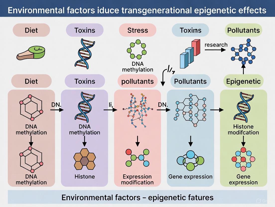
Diagram 1: Environmental triggers and their epigenetic pathways, showing how different stressors influence specific mechanisms leading to functional changes and potential inheritance.
Toxicants as Epigenetic Inducers
Mechanisms of Toxin-Induced Epigenetic Alterations
Environmental toxicants, including air pollutants, endocrine disruptors, heavy metals, and recreational substances such as tobacco smoke and alcohol, can induce profound epigenetic changes through multiple mechanisms [19]. These toxicants often directly interact with epigenetic regulatory machinery, modifying the activity of DNMTs, histone-modifying enzymes, and RNA interference pathways.
Notably, many toxins trigger oxidative stress responses that subsequently alter the epigenetic landscape. For example, arsenic exposure has been shown to induce transgenerational effects on learning and memory in rats through a crosstalk between arsenic methylation, hippocampal metabolism, and histone modifications [24]. Similarly, nicotine exposure in male mice produces behavioral impairment across multiple generations of descendants, suggesting stable inheritance of nicotine-induced epigenetic marks [24].
Transgenerational Inheritance of Toxicant-Induced Effects
The potential for transgenerational epigenetic inheritance of toxicant-induced effects remains a subject of intense investigation and debate. In mammals, efficient epigenetic reprogramming during gametogenesis and early embryogenesis typically erases most acquired epigenetic marks [23]. However, some marks may evade this reprogramming, particularly those located at genomic regions resistant to demethylation, such as centromeric satellites and imprinted loci [5].
A critical consideration in this field is distinguishing between true transgenerational inheritance versus intergenerational effects. Intergenerational effects involve direct exposure of the germline (F1 generation) when a pregnant F0 female is exposed, affecting the F2 generation. In contrast, transgenerational effects manifest in the F3 generation and beyond without direct exposure [5]. While compelling examples exist in plants and invertebrates, evidence in mammals is more limited. The Agouti mouse model represents one of the best-characterized examples, where variable DNA methylation at a transposon inserted near a coat color gene shows modest heritability [6].
Table 2: Selected Toxicants and Their Documented Epigenetic Effects
| Toxicant Category | Specific Examples | Documented Epigenetic Changes | Potential Health Outcomes |
|---|---|---|---|
| Air Pollutants | Particulate matter, polycyclic aromatic hydrocarbons | Altered global DNA methylation, histone modifications in stress response genes | Respiratory diseases, cardiovascular impairment, accelerated aging |
| Heavy Metals | Arsenic, lead, cadmium | DNA methylation changes in genes involved in oxidative stress response and DNA repair | Cognitive deficits, cardiovascular disease, increased cancer risk |
| Endocrine Disruptors | Bisphenol A, phthalates | Altered methylation of genes involved in hormonal signaling and development | Reproductive disorders, metabolic syndrome, developmental abnormalities |
| Recreational Substances | Tobacco smoke, alcohol | Genome-wide DNA methylation changes, histone modifications in addiction-related pathways | Addiction, cardiovascular disease, cancer, behavioral disorders |
Nutritional Stress and Epigenetic Modulation
Prenatal and Early-Life Nutritional Programming
Early life represents a period of exceptional epigenetic plasticity, during which nutritional factors can establish lasting epigenetic patterns that influence health trajectories into adulthood [20]. The Developmental Origins of Health and Disease (DOHaD) hypothesis formalizes this concept, proposing that early life nutritional conditions program an individual's disease risk in later life [25].
According to DOHaD principles, the developing fetus utilizes environmental cues, including nutrient availability, to determine an optimal phenotype for survival in the anticipated postnatal environment. However, when a mismatch occurs between prenatal predictions and actual postnatal conditions, this programming becomes maladaptive, increasing disease risk [25]. This nutritional programming is mediated through epigenetic mechanisms that establish stable gene expression patterns.
Specific Nutritional Influences on Epigenetic Machinery
Specific nutrients directly participate in epigenetic modifications as methyl donors, enzyme co-factors, or metabolic regulators. Methyl donors such as folate, choline, and betaine provide methyl groups for DNA and histone methylation reactions. Micronutrients including vitamin B12, zinc, and selenium serve as essential cofactors for epigenetic enzymes such as DNMTs and histone deacetylases (HDACs).
The relationship between nutrition and epigenetics is bidirectional; while nutrients influence epigenetic states, epigenetic mechanisms also regulate nutrient metabolism. For instance, DNA methylation patterns of metabolic genes can influence how individuals respond to dietary interventions, contributing to the variable effectiveness of nutritional therapies across populations [26].
Psychological Trauma as an Epigenetic Inducer
Neurobiological Pathways Linking Trauma and Epigenetics
Psychological trauma, particularly during sensitive developmental windows, can induce stable epigenetic changes that shape long-term mental and physical health outcomes [20] [24]. The primary neurobiological pathway mediating this relationship is the hypothalamic-pituitary-adrenal (HPA) axis, which regulates stress response through glucocorticoid signaling [20].
Early life stress and trauma exposure have been associated with DNA methylation changes in key HPA axis regulatory genes, including the glucocorticoid receptor gene (NR3C1) and FKBP5 [20]. These epigenetic modifications alter HPA axis function, leading to either hyperactive or blunted stress responses that predispose individuals to psychiatric disorders such as depression, anxiety, and post-traumatic stress disorder (PTSD) [20] [24].
The diathesis-stress model provides a framework for understanding how genetic vulnerabilities interact with environmental stressors, with epigenetic mechanisms serving as the molecular interface of this interaction [24]. This model suggests that individuals with genetic predispositions to mental illness may require specific environmental triggers to manifest the disorder, with epigenetic changes mediating this process.
Intergenerational and Transgenerational Transmission of Trauma
Emerging evidence suggests that the epigenetic effects of psychological trauma may extend across generations. A groundbreaking 2025 study with three generations of Syrian refugee families identified distinct epigenetic signatures of violence exposure across generations, including germline-associated differential methylation [25]. This study compared families with different timing of violence exposure (1980 vs. 2011) and found 14 differentially methylated positions associated with germline exposure and 21 with direct violence exposure [25].
Notably, most of these differentially methylated positions showed the same directionality in DNA methylation changes across germline, prenatal, and direct exposures, suggesting a common epigenetic response to violence [25]. The study also identified epigenetic age acceleration in children with prenatal exposure to violence, highlighting the particular vulnerability of the in utero developmental period [25].
Animal models provide mechanistic insights into how trauma-induced epigenetic changes might be transmitted. In mice, chronic psychosocial stress alters DNA methylation patterns in male germ cells, with these changes potentially transmitted to offspring [24]. Similarly, early trauma in mice (unpredictable maternal separation and stress) leads to altered miRNA expression in sperm and behavioral changes that persist across multiple generations [5].
Diagram 2: Psychological trauma epigenetic pathway, illustrating the neurobiological and molecular mechanisms linking stress exposure to health outcomes and transgenerational effects.
Experimental Approaches and Methodologies
Epigenome-Wide Association Studies (EWAS)
Epigenome-wide association studies (EWAS) have emerged as a powerful approach for identifying epigenetic signatures associated with environmental exposures. These studies typically utilize microarray-based platforms, such as the Illumina EPIC BeadChip which assays over 850,000 CpG sites, to conduct comprehensive DNA methylation analyses across the genome [25].
The Syrian refugee trauma study exemplifies a robust EWAS design, incorporating a three-generation cohort with contrasting developmental exposures to violence—direct exposure, prenatal exposure, and germline exposure [25]. This study employed a two-stage analytic approach: first using robust linear regression to identify differentially methylated positions associated with violence trauma, followed by generalized estimating equations (GEE) to account for family clustering [25]. Significant hits were determined using strict Bonferroni correction for multiple testing (p value < 6.5E-8) [25].
Emerging Technologies and Novel Approaches
Technological advances continue to enhance our ability to detect and interpret epigenetic changes. Next-generation sequencing (NGS) methods now enable base-resolution mapping of DNA methylation, histone modifications, and chromatin accessibility across the entire genome. Single-cell epigenomics is revolutionizing the field by allowing researchers to examine epigenetic heterogeneity within complex tissues and track epigenetic dynamics during development.
Liquid biopsies that analyze extracellular vesicles (EVs) represent another promising approach. As Andrea Baccarelli explains, "Extracellular vesicles are fascinating because they offer a way to study tissues that we can't easily access through traditional methods" [22]. These tiny, membrane-bound vesicles carry molecular messages (RNA, proteins, lipids) that reflect the health and function of their cells of origin, including neurons in the brain, allowing noninvasive assessment of organ-specific epigenetic responses [22].
The Scientist's Toolkit: Essential Research Reagents and Platforms
Table 3: Key Research Reagents and Platforms for Epigenetic Studies
| Tool Category | Specific Examples | Primary Applications | Technical Considerations |
|---|---|---|---|
| Methylation Analysis Platforms | Illumina EPIC BeadChip, whole-genome bisulfite sequencing | Genome-wide DNA methylation profiling, differential methylation analysis | Coverage limitations (BeadChip) vs. comprehensive mapping (sequencing) |
| Histone Modification Tools | ChIP-seq kits, histone modification-specific antibodies | Mapping histone modifications genome-wide, quantifying specific marks | Antibody specificity critical, requires high-quality chromatin preparation |
| Non-coding RNA Analysis | Small RNA sequencing, miRNA microarrays | Profiling miRNA and other small non-coding RNAs | Specialized library preparation needed for small RNAs |
| Sample Preservation | PAXgene Blood RNA tubes, FFPE tissue protocols | Preserving epigenetic marks in clinical samples | FFPE can cause nucleic acid fragmentation requiring specialized repair protocols |
| Data Analysis Platforms | R/Bioconductor packages, specialized EWAS software | Statistical analysis of epigenetic data, correcting for cell type heterogeneity | Computational intensity varies by approach, cell type deconvolution often necessary |
Implications for Drug Development and Therapeutic Interventions
Epigenetic Biomarkers in Clinical Applications
Epigenetic biomarkers offer significant potential for advancing clinical practice, particularly in the realms of disease diagnosis, prognosis, and treatment monitoring [26]. These biomarkers provide several advantages over genetic markers, including their dynamic nature, responsiveness to environmental influences, and ability to provide functional information about gene regulation [26].
In oncology, several epigenetic biomarkers have already been translated to clinical use. The SEPT9 methylation test (Epi proColon) for colorectal cancer detection demonstrates how DNA methylation signatures can be leveraged for non-invasive cancer screening [21]. Similarly, CDO1 gene hypermethylation shows promise as a diagnostic marker for multiple cancer types, detectable in various body fluids including plasma and urine [21].
Beyond cancer, epigenetic biomarkers hold potential for assessing mental health risks and treatment responses. Research indicates that changes in gene expression within limbic brain regions associated with depression and stress-related disorders involve aberrant epigenetic regulation [24]. Furthermore, antidepressant medications may exert their therapeutic effects, at least partially, through epigenetic mechanisms [24].
Epigenetic-Targeted Therapeutics
The reversible nature of epigenetic modifications makes them attractive targets for therapeutic intervention. Epigenetic drugs currently in clinical use primarily target DNA methyltransferases (e.g., azacitidine, decitabine) and histone deacetylases (e.g., vorinostat, romidepsin) [21]. However, these first-generation epigenetic therapies lack specificity, leading to broad genome-wide effects and significant side effects.
Next-generation epigenetic therapies aim for greater precision by targeting specific readers, writers, or erasers of epigenetic marks. The development of precision environmental health (PEH) approaches represents a complementary strategy, focusing on preventing environmentally-induced epigenetic changes before they contribute to disease pathogenesis [22]. As Andrea Baccarelli explains, "True health develops during the months, years, and lifetime before someone gets a diagnosis — the times when we are best poised to intervene" [22].
Diagram 3: Germline reprogramming and epigenetic inheritance, showing mechanisms by which epigenetic marks escape erasure and the strength of evidence across species.
Environmental triggers—including toxicants, nutritional stress, and psychological trauma—function as potent epigenetic inducers that shape disease susceptibility and health trajectories across the lifespan. The emerging evidence for transgenerational epigenetic inheritance of environmentally-induced effects represents a fundamental shift in our understanding of heredity, suggesting that ancestral experiences can influence offspring biology without changes to DNA sequence.
While the field has made remarkable progress, significant challenges remain. The mammalian germline undergoes extensive epigenetic reprogramming, creating formidable barriers to transgenerational inheritance [23] [6]. Many reported effects in mammals are actually intergenerational rather than truly transgenerational, reflecting direct exposure of the germline rather than stable inheritance across multiple unexposed generations [5]. Furthermore, as noted in a 2024 critical perspective, "the evidence for many potentially important forms of environmentally induced epigenetic inheritance remains inconclusive" [6].
Future research directions should prioritize standardized methodologies for assessing transgenerational epigenetic inheritance, improved epigenetic editing tools for causal validation, and expanded longitudinal multi-generational cohorts in human populations. The development of an "epigenetic score meter"—a tool that can disentangle the relationship between genetic and environmental influences—represents a promising approach for advancing precision medicine applications [24].
As the field evolves, integrating epigenetic perspectives into public health strategies offers the potential to move beyond treatment toward genuine prevention of environmentally-mediated diseases. By understanding how environmental triggers become biologically embedded through epigenetic mechanisms, we can develop more effective interventions to promote health across generations.
Epigenetic reprogramming in the mammalian germline involves two extensive waves of genome-wide DNA demethylation to reset epigenetic information for totipotency. However, specific genomic regions resist this erasure, retaining epigenetic marks that can be transmitted transgenerationally. This whitepaper examines the molecular mechanisms that enable certain epigenetic signatures to escape developmental reprogramming, focusing on the characteristics of resistant genomic loci and the experimental evidence supporting this phenomenon. Within the context of environmental epigenetics, we explore how exposure to various factors—including toxicants, nutrients, and other stressors—can induce stable epigenetic alterations in the germline that bypass reprogramming barriers. These escaped epimutations are associated with heritable disease susceptibilities and phenotypic changes, presenting novel considerations for drug development and therapeutic targeting.
The mammalian genome undergoes two global epigenetic reprogramming events during early development: first in primordial germ cells (PGCs), the precursors to sperm and eggs, and second in the pre-implantation embryo after fertilization [27] [28]. These reprogramming waves involve massive DNA demethylation through both passive (replication-dependent) and active (enzyme-mediated) mechanisms, erasing most epigenetic marks to restore totipotency and reestablish developmental plasticity [27]. The reprogramming process in PGCs is particularly comprehensive, with global DNA methylation levels decreasing to less than 5%,
Despite this genome-wide erasure, certain genomic regions exhibit resistance to demethylation, maintaining their epigenetic signatures through both reprogramming events [28]. These "escapee" regions include imprinted genes, transposable elements, and other sequences with potential regulatory significance [27] [28]. Environmentally induced epigenetic modifications that strategically locate within these resistant regions can thereby evade erasure and become stably inherited across generations, providing a plausible molecular mechanism for transgenerational epigenetic inheritance (TEI) of acquired traits and disease susceptibilities [2] [29] [28].
Molecular Mechanisms of Epigenetic Escape
DNA Methylation and Demethylation Pathways
DNA methylation primarily occurs at CpG dinucleotides, resulting in 5-methylcytosine (5mC), which is generally associated with transcriptional repression. The DNA methyltransferases DNMT3A and DNMT3B perform de novo methylation, while DNMT1 acts as a maintenance methyltransferase during cell division [27]. Active demethylation involves ten-eleven translocation (TET) enzymes (TET1, TET2, TET3) that oxidize 5mC to 5-hydroxymethylcytosine (5hmC), 5-formylcytosine (5fC), and 5-carboxycytosine (5caC), which can then be excised by thymine-DNA glycosylase (TDG) in conjunction with base excision repair [27].
During PGC development, global demethylation occurs through both active and passive mechanisms. Passive demethylation results from repression of DNMTs and UHRF1 (which directs DNMT1 to replication foci), while active demethylation is associated with increased TET family expression [27]. Human PGCs specifically repress UHRF1, DNMT3A, and DNMT3B, while enriching TET1 and TET2 compared to surrounding somatic cells [27].
Genomic Regions Resistant to Reprogramming
The following table summarizes the primary genomic regions and sequence elements that demonstrate resistance to epigenetic reprogramming:
Table 1: Genomic Regions Resistant to Epigenetic Reprogramming
| Resistant Region | Resistance Mechanism | Functional Significance | Representative Examples |
|---|---|---|---|
| Imprinted Genes | Protection from TET-mediated oxidation; histone modifications maintain methylation | Maintain parental-origin-specific expression; crucial for development | Various imprinted loci maintaining differential methylation [28] |
| Transposable Elements (TEs) | Dense methylation prevents reactivation and retrotransposition | Genome stability maintenance | IAP elements in agouti gene [28]; LINE elements in sheep [28] |
| Subtolomeric Regions | Specific chromatin context; positional effects | Chromosome stability | Pericentromeric repeats; subtelomeric regions [28] |
| Low-Complexity Repeats | Unknown; potentially related to chromatin accessibility | Possible role in timing of gene expression during development | Simple sequence repeats in sheep [28] |
| Non-Imprinted Genes | Specific DNA binding factors; histone mark enrichment | Developmental processes; disease susceptibility | Genes affecting growth, fertility, neural development [28] |
Research in sheep models has revealed that a significant proportion (63.5%) of transgenerationally inherited differentially methylated cytosines (DMCs) reside within repetitive element regions, with the majority located in long interspersed nuclear elements (LINEs) [28]. This suggests that the genomic and chromatin context significantly influences susceptibility to epigenetic escape.
Experimental Evidence for Escape Mechanisms
Key Studies Demonstrating Epigenetic Escape
Multiple experimental approaches across species have provided compelling evidence for epigenetic escape mechanisms. The following table summarizes pivotal studies in this field:
Table 2: Key Experimental Evidence for Epigenetic Escape Mechanisms
| Study System | Environmental Exposure | Escaped Epigenetic Marks | Transgenerational Phenotype | Reference |
|---|---|---|---|---|
| Mouse (Agouti) | Maternal methyl donor diet | DNA methylation at IAP transposon upstream of agouti gene | Altered coat color (yellow), obesity | [28] |
| Mouse (AxinFu) | Methyl donor supplementation | DNA methylation at TE within AxinFu allele | Kinked tail phenotype | [28] |
| Sheep | Paternal methionine supplementation | 107 DMCs in CG, CHH, and CHG contexts | Altered growth traits, male fertility | [28] |
| Rat | Various environmental toxicants (plastics, pesticides, jet fuel) | Disease-specific differential DNA methylation regions (DMRs) in sperm | Kidney disease, prostate disease, testis disease, obesity, pubertal abnormalities | [2] |
| Human PGCs | In vivo development analysis | Specific histone modification patterns (H3K27me3 retention) | Potential impact on germ cell differentiation | [27] |
Characteristics of Escaped Regions in Sheep Model
A detailed analysis of transgenerationally inherited DMCs in a sheep model exposed to paternal methionine supplementation revealed several key characteristics of regions escaping epigenetic reprogramming:
- Genomic Distribution: 65% located in intergenic regions, 33% in intronic regions, and 2% in promoter regions [28]
- Sequence Context: 82 of 107 TEI DMCs (76.6%) were in CG context, 20 (18.7%) in CHH, and 5 (4.7%) in CHG context [28]
- Functional Associations: Genes associated with these escaped regions impact growth, development, male fertility, cardiac disorders, and neurodevelopment [28]
- Neurological Links: Interestingly, 21 of 34 transgenerationally methylated genes have associations with neural development and brain disorders, including autism, schizophrenia, bipolar disease, and intellectual disability, suggesting a potential genetic overlap between brain and infertility disorders [28]
Experimental Protocols for Studying Escape Mechanisms
Methylated DNA Immunoprecipitation Sequencing (MeDIP-Seq)
Purpose: To comprehensively map genome-wide DNA methylation patterns in germ cells and evaluate resistance to reprogramming.
Detailed Methodology:
- Sperm Collection: Collect sperm from F0, F1, F2, and F3 generation males to distinguish transgenerational from intergenerational effects
- DNA Extraction and Fragmentation: Isolate genomic DNA and fragment to 100-500bp using sonication or enzymatic digestion
- Methylated DNA Immunoprecipitation: Incubate fragmented DNA with 5-methylcytosine-specific antibody; pull down immunoprecipitated methylated DNA fragments
- Library Preparation and Sequencing: Prepare sequencing libraries from immunoprecipitated DNA; sequence using high-throughput platforms
- Bioinformatic Analysis: Map sequences to reference genome; identify differentially methylated regions (DMRs) with statistical significance; use 1kb DMR size to improve bioinformatics accuracy [2]
Key Considerations: Updated MeDIP procedures with advanced reagents improve reproducibility and accuracy compared to earlier methodologies [2].
Transgenerational Inheritance Study Design
Purpose: To distinguish true transgenerational epigenetic inheritance from intergenerational effects.
Standard Protocol:
- F0 Generation Exposure: Expose gestating female mammals during fetal gonadal development (e.g., embryonic days 8-14 in rats) to environmental factors
- F1 Generation: Breed exposed F0 females to generate F1 offspring; these are directly exposed as germ cells in utero
- F2 Generation: Breed F1 males and females to generate F2 offspring; in females, this represents the first unexposed generation
- F3 Generation: Breed F2 males and females to generate F3 offspring; the first truly transgenerational cohort with no direct exposure [2] [29]
Critical Consideration: True transgenerational inheritance in mammals requires demonstration of inherited phenotypes and epimutations to at least the F3 generation after maternal exposure or F2 after paternal exposure, excluding direct exposure effects [29].
Visualization of Escape Mechanisms
Germline Reprogramming and Escape Pathways
Diagram 1: Germline Reprogramming and Escape Pathways. This diagram illustrates how environmental exposures induce germline epigenetic modifications that face extensive reprogramming in primordial germ cells (PGCs). While most epigenetic marks are erased (green pathway), resistant genomic regions enable epigenetic escape (red pathway), leading to transgenerational inheritance.
Genomic Distribution of Escaped Epimutations
Diagram 2: Genomic Distribution of Escaped Epimutations. This diagram visualizes the genomic distribution and functional impact of transgenerationally inherited differentially methylated cytosines (DMCs) based on data from a sheep nutritional epigenetics study.
The Scientist's Toolkit: Essential Research Reagents
Table 3: Essential Research Reagents for Studying Epigenetic Escape
| Reagent/Category | Specific Examples | Research Application | Key Function |
|---|---|---|---|
| DNA Methylation Inhibitors | 5-azacytidine, zebularine | Experimental manipulation of DNA methylation | DNMT inhibition; creates hypomethylated state for comparison |
| Methylation Detection Kits | Methylated DNA Immunoprecipitation (MeDIP) kits, bisulfite conversion kits | Mapping genome-wide methylation patterns | Selective enrichment or conversion for methylation analysis |
| TET Enzyme Modulators | Vitamin C (TET activator), TET inhibitors | Manipulating active demethylation pathways | Studying TET-mediated oxidation in reprogramming |
| Antibodies for Epigenetic Marks | 5-methylcytosine, 5-hydroxymethylcytosine, H3K27me3, H3K4me3 | Immunostaining, MeDIP, ChIP experiments | Detection and enrichment of specific epigenetic modifications |
| Bisulfite Sequencing Kits | EZ DNA Methylation kits, Pyrosequencing kits | Single-base resolution methylation analysis | Converting unmethylated cytosines to uracils while preserving methylated cytosines |
| In Vitro PGC Culture Systems | Human PGC-like cells (hPGCLCs) | Modeling early human germline development in vitro | Studying reprogramming dynamics without ethical constraints of human embryos |
Implications for Disease Etiology and Drug Development
The escape of epigenetic marks from germline reprogramming provides a plausible mechanism for the transgenerational inheritance of disease susceptibility. Studies have demonstrated that exposure to various environmental toxicants—including hydrocarbons, dioxins, pesticides, plastics, and herbicides—promotes the epigenetic transgenerational inheritance of pathologies affecting the kidney, prostate, testes, pubertal development, and metabolic function [2]. Importantly, the disease-specific differentially methylated regions (DMRs) in sperm are exposure-specific for each pathology with negligible overlap, suggesting that different environmental exposures influence unique subsets of DMRs and genes to promote the developmental origins of disease [2].
For drug development professionals, these escape mechanisms present both challenges and opportunities. The stable inheritance of epigenetic states suggests that:
- Early-life environmental exposures may create disease susceptibilities that manifest generations later
- Epigenetic biomarkers in sperm may serve as predictors for transgenerational disease risks
- Therapeutic interventions targeting epigenetic modifiers (TET enzymes, DNMTs, histone modifiers) may potentially reverse inherited epimutations
- Drug safety profiles may need to consider potential transgenerational epigenetic effects
The nervous system appears particularly vulnerable to transgenerational epigenetic influences, with numerous studies reporting inherited behavioral phenotypes and stress responses [29]. This aligns with findings that genes escaping reprogramming are enriched for functions in nervous system development and association with brain disorders including autism, schizophrenia, and bipolar disease [28].
Epigenetic escape from germline reprogramming represents a sophisticated molecular mechanism enabling the transgenerational transmission of environmentally acquired information. Specific genomic regions—including imprinted genes, transposable elements, and other sequences with particular chromatin contexts—demonstrate inherent resistance to the extensive DNA demethylation that characterizes PGC development. When environmentally induced epigenetic modifications strategically target these resistant regions, they can bypass reprogramming barriers and become stably inherited across generations.
The growing evidence for this phenomenon necessitates a expanded framework for disease etiology that integrates environmental epigenetics with traditional genetic models. For researchers and drug development professionals, understanding these escape mechanisms provides critical insights into the molecular basis of transgenerational disease inheritance and potential avenues for therapeutic intervention. Future research should focus on precisely characterizing the molecular signatures of resistant regions, developing more sophisticated models for studying human germline epigenetics, and exploring pharmacological approaches to modulate these inherited epigenetic states.
The field of transgenerational epigenetics has revolutionized our understanding of how environmental exposures can shape health and disease across generations. This paradigm challenges the conventional dogma that inheritance is solely governed by DNA sequence, revealing that environmental factors can induce epigenetic modifications that are transmitted to subsequent generations. These discoveries provide a mechanistic bridge between nature and nurture, explaining how parental and ancestral experiences can influence offspring biology without altering the genetic code itself [30].
This whitepaper examines three foundational research areas that have been instrumental in establishing the principles of environmental epigenetics and transgenerational inheritance. The Agouti mouse model serves as a quintessential epigenetic biosensor, demonstrating how nutritional and chemical exposures during gestation can produce stable, heritable changes in phenotype through DNA methylation alterations. Human studies of the Dutch Hunger Winter cohort provide compelling evidence for fetal metabolic programming in response to prenatal nutritional deprivation. Finally, research on Holocaust survivors and their descendants reveals how severe psychological trauma can leave epigenetic signatures that are transmitted to subsequent generations, altering stress reactivity and social-emotional functioning.
Together, these seminal studies provide a comprehensive framework for understanding how diverse environmental exposures—from nutrients and toxicants to profound psychological stress—can produce lasting epigenetic changes with transgenerational health implications.
The Agouti Mouse Model: An Epigenetic Biosensor
Experimental Model and Molecular Mechanism
The viable yellow agouti (Avy) mouse represents a cornerstone model in environmental epigenetics research. This model centers on a metastable epiallele resulting from the insertion of an intracisternal A particle (IAP), a murine retrotransposon, upstream of the transcription start site of the Agouti gene [30]. The wild-type Agouti gene encodes a paracrine signaling molecule that regulates melanin production, typically resulting in a brown coat with a sub-apical yellow band on each hair shaft.
The Avy allele contains a cryptic promoter in the proximal end of the IAP that drives constitutive, ectopic expression of Agouti protein. This ectopic expression produces distinct phenotypic outcomes: yellow fur (when the IAP is unmethylated), pseudoagouti brown fur (when the IAP is methylated), or mottled variations (with intermediate methylation) [30]. Importantly, the degree of CpG methylation within the IAP's long terminal repeat (LTR) correlates inversely with ectopic Agouti expression and is established stochastically during early embryonic development, making it highly sensitive to environmental influences during critical developmental windows.
Key Experimental Protocols
- Animal Model and Breeding Scheme: Female a/a mice are bred with Avy/a males, producing isogenic offspring with identical Avy alleles but potentially different epigenetic regulations. This genetic uniformity allows researchers to attribute phenotypic variation to epigenetic rather than genetic differences [30].
- Exposure Paradigms: Gestating F0 dams are exposed to specific environmental factors during the period of fetal gonadal sex determination (approximately embryonic days 8-14 in mice), when epigenetic marks are being established and are particularly vulnerable to reprogramming.
- Phenotypic Assessment: Coat color is scored in Avy/a offspring as a visual biomarker of the underlying epigenetic status. Yellow coat indicates hypomethylation of the Avy IAP, while pseudoagouti coat indicates hypermethylation.
- Molecular Analysis: DNA methylation at specific CpG sites within the Avy IAP LTR is quantified using bisulfite sequencing in tissues derived from all three germ layers to confirm that epigenetic changes were established early in development.
- Long-Term Health Monitoring: Offspring are monitored for adult-onset conditions including obesity, diabetes, and tumorigenesis, which are associated with ectopic Agouti expression.
Seminal Findings and Transgenerational Implications
Research using the Agouti mouse model has yielded several groundbreaking discoveries:
- Nutritional Effects: Maternal dietary supplementation with the soy phytoestrogen genistein (250 mg/kg diet) shifted offspring coat color toward pseudoagouti and increased methylation at six CpG sites within the Avy IAP. This hypermethylation persisted into adulthood and protected offspring from obesity [30].
- Toxicant Effects: Maternal exposure to bisphenol A (BPA at 50 mg/kg diet) shifted offspring coat color toward yellow and decreased methylation at nine CpG sites within the Avy IAP. This hypomethylating effect was also observed at another metastable epiallele (CabpIAP), suggesting BPA's epigenetic impact is not gene-locus specific [30].
- Nutritional Protection: The BPA-induced hypomethylation was abolished when mothers received supplemental methyl donors (folic acid, betaine, vitamin B12, and choline) or the phytoestrogen genistein alongside BPA exposure, demonstrating that dietary interventions can counteract environmental toxicants [30].
The Agouti model provides critical evidence that epigenetic marks established during early development are mitotically heritable and can produce lasting phenotypic consequences, serving as a mechanistic link between early environmental exposures and adult disease susceptibility.
Dutch Hunger Winter Studies: Human Famine Epigenetics
Historical Cohort and Study Design
The Dutch Hunger Winter of 1944-1945 represents a unique natural experiment in human transgenerational epigenetics. This six-month famine occurred during the German embargo of the western Netherlands, creating a well-defined period of severe nutritional deprivation in an otherwise well-nourished population. The cohort design allows researchers to compare individuals exposed to famine at different gestational stages with their unexposed same-sex siblings, controlling for genetic and familial confounding factors [31].
Key aspects of this historical cohort include:
- Precise Exposure Timing: Detailed birth records enable researchers to determine the exact gestational timing of famine exposure for each individual.
- Sibling Controls: The use of unexposed siblings of the same sex provides ideal matched controls with similar genetic backgrounds and postnatal environments.
- Long-Term Follow-Up: Health outcomes and biological samples have been collected from these individuals throughout their lives, with many studies conducted when participants were approximately 60 years old.
Methodological Approach
- Cohort Identification: Researchers identified individuals born in specific institutions in western Netherlands cities immediately after the famine, with well-documented birth records and medical histories.
- Clinical Phenotyping: Comprehensive metabolic assessments were conducted, including measurements of blood glucose, insulin sensitivity, lipid profiles, blood pressure, and body mass index.
- Epigenome-Wide Analysis: DNA from peripheral blood was analyzed using array-based or sequencing-based technologies to assess genome-wide methylation patterns at over 1.2 million CpG sites [31].
- Statistical Analysis: Robust statistical models compared methylation patterns between exposed individuals and their unexposed siblings, while controlling for potential confounders including cell type composition.
Key Epigenetic and Health Findings
- Differential Gene Regulation: Individuals prenatally exposed to famine showed differential DNA methylation at multiple genomic loci compared to their unexposed siblings, with particularly pronounced effects on genes involved in growth and development [31].
- Metabolic Consequences: These epigenetic changes were associated with adverse metabolic outcomes in adulthood, including elevated LDL cholesterol, increased body mass index, and higher rates of conditions like hyperglycemia and type 2 diabetes [31].
- Transgenerational Effects: Subsequent research on the Chinese famine (1959-1961) revealed that prenatal exposure to starvation was associated with elevated risks of hyperglycemia, type 2 diabetes, renal dysfunction, and chronic kidney disease not only in the directly exposed generation (F1) but also in their offspring (F2), particularly when both parents had been exposed [32].
The Dutch Hunger Winter studies provide the most compelling human evidence that prenatal nutritional environment can program the epigenome with lasting consequences for metabolic health, potentially transmitted across multiple generations.
Holocaust Survivor Research: Trauma-Associated Epigenetic Transmission
Study Populations and Design
Research on Holocaust survivors and their descendants has provided groundbreaking insights into how severe psychological trauma can produce intergenerational epigenetic effects. The seminal work by Rachel Yehuda and colleagues compared Holocaust survivors (with and without PTSD) and their adult offspring to Jewish control families living outside Europe during World War II [33] [34]. More recent studies have expanded to include third- and fourth-generation descendants [35].
Key aspects of the research design include:
- Rigorous Trauma Assessment: Detailed clinical interviews and standardized assessments (e.g., Clinician-Administered PTSD Scale) were used to characterize trauma exposure and psychiatric symptoms.
- Multi-Generational Approach: Studies examined epigenetic patterns across multiple generations to distinguish direct exposure effects from transgenerational transmission.
- Control for Direct Trauma: Careful screening ensured that offspring themselves had not experienced significant direct trauma that could confound results.
Molecular and Psychological Assessment Methods
- Epigenetic Analysis: DNA was typically extracted from peripheral blood mononuclear cells (PBMCs) or saliva. Target gene approaches initially focused on candidate genes related to stress regulation, particularly FKBP5, a key regulator of glucocorticoid receptor sensitivity [33]. More recent studies have expanded to include genes related to the oxytocin system and sympathetic nervous system [35].
- DNA Methylation Quantification: Bisulfite sequencing or pyrosequencing was used to quantify methylation at specific CpG sites within target genes.
- Stress Physiology Assessment: Hypothalamic-pituitary-adrenal (HPA) axis function was evaluated through cortisol measurements and dexamethasone suppression tests.
- Comprehensive Psychometric Evaluation: Validated instruments assessed PTSD symptoms, depression, anxiety, attachment styles, resilience, and quality of social-emotional ties [35].
Intergenerational and Transgenerational Findings
- Opposing Methylation Patterns in Parent-Child Pairs: Holocaust survivors showed approximately 10% higher methylation at a specific FKBP5 intronic region compared to controls, while their offspring showed 7.7% lower methylation at the same site [33]. This opposing pattern suggests complex adaptive mechanisms in intergenerational trauma transmission.
- Altered Stress Regulation: The epigenetic changes were associated with functional alterations in HPA axis regulation, with survivors showing cortisol suppression characteristic of PTSD, while their offspring exhibited enhanced stress reactivity [33].
- Third- and Fourth-Generation Adaptations: Recent research on third- and fourth-generation descendants reveals a dual pattern of both vulnerability and resilience. These individuals show DNA methylation patterns associated with stronger activation of the oxytocin system (enhancing social bonding) alongside distinct methylation in CRH, CRHBP, FKBP5, and NR3C1 genes linked to increased HPA axis activation and stress reactivity [35].
- Psychological Resilience: Despite these biological changes, third- and fourth-generation descendants do not show elevated levels of psychopathology, suggesting the development of successful adaptive mechanisms and resilience [35].
Comparative Analysis of Seminal Studies
Table 1: Comparative Analysis of Seminal Transgenerational Epigenetic Studies
| Research Aspect | Agouti Mouse Model | Dutch Hunger Winter | Holocaust Survivor Research |
|---|---|---|---|
| Environmental Exposure | Nutritional compounds (genistein), environmental toxicants (BPA) | Severe famine/nutritional deprivation | Severe psychological trauma |
| Species | Mouse (Mus musculus) | Human | Human |
| Key Epigenetic Mechanism | DNA methylation of IAP retrotransposon | DNA methylation of growth and developmental genes | DNA methylation of stress-related genes (FKBP5, NR3C1) and oxytocin system |
| Primary Tissue Analyzed | Multiple tissues from all germ layers | Peripheral blood | Peripheral blood mononuclear cells, saliva |
| Transgenerational Evidence | Intergenerational (F1) demonstrated; transgenerational potential suggested | Intergenerational (F1) and transgenerational (F2) effects observed | Intergenerational (F1) and transgenerational (F2, F3, F4) effects observed |
| Key Health Outcomes | Coat color change, obesity, diabetes, tumorigenesis | Metabolic disease, cardiovascular risk, altered growth | Altered stress reactivity, psychopathology risk, social-emotional adaptations |
| Strengths | Controlled genetics, causal inference, mechanistic insights | Well-defined exposure in human population, sibling controls | Rare human trauma model, multi-generational design, psychological and biological integration |
| Limitations | Mouse model may not fully recapitulate human biology | Retrospective design, potential confounding factors | Complex psychosocial confounding, tissue specificity of epigenetic marks |
Experimental Pathways and Workflows
Agouti Mouse Experimental Workflow
Human Cohort Study Design
Stress Pathway Epigenetic Regulation
Research Reagent Solutions and Methodologies
Table 2: Essential Research Reagents and Methodologies for Transgenerational Epigenetic Studies
| Research Tool Category | Specific Examples | Research Application |
|---|---|---|
| Animal Models | Avy mouse model, Outbred rat models | Controlled genetic background for environmental exposure studies |
| Epigenetic Analysis Reagents | Bisulfite conversion kits, Methylated DNA immunoprecipitation (MeDIP) reagents, DNA methyltransferase inhibitors, HDAC inhibitors | Detection and manipulation of DNA methylation and histone modifications |
| Molecular Biology Kits | Pyrosequencing kits, Whole-genome bisulfite sequencing kits, ChIP-seq kits, RNA-seq kits | Comprehensive epigenomic and transcriptomic profiling |
| Cell Culture Systems | Primary germ cells, Embryonic stem cells, Sperm and oocyte culture systems | In vitro mechanistic studies of epigenetic inheritance |
| Bioinformatics Tools | Epigenome-wide association study (EWAS) pipelines, Differential methylation analysis software, Multigenerational statistical models | Analysis of complex epigenetic datasets across generations |
| Physiological Assays | Cortisol ELISA kits, Glucose tolerance test reagents, Metabolic cages, Behavioral assessment tools | Phenotypic characterization of transgenerational effects |
The seminal studies explored in this whitepaper—from the Agouti mouse model to the Dutch Hunger Winter and Holocaust survivor research—collectively establish a robust foundation for understanding how environmental exposures can induce transgenerational epigenetic effects. These diverse research approaches demonstrate that both physical environmental factors (nutrition, toxicants) and psychological experiences (trauma) can produce stable epigenetic modifications that influence health and disease susceptibility across multiple generations.
The Agouti mouse model provides a mechanistic framework for understanding how environmental exposures during critical developmental windows can reprogram the epigenome. The Dutch Hunger Winter studies offer compelling human evidence for developmental metabolic programming through epigenetic mechanisms. The Holocaust survivor research reveals the complex interplay between biological and psychological factors in trauma transmission, highlighting both vulnerability and resilience pathways.
For researchers and drug development professionals, these findings have profound implications. They suggest that comprehensive understanding of disease etiology must consider ancestral environmental exposures and their epigenetic legacies. Furthermore, they highlight potential opportunities for developing epigenetic-based diagnostics and therapeutics that could mitigate transgenerational disease risk. As the field advances, integrating these perspectives will be essential for developing truly personalized medicine approaches that account for both genetic and environmental inheritance across generations.
Research Models and Analytical Approaches for Studying Transgenerational Epigenetics
Transgenerational epigenetic inheritance (TEI) represents a paradigm shift in understanding how environmental exposures influence physiology and disease risk across generations. This whitepaper examines how three key model organisms—C. elegans, rodents, and plants—provide complementary insights into the molecular mechanisms underlying this phenomenon. We synthesize current research demonstrating how environmental factors including pathogens, toxicants, and dietary changes can induce heritable epigenetic changes that persist for multiple generations. The distinct advantages of each system enable researchers to unravel conserved epigenetic pathways while addressing organism-specific inheritance mechanisms. This technical guide provides a comprehensive overview of experimental approaches, key findings, and methodological considerations for investigating environmentally induced transgenerational epigenetic effects across these model systems.
Transgenerational epigenetic inheritance (TEI) is defined as the germline transmission of epigenetic information between generations in the absence of continued environmental exposure [1]. This phenomenon represents a nongenetic form of inheritance wherein environmental experiences of one generation can influence gene expression and phenotype in subsequent, unexposed generations. The field has evolved from historical controversies to establishing mechanistic foundations through studies in model organisms that provide tractable systems for investigating these complex processes.
Epigenetic mechanisms include DNA methylation, histone modifications, noncoding RNAs, and chromatin remodeling complexes that regulate gene expression without altering the underlying DNA sequence [1] [4]. While these mechanisms normally ensure proper cellular differentiation and gene regulation during development, they can also be influenced by environmental exposures. When such environmentally induced epigenetic alterations escape the typical reprogramming events that occur during gametogenesis and early embryogenesis, they may be transmitted to subsequent generations.
The definition of "transgenerational" varies depending on the exposed organism. When a gestating female mammal (F0 generation) is exposed, the F1 generation fetus and the F2 generation germline are also directly exposed; therefore, observation of effects in the F3 generation (great-grand offspring) is required to demonstrate a true transgenerational phenomenon [1]. In contrast, for exposures in adult organisms, the F2 generation represents the first truly transgenerational cohort. This distinction is critical for proper experimental design and interpretation of transgenerational inheritance studies.
Caenorhabditis elegans as a Model System
The nematode C. elegans provides a powerful model for TEI research due to its short generation time (approximately 3 days), well-characterized genetics, and suitability for large-scale screening approaches. Several landmark studies have demonstrated the transmission of acquired traits across multiple generations in this organism, providing insights into conserved epigenetic mechanisms.
Key Advantages and Experimental Approaches
C. elegans offers several distinct advantages for epigenetic research:
- Rapid generation time enables investigation of multiple generations within weeks
- Self-fertilizing hermaphrodites facilitate the maintenance of epigenetic states without genetic recombination
- Conserved epigenetic machinery including RNAi pathways, histone modifiers, and DNA methylation regulators
- Transparent body allows in vivo observation of fluorescent reporters and cellular processes
- Complete cell lineage map and well-annotated genome support mechanistic studies
Recent evidence indicates that environmental stressors including methylmercury, arsenite, starvation, heat, bacterial infection, and mitochondrial inhibitors can produce profound effects on the epigenome that persist for multiple generations [36]. The experimental approaches commonly used in C. elegans epigenetic research include avoidance assays, fluorescence imaging of epigenetic reporters, genetic manipulation through RNAi and mutant strains, and high-throughput sequencing of epigenetic marks.
Mechanistic Insights from C. elegans Studies
Learned Avoidance of Pathogens
One of the most compelling examples of TEI in C. elegans involves the learned avoidance of pathogenic bacteria. Worms exposed to Pseudomonas aeruginosa (strain PA14) learn to avoid this pathogen, and this behavioral adaptation can be transmitted for up to four generations without re-exposure [37]. The bacterial small RNA P11 has been identified as necessary and sufficient to induce this transgenerational inheritance [37] [36]. The molecular pathway involves the RNA interference PIWI-interacting RNA (piRNA) pathway, Cer1 retrotransposon particles, and histone methylation to downregulate maco-1, a gene functioning in sensory neurons that regulates chemotaxis [36].
The standard avoidance assay protocol involves:
- Training phase: 24-hour exposure of parental generation (P0) to PA14 bacteria on slow-killing assay plates
- Transfer: Collection and washing of worms after training, with transfer to standard OP50 E. coli food source
- Testing: Placement of worms on assay plates with PA14 and OP50 at opposite ends, with sodium azide immobilizer added to each spot to capture initial choices
- Scoring: Counting worms in proximity to each bacterial spot after one hour
- Generational propagation: Collecting eggs from each generation to continue the lineage without re-exposure
This experimental paradigm has revealed that the initial attraction to PA14 in naive animals is converted to avoidance in trained animals and their F1 and F2 descendants [37]. Discrepancies in reproducibility between research groups have highlighted the importance of methodological details, particularly the use of sodium azide versus temperature shift for immobilization, which can affect the potential for additional learning during the assay itself [37].
Inheritance of Associative Memories
Beyond pathogen avoidance, C. elegans can transmit associative memories across generations. A recent study demonstrated that worms trained to associate the attractive odorant isoamyl alcohol (IAA) with starvation conditions passed this associative memory to F1 progeny, who showed odor-evoked avoidance behavior despite never having experienced starvation [38]. Furthermore, both F1 and F2 descendants exhibited odor-evoked cellular stress responses, manifested by translocation of DAF-16/FOXO to nuclei.
The molecular pathways involved in this inheritance include:
- Histone modifications: H3K9 mono/di-methylation (MET-2), H3K9 tri-methylation (SET-25), and H3K36 methylation (MET-1)
- Small RNA pathways: Argonaute proteins NRDE-3 and HRDE-1
- Neuropeptide signaling: EGL-3 dependent neuropeptide secretion
Notably, sperm—but not oocytes—were found to transmit these acquired cellular changes [38]. This research demonstrates that in C. elegans, both behavioral memories and associated cellular stress responses can be transmitted across generations through specific epigenetic mechanisms.
Protein-Based Epigenetic Inheritance
Beyond the well-established mechanisms involving small RNAs and histone modifications, recent research has uncovered a novel protein-based epigenetic inheritance mechanism in C. elegans. Amyloid-like protein aggregates called "herasomes" can transmit phenotypic traits across generations without changes to the DNA sequence [39]. These structures form in the germline and can induce transgenerational changes in fertility and sex determination when the mstr-1 and mstr-2 genes are mutated.
This discovery expands the potential mechanisms of epigenetic inheritance beyond nucleic acid-based processes and may have implications for understanding protein-based inheritance in other organisms, including humans where amyloid accumulation is associated with neurodegenerative diseases.
Figure 1: Molecular pathway of associative memory inheritance in C. elegans demonstrating key epigenetic mechanisms involved in transgenerational transmission.
Experimental Protocols for C. elegans TEI Research
Transgenerational Learned Avoidance Assay
Materials: PA14 and OP50 bacterial strains, NGM assay plates, sodium azide solution, synchronized L4 larval stage worms.
Procedure:
- Grow PA14 cultures overnight in LB broth at 37°C with shaking.
- Spot 5μl of PA14 and OP50 on opposite sides of 6cm NGM assay plates. Add 1μl of 1M sodium azide to each spot.
- Train parental (P0) generation by transferring ~100 young adult worms to PA14-seeded plates for 24 hours at 20°C.
- Collect trained worms, wash 3x in M9 buffer to remove bacteria.
- Transfer subset of trained worms to assay plates, count distribution after 1 hour.
- For subsequent generations, transfer eggs from trained worms to fresh OP50 plates. Repeat testing at each generation without re-exposure.
- Compare avoidance index: (number at OP50 - number at PA14) / total worms.
Key considerations: Maintain consistent bacterial growth conditions as P11 small RNA expression varies with PA14 growth phase [37]. Use sodium azide rather than temperature shift for immobilization to prevent additional learning during assay.
Associative Memory Training Protocol
Materials: Isoamyl alcohol (IAA), 1% agarose assay plates, starvation plates (lacking peptone).
Procedure:
- Prepare spaced training protocol: 1-hour starvation with IAA exposure, followed by 1-hour recovery on food without IAA, repeated 3-5 times.
- Test avoidance behavior by placing single worms on assay plates with IAA spot versus control spot.
- Score avoidance as transitions from forward to backward locomotion upon encountering IAA gradient.
- For cellular stress assays, use transgenic strains expressing DAF-16::GFP to monitor nuclear translocation.
- Collect eggs from avoiding worms to establish subsequent generations.
Key considerations: Pre-select P0 animals that show strong avoidance to establish lineages. Use mock-trained controls with starvation but no IAA pairing.
Rodent Models for Transgenerational Epigenetics
Rodent models, particularly rats and mice, provide essential insights into mammalian TEI with direct relevance to human health and disease. These systems enable investigation of complex physiological outcomes and tissue-specific epigenetic changes in organisms with closer evolutionary relationships to humans.
Key Advantages and Experimental Approaches
Rodent models offer several critical advantages for epigenetic research:
- Mammalian physiology with closer relevance to human systems
- Similar epigenetic reprogramming events during gametogenesis and embryogenesis
- Ability to study complex disease phenotypes including metabolic disorders, cancer, and reproductive abnormalities
- Tissue-specific analyses across multiple organ systems
- Well-established genetic tools including inbred strains, knockout models, and genome editing
The classic experimental design for rodent TEI studies involves exposure during critical developmental windows, particularly during gonadal sex determination when the germline undergoes extensive epigenetic reprogramming. Common exposure paradigms include endocrine disruptors, nutritional interventions, and stress regimens.
Mechanistic Insights from Rodent Studies
Environmental Toxicants and Reproductive Disease
Seminal studies using the endocrine disruptor vinclozolin demonstrated that exposure of gestating female rats during gonadal sex determination (E8-E14) could induce reproductive abnormalities persisting for multiple generations [1]. These transgenerational effects included decreased sperm counts, increased apoptosis in testicular germ cells, and increased incidence of adult-onset diseases such as prostate and kidney abnormalities.
The molecular mechanism involves altered DNA methylation patterns in the sperm epigenome that are transmitted across generations [1]. Subsequent studies have shown that other environmental toxicants including DDT, plastics components (BPA, phthalates), and hydrocarbon mixtures can produce similar transgenerational effects on reproductive health.
The following table summarizes key environmental exposures shown to induce transgenerational effects in rodent models:
Table 1: Environmental Exducers of Transgenerational Epigenetic Inheritance in Rodent Models
| Toxicant/Exposure | Transgenerational Effects Observed | Proposed Epigenetic Mechanism | References |
|---|---|---|---|
| Vinclozolin | Decreased sperm count/motility, testis apoptosis, prostate/kidney disease | Altered sperm DNA methylation patterns | [1] |
| DDT | Decreased sperm count, testis apoptosis | Germline epigenetic alterations | [1] |
| BPA and Phthalates | Seminiferous tubule atrophy, tubule vacuoles, germ cell agenesis | DNA methylation changes | [1] |
| Jet Fuel/Hydrocarbon Mixture | Increased testis apoptosis | Epigenetic reprogramming of male germline | [1] |
| Benzo[a]pyrene | Seminiferous tubule defects | Not specified | [1] |
| Permethrin/DEET | Seminiferous tubule atrophy, vacuoles, germ cell agenesis | Not specified | [1] |
The Agouti Mouse Model
The Agouti viable yellow (Aᵛʸ) mouse represents one of the best-characterized examples of TEI in mammals. In this model, a retrotransposon inserted upstream of the Agouti gene shows variable DNA methylation that correlates with coat color ranging from yellow (hypomethylated) to pseudoagouti (hypermethylated) [6]. This epigenetic variability is heritable and demonstrates that transposable elements can serve as substrates for TEI in mammals.
Early claims that maternal diet could influence the epigenetic status of the Aᵛʸ allele in offspring have been challenged by subsequent larger studies that failed to validate these findings [6]. This highlights the importance of robust experimental design and replication in TEI research. A broader screen for similar metastable epialleles in the mouse genome found that the Aᵛʸ locus is exceptional rather than representative of widespread epigenetic inheritance mechanisms [6].
Experimental Protocols for Rodent TEI Research
Multigenerational Exposure Design
Materials: Timed-pregnant females, exposure compounds, appropriate vehicle controls.
Procedure:
- Expose pregnant F0 females during critical developmental window (e.g., E8-E14 for gonadal sex determination in rats).
- Cross resulting F1 offspring to generate F2 generation.
- Cross F2 offspring to generate F3 generation - the first truly transgenerational cohort.
- For adult exposures, begin with F0 adults and examine F2 offspring as transgenerational cohort.
- Collect tissues across generations for phenotypic analysis and epigenetic profiling.
Key considerations: Use appropriate vehicle controls and maintain consistent housing conditions. Large cohort sizes are needed due to potential incomplete penetrance. Blind phenotypic assessments to prevent bias.
Epididymal Sperm Collection for Epigenetic Analysis
Materials: Dissection tools, PBS buffer, tissue homogenizer, DNA/RNA extraction kits.
Procedure:
- Euthanize adult male rodents according to IACUC protocols.
- Isister epididymides and transfer to PBS buffer.
- Puncture epididymides and incubate at 37°C for 15 minutes to allow sperm swim-out.
- Filter sperm suspension through 40μm cell strainer to remove tissue debris.
- Centrifuge at 5000xg for 10 minutes to pellet sperm.
- Extract DNA for bisulfite sequencing or RNA for small RNA sequencing.
Key considerations: Include somatic cell controls to assess purity of sperm preparation. Use appropriate preservation methods for different epigenetic analyses.
Plant Models for Epigenetic Inheritance
Plants exhibit robust transgenerational epigenetic inheritance and provide unique insights into mechanisms that may be conserved in other kingdoms. Their sessile nature has likely selected for enhanced epigenetic plasticity as an adaptive strategy to cope with environmental challenges.
Key Advantages and Experimental Approaches
Plant models offer several distinctive advantages for epigenetic research:
- Absence of dedicated germline allows more direct transmission of somatic epigenetic states
- Easily observable phenotypic markers for epigenetic states (e.g., flower symmetry, pigmentation)
- Well-characterized epigenetic mutants with defects in DNA methylation and histone modification pathways
- Rapid generation times for some species (e.g., Arabidopsis)
- Natural epialleles that can be studied in ecological contexts
Common experimental approaches in plant epigenetic research include analysis of natural epialleles, induction of epigenetic changes through environmental stress, characterization of epigenetic mutants, and crossing experiments to assess mitotic and meiotic stability of epigenetic states.
Mechanistic Insights from Plant Studies
DNA Methylation Pathways
Plants maintain complex DNA methylation patterns involving three distinct pathways:
- CG methylation maintenance by MET1 (DNA Methyltransferase 1)
- CHG methylation through the CMT3/SUVH pathway involving histone H3K9 methylation
- RNA-directed DNA methylation (RdDM) for de novo methylation and CHH context maintenance
This intricate system allows for stable inheritance of DNA methylation patterns while maintaining plasticity in response to environmental signals [40]. DNA methylation in plants serves to regulate gene expression, particularly at transposable elements where it maintains genome stability.
Natural Epialleles and Paramutation
Several well-characterized natural epialleles demonstrate the phenotypic consequences of TEI in plants:
- The Lcyc gene in Linaria vulgaris controls floral symmetry. Increased DNA methylation in the promoter region converts flowers from bilateral to radial symmetry, a change that is heritable [41] [40].
- The SUPERMAN (SUP) gene in Arabidopsis thaliana displays epigenetic alleles (clark kent alleles) associated with hypermethylation and changes in floral organ number [40].
- Paramutation at the b1 and r1 loci in maize involves meiotically heritable changes in gene expression mediated by interactions between alleles [41].
These examples illustrate how stable epigenetic variants can produce heritable phenotypic diversity in natural plant populations, potentially contributing to adaptation.
Figure 2: DNA methylation maintenance pathways in plants showing three distinct mechanisms for perpetuating methylation patterns across generations.
Stress-Induced Epigenetic Changes
Environmental stresses including drought, UV radiation, cold, and pathogens can trigger heritable epigenetic changes in plants [41] [40]. These changes often involve alterations in DNA methylation patterns and increased recombination frequencies, potentially generating genetic diversity that may be adaptive under stressful conditions.
This stress-responsive epigenetic plasticity may represent an evolutionary adaptation for sessile organisms to cope with rapidly changing environments. The transmission of stress-induced epigenetic states across generations could provide a form of "molecular memory" that prepares offspring for similar environmental challenges.
Experimental Protocols for Plant TEI Research
DNA Methylation Analysis
Materials: Plant tissue, DNA extraction kits, bisulfite conversion kits, PCR reagents.
Procedure:
- Extract genomic DNA from plant tissue of interest.
- Treat DNA with sodium bisulfite to convert unmethylated cytosines to uracils.
- Perform PCR amplification of target regions with bisulfite-specific primers.
- Clone PCR products and sequence multiple clones OR use deep sequencing approaches.
- Analyze methylation patterns by comparing sequence to unconverted control.
Key considerations: Include controls for complete bisulfite conversion. For genome-wide analyses, use whole-genome bisulfite sequencing or methylated DNA immunoprecipitation.
Cross-Generational Stress Experiments
Materials: Arabidopsis or other plant model seeds, growth chambers, stress treatments.
Procedure:
- Grow F0 plants under controlled conditions until specific developmental stage.
- Apply stress treatment (e.g., drought, heat, pathogen infection).
- Collect seeds from stressed plants to generate F1 generation.
- Grow F1 plants without stress and collect F2 seeds.
- Continue for multiple generations without stress exposure.
- Assess phenotypic and epigenetic changes at each generation.
Key considerations: Use identical growth conditions for all generations except the stress treatment. Maintain parallel unstressed control lineages.
Comparative Analysis Across Model Systems
While each model organism provides unique insights, comparative analysis reveals both conserved principles and system-specific mechanisms of TEI. Understanding these commonalities and differences enhances our overall understanding of epigenetic inheritance across biological kingdoms.
Conserved Epigenetic Mechanisms
Several epigenetic mechanisms appear to be broadly conserved across model systems:
- Histone modifications: H3K9 and H3K27 methylation often associate with transgenerational gene silencing
- Small RNA pathways: RNAi machinery facilitates sequence-specific epigenetic regulation
- DNA methylation: Though more prominent in plants and vertebrates, plays a role in TEI across systems
- Chromatin remodelers: ATP-dependent complexes regulate epigenetic states across kingdoms
These conserved mechanisms suggest deep evolutionary origins for epigenetic regulation, with TEI potentially arising as a byproduct of systems that evolved for developmental gene regulation and genome defense.
System-Specific Features
Important differences between model systems reflect their distinct biology:
- Reprogramming extent: Mammals undergo extensive epigenetic reprogramming during gametogenesis and early development, while plants and C. elegans have more permeable barriers to transgenerational inheritance.
- Germline development: Plants lack a dedicated germline, potentially facilitating somatic-to-germline information transfer.
- Adaptive significance: TEI may serve different biological functions in different organisms—primarily as a bet-hedging strategy in plants and invertebrates, versus a developmental programming mechanism in mammals.
The following table compares key features of the three model systems for TEI research:
Table 2: Comparative Analysis of Model Systems for Transgenerational Epigenetic Inheritance Studies
| Feature | C. elegans | Rodent Models | Plant Systems |
|---|---|---|---|
| Generation Time | ~3 days | ~2-3 months | ~1-3 months (Arabidopsis) |
| Epigenetic Reprogramming | Limited | Extensive during gametogenesis | Intermediate with tissue-specific patterns |
| Key Epigenetic Mechanisms | Small RNAs, histone modifications, amyloids | DNA methylation, histone modifications, noncoding RNAs | DNA methylation, histone modifications, RNA-directed DNA methylation |
| Typical Generations Studied | 4-10+ | 3-4 | 3-6 |
| Advantages | Rapid generation, genetic tractability, conserved pathways | Mammalian physiology, clinical relevance, tissue complexity | Visual phenotypes, natural epialleles, ecological relevance |
| Limitations | Evolutionary distance from humans, simplified physiology | Long generation time, ethical considerations, cost | Plant-specific mechanisms, limited organ complexity |
| Primary Research Applications | Mechanism discovery, pathway analysis, high-throughput screening | Disease modeling, toxicology, developmental programming | Environmental adaptation, agricultural applications, basic mechanisms |
Technical Considerations for Cross-System Comparisons
When comparing TEI across model systems, several technical considerations are essential:
- Generation definitions: The distinction between intergenerational and transgenerational effects differs between systems, particularly for in utero exposures in mammals.
- Epigenetic assay compatibility: Methods for profiling DNA methylation, histone modifications, and RNA populations may require optimization for different organisms.
- Environmental controls: Standardizing environmental conditions is critical as subtle variations can influence epigenetic states.
- Statistical power: Effect sizes for TEI are often modest, requiring appropriate sample sizes and replication.
The Scientist's Toolkit: Essential Research Reagents
This section provides a comprehensive overview of key reagents, model systems, and methodologies essential for investigating transgenerational epigenetic inheritance across model organisms.
Table 3: Essential Research Reagents and Resources for Transgenerational Epigenetic Inheritance Studies
| Category | Specific Reagents/Models | Application/Function | Example Use Cases |
|---|---|---|---|
| Model Organisms | C. elegans N2 (wild type), AU1 (PA14 avoidance) | Behavioral epigenetics, high-throughput screening | Learned avoidance assays, associative memory |
| C. elegans mutant strains (met-2, set-25, nrde-3, hrde-1) | Epigenetic pathway dissection | Mechanism testing in learned behavior inheritance | |
| Arabidopsis thaliana ecotypes, ddm1, met1 mutants | Plant epigenetic mechanisms, DNA methylation studies | Flower development epialleles, stress inheritance | |
| Linaria vulgaris (wild type and natural Lcyc epiallele) | Natural epiallele characterization | Floral symmetry epigenetic regulation | |
| Agouti (Aᵛʸ) mice, Sprague-Dawley rats | Mammalian TEI, toxicology studies | Environmental toxicant effects across generations | |
| Molecular Tools | Anti-5-methylcytosine antibodies, MeDIP kits | DNA methylation detection and enrichment | Genome-wide methylation profiling |
| Histone modification antibodies (H3K9me, H3K27me, H3K4me) | Chromatin state analysis | ChIP-seq for histone modification transmission | |
| Small RNA sequencing kits | piRNA, siRNA profiling | Small RNA pathway analysis in TEI | |
| Bisulfite conversion kits | DNA methylation analysis at single-base resolution | Targeted and whole-genome bisulfite sequencing | |
| DAF-16::GFP transgenic strains (C. elegans) | Cellular stress response monitoring | Nuclear translocation assays in associative memory | |
| Assay Systems | Pseudomonas aeruginosa PA14 | Pathogen avoidance training | C. elegans learned avoidance paradigm |
| Sodium azide immobilization solution | Behavioral assay fixation | Preventing additional learning during avoidance tests | |
| Isoamyl alcohol (IAA) | Olfactory conditioning | Associative memory training in C. elegans | |
| Vinclozolin, DDT, BPA, phthalates | Environmental toxicant exposure | Rodent transgenerational disease models | |
| Drought, heat, UV stress chambers | Plant environmental stress applications | Inducing epigenetic changes in plant models |
The study of transgenerational epigenetic inheritance through model organisms has transformed our understanding of inheritance and gene-environment interactions. C. elegans provides unparalleled advantages for mechanistic dissection of conserved epigenetic pathways, rodent models offer essential insights into mammalian physiology and disease relevance, and plant systems reveal unique adaptations for environmental responsiveness. Together, these complementary approaches continue to unravel the complex molecular machinery underlying the transmission of environmental experiences across generations.
Future research directions will likely include:
- Integration of single-cell multi-omics approaches to resolve cellular heterogeneity in epigenetic inheritance
- Development of improved tools for manipulating specific epigenetic marks in spatiotemporal patterns
- Exploration of the potential for reversing or erasing maladaptive transgenerational epigenetic memories
- Investigation of the evolutionary significance of TEI across diverse species and ecological contexts
- Translation of basic mechanistic insights into strategies for mitigating environmentally induced transgenerational disease risk
As methodologies continue to advance, these model systems will remain essential for addressing fundamental questions about how environmental experiences shape biological trajectories across generations, with profound implications for evolutionary biology, medicine, and public health.
Transgenerational epigenetic inheritance describes the phenomenon where environmental exposures (e.g., toxicants, stress, diet) can induce phenotypic changes or disease susceptibilities that are transmitted to subsequent generations through epigenetic mechanisms, without continued direct exposure [42] [1]. This phenomenon challenges traditional genetics and represents a critical component in understanding the full scope of disease etiology. The accurate design of such studies hinges on the precise definition of generations (F0, F1, F2, F3), which is contingent upon the timing of the initial environmental insult [43]. A foundational understanding of these generational classifications is essential for distinguishing between direct exposure effects and bona fide germline-mediated transgenerational inheritance, a distinction that is paramount for researchers and drug development professionals investigating the long-term impact of environmental factors [43] [1].
Defining Generations for Different Exposure Scenarios
The core of transgenerational study design lies in differentiating between multigenerational (direct exposure of multiple generations) and transgenerational (germline transmission without direct exposure) phenomena. The generational level required to confirm a transgenerational effect depends entirely on whether the initial exposure occurred in a gestating or a postnatal/adult organism [43] [1].
Table 1: Generational Definitions and Direct Exposure Status
| Generation | Definition | Directly Exposed in Gestating F0 Exposure? | Directly Exposed in Postnatal F0 Exposure? |
|---|---|---|---|
| F0 | The initially exposed generation | Yes | Yes |
| F1 | The offspring of the F0 generation | Yes (as embryo/fetus) | No (germline only) |
| F2 | The grand-offspring of the F0 generation | Yes (as germline in F1 fetus) | First Non-Exposed |
| F3 | The great-grand-offspring of the F0 generation | First Non-Exposed | Not Applicable (N/A) |
Scenario 1: Exposure of a Gestating Female (F0)
When a pregnant female (F0) is exposed, three generations are directly exposed simultaneously: the F0 mother, the F1 offspring developing in utero, and the primordial germ cells (F2 generation) within the F1 fetus [43] [1]. These primordial germ cells are the precursors to the sperm or eggs that will ultimately produce the F2 generation. Consequently, any phenotypic alterations observed in the F1 or F2 generations could be the result of direct exposure effects, rather than epigenetic inheritance.
Therefore, to demonstrate a true transgenerational epigenetic inheritance phenomenon in this scenario, the effect must be observed in the F3 generation (the great-grand-offspring), which is the first generation without any possibility of direct exposure [43] [1]. For example, in a seminal study, pregnant female rats (F0) exposed to the endocrine disruptor vinclozolin transmitted increased disease susceptibility to their F1-F3 progeny, with the F3 generation confirming the transgenerational inheritance of the phenotype [1].
Scenario 2: Exposure of a Postnatal or Adult Individual (F0)
When exposure occurs in a postnatal or adult individual (F0), the number of directly exposed generations is reduced. In this scenario, the F0 individual and their germ cells (which will form the F1 generation) are directly exposed [43]. Thus, the F1 generation could be affected either by direct exposure (via the germ cells) or by inherited epigenetic changes.
The F2 generation (the grand-offspring) is then the first generation that is entirely unexposed, as it is derived from the germline of the F1 generation [43]. The manifestation of a phenotype in this F2 generation is considered conclusive evidence for a transgenerational effect following adult exposure. This experimental design is less common in the literature, as the period of germ cell development in utero is recognized as a particularly sensitive window for environmental insults to reprogram the epigenome [1].
Diagram 1: Generational exposure following F0 gestating female exposure. The F3 generation is the first without direct exposure.
Methodologies for Transgenerational Epigenetic Analysis
Core Experimental Protocols
Robust transgenerational studies require integrated methodological approaches spanning exposure paradigms, phenotypic assessment, and molecular analysis.
Animal Model Exposure Protocol: A widely adopted protocol involves exposing pregnant rat or mouse dams (F0) during the critical period of fetal gonadal sex determination. For rats, this is typically embryonic days 8-14 (E8-E14) [2]. The transient nature of this exposure is critical to isolate its long-term effects. The F1 offspring are bred to generate the F2 generation, which are then bred to produce the F3 generation. All lineages are maintained without any further exposure to the initial stressor [2]. To avoid inbreeding and its confounding effects, unrelated males and females from within the exposure lineage should be interbred [2].
Phenotypic Assessment: Animals across generations (typically F1-F3) are aged to adulthood (e.g., 1 year in rats) to assess for the late-onset disease phenotypes characteristic of transgenerational inheritance. Pathological analysis involves systematic histopathological examination of tissues such as testis, prostate, and kidney by multiple blinded investigators to identify diseases like spermatogenic defects, tumors, and immune abnormalities [43] [2].
Epigenetic Analysis - Methylated DNA Immunoprecipitation Sequencing (MeDIP-Seq): This is a key technique for identifying transgenerational sperm epimutations [2].
- DNA Extraction and Fragmentation: High-quality genomic DNA is isolated from sperm (or other tissues) and randomly fragmented by sonication.
- Immunoprecipitation: The fragmented DNA is incubated with a specific antibody that binds to 5-methylcytosine (5-mC). The antibody-methylated DNA complexes are then captured using magnetic beads.
- Washing and Elution: Beads are washed to remove non-specifically bound DNA, and the methylated DNA is eluted.
- Library Preparation and Sequencing: The eluted DNA is used to construct a sequencing library, which is then subjected to high-throughput sequencing.
- Bioinformatic Analysis: Sequencing reads are aligned to a reference genome to identify Differential DNA Methylation Regions (DMRs). In transgenerational studies, a 1 kb DMR size is often used for robust bioinformatic analysis [2]. These DMRs serve as potential epigenetic biomarkers for the transmitted phenotype.
The Scientist's Toolkit: Key Research Reagents
Table 2: Essential Reagents and Kits for Transgenerational Epigenetic Research
| Research Reagent / Kit | Primary Function in Transgenerational Studies |
|---|---|
| DNA Methyltransferase (DNMT) Inhibitors (e.g., 5-Azacytidine, Decitabine) | Investigate the mechanistic role of DNA methylation; used to test if reversing methylation prevents phenotype transmission [44]. |
| Histone Deacetylase (HDAC) Inhibitors (e.g., Trichostatin A, Vorinostat) | Probe the functional role of histone acetylation in epigenetic inheritance and gene expression [44]. |
| MeDIP Kit | Core reagent for the MeDIP-Seq protocol, containing the 5-mC antibody and magnetic beads for immunoprecipitation of methylated DNA [2]. |
| Bisulfite Conversion Kit | Essential for validating MeDIP-Seq findings. Chemically converts unmethylated cytosines to uracils, allowing for single-base-pair resolution mapping of DNA methylation. |
| DNMT & TET Antibodies | Used in Western blotting or immunofluorescence to assess protein levels of epigenetic "writers" (DNMTs) and "erasers" (TET enzymes) across generations [44]. |
| Methylation-Specific PCR (MSP) Primers | Custom-designed primers to confirm and quantify DNA methylation status at specific DMRs identified by genome-wide assays. |
Challenges and Future Directions
Despite compelling evidence from animal models, demonstrating environmentally induced epigenetic transgenerational inheritance in humans remains challenging [42]. The primary obstacles include the lengthy follow-up required across at least three generations, the difficulty in controlling for multigenerational confounding factors (e.g., shared environment and lifestyle), and the complexity of obtaining appropriate human biospecimens, particularly germ cells [42] [45].
Future research must leverage multi-omics approaches, integrating epigenomics with genomics and transcriptomics to disentangle the interactions between genetic and epigenetic factors [42] [2]. Furthermore, advanced statistical methods, such as causal mediation analysis and mendelian randomization using methylation quantitative trait loci (mQTLs), are being developed to strengthen causal inference in human population studies [42]. As the field progresses, a deeper understanding of transgenerational inheritance will be crucial for informing public health policies, disease prevention strategies, and the development of novel epigenetic therapeutics [44] [4].
The paradigm of biological inheritance has expanded beyond the genetic code to encompass epigenetic modifications that can be induced by environmental factors and transmitted across generations. This phenomenon, known as environmentally induced epigenetic transgenerational inheritance, provides a mechanistic explanation for how ancestral exposures to environmental factors can influence disease susceptibility in subsequent generations without continued direct exposure [1] [2]. Understanding these complex processes requires moving beyond single-omics approaches to integrated multi-omics strategies that simultaneously profile the epigenome, genome, and transcriptome across generational lineages. Multi-omics research represents a transformative approach that integrates data from genomics, transcriptomics, proteomics, epigenomics, and other domains to reveal comprehensive insights into biological systems [46]. This technical guide examines the current methodologies, analytical frameworks, and experimental protocols for employing multi-omics integration to decipher transgenerational epigenetic inheritance patterns and their implications for disease etiology.
The clinical and research significance of this field is substantial. Non-communicable diseases (NCDs) such as cardiovascular diseases, cancers, chronic respiratory diseases, and diabetes pose a significant global health challenge, accounting for the majority of fatalities and disability-adjusted life years worldwide [47]. These diseases arise from complex interactions between genetic, behavioral, and environmental factors, with early-life environmental exposures potentially establishing epigenetic patterns that persist across generations [2] [4]. Multi-omics technologies enable researchers to explore these interactions comprehensively, offering unprecedented opportunities to identify diagnostic biomarkers, elucidate disease mechanisms, and develop targeted therapeutic interventions [48] [47].
Molecular Mechanisms of Transgenerational Epigenetic Inheritance
Foundational Epigenetic Processes
Epigenetics is defined as "molecular factors and processes around DNA that regulate genome activity independent of DNA sequence, and are mitotically stable" [2]. The principal epigenetic mechanisms include:
- DNA methylation: The addition of a methyl group to cytosine residues in CpG dinucleotides, primarily catalyzed by DNA methyltransferases (DNMT1, DNMT3A, DNMT3B) [4]. This modification typically results in gene silencing when it occurs in promoter regions.
- Histone modifications: Post-translational modifications to histone proteins, including methylation, acetylation, and phosphorylation, which alter chromatin structure and DNA accessibility [4].
- Non-coding RNAs: RNA molecules that regulate gene expression through binding to the genome and mRNA transcripts, including microRNAs, long non-coding RNAs, and piwi-interacting RNAs [4].
- Chromatin structure: The three-dimensional organization of DNA within the nucleus, which influences gene expression by controlling access to transcriptional machinery [4].
These epigenetic processes are dynamically influenced by environmental exposures, including diet, pollutants, stress, and toxicants, allowing the environment to shape gene expression and cellular function throughout an individual's lifespan and across generations [4].
Transgenerational Inheritance Framework
For a phenotypic or epigenetic trait to be considered transgenerationally inherited, it must persist in generations that were not directly exposed to the initial environmental trigger. The experimental framework for establishing this involves:
- Critical exposure windows: Exposure during fetal gonadal sex determination (embryonic days 8-14 in rats) when primordial germ cells undergo epigenetic reprogramming [1] [2].
- Generational analysis: When a gestating F0 generation female is exposed, the F1 generation fetus and the F2 generation germline are also directly exposed. Therefore, observation of phenotypes in the F3 generation (great-grand-offspring) is required to demonstrate true transgenerational inheritance [1].
- Germline epigenetic reprogramming: During development, there are two major waves of epigenetic reprogramming—in primordial germ cells and during preimplantation—where most epigenetic marks are erased and reestablished. Environmentally induced epimutations that escape this reprogramming can be transmitted to subsequent generations [1] [49].
Table 1: Environmental Toxicants Demonstrating Transgenerational Inheritance Effects
| Toxicant | Transgenerational Reproductive Disease Observed | Key References |
|---|---|---|
| Vinclozolin (fungicide) | Decreased sperm count, testis abnormalities, prostate disease | [1] |
| DDT (insecticide) | Germ cell apoptosis, decreased sperm motility | [1] |
| Plastics (BPA, phthalates) | Seminiferous tubule atrophy, tubule vacuoles, germ cell agenesis | [1] [2] |
| Jet fuel (JP8) | Increased germ cell apoptosis, kidney disease | [2] |
| Glyphosate (herbicide) | Kidney disease, prostate disease, obesity, pubertal abnormalities | [2] |
| Dioxin (TCDD) | Multiple disease pathologies including testis, kidney, and prostate disease | [2] |
Multi-Omics Technologies for Profiling Across Generations
Genomic and Epigenomic Profiling Technologies
Next-generation sequencing (NGS) technologies have revolutionized our capacity to profile genomic and epigenomic features across generations. Key technologies include:
- Whole genome sequencing (WGS): Provides comprehensive analysis of genetic variants including SNPs, insertions-deletions, and structural variants [48] [47]. Advancements in NGS platforms like Illumina's NovaSeq technology can now generate 20-52 billion reads per run with maximum read lengths of up to 2×250 bp [48].
- Methylated DNA immunoprecipitation sequencing (MeDIP-Seq): An antibody-based method to enrich for methylated DNA regions followed by sequencing, enabling genome-wide DNA methylation analysis [2]. Updated MeDIP procedures with advanced reagents have improved reproducibility and accuracy in identifying differential methylated regions (DMRs) [2].
- Chromatin Immunoprecipitation sequencing (ChIP-seq): Maps histone modifications and transcription factor binding sites genome-wide. The Roadmap Epigenomics Project has generated over 300 chromatin state maps across diverse human tissues and cell types [50].
- Assay for Transposase-Accessible Chromatin sequencing (ATAC-seq): Identifies open chromatin regions, providing insights into chromatin accessibility and regulatory elements.
Transcriptomic and Proteomic Technologies
- RNA sequencing (RNA-seq): Enables comprehensive profiling of the transcriptome, including coding and non-coding RNAs. Single-cell RNA-seq technologies now allow transcriptional profiling at single-cell resolution [46].
- Spatial transcriptomics: Emerging technologies that preserve spatial information in tissues while capturing transcriptomic data [46].
- Mass spectrometry-based proteomics: Identifies and quantifies protein abundance and post-translational modifications, providing a direct readout of cellular function [47].
Integration with Environmental Exposure Data
The exposome concept encompasses the measure of all environmental exposures—including diet, behaviors, stress, pathogens, and pollutants—experienced throughout an individual's lifetime and how these relate to biology and health [4]. Multi-omics approaches are increasingly being integrated with exposomic data to understand gene-environment interactions that underpin transgenerational inheritance patterns [47].
Data Integration and Analytical Approaches
Computational Integration Strategies
Integrating multi-omics datasets presents significant computational challenges due to the inherent complexity, heterogeneity, and massive scale of the data. Primary integration strategies include:
- Early integration: Datasets from different omics layers are concatenated by features or conditions before applying analytical methods [51]. OmicsTIDE employs early integration by concatenating two omics datasets by condition and clustering the concatenated matrix [51].
- Late integration: Patterns are identified in each omics layer separately, then combined as input for regression or classification models [51].
- Network integration: Multiple omics datasets are mapped onto shared biochemical networks to improve mechanistic understanding [46]. In this approach, analytes (genes, transcripts, proteins, metabolites) are connected based on known interactions, such as transcription factors mapped to the transcripts they regulate [46].
Artificial Intelligence and Machine Learning
Advances in artificial intelligence (AI) and machine learning (ML) are enabling more effective integration of multi-omics data:
- Dimensionality reduction: Techniques such as Principal Component Analysis (PCA) effectively segregate cells and tissues based on fundamental characteristics, as demonstrated in chromatin state maps where PCA projections of histone modifications distinctly separate pluripotent stem cells from other cell types [50].
- Clustering algorithms: Identify trends—defined as sets of omics-entities that follow distinct trajectories across conditions—within and between omics layers [51].
- Predictive modeling: Machine learning approaches harness multi-omics data to build predictive models of disease course, drug efficacy, and treatment outcomes [46] [47].
Table 2: Multi-Omics Data Classification Framework
| Classification Dimension | Categories | Description |
|---|---|---|
| Attribute Type | Categorical | Different bases of SNPs in genomics research |
| Quantitative | Expression levels of genes, proteins, metabolites | |
| Experimental Design | Intra-omics, inter-condition | Studies within an omics layer across different conditions |
| Inter-omics, intra-condition | Studies across omics layers within the same biological condition | |
| Dataset Connection | Common keys | Datasets share identifiers (e.g., gene names) for direct comparison |
| No common keys | Direct comparison not possible without additional mapping | |
| Omics Layers | Two omics layers | Comparison of two omics types (e.g., transcriptomics and proteomics) |
| Multi-omics (>2 layers) | Integration of more than two omics types for complex pattern discovery |
Experimental Protocols for Transgenerational Multi-Omics Research
Animal Model Generation Protocol
The established protocol for studying environmentally induced epigenetic transgenerational inheritance involves:
- Animal model selection: Outbred rats are commonly used to minimize inbreeding artifacts and optimize pathology observations [2].
- Exposure protocol: F0 generation gestating females are transiently exposed to specific environmental toxicants during fetal gonadal sex determination (embryonic days 8-14 in rats) [1] [2].
- Breeding scheme: F1 generation offspring are bred at 3 months to produce the F2 generation, which are then bred to generate the F3 generation. Interbreeding unrelated males and females within exposure lineages avoids inbreeding and optimizes pathology observation [2].
- Tissue collection: Animals are aged to 1 year to assess pathology and disease phenotypes, then tissues (testis, prostate, kidney, liver, heart, ovary, uterus) and sperm are collected for analysis [49] [2].
Multi-Omics Profiling Workflow
The integrated workflow for multi-omics profiling across generations includes:
- Sample preparation: Tissue-specific optimization of protocols for different omics assays, accommodating limited sample availability [50].
- Multi-omics data generation:
- DNA methylation analysis: Using updated MeDIP-Seq protocols with improved reagents for reproducibility [2].
- Chromatin state mapping: ChIP-seq for histone modifications (H3K4me3, H3K27me3, H3K9me3, H3K36me3, H3K9ac, H3K27ac) [50].
- Transcriptome profiling: RNA-seq of multiple tissues to identify transgenerational transcriptomes [49].
- Pathology assessment: Histological analysis by multiple blinded investigators examining larger tissue section regions for abnormal histology and pathology [2].
Diagram 1: Transgenerational Multi-Omics Experimental Workflow. This diagram illustrates the integrated workflow from initial environmental exposure through multi-omics profiling and data analysis to identify transgenerational disease biomarkers.
Data Integration and Analysis Protocol
- Quality control: Microarray and sequencing data are compared for quality control, eliminating outliers [49].
- Differential analysis: For transcriptome data, differentially expressed genes are identified with a minimum 1.2-fold change in expression and statistical significance of P < 0.05 [49].
- Epigenomic analysis: Differential methylated regions (DMRs) are identified with a 1 kb size threshold to improve bioinformatics accuracy [2].
- Network analysis: Gene bionetwork analysis identifies modules with coordinated gene expression and unique gene networks regulating tissue-specific gene expression and function [49].
The Scientist's Toolkit: Essential Research Reagents and Materials
Table 3: Essential Research Reagents for Transgenerational Multi-Omics Studies
| Reagent/Material | Application | Technical Specification |
|---|---|---|
| Illumina NovaSeq Series | High-throughput sequencing | Output: 6-16 Tb, 20-52 billion reads/run, read lengths up to 2×250 bp [48] |
| Methylated DNA Immunoprecipitation (MeDIP) Kit | DNA methylation analysis | Updated protocols with advanced reagents for improved reproducibility and accuracy [2] |
| Chromatin Immunoprecipitation (ChIP) Grade Antibodies | Histone modification mapping | Specific antibodies for H3K4me3, H3K27me3, H3K9me3, H3K36me3, H3K9ac, H3K27ac [50] |
| Single-Cell Multi-Omics Platforms | Single-cell resolution analysis | Enables correlated genomic, transcriptomic, and epigenomic measurements from same cells [46] |
| Long-Read Sequencing Technologies | Complex genomic regions | Examination of complex parts of genome and full-length transcripts [46] |
| OmicsTIDE Visualization Tool | Multi-omics data exploration | Web tool for interactive comparison of gene-based quantitative omics data [51] |
Analytical Visualization and Data Interpretation
Trend Analysis and Visualization
The Omics Trend-comparing Interactive Data Explorer (OmicsTIDE) provides a framework for visualizing and interpreting multi-omics trends:
- Trend definition: A set of omics-entities that follow a distinct trajectory across at least two conditions [51].
- Profile plots: Visualize trends as parallel coordinate plots, showing expression patterns across conditions [51].
- Sankey diagrams: Enable interactive pairwise trend comparison to discover concordant and discordant trends between omics layers [51].
- Functional analysis: Gene Ontology enrichment analysis of trend subsets reveals biological context and significance [51].
Diagram 2: Multi-Omics Data Analysis Pipeline. This diagram outlines the key steps in processing, integrating, and interpreting multi-omics data to identify biologically significant patterns across generational samples.
Chromatin State Transition Analysis
Principal Component Analysis (PCA) of chromatin state maps effectively segregates cell types based on fundamental characteristics. Key transitions include:
- Developmental specification: Accompanied by progressive chromatin restriction as the default state transitions from dynamic remodeling to generalized compaction [50].
- Culture-induced transitions: Exposure to serum in vitro triggers distinct chromatin reorganization, including de novo establishment of domains with features of constitutive heterochromatin [50].
- Pluripotency signature: Pluripotent stem cells show marked separation from other cell types in H3K27me3 and H3K4me1 projections, indicating global chromatin reorganization [50].
Future Directions and Clinical Translation
Advancing Multi-Omics Technologies
The field of multi-omics is rapidly evolving, with several key trends shaping its future:
- Single-cell multi-omics: Moving beyond bulk tissue analysis to single-cell resolution, enabling investigation of cellular heterogeneity in transgenerational inheritance [46].
- Long-read sequencing: Application of technologies like PacBio and Oxford Nanopore to examine complex genomic regions and full-length transcripts [46].
- Spatial multi-omics: Integration of spatial information with multi-omics data to preserve tissue architecture context [46].
- Protein integration: Combining extracellular and intracellular protein measurements with nucleic acid analyses for comprehensive functional understanding [46].
Clinical Applications and Precision Medicine
Multi-omics approaches are increasingly being translated into clinical applications:
- Liquid biopsies: Analysis of cell-free DNA, RNA, proteins, and metabolites for non-invasive disease detection and monitoring [46]. Initially focused on oncology, these applications are expanding into other medical domains [46].
- Patient stratification: Integrating molecular data with clinical measurements to predict disease progression and optimize treatment plans [46].
- Precision medicine: Utilizing multi-omics profiles to develop targeted prevention, diagnostic, and treatment strategies tailored to individual genetic and environmental backgrounds [48] [47].
Addressing Research Challenges
Critical challenges must be addressed to advance transgenerational multi-omics research:
- Data integration and standardization: Developing robust protocols for data integration and establishing standards to ensure reproducibility and reliability [46] [47].
- Computational infrastructure: Creating appropriate computing and storage infrastructure, including federated computing specifically designed for multi-omics data [46].
- Diversity and representation: Addressing the severe underrepresentation of non-European genetic ancestries in most omics datasets to ensure findings are broadly applicable and to reduce health disparities [48] [47].
- Collaborative frameworks: Fostering collaboration among academia, industry, and regulatory bodies to drive innovation, establish standards, and create frameworks supporting clinical application of multi-omics [46].
Multi-omics integration provides unprecedented capabilities for profiling the epigenome, genome, and transcriptome across generations, offering powerful insights into the mechanisms of environmentally induced epigenetic transgenerational inheritance. The synergistic application of genomic, epigenomic, and transcriptomic technologies—coupled with advanced computational integration methods and AI-driven analytics—enables researchers to decipher the complex molecular interplay underlying transgenerational phenotypes and disease susceptibility. As the field continues to evolve with advancements in single-cell technologies, spatial multi-omics, and computational methods, multi-omics approaches will increasingly illuminate the intricate connections between ancestral environmental exposures, epigenetic regulation, and disease etiology across generations. These insights hold significant promise for advancing precision medicine, developing targeted therapeutic interventions, and ultimately breaking cycles of transgenerational disease transmission.
In the study of how environmental factors induce transgenerational epigenetic effects, the precise separation and analysis of germline and somatic cellular compartments is a foundational requirement. The germline represents the lineage of cells capable of transmitting genetic and epigenetic information to subsequent generations, while somatic cells constitute the vast majority of body tissues but do not contribute DNA to offspring [52]. This distinction is critically important in environmental epigenetics because epigenetic transgenerational inheritance requires the germline transmission of epigenetic information between generations in the absence of direct environmental exposures [53]. Without rigorous strategies to isolate these compartments, researchers cannot definitively distinguish true transgenerational inheritance from intergenerational effects or somatic cell mitotic stability.
The challenge extends beyond simple separation to understanding the distinct biological behaviors of these compartments. Environmental factors such as toxicants, nutrition, and stress can promote stable epigenomes and modified phenotypes through both somatic stability and germline transmission [53]. However, only germline-mediated transmission can produce effects in unexposed descendants, making the isolation of germ cells particularly crucial for transgenerational studies. This technical guide provides comprehensive methodologies for isolating and analyzing germline and somatic cells within the specific context of environmental epigenetics research.
Fundamental Biological distinctions Between Germline and Somatic Compartments
Origin, Function, and Mutational Landscape
Table 1: Core Biological Differences Between Germline and Somatic Cellular Compartments
| Characteristic | Germline | Somatic |
|---|---|---|
| Definition | Cells that give rise to gametes (sperm and oocytes) and can transmit information to offspring | All body cells except germ cells; not transmitted to offspring |
| Mutation Rate | ~1.2×10⁻⁸ mutations per bp per generation (human) [54] | ~2.8×10⁻⁷ mutations per bp (human fibroblasts) [54] |
| Epigenetic Reprogramming | Undergoes genome-wide demethylation followed by remethylation during development [29] | Generally stable epigenetic patterns with tissue-specific differentiation |
| Environmental Sensitivity | Specific windows of susceptibility (e.g., gonadal sex determination) [53] | Varies by tissue type and developmental stage |
| Inheritance Potential | Constitutional variants present in all cells and heritable [52] | Somatic variants not heritable; can cause mosaicism [52] |
The mutational divergence between germline and somatic cells is particularly striking. Direct comparisons in both humans and mice reveal that the somatic mutation rate is almost two orders of magnitude higher than the germline mutation rate [54]. This dramatic difference highlights the "privileged status" of germline genome integrity maintenance and has significant implications for how each compartment responds to environmental insults. In mice, the corrected germline mutation rate is approximately 1.2×10⁻¹⁰ mutations per base pair per mitosis, compared to a somatic rate of 8.1×10⁻⁹ in fibroblasts. Similarly, humans show a germline rate of 3.3×10⁻¹¹ versus a somatic rate of 2.66×10⁻⁹ [54].
The distinction becomes particularly crucial in disease contexts such as cancer, where somatic mutations accumulate in specific tissues, while germline variants in cancer predisposition genes like BRCA1/2 are present in all cells and can be inherited [52] [55]. This compartmentalization directly impacts experimental design for environmental epigenetics studies, as the detection of true transgenerational inheritance requires demonstrating transmission through the germline independent of direct exposure or somatic stability mechanisms.
Implications for Transgenerational Epigenetic Inheritance Research
For environmental epigenetic studies, the germline-somatic distinction determines whether a phenotype represents true transgenerational inheritance. When a gestating female (F0 generation) is exposed, the F1 embryo and the germ cells within that embryo that will produce the F2 generation are all directly exposed. Therefore, only phenotypes appearing in the F3 generation and beyond can be considered evidence of epigenetic transgenerational inheritance through the germline [53] [29]. In contrast, if an environmental exposure modifies the epigenome of a somatic cell during a critical developmental window, the somatic epigenetic mitotic stability ensures this modification is replicated throughout the cell's lineage, potentially affecting organ function and disease susceptibility later in life without involving germline transmission [53].
Strategic Approaches to Cell Isolation
Germline Cell Isolation Techniques
Murine Primordial Germ Cell (PGC) Isolation
The isolation of primordial germ cells (PGCs) from murine embryos during the critical period of gonadal sex determination represents a key methodology for studying how environmental exposures during gestation establish epigenetic transgenerational inheritance.
Protocol:
- Timed Mating: Set up timed matings, with the day of vaginal plug detection designated as embryonic day 0.5 (E0.5)
- Embryo Dissection: Sacrifice pregnant dams at E10.5-E13.5, corresponding to PGC migration and gonadal colonization
- Tissue Dissociation: Isolve embryonic gonadal ridges and dissociate using enzymatic treatment (0.05% trypsin-EDTA with 0.1% DNase I) at 37°C for 5-7 minutes
- Fluorescence-Activated Cell Sorting (FACS): Stain dissociated cells with anti-SSEA-1 (for early PGCs) or anti-c-KIT antibodies (for later PGCs) and sort using a high-speed cell sorter
- Purity Assessment: Validate sort purity through alkaline phosphatase staining or expression analysis of germline-specific markers (Dazl, Mvh, Blimp1)
Critical Considerations: The period of gonadal sex determination (E10.5-E13.5 in mice) represents a critical window when environmental toxicants can alter the epigenetic programming of the male germline, creating permanently imprinted epigenetic marks that can be transmitted transgenerationally [53].
Sperm Isolation from Mature Testes and Epididymides
Mature spermatozoa provide a clinically accessible germline cell type for epigenetic analysis in transgenerational studies.
Protocol:
- Tissue Collection: Dissect testes and epididymides from euthanized adult males
- Sperm Release: Puncture caudal epididymides and incubate in physiological saline at 37°C for 15 minutes to allow sperm swim-out
- Somatic Cell Depletion: Resuspend cell suspension in red blood cell lysis buffer (155 mM NH₄Cl, 10 mM KHCO₃, 0.1 mM EDTA) for 5 minutes to eliminate contaminating somatic cells
- Density Gradient Centrifugation: Layer cell suspension on discontinuous density gradient (35%/70% Percoll) and centrifuge at 800×g for 30 minutes
- Sperm Collection: Collect sperm from the 70% gradient layer and wash twice with PBS
- Viability Assessment: Assess motility and morphology under phase-contrast microscopy
This methodology has been successfully employed in studies where exposure of gestating females to environmental toxicants like vinclozolin or plastic compounds resulted in transgenerational transmission of DNA methylation biomarkers in sperm across multiple generations [56].
C. elegans Germline Isolation for High-Throughput Screening
The nematode C. elegans provides a powerful model for high-throughput screening of environmental toxicants on germline function.
Protocol:
- Strain Selection: Utilize germline-specific reporter strains (e.g., Pxol-1::gfp for X-chromosome nondisjunction detection) with collagen mutations (col-121) to increase cuticle permeability [57]
- Chemical Exposure: Expose synchronized L4 larval populations to environmental chemicals in liquid culture
- High-Throughput Sorting: Use large-object flow cytometry (COPAS Biosort) to isolate worms based on germline-specific fluorescence signals
- Downstream Analysis: Process sorted populations for aneuploidy assessment, DNA damage markers, or transcriptional analysis
This platform has identified numerous environmental chemicals (including phthalates and pesticides) that induce germline aneuploidy and meiotic defects, with concentrations correlating well with mammalian reproductive endpoints [57].
Somatic Tissue Isolation for Comparative Analysis
Primary Fibroblast Isolation for Somatic Mutation Studies
Primary dermal fibroblasts provide an excellent model for studying somatic mutation accumulation and epigenetic changes.
Protocol:
- Tissue Source: Obtain dermal biopsies from experimental subjects or isolate from euthanized animals
- Enzymatic Dissociation: Mince tissue finely and digest with 0.1% collagenase type IV in DMEM at 37°C for 2-4 hours
- Cell Culture: Plate dissociated cells in fibroblast growth medium (DMEM with 10% FBS, 1% penicillin-streptomycin)
- Early Passage Use: Utilize cells at passage 3-5 to minimize culture-induced artifacts
- Single-Cell Cloning: For mutation rate studies, isolate single cells by limiting dilution or FACS for whole genome amplification and sequencing
This approach enabled the first direct comparison of germline and somatic mutation rates, revealing the significantly higher mutation burden in somatic cells [54].
Tissue-Specific Somatic Cell Isolation from Solid Organs
For studies examining tissue-specific epigenetic responses to environmental exposures, precise isolation of target somatic cells is essential.
Protocol:
- Perfusion and Dissociation: Perfuse animals with cold PBS to remove blood components, then rapidly dissect target organs (liver, kidney, brain)
- Mechanical Disruption: Mince tissues into 1-2 mm³ pieces using sterile scalpels
- Enzymatic Digestion: Use tissue-specific enzyme cocktails (e.g., collagenase/hyaluronidase for liver, papain for neural tissue)
- Cell Separation: Pass digested tissue through cell strainers (70 μm then 40 μm) to obtain single-cell suspensions
- Cell-Type Specific Isolation: Employ antibody-based magnetic sorting (MACS) or FACS with cell-type-specific surface markers
Analytical Methodologies for Germline and Somatic Epigenetic profiling
Epigenomic Mapping Techniques
Table 2: Analytical Approaches for Germline vs. Somatic Epigenetic profiling
| Methodology | Germline Application | Somatic Application | Transgenerational Relevance |
|---|---|---|---|
| Whole Genome Bisulfite Sequencing | Identify imprinted-like epialleles in sperm that escape reprogramming [53] | Tissue-specific methylation patterns in response to environmental exposures | Distinguish true transgenerational inheritance from somatic stability |
| ChIP-Sequencing | Histone retention analysis in sperm (H3K4me3, H3K27me3) [29] | Chromatin state dynamics in differentiated somatic tissues | Identify chromatin marks that evade epigenetic reprogramming |
| Small RNA Sequencing | Sperm tRNA-derived fragments and miRNAs as potential carriers of epigenetic information [29] | Somatic tissue miRNA responses to environmental stressors | Characterize RNA-mediated transgenerational inheritance mechanisms |
| Mutational Signature Analysis | Germline variant impact on somatic mutation processes [55] | Somatic fingerprint identification in cancers and aged tissues [58] | Link ancestral exposures to somatic disease risk in descendants |
Integrative Analysis of Germline-Somatic Interactions
Advanced computational approaches now enable the integrated analysis of germline and somatic data to understand how inherited variants influence somatic mutation processes.
Bayesian Hidden Genome Topic Model: This statistical methodology identifies associations between germline variants and somatic mutation patterns by:
- Meta-feature Embedding: Aggregating rare somatic variants using biological contexts (e.g., gene labels, SBS categories)
- Multi-modality Integration: Simultaneously analyzing different mutation types (SNVs, CNAs, indels)
- Topic Modeling: Applying natural language processing techniques to identify distinctive somatic profiles linked to specific germline variants or environmental exposures [58]
This approach has successfully characterized somatic tumor fingerprints in breast cancer patients with germline BRCA1/2 mutations and in head and neck cancer patients with HPV exposure, demonstrating the power of integrated germline-somatic analysis [58].
Experimental Design and Workflow visualization
Transgenerational Study Design for Environmental Epigenetics
Diagram 1: Transgenerational Experimental Workflow - This diagram illustrates the critical breeding scheme required to distinguish true transgenerational inheritance from intergenerational effects, with parallel analysis of germline and somatic tissues in the unexposed F3 generation.
Integrated Germline-Somatic Analysis Pipeline
Diagram 2: Integrated Analysis Pipeline - This workflow shows the comprehensive approach from sample collection through functional validation that enables researchers to distinguish germline-specific from somatic-specific epigenetic responses to environmental exposures.
Research Reagent Solutions for Germline-Somatic Studies
Table 3: Essential Research Reagents for Germline and Somatic Cell Isolation and Analysis
| Reagent/Category | Specific Examples | Application | Germline/Specificity |
|---|---|---|---|
| Cell Surface Markers | Anti-SSEA-1, Anti-c-KIT, Anti-ITGA6 | Germ cell isolation and purification | Primordial germ cells, spermatogonia |
| Epigenetic Enzymes | DNMT inhibitors, TET activators, HDAC inhibitors | Functional testing of epigenetic mechanisms | Both compartments |
| Environmental Toxicants | Vinclozolin, Bisphenol A, Phthalates, Dioxin [53] [57] | Induction of epigenetic changes | Both compartments, transgenerational studies |
| Model Organisms | C. elegans (Pxol-1::gfp; col-121), Mouse inbred strains, Zebrafish | High-throughput screening, mechanistic studies | Germline-focused models |
| Sequencing Kits | Whole genome bisulfite sequencing, Small RNA library prep, Single-cell RNA-seq | Epigenomic and transcriptomic profiling | Compartment-specific analysis |
| Bioinformatics Tools | Bayesian topic models [58], Mutational signature analysis [55] | Integrated germline-somatic data analysis | Distinguishing inheritance mechanisms |
The strategic isolation and analysis of germline versus somatic cells represents a critical methodological foundation for advancing our understanding of environmental epigenetics and transgenerational inheritance. The techniques and approaches outlined in this guide enable researchers to distinguish true germline-mediated transgenerational inheritance from somatic stability and intergenerational exposures. As the field progresses, integrated analysis of both compartments will continue to reveal how environmental exposures experienced by ancestors shape disease susceptibility and phenotypic variation in their descendants through epigenetic mechanisms. The ongoing refinement of cell isolation strategies and analytical methods will undoubtedly provide deeper insights into the complex interplay between environment, epigenetics, and inheritance across generations.
Environmental epigenetics represents a paradigm shift in understanding how environmental factors influence disease etiology across generations. The core premise is that various environmental exposures—including endocrine disruptors, pesticides, and hydrocarbons—can induce epigenetic modifications in the germline that are transmitted to subsequent generations, a phenomenon termed epigenetic transgenerational inheritance [1] [2]. This process involves the germline transmission of altered epigenetic information between generations in the absence of continued environmental exposures, leading to increased susceptibility to various diseases in offspring [1]. For this transgenerational inheritance to occur, environmental exposures must target the developing germline during critical windows of epigenetic reprogramming, such as fetal gonadal sex determination, when the germ cell epigenome is particularly vulnerable to reprogramming [1] [4].
The molecular mechanisms driving this inheritance pattern involve all major epigenetic processes, including DNA methylation, histone modifications, chromatin remodeling, and non-coding RNA expression [4]. Among these, DNA methylation has been most extensively studied in transgenerational inheritance models. Environmentally induced alterations in the sperm epigenome, specifically differential DNA methylation regions (DMRs), have been identified as key biomarkers and potential mechanistic drivers of transgenerational disease phenotypes [2]. The emerging field of single-cell epigenomics now provides unprecedented resolution to investigate these phenomena at the cellular level, revealing heterogeneity in epigenetic responses to environmental exposures and enabling the identification of rare cell populations that might be critical in transmission pathways [59] [60].
Technical Foundations of Epigenomic Profiling
Core Epigenetic Mechanisms
Eukaryotic cells employ several interconnected epigenetic mechanisms to regulate gene expression without altering the underlying DNA sequence. The four major epigenetic layers include: (1) DNA methylation, which involves the addition of methyl groups to cytosine bases, primarily at CpG dinucleotides; (2) histone modifications, comprising post-translational alterations to histone proteins that influence chromatin structure; (3) chromatin accessibility and compaction, determining DNA availability for transcriptional machinery; and (4) nuclear organization, encompassing the three-dimensional architecture of the genome within the nucleus [61]. These mechanisms work in concert to establish and maintain cell identity, and their disruption by environmental factors can lead to stable alterations in gene expression programs that may be transmitted across generations [62] [4].
Evolution of Epigenomic Technologies
The field of epigenomics has evolved from locus-specific analyses to genome-wide profiling methods, enabled by technological advances in sequencing and molecular biology. Early approaches relied on microarray platforms that provided limited coverage of epigenetic marks [61]. The advent of next-generation sequencing (NGS) revolutionized epigenomic profiling, allowing comprehensive mapping of DNA methylation, histone modifications, and chromatin accessibility at base-pair resolution [61] [63]. More recently, single-cell epigenomic methods have emerged, revealing unprecedented cellular heterogeneity in epigenetic states and enabling the study of rare cell populations that were previously masked in bulk analyses [64] [60]. The latest innovations include multi-omic single-cell technologies that simultaneously profile multiple epigenetic layers in the same cell, as well as spatial epigenomic methods that preserve tissue architecture while mapping epigenetic landscapes [65] [63].
Table 1: Evolution of Epigenomic Profiling Technologies
| Era | Primary Technologies | Key Advancements | Limitations |
|---|---|---|---|
| Early Period (Pre-2000s) | Restriction enzyme-based methods, MSP, Southern blot | Locus-specific analysis, discovery of fundamental epigenetic mechanisms | Low throughput, limited genomic coverage, requires prior knowledge of target regions |
| Genome-Wide Era (2000-2015) | Microarray platforms, NGS-based methods (ChIP-seq, WGBS, MeDIP-seq, RRBS) | Genome-wide coverage, higher throughput, established reference epigenomes | Population averaging masks cellular heterogeneity, requires large cell numbers |
| Single-Cell Revolution (2015-Present) | scBS-seq, scATAC-seq, scChIC-seq, scNOME-seq | Resolution of cellular heterogeneity, analysis of rare cell populations, identification of novel cell states | Lower coverage per cell, higher costs, computational complexity |
| Multi-Omic Integration (Present-Future) | scEpi2-seq, scM&T-seq, CITE-seq, spatial epigenomics | Combined epigenetic and transcriptomic profiling in single cells, tissue context preservation | Technical integration challenges, data interpretation complexity, cost barriers |
High-Throughput Bisulfite Sequencing Methodologies
Fundamental Principles and Bulk Approaches
Bisulfite sequencing is widely considered the gold-standard method for DNA methylation analysis, providing single-base resolution and absolute quantification of methylation levels [61] [64]. The technique relies on the differential sensitivity of cytosines to sodium bisulfite conversion, whereby unmethylated cytosines are deaminated to uracils (read as thymines after PCR amplification), while methylated cytosines remain unchanged [61]. This chemical treatment creates sequence polymorphisms that can be detected through sequencing, allowing precise mapping of methylated cytosines across the genome.
The most comprehensive approach, whole-genome bisulfite sequencing (WGBS), applies this principle to the entire genome, enabling unbiased assessment of methylation patterns at single-base resolution [61]. However, WGBS requires substantial sequencing depth and can be cost-prohibitive for large sample sizes. To address this limitation, reduced representation bisulfite sequencing (RRBS) was developed, which uses restriction enzymes to enrich for CpG-dense regions (approximately 1-5% of the genome), significantly reducing sequencing costs while maintaining coverage of key regulatory elements such as promoters and CpG islands [61] [64]. For even more targeted approaches, capture-based methods like bisulfite padlock probes (BSPP) or hybridization capture (e.g., Illumina's TruSeq Methyl Capture EPIC) enable focused analysis of specific genomic regions of interest, further optimizing costs for clinical or applied research settings [61].
Single-Cell Bisulfite Sequencing Innovations
The adaptation of bisulfite sequencing to single-cell resolution has presented significant technical challenges, primarily due to DNA degradation during bisulfite conversion and the limited starting material from individual cells [64]. Early single-cell bisulfite sequencing methods employed post-bisulfite adapter tagging (PBAT), where bisulfite conversion is performed before library preparation to prevent destruction of adapter-tagged fragments [64]. This approach enabled measurement of methylation at up to 50% of CpG sites in a single cell, revealing substantial heterogeneity in distal enhancer methylation that was previously masked in bulk analyses [64].
More recently, droplet-based microfluidic technologies have dramatically improved the throughput and scalability of single-cell methylome profiling. The Drop-BS platform exemplifies this advancement, enabling simultaneous preparation of bisulfite sequencing libraries for up to 10,000 single cells within two days [59]. This technology encapsulates single nuclei and barcode beads into water-in-oil droplets that serve as microreactors for cell lysis, DNA fragmentation, barcoding, and bisulfite conversion. The ultrahigh throughput offered by droplet microfluidics addresses a critical need in single-cell epigenomics—the capacity to profile sufficiently large cell populations (10^4-10^5 cells) to adequately capture cellular heterogeneity in complex tissues like brain tumors [59].
Table 2: Comparison of Bisulfite Sequencing Methodologies
| Method | Resolution | Coverage | Throughput | Best Applications | Key Limitations |
|---|---|---|---|---|---|
| WGBS | Single-base | Genome-wide | Low to moderate (bulk) | Reference methylomes, discovery of novel DMRs | High sequencing costs, computationally intensive |
| RRBS | Single-base | CpG-rich regions (~1-5% of genome) | Moderate (bulk) | Population studies, clinical biomarker validation | Limited coverage of regulatory elements outside CpG islands |
| Methylation Arrays | Single-CpG | Predefined CpG sites (~850,000 sites) | High | Epidemiological studies, clinical diagnostics | Targeted coverage only, inability to detect novel regions |
| scBS-seq | Single-base | ~50% of CpGs per cell | Low (hundreds of cells) | Cellular heterogeneity studies, embryonic development | Limited throughput, high per-cell cost |
| Drop-BS | Single-base | Varies per cell | Very high (up to 10,000 cells) | Complex tissues, tumor heterogeneity, cell atlas construction | Specialized equipment required, protocol complexity |
Experimental Protocol: Drop-BS for Single-Cell Methylome Profiling
The Drop-BS protocol involves five major steps and three microfluidic devices to generate Illumina-compatible bisulfite sequencing libraries from single nuclei [59]:
Single-Cell Encapsulation and DNA Fragmentation: Single nuclei are encapsulated into droplets (~35 μm diameter) with lysis buffer containing micrococcal nuclease (MNase) for nuclear lysis and genomic DNA fragmentation. Conditions are optimized to maximize single-cell occupancy (~10% of droplets) while minimizing doublets (<0.5%). MNase digestion is carefully calibrated—over-digestion produces fragments too short for library construction, while under-digestion yields insufficient input material.
Droplet Pairing and Fusion: A separate population of droplets containing single barcode beads and end-repair/ligation reagents is generated. These are paired with cell-containing droplets using a droplet fusion device, and fusion is triggered via dielectrophoresis (DEP), achieving approximately 80% fusion efficiency.
DNA Barcoding and Ligation: Fused droplets are collected and exposed to UV light, cleaving photocleavable linkers to release barcoded oligonucleotides from the beads. These barcodes are appended to fragmented DNA via ligation, enabling each fragment to be traced back to its cell of origin after pooling.
Droplet-Based Bisulfite Conversion: A critical innovation in Drop-BS is the performance of bisulfite conversion within droplets, which increases final library concentration by approximately 9-fold compared to conventional tube-based conversion. Droplets containing barcoded DNA and bisulfite reagents are generated and incubated for conversion, achieving a 99.0% conversion rate as validated using unmethylated lambda DNA.
Library Amplification and Sequencing: After breaking the droplets, the converted DNA undergoes random priming and indexing PCR to generate the final sequencing library with Illumina-compatible indices.
The entire protocol requires approximately two days, with microfluidic device operations completed within 1.3 hours for processing 2,000 cells. Throughput can be scaled to 10,000 cells by extending device running times or processing multiple batches [59].
Chromatin Immunoprecipitation (ChIP) and Histone Modification Profiling
Principles and Bulk ChIP-Sequencing
Chromatin Immunoprecipitation followed by sequencing (ChIP-seq) is the cornerstone method for mapping histone modifications and transcription factor binding sites genome-wide [61]. The technique involves cross-linking proteins to DNA, fragmenting chromatin, immunoprecipitating protein-DNA complexes with specific antibodies, and then sequencing the bound DNA fragments [61]. The quality of ChIP-seq data heavily depends on antibody specificity, with rigorous validation required to ensure minimal off-target binding. Bulk ChIP-seq has been instrumental in establishing reference epigenomes and identifying characteristic histone modification patterns associated with various genomic elements, such as H3K4me3 at active promoters, H3K27ac at enhancers, and H3K27me3 at facultative heterochromatin [61].
Single-Cell Histone Profiling Methods
Adapting ChIP-seq to single-cell resolution has been particularly challenging due to the high background noise caused by nonspecific antibody pull-down, which becomes more pronounced with decreasing target antigen [64]. Innovative approaches have emerged to overcome this limitation. Droplet-based microfluidics has been successfully applied to single-cell ChIP-seq by performing immunoprecipitation on chromatin from a pool of single cells that have already undergone MNase digestion and barcoding [64]. In this approach, the pull-down is effectively performed on thousands of cells simultaneously, with cellular identity preserved through barcoding.
More recent advancements include single-cell Epi2-seq (scEpi2-seq), which enables joint profiling of histone modifications and DNA methylation in the same single cell [65]. This multi-omic approach leverages TET-assisted pyridine borane sequencing (TAPS) for bisulfite-free DNA methylation detection alongside antibody-based histone modification profiling. After single-cell isolation and permeabilization, a protein A-MNase (pA-MNase) fusion protein is tethered to specific histone modifications using antibodies. MNase digestion is then initiated by adding Ca²⁺, generating fragments that are subsequently processed for adaptor ligation with cellular barcodes before TAPS conversion and library preparation [65]. This method provides valuable insights into epigenetic interactions during cell type specification and reveals how DNA methylation maintenance is influenced by local chromatin context [65].
Integrated Single-Cell Epigenomic Profiling Platforms
Multi-Omic Single-Cell Technologies
The recognition that multiple epigenetic layers interact to regulate gene expression has driven the development of multi-omic single-cell technologies that simultaneously capture different types of epigenetic information from the same cell [65] [60]. These integrated approaches are particularly valuable for studying transgenerational epigenetic inheritance, where coordinated changes across multiple epigenetic domains may be involved in stable transmission of epigenetic states.
The scEpi2-seq platform represents a significant advancement in this area, providing a readout of both histone modifications (H3K9me3, H3K27me3, or H3K36me3) and DNA methylation at single-cell and single-molecule resolution [65]. Application of this technology in the mouse intestine has yielded insights into epigenetic interactions during cell type specification, revealing that differentially methylated regions demonstrate independent cell-type regulation in addition to H3K27me3 regulation, reinforcing that CpG methylation acts as an additional layer of control in facultative heterochromatin [65].
Another innovative approach, scM&T-seq, enables parallel whole-genome bisulfite sequencing and RNA-seq from the same single cell through physical separation of poly-A mRNA from genomic DNA [64]. This allows direct investigation of links between epigenetic and transcriptional heterogeneity within individual cells, providing powerful insights into how epigenetic states correlate with phenotypic outcomes.
Workflow Visualization: Single-Cell Multi-Omic Epigenetic Profiling
Research Reagent Solutions for Single-Cell Epigenomics
Table 3: Essential Research Reagents for Single-Cell Epigenomic Profiling
| Reagent Category | Specific Examples | Function | Technical Considerations |
|---|---|---|---|
| Cell/Nucleus Isolation | Dissociation enzymes, DAPI, SYTOX Green, PBS | Cell integrity preservation, viability assessment | Optimization required for different tissue types; nuclear integrity critical for epigenetics |
| Barcoding Systems | Barcode beads (Drop-BS), combinatorial indexing | Cellular origin identification, sample multiplexing | Barcode diversity must exceed cell number; minimal barcode hopping |
| Epigenetic Enzymes | Micrococcal nuclease (MNase), Tn5 transposase | Chromatin fragmentation, tagmentation | Titration required for optimal fragment size distribution |
| Conversion Reagents | Sodium bisulfite, TET enzymes, pyridine borane | Chemical conversion of epigenetic marks | Conversion efficiency monitoring essential; DNA damage minimization |
| Antibodies | Anti-H3K27me3, Anti-H3K9me3, Anti-5mC | Specific targeting of epigenetic marks | Validation for ChIP-grade specificity; lot-to-lot consistency |
| Library Preparation | T7 promoter adapters, UMIs, PCR reagents | Amplification, sequencing compatibility | Minimization of PCR duplicates; maintenance of epigenetic information |
Applications in Transgenerational Epigenetic Inheritance Research
Investigating Environmentally Induced Epigenetic Inheritance
High-throughput epigenomic technologies have revolutionized research on environmentally induced transgenerational inheritance, providing mechanistic insights into how ancestral exposures to environmental factors can promote disease susceptibility in subsequent generations [1] [2]. Animal studies have demonstrated that exposure to various environmental toxicants—including vinclozolin, plastics, pesticides, dioxin, and jet fuel—during critical windows of fetal germline development can induce epigenetic transgenerational inheritance of disease phenotypes [1] [2] [62]. These transgenerational effects include increased incidence of testis disease, prostate disease, kidney disease, obesity, and pubertal abnormalities in the F3 generation (great-grand offspring) and beyond [2].
Single-cell epigenomic approaches are particularly valuable in this context because they can identify rare subpopulations of germ cells that may carry and transmit environmentally induced epigenetic alterations. For example, studies using these technologies have revealed that transgenerational sperm epimutations are exposure-specific, with distinct differential DNA methylation regions (DMRs) associated with different ancestral exposures [2]. This exposure-specificity suggests that a large number of epigenetic loci and associated genes are involved in specific pathologies, and various environmental exposures influence unique subsets of DMRs and genes to promote the transgenerational developmental origins of disease [2].
Workflow Visualization: Transgenerational Inheritance Study Design
Key Findings and Implications
Applications of high-throughput epigenomic technologies in transgenerational inheritance research have yielded several fundamental insights. First, germline epigenetic reprogramming during specific developmental windows (particularly during fetal gonadal sex determination) appears to be a critical period when environmental exposures can induce permanent epigenetic alterations that are transmitted to subsequent generations [1] [4]. Second, these environmentally induced epigenetic alterations in the germline manifest as sperm epimutations—specific DMRs that are retained in the F3 generation and associated with specific disease phenotypes [2]. Third, there appears to be significant exposure-specificity in these epimutations, with different environmental toxicants inducing distinct sets of DMRs associated with the same disease pathologies [2].
From a methodological perspective, single-cell epigenomic approaches have revealed substantial cellular heterogeneity in epigenetic responses to environmental exposures, suggesting that only specific subpopulations of germ cells may be susceptible to environmentally induced epigenetic reprogramming or capable of transmitting these alterations to the next generation [59] [60]. This heterogeneity has important implications for study design, emphasizing the need for single-cell approaches with sufficient throughput to capture rare cell populations that might be critical in transgenerational inheritance.
Future Perspectives and Concluding Remarks
The rapid advancement of high-throughput epigenomic technologies is poised to transform our understanding of transgenerational epigenetic inheritance and its role in disease etiology. Several emerging trends are likely to shape future research in this field. Spatial epigenomics approaches that preserve tissue architecture while mapping epigenetic landscapes will provide critical context for understanding how environmentally induced epigenetic alterations manifest in specific tissue microenvironments [63]. The continued development of multi-omic single-cell technologies that simultaneously capture genomic, epigenomic, transcriptomic, and proteomic information from the same cell will enable more comprehensive understanding of the relationships between different molecular layers in transgenerational inheritance [65] [60].
From a clinical perspective, the identification of exposure-specific epigenetic biomarkers in accessible tissues (e.g., blood, sperm) holds promise for assessing individual susceptibility to environmentally induced disease and developing early intervention strategies [63]. The translation of these biomarkers to human populations will require careful validation in epidemiological studies and consideration of ethical implications.
In conclusion, high-throughput techniques for bisulfite sequencing, ChIP, and single-cell epigenomic profiling have dramatically expanded our ability to investigate the mechanisms underlying environmentally induced transgenerational epigenetic inheritance. These technologies have revealed unprecedented complexity in epigenetic responses to environmental exposures and have identified specific epigenetic signatures associated with transgenerational disease phenotypes. As these methods continue to evolve, they will undoubtedly provide further insights into how ancestral environments shape disease risk in subsequent generations, potentially informing new strategies for disease prevention and intervention.
Navigating Challenges and Confounding Factors in Transgenerational Epigenetic Research
A paradigm shift is occurring in our understanding of inheritance, moving beyond purely genetic models to include epigenetic mechanisms that can potentially transmit environmentally induced phenotypes across generations. This transgenerational epigenetic inheritance refers to the transmission of epigenetic information through the germline in the absence of direct environmental exposure, leading to increased disease risk in unexposed offspring generations [42]. However, a formidable challenge persists: distinguishing true causal epigenetic inheritance from spurious correlations arising from shared environmental and cultural factors. This distinction represents a critical methodological frontier for researchers, scientists, and drug development professionals working to validate environmental inductions of transgenerational epigenetic effects.
The complexity of this task is underscored by ongoing scientific debate. While transgenerational epigenetic inheritance has been well-documented in plants and invertebrate animals, its occurrence in mammals—and humans in particular—remains a matter of controversial debate, mostly because its study is confounded by genetic, ecological, and cultural inheritance [56] [66]. In humans, parents and offspring share not only genes but also environments, lifestyles, and social structures. When epigenetic patterns and disease phenotypes co-segregate across generations, it is exceptionally difficult to determine whether these patterns have been transmitted through the germline or established anew in each generation by the action of shared genes and shared environments [66]. This whitepaper provides a comprehensive technical guide to methodological approaches that can control for these confounding factors, enabling more rigorous causal inference in transgenerational epigenetic research.
Defining the Confounding Landscape
Types of Inheritance and Confounding Mechanisms
Precisely defining inheritance types is essential for identifying confounding pathways. The following terminology establishes a precise framework for discussing transmission mechanisms:
- Transgenerational Inheritance: Occurs when epigenetic changes are transmitted to generations that were never directly exposed to the initial environmental trigger. In mammals, this requires studying at least the F3 generation when exposure occurs in a pregnant F0 female, or the F2 generation when exposure occurs in the F0 male, to exclude direct exposure effects on the germline [42] [66].
- Intergenerational Inheritance: Involves direct exposure of the fetus (F1) and its germ cells (which will form the F2 generation) to environmental factors in utero. This is distinct from true transgenerational inheritance as subsequent generations may still experience direct exposure effects [42].
- Genetic Inheritance: Transmission of DNA sequence variants that directly cause phenotypic variation.
- Ecological and Cultural Inheritance: Transmission of behaviors, social structures, physical environments, and nutritional patterns that can independently shape the epigenome in each generation [66].
Confounding arises when these inheritance mechanisms produce similar phenotypic outcomes but through fundamentally different causal pathways. A confounder is an extraneous variable that is associated with both the treatment/exposure and the outcome, potentially creating the illusion of a causal relationship where none exists [67] [68]. In the context of transgenerational epigenetics, shared environment and lifestyle factors across generations represent potent confounders that must be measured and controlled [42].
Visualizing the Confounding Problem
The diagram below illustrates the complex relationships between exposures, epigenetic states, and confounding factors across generations, highlighting the challenge of distinguishing causal epigenetic inheritance from spurious correlations.
As illustrated, the central challenge is distinguishing the causal epigenetic inheritance pathway (green) from confounding environmental and cultural transmission pathways (red). The shared environment can independently influence both epigenetic states and phenotypic outcomes across generations, creating correlations that mimic transgenerational inheritance.
Methodological Approaches for Controlling Confounding
Study Design Controls
Strategic study design represents the first line of defense against confounding in transgenerational epigenetic research. The following approaches are employed to minimize confounding before data collection:
- Randomization: Random assignment of study subjects to exposure categories helps break links between exposure and potential confounders by generating groups that are fairly comparable with respect to both known and unknown confounding variables [67].
- Restriction: Limiting study participation to subjects with similar characteristics for potential confounding variables (e.g., only one sex, specific age range, or particular socioeconomic status) eliminates variation in the confounder [67].
- Matching: In case-control studies, selecting comparison groups with similar distributions of potential confounders (e.g., matching cases and controls by age, sex, and socioeconomic factors) helps control for these variables [67].
For animal studies, specific design considerations are critical:
- Multigenerational Sampling: Studying at least three generations (F0-F3) when exposing pregnant females to distinguish true transgenerational inheritance from intergenerational effects [42] [66].
- Germline Analysis: Direct examination of epigenetic marks in purified germ cells to confirm their presence in the transmission vehicle [66].
- In Vitro Fertilization (IVF) and Embryo Transfer: Using assisted reproductive technologies to exclude effects mediated by maternal physiology, behavior, or intrauterine environment [66].
- Foster Mother Studies: Cross-fostering offspring to control for postnatal maternal effects and behavioral transmission [42].
Table 1: Study Design Controls for Specific Confounding Types
| Confounding Threat | Study Design Solution | Experimental Example |
|---|---|---|
| Intrauterine Exposure | Study F3 generation (maternal exposure) or F2 generation (paternal exposure) | Expose pregnant F0 rats to BDE-47; assess reproductive parameters in F3 males [69] |
| Cultural & Behavioral Transmission | Cross-fostering, IVF, and embryo transfer | Use foster mothers for F1 offspring to eliminate postnatal behavioral transmission |
| Shared Adult Environment | Controlled laboratory environments across generations | Maintain consistent diet, housing, and social conditions for all generations |
| Genetic Drift | Use inbred strains in animal studies or control for genetic relatedness in human studies | Employ genetically identical animals to eliminate genetic confounding |
Statistical Controls and Analytical Methods
When experimental design cannot fully eliminate confounding, statistical methods provide a crucial secondary approach. These techniques adjust for potential confounders during data analysis:
- Stratification Analysis: This approach fixes the level of confounders and evaluates exposure-outcome associations within each stratum. The Mantel-Haenszel estimator then provides an adjusted result across strata. A difference between crude and stratified results indicates likely confounding [67].
- Multivariate Regression Models: These models can handle numerous covariates simultaneously:
- Logistic Regression: Produces odds ratios controlled for multiple confounders (adjusted odds ratios) [67].
- Linear Regression: Examines associations between multiple covariates and continuous outcomes while adjusting for confounders [67].
- Analysis of Covariance (ANCOVA): Combines ANOVA and regression to test whether factors affect outcomes after removing variance accounted for by quantitative covariates [67].
- Causal Mediation Analysis: Used to evaluate the role of parental phenotypic changes in the association between grandparental exposure and grandchild disease risk [42].
- Inverse Probability Weighting (IPW): Accounts for selection bias, particularly when subfertility or cohort retention issues may influence study population selection [42].
- Methylation Quantitative Trait Loci (mQTL)-Based Mendelian Randomization: Uses genetic variants as instrumental variables to assess potential causal relationships between epigenetic modifications and outcomes while controlling for unmeasured confounding [42].
Table 2: Statistical Methods for Addressing Confounding in Transgenerational Studies
| Statistical Method | Primary Use Case | Key Considerations |
|---|---|---|
| Stratification | Controlling for 1-2 categorical confounders with limited strata | Becomes impractical with multiple confounders or continuous variables |
| Multivariate Regression | Simultaneously controlling for multiple confounders of different types | Requires adequate sample size; assumptions about functional form |
| Causal Mediation Analysis | Decomposing direct and indirect effects in multigenerational pathways | Carefully control for confounders of mediator-outcome relationships |
| mQTL Mendelian Randomization | Assessing causality in epigenetic-phenotype associations | Requires suitable genetic instruments; addresses reverse causation |
Measurement and Technical Controls
Accurate measurement and technical controls are essential for reducing information bias in transgenerational epigenetic studies:
- Objective Exposure Assessment: Using biomarkers or historical records rather than self-report or recall to reduce measurement error and recall bias, especially when the offspring is diseased [42].
- Cell-Type Specific Analysis: Isolating target cells or performing cell-type estimation in analyses, as the epigenome is cell-type specific and heterogeneous tissue samples can dilute associations [42] [4].
- Batch Effect Control: Using consistent intraplate and interplate controls, randomizing plates, and processing samples from the same family in the same batch to minimize technical variation [42].
- Germ Cell Purity Verification: Using swim-up techniques for sperm or micromanipulation to purify germ cells to the highest purity, then verifying absence of somatic contamination through methylation analysis of imprinted genes [66].
Experimental Protocols for Validated Transgenerational Inheritance
Comprehensive Workflow for Controlled Transgenerational Studies
The following integrated experimental workflow incorporates multiple confounding controls to provide robust evidence for transgenerational epigenetic inheritance:
Case Example: Avian Transgenerational Model
A recent multigenerational chicken study demonstrates the application of rigorous controls to investigate transgenerational epigenetic effects [70]. This study examined the impact of in ovo stimulation with synbiotic and choline on gonadal tissue across three generations of Green-legged Partridgelike chickens, implementing several key methodological controls:
- Generational Framework: The study extended to the F3 generation, with careful tracking of exposure groups across generations.
- Controlled Administration: Bioactive substances were precisely administered on the 12th day of embryonic development, ensuring consistent timing and dosage.
- Exposure Group Differentiation: The experimental design included groups that received exposure only in the F1 generation (SYNs and SYNCHs) and groups that received repeated exposure in every generation (SYNr and SYNCHr), allowing researchers to distinguish between the effects of ancestral versus direct exposure.
- Molecular Phenotyping: Researchers performed both transcriptomic profiling (RNA sequencing) and epigenomic analysis (reduced representation bisulfite sequencing) on gonadal tissues, providing multi-layered molecular evidence.
- Integration of Molecular Data: Combined analysis of gene expression and DNA methylation data identified coordinated changes, with the single co-administration of synbiotic and choline in F1 embryos (SYNCHs) leading to 1,897 differentially expressed genes and 786 differentially methylated regions in F3 gonads.
This comprehensive approach enabled researchers to document cumulative exposure effects, with repeated administration across generations resulting in an even greater number of epigenetic alterations (2,804 differentially expressed genes and 2,880 differentially methylated regions in F3), providing compelling evidence for true transgenerational inheritance rather than simple correlation [70].
The Scientist's Toolkit: Essential Research Reagents and Methods
Table 3: Key Research Reagent Solutions for Transgenerational Epigenetic Studies
| Reagent/Method | Primary Function | Technical Considerations |
|---|---|---|
| Reduced Representation Bisulfite Sequencing (RRBS) | High-throughput DNA methylation analysis | Cost-effective; covers CpG-rich regions; used in avian transgenerational study [70] |
| Whole-Genome Bisulfite Sequencing | Comprehensive DNA methylation profiling | More expensive; provides complete methylome coverage; used in BDE-47 rat study [69] |
| Synbiotic PoultryStar | Modulate gut microbiota and nutriepigenetic effects | Contains prebiotic (inulin) and probiotic mixture; administered in ovo in avian model [70] |
| Sperm RNA Extraction & Purification | Isolate RNA mediators for epigenetic inheritance | Requires verification of germ cell purity; used in sperm RNA injection studies [66] |
| CRISPR/Cas9 Epigenome Editors | Targeted epigenetic modification for functional validation | Can introduce genetic confounders; requires careful controls [56] [66] |
| In Ovo Injection System | Precise prenatal administration in avian models | Enables controlled timing and dosage of embryonic exposures [70] |
| Cell Sorting Technologies | Purify specific cell types for epigenetic analysis | Essential for germ cell isolation; reduces cellular heterogeneity confounding [42] |
Distinguishing causation from correlation in transgenerational epigenetic inheritance research requires methodical attention to confounding control at every stage of study design, implementation, and analysis. The approaches outlined in this technical guide—including multigenerational study designs, statistical control methods, molecular verification techniques, and functional validation strategies—provide a robust framework for advancing this complex field. As research progresses, emerging technologies like single-cell multi-omics, improved epigenome editing, and enhanced computational models will further strengthen causal inference. For drug development professionals and researchers, implementing these rigorous methodologies is essential for translating observations of transgenerational epigenetic effects into validated therapeutic targets and understanding the true impact of environmental exposures on future generations.
In the study of environmentally induced transgenerational epigenetic effects, robust experimental design is paramount. Such research seeks to understand how exposures to environmental factors can cause epigenetic changes that are heritable across multiple generations, without continued exposure [1] [2]. This field inherently involves complex, multi-generational studies that are exceptionally vulnerable to technical artifacts. Three major technical challenges can compromise data integrity and interpretation: batch effects, sample degradation, and cell-type heterogeneity. Effectively managing these factors is not merely a procedural formality but a fundamental prerequisite for producing reliable and reproducible science. This guide provides an in-depth examination of these considerations, offering detailed methodologies and solutions tailored for researchers, scientists, and drug development professionals in this field.
Batch Effects: Identification and Correction
Understanding Batch Effects
Batch effects are defined as unwanted technical variations introduced by differences in laboratories, instrumentation, reagent batches, or personnel [71]. In transgenerational studies, where samples may be processed over months or years across different cohorts of animals, these effects can easily become confounded with biological factors of interest, such as exposure groups or generational lineages. If uncorrected, they can produce spurious findings or mask genuine heritable epigenetic signals.
Recent advancements in quantifying batch effects highlight that they can impact genomic features unevenly. For instance, in single-cell data, a portion of highly batch-sensitive genes can dominate the overall technical variation, while other genes remain largely unaffected [72]. This underscores the importance of feature-specific, rather than global, assessments of batch effect magnitude.
Benchmarking Correction Strategies
A comprehensive benchmark study for mass spectrometry-based proteomics provides a framework applicable to epigenomics. The study evaluated batch-effect correction at different data levels (precursor, peptide, and protein) and found that protein-level correction was the most robust strategy [71]. This suggests that in epigenetic analyses, correcting data at a more functionally integrated level (e.g., differentially methylated regions) may be more effective than correcting raw sequencing reads.
The study assessed seven batch-effect correction algorithms (BECAs) under balanced and confounded scenarios, combining them with three quantification methods [71]. The performance of these algorithms is summarized in the table below.
Table 1: Overview of Batch-Effect Correction Algorithms (BECAs)
| Algorithm | Underlying Principle | Reported Strengths | Considerations for Epigenetics |
|---|---|---|---|
| Combat | Empirical Bayesian method to modify mean shift across batches [71]. | Widely adopted; effective for mean shifts. | Assumes a linear batch effect model. |
| Median Centering | Centers the median of each batch to a common reference. | Simple and computationally efficient [71]. | May not correct for variance differences. |
| Ratio | Scales intensities using ratios against a universal reference [71]. | Superior performance in confounded designs [71]. | Requires high-quality reference standards. |
| RUV-III-C | Linear regression to estimate and remove unwanted variation [71]. | Uses negative controls to guide correction. | Dependent on the selection of appropriate control features. |
| Harmony | Iterative clustering based on PCA to calculate cluster-specific correction factors [71]. | Effective for complex, non-linear batch effects. | Originally designed for single-cell data. |
| WaveICA2.0 | Multi-scale decomposition to remove batch effects based on injection order [71]. | Corrects for signal drift over time. | Requires injection order metadata. |
| NormAE | Deep learning-based correction of non-linear batch factors [71]. | Can model complex non-linear relationships. | "Black box" nature; requires large datasets for training. |
Recommended Experimental Protocol for Batch-Effect Management
Study Design:
- Randomization: Fully randomize samples from different exposure lineages, generations, and biological groups across processing batches.
- Balancing: If confounding is unavoidable, ensure the design is balanced to enable statistical disentanglement of batch and biological effects.
- Reference Materials: Incorporate universal reference samples (e.g., commercial DNA methylation standards or a pooled sample from all groups) in every batch to monitor technical variation and facilitate ratio-based corrections [71].
Quality Control:
- Pre-correction Diagnostics: Use Principal Variance Component Analysis (PVCA) to quantify the proportion of variance attributable to batch versus biological factors before applying corrections [71].
- Feature-based Assessment: For single-cell or feature-level analyses, employ metrics like the Group Technical Effect to identify genes or genomic regions that are highly sensitive to batch effects [72].
Correction and Validation:
- Algorithm Selection: Choose a BECA based on your experimental design (e.g., Ratio-based methods for confounded designs) and data type.
- Post-correction Evaluation: Assess the success of correction using metrics like Signal-to-Noise Ratio to ensure biological group separation has been maintained or improved [71]. Visualize data using PCA plots before and after correction.
Sample Degradation: Impact on Epigenetic Marks
Mechanisms and Consequences of Degradation
Sample degradation is a dynamic process that directly threatens the integrity of epigenetic marks, including DNA methylation. DNA degradation occurs through several mechanisms:
- Hydrolysis: The breakdown of chemical bonds by water, leading to depurination or single-strand breaks.
- Oxidation: Damage from reactive oxygen species that can modify bases and break the DNA backbone.
- Enzymatic Activity: Endogenous nucleases become active post-mortem or upon cell death, fragmenting DNA [73].
The stability of DNA is a key advantage in forensic and transgenerational studies, as it is more stable than RNA or proteins [73]. However, degradation still poses significant challenges. Notably, the type of DNA affects its resilience; mitochondrial DNA, due to its circular structure and multiple copies per cell, often degrades at a different rate compared to nuclear DNA [73]. In transgenerational research, which often relies on biobanked tissues or sperm, understanding and controlling for degradation is critical, as it can lead to:
- False Negatives: Failure to amplify target regions in assays like bisulfite sequencing.
- Biased Quantification: Over- or under-representation of certain genomic regions due to non-random fragmentation.
- Loss of Sample: Severely degraded samples may be unusable for certain high-resolution epigenetic analyses.
Assessing and Mitigating Degradation
Table 2: Factors Influencing DNA Degradation and Mitigation Strategies
| Factor | Impact on Degradation | Mitigation Strategy |
|---|---|---|
| Temperature | Higher temperatures dramatically accelerate degradation rates [73]. | Store samples at -80°C or in liquid nitrogen; minimize freeze-thaw cycles. |
| Humidity | High humidity promotes hydrolytic damage and microbial growth [73]. | Use desiccants during storage and ensure samples are properly dried or stored in a non-aqueous medium. |
| Ultraviolet (UV) Radiation | UV radiation causes pyrimidine dimers and strand breaks [73]. | Use amber tubes and minimize sample exposure to direct light during collection and processing. |
| Post-mortem Interval (PMI) | Longer PMIs correlate with increased fragmentation [73]. | Standardize tissue collection protocols to minimize PMI in animal studies. |
| Chemical Environment | Exposure to acids, bases, or oxidizing agents damages DNA. | Use neutral-pH buffers for storage and ensure equipment is nuclease-free. |
Experimental Protocol for DNA Integrity Assessment:
- Quality Control Check: Always assess DNA integrity prior to epigenetic analysis. Standard methods include:
- Gel Electrophoresis: Visualizes the extent of DNA fragmentation (e.g., a smear versus a tight, high-molecular-weight band).
- Bioanalyzer/TapeStation: Provides an objective DNA Integrity Number (DIN) or RNA Integrity Number (RIN), which are highly correlated with the success of downstream assays.
- Targeted Assays: For severely degraded samples, consider methods designed for low-input and fragmented DNA, such as:
- Bisulfite Sequencing for Degraded DNA: Assays that target shorter amplicons.
- Methylated DNA Immunoprecipitation (MeDIP): This protocol, used in transgenerational inheritance studies, can be applied to fragmented DNA, as it involves pulling down methylated DNA fragments for sequencing [2].
- Data Analysis Adjustment: In sequencing data, be aware of potential biases. Check for correlations between DNA quality metrics (like DIN) and key outcome variables (e.g., global methylation levels), and include quality metrics as covariates in statistical models if necessary.
Cell-Type Heterogeneity in Epigenetic Analyses
The Challenge of Cellular Mixture
Most tissues are composed of multiple cell types, each with a distinct epigenetic landscape. Analyzing bulk tissue samples can therefore mask cell-type-specific epigenetic changes induced by environmental exposures. This is particularly problematic in transgenerational studies, where an observed epigenetic shift in a bulk tissue analysis could be due to a change in the proportion of cell types rather than a genuine change in the epigenetic mark within a specific cell lineage [74] [75].
Computational Deconvolution and Single-Cell Approaches
To address this, computational deconvolution methods have been developed. These algorithms use reference epigenomes or transcriptomes from pure cell types to estimate the proportions of each cell type in a bulk sample and, in some cases, infer cell-type-specific epigenetic signals.
Table 3: Computational Methods to Address Cell-Type Heterogeneity
| Method Category | Examples | Key Principle | Application Context |
|---|---|---|---|
| Reference-based Deconvolution | CARD [75], STRIDE [75], Cell2location [75], SPOTlight [75] | Uses a predefined signature matrix of cell-type-specific markers to estimate cell type proportions from mixed signals. | Bulk tissue DNA methylation or gene expression data. |
| Spatial Transcriptomics Integration | DeCoST [75] | Integrates spatial location information with gene expression to improve deconvolution accuracy in spatial transcriptomics data. | Resolving cellular heterogeneity within the context of tissue architecture. |
| Modeling Perturbation States | PME Model [74] | Uses a continuous perturbation score derived from single-cell data to model heterogeneous cellular responses to stimuli, enhancing detection of context-specific genetic regulation. | Identifying response expression quantitative trait loci in single-cell data after environmental perturbation. |
Experimental Protocol for Accounting for Cell-Type Heterogeneity:
- Study Design Choice:
- Bulk Tissue with Deconvolution: If using bulk assays, plan for parallel cell sorting or single-cell RNA sequencing to generate a cell-type-specific reference signature for your tissue of interest. This is crucial for accurate deconvolution.
- Single-Cell/Single-Nucleus Epigenomics: Where feasible and budget-allowing, directly profile epigenetic marks (e.g., scATAC-seq, snmC-seq) at the single-cell level. This is the most direct and powerful way to resolve heterogeneity.
- Data Analysis:
- For Bulk Data: Apply a validated deconvolution algorithm suitable for your data type (e.g., DNA methylation microarray vs. RNA-seq). Always report the estimated cell type proportions as a key sample covariate.
- For Single-Cell Data: Explicitly model cell-level variation. As demonstrated in a 2025 study, modeling a continuous perturbation state at the single-cell level, rather than assuming a binary state, can increase the detection of response expression quantitative trait loci by an average of 36.9% [74]. This approach is highly relevant for detecting subtle, exposure-induced epigenetic changes across diverse cell states.
The Scientist's Toolkit: Essential Reagents and Materials
Table 4: Key Research Reagent Solutions for Technical Challenges
| Reagent/Material | Function | Technical Consideration |
|---|---|---|
| Universal Reference Materials | Technical controls for batch-effect monitoring and correction [71]. | Should be aliquoted from a single, large batch to ensure consistency across all experimental runs. |
| DNA/RNA Stabilization Buffers | Preserve nucleic acid integrity at collection from fresh tissues or sperm [73]. | Critical for field work or when immediate freezing at -80°C is not possible. |
| Methylated & Unmethylated DNA Controls | Positive controls for bisulfite conversion assays and MeDIP efficiency. | Essential for validating that observed methylation changes are biological and not technical artifacts. |
| Cell Sorting Antibodies & Kits | Isolation of specific cell populations for pure epigenetic analysis. | Enables generation of cell-type-specific reference data for deconvolution or direct profiling. |
| Single-Cell Partitioning & Library Prep Kits | For generating single-cell/nucleus epigenomic or transcriptomic libraries. | Allows for direct interrogation of cell-type heterogeneity and identification of rare cell populations. |
| Nuclease-Free Water & Tubes | Prevents exogenous degradation of samples during processing. | A basic but critical precaution to maintain sample quality. |
Integrated Experimental Workflow
The following diagram illustrates a robust integrated workflow that incorporates the technical considerations discussed to minimize artifacts in transgenerational epigenetic studies.
The rigorous investigation of environmental factors inducing transgenerational epigenetic effects demands a proactive and integrated approach to technical challenges. Batch effects, sample degradation, and cell-type heterogeneity are not peripheral concerns but central to data integrity. By implementing robust experimental designs—featuring randomization and reference standards—employing careful sample handling and quality control, and leveraging advanced computational correction and deconvolution methods, researchers can significantly enhance the validity, reproducibility, and interpretability of their findings. Mastering these technical considerations is essential for uncovering the precise epigenetic mechanisms that bridge environmental exposures and heritable health outcomes.
Research into how environmental factors induce transgenerational epigenetic effects represents a paradigm shift in understanding disease etiology. The core challenge in this field lies in establishing causal relationships between ancestral exposures and heritable health outcomes, rather than merely observing associations. Environmental toxicants such as vinclozolin, plastics, pesticides, and hydrocarbons have been shown to promote the epigenetic transgenerational inheritance of disease through exposure-specific epigenetic alterations in the germline [1] [2]. These findings necessitate analytical frameworks that can disentangle complex causal pathways. Causal mediation analysis (CMA) and inverse probability weighting (IPW) provide robust statistical methodologies for investigating these mechanisms. CMA allows researchers to decompose the total effect of an exposure into direct and indirect effects mediated through epigenetic mechanisms, while IPW adjusts for confounding variables that could otherwise bias these estimates. Together, these approaches enable researchers to move beyond correlation to establish causality in the relationship between environmental exposures, epigenetic modifications, and transgenerational health outcomes, providing critical insights for both public health policy and drug development targeting epigenetic mechanisms.
Scientific Foundation: Environmental Epigenetics and Transgenerational Inheritance
Environmental Induction of Transgenerational Effects
The developmental origins of health and disease (DOHaD) paradigm posits that environmental exposures during critical developmental windows can program long-term health trajectories. When these exposures impact the germline, effects may persist transgenerationally. The true transgenerational phenomenon requires examination of generations not directly exposed. When a gestating female (F0 generation) is exposed, the F1 generation fetus and its germ cells (which will form the F2 generation) are also directly exposed; thus, the F3 generation represents the first non-exposed generation [1]. Studies have demonstrated that various environmental toxicants promote transgenerational inheritance of pathologies including testicular disease, ovarian disease, kidney disease, prostate disease, obesity, and pubertal abnormalities [2].
The key epigenetic reprogramming periods susceptible to environmental disruption include:
- Primordial germ cell development: During gonadal sex determination, the DNA methylation of primordial germ cells is largely erased and subsequently reestablished
- Pre-implantation embryo: After fertilization, both paternal and maternal genomes undergo demethylation waves to regain totipotency, followed by remethylation [4]
- Tissue differentiation: Specific epigenetic patterns are established during cellular differentiation
Environmental exposures during these critical windows can permanently alter the epigenomic programming of germ cells, creating epimutations that can be transmitted to subsequent generations [1] [76].
Molecular Mechanisms of Epigenetic Inheritance
Epigenetic modifications provide the molecular substrate for transgenerational inheritance. The major epigenetic mechanisms include:
Table 1: Major Epigenetic Mechanisms in Transgenerational Inheritance
| Mechanism | Description | Role in Transgenerational Inheritance |
|---|---|---|
| DNA Methylation | Addition of methyl groups to cytosine bases in CpG dinucleotides | Heritable silencing of gene expression; most studied in transgenerational studies |
| Histone Modifications | Post-translational modifications of histone proteins (acetylation, methylation, phosphorylation) | Alters chromatin accessibility and gene expression |
| Non-coding RNAs | Regulatory RNA molecules (miRNA, siRNA, piRNA) | Post-transcriptional gene regulation; can be present in gametes |
| Chromatin Structure | Higher-order organization of DNA-protein complexes | Influences genome accessibility and function |
These epigenetic marks can be altered by environmental exposures such as endocrine disruptors, heavy metals, air pollutants, and nutritional factors [76] [4]. The resulting epimutations in sperm and egg cells can transmit disease susceptibility to subsequent generations without changes to the DNA sequence itself [2].
Causal Mediation Analysis: Principles and Applications
Conceptual Foundation and Definitions
Causal mediation analysis provides a formal framework for investigating the mechanisms through which an exposure affects an outcome. In the context of transgenerational epigenetics, CMA helps determine to what extent ancestral environmental exposures affect descendant health outcomes through epigenetic mediators rather than through other pathways.
The key estimands in CMA are defined using the potential outcomes framework:
- Total Effect (TE): The overall effect of exposure A on outcome Y
- Natural Direct Effect (NDE): The effect of A on Y not mediated through M
- Natural Indirect Effect (NIE): The effect of A on Y that operates through mediator M [77]
These effects are defined formally as:
TE = E[Y(1) - Y(0)]NDE = E[Y(1, M(0)) - Y(0, M(0))]NIE = E[Y(1, M(1)) - Y(1, M(0))]
where Y(a) represents the potential outcome when A is set to a, and M(a) represents the potential value of the mediator when A is set to a [77].
Methodological Approaches to Causal Mediation
Several statistical approaches have been developed for estimating causal mediation effects:
Table 2: Approaches to Causal Mediation Analysis
| Approach | Key Principle | Applicability | Limitations |
|---|---|---|---|
| Regression-Based | Models outcome and mediator using regression equations | Continuous and binary outcomes/mediators | Requires specific interaction terms; sensitive to model misspecification |
| Inverse Odds Ratio Weighting (IORW) | Uses odds ratio weights to create pseudo-populations | Various outcome and mediator types | Less efficient with rare mediators |
| Natural Effect Models | Directly parameterizes natural direct and indirect effects | Direct estimation of path-specific effects | Complex implementation |
| Marginal Structural Models (MSM) | Uses weighted estimation to account for time-varying confounding | Longitudinal data with time-varying confounders | Requires correct weight specification |
| G-Computation | Simulates counterfactual outcomes under different exposure scenarios | Flexible for various data structures | Computationally intensive |
The CMAverse R package implements all these approaches, supporting a single exposure, multiple sequential mediators, and a single outcome, making it particularly suitable for complex epigenetic mediation pathways [78].
Implementing Causal Mediation Analysis in Epigenetic Research
For researchers investigating transgenerational epigenetic effects, implementing CMA involves several critical steps:
Define the Causal Question: Precisely specify the exposure (e.g., ancestral vinclozolin exposure), mediator (e.g., DNA methylation at specific loci), and outcome (e.g., testis disease in F3 generation)
Specify Assumptions:
- No Unmeasured Exposure-Outcome Confounding
- No Unmeasured Mediator-Outcome Confounding
- No Unmeasured Exposure-Mediator Confounding
- No Mediator-Outcome Confounding Affected by Exposure
Select Appropriate Models: Based on the measurement scale of variables and study design
Conduct Sensitivity Analyses: Assess robustness to violations of assumptions, particularly unmeasured confounding [77] [78]
The following diagram illustrates the core causal structure for mediation analysis in transgenerational epigenetic studies:
Diagram 1: Causal structure for mediation analysis in transgenerational epigenetic studies. Path a represents the effect of ancestral exposure on epigenetic mediators; path b represents the effect of epigenetic mediators on health outcomes; path c' represents the direct effect of ancestral exposure not through the measured epigenetic mediators.
Inverse Probability Weighting: Theory and Implementation
Foundations of IPW
Inverse probability weighting is a method to adjust for confounding in observational studies by creating a pseudo-population in which the exposure is independent of measured confounders. IPW uses the propensity score—the probability of receiving the exposure given observed covariates—to weight observations [79] [80].
The IPW estimator for the average treatment effect is:
μ̂_{a,n}^IPWE = 1/n Σ_{i=1}^n Y_i * 1_{A_i=a} / p̂_n(A_i|X_i)
where p̂_n(A_i|X_i) is the estimated propensity score [81].
In transgenerational studies, IPW can adjust for potential confounding factors that influence both ancestral exposures and descendant outcomes, such as maternal age, litter effects, or housing conditions in animal studies, or socioeconomic status, geography, and lifestyle factors in human studies.
Constructing Inverse Probability Weights
Implementing IPW involves two key stages:
Propensity Score Estimation:
- Model the probability of exposure given covariates:
P(A=1|X) - Use logistic regression, including all suspected confounders
- Assess model adequacy and overlap in propensity scores between groups
- Model the probability of exposure given covariates:
Weight Construction:
- For exposed individuals:
weight = 1 / P(A=1|X) - For unexposed individuals:
weight = 1 / (1 - P(A=1|X)) - Stabilized weights can reduce variability:
weight = P(A=1) / P(A=1|X)for exposed andweight = (1-P(A=1)) / (1-P(A=1|X))for unexposed [80]
- For exposed individuals:
The application of these weights creates a pseudo-population where the distribution of measured covariates is balanced between exposure groups, mimicking a randomized experiment [79] [80].
Addressing Complex Sampling with IPW
In transgenerational epigenetic studies, complex sampling designs are common, requiring special consideration for sampling weights. Recent methodological work recommends:
- Using sampling weights in both stages: When combining IPW with sampling weights, use sampling weights in both the propensity score estimation and outcome model stages for minimal bias [77]
- Weight truncation: Address extreme weights that can increase variance by truncating or trimming very large weights
- Balance assessment: Evaluate covariate balance before and after weighting using standardized mean differences and variance ratios
The following workflow illustrates the IPW process in transgenerational epigenetic studies:
Diagram 2: Inverse probability weighting workflow for transgenerational epigenetic studies. The process transforms observed confounded data into a pseudo-population with balanced covariates, enabling unconfounded effect estimation.
Integrated Analytical Framework for Transgenerational Epigenetics
Combining CMA and IPW in Epigenetic Studies
The integration of causal mediation analysis and inverse probability weighting provides a powerful framework for investigating transgenerational epigenetic mechanisms. This combined approach addresses both confounding of the exposure-outcome relationship and enables decomposition of direct and indirect effects through epigenetic mediators.
The recommended analytical strategy involves:
- Addressing Confounding: Use IPW to create weights that balance pre-exposure covariates across exposure groups
- Mediation Analysis: Implement CMA using the weights from step 1 to estimate natural direct and indirect effects
- Sensitivity Analysis: Assess robustness to unmeasured confounding using methods like E-values [77] [78]
For longitudinal epigenetic studies with repeated measures of mediators and time-varying confounding, marginal structural models with time-varying weights are particularly appropriate [80].
Experimental Design Considerations
Robust application of these methods requires careful experimental design:
- Transgenerational Cohort Design: Properly define generational exposures, with F3 as the first truly transgenerational generation in maternal lineage exposures [1]
- Mediator Measurement: Select appropriate epigenetic measures (DNA methylation, histone modifications, non-coding RNA) with consideration of timing and tissue specificity
- Confounder Assessment: Identify and measure potential confounders a priori based on biological knowledge
Table 3: Research Reagent Solutions for Transgenerational Epigenetic Studies
| Reagent/Material | Function | Application in Transgenerational Studies |
|---|---|---|
| Methylated DNA Immunoprecipitation (MeDIP) | Enrichment of methylated DNA regions | Genome-wide DNA methylation analysis in sperm and tissues |
| Bisulfite Conversion Reagents | Convert unmethylated cytosines to uracils | Single-base resolution DNA methylation analysis |
| Histone Modification Antibodies | Immunoprecipitation of specific histone marks | Chromatin immunoprecipitation for histone modifications |
| small RNA Sequencing Kits | Library preparation for small RNA sequencing | Sperm miRNA and other small RNA analysis |
| Propensity Score Modeling Software (R, SAS, Stata) | Estimate probability of exposure given covariates | Inverse probability weighting implementation |
Causal mediation analysis and inverse probability weighting provide essential methodological frameworks for advancing research into environmentally induced transgenerational epigenetic inheritance. These approaches enable researchers to move beyond observed associations to make causal inferences about the mechanisms linking ancestral environmental exposures to descendant health outcomes. As the field progresses, integrating these methods with emerging multi-omics technologies and novel experimental designs will further elucidate the complex pathways through of environmental exposures across generations. This knowledge is critical for developing targeted interventions and therapeutics that can mitigate the transgenerational impacts of environmental exposures.
The nematode C. elegans has emerged as a pivotal model organism for studying transgenerational epigenetic inheritance, particularly in the context of learned pathogen avoidance. Recent independent validation studies have confirmed that learned avoidance of Pseudomonas aeruginosa PA14 can be transmitted to multiple generations, providing crucial insights into environmental induction of epigenetic effects. However, significant challenges in reproducibility have emerged across laboratories, highlighting the exquisite sensitivity of behavioral assays to methodological variations. This technical review examines the core factors influencing reproducibility in C. elegans learned avoidance studies, presents standardized protocols, and offers evidence-based recommendations to enhance reliability in transgenerational epigenetic research. The lessons derived from this paradigm extend to broader considerations for behavioral epigenetics and high-throughput drug screening using invertebrate model systems.
Caenorhabditis elegans represents an ideal model organism for investigating transgenerational epigenetic inheritance due to its short generation time, genetic tractability, and conserved molecular pathways. The learned avoidance paradigm, wherein worms trained to avoid pathogenic bacteria transmit this behavior to subsequent generations, has become a cornerstone model for studying how environmental factors induce heritable epigenetic changes [82] [37]. This phenomenon was first systematically documented by the Murphy laboratory, which demonstrated that exposure to Pseudomonas aeruginosa PA14 triggers learned avoidance persisting for multiple generations without re-exposure [83].
The molecular mechanisms underlying this transgenerational inheritance involve small RNA pathways and dsRNA transport proteins SID-1 and SID-2 [82]. Recent research has identified a specific small RNA, P11, produced by PA14, as necessary and sufficient for inducing transgenerational inheritance of learned avoidance [37]. This discovery provides a molecular foothold for understanding how environmental exposures generate epigenetic memories that persist across generations.
However, the field has faced significant reproducibility challenges. The Hunter group reported inability to replicate persistence of avoidance beyond the F1 generation, highlighting substantial methodological sensitivities in the assay system [82] [37]. Recent independent validation studies have reconciled these discrepancies by identifying critical methodological factors, offering a path toward standardized protocols for the field [82] [83].
The Learned Avoidance Paradigm: Case Study in Reproducibility
Conflicting Findings and Methodological Divergence
The reproducibility crisis in C. elegans learned avoidance research emerged when Gainey et al. (2025) of the Hunter group reported failure to detect avoidance behavior beyond the F1 generation, contrary to earlier findings from the Murphy group [82]. This discrepancy prompted rigorous investigation into methodological differences between laboratories, revealing that seemingly minor variations in protocol dramatically impact experimental outcomes.
Key Points of Contention:
- The Murphy group consistently observed transgenerational inheritance through F2-F4 generations
- The Hunter group reported robust F1 inheritance but inconsistent F2 effects
- Independent validation by Vidal-Gadea et al. confirmed F2 inheritance using Murphy protocols [83]
Critical Methodological Insights
Comparative analysis revealed that the omission of sodium azide in immobilization procedures completely abolished detection of inherited avoidance in F2 generations [82] [37]. The Murphy group proposed that sodium azide captures initial choice behavior before secondary learning during the assay, while temperature-shift methods allow additional learning that confounds inherited avoidance measurements [37].
Additionally, differences in bacterial culture conditions, particularly those affecting P11 small RNA expression in PA14, were identified as potential factors influencing reproducibility [37]. The attraction of naive worms to PA14 – a crucial baseline measurement – was inconsistently observed across laboratories, further complicating interpretation of avoidance behavior [37].
Quantitative Analysis of Reproducibility Factors
Table 1: Comparative Analysis of Methodological Variables in Learned Avoidance Studies
| Methodological Factor | Murphy Protocol | Hunter Protocol | Vidal-Gadea Validation | Impact on Reproducibility |
|---|---|---|---|---|
| Immobilization Method | Sodium azide (400mM-1M) | Temperature shift (4°C) | Sodium azide (400mM) | Critical: Azide captures initial choice; temperature allows additional learning |
| Bacterial Culture OD600 | Standardized to 1.0 | Not specified | Standardized to 1.0 | High: Affects pathogenicity and small RNA expression |
| Synchronization Buffer | M9 buffer | M9 buffer | Liquid NGM buffer | Moderate: Osmotic stress may affect behavioral responses |
| Assay Duration | 1 hour | 1 hour | 1 hour | Critical: Sufficient for choice but limits secondary learning |
| PA14 Culture Age | <16 hours, no biofilm | Not specified | <16 hours, no biofilm | High: Affects bacterial behavior and P11 expression |
Table 2: Behavioral Choice Indices Across Generations in Validation Study
| Generation | Choice Index (Mean ± SEM) | Statistical Significance vs. Naive | Sample Size (n assays) | Attenuation Pattern |
|---|---|---|---|---|
| Naive (P0) | -0.4 ± 0.05 | Reference | 20 | Baseline attraction to PA14 |
| Trained (P0) | 0.6 ± 0.07 | p<0.001 | 20 | Strong avoidance after training |
| F1 | 0.45 ± 0.06 | p<0.01 | 20 | Significant but reduced avoidance |
| F2 | 0.35 ± 0.05 | p<0.05 | 45 | Further attenuated but significant |
Standardized Experimental Protocols
Bacterial Culture and Preparation
Pseudomonas aeruginosa PA14 Culture:
- Inoculate 6mL LB medium (pH 7.5) with single PA14 colony
- Incubate overnight at 37°C with shaking at 250rpm
- Do not exceed 16 hours incubation to prevent biofilm formation
- Dilute to OD600 = 1.0 in fresh LB media prior to seeding
- Seed training plates (10cm NGM) with 1mL bacterial culture
- Incubate seeded plates at 25°C for 48 hours before use [82] [83]
Escherichia coli OP50 Culture:
- Prepare identical to PA14 but without biofilm precautions
- Use as control food source and for choice assay spots [83]
C. elegans Maintenance and Synchronization
Worm Maintenance:
- Maintain wild-type N2 C. elegans on NGM plates seeded with OP50
- Keep at standard temperature of 20°C
- Passage regularly to avoid overcrowding and starvation [82] [83]
Synchronization Protocol:
- Collect gravid adults from OP50-seeded plates by washing with liquid NGM buffer
- Add standard bleach solution and nutate gently for 5-10 minutes
- Wash embryo pellet 2-3 times with liquid NGM after centrifugation
- Plate 250-350 eggs per OP50-seeded NGM plate
- Incubate at 20°C for 48-52 hours until late L4 stage [82] [83]
Note: Liquid NGM buffer matches the pH (6.0) and ionic composition of solid NGM culture plates, unlike M9 buffer (pH 7.0), potentially reducing osmotic stress [82].
Training and Transgenerational Inheritance Assay
Training Phase:
- Transfer late L4 worms to PA14 training plates
- Incubate at 20°C for 24 hours
- Collect trained adults for testing and embryo collection [82]
Choice Assay Setup:
- Prepare choice assay plates (6cm NGM) with 25μL spots of OP50 and PA14 on opposite sides
- Place spots 24 hours prior to assays to allow lawn establishment
- Add 1.0μL of 400mM sodium azide to each bacterial spot before adding worms
- Place approximately 50-100 worms at center of choice plate
- Allow free movement for 1 hour [82] [83]
Choice Index Calculation:
Where:
- CI > 0 indicates avoidance of PA14
- CI < 0 indicates preference for PA14
- CI = 0 indicates no preference [82] [83]
Transgenerational Testing:
- F1 generation: Synchronized progeny from trained adults, developed on OP50 without PA14 exposure
- F2 generation: Derived from untrained F1 adults, synchronized similarly
- Test both generations using identical choice assay conditions [82]
Visualization of Experimental Workflows and Methodological Factors
The Scientist's Toolkit: Essential Research Reagents
Table 3: Key Research Reagents for Learned Avoidance Assays
| Reagent/Resource | Specifications | Function in Assay | Critical Parameters |
|---|---|---|---|
| C. elegans strain | Wild-type N2 (Caenorhabditis Genetics Center) | Model organism for transgenerational studies | Avoid genetic drift; regular outcrossing |
| Bacterial strain: PA14 | Pseudomonas aeruginosa PA14 | Pathogenic training stimulus | Culture <16hr; OD600=1.0; monitor P11 expression |
| Bacterial strain: OP50 | Escherichia coli OP50 | Control food source | Standard laboratory maintenance |
| Sodium azide solution | 400mM in choice assay | Immobilization at point of choice | Critical concentration; avoid premature paralysis |
| Liquid NGM buffer | pH 6.0, matches NGM plates | Synchronization medium | Reduces osmotic stress vs. M9 buffer |
| Choice assay plates | 6cm NGM with opposing spots | Behavioral testing arena | Standardize spot size (25μL) and placement |
Advanced Methodological Considerations
High-Throughput Behavioral Phenotyping
Recent advances in high-throughput screening technologies have transformed C. elegans behavioral analysis. Automated systems like the COPAS BIOSORT enable large-scale worm handling, while machine learning approaches facilitate detection of subtle behavioral phenotypes that may escape conventional analysis [84] [85].
Machine Learning-Enhanced Phenotyping:
- Traditional feature extraction (Tierpsy Tracker) coupled with Random Forest classifiers provides high accuracy in detecting behavioral phenotypes [84]
- Deep learning approaches using CNN-Transformer architectures can directly analyze video sequences of worm behavior [84]
- These approaches enable detection of non-linear patterns and complex interactions between multiple behavioral features [84]
Applications in Drug Discovery and Repurposing
The reproducibility of C. elegans behavioral assays has significant implications for drug discovery pipelines. High-throughput behavioral phenotyping has been successfully applied to systematic drug repurposing screens using FDA-approved compound libraries [85] [86].
Key Advances:
- Multidimensional behavioral fingerprinting can capture phenotypic patterns across diverse genetic backgrounds [85] [86]
- Standardized 16-minute behavioral assays can detect mutant phenotypes in 25 different C. elegans disease models simultaneously [85]
- Drug combination assays using C. elegans enable efficient evaluation of synergistic therapeutic effects [87]
The C. elegans learned avoidance paradigm offers powerful insights into transgenerational epigenetic inheritance, but requires meticulous attention to methodological details to ensure reproducibility. Based on comparative analysis of conflicting studies and independent validations, the following best practices are recommended:
- Standardize immobilization methods using sodium azide rather than temperature shifts to capture initial choice behavior
- Control bacterial culture conditions meticulously, including culture age, optical density, and monitoring of key virulence factors like P11 small RNA
- Implement standardized synchronization protocols using liquid NGM buffer to minimize osmotic stress
- Include appropriate controls in each experiment, including naive worm attraction to PA14 as a baseline
- Adopt high-throughput phenotyping technologies with machine learning analysis to detect subtle behavioral patterns
- Document and report all methodological details including reagent sources, buffer compositions, and timing parameters
These practices will enhance reproducibility not only in learned avoidance studies but across C. elegans behavioral genetics and epigenetics research, ultimately strengthening the validity of findings in transgenerational inheritance and supporting more reliable drug discovery pipelines.
Evaluating Evidence Across Species and Exploring Clinical Translation
Transgenerational epigenetic inheritance (TEI) describes the phenomenon where environmentally induced biochemical modifications to DNA and associated histone proteins, without altering the underlying DNA sequence, are passed down to subsequent generations. These modifications, which include DNA methylation and histone alterations, can result in profound changes in gene expression and phenotype. The transmission of this "molecular memory" of ancestral environmental exposures represents a potential non-genetic mechanism of inheritance and adaptation. The field is rapidly advancing, with compelling and well-documented evidence in plants and invertebrate animals standing in stark contrast to the ongoing, complex debate surrounding its occurrence and significance in mammals. This divergence is largely rooted in fundamental biological differences, such as the extent of epigenetic reprogramming during gametogenesis and embryogenesis, which is more extensive in mammals. This review provides a comparative analysis of the evidence, explores the mechanistic underpinnings, and details the experimental approaches that define the current state of the field, framed within a broader thesis on how environmental factors induce transgenerational epigenetic effects.
Mechanisms of Epigenetic Inheritance and Environmental Induction
At its core, epigenetic regulation controls gene expression and maintains cell identity. The primary mechanisms include DNA methylation, the addition of a methyl group to cytosine residues in CpG dinucleotides; histone modifications, such as methylation and acetylation, which alter chromatin structure; and regulation by non-coding RNAs (e.g., miRNA, lncRNA) [4]. Environmental exposures—from diet and toxins to psychological stress—can interface with these mechanisms through conserved molecular pathways.
A key pathway involves immune–epigenetic crosstalk. Environmental pollutants such as heavy metals, particulate matter (PM2.5), and endocrine-disrupting chemicals (EDCs) are sensed by innate immune receptors like the aryl hydrocarbon receptor (AHR) and Toll-like receptor 4 (TLR4). This activation triggers downstream signaling (e.g., NF-κB) and the generation of reactive oxygen species (ROS), leading to the secretion of pro-inflammatory cytokines like IL-6 and TNF-α. These immune-derived signals can directly influence the epigenetic machinery; for instance, IL-6 can downregulate DNA methyltransferases (DNMTs), while ROS can inhibit histone deacetylases (HDACs). This convergence of immune activation and epigenetic remodeling establishes a durable molecular signature of exposure [88]. The diagram below illustrates this conserved pathway from environmental exposure to epigenetic alteration.
For these modifications to have evolutionary or long-term health consequences, they must be transmitted across generations. This involves evading two major waves of epigenetic reprogramming that occur in mammalian development: one in primordial germ cells and another post-fertilization, which largely erases somatic epigenetic marks to establish totipotency. In plants and many invertebrates, this reprogramming is less comprehensive, allowing environmentally induced epimutations to be more readily transmitted [56] [4]. The stability of these marks and their carriage through the germline represent the fundamental basis of TEI.
Robust Evidence in Plants and Invertebrates
Key Experimental Findings
Research in plant and invertebrate models has yielded robust, reproducible evidence for TEI, often demonstrating a clear adaptive benefit.
- Plants: A 2025 study on Fagopyrum species (buckwheat) exposed to gamma-ray irradiation found that this stressor caused significant alterations in the pattern and level of DNA methylation in the roots of the first generation (M1). Notably, these methylation changes were also observed in the third generation (M3), demonstrating transgenerational inheritance. The response was species-specific, with F. esculentum showing more persistent changes than F. tataricum, highlighting how life history (e.g., cross-pollinating vs. self-pollinating) can influence epigenetic outcomes [89]. Furthermore, studies on plants exposed to toxic metals like lead show that the parental environment can program offspring growth strategies, directing them to grow in non-contaminated areas, an clear adaptive advantage [56].
- Invertebrates: In Daphnia magna (water flea), exposure to toxic copper in the F0 generation led to the same modified transcriptional patterns in the F1, F2, and F3 generations. The modified genes were involved in critical stress-response pathways, including DNA repair, mitigation of oxidative stress, detoxification, and circadian clock functioning. This consistent, multi-generational transcriptional profile provides strong evidence for a stable epigenetic program governing phenotypic response [56].
Detailed Experimental Protocol: Plant Gamma-Ray Irradiation
To illustrate the methodological rigor in this field, the following workflow details a standard protocol for inducing and assessing transgenerational epigenetic effects in plants [89].
Key Steps Explained:
- Treatment (F0): Seeds are exposed to a stressor, such as gamma-ray irradiation at various doses (e.g., 75-600 Gy). This step induces genetic and epigenetic alterations.
- Generation of M1: Treated seeds are germinated and grown under standardized greenhouse conditions (controlled temperature, photoperiod, light intensity). The resulting plants are the first mutant generation (M1).
- M1 Analysis: Root meristems from M1 plants are harvested for analysis. This typically includes:
- Genome Instability Assays: TUNEL assay for DNA fragmentation; micronucleus test for chromosomal damage; flow cytometry for cell cycle profile.
- Epigenetic Analysis: Quantitative and qualitative assessment of global DNA methylation using techniques like immunolocalization on histological sections or MSAP.
- Generational Progression: M1 plants are self-pollinated or cross-pollinated to produce the M2 generation, which is similarly grown and then propagated to produce the M3 generation. The M3 is considered the first true transgenerational generation, as it is the first not directly exposed to the original stressor.
- Transgenerational Analysis (M3): The same genomic and epigenomic analyses performed on M1 are repeated on the M3 generation and compared to untreated controls. Persistence of epigenetic changes and genomic instability in M3 provides evidence for TEI.
The Scientist's Toolkit: Key Reagents for Plant TEI Research
Table 1: Essential Research Reagents for Plant Transgenerational Epigenetic Studies
| Reagent / Material | Function in Experimental Protocol |
|---|---|
| Gamma Radiation Source | Physical mutagen and stressor to induce genetic and epigenetic instability in seeds (F0 generation). |
| Fagopyrum esculentum & F. tataricum Seeds | Model plant organisms with short life cycles and differing pollination strategies (cross- vs. self-pollinating). |
| Methanol:Acetic Acid (3:1) | Fixative solution used to preserve root tip cellular architecture for cytogenetic preparation. |
| Pectinase & Cellulase Enzymes | Digestive enzyme mixture used to break down cell walls in root tips, allowing for the creation of meristematic cell preparations. |
| TUNEL Assay Kit | Contains terminal deoxynucleotidyl transferase (TdT) and fluorescently labelled nucleotides to enzymatically label and detect DNA strand breaks (a marker of genomic instability). |
| Anti-5-Methylcytosine Antibody | Primary antibody used for the immunolocalization and quantification of global DNA methylation levels on cytological preparations. |
The Ongoing Debate and Complex Evidence in Mammals
The Evidentiary Bar and Major Challenges
In contrast to plants and invertebrates, the occurrence of TEI in mammals remains a subject of intense debate. The core of the controversy lies in the stringent criteria required for definitive proof and the confounding biological factors unique to mammals.
A critical review of 80 published studies claiming to demonstrate TEI in mammals found that the majority lacked necessary evidence, such as the inheritance of the same epimutations across generations, corresponding gene expression changes, and confirmation of the persistence of these marks in germ cells of each generation [56]. Key challenges include:
- Extensive Epigenetic Reprogramming: The near-complete erasure and re-establishment of DNA methylation marks in mammalian primordial germ cells and pre-implantation embryos create a significant barrier to the transmission of epigenetic information [56] [90].
- Distinguishing TEI from Intergenerational Effects: Many studies confuse true TEI (transmission beyond the directly exposed generation, e.g., to F3) with intergenerational effects, where the exposure directly affects the gestating fetus and its germ cells (F1) [56].
- Genetic vs. Epigenetic Causation: It is difficult to rule out the possibility that observed heritable phenotypes are driven by selective genetic mutations rather than pure epigenetic mechanisms. For example, CRISPR/Cas9-based epigenetic editing tools can sometimes introduce unintended genetic changes that could be the true cause of inheritance [56].
Contrasting Evidence and Emerging Insights
Despite these challenges, some studies present evidence supporting the possibility of TEI in mammals, though often with complex and non-canonical mechanisms.
Supporting Evidence:
- A study on gestating rats (F0) exposed to plastic-derived compounds identified specific DNA methylation biomarkers in the sperm of the F3 generation, which were linked to transgenerational diseases like testis and kidney pathologies [56].
- Paternal exposure to a high-fat diet in mice led to metabolic syndrome in female offspring, mediated by alterations in sperm miRNA content [12]. Similarly, paternal stress can reprogram offspring hypothalamic-pituitary-adrenal (HPA) axis regulation through changes in sperm miRNAs [12].
- Folate supplementation in F0 mice enhanced axon regeneration in unsupplemented F1-F3 progeny after spinal cord injury, suggesting a beneficial, inherited epigenetic program [56].
Complex and Contrary Findings:
- Sequential exposure of multiple generations (F0-F3) of rats to various toxicants resulted in sperm DNA methylation regions that showed little overlap between generations, indicating "continual baseline reprogramming." Only in the unexposed F4 and F5 generations did the sperm methylome stabilize, yet pathologies were still observed, suggesting a complex, non-linear relationship between specific epimutations and phenotype [56].
- Some researchers argue that while evidence exists for the transgenerational inheritance of phenotypes, direct evidence for the stable inheritance of specific epimutations in mammals remains limited [56].
The following tables synthesize the quantitative and qualitative differences in TEI evidence across kingdoms.
Table 2: Comparative Analysis of Transgenerational Epigenetic Inheritance Evidence
| Aspect | Plants & Invertebrates | Mammals |
|---|---|---|
| Strength of Evidence | Strong, well-documented, and reproducible [56] [89]. | Contentious, with ongoing debate and calls for more rigorous proof [56]. |
| Key Supporting Data | Persistent DNA methylation changes across 3+ generations; conserved stress-response transcriptional programs [56] [89]. | Mainly phenotypic and disease transmission to F3; some documented sperm miRNA and DNA methylation changes [56] [12]. |
| Plausibility & Mechanism | High; less extensive reprogramming facilitates transmission [90]. | Low; extensive germline and embryonic reprogramming poses a significant barrier [56] [4]. |
| Adaptive Value Demonstrated | Yes (e.g., directed growth away from lead contamination) [56]. | Debated; often associated with disease and metabolic dysfunction [56] [12]. |
Table 3: Quantitative Data from Select Transgenerational Studies
| Study Model | Exposure/Stressor | Key Transgenerational Finding (F3 or later) | Measurement Technique |
|---|---|---|---|
| F. esculentum (Plant) [89] | Gamma-rays (150 Gy) | Significant change in DNA methylation level vs. control | DNA methylation immunolocalization |
| F. tataricum (Plant) [89] | Gamma-rays (150 Gy) | No significant change in DNA methylation level vs. control | DNA methylation immunolocalization |
| Daphnia magna (Invertebrate) [56] | Toxic Copper | Same modified transcriptional patterns (F1-F3) | RNA analysis of stress-response genes |
| Rat (Mammal) [56] | Plastic-derived Compounds | DNA methylation biomarkers for disease in F3 sperm | Whole Genome Bisulfite Sequencing |
| Mouse (Mammal) [56] | Paternal High-Fat Diet | Altered metabolic phenotype in F2 offspring; altered miRNA in F0 sperm | Phenotypic screening, miRNA sequencing |
The comparative analysis reveals a stark dichotomy: TEI is a well-established and likely adaptive phenomenon in plants and invertebrates, whereas in mammals, its existence and mechanistic basis remain a frontier of biological research. For the field to advance, particularly in mammals, future studies must adhere to rigorous standards, including large sample sizes, transgenerational designs (to at least the F3 generation), deep sequencing of germline epigenomes across generations, and meticulous efforts to rule out genetic confounding [90]. The implications of resolving this debate are profound. Confirmed TEI in humans would fundamentally alter our understanding of heredity and disease etiology, suggesting that the environmental experiences of our ancestors could directly influence our health today. This would have significant consequences for drug development, shifting focus towards epigenetic therapies and encouraging a more comprehensive view of disease risk that incorporates ancestral environmental history. Finally, it would underscore the profound and long-lasting public health consequences of environmental pollution and injustice, as toxicant exposures could potentially cast a shadow over the health of generations to come.
Cohort studies represent a cornerstone of observational research, enabling the investigation of potential causes of disease without any active intervention by the researcher. In a cohort study, a defined group of participants who do not have the outcome of interest is identified and followed over time to evaluate the relationship between exposures and outcomes [91]. These studies are particularly valued for their ability to establish a temporal sequence between exposure and outcome, providing a stronger foundation for causal inference than other observational designs like cross-sectional or case-control studies [92] [93].
Within the burgeoning field of environmental epigenetics, cohort studies provide an essential framework for investigating how environmental factors can induce transgenerational epigenetic effects. They offer a powerful, and often ethically necessary, methodology for studying how exposures experienced by one generation can correlate with molecular and phenotypic changes in subsequent, unexposed generations, thereby shaping the health trajectories of populations [56] [4].
Section 1: Core Principles and Design of Cohort Studies
Fundamental Design
The fundamental design of a cohort study begins with the selection of a group of participants free of the outcome of interest. These participants are then classified based on their exposure status (exposed vs. unexposed) and followed over a period to evaluate the occurrence of the outcome [91]. The basic structure and flow of this design are illustrated below.
Types of Cohort Studies
Cohort studies are primarily categorized based on the timing of data collection relative to the initiation of the study.
- Prospective Cohort Studies: Participants are enrolled before the outcome occurs and are followed into the future. This design is considered the gold standard for its ability to clearly establish temporality, control data collection methods, and reduce recall bias. However, it is often time-consuming, costly, and vulnerable to loss of participants over time [91] [94].
- Retrospective Cohort Studies: Researchers use existing records (e.g., medical charts, historical data) to identify a cohort based on past exposure status and then reconstruct the occurrence of outcomes up to the present. This approach is faster and less expensive than a prospective design but is constrained by the quality and completeness of the pre-existing data and is more susceptible to information biases [91] [93].
Table 1: Comparison of Prospective and Retrospective Cohort Designs
| Feature | Prospective Cohort Study | Retrospective Cohort Study |
|---|---|---|
| Temporal Direction | Forward in time | Backward in time |
| Data Collection | Planned and initiated at study start | Relies on pre-existing data |
| Time & Cost | High | Relatively low |
| Control over Data | High | Limited |
| Primary Bias | Loss to follow-up | Information and selection bias |
| Ideal for | New research questions, direct measurement | Rapid analysis of established cohorts/records |
Section 2: Strengths of Cohort Studies in Epigenetic Research
The strengths of cohort studies make them exceptionally well-suited for investigating the complex relationship between environmental exposures and transgenerational epigenetic effects.
Establishing Temporality
The longitudinal nature of cohort studies, where exposure is confirmed before the outcome is observed, is critical for supporting causal inference. In transgenerational research, this is paramount for establishing that an ancestor's exposure (e.g., to a toxin or nutritional shift) precedes the epigenetic modifications (e.g., DNA methylation changes) observed in subsequent generations [91] [93].
Studying Multiple Outcomes
A single exposure can be studied for its relationship with numerous outcomes. For instance, a cohort established to study the effects of a paternal exposure to an environmental toxicant can be investigated for correlations with various offspring phenotypes, including metabolic syndrome, neurobehavioral disorders, and reproductive health, all within the same study population [91].
Ethical Applicability
Randomly assigning human subjects to potentially harmful environmental exposures (e.g., endocrine disruptors, severe psychological stress) is unethical. Cohort studies observe exposures that occur naturally, making them the only viable and ethical method to study the human health impacts of such factors across generations [95].
Efficiency for Rare Exposures
Cohort studies are an efficient design for investigating rare exposures. Researchers can actively seek out and enroll individuals with a specific, uncommon exposure (e.g., a particular occupational hazard or a unique dietary practice) and then follow them to identify potential health outcomes [91] [93].
Table 2: Key Strengths and Their Application to Transgenerational Epigenetics
| Strength | Description | Relevance to Transgenerational Epigenetics |
|---|---|---|
| Temporality | Exposure is documented before outcome occurs. | Essential for linking ancestral exposure to offspring epigenetic marks. |
| Multiple Outcomes | One exposure can be linked to many disease endpoints. | Allows investigation of diverse phenotypes (e.g., metabolic, neurological) from a single exposure. |
| Ethical Design | Observes natural exposure; no intervention. | Only feasible way to study harmful exposures (e.g., toxins, famine) in humans. |
| Rare Exposure Efficiency | Effective for studying uncommon exposures. | Enables focused study on groups with specific occupational or environmental exposures. |
Despite their strengths, cohort studies have inherent limitations that must be carefully considered in their design and interpretation, especially in long-term epigenetic research.
Attrition Bias (Loss to Follow-up)
In prospective cohort studies, participants may leave the study over time due to relocation, loss of interest, or death. If the loss of participants is systematic and related to both the exposure and the outcome, it can introduce attrition bias, distorting the true association. Maintaining follow-up with over 80% of the original cohort is often recommended to minimize this risk [96] [93].
Selection Bias
Selection bias occurs when the relationship between exposure and outcome is different for those who participate in the study and those who do not. For example, in a retrospective cohort study on paternal stress and offspring depression, if records for highly stressed fathers who also had children with depression are less complete, these pairs might be systematically excluded, leading to an underestimation of the true effect [96].
Confounding
Confounding is a situation where a third variable, associated with both the exposure and the outcome, creates a spurious association. In transgenerational studies, socioeconomic status is a classic confounder, as it can influence both the likelihood of environmental exposure (e.g., living in a polluted area) and health outcomes in offspring through pathways unrelated to epigenetics (e.g., access to healthcare, nutrition) [96] [95].
Methodological and Practical Challenges
- Time and Cost: Prospective cohort studies, especially those spanning generations, require a substantial long-term investment of resources and time [91] [95].
- Inefficiency for Rare Outcomes: Unless the cohort is very large, cohort studies are inefficient for investigating rare outcomes [92] [93].
- Measurement Error: In retrospective studies, researchers have no control over how exposure or outcome data were originally collected, which can lead to misclassification [91] [94].
Section 4: Correlational Findings in Transgenerational Epigenetics
Human cohort studies have identified compelling correlations between ancestral exposures and offspring health, providing the foundational evidence for transgenerational epigenetic effects. The following diagram synthesizes key exposure-outcome pathways investigated in this field.
Table 3: Select Correlational Findings from Human and Model Organism Cohort Studies
| Ancestral Exposure | Correlated Offspring Phenotype | Proposed Epigenetic Mechanism | Study Type / Model |
|---|---|---|---|
| Paternal Obesity | Metabolic dysfunction, altered glucose metabolism | Alterations in sperm miRNA and DNA methylation at metabolic gene regulators | Human Cohort & Rodent Models [12] [56] |
| Paternal Stress | Anxiety-like behavior, dysregulated HPA axis stress response | Changes in sperm miRNA content and DNA methylation of stress-response genes | Rodent Cohort Studies [12] |
| Paternal BPA Exposure | Impaired glucose tolerance, testicular pathologies | Altered DNA methylation at imprinted genes (e.g., Igf2) | Rodent Cohort Studies [12] |
| Paternal Smoking | Increased asthma risk, low birth weight | Hypomethylation in immune-related genes in offspring cord blood | Human Cohort Studies [12] |
| Overgeneration undernutrition | Altered cardiovascular disease risk | Putative stable epigenetic reprogramming | Human Cohort (Historical famine studies) [4] |
Section 5: Experimental Protocols and Research Toolkit
Generic Protocol for a Prospective Human Cohort Study on Transgenerational Effects
This protocol outlines key steps for establishing a cohort to investigate paternal environmental exposures.
- Hypothesis & Aims Formulation: Define a clear hypothesis (e.g., "Paternal pre-conceptual exposure to air particulate matter (PM2.5) is associated with altered DNA methylation patterns in offspring.").
- Cohort Recruitment & Baseline Data Collection:
- Recruit a cohort of male participants (the F0 generation) prior to conception of their children (F1).
- Collect detailed exposure data: Questionnaires (diet, stress, substance use), biometrics (BMI, blood pressure), and biological samples (blood, semen) for biomarker assessment (e.g., plasma levels of pollutants).
- Obtain informed consent for long-term follow-up and analysis of own and future offspring's data.
- Follow-up and Offspring Enrollment:
- Follow F0 participants through their partner's pregnancy.
- Upon birth, enroll the F1 offspring into the cohort.
- Collect biological samples from the F1 generation (e.g., cord blood, placenta, buccal swabs) for epigenetic analysis.
- Longitudinal Offspring Assessment:
- Follow the F1 offspring over time (childhood, adolescence) to assess health outcomes (growth, metabolic panels, neurodevelopmental assessments).
- Periodically collect additional biological samples for ongoing epigenetic profiling.
- Data Analysis:
- Exposure-Outcome: Compare the incidence of F1 health outcomes between F0 fathers with high and low exposure levels.
- Molecular Correlates: Perform epigenome-wide association studies (EWAS) on F1 samples to identify DNA methylation patterns correlated with F0 paternal exposure.
The Scientist's Toolkit: Key Reagent Solutions
Research in environmental epigenetics relies on a suite of specialized reagents and tools.
Table 4: Essential Research Reagents for Epigenetic Cohort Studies
| Reagent / Tool | Function | Application in Epigenetic Analysis |
|---|---|---|
| Bisulfite Conversion Kits | Chemically converts unmethylated cytosines to uracils, while methylated cytosines remain unchanged. | The cornerstone for differentiating methylated from unmethylated DNA prior to sequencing or PCR. |
| DNA Methyltransferases (DNMTs) and Inhibitors | Enzymes (DNMT1, DNMT3A/B) establish/maintain methylation patterns. Inhibitors (e.g., 5-Azacytidine) cause DNA hypomethylation. | Used to manipulate methylation states in vitro or in vivo to test causal effects of specific methylation marks. |
| Histone Modification Specific Antibodies | Immunorecognition of specific histone post-translational modifications (e.g., H3K4me3, H3K27ac). | Essential for Chromatin Immunoprecipitation (ChIP) assays to map histone modifications across the genome. |
| Next-Generation Sequencing (NGS) | High-throughput sequencing of DNA or RNA. | Used for whole-genome bisulfite sequencing (WGBS), ChIP-seq, and RNA-seq to profile the epigenome and transcriptome at scale. |
| Epigenetic Microarrays | Array-based platforms containing probes for CpG sites across the genome. | A cost-effective method for conducting EWAS on large numbers of cohort samples (e.g., Illumina EPIC array). |
| CRISPR/dCas9 Epigenetic Editors | A catalytically dead Cas9 fused to epigenetic effector domains (e.g., methyltransferases, acetyltransferases). | Allows for targeted editing of epigenetic marks at specific genomic loci to establish causality. |
Human cohort studies, with their robust design for establishing temporality and investigating multiple outcomes, are indispensable for uncovering the correlational links between environmental exposures and transgenerational epigenetic effects. Their strengths are perfectly suited to the slow, complex nature of this research, while their limitations—particularly concerning confounding, bias, and practicality—require diligent methodological rigor. The correlational findings generated from these studies provide the critical foundation for generating hypotheses about epigenetic mechanisms. These hypotheses can then be tested using the advanced tools in the molecular biologist's toolkit, moving from correlation toward causation and ultimately informing public health strategies to mitigate the cross-generational impacts of the environment.
Epigenetics, the study of heritable changes in gene expression that do not involve alterations to the underlying DNA sequence, has revolutionized our understanding of gene regulation and disease pathogenesis [97]. The primary epigenetic mechanisms include DNA methylation, histone modifications, chromatin remodeling, and regulation by non-coding RNAs [97] [98]. These mechanisms collectively form an intricate regulatory network that controls cellular identity, proliferation, and survival by modulating chromatin architecture and accessibility [99] [98]. In recent years, the reversible nature of epigenetic modifications has positioned them as attractive therapeutic targets, leading to the development of novel pharmacological agents across diverse disease contexts, particularly in oncology [100] [98].
The context of environmental factors inducing transgenerational epigenetic effects adds profound significance to this field. Research demonstrates that environmental exposures can leave lasting epigenetic marks that may be transmitted to subsequent generations, influencing disease susceptibility across the lifespan [11]. This paradigm underscores the critical importance of understanding epigenetic machinery for developing interventions that can potentially reverse or mitigate these inherited epigenetic alterations.
DNA Methyltransferase (DNMT) Inhibitors
Fundamentals of DNA Methylation
DNA methylation involves the covalent addition of a methyl group to the fifth carbon of cytosine bases primarily within CpG dinucleotides, forming 5-methylcytosine (5mC) [101] [98]. This modification is catalyzed by DNA methyltransferases (DNMTs), with DNMT1 responsible for maintaining methylation patterns during DNA replication, and DNMT3A and DNMT3B performing de novo methylation [101]. The DNMT3L protein, while lacking catalytic activity, serves as a crucial stimulator of de novo methylation by forming complexes with DNMT3A and DNMT3B [102]. DNA methylation typically leads to transcriptional repression by recruiting proteins that promote chromatin condensation and by physically impeding transcription factor binding [98].
In cancer, widespread genome hypomethylation coexists with localized hypermethylation of CpG islands in promoter regions of tumor suppressor genes, leading to their silencing [101]. This aberrant methylation landscape contributes to genomic instability, oncogene activation, and loss of cellular homeostasis, driving tumorigenesis and therapeutic resistance [101] [98].
DNMT Inhibitors: Mechanisms and Applications
DNMT inhibitors (DNMTis) represent a class of epigenetic therapeutics that reverse hypermethylation-induced gene silencing. These compounds are categorized based on their chemical structures and mechanisms of action:
Table 1: Classes of DNMT Inhibitors
| Class | Mechanism | Examples | Clinical Status |
|---|---|---|---|
| Nucleoside Analogues | Incorporate into DNA and trap DNMT enzymes | Azacitidine, Decitabine, Guadecitabine | FDA-approved for myelodysplastic syndromes |
| Non-Nucleoside Inhibitors | Directly bind DNMT catalytic pocket without incorporation | RG108, SGI-1027 | Preclinical development |
| Natural Products | Direct or indirect inhibition often with pleiotropic effects | Curcumin, EGCG, Bioactive compounds from Panch Phoron spices | Investigational/Preclinical |
The nucleoside analogues, such as azacitidine and decitabine, remain the most established DNMT inhibitors in clinical practice. These compounds are incorporated into DNA during replication and form covalent complexes with DNMTs, leading to their proteasomal degradation and subsequent global DNA hypomethylation [101]. Guadecitabine, a next-generation dinucleotide inhibitor resistant to cytidine deaminase degradation, demonstrates prolonged exposure and improved efficacy in clinical trials for acute myeloid leukemia and myelodysplastic syndrome [101].
Recent drug discovery efforts have expanded to include natural products and synthetic non-nucleoside compounds. Virtual screening and molecular dynamics simulations have identified several promising phytoconstituents from traditional spice blends like Panch Phoron (containing cumin, fennel, fenugreek, mustard, and black cumin seeds) that show potent binding affinity to DNMT1, DNMT3A, and DNMT3B [102]. These natural compounds offer potential therapeutic avenues with potentially fewer side effects than synthetic agents.
Experimental Protocol: Assessing DNMT Inhibitor Efficacy
Objective: To evaluate the effects of DNMT inhibitors on DNA methylation status and gene re-expression.
Methodology:
- Cell Treatment: Treat cancer cell lines (e.g., HCT116 colorectal cancer cells) with DNMT inhibitors at varying concentrations (0.5-10 μM) for 72-96 hours, with fresh drug replenished every 24 hours.
- DNA Extraction and Methylation Analysis:
- Extract genomic DNA using commercial kits.
- Perform whole-genome bisulfite sequencing (WGBS) or Illumina Infinium MethylationEPIC array analysis for genome-wide methylation profiling.
- Conduct pyrosequencing of candidate gene promoters (e.g., tumor suppressor genes known to be hypermethylated in the model system) for targeted quantification.
- RNA Extraction and Expression Analysis:
- Extract total RNA and synthesize cDNA.
- Perform quantitative RT-PCR to measure re-expression of silenced tumor suppressor genes.
- Conduct RNA-seq for transcriptome-wide analysis.
- Functional Assays:
- Assess cell viability using MTT or CellTiter-Glo assays.
- Evaluate apoptosis via annexin V/propidium iodide staining and flow cytometry.
- Measure clonogenic capacity through colony formation assays.
Key Reagents:
- DNMT inhibitors (azacitidine, decitabine, or novel compounds)
- Cell culture media and supplements
- DNA/RNA extraction kits
- Bisulfite conversion kit
- SYBR Green qPCR Master Mix
- Apoptosis detection kit
Histone Deacetylase (HDAC) Inhibitors
Histone Acetylation Dynamics
Histone acetylation represents a fundamental epigenetic mechanism regulated by the opposing activities of histone acetyltransferases (HATs) and histone deacetylases (HDACs) [100] [103]. Acetylation of lysine residues on histone tails neutralizes their positive charge, reducing affinity for negatively charged DNA and resulting in chromatin relaxation and transcriptional activation [100] [103]. HDACs remove acetyl groups, leading to chromatin condensation and transcriptional repression [104].
The 18 human HDACs are categorized into four classes based on structure and mechanism:
- Class I (HDAC1, 2, 3, 8): Ubiquitously expressed nuclear enzymes
- Class IIa (HDAC4, 5, 7, 9) and IIb (HDAC6, 10): Shuttle between nucleus and cytoplasm
- Class III (SIRT1-7): NAD+-dependent sirtuins
- Class IV (HDAC11): Shares features with both Class I and II [100] [103]
Aberrant HDAC expression and activity are documented across numerous cancers, contributing to uncontrolled proliferation, evasion of apoptosis, and therapeutic resistance [100] [104] [103]. This established HDACs as promising targets for epigenetic therapy.
HDAC Inhibitors: Structural Classes and Therapeutic Applications
HDAC inhibitors (HDACis) comprise a structurally diverse family of compounds that typically share three key pharmacophoric elements: a zinc-binding group that chelates the catalytic Zn2+ ion, a hydrophobic cap group that interacts with the rim of the HDAC active site, and a linker region that connects these motifs [100] [104]. These compounds are classified based on their chemical structure:
Table 2: Structural Classes of HDAC Inhibitors
| Class | Zinc-Binding Group | Examples | Approval Status | Key Features |
|---|---|---|---|---|
| Hydroxamates | Hydroxamic acid | Vorinostat (SAHA), Belinostat, Panobinostat, WMJ-J-09 | FDA-approved (multiple) | Broad-spectrum inhibition, potent anti-tumor activity |
| Benzamides | Ortho-amino anilide | Entinostat, Tucidinostat | FDA-approved/Investiga-tional | Greater class I selectivity |
| Short-chain Fatty Acids | Carboxylate | Valproic acid, Sodium butyrate | Investigational | Weak inhibitors, require high doses |
| Cyclic Peptides | Thiol or hydroxamate | Romidepsin | FDA-approved | Potent class I selective prodrug |
First-generation pan-HDAC inhibitors like vorinostat and romidepsin demonstrated clinical efficacy in hematological malignancies but showed limited activity in solid tumors and exhibited toxicity profiles that restricted their utility [100] [103]. Current research focuses on developing isoform-selective HDAC inhibitors to improve therapeutic indices and minimize off-target effects [100] [103].
Promisingly, HDACis like WMJ-J-09, a novel hydroxamate-based inhibitor, demonstrate potent anti-tumor effects in colorectal cancer models through multifaceted mechanisms including cell cycle arrest, apoptosis induction, α-tubulin acetylation, and survivin downregulation [105]. In vivo studies show significant reduction of HCT116 xenograft growth, highlighting the translational potential of next-generation HDAC inhibitors [105].
Experimental Protocol: Evaluating HDAC Inhibitor Activity
Objective: To assess the cellular effects and mechanism of action of HDAC inhibitors.
Methodology:
- Viability and Cytotoxicity Assays:
- Treat cancer cell lines with HDAC inhibitors (0.1-10 μM) for 24-72 hours.
- Measure cell viability using MTT assay.
- Determine IC50 values using non-linear regression analysis.
- Cell Cycle Analysis:
- Harvest treated cells, fix in ethanol, and stain with propidium iodide.
- Analyze DNA content by flow cytometry to determine cell cycle distribution.
- Apoptosis Detection:
- Stain cells with annexin V-FITC and propidium iodide.
- Quantify early and late apoptotic populations by flow cytometry.
- Detect caspase activation and PARP cleavage by western blotting.
- HDAC Activity and Acetylation Status:
- Measure global HDAC activity using fluorometric HDAC activity assays.
- Evaluate acetylation of histones (H3, H4) and non-histone proteins (α-tubulin, p53) by western blotting.
- Microtubule Assembly Assessment:
- Examine tubulin distribution by immunofluorescence microscopy using β-tubulin antibodies.
- Fractionate cell lysates to separate soluble and polymerized tubulin by western blot.
Key Reagents:
- HDAC inhibitors (vorinostat, selective inhibitors, or novel compounds)
- MTT reagent
- Propidium iodide
- Annexin V-FITC apoptosis detection kit
- Antibodies against acetyl-histone H3, acetyl-α-tubulin, cleaved PARP, cleaved caspase-3
- Tubulin polymerization assay kit
Diagram Title: HDAC Inhibitor Mechanism in Cancer Cells
Novel Epigenetic Editors
Precision Epigenome Editing Technologies
Beyond pharmacological inhibition, recent advances in epigenetic editing enable precise, targeted manipulation of epigenetic marks at specific genomic loci [97]. These technologies primarily utilize engineered zinc finger proteins (ZFPs), transcription activator-like effectors (TALEs), or clustered regularly interspaced short palindromic repeats (CRISPR)-Cas9 systems fused to epigenetic effector domains [97] [99].
The CRISPR-dCas9 system, employing a catalytically inactive Cas9 (dCas9), serves as a programmable platform for recruiting epigenetic modifiers to DNA sequences complementary to guide RNAs [99]. This approach allows for precise manipulation of DNA methylation and histone modifications at individual genes or regulatory elements, enabling functional studies of specific epigenetic marks and potential therapeutic applications [97] [99].
Epigenetic Editor Platforms and Applications
Key epigenetic editing platforms include:
- CRISPR-dCas9 DNA Methylation Editors: Fusion of dCas9 to DNMT3A catalytic domain for targeted DNA methylation or to TET1 catalytic domain for targeted DNA demethylation [99].
- CRISPR-dCas9 Histone Modification Editors: Fusion of dCas9 to histone acetyltransferases (p300), histone methyltransferases, or histone demethylases for targeted histone modification [97].
- Transcriptional Regulators: Fusion of dCas9 to transcriptional repressor domains (KRAB, DNMT3A) or activator domains (VP64, p65) for gene silencing or activation [97].
- Multiplexed Epigenetic Editing: Simultaneous targeting of multiple epigenetic pathways to achieve stable gene expression changes [97].
These tools have been successfully employed to investigate causal relationships between specific epigenetic marks and gene expression, model disease-associated epigenetic states, and develop potential therapeutic strategies for cancer, genetic disorders, and other diseases linked to epigenetic dysregulation [97] [99].
Experimental Protocol: Targeted DNA Demethylation Using CRISPR-dCas9-TET1
Objective: To reactivate a silenced tumor suppressor gene through targeted DNA demethylation.
Methodology:
- Guide RNA Design: Design and clone 3-5 guide RNAs targeting CpG-rich regions within the promoter of the tumor suppressor gene of interest.
- Plasmid Construction: Clone the dCas9-TET1 catalytic domain fusion construct into a mammalian expression vector.
- Cell Transfection: Co-transfect the dCas9-TET1 construct with guide RNA plasmids into cancer cells using an appropriate transfection method.
- Validation of Epigenetic Editing:
- Harvest genomic DNA 72-96 hours post-transfection.
- Perform bisulfite sequencing of the targeted region to assess DNA methylation changes.
- Analyze gene expression by qRT-PCR and western blotting.
- Functional Assessment:
- Evaluate effects on cell proliferation using MTT or CellTiter-Glo assays.
- Assess apoptosis induction by annexin V/propidium iodide staining.
- Measure clonogenic capacity through colony formation assays.
Key Reagents:
- dCas9-TET1 fusion construct
- Guide RNA expression vectors
- Transfection reagent
- DNA extraction kit
- Bisulfite conversion kit
- PCR primers for targeted bisulfite sequencing
- Antibodies for target protein detection
Diagram Title: Epigenetic Editing Experimental Workflow
The Scientist's Toolkit: Essential Research Reagents
Table 3: Key Research Reagents for Epigenetic Studies
| Reagent Category | Specific Examples | Research Application |
|---|---|---|
| DNMT Inhibitors | Azacitidine, Decitabine, RG108 | Demethylation studies, tumor suppressor gene re-expression |
| HDAC Inhibitors | Vorinostat (SAHA), Romidepsin, WMJ-J-09, Tubastatin A | Histone acetylation modulation, selective HDAC inhibition |
| HAT Inhibitors | Anacardic acid, Garcinol | Histone acetylation mechanistic studies |
| Epigenetic Editing Systems | dCas9-DNMT3A, dCas9-TET1, dCas9-p300 | Targeted epigenome modification, causal epigenetics |
| Methylation Analysis | Bisulfite conversion kits, Methylation-specific PCR primers, 5mC antibodies | DNA methylation quantification |
| Acetylation Detection | Anti-acetyl-histone H3/K9/K14, anti-acetyl-α-tubulin antibodies | Histone and non-histone acetylation assessment |
| Cell Viability/Proliferation | MTT, CellTiter-Glo, BrdU incorporation kits | Anti-tumor efficacy screening |
| Apoptosis Detection | Annexin V-FITC/propidium iodide kits, caspase activity assays | Cell death mechanism studies |
| Chromatin Analysis | ChIP kits, ATAC-seq reagents, MNase | Chromatin accessibility and structure studies |
The field of epigenetic therapeutics continues to evolve rapidly, with DNMT inhibitors, HDAC inhibitors, and novel epigenetic editors representing three distinct but complementary approaches to manipulating the epigenome for therapeutic benefit. Current research focuses on enhancing the specificity of these interventions to improve therapeutic indices and minimize off-target effects [100] [97] [98].
Future directions include developing isoform-selective epigenetic inhibitors with refined safety profiles, rational combination therapies that target multiple epigenetic pathways simultaneously and integrating epigenetic agents with conventional chemotherapy, radiotherapy, and immunotherapy to overcome therapeutic resistance [104] [98]. The emerging field of epigenetic engineering offers unprecedented precision for correcting disease-associated epigenetic marks, potentially enabling durable therapeutic effects with single interventions [97] [99].
In the context of environmental epigenetics and transgenerational inheritance, these therapeutic paradigms hold promise for potentially reversing environmentally-induced epigenetic alterations that contribute to disease susceptibility across generations [11]. As our understanding of epigenetic mechanisms deepens and technologies advance, epigenetic therapeutics are poised to play an increasingly prominent role in precision medicine approaches for cancer and other complex diseases.
The recognition that environmental factors can induce stable epimutations transgenerationally has profound implications for disease risk assessment. This whitepaper provides a technical guide for discovering and validating epigenetic biomarkers, with a specific focus on DNA methylation signatures that withstand mitotic division and can be transmitted meiotically. We detail experimental frameworks for identifying these robust epigenetic marks, emphasizing their application within environmental epigenetics and transgenerational research. The integration of these biomarkers into risk assessment models promises to revolutionize preventative medicine by enabling early detection of disease susceptibility long before clinical symptoms manifest.
Epigenetics is defined as "molecular factors and processes around DNA that regulate genome activity independent of DNA sequence, and are mitotically stable" [2]. Unlike genetic mutations, which alter the DNA sequence itself, epimutations modify molecular processes around DNA—including DNA methylation, histone modifications, and non-coding RNA expression—to regulate gene activity without changing the underlying genetic code [1] [2]. The critical distinction for biomarker discovery lies in identifying those epimutations that demonstrate remarkable stability across cell divisions and, in some cases, across generations.
The potential of epigenetic biomarkers for clinical translation significantly surpasses that of traditional genetic biomarkers. Genome-wide association studies (GWAS) generally find that less than 2% of specific diseased populations have correlated DNA sequence mutations, whereas epigenome-wide association studies (EWAS) frequently identify epigenetic alterations present in 90-95% of individuals with pathology [106]. This high-frequency association makes epimutations particularly valuable for developing sensitive diagnostic and prognostic tools.
Environmentally induced epigenetic transgenerational inheritance provides a mechanistic link for how ancestral exposures to environmental factors can promote disease susceptibility in subsequent generations [1] [2]. This phenomenon occurs when environmental triggers during critical developmental windows (such as fetal gonadal development) reprogram the germline epigenome, enabling the transmission of acquired epimutations to offspring who were never directly exposed [1]. These stably transmitted epimutations serve as powerful biomarkers for assessing disease risk originating from ancestral environmental exposures.
Technical Foundations of Stable Epimutations
Molecular Basis of Epimutations
Stable epimutations primarily manifest as alterations in DNA methylation patterns, particularly at CpG islands and gene promoter regions. From a biomarker perspective, the most valuable epimutations are those that persist through extensive epigenetic reprogramming events during development [4]. After fertilization, the mammalian embryo undergoes global demethylation, erasing most parental epigenetic marks, followed by remethylation events that establish new epigenetic patterns. The epimutations that survive this reprogramming process are of particular interest for transgenerational disease risk assessment [4].
The molecular machinery responsible for establishing and maintaining these stable epimutations includes DNA methyltransferases (DNMTs): DNMT1 maintains existing methylation patterns during cell division, while DNMT3A and DNMT3B establish new methylation patterns in response to environmental cues [4]. Additional epigenetic mechanisms include histone modifications (methylation, acetylation, phosphorylation) that alter chromatin structure and accessibility, and non-coding RNAs that can regulate gene expression and potentially mediate transgenerational inheritance [1] [2].
Characteristics Distinguishing Stable vs. Transient Epimutations
For effective biomarker development, it is crucial to distinguish stable epimutations from transient epigenetic changes. Stable epimutations demonstrate persistence through multiple cell divisions, resistance to developmental reprogramming events, association with altered gene expression patterns, and transmission across generations in cases of germline incorporation [1] [2] [4]. In contrast, transient epimutations typically represent temporary adaptations to environmental stimuli, are reversed when the stimulus is removed, show tissue-specific patterns without germline transmission, and often reflect current exposures rather than inherited risk [107].
Table 1: Characteristics Distinguishing Stable vs. Transient Epimutations
| Characteristic | Stable Epimutations | Transient Epimutations |
|---|---|---|
| Persistence | Maintained through multiple cell divisions | Temporary, reversible changes |
| Reprogramming Resistance | Survive epigenetic reprogramming during development | Erased during reprogramming events |
| Tissue Distribution | Often present in multiple tissues | Typically tissue-specific |
| Generational Transmission | Can be transmitted transgenerationally via germline | Not transmitted to subsequent generations |
| Environmental Influence | Result from specific exposure during critical developmental windows | Reflect current or recent environmental exposures |
| Utility as Biomarker | Predictive of disease susceptibility | Indicative of current exposure or disease state |
Experimental Framework for Identifying Stable Epimutations
Study Design Considerations
Robust discovery of stable epimutations requires carefully controlled experimental designs that account for environmental exposures, generational effects, and appropriate validation. Transgenerational studies require particular attention to proper generational spacing. When a gestating female (F0 generation) is exposed to an environmental factor, the F1 generation fetus and the germ cells that will produce the F2 generation are also directly exposed. Therefore, observation of the F3 generation (great-grand-offspring) is necessary to confirm true transgenerational inheritance, as this generation has had no direct exposure [1].
For human studies, Epigenome-Wide Association Studies (EWAS) represent a powerful approach for identifying epigenetic biomarkers associated with disease states or environmental exposures. High-quality EWAS should include a discovery cohort and an independent validation cohort, have sufficient sample size to detect realistic effect sizes, apply proper adjustment for multiple testing and cell-type heterogeneity, and provide access to raw data according to FAIR principles [107].
The five-phase framework for biomarker implementation in healthcare provides a structured approach for translational research [107]: Phase 1 involves preclinical exploratory studies; Phase 2 assesses clinical assays for disease detection; Phase 3 includes retrospective longitudinal studies; Phase 4 comprises prospective screening studies; and Phase 5 involves prospective intervention studies that demonstrate health benefits.
Methodological Approaches
DNA Methylation Analysis Techniques
DNA methylation represents the most stable and well-characterized epigenetic mark for biomarker development [107]. The following table summarizes key methodological approaches for DNA methylation analysis:
Table 2: DNA Methylation Analysis Techniques for Biomarker Discovery
| Method | Principle | Throughput | Applications | Advantages/Limitations |
|---|---|---|---|---|
| Bisulfite Sequencing | Chemical conversion of unmethylated cytosines to uracils | Targeted to genome-wide | Gold standard for base-resolution methylation analysis | High accuracy; but harsh DNA treatment |
| Methylated DNA Immunoprecipitation (MeDIP) | Antibody-based enrichment of methylated DNA | Genome-wide | DMR discovery; requires validation | Genome coverage without bisulfite; lower resolution |
| Methylation Microarrays | Probes for CpG sites following bisulfite conversion | High-throughput | EWAS in large cohorts | Cost-effective for large studies; limited genomic coverage |
| Bisulfite Pyrosequencing | Sequencing by synthesis of bisulfite-converted DNA | Targeted | Validation of candidate regions | Quantitative; absolute methylation levels |
| (q)MSP | Methylation-specific PCR after bisulfite conversion | Targeted | Clinical validation of biomarkers | Highly sensitive; quantitative |
For studies of transgenerational inheritance, sperm DNA methylation analysis is particularly valuable, as the sperm epigenome can capture environmentally-induced epimutations that are transmitted to subsequent generations [1] [2]. Advanced methods like MeDIP-seq followed by next-generation sequencing provide comprehensive coverage for identifying differential methylated regions (DMRs) associated with transgenerational disease [2].
Workflow for Transgenerational Epimutation Discovery
The following diagram illustrates a comprehensive workflow for identifying stable epimutations in transgenerational studies:
Discovery Workflow for Transgenerational Epimutations
Analysis and Validation of Candidate Epimutations
Bioinformatics and Statistical Considerations
Robust identification of stable epimutations requires sophisticated bioinformatics approaches. For Differential Methylated Region (DMR) analysis, the following parameters are critical: DMR size (typically ≥1kb for transgenerational studies), statistical significance (p-value with multiple test correction), magnitude of methylation difference (minimum Δβ), and genomic context (promoter, enhancer, gene body) [2].
Cell-type heterogeneity represents a major confounding factor in epigenetic studies, as epigenetic patterns are highly cell-specific. Failure to properly adjust for cell composition can lead to false positives in EWAS [107]. Reference-based and reference-free computational methods have been developed to estimate and adjust for cell-type proportions in heterogeneous samples like blood or complex tissues.
For transgenerational studies, additional considerations include lineage-specific effects (maternal vs. paternal transmission), sex-specific analyses, and integration with genetic variants (methylation quantitative trait loci - meQTLs) that may influence epigenetic patterns [2].
Validation Strategies
Candidate epimutations require rigorous validation before advancing as biomarkers:
Technical validation confirms the methylation status using an independent method (e.g., bisulfite pyrosequencing for MeDIP-seq hits). Biological validation assesses stability across timepoints and correlation with gene expression. Functional validation may involve in vitro or in vivo models to establish causal relationships. Clinical validation demonstrates association with disease phenotypes in independent cohorts [107].
For transgenerational biomarkers, additional validation includes demonstrating germline transmission (presence in sperm or oocytes) and persistence across multiple generations (F1-F3) in the absence of continued exposure [1] [2].
Research Reagent Solutions
The following table outlines essential research reagents for epimutation discovery and validation:
Table 3: Essential Research Reagents for Epimutation Discovery
| Reagent Category | Specific Examples | Application Notes |
|---|---|---|
| DNA Methylation Kits | Bisulfite conversion kits (EZ DNA Methylation kits); MeDIP kits | Bisulfite conversion efficiency critical; antibody specificity for MeDIP |
| Enzymes | Restriction enzymes (MSRE); DNA methyltransferases; TET enzymes | MSRE for locus-specific methylation assessment; DNMTs for functional studies |
| Antibodies | 5-methylcytosine; 5-hydroxymethylcytosine; histone modification-specific antibodies | Specificity validation essential; species compatibility |
| Array/RRBS Platforms | Illumina MethylationEPIC BeadChip; Agilent SureSelect Methyl-Seq | EPIC array covers >850,000 CpG sites; target enrichment for specific loci |
| NGS Library Prep | Bisulfite sequencing kits; oxidative bisulfite sequencing kits | Optimized for converted DNA; unique molecular identifiers for single-cell |
| qPCR/qMSP Reagents | Methylation-specific PCR primers; SYBR green/TAQMAN master mixes | Primer design critical for bisulfite-converted DNA; optimization required |
| Cell Isolation Kits | Sperm isolation kits; immune cell separation kits; nuclei extraction | Cell purity critical for tissue-specific epimutations |
| Bioinformatics Tools | Bismark; MethylKit; SeSAMe; DMRcate | Alignment, quality control, and differential methylation analysis |
Data Interpretation and Integration
Systems Epigenetics Approach
A systems-level framework is essential for interpreting epimutation data in the context of disease risk. The following diagram illustrates a multiscale systems biology approach to understanding environmentally-induced transgenerational disease etiology:
Systems Epigenetics of Transgenerational Inheritance
This framework integrates environmental exposures during critical developmental windows with molecular epigenetic changes, leading to altered cellular function, tissue pathology, and ultimately organismal disease. The germline transmission of epimutations completes the transgenerational cycle, enabling the perpetuation of disease risk across generations without additional exposure [2].
Integration with Genetic and Exposure Data
Stable epimutations do not exist in isolation but interact with genetic predisposition and environmental exposures. The integration of epigenomic data with genetic information helps distinguish primary epimutations from those secondary to genetic sequence variation. Similarly, correlation with exposure data (chemical exposures, nutrition, stress) helps establish environmental triggers for specific epigenetic patterns [2] [4].
Machine learning approaches are increasingly valuable for integrating these multidimensional datasets. Recent studies have successfully used algorithms such as random forests, gradient boosting machines, and neural networks to model the complex relationships between epigenetic biomarkers, environmental factors, and disease outcomes [108]. These models can identify the most predictive epigenetic features for specific diseases and generate risk scores for clinical application.
The discovery of stable epimutations for disease risk assessment represents a paradigm shift in our understanding of how environmental exposures impact health across generations. The technical frameworks outlined in this whitepaper provide researchers with robust methodologies for identifying, validating, and implementing epigenetic biomarkers in transgenerational studies. As the field advances, the integration of epigenetic biomarkers into clinical risk assessment models will enable earlier detection of susceptibility and more personalized preventative strategies, ultimately shifting healthcare from reaction to prevention.
The therapeutic application of epigenetic principles, while firmly established in oncology, holds significant untapped potential for treating non-malignant diseases. This potential is amplified when considered within the framework of diseases influenced by environmental factors and their transgenerational epigenetic effects. This whitepaper delineates the molecular mechanisms underpinning epigenetic regulation, reviews the promising applications of epigenetic therapies in neurological, psychiatric, and autoimmune disorders, and details the experimental methodologies for validating these targets. Furthermore, we discuss the challenges in clinical translation and outline a pathway for leveraging epigenetic biomarkers and targeted therapies to address diseases rooted in environmental exposures and potentially inherited epigenetic alterations.
Epigenetics refers to the study of heritable changes in gene expression that do not involve alterations to the underlying DNA sequence [44]. These mechanisms form a crucial regulatory system that allows an organism to adapt to environmental cues, mediate cellular differentiation during development, and maintain tissue-specific gene expression throughout life [109] [44]. The epigenome is dynamically regulated by a suite of enzymes categorized by their function: writers (e.g., DNA methyltransferases, histone acetyltransferases) add chemical groups to DNA or histones; erasers (e.g., TET enzymes, histone deacetylases) remove these modifications; and readers (e.g., proteins with bromodomains, methyl-CpG-binding domains) interpret these marks and recruit downstream effector proteins [44] [110].
Critically, dysregulation of these epigenetic processes is a hallmark of numerous diseases beyond cancer. Evidence increasingly links environmental toxins, nutritional factors, and traumatic experiences to pathogenic epigenetic alterations [109] [111] [112]. Such environmentally induced epigenetic changes can sometimes escape the genome-wide reprogramming that occurs during early embryogenesis and germ cell development, leading to intergenerational (F1-F2) or even transgenerational (F3 and beyond) inheritance of disease susceptibility [56] [112] [45]. This paradigm provides a compelling biological basis for the development of epigenetic therapies aimed at reversing these dysregulated marks, thereby offering a novel approach to treating a wide array of complex diseases.
Molecular Mechanisms and Environmental Triggers
Core Epigenetic Mechanisms
The primary epigenetic mechanisms include DNA methylation, histone modifications, and regulation by non-coding RNAs. The interplay between these systems creates a complex regulatory network that controls chromatin state and gene expression.
- DNA Methylation: This process involves the covalent addition of a methyl group to the 5' position of cytosine residues, primarily within CpG dinucleotides, forming 5-methylcytosine (5mC). This modification is generally associated with gene silencing. The methylation mark is established by DNA methyltransferases (DNMTs) and can be actively reversed by Ten-eleven translocation (TET) enzymes, which oxidize 5mC to 5-hydroxymethylcytosine (5hmC) and further derivatives, initiating the demethylation pathway [44] [112].
- Histone Modifications: Histones are subject to a wide array of post-translational modifications—including acetylation, methylation, phosphorylation, and ubiquitination—on their N-terminal tails. These modifications alter chromatin accessibility; for instance, histone acetylation is typically associated with open, transcriptionally active chromatin, while certain methylation marks (e.g., H3K27me3) are linked to facultative heterochromatin and gene repression [44] [112].
- Non-Coding RNAs (ncRNAs): ncRNAs, such as microRNAs (miRNAs) and long non-coding RNAs (lncRNAs), regulate gene expression at the transcriptional and post-transcriptional levels. They can guide chromatin-modifying complexes to specific genomic loci or target mRNAs for degradation, thereby fine-tuning gene expression programs crucial for development and homeostasis [44] [112].
Environmental Programming of the Epigenome
Environmental factors can induce persistent changes in the epigenome, particularly during susceptible windows of development such as the prenatal, perinatal, and peripubertal periods [109]. The concept of "lifelong editing of early-life epigenetic memories" suggests that initial epigenetic marks established by an early exposure can be modified later in life by hormonal changes or subsequent environmental insults, ultimately influencing disease risk in adulthood [109].
Table 1: Environmental Toxins and Associated Epigenetic Changes in Neurodegenerative Diseases
| Disease | Environmental Toxins | Observed Epigenetic Changes |
|---|---|---|
| Alzheimer's Disease (AD) | Air pollutants, toxic metals (As, Cd, Mn, Hg), organic chemicals (pesticides) | DNA hyper- or hypomethylation in patient studies; histone modifications linked to microglia-induced inflammation [111]. |
| Parkinson's Disease (PD) | Air pollutants, toxic metals (As, Cd, Mn, Hg), organic chemicals (pesticides) | DNA hyper- or hypomethylation in patient studies; histone modifications linked to microglia-induced inflammation [111]. |
| Amyotrophic Lateral Sclerosis (ALS) | Air pollutants, toxic metals (As, Cd, Mn, Hg), organic chemicals (pesticides) | DNA hyper- or hypomethylation in patient studies; histone modifications linked to microglia-induced inflammation [111]. |
The following diagram illustrates the conceptual link between environmental exposure, epigenetic reprogramming, and the potential for transgenerational inheritance, which underpins the therapeutic rationale.
Promising Therapeutic Domains Beyond Cancer
Neurodegenerative and Neuropsychiatric Disorders
The role of epigenetic dysregulation is strongly evidenced in disorders of the brain, a complex organ where long-term gene expression patterns underpin function and plasticity.
- Neurodegenerative Diseases: As summarized in Table 1, numerous environmental toxins have been associated with AD, PD, and ALS, and these exposures are linked to specific epigenetic changes, such as DNA methylation alterations in patients [111]. For instance, studies have found that toxins can drive DNA methylation changes associated with microglia-induced inflammation, a common pathway in neurodegeneration [111].
- Psychiatric Disorders and Trauma: Exposure to trauma, particularly during early life, can lead to durable epigenetic modifications in stress-response pathways. For example, childhood abuse has been correlated with altered DNA methylation of the glucocorticoid receptor (NR3C1) gene promoter in the brain, leading to dysregulated hypothalamic-pituitary-adrenal (HPA) axis function [109] [112]. Similarly, decreased methylation of the FKBP5 gene, a regulator of glucocorticoid receptor sensitivity, has been observed in descendants of Holocaust survivors and is associated with an increased risk for PTSD and anxiety disorders [112].
Autoimmune, Inflammatory, and Metabolic Diseases
Epigenetic mechanisms are fundamental to immune cell differentiation and function. Dysregulation can lead to a breakdown of tolerance and promote inflammatory states.
- Autoimmunity and Inflammation: While epigenetic therapies have shown limited success in autoimmunity to date, the strong evidence of epigenetic dysregulation in these diseases makes them a prime target for more specific, next-generation therapies [110]. HDAC inhibitors have demonstrated proof-of-concept efficacy in modulating inflammatory responses in preclinical models.
- Metabolic Disorders: Conditions like type 2 diabetes and obesity have been linked to early-life nutritional stress, in line with the Barker hypothesis of developmental origins of health and disease [109] [56]. Studies in animal models have shown that a high-fat diet in F0 female mice can lead to epigenetic changes in neural stem and progenitor cells that persist into the F3 generation, affecting metabolic physiology in descendants [56].
Experimental Protocols for Validating Epigenetic Therapies
The transition from bench to bedside requires a robust pipeline for target identification, validation, and therapeutic testing. The following workflow and detailed protocols provide a framework for this process.
Protocol 1: Genome-Wide DNA Methylation Profiling
Objective: To identify differentially methylated regions (DMRs) associated with a specific disease or environmental exposure in target tissues or biofluids.
Methodology:
- Sample Preparation: Extract genomic DNA from target tissue (e.g., post-mortem brain, blood, or saliva). For transgenerational studies, collect samples from F0 (exposed) and F1-F3 (potentially affected) generations. Bisulfite conversion is critical, which converts unmethylated cytosines to uracils while leaving methylated cytosines unchanged.
- Genome-Wide Analysis: Use array-based platforms (e.g., Illumina's Infinium MethylationEPIC BeadChip) or next-generation sequencing-based methods (e.g., Whole Genome Bisulfite Sequencing, WGBS) for unbiased single-base resolution methylation analysis.
- Data Analysis: Process raw data with bioinformatic pipelines (e.g., R packages
minfiormethylKit). Normalize data, then identify DMRs with statistically significant methylation differences between case and control groups. Integrate with RNA-seq data to correlate methylation changes with gene expression.
Protocol 2: Functional Validation with Epigenetic Editing
Objective: To establish a causal relationship between a specific epigenetic mark and a gene expression or phenotypic change.
Methodology:
- System Design: Utilize a CRISPR/dCas9 system fused to an epigenetic effector domain (e.g., dCas9-DNMT3A for targeted methylation or dCas9-TET1 for targeted demethylation). Design sgRNAs to target the genomic region of interest identified in Protocol 1.
- Cell Transfection/Transduction: Introduce the dCas9-epigenetic effector and sgRNA constructs into relevant cell lines (e.g., neuronal progenitors, immune cells) via lentiviral transduction or lipid-based transfection.
- Validation: After 72-96 hours, harvest cells.
- Molecular Validation: Confirm on-target epigenetic changes using bisulfite pyrosequencing (for DNA methylation) or ChIP-qPCR (for histone modifications). Assess consequent gene expression changes by RT-qPCR or RNA-seq.
- Phenotypic Validation: Perform functional assays relevant to the disease, such as measuring cytokine secretion, neuronal differentiation, or apoptosis resistance.
Protocol 3: Assessing Transgenerational Inheritance in Animal Models
Objective: To determine if an environmentally induced epigenetic alteration and its associated phenotype can be transmitted to subsequent, unexposed generations.
Methodology:
- Animal Exposure: Expose gestating female rodents (F0 generation) to the environmental stressor or toxin of interest during a critical developmental window (e.g., embryonic day 8-14).
- Breeding Scheme: Breed the exposed F0 females to generate F1 offspring. The F1 embryos are directly exposed in utero. To isolate germline transmission, breed the F1 animals to generate F2, and subsequently the F2 to generate the F3 generation. The F3 generation is the first considered truly transgenerational, as they were never directly exposed.
- Analysis: In the F1, F2, and F3 generations, assess:
- Sperm/Tissue Analysis: Examine epigenetic marks (e.g., DNA methylation, histone modifications) in germ cells (sperm) and somatic tissues (e.g., brain, liver).
- Phenotypic Analysis: Evaluate for behavioral, metabolic, or pathological phenotypes consistent with the original exposure.
- Criterion for Evidence: As highlighted in recent reviews, unequivocal evidence requires demonstrating inheritance of the same epimutations across generations, associated gene expression changes, and confirmation of the epimutation in the germline of each generation [56].
The Scientist's Toolkit: Key Reagent Solutions
Table 2: Essential Research Reagents for Epigenetic Therapy Development
| Reagent / Solution | Function & Application |
|---|---|
| Bisulfite Conversion Kits | Critical for DNA methylation analysis; deaminates unmethylated cytosine to uracil, allowing for the discrimination of methylated vs. unmethylated bases via sequencing or PCR. |
| DNMT/HDAC Inhibitors | Tool compounds for proof-of-concept studies. Examples: 5-azacitidine (DNMTi), Vorinostat (HDACi). Used to test the functional consequence of global epigenetic modulation in cellular and animal models. |
| CRISPR/dCas9 Epigenetic Effector Systems | Modular platforms for targeted epigenetic editing. Plasmids or viral particles encoding dCas9 fused to domains like DNMT3A (writer) or TET1 (eraser) are used for functional validation of specific epigenetic marks. |
| Infinium Methylation BeadChip Arrays | Microarray platforms for robust, cost-effective genome-wide DNA methylation profiling at single-base resolution, ideal for large cohort biomarker discovery. |
| Class- and Isoform-Selective Small Molecule Inhibitors | Second-generation epigenetic drugs with improved specificity (e.g., SGI-110, a second-generation DNMTi; KAT/Tip60 inhibitors like TH1834). Used for more targeted therapeutic testing. |
| Epigenetic Clock Panels | Targeted assays (e.g., via pyrosequencing or ddPCR) to measure biological age based on DNA methylation patterns at specific CpG sites, used as a biomarker of aging and healthspan. |
Clinical Translation: Challenges and Future Directions
Translating epigenetic discoveries into approved therapies faces several hurdles. The first generation of epigenetic drugs, including DNMT and HDAC inhibitors, are plagued by poor pharmacokinetics and toxicity due to their broad, genome-wide effects and lack of cellular specificity [110]. Furthermore, in the context of transgenerational epigenetics, establishing causality in human studies is profoundly challenging due to countless confounding environmental and social factors [112] [45].
Future success hinges on the development of highly specific therapeutic modalities. These include:
- Next-Generation Small Molecules: Isoform-selective inhibitors and heterobifunctional degraders (e.g., PROTACs) that target epigenetic readers, writers, or erasers with greater precision [44] [110].
- Targeted Epigenetic Editing: The use of in vivo delivered CRISPR/dCas9 systems to correct disease-specific epimutations at their source, offering the potential for durable, locus-specific reprogramming without off-target effects [110].
- Liquid Biopsy Biomarkers: The development of non-invasive epigenetic biomarkers detectable in blood or urine is crucial for early diagnosis and monitoring treatment response, especially for diseases like neurodegeneration [111] [113]. For example, epigenetic leukocyte counts and epigenetic clocks are already nearing clinical application [113].
By focusing on specificity and leveraging advanced biomarkers, the immense potential of epigenetic therapies for treating environmentally influenced and complex inherited diseases beyond oncology can be systematically realized.
Conclusion
The evidence for environmentally induced transgenerational epigenetic inheritance is compelling, with well-established mechanisms in plants and invertebrates and growing, though complex, support in mammals. The field is advancing beyond correlation to establish causation through sophisticated multi-generational study designs and multi-omics integration. Future research must prioritize standardized protocols, improved cellular specificity, and understanding of the complex interplay between genetics, epigenetics, and environment. For biomedical and clinical research, these findings underscore that environmental health has implications spanning multiple generations, offering novel avenues for disease prevention, early risk detection, and the development of targeted epigenetic therapies. The translation of this knowledge into clinical practice holds promise for addressing the rising burden of complex diseases linked to ancestral environmental exposures.
References
- 1. Environmentally Induced Epigenetic Transgenerational ... [https://pmc.ncbi.nlm.nih.gov/articles/PMC6058737/]
- 2. Environmental induced transgenerational inheritance ... [https://www.nature.com/articles/s41598-022-09336-0]
- 3. Inter- or transgenerational epigenetic inheritance? [https://thisisepigenetics.ca/about-epigenetics/intergenerational-versus-transgenerational-epigenetic-inheritance-parental]
- 4. Environmental exposures influence multigenerational ... [https://clinicalepigeneticsjournal.biomedcentral.com/articles/10.1186/s13148-024-01762-3]
- 5. Transgenerational epigenetic inheritance [https://en.wikipedia.org/wiki/Transgenerational_epigenetic_inheritance]
- 6. Transgenerational epigenetic inheritance: a critical ... [https://www.frontiersin.org/journals/epigenetics-and-epigenomics/articles/10.3389/freae.2024.1434253/full]
- 7. Inter- and transgenerational epigenetic inheritance [https://pmc.ncbi.nlm.nih.gov/articles/PMC4456695/]
- 8. Transgenerational and intergenerational epigenetic ... [https://www.sciencedirect.com/science/article/pii/S0091674918310583]
- 9. Epigenetic memory: The role of the crosstalk between ... [https://pmc.ncbi.nlm.nih.gov/articles/PMC12475858/]
- 10. Epigenetics, genomics imprinting and non-coding RNAs [https://www.sciencedirect.com/science/article/abs/pii/S1877117323000352]
- 11. Environmental Epigenetics 2025 update - PMC [https://pmc.ncbi.nlm.nih.gov/articles/PMC11879091/]
- 12. Epigenetic effects of paternal environmental exposures ... [https://www.sciencedirect.com/science/article/abs/pii/S0168952525001076]
- 13. From DNA Methylation and Histone Modifications to Non- ... [https://www.mdpi.com/2076-3417/15/18/9940]
- 14. Epigenetic regulation of brain development, plasticity, and ... [https://www.nature.com/articles/s41386-025-02179-z]
- 15. Mechanism of non-coding RNA regulation of DNMT3A [https://epigeneticsandchromatin.biomedcentral.com/articles/10.1186/s13072-025-00574-w]
- 16. The Role of microRNAs and Long Non-coding RNAs in ... [https://www.frontiersin.org/journals/immunology/articles/10.3389/fimmu.2025.1695894/full]
- 17. The role of long non-coding RNAs in mediating epigenetic ... [https://pubmed.ncbi.nlm.nih.gov/40493992/]
- 18. Targeting epigenetic and post-translational modifications of ... [https://pubmed.ncbi.nlm.nih.gov/40258841/]
- 19. Review Epigenetics: A link between toxicants and diseases [https://www.sciencedirect.com/science/article/pii/S2589004225008740]
- 20. Epigenetic Modifications in Stress Response Genes ... [https://pmc.ncbi.nlm.nih.gov/articles/PMC6857662/]
- 21. Research progress and applications of epigenetic ... [https://www.frontiersin.org/journals/pharmacology/articles/10.3389/fphar.2024.1308309/full]
- 22. Epigenetics and the exposome: A new era of disease prevention [https://www.niehs.nih.gov/news/factor/2025/2/feature/4-feature-epigenetics-and-the-exposome]
- 23. Transgenerational Epigenetic Inheritance: myths and ... [https://pmc.ncbi.nlm.nih.gov/articles/PMC4020004/]
- 24. Interplay between genes and social environment [https://www.nature.com/articles/s41420-025-02580-z]
- 25. Epigenetic signatures of intergenerational exposure to ... [https://www.nature.com/articles/s41598-025-89818-z]
- 26. Epigenetic biomarkers: Current strategies and future ... [https://pmc.ncbi.nlm.nih.gov/articles/PMC6733278/]
- 27. Epigenetic reprogramming in mouse and human primordial ... [https://www.nature.com/articles/s12276-024-01359-z]
- 28. Characterization of genomic regions escaping epigenetic ... [https://pmc.ncbi.nlm.nih.gov/articles/PMC10944287/]
- 29. Transgenerational Non-Genomic Transmission of Acquired ... [https://www.sciencedirect.com/science/article/pii/S0197018625001615]
- 30. The agouti mouse model: an epigenetic biosensor for ... [https://pmc.ncbi.nlm.nih.gov/articles/PMC2822875/]
- 31. Dutch Famine Affected Regulation of Growth Genes [https://www.publichealth.columbia.edu/news/dutch-famine-affected-regulation-growth-genes]
- 32. The hunger strikes back: an epigenetic memory for ... [https://www.nature.com/articles/s41418-023-01159-4]
- 33. Study finds epigenetic changes in children of Holocaust ... [https://www.research.va.gov/currents/1016-3.cfm]
- 34. Study of Holocaust survivors finds trauma passed on to ... [https://www.theguardian.com/science/2015/aug/21/study-of-holocaust-survivors-finds-trauma-passed-on-to-childrens-genes]
- 35. From trauma to resilience: psychological and epigenetic ... [https://www.nature.com/articles/s41598-025-12085-5]
- 36. Caenorhabditis Elegans as a Model for Environmental ... [https://pmc.ncbi.nlm.nih.gov/articles/PMC11743352/]
- 37. Transgenerational Epigenetic Inheritance: Twists and turns ... [https://elifesciences.org/articles/109427]
- 38. Inheritance of associative memories and acquired cellular ... [https://www.nature.com/articles/s41467-023-39804-8]
- 39. U of T Scientists Discover Protein-Driven Inheritance in Worm [https://moleculargenetics.utoronto.ca/news/derry-lab-u-t-scientists-discover-protein-driven-inheritance-worm]
- 40. Transgenerational epigenetic inheritance in plants [https://en.wikipedia.org/wiki/Transgenerational_epigenetic_inheritance_in_plants]
- 41. Transgenerational epigenetic inheritance in plants - PMC [https://pmc.ncbi.nlm.nih.gov/articles/PMC4359895/]
- 42. Transgenerational epigenetic inheritance: Perspectives ... [https://pmc.ncbi.nlm.nih.gov/articles/PMC10795545/]
- 43. What is an Epigenetic Transgenerational Phenotype? F3 or ... [https://pmc.ncbi.nlm.nih.gov/articles/PMC2249610/]
- 44. Epigenetics-targeted drugs: current paradigms and future ... [https://www.nature.com/articles/s41392-024-02039-0]
- 45. Present and future challenges for the investigation of ... [https://www.sciencedirect.com/science/article/pii/S0160412023000491]
- 46. 2025 Trends: Multiomics [https://www.genengnews.com/topics/omics/2025-trends-multiomics/]
- 47. Multi-omics approaches for understanding gene-environment ... [https://pmc.ncbi.nlm.nih.gov/articles/PMC11786457]
- 48. Genomics and multiomics in the age of precision medicine [https://www.nature.com/articles/s41390-025-04021-0]
- 49. Epigenetic transgenerational inheritance of somatic ... [https://pmc.ncbi.nlm.nih.gov/articles/PMC3491419/]
- 50. Genome-wide chromatin state transitions associated with ... [https://pmc.ncbi.nlm.nih.gov/articles/PMC3563935/]
- 51. OmicsTIDE: interactive exploration of trends in multi-omics data [https://pmc.ncbi.nlm.nih.gov/articles/PMC9869718/]
- 52. Constitutional (germline) vs somatic (tumour) variants [https://www.genomicseducation.hee.nhs.uk/genotes/knowledge-hub/constitutional-germline-vs-somatic-tumour-variants/]
- 53. Environmental epigenetic transgenerational inheritance ... [https://pmc.ncbi.nlm.nih.gov/articles/PMC5703187/]
- 54. Differences between germline and somatic mutation rates ... [https://www.nature.com/articles/ncomms15183]
- 55. The impact of rare germline variants on human somatic ... [https://www.nature.com/articles/s41467-022-31483-1]
- 56. Adaptation and changing phenotypes through ... [https://pmc.ncbi.nlm.nih.gov/articles/PMC12351697/]
- 57. Assessing effects of germline exposure to environmental ... [https://journals.plos.org/plosgenetics/article?id=10.1371/journal.pgen.1007975]
- 58. Identifying Somatic Fingerprints of Cancers Defined ... [https://pmc.ncbi.nlm.nih.gov/articles/PMC11522022/]
- 59. Droplet-based bisulfite sequencing for high-throughput ... [https://www.nature.com/articles/s41467-023-40411-w]
- 60. Single-cell sequencing technology applied to epigenetics for ... [https://clinicalepigeneticsjournal.biomedcentral.com/articles/10.1186/s13148-023-01574-x]
- 61. A Comparative Overview of Epigenomic Profiling Methods [https://www.frontiersin.org/journals/cell-and-developmental-biology/articles/10.3389/fcell.2021.714687/full]
- 62. Epigenetic transgenerational actions of environmental ... [https://www.sciencedirect.com/science/article/abs/pii/S1043276009002185]
- 63. Advances in Epigenomic Sequencing and Their Applications ... [https://pmc.ncbi.nlm.nih.gov/articles/PMC12569958/]
- 64. Single-cell epigenomics: powerful new methods for ... [https://genomebiology.biomedcentral.com/articles/10.1186/s13059-016-0944-x]
- 65. Single-cell multi-omic detection of DNA methylation and ... [https://www.nature.com/articles/s41592-025-02847-4]
- 66. A critical view on transgenerational epigenetic inheritance ... [https://www.nature.com/articles/s41467-018-05445-5]
- 67. How to control confounding effects by statistical analysis [https://pmc.ncbi.nlm.nih.gov/articles/PMC4017459/]
- 68. Confounding / Confounder (Variable) [https://causalwizard.app/inference/article/confounding]
- 69. Epigenetic transgenerational effects of prenatal exposure ... [https://www.sciencedirect.com/science/article/pii/S0278691525004843]
- 70. Transgenerational effects of in ovo stimulation with ... [https://www.nature.com/articles/s41598-025-16387-6]
- 71. Protein-level batch-effect correction enhances robustness ... [https://www.nature.com/articles/s41467-025-64718-y]
- 72. Quantifying batch effects for individual genes in single-cell ... [https://pubmed.ncbi.nlm.nih.gov/40579473/]
- 73. An overview of DNA degradation and its implications in ... [https://ejfs.springeropen.com/articles/10.1186/s41935-024-00389-y]
- 74. Modeling heterogeneity in single-cell perturbation states ... [https://www.nature.com/articles/s41588-025-02344-6]
- 75. unveiling cell type heterogeneity in spatial transcriptomics ... [https://pmc.ncbi.nlm.nih.gov/articles/PMC12459264/]
- 76. Influence of environmental exposure on human epigenetic ... [https://pmc.ncbi.nlm.nih.gov/articles/PMC4286705/]
- 77. Sampling weighting strategies in causal mediation analysis [https://bmcmedresmethodol.biomedcentral.com/articles/10.1186/s12874-024-02262-x]
- 78. CMAverse - Causal Mediation Analysis [https://bs1125.github.io/CMAverse/]
- 79. Inverse Probability Weighting [https://www.publichealth.columbia.edu/research/population-health-methods/inverse-probability-weighting]
- 80. An introduction to inverse probability of treatment weighting in ... [https://pmc.ncbi.nlm.nih.gov/articles/PMC8757413/]
- 81. Inverse probability weighting [https://en.wikipedia.org/wiki/Inverse_probability_weighting]
- 82. Independent validation of transgenerational inheritance of ... [https://pmc.ncbi.nlm.nih.gov/articles/PMC12604853/]
- 83. Independent validation of transgenerational inheritance ... [https://elifesciences.org/articles/107034]
- 84. High-throughput behavioral screening in Caenorhabditis ... [https://www.nature.com/articles/s41598-025-10370-x]
- 85. High-throughput behavioural phenotyping of 25 C. elegans ... [https://bmcbiol.biomedcentral.com/articles/10.1186/s12915-025-02368-8]
- 86. High-throughput tracking enables systematic phenotyping ... [https://elifesciences.org/articles/92491]
- 87. Drug combination assays using Caenorhabditis elegans as ... [https://www.sciencedirect.com/science/article/abs/pii/S1056871925000036]
- 88. Immune–Epigenetic Effects of Environmental Pollutants [https://www.mdpi.com/1467-3045/47/9/703]
- 89. Transgenerational genetic and epigenetic changes induced ... [https://bmcplantbiol.biomedcentral.com/articles/10.1186/s12870-025-07033-4]
- 90. Epigenetic variation in light of population genetic practice [https://www.nature.com/articles/s41467-025-55989-6]
- 91. Methodology Series Module 1: Cohort Studies - PMC [https://pmc.ncbi.nlm.nih.gov/articles/PMC4763690/]
- 92. Cohort Study [https://brookbushinstitute.com/glossary/cohort-study]
- 93. Observational Studies: Cohort and Case-Control Studies [https://pmc.ncbi.nlm.nih.gov/articles/PMC2998589/]
- 94. What is a Cohort Study? | Importance, Types, Pros & Cons [https://atlasti.com/research-hub/cohort-study]
- 95. Cohort study: What are they, examples, and types [https://www.medicalnewstoday.com/articles/281703]
- 96. Limitations and Biases in Cohort Studies [https://www.intechopen.com/chapters/59393]
- 97. Precision scalpels for the epigenome: next-gen editing tools in ... [https://pmc.ncbi.nlm.nih.gov/articles/PMC12209235/]
- 98. Targeting epigenetic regulators as a promising avenue to ... [https://www.nature.com/articles/s41392-025-02266-z]
- 99. Clinical epigenetics: recent advances and opportunities [https://epicom.biomedcentral.com/articles/10.1186/s43682-025-00038-y]
- 100. Recent Insights into the Creation of Histone Deacetylase ... [https://pmc.ncbi.nlm.nih.gov/articles/PMC12429223/]
- 101. The Role of the DNA Methyltransferase Family and the ... [https://pmc.ncbi.nlm.nih.gov/articles/PMC11854558/]
- 102. Identification of DNMT modulators as potential anti-cancer ... [https://www.sciencedirect.com/science/article/abs/pii/S2212429225011538]
- 103. Mechanisms of HDACs in cancer development [https://www.frontiersin.org/journals/immunology/articles/10.3389/fimmu.2025.1529239/full]
- 104. Unlocking the therapeutic potential of HDAC inhibitors in ... [https://www.sciencedirect.com/science/article/abs/pii/S1040842825003087]
- 105. The hydroxamate based HDAC inhibitor WMJ-J-09 induces ... [https://www.nature.com/articles/s41598-025-04714-w]
- 106. Epigenetic biomarkers for disease susceptibility and ... [https://www.sciencedirect.com/science/article/pii/S1550413123004473]
- 107. Clinical promise and applications of epigenetic biomarkers [https://pmc.ncbi.nlm.nih.gov/articles/PMC11682640/]
- 108. The relationship between epigenetic biomarkers and ... [https://pmc.ncbi.nlm.nih.gov/articles/PMC11968389/]
- 109. Environmental Epigenetics and Its Implication on Disease Risk and Health Outcomes - PMC [https://pmc.ncbi.nlm.nih.gov/articles/PMC4021822/]
- 110. Drugging the epigenome in the age of precision medicine [https://clinicalepigeneticsjournal.biomedcentral.com/articles/10.1186/s13148-022-01419-z]
- 111. Epigenetic Biomarkers Driven by Environmental Toxins ... [https://pmc.ncbi.nlm.nih.gov/articles/PMC11860158/]
- 112. Epigenetic Echoes: Bridging Nature, Nurture, and Healing Across Generations - PMC [https://pmc.ncbi.nlm.nih.gov/articles/PMC11989090/]
- 113. How to Translate DNA Methylation Biomarkers Into Clinical ... [https://www.frontiersin.org/journals/cell-and-developmental-biology/articles/10.3389/fcell.2022.854797/full]