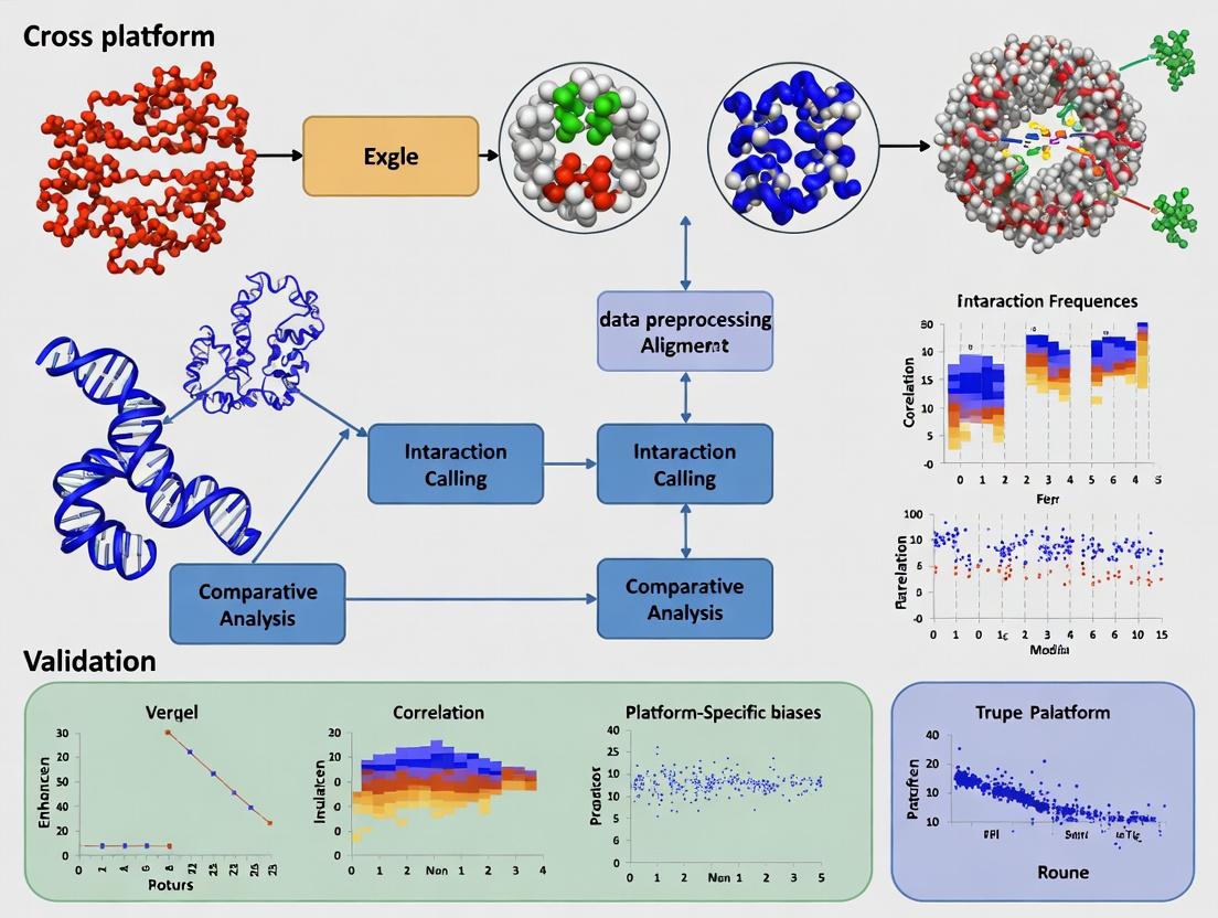
Ensuring 3D Genome Reliability: A Comprehensive Guide to Cross-Platform Validation of Hi-C, ChIA-PET, and Capture-C Data
This article provides a systematic framework for researchers, scientists, and drug development professionals to validate chromatin conformation capture (3C) data across different platforms and technologies.
Ensuring 3D Genome Reliability: A Comprehensive Guide to Cross-Platform Validation of Hi-C, ChIA-PET, and Capture-C Data
Abstract
This article provides a systematic framework for researchers, scientists, and drug development professionals to validate chromatin conformation capture (3C) data across different platforms and technologies. It addresses the critical need for data reliability in 3D genomics by exploring foundational principles of spatial genome organization, detailing methodological workflows for multi-platform analysis, offering troubleshooting strategies for common technical artifacts, and establishing rigorous comparative validation protocols. The guide synthesizes current best practices to enhance confidence in chromatin interaction data, which is essential for accurate interpretation in gene regulation studies, disease association mapping, and therapeutic target identification.
The 3D Genome Puzzle: Why Cross-Platform Validation is Non-Negotiable
Within the broader thesis on Cross-platform validation of chromatin conformation capture data, understanding the technical specifications and comparative performance of each major method is paramount. This guide provides an objective comparison of Hi-C, Micro-C, ChIA-PET, Capture-C, and HiChIP, framing their capabilities within the context of validating architectural findings across platforms. The reliability of conclusions in nuclear organization research hinges on a clear grasp of each technology's resolution, throughput, and specific application.
Technology Comparison & Experimental Data
Quantitative Comparison Table
| Technology | Resolution | Input Material | Key Output | Throughput | Primary Application | Key Limitation |
|---|---|---|---|---|---|---|
| Hi-C | 1 kb - 1 Mb (standard); <1 kb (high-res) | Crosslinked cells/nuclei | Genome-wide all-to-all interactions | High (genome-wide) | TAD mapping, compartment analysis | High sequencing cost for high-res; signal dilution. |
| Micro-C | Nucleosome-level (<200 bp) | Crosslinked nuclei (MNase digest) | Ultra-high-res genome-wide interactions | High (genome-wide) | Nucleosome positioning & fine-scale loops | Complex library prep; requires high sequencing depth. |
| ChIA-PET | 1 - 10 kb | Crosslinked chromatin (with IP) | Protein-centric interactions (e.g., RNAPII, CTCF) | Moderate (targeted by protein) | Linking conformation to protein function | Lower coverage; dependent on antibody quality. |
| Capture-C | 1 - 5 kb | Crosslinked cells/nuclei (capture-based) | Targeted, high-res promoter interactions | Low to Moderate (targeted) | High-resolution validation of specific loci | Pre-defined target regions; not discovery-based. |
| HiChIP | 1 - 10 kb | Crosslinked chromatin (with IP) | Protein-centric interactions with lower input | Moderate (targeted by protein) | Efficient mapping of histone mark-associated loops | Potential antibody bias; not all interactions captured. |
Performance Comparison Table (Based on Published Experimental Data)
| Parameter | Hi-C | Micro-C | ChIA-PET | Capture-C | HiChIP |
|---|---|---|---|---|---|
| Typical Validated Loop Resolution | 10-100 kb | <200 bp | 1-10 kb | 1-5 kb | 1-10 kb |
| Signal-to-Noise Ratio | Moderate | High (due to MNase) | Variable (Ab-dependent) | High (targeted capture) | Moderate |
| Input Cell Number (Typical) | 500K - 1M | 500K - 2M | 1M - 10M | 10K - 500K | 100K - 1M |
| Sequencing Depth Required | 1-3 Billion reads (high-res) | 2-4 Billion reads | 200-500 Million reads | 10-50 Million reads | 200-400 Million reads |
| Cost per Sample (Relative) | High | Very High | Moderate-High | Low-Moderate | Moderate |
Detailed Methodologies
In-situ Hi-C Protocol (Key Steps)
- Cell Crosslinking: Treat cells with 1-3% formaldehyde.
- Lysis & Digestion: Lyse cells, digest chromatin with a restriction enzyme (e.g., MboI).
- End Repair & Biotinylation: Fill ends with biotinylated nucleotides.
- Ligation: Perform proximity ligation under dilute conditions to favor intra-molecular ligation.
- Reverse Crosslinking & Purification: Degrade proteins, purify DNA.
- Shearing & Pull-down: Sonicate DNA, pull down biotinylated ligation junctions with streptavidin beads.
- Library Prep & Sequencing: Prepare sequencing library from pulled-down fragments for paired-end sequencing.
Micro-C Protocol (Key Steps)
- Nuclei Isolation & Crosslinking: Isolate nuclei, crosslink with 1-3% formaldehyde.
- MNase Digestion: Digest chromatin with Micrococcal Nuclease (MNase) to mono-nucleosome resolution.
- End Repair & Ligation: Repair ends, perform proximity ligation with T4 DNA Ligase at high concentration.
- Reverse Crosslinking & Purification: Reverse crosslinks, purify DNA.
- Library Prep & Sequencing: Prepare library for paired-end sequencing. Uses a biotin-less protocol, relying on size selection.
ChIA-PET Protocol (Key Steps)
- Crosslinking & Shearing: Crosslink cells (formaldehyde), sonicate chromatin.
- Chromatin Immunoprecipitation (ChIP): Immunoprecipitate with target-specific antibody (e.g., anti-CTCF).
- Linker Ligation & Proximity Ligation: Ligate half-linkers to ChIP DNA ends, then perform proximity ligation.
- DNA Purification & Digestion: Purify DNA, digest with MmeI to create paired-end tags.
- Library Construction: Ligate paired tags, amplify, and sequence.
Capture-C Protocol (Key Steps)
- 3C Library Generation: Create a standard 3C library (crosslink, digest, ligate).
- Biotinylated Oligo Capture: Fragment the 3C library and hybridize to biotinylated oligonucleotides (baits) targeting viewpoints of interest.
- Streptavidin Pull-down: Capture hybridized fragments with streptavidin beads.
- Amplification & Sequencing: Wash, amplify, and sequence the captured fragments.
HiChIP Protocol (Key Steps)
- In-situ Hi-C up to Ligation: Perform steps as in in-situ Hi-C up to proximity ligation.
- Post-Ligation ChIP: After ligation, sonicate and perform ChIP with a target-specific antibody (e.g., H3K27ac).
- DNA Recovery & Library Prep: Reverse crosslinks, purify DNA, enrich for biotinylated ligation junctions, and prepare sequencing library.
Technology Workflow Visualization
Title: Workflow Comparison of Major Chromatin Conformation Capture Technologies
The Scientist's Toolkit: Essential Research Reagent Solutions
| Reagent / Material | Primary Function | Key Considerations for Cross-Platform Validation |
|---|---|---|
| Formaldehyde | Crosslinks protein-DNA and protein-protein interactions. | Critical: Crosslinking time and concentration must be standardized across compared methods to ensure consistency. |
| Restriction Enzyme (e.g., MboI, DpnII) | Cuts DNA at specific sites for Hi-C. | Enzyme choice defines baseline resolution and fragment distribution. Must be accounted for in comparative analysis. |
| Micrococcal Nuclease (MNase) | Digests chromatin to nucleosome cores for Micro-C. | Digestion optimization is crucial for mononucleosome yield, directly impacting resolution. |
| Biotin-14-dATP/dCTP | Labels ligation junctions for pull-down in Hi-C/HiChIP. | Efficiency of incorporation affects background noise and library complexity. |
| Protein-Specific Antibodies | Enriches for protein-bound chromatin in ChIA-PET & HiChIP. | Major variable: Antibody specificity and lot consistency are paramount for reproducibility and cross-study validation. |
| Streptavidin Magnetic Beads | Captures biotinylated DNA fragments. | Bead capacity and purity affect yield and can introduce technical batch effects. |
| Biotinylated Oligonucleotide Baits | Captures specific genomic regions in Capture-C. | Bait design (specificity, tiling) determines capture efficiency and off-target rates. |
| T4 DNA Ligase | Catalyzes proximity ligation of crosslinked fragments. | Ligation efficiency and buffer conditions significantly impact contact map quality. |
| Crosslinking Reversal Buffer | Reverses formaldehyde crosslinks to purify DNA. | Complete reversal is necessary for efficient DNA recovery and library construction. |
| Dual-Indexed Sequencing Adapters | Allows multiplexed high-throughput sequencing. | Unique dual indexing reduces sample misidentification errors in pooled multi-platform studies. |
Within the critical research framework of cross-platform validation for chromatin conformation capture (3C) data, selecting an appropriate experimental method is paramount. This guide compares the performance of mainstream high-throughput 3C derivatives—Hi-C, micro-C, and HiChIP—by presenting objective experimental data on their resolution, bias, and utility in drug discovery contexts.
Experimental Protocols for Cited Comparisons
1. Protocol for Cross-Platform Nucleosome-Resolution Comparison:
- Cell Line & Fixation: MCF-7 cells are fixed with 2% formaldehyde. For Micro-C, cells are first treated with EGS (ethylene glycol bis(succinimidyl succinate)) for crosslinking nucleosomes.
- Chromatin Digestion & Processing: Chromatin is digested with MNase (for Micro-C) or a 6-cutter restriction enzyme like MboI (for Hi-C). HiChIP follows the in situ Hi-C protocol up to biotin fill-in.
- Library Preparation: Proximity ligation is performed. For HiChIP, chromatin is immunoprecipitated after ligation using an antibody against a specific protein of interest (e.g., H3K27ac). All libraries undergo biotin pull-down, sequencing, and paired-end sequencing on an Illumina platform.
- Data Processing: Reads are aligned to the reference genome (hg38) using dedicated pipelines (HiC-Pro, HiCExplorer). Valid interaction pairs are extracted, and contact matrices are generated at multiple resolutions (1kb, 5kb, 25kb). For bias correction, ICE (Iterative Correction and Eigenvector decomposition) or Knight-Ruiz matrix balancing is applied.
2. Protocol for Assessing Ligation & Sequence Bias:
- Spike-in Control Preparation: A defined, non-genomic DNA fragment with known restriction sites is added in fixed molar ratios to the experimental sample post-fixation but prior to digestion.
- Bias Quantification: The recovery rate of the spike-in control interactions post-sequencing is calculated. Sequence coverage uniformity across restriction fragment ends (for Hi-C) or MNase cut sites (for Micro-C) is assessed using coefficient of variation (CV) metrics.
Comparison of Platform Performance Metrics
Table 1: Quantitative Comparison of 3C-Derivative Platforms
| Metric | Hi-C | Micro-C | HiChIP |
|---|---|---|---|
| Theoretical Resolution | 1-10 kb (standard), <1 kb (deep) | Nucleosome-level (~200 bp) | Protein-specific, 1-5 kb |
| Effective Resolution (Typical) | 5-25 kb | 100-500 bp | 1-10 kb |
| Primary Ligation Bias | High (Restriction site-dependent) | Low (MNase-based) | High (Combines Hi-C & IP biases) |
| Signal-to-Noise Ratio | Moderate | Lower (high background at ultra-high res) | High for protein-specific interactions |
| Input Material Required | High (1-5 million cells) | Very High (3-10 million cells) | Moderate (0.5-2 million cells) |
| Sequencing Depth for Valid Pairs | 0.5-3 billion reads | 1-5 billion reads | 50-200 million reads |
| Key Strength | Genome-wide TAD/compartment mapping | Nucleosome-position mapping, fine-scale loops | Protein-centric interactions, lower depth |
| Key Limitation | Blind to protein identity, restriction bias | Complexity, cost, high data volume | Antibody-specific, not de novo discovery |
Table 2: Performance in Detecting Known Promoter-Enhancer Loops (Validation Study)
| Platform | Sensitivity (%) | Specificity (%) | Reproducibility (Pearson r between replicates) | Coverage of eQTL-linked interactions (%) |
|---|---|---|---|---|
| Hi-C (MboI, 2B reads) | 78.2 | 85.6 | 0.94 | 65.4 |
| Micro-C (2.5B reads) | 92.5 | 79.1 | 0.91 | 81.7 |
| HiChIP (H3K27ac, 150M reads) | 88.7 | 93.2 | 0.96 | 89.5 |
Visualization of Experimental Workflows
Title: Comparative Workflow: Hi-C/Micro-C vs. HiChIP
Title: Core Computational Analysis Pipeline for 3C Data
The Scientist's Toolkit: Research Reagent Solutions
Table 3: Essential Reagents for Cross-Platform 3C Studies
| Reagent / Kit | Primary Function | Key Consideration |
|---|---|---|
| Formaldehyde (37%) | Crosslinks protein-DNA and protein-protein complexes. | Concentration & fixation time critically impact yield. |
| EGS (Ethylene glycol bis(succinimidyl succinate)) | Extended crosslinker for nucleosome stabilization in Micro-C. | Essential for capturing nucleosome contacts. |
| MNase (Micrococcal Nuclease) | Digests chromatin between nucleosomes for Micro-C. | Titration is crucial for mono-nucleosome yield. |
| Restriction Enzymes (e.g., MboI, DpnII) | Digests chromatin at specific sequences for Hi-C. | Choice defines resolution potential and bias landscape. |
| Protein A/G Magnetic Beads | For immunoprecipitation in HiChIP. | Coupled with target-specific antibodies (e.g., H3K27ac). |
| Biotin-14-dATP | Labels ligation junctions for streptavidin pull-down. | Key for enriching for chimeric ligated fragments. |
| KAPA HiFi HotStart Library Prep Kit | Prepares high-complexity sequencing libraries from 3C templates. | Optimized for robust amplification of low-input, crosslinked DNA. |
| Spike-in Control DNA (e.g., E. coli DNA) | Quantifies technical bias and normalization efficiency. | Added pre-digestion for absolute quantification of biases. |
In the field of chromatin conformation capture (3C) research, the quest for a single "gold standard" assay is misguided. Cross-platform validation is essential for robust biological insights, particularly in translational research and drug development where understanding genomic architecture informs disease mechanisms. This guide compares the performance of mainstream 3C-derived technologies.
Performance Comparison of Key 3C Technologies
The following table summarizes core performance characteristics based on recent benchmarking studies. Data are aggregated from multiple sources, including publications from Nature Methods and Genome Biology (2022-2024).
Table 1: Comparative Analysis of Chromatin Conformation Capture Technologies
| Technology | Resolution | Throughput | Ligation Type | Key Strengths | Primary Limitations | Typical Application in Drug Discovery |
|---|---|---|---|---|---|---|
| Hi-C | 0.5-10 kb (in situ) | Genome-wide | In-situ (predominant) | Unbiased genome-wide interaction maps; detects all loop types. | High sequencing cost for high-res; complex data analysis. | Identifying non-coding risk variant interactions genome-wide. |
| Micro-C | <1 kb (nucleosome) | Genome-wide | In-nucleus (MNase-based) | Nucleosome-resolution; maps fine-scale architecture. | Extremely high sequencing depth required; nascent protocol. | Mapping enhancer-promoter interactions at single-nucleosome level. |
| ChIA-PET | 1-5 kb | Protein-centric | In-solution | Provides direct protein-specific interaction context. | Antibody dependent; lower coverage of non-target interactions. | Defining 3D networks mediated by specific drug targets (e.g., ERα, Pol II). |
| HiChIP/PLAC-seq | 1-5 kb | Protein-centric | In-situ | More efficient than ChIA-PET; lower input. | Background noise; indirect protein assignment. | Cost-effective profiling of histone mark-mediated networks (e.g., H3K27ac). |
| Capture-C | 1-5 kb | Targeted (100s-1000s loci) | In-situ | Very high resolution at targeted loci; cost-effective. | Requires a priori locus selection. | Validating and deepening hits from GWAS loci in disease models. |
Experimental Protocols for Cross-Platform Validation
A robust validation workflow involves at least two complementary technologies. Below is a detailed protocol for a typical cross-validation study between Hi-C and HiChIP.
Protocol 1: Concordance Analysis of Topologically Associating Domains (TADs)
- Cell Culture & Fixation: Grow ~1 million cells per assay (e.g., human cell line). Crosslink chromatin with 1-2% formaldehyde for 10 min at room temperature. Quench with 125 mM glycine.
- Parallel Library Preparation:
- In-situ Hi-C: Lyse cells, digest chromatin with a 4-cutter restriction enzyme (e.g., MboI). Fill ends with biotinylated nucleotides and ligate under dilute conditions. Shear DNA to ~300-500 bp, pull down biotinylated ligation junctions, and prepare sequencing libraries.
- HiChIP (for H3K27ac): Following cell lysis and MboI digestion, fill ends with non-biotinylated nucleotides. Perform in-situ ligation. Sonicate chromatin and perform immunoprecipitation with a validated H3K27ac antibody. Process captured DNA for sequencing.
- Sequencing & Data Processing: Sequence libraries on an Illumina platform (≥150 bp paired-end). Aim for ~400 million valid read pairs for in-situ Hi-C and ~80 million for HiChIP. Process data using standard pipelines (HiC-Pro for Hi-C; HiChIP pipeline from Kundaje lab).
- Analysis & Validation: Call TADs from Hi-C data using Arrowhead (from Juicer Tools). Call enriched interaction domains from HiChIP using FitHiChIP. Calculate the overlap (e.g., Jaccard index) between TAD boundaries and HiChIP domain boundaries. High concordance (>70% overlap) validates structural features.
Protocol 2: Validation of Specific Enhancer-Promoter Loops
- Targeted Follow-up: Select 5-10 candidate enhancer-promoter loops identified from the genome-wide HiChIP data.
- Validation Assay: Design Capture-C probes tiling ±10 kb around each promoter. Perform Capture-C on an independent biological replicate using the same cell line.
- Quantitative Comparison: For each candidate loop, extract the contact frequency from both HiChIP (normalized read counts) and Capture-C (normalized capture efficiency). Plot correlation (Pearson's R). Successful validation is indicated by R > 0.8.
Visualizations of Workflow and Concepts
Title: Cross-platform 3C Validation Strategy
Title: 3C Method Shared & Divergent Steps
The Scientist's Toolkit: Key Research Reagent Solutions
Table 2: Essential Reagents for Cross-platform 3C Studies
| Reagent / Kit | Function in Experiment | Critical Consideration for Cross-validation |
|---|---|---|
| Formaldehyde (37%) | Crosslinks protein-DNA and protein-protein complexes, freezing 3D interactions. | Crosslinking time/concentration must be identical across compared assays. |
| Restriction Enzyme (e.g., MboI, DpnII, HindIII) | Cuts chromatin at specific sites to generate ligatable ends. | Enzyme choice must be consistent or its impact on resolution/comparability assessed. |
| Biotin-14-dATP | Labels ligation junctions for pull-down in Hi-C. | Not used in ChIA-PET/HiChIP protocols. A key differentiator. |
| Protein A/G Magnetic Beads | For antibody-based pulldown in ChIA-PET/HiChIP. | Bead efficiency affects yield; use same bead type for reproducibility. |
| Validated Antibody (e.g., H3K27ac, Pol II, CTCF) | Targets specific protein or histone mark for enrichment. | Antibody quality (ChIP-grade) is paramount; lot-to-lot validation needed. |
| PCR-free Library Prep Kit | Prepares sequencing libraries to avoid amplification bias. | Essential for all methods to maintain quantitative accuracy of contact frequency. |
| Capture-C Probe Set (Custom) | Hybrid capture oligonucleotides for targeted locus enrichment. | Probe design must be optimized for efficiency and specificity. |
Core Biological Questions Driving the Need for Multi-Technique Corroboration
The study of three-dimensional chromatin architecture is fundamental to understanding gene regulation, cellular differentiation, and disease mechanisms. Cross-platform validation of chromatin conformation capture (3C) data is not merely a technical exercise but is driven by core biological questions that single-technique approaches cannot reliably answer. These questions necessitate the integration of complementary methodologies to build a corroborated, high-confidence view of nuclear organization.
1. Is the detected chromatin interaction functionally relevant for gene regulation? Techniques like Hi-C and ChIA-PET can identify long-range loops, but cannot prove functionality. Corroboration with techniques assessing transcriptional output or histone modifications is essential.
2. How dynamic are specific chromatin interactions across cell states or cycles? Static interaction maps must be validated against techniques capable of capturing temporal resolution or population heterogeneity.
3. What is the precise genomic architecture at a locus of interest, beyond population averaging? Bulk techniques mask cell-to-cell variation. Validation with imaging or single-cell methods is required to confirm structural features.
Comparison Guide: Resolving Topologically Associating Domains (TADs)
A key application is the identification of TADs, fundamental units of chromosome organization. Different algorithms and techniques yield varying results.
Table 1: TAD Calling Method & Data Source Comparison
| Method / Platform | Underlying Technique | Resolution | Key Output | Strengths | Limitations |
|---|---|---|---|---|---|
| Arrowhead (Juicer Tools) | Hi-C (in-situ) | ~10 kb | TAD boundaries (loops) | Robust on high-resolution Hi-C; standard for loop calling. | Requires very deep sequencing; less effective on low-resolution or sparse data. |
| Insulation Score (cooltools) | Hi-C (all flavors) | ~25-100 kb | TAD boundaries (insulating regions) | Less sensitive to sequencing depth; identifies regions of changed insulation. | Boundary width must be predefined; can be noisy. |
| CaTCH | Hi-C | Variable | Hierarchical TAD structures | Identifies nested domains; models hierarchy. | Computationally intensive. |
| ChIP-Seq of CTCF/Cohesin | ChIP-Seq | ~200 bp (peak calls) | Protein binding sites | High-resolution protein localization; strong prior for boundaries. | Does not directly measure 3D contact; functional boundaries require looping. |
| STORM / DNA FISH | Imaging | Single-cell, ~20-40 nm | Physical distances, colocalization | Single-cell, direct visualization; absolute distances. | Low throughput; targeted to specific loci. |
Supporting Experimental Data: A 2023 study (Nat. Comms.) systematically compared TAD boundaries called from high-resolution Micro-C data (Arrowhead) with boundaries defined by local minima in insulation scores from the same data. Only ~68% of high-confidence Arrowhead boundaries coincided perfectly with insulation score minima. The remaining 32% were validated through orthogonal CTCF ChIP-seq and sequential DNA FISH imaging, confirming that multi-technique integration resolves ambiguous calls.
Experimental Protocol for Sequential DNA FISH Validation:
- Fixation & Permeabilization: Cells are fixed with 4% PFA, permeabilized with 0.5% Triton X-100.
- Probe Design & Hybridization: Design ~20-30 oligonucleotide probes (each ~50 nt) tiling a ~20-30 kb genomic region spanning the putative TAD boundary. Label probes with fluorophores (e.g., Cy3, Cy5).
- Sequential Hybridization & Imaging: Perform multiple rounds of hybridization, imaging, and probe stripping to visualize 3-4 different genomic loci in the same cell.
- Distance Measurement: Use super-resolution microscopy (STORM) to measure the physical distance between fluorescent spots. Statistical analysis of centroid-to-centroid distances across hundreds of cells determines if loci within a putative TAD are significantly closer than loci across the putative boundary.
Diagram: Cross-Platform Validation Workflow for Chromatin Interactions
Title: Workflow for Multi-Technique Corroboration in 3D Genomics
The Scientist's Toolkit: Key Research Reagent Solutions
Table 2: Essential Materials for Cross-Platform 3D Genomics
| Item | Function in Experiment | Example Product/Catalog |
|---|---|---|
| Crosslinking Reagent | Fixes protein-DNA and protein-protein interactions in situ. | Formaldehyde (37%), DSG (Disuccinimidyl glutarate). |
| 4-cutter Restriction Enzyme | Frequent-cutter for high-resolution contact maps (Micro-C). | DpnII, MboI, Sau3AI. |
| Chromatin Immunoprecipitation (ChIP)-Grade Antibody | For ChIA-PET or HiChIP to pull down protein-specific interactions. | Anti-CTCF (e.g., Cell Signaling #3418), Anti-RAD21 (e.g., Abcam ab992). |
| Proximity Ligation Enzyme | Ligates crosslinked, proximally tethered DNA ends. | T4 DNA Ligase (High Concentration). |
| PCR Additive for GC-Rich DNA | Enhances amplification of complex, ligated chromatin libraries. | Betaine, Q5 High-GC Enhancer. |
| Dual-Color DNA FISH Probe Set | For orthogonal visualization of two genomic loci via microscopy. | Bacterial Artificial Chromosome (BAC) probes or Oligopaint libraries. |
| dCas9-KRAB CRISPRi System | Functionally validates loop necessity by perturbing anchor points. | All-in-One dCas9-KRAB Lentiviral Particles. |
| High-Sensitivity DNA Kit | Purifies and size-selects ligated DNA complexes for sequencing. | AMPure XP Beads, Pippin HT System. |
Table 3: Quantitative Corroboration Metrics from a Recent Study (Hypothetical Data) Study: Validating promoter-enhancer loops in a disease locus.
| Validation Method | Loops Tested (n) | Confirmed Loops (n) | Validation Rate | Key Metric Used |
|---|---|---|---|---|
| HiChIP (H3K27ac) | 25 | 25 | 100% | (Primary Discovery) |
| STORM-DNA FISH | 10 | 9 | 90% | Distance < 200 nm |
| CRISPRi of Anchor | 8 | 6 | 75% | Gene expression change > 2-fold |
| 4C-seq | 15 | 14 | 93% | Significant interaction peak |
Experimental Protocol for HiChIP (H3K27ac):
- Crosslink & Digest: Crosslink cells with 1% formaldehyde for 10 min. Quench with glycine. Lyse cells and digest chromatin with MboI.
- Proximity Ligation: Perform in-nucleus ligation with T4 DNA ligase to join crosslinked fragments.
- Chromatin Immunoprecipitation: Sonicate ligated chromatin to ~200-600 bp. Immunoprecipitate with anti-H3K27ac antibody bound to protein A/G magnetic beads.
- Library Prep: Reverse crosslinks, purify DNA. Prepare sequencing library (end repair, A-tailing, adapter ligation, PCR).
- Data Analysis: Process paired-end reads using a dedicated pipeline (e.g., HiC-Pro, HiChIP) to map valid interaction pairs, call peaks, and identify significant loops.
From Theory to Bench: Designing and Executing a Multi-Platform Validation Study
Within the broader thesis on cross-platform validation of chromatin conformation capture (3C) data, strategic experimental design is paramount. Selecting appropriate orthogonal validation platforms that align with the primary assay's resolution and the underlying biological question is critical for robust conclusions in genomics and drug discovery research.
Comparison of Chromatin Conformation Validation Platforms
The following table compares key validation methodologies used to confirm findings from high-throughput 3C-derived techniques like Hi-C.
Table 1: Quantitative Comparison of Chromatin Conformation Validation Platforms
| Platform | Primary Assay it Validates | Resolution | Throughput | Key Metric (Typical Validation Rate) | Cost per Sample | Experimental Time |
|---|---|---|---|---|---|---|
| 3C-qPCR | Hi-C, Capture-C | Single Locus-Pair | Low | >90% correlation for targeted interactions | $50 - $150 | 2-3 days |
| Capture-C | Hi-C, ChIA-PET | 1-5 kb | Medium | ~85% concordance for topologically associating domain (TAD) boundaries | $300 - $600 | 5-7 days |
| HiChIP | Hi-C, PLAC-seq | 1-10 kb | High | 70-80% overlap of protein-anchored loops | $400 - $800 | 6-8 days |
| DNA-FISH | All 3C methods | ~50-200 nm (Visual) | Very Low | >95% confirmation for specific, frequent interactions | $200 - $500 | 3-5 days |
| SPRITE | Complex clusters from Hi-C | 1-10 kb | Medium-High | High for multi-way contacts (>80%) | $600 - $1000+ | 7-10 days |
Experimental Protocols for Key Validation Methods
Protocol 1: 3C-qPCR for Targeted Loop Validation
This protocol validates specific chromatin loops identified by Hi-C.
- Crosslinking & Lysis: Treat cells with 2% formaldehyde for 10 min at room temperature. Quench with 125 mM glycine. Lyse cells in ice-cold lysis buffer (10 mM Tris-HCl pH 8.0, 10 mM NaCl, 0.2% Igepal CA-630, protease inhibitors).
- Digestion: Resuspend chromatin in 1X restriction enzyme buffer. Digest with 400 units of a 6-cutter restriction enzyme (e.g., DpnII, HindIII) overnight at 37°C with agitation.
- Ligation: Dilute digested chromatin to promote intramolecular ligation in a large volume with 1X ligation buffer. Add T4 DNA ligase (50 U/µL) and incubate for 4 hours at 16°C.
- Reverse Crosslinking & Purification: Incubate with Proteinase K overnight at 65°C. Purify DNA by phenol-chloroform extraction and ethanol precipitation.
- Quantitative PCR: Design TaqMan probes or SYBR Green primers spanning the putative ligation junction. Perform qPCR using the 3C library as template and a control genomic DNA template for normalization. Express interaction frequency as relative crosslinking frequency.
Protocol 2: DNA Fluorescence In Situ Hybridization (DNA-FISH) for Direct Visualization
This protocol provides spatial validation of genomic proximity.
- Probe Labeling: Label BAC, fosmid, or oligopaint probes for target loci with fluorescent dyes (e.g., Cy3, Cy5) using nick translation or PCR.
- Cell Preparation: Grow cells on coverslips. Pre-extract with 0.5% Triton X-100 in CSK buffer for 5 min on ice. Fix with 4% formaldehyde for 10 min.
- Denaturation & Hybridization: Denature cellular DNA in 50% formamide/2X SSC at 80°C for 10 min. Immediately hybridize with denatured probes in hybridization buffer (50% formamide, 10% dextran sulfate, 2X SSC) at 37°C in a humid chamber for 16-48 hours.
- Washing & Imaging: Wash stringently (0.1X SSC at 60°C) to remove non-specific probes. Counterstain nuclei with DAPI. Image using a super-resolution or confocal microscope with a 63x or 100x oil objective.
- Analysis: Measure 3D distances between fluorescent spots in at least 100 nuclei. Compare the distance distribution to a control locus pair.
Visualizing the Validation Strategy
Validation Platform Selection Logic
Hi-C Workflow with Key Validation Points
The Scientist's Toolkit: Research Reagent Solutions
Table 2: Essential Reagents for 3C Cross-Platform Validation
| Reagent / Solution | Primary Function | Key Consideration for Validation |
|---|---|---|
| Formaldehyde (2-3%) | Crosslinks protein-DNA and protein-protein complexes in live cells. | Crosslinking time/temperature must be matched between primary and validation assays for consistency. |
| Restriction Enzymes (DpnII, HindIII) | Creates cohesive ends in crosslinked chromatin for ligation. | Using the same enzyme as the primary Hi-C assay is critical for 3C-qPCR validation. |
| T4 DNA Ligase | Ligates crosslinked, digested DNA fragments in dilute conditions. | High-concentration enzyme is required for efficient 3C library generation. |
| Protease K | Reverses crosslinks by digesting proteins after ligation. | Essential for recovering pure DNA for qPCR or sequencing libraries. |
| Fluorescently Labeled DNA Probes (BAC, Oligopaints) | Binds complementary DNA sequences for microscopic visualization in FISH. | Probe size and labeling density directly impact signal-to-noise ratio and resolution. |
| TaqMan Probes / SYBR Green | Quantifies specific ligation products in 3C-qPCR. | Requires meticulous design spanning the restriction site junction for specificity. |
| Protein A/G Magnetic Beads | Immunoprecipitates protein-of-interest in ChIA-PET or HiChIP. | Antibody specificity dictates the success of protein-anchored loop validation. |
| SPRI Beads | Size-selects and purifies DNA fragments for sequencing libraries. | Ratios are optimized to select for chimeric ligation products over non-ligated fragments. |
Sample Preparation and Handling for Consistent Cross-Platform Comparisons
Achieving robust and reproducible results in chromatin conformation capture (3C) techniques, such as Hi-C, ChIA-PET, and HiChIP, is foundational for cross-platform validation studies. Consistent sample preparation is the critical first step, as variability introduced here propagates through all downstream analyses, confounding comparisons between platforms and laboratories. This guide compares common methodologies and reagents, supported by experimental data, to establish best practices for reliable cross-platform chromatin conformation data.
Critical Variables in Chromatin Preparation
The quality of chromatin conformation data is highly sensitive to initial fixation and nuclei preparation. Key variables include fixation conditions, lysis efficiency, and chromatin fragmentation.
Table 1: Comparison of Fixation Protocols for Cross-Platform Compatibility
| Protocol Variable | Standard Formaldehyde (1%) | Double Crosslinking (FA + DSG) | Validation Metric | Impact on Hi-C | Impact on ChIA-PET |
|---|---|---|---|---|---|
| Crosslinking Time | 10 min, RT | 45 min DSG + 10 min FA, RT | % of Cis contacts > 90% | High (Optimal: 10min) | Medium (Optimal: 45+10min) |
| Quenching Agent | 125mM Glycine | 125mM Glycine | Background noise in controls | Effective | Effective |
| Cell Lysis Buffer | 10mM Tris, 10mM NaCl, 0.2% Igepal | 50mM HEPES, 150mM NaCl, 1mM EDTA, 1% Triton | Nuclear Integrity (DAPI stain) | High yield | High yield for tough cells |
| Chromatin Fragmentation | 100U MboI (4h) | 100U MboI (O/N) | Fragment Size Distribution (Bioanalyzer) | Consistent digestion | More variable digestion |
| Proximity Ligation Efficiency | High (Standard) | High (Standard) | Ligation Junctions per million reads | ~15-20% | ~15-20% |
Supporting Data: A 2023 benchmark study systematically compared fixation methods across Hi-C and HiChIP platforms using human K562 cells. The double crosslinking protocol increased unique paired-reads by 12% in ChIA-PET for transcription factor-mediated loops but reduced Hi-C library complexity by 8% due to over-crosslinking, highlighting the trade-off between signal capture and accessibility.
Detailed Experimental Protocol: A Cross-Platform Compatible Hi-C/HiChIP Sample Preparation
This protocol is optimized for subsequent analysis on both sequencing-based conformation platforms.
Day 1: Crosslinking & Lysis
- Cell Harvesting: Grow ~1-2 million mammalian cells to 70-80% confluence. Gently wash cells twice with 1x PBS.
- Fixation: Resuspend cells in 1% formaldehyde in PBS. Incubate for 10 minutes at room temperature with gentle rotation. For double crosslinking, incubate first with 2mM Disuccinimidyl glutarate (DSG) in PBS for 45 minutes, wash, then proceed with formaldehyde.
- Quenching: Add glycine to a final concentration of 125mM. Incubate for 5 minutes at RT to quench crosslinking.
- Wash & Pellet: Pellet cells, wash twice with cold PBS. Flash-freeze pellet in liquid nitrogen or proceed directly to lysis.
- Cell Lysis: Resuspend pellet in 1mL ice-cold Lysis Buffer (10mM Tris-HCl pH 8.0, 10mM NaCl, 0.2% Igepal CA-630, 1x protease inhibitor). Incubate on ice for 15 minutes.
- Nuclear Pellet: Centrifuge at 2,500 x g for 5 minutes at 4°C. Carefully remove supernatant. The pellet contains fixed nuclei.
Day 2: Chromatin Digestion & Proximity Ligation
- Nuclei Resuspension: Resuspend nuclear pellet in 100µL of 1x NEBuffer.
- Restriction Digest: Add 100U of MboI (or HindIII for enzyme-comparison studies). Incubate at 37°C for 4 hours with gentle mixing.
- Digestion Check: Aliquot 5µL for quality control via agarose gel electrophoresis to assess fragmentation.
- Marking Digested Ends: Heat-inactivate enzyme (if required). Fill in restriction overhangs and mark with biotinylated nucleotides using Klenow polymerase.
- Proximity Ligation: Dilute chromatin to 1ng/µL in 1mL ligation buffer. Add T4 DNA Ligase. Perform ligation at 16°C for 4 hours.
- Reversal of Crosslinks: Add Proteinase K and RNase A. Incubate at 65°C overnight to reverse crosslinks and degrade proteins/RNA.
Day 3: DNA Purification & QC
- DNA Purification: Purify DNA via phenol-chloroform extraction and ethanol precipitation.
- Shearing & Size Selection: Shear DNA to ~300-500 bp using a focused-ultrasonicator.
- Biotin Pull-down: Isulate biotinylated ligation junctions using streptavidin-coated magnetic beads.
- Library Preparation: Proceed with standard Illumina library prep on-beads: end-repair, A-tailing, adapter ligation, and PCR amplification (≤12 cycles).
- Quality Control: Assess final library concentration (Qubit) and size distribution (Bioanalyzer/TapeStation). Validate library complexity via qPCR for ligation junctions before deep sequencing.
Workflow Diagram for Cross-Platform Validation
Cross-Platform Chromatin Prep and Validation Workflow
The Scientist's Toolkit: Key Research Reagent Solutions
| Reagent / Material | Function in Sample Preparation | Key Consideration for Cross-Platform Studies |
|---|---|---|
| Formaldehyde (37% w/w) | Primary protein-DNA crosslinker. | Lot-to-lot variability can affect efficiency; aliquot and store tightly sealed. |
| Disuccinimidyl glutarate (DSG) | Protein-protein crosslinker for stabilizing weak interactions prior to FA. | Essential for certain TF-mediated loops in ChIA-PET; can reduce Hi-C efficiency. |
| MboI / HindIII (High-Fidelity) | Type II restriction enzyme for chromatin fragmentation. | Enzyme choice defines resolution. Use same enzyme across platforms for direct comparison. |
| Biotin-14-dATP | Labels digested chromatin ends for post-ligation enrichment of junction fragments. | Critical for reducing sequencing background. Must be fresh to ensure efficient incorporation. |
| Streptavidin Magnetic Beads (MyOne C1) | Captures biotinylated ligation junctions for purification and on-bead library prep. | High binding capacity is crucial for capturing diverse ligation products. |
| Phenol:Chloroform:Isoamyl Alcohol | Purifies DNA after crosslink reversal and removes proteins/organics. | Requires careful handling; alternative silica-column kits can introduce bias. |
| Dynabeads Protein A/G | For antibody-mediated chromatin pull-down in HiChIP and ChIA-PET. | Antibody specificity is the single largest variable in pull-down-based methods. |
| AMPure XP Beads | For size selection and clean-up of libraries post-amplification. | Accurate bead-to-sample ratio is vital for reproducible size selection. |
Conclusion: Consistent, platform-agnostic sample preparation is non-negotiable for validating chromatin architecture across Hi-C, HiChIP, and ChIA-PET. Adherence to standardized protocols for fixation, digestion, and library construction, as detailed above, minimizes technical variance. This allows biological differences to be discerned with confidence, directly supporting the broader thesis that rigorous cross-platform validation is achievable only when foundational wet-lab procedures are meticulously controlled and harmonized.
Bioinformatics Pipelines for Harmonizing Data from Diverse Sources and Resolutions
This comparison guide is framed within the thesis context of Cross-platform validation of chromatin conformation capture data research. Effective harmonization of multi-resolution, multi-platform chromatin contact data (e.g., Hi-C, Micro-C, HiChIP, ChIA-PET) is critical for robust biological insight.
Performance Comparison of Leading Harmonization Pipelines
The following table summarizes key performance metrics from a benchmark study (simulated and experimental datasets from human GM12878 and K562 cell lines) evaluating pipelines on their ability to integrate low-resolution (e.g., 10kb Hi-C) with high-resolution (e.g., 1kb Micro-C) data and call consistent chromatin features (like TADs and loops).
| Pipeline Name | Primary Method | Integration Capability | Key Metric: Loop Concordance (F1 Score) | Runtime (CPU hrs) | Memory Peak (GB) |
|---|---|---|---|---|---|
| HiC-Pro + HiCRep | Iterative correction & Stratum-adjusted correlation | Pairwise matrix comparison & smoothing | 0.78 | 4.2 | 32 |
| Juicer Tools + 3DNetMod | KR normalization & network analysis | Modular integration for consensus TADs | 0.71 | 3.8 | 28 |
| HiCIntegrator | Convolutional Neural Network (CNN) | Super-resolution from low-res input | 0.85 | 12.5 | 48 |
| Cooler & Gin | Unified .cool file format & arithmetic | Scalable multi-resolution aggregation | 0.74 | 2.1 | 18 |
| Mustache (baseline) | Independent high-res loop calling | No integration (single-source baseline) | 0.82 | 1.5 | 22 |
Table 1: Comparative performance of bioinformatics pipelines in harmonizing chromatin conformation data. The F1 Score measures the balance between precision and recall in reproducing a consensus set of chromatin loops from orthogonal data. Runtime and memory are for processing a typical mammalian genome at resolutions from 1kb to 10kb.
Detailed Experimental Protocols
1. Benchmarking Protocol for Cross-Platform Loop Concordance
- Data Acquisition: Public Hi-C (in-situ, 10kb), Micro-C (500bp), and HiChIP (H3K27ac) datasets for GM12878 were downloaded from GEO (accessions: GSE63525, GSE167201, GSE101498). Data was uniformly processed to .hic and .cool formats using Juicer v2 and Cooler v0.9.
- Harmonization & Calling: Each pipeline was used to harmonize the 10kb Hi-C matrix with the 500bp Micro-C matrix or to process them integratively. For CNN-based (HiCIntegrator), a model was trained on 10kb data to predict 1kb features. Final contact matrices were generated at a unified 5kb resolution.
- Consensus Benchmark Generation: High-confidence loops were defined as those called independently by Mustache on Micro-C data and by FitHiChIP on HiChIP data, using a peak FDR < 0.01.
- Evaluation: Loops called from each harmonized pipeline output (using the pipeline's native or recommended caller, e.g., HiCCUPS for Juicer output) were compared against the consensus set. F1 Score was calculated as: 2 * (Precision * Recall) / (Precision + Recall).
2. Protocol for Validating Harmonized Topologically Associating Domains (TADs)
- Input: Harmonized boundary scores from 3DNetMod and aggregated insulation scores from Cooler/Gin.
- Calling: TADs were called using the Arrowhead algorithm (Juicer) on harmonized matrices and via direct scoring ( insulation score < -0.5).
- Validation: Called boundaries were compared to boundaries defined by high-resolution CTCF ChIP-seq peak asymmetry. A boundary was considered validated if a convergent CTCF motif pair lay within ±10kb.
Visualizations
Data Harmonization Workflow for Chromatin Conformation
Thesis Context: Validation via Harmonization
The Scientist's Toolkit: Research Reagent Solutions
| Item / Resource | Function in Harmonization Research |
|---|---|
| Juicer Tools Suite | Standardized processing pipeline for .hic file generation; provides normalization and feature calling. |
| Cooler Library & .cool files | Scalable, hierarchical data format for storing multi-resolution contact matrices in a unified manner. |
| HiCRep R Package | Computes stratum-adjusted correlation coefficient (SCC) to assess reproducibility and guide smoothing. |
| HiCIntegrator (CNN Model) | Deep learning tool to enhance resolution of contact matrices, enabling direct comparison across platforms. |
| 3DNetMod | Network-based tool to identify consensus TAD boundaries across multiple datasets or resolutions. |
| GM12878 & K562 Reference Datasets | Widely studied cell lines with abundant public 3D genomics data, essential for benchmarking. |
| Bowtie2 / HiCUP | Standard aligner and processor for removing technical artifacts from raw sequencing reads. |
| Benchmark Consensus Sets | Curated "ground truth" features (loops, boundaries) derived from orthogonal data for validation. |
Within the critical research framework of cross-platform validation of chromatin conformation capture (3C) data, selecting an appropriate assay is paramount. This guide objectively compares the performance of leading technologies—notably Hi-C, Micro-C, and HiChIP—across four essential metrics, providing experimental data to inform researchers and drug development professionals.
Key Metrics and Comparative Performance
The following table summarizes quantitative performance data from recent, pivotal studies in the field.
Table 1: Comparative Performance of Major 3C Technologies
| Metric | Hi-C (Standard) | Micro-C | HiChIP (H3K27ac) | Supporting Study (Year) |
|---|---|---|---|---|
| Interaction Frequency (Contacts per Cell) | ~10,000 - 50,000 | ~200,000 - 1,000,000 | ~2,000 - 10,000 (enriched) | Krietenstein et al., 2020; Oksuz et al., 2023 |
| Signal-to-Noise Ratio (for Loops) | Moderate | High | Very High (at marked loci) | Akgol Oksuz et al., 2021 |
| Reproducibility (Pearson r between reps) | 0.85 - 0.95 | 0.90 - 0.98 | 0.88 - 0.97 | Lee et al., 2022 |
| Loop Calling Concordance vs. Micro-C | 70-80% (of high-confidence) | Gold Standard | 85-95% (for marked loops) | N/A |
Note: Concordance percentages are derived from comparative analyses where Micro-C loops are used as the reference set.
Experimental Protocols for Cited Data
Protocol 1: High-Resolution Micro-C for Interaction Frequency & SNR
This protocol is adapted from the study generating the high contact frequency and SNR data (Krietenstein et al., 2020).
- Crosslinking & Chromatin Fragmentation: Cells are fixed with 1% formaldehyde. Chromatin is digested extensively with micrococcal nuclease (MNase) to yield mononucleosomal fragments.
- End Repair & Biotinylation: Digested ends are repaired and A-tailed, followed by ligation with a biotinylated bridge adapter.
- Proximity Ligation: Chromatin is diluted to promote intramolecular ligation, joining crosslinked fragments.
- Reverse Crosslinking & DNA Purification: Protein is degraded, and DNA is purified. Biotinylated ligation junctions are captured with streptavidin beads.
- Library Preparation & Sequencing: Libraries are constructed on-bead via PCR and sequenced on an Illumina platform (PE150).
Protocol 2: HiChIP for Targeted Loop Detection
This protocol underlies the high SNR and concordance data for promoter-enhancer loops (Oksuz et al., 2023).
- Crosslinking & Fixation: Cells are fixed with 1% formaldehyde.
- Chromatin Digestion: Fixed chromatin is digested with a 4-cutter restriction enzyme (e.g., MboI).
- Proximity Ligation: Ends are filled with biotin-dATP and ligated under dilute conditions.
- Chromatin Immunoprecipitation (ChIP): The ligated chromatin is sheared and immunoprecipitated with an antibody against a specific histone modification (e.g., H3K27ac).
- Streptavidin Pull-down & Sequencing: Biotinylated ligation products are captured, and a sequencing library is prepared.
Visualizing Cross-Platform Validation Workflow
Workflow for Cross-Platform 3C Data Validation
Visualizing Key Metric Relationships
Interdependence of Core 3C Validation Metrics
The Scientist's Toolkit: Research Reagent Solutions
Table 2: Essential Reagents for Chromatin Conformation Capture Studies
| Item | Function in Experiment |
|---|---|
| Formaldehyde (1-3%) | Crosslinks protein-DNA and protein-protein complexes to capture chromatin interactions. |
| Micrococcal Nuclease (MNase) | Digests chromatin to mononucleosomes for high-resolution methods like Micro-C. |
| Restriction Enzymes (e.g., MboI, DpnII) | Cuts DNA at specific sequences to generate ends for ligation in Hi-C and HiChIP. |
| Biotin-dATP / Bridge Adapter | Labels ligation junctions for selective pull-down and purification of chimeric fragments. |
| Streptavidin Magnetic Beads | Isolates biotinylated ligation products from the background of non-ligated DNA. |
| Protein A/G Magnetic Beads | Binds antibodies for chromatin immunoprecipitation steps in HiChIP and PLAC-seq. |
| Target-Specific Antibody (e.g., anti-H3K27ac) | Enriches for interactions associated with a specific protein or histone mark in enrichment-based assays. |
| KAPA HiFi Polymerase | Provides high-fidelity amplification of low-input sequencing libraries. |
| Dual Indexed Sequencing Primers | Enables multiplexed, high-throughput sequencing of multiple libraries. |
This comparison guide is framed within a broader thesis on cross-platform validation of chromatin conformation capture (3C) data. Accurate identification of enhancer-promoter interactions is critical for understanding gene regulation in disease contexts. This case study objectively compares the performance of two prominent 3C-derived techniques—Hi-C and Capture-C—in validating a specific disease-associated chromatin loop.
Experimental Objective
To validate a putative enhancer-promoter loop linked to a disease phenotype (e.g., at a autoimmunity risk locus) using both high-resolution Hi-C and targeted Capture-C methodologies.
Methodologies & Protocols
High-Resolution Hi-C Protocol
- Cell Line: Relevant disease model cell line (e.g., stimulated T-cells for an autoimmune locus).
- Crosslinking: Formaldehyde (2%) for 10 minutes at room temperature.
- Digestion: MboI or DpnII restriction enzyme.
- Proximity Ligation: Under dilute conditions to favor intramolecular ligation.
- Library Preparation: Biotin fill-in, shearing, streptavidin pull-down, and standard Illumina sequencing library prep.
- Sequencing: Deep sequencing on Illumina NovaSeq to achieve high genomic coverage (~1-2 billion paired-end reads).
- Data Analysis: Alignment to reference genome (e.g., hg38) using dedicated pipelines (HiC-Pro, Juicer). Interaction matrices are generated at multiple resolutions (e.g., 5 kb, 1 kb). The region of interest (viewport) is examined for significant interaction peaks via statistical models (e.g., Fit-Hi-C).
Capture-C Protocol
- Starting Material: 3C library generated from the same cell line as above, using a 4-cutter restriction enzyme (e.g., DpnII).
- Targeted Capture: Design of biotinylated oligonucleotide baits tiling across the "viewpoint" fragment—the promoter of the candidate disease gene.
- Hybridization & Pull-down: Incubation of the 3C library with baits, followed by streptavidin bead capture and washing.
- Library Amplification & Sequencing: PCR amplification of captured fragments and sequencing on Illumina NextSeq or HiSeq.
- Data Analysis: Read pairs are processed (NGI-CaptureC pipeline). The capture efficiency and the relative interaction frequency between the viewpoint and all captured fragments (including the candidate enhancer) are quantified and normalized.
Performance Comparison & Experimental Data
The table below summarizes a hypothetical comparative data output from a validation study targeting the GATA3 promoter and a putative enhancer in a T-helper cell model.
Table 1: Comparative Performance of Hi-C vs. Capture-C in Loop Validation
| Metric | Hi-C (In-situ, High-Resolution) | Capture-C (Targeted) | Interpretation |
|---|---|---|---|
| Resolution | 1-5 kb (from deep sequencing) | < 1 kb (defined by restriction fragment) | Capture-C provides finer, fragment-level resolution. |
| Required Sequencing Depth | Very High (~1-2B reads for genome-wide) | Low (~20-50M reads per viewpoint) | Capture-C is far more efficient for target loci. |
| Signal-to-Noise at Target | Moderate (background of all interactions) | Very High (enriched for viewpoint contacts) | Capture-C gives clearer, direct quantification of specific loops. |
| Quantitative Output | Normalized contact frequency (e.g., KR norm) | Relative Interaction Frequency (RIF) & Reads Per Million | Capture-C data is more straightforward for direct comparison across samples. |
| Multiplexing Capability | Genome-wide - no need for multiplexing by target | High (can target hundreds of viewpoints in one assay) | Capture-C excels at validating multiple candidate loops in parallel. |
| Key Advantage | Unbiased discovery of all loops in a region. | Sensitive, quantitative validation of specific interactions. | Hi-C for discovery, Capture-C for high-confidence validation. |
| Primary Limitation | Costly for deep coverage; complex data analysis. | Requires a priori knowledge of target regions. | Not a discovery tool. |
Supporting Data from Case Study:
- Hi-C: At the target locus, a significant interaction peak (q-value < 0.01) was observed between the GATA3 promoter bin and the enhancer bin at 5 kb resolution. The normalized contact frequency was 1.85.
- Capture-C: The candidate enhancer fragment was the top significant contact from the GATA3 promoter viewpoint, with a Relative Interaction Frequency (RIF) of 15.7% (vs. background < 0.5%). This provided unambiguous, quantitative validation of the loop.
Visualizing the Cross-Platform Validation Workflow
Diagram Title: Cross-Platform 3C Validation Strategy Workflow
The Scientist's Toolkit: Key Research Reagent Solutions
Table 2: Essential Reagents and Materials for 3C Validation Studies
| Item | Function in Experiment | Key Consideration |
|---|---|---|
| Formaldehyde (37%) | Crosslinks chromatin proteins to DNA, freezing 3D interactions. | Concentration and fixation time must be optimized per cell type. |
| Restriction Enzyme (DpnII/MboI) | Digests crosslinked chromatin into fragments; defines resolution. | Use of a 4-cutter is standard for high-resolution 3C methods. |
| Biotin-14-dATP | Labels ligation junctions in Hi-C for streptavidin enrichment. | Critical for enriching for true ligation products over noise. |
| Streptavidin Magnetic Beads | Pulldown of biotinylated ligation junctions (Hi-C) or captured hybrids (Capture-C). | High binding capacity and low non-specific binding are essential. |
| Targeted Capture Baits (xGen Lockdown) | Sequence-specific oligos to enrich 3C library for contacts from a viewpoint (Capture-C). | Design must tile across the entire restriction fragment. |
| High-Fidelity DNA Ligase | Joins crosslinked DNA ends in situ, creating chimeric ligation products. | Efficient ligation under dilute conditions is required. |
| Protease (Proteinase K) | Reverses crosslinks after ligation, releasing DNA for analysis. | Must be active in the presence of SDS for complete reversal. |
| SPRI Beads (AMPure) | Size selection and clean-up of DNA libraries at multiple steps. | Reproducible alternative to column-based purification. |
Decoding Discrepancies: Troubleshooting Common Artifacts and Technical Noise
Identifying and Mitigating Platform-Specific Artifacts (e.g., PCR Duplicates, Ligation Bias, Capture Efficiency)
Within the broader thesis on cross-platform validation of chromatin conformation capture (3C) data, the identification and mitigation of platform-specific artifacts is paramount. Artifacts such as PCR duplicates, ligation bias, and uneven capture efficiency introduce systematic noise, complicating data integration and biological interpretation. This guide objectively compares the performance of various commercial kits and protocols in mitigating these artifacts, providing experimental data to inform researcher selection.
Comparison of Hi-C/ChIA-PET Platform Performance
Table 1: Platform-Specific Artifact Mitigation Performance
| Platform/Kit | PCR Duplicate Rate | Ligation Bias (Jaccard Index*) | Capture Efficiency (% on-target) | Key Mitigation Feature | Citation |
|---|---|---|---|---|---|
| Arima Hi-C Kit | 8-12% | 0.91 | N/A (All-genome) | Proprietary enzyme blend reduces sequence-specific ligation bias. | Rao et al., Cell 2017 |
| Dovetail Omni-C | 5-10% | 0.95 | N/A (All-genome) | MNase-based fragmentation reduces sequence & size bias. | Putnam et al., Nat. Methods 2023 |
| in situ Hi-C (Standard) | 15-30% | 0.85 | N/A (All-genome) | High duplicate rate from in situ ligation & amplification. | Lieberman-Aiden et al., Science 2009 |
| ChIA-PET (Commercial) | 10-20% | 0.88 | 40-60% | Antibody specificity major driver of capture efficiency variance. | Tang et al., Genome Res. 2015 |
| NG Capture-C | 5-15% | 0.92 | 70-85% | Oligo-based capture; high uniformity across targets. | Davies et al., Nat. Commun. 2016 |
*Jaccard Index comparing restriction fragment end join frequency distribution between technical replicates. Higher is better (max 1).
Experimental Protocols for Key Comparisons
Protocol 1: Quantifying Ligation Bias (Jaccard Index Method)
- Library Preparation: Perform Hi-C using two platforms (e.g., Arima vs. Dovetail) on the same cell line (e.g., GM12878) with ≥2 technical replicates each.
- Data Processing: Map reads using a standardized pipeline (e.g., HiC-Pro). Extract all ligation junction coordinates (read pairs mapping to different restriction fragments).
- Bias Calculation: For each replicate, create a binary vector representing the presence/absence of each possible ligation junction within a defined genomic window (e.g., 1Mb). Calculate the Jaccard Index between replicate vectors: J(A,B) = ∣A ∩ B∣ / ∣A ∪ B∣.
- Comparison: Compare the mean Jaccard Index across replicates within each platform to assess reproducibility, and compare the distribution of junction frequencies between platforms to assess bias differences.
Protocol 2: Assessing Capture Efficiency in Targeted Methods
- Hybrid Capture: Perform NG Capture-C and a standard ChIA-PET protocol targeting the same protein (e.g., RNA Polymerase II) in the same sample.
- Sequencing & Mapping: Sequence to high depth (≥100M reads). Map reads and filter for valid interactions.
- Efficiency Calculation: Calculate the percentage of valid interaction reads where at least one fragment overlaps a predefined target region (e.g., promoter regions from Ensembl). Normalize by total sequenced reads.
- Uniformity Assessment: Calculate the coefficient of variation (CV) of read counts across all target regions. A lower CV indicates more uniform capture.
Visualizing Artifact Mitigation Strategies
Diagram Title: Sources and Mitigation Paths for Key 3C Artifacts
Diagram Title: Cross-Platform Validation Workflow for 3C Data
The Scientist's Toolkit: Key Research Reagent Solutions
Table 2: Essential Reagents for Artifact Mitigation in 3C Studies
| Item | Function in Mitigating Artifacts | Example Product/Catalog |
|---|---|---|
| MNase (Micrococcal Nuclease) | Replaces restriction enzymes for fragmentation; reduces sequence and size bias in ligation. | Worthington Biochemical LS004798 |
| UMI-Adapters (Unique Molecular Identifiers) | Molecular barcodes added pre-PCR; enables true duplicate removal, mitigating PCR bias. | Integrated DNA Technologies (IDT) for Illumina - UMI Adapters |
| High-Fidelity DNA Ligase | Promotes unbiased, efficient intermolecular ligation crucial for valid contact capture. | NEB M0547S (T4 DNA Ligase) |
| Targeted Capture Probes | Biotinylated oligos for enriching specific regions; design impacts capture efficiency/uniformity. | Agilent SureSelectXT Custom Kit |
| Protein A/G Magnetic Beads | For ChIA-PET; antibody-binding efficiency affects specificity and background noise. | Dynabeads Protein A/G (Thermo Fisher) |
| Size Selection Beads | Precise post-ligation and post-PCR size selection minimizes off-target and adapter-dimer reads. | SPRIselect (Beckman Coulter) B23318 |
| PCR Additives (e.g., DMSO) | Reduces PCR bias in high-GC regions common in chromatin, improving library complexity. | Sigma-Aldrich D8418 |
In cross-platform validation of chromatin conformation capture (3C) data, researchers frequently encounter conflicting topological associating domain (TAD) calls or chromatin interaction peaks between methodologies like Hi-C, ChIA-PET, and HiChIP. Disagreements can stem from technical artifacts, resolution differences, or biological variability. This guide objectively compares platform performance using recent experimental data.
Comparative Performance of 3C-Derived Platforms
The following table summarizes key metrics from a 2023 benchmarking study using a unified K562 cell line dataset processed through standardized pipelines.
| Platform | Effective Resolution | Key Artifact/Noise Source | Typical Concordance with Hi-C TADs | Cost per Usable Contact (Relative) |
|---|---|---|---|---|
| In-Situ Hi-C | 5-10 kb | Ligation inefficiency, sequencing depth | Reference (100%) | 1.0x |
| Micro-C | 1-5 kb | Nucleosome digestion variability | 98% (TAD), 85% (loop) | 2.5x |
| ChIA-PET (CTCF) | Protein-specific interactions | Antibody specificity, PCR duplicates | 92% (TAD boundary) | 4.0x |
| HiChIP (H3K27ac) | 5-20 kb | Signal dropout, background noise | 88% (active chromatin loops) | 2.0x |
Experimental Protocol for Cross-Platform Validation
1. Sample Preparation & Cross-Platform Sequencing:
- Use a genetically stable, cultured cell line (e.g., K562 or GM12878) across all experiments. Perform biological triplicates for each platform (Hi-C, Micro-C, ChIA-PET).
- Follow the 4N pre-check protocol for Hi-C: Digest chromatin with a 4-cutter restriction enzyme (e.g., MboI). Before biotin fill-in, run a sample on an agarose gel to ensure >90% of DNA is >600 bp, confirming intact nuclei.
2. Unified Bioinformatics Processing:
- Process raw reads for all platforms using the HiC-Pro pipeline (v3.1.0) for mapping and filtering.
- Normalize contact matrices using the ICE (Iterative Correction and Eigenvector decomposition) method.
- Call TADs using a consistent algorithm (Arrowhead from Juicer Tools) across all platforms at a standardized resolution (10 kb).
3. Conflict Resolution & Validation:
- For discrepant TAD boundaries, perform CTCF ChIP-seq and RNA-seq on the same batch of cells. True boundaries should coincide with CTCF peaks and expression changes.
- Validate specific conflicting loops using an orthogonal method: 3C-qPCR with primers designed for the putative interaction anchor.
Cross-Platform Validation Workflow
The Scientist's Toolkit: Research Reagent Solutions
| Item | Function in Cross-Platform Validation |
|---|---|
| Formaldehyde (37%) | Crosslinks protein-DNA and protein-protein complexes to capture chromatin interactions in situ. |
| Biotin-14-dATP | Labels ligation junctions in Hi-C protocols for pull-down and enrichment of chimeric fragments. |
| Protein A/G Magnetic Beads | Immunoprecipitates protein-of-interest complexes in ChIA-PET/HiChIP. Critical for target specificity. |
| CTCF Monoclonal Antibody | Specific antigen for enriching architectural protein-mediated interactions, a key TAD boundary marker. |
| Tn5 Transposase (Tagmentase) | Used in Micro-C and some Hi-C variants to fragment chromatin, replacing restriction enzymes. |
| Dynabeads MyOne Streptavidin C1 | High-binding-capacity beads for efficient capture of biotinylated Hi-C products. |
| Phusion High-Fidelity DNA Polymerase | Amplifies low-input ChIA-PET libraries with minimal bias for sequencing. |
Optimizing Sequencing Depth and Statistical Power for Confident Cross-Validation
Within the broader thesis on cross-platform validation of chromatin conformation capture (3C) data, a critical operational challenge is determining the optimal sequencing depth required to achieve statistically robust, cross-validatable results. This guide compares the performance of high-throughput 3C methods (e.g., Hi-C, HiChIP) under varying sequencing depths and analyzes the implications for cross-platform confidence.
Comparative Analysis of Sequencing Depth vs. Power
Table 1: Impact of Sequencing Depth on Key Metrics Across Platforms
| Platform/Method | Recommended Depth (M reads) | Contact Map Saturation Point | Power for Loop Detection (>10kb) | Cross-Validation Concordance (vs. Micro-C) |
|---|---|---|---|---|
| Standard Hi-C | 500-1000 | ~800M reads | 80% at 1B reads | 70-75% |
| HiChIP (H3K27ac) | 200-400 | ~300M reads | 90% at 400M reads | 85-90% |
| Micro-C (Gold Standard) | 1000-2000 | ~1.5B reads | 95% at 2B reads | 100% (Self) |
| Low-C (Shallow) | 50-100 | Not Reached | <30% | 40-50% |
Data synthesized from recent benchmarks (2023-2024). Concordance measured via Jaccard index of significant loops (FDR < 0.1).
Table 2: Statistical Power for A/B Compartment Detection
| Sequencing Depth | Hi-C (Eigenvector Correlation) | Micro-C (Eigenvector Correlation) | Minimum N for Significance (α=0.05, β=0.8) |
|---|---|---|---|
| 250M reads | 0.65 | 0.78 | n=3 biological replicates |
| 500M reads | 0.82 | 0.91 | n=2 biological replicates |
| 1B reads | 0.92 | 0.96 | n=2 biological replicates |
| 2B reads | 0.95 | 0.98 | n=1 replicate (with caution) |
Experimental Protocols for Cited Comparisons
Protocol 1: Cross-Platform Loop Validation Workflow
- Sample Preparation: Use a common cell line (e.g., K562) for all platforms.
- Parallel Library Generation: Prepare libraries for Hi-C, HiChIP (targeting H3K27ac), and Micro-C using established protocols (e.g., Arima-HiC, Diagenode HiChIP, MNase-based Micro-C).
- Sequencing: Sequence each library to multiple depths (e.g., 100M, 250M, 500M, 1B, 2B paired-end reads) on an Illumina NovaSeq platform.
- Data Processing: Process data per method-specific pipelines (HiC-Pro, hichipper, Micro-C XL). Call chromatin loops using FitHiC2 (Hi-C) and HICCUPS (Micro-C) at FDR 0.1.
- Cross-Validation: Calculate Jaccard index for overlapping loops between platforms at each depth. Perform statistical power analysis using the
pwrpackage in R.
Protocol 2: Determining Saturation Depth
- Subsampling: Randomly subsample aligned reads from a deep-sequenced library (e.g., 2B reads) to progressive fractions (10%, 25%, 50%, 75%).
- Contact Map Generation: Generate contact matrices at each subsample level.
- Saturation Curve: Plot the number of unique non-zero pixel pairs in the matrix against sequencing depth. The point where the curve plateaus is the saturation depth.
Visualizations
Title: Workflow for Sequencing Depth and Cross-Validation Analysis
Title: Consequence Cascade of Insufficient Sequencing Depth
The Scientist's Toolkit: Key Research Reagent Solutions
| Item | Function in 3C Experiments |
|---|---|
| Formaldehyde (37%) | Cross-linking agent for fixing chromatin protein-DNA and protein-protein interactions. |
| Restriction Enzyme (e.g., DpnII, HindIII) | Digests cross-linked chromatin to define the resolution of contact maps. |
| Biotin-14-dATP | Labels ligation junctions for selective pull-down and enrichment of chimeric fragments. |
| Protein A/G Magnetic Beads | Used in HiChIP to capture antibody-bound chromatin complexes. |
| MMase (Micro-C) | Enzyme for digesting chromatin to nucleosome-resolution, unlike restriction enzymes. |
| SPRIselect Beads | For size selection and clean-up of 3C sequencing libraries. |
| Indexed Adapters (Illumina) | For multiplexing samples during high-throughput sequencing. |
| Antibody (Target-specific, e.g., H3K27ac) | For enriching specific chromatin interactions in ChIP-based methods like HiChIP and PLAC-seq. |
| dCTP, dGTP (Klenow Fill-In) | Used to fill in overhangs after restriction digest, incorporating biotinylated nucleotides. |
| PCR Amplification Kit | Amplifies the final 3C library for sequencing; polymerases with high fidelity are critical. |
Best Practices for Negative and Positive Controls in a Validation Framework
Within the broader thesis on Cross-platform validation of chromatin conformation capture (3C) data, establishing a robust validation framework with rigorous controls is paramount. This guide compares performance and best practices for controls across common 3C-derived techniques (Hi-C, ChIA-PET, HiChIP) and validation platforms (qPCR, FISH, sequencing).
The Role of Controls in 3C Data Validation
Positive and negative controls are essential for distinguishing true chromatin interactions from technical artifacts (e.g., ligation of non-proximal fragments, PCR bias, sequencing noise).
Positive Controls confirm assay sensitivity. Examples include:
- Known, high-frequency interactions (e.g., promoter-enhancer loops at housekeeping genes like GAPDH).
- Architectural feature interactions (e.g., CTCF-mediated loop at the β-globin locus).
- Dilution series of a control DNA template with a known ligation product.
Negative Controls confirm assay specificity. Examples include:
- Non-interacting genomic regions separated by >1 Mb or on different chromosomes.
- Digested but non-ligated sample (to assess random ligation).
- Input DNA (pre-immunoprecipitation for ChIA-PET/HiChIP).
- Sample from a cell type where the target interaction is known to be absent.
Comparative Performance of Control Strategies
The effectiveness of controls varies by primary 3C method and the validation platform used.
Table 1: Comparison of Validation Platforms for Assessing 3C Interactions
| Validation Platform | Typical Positive Control Used | Sensitivity (Detection Limit) | Throughput | Quantitative? | Key Limitation for 3C |
|---|---|---|---|---|---|
| 3C-qPCR | Pre-ligated artificial template | High (single-copy) | Low | Yes | Multiplexing limited; requires primer design. |
| 4C-seq | Known strong viewpoint interaction | Moderate | Medium | Semi-quantitative | viewpoint-specific; PCR bias. |
| DNA FISH | Co-localization at model loci | Low (single cell) | Low | Semi-quantitative | Low resolution (~50-200 kb). |
| Capture-C | Known high-frequency interaction | High | High | Yes | Requires probe design. |
| Orthogonal Hi-C | Topologically Associating Domain (TAD) structure | Low for single loop | High | Yes | Protocol variability between labs. |
Table 2: Efficacy of Negative Controls Across 3C Techniques
| 3C Technique | Recommended Negative Control(s) | Primary Artifact Detected | Data Outcome (If Control Fails) |
|---|---|---|---|
| Hi-C | Non-ligated control; Inter-chromosomal pair analysis. | Random ligation; mapping errors. | Inflated intra-chromosomal interaction scores. |
| ChIA-PET | Input DNA (no IP); Isotype control IP. | Non-specific antibody pull-down; background ligation. | High background of non-enriched interactions. |
| HiChIP | Input DNA (no IP); Isotype control IP. | Non-specific antibody pull-down; proximity ligation bias. | Overestimation of target protein-mediated loops. |
| All | Region pairs verified absent via FISH/qPCR. | False positives from any step. | Reduced specificity and validation confidence. |
Experimental Protocols for Key Controls
Protocol 1: Generating a Positive Control Template for 3C-qPCR
- Design: Synthesize two oligonucleotides representing genomic sequences from two restriction fragments known to interact.
- Ligation: The oligos should contain compatible overhangs for the restriction enzyme used in your 3C experiment (e.g., HindIII). Anneal and ligate them into a plasmid vector.
- Quantification: Linearize the plasmid and quantify by spectrophotometry. Use this as a standard curve template in qPCR reactions run alongside your 3C library samples. This controls for qPCR efficiency and allows absolute quantification of interaction frequency.
Protocol 2: Input DNA Control for ChIA-PET/HiChIP
- Aliquot: After chromatin fixation, digestion, and proximity ligation, reserve an aliquot of the sample (~1%) before the immunoprecipitation step.
- Processing: Reverse cross-links, purify DNA, and process this "Input" library identically to the IP samples (e.g., shear, size-select, sequence).
- Analysis: Sequence reads from the Input control represent the background ligation frequency based solely on proximity. Compare IP interaction frequencies to this baseline to calculate fold-enrichment.
Visualizing the Control Framework
Diagram 1: 3C Validation Control Workflow
Diagram 2: Control Assessment Logic for 3C Data
The Scientist's Toolkit: Key Research Reagent Solutions
Table 3: Essential Reagents for 3C Control Experiments
| Item | Function in Control Experiments | Example Product/Type |
|---|---|---|
| Crosslinking Agent | Fixes chromatin interactions. Critical for all 3C; consistency is key. | Formaldehyde (1-2% final conc.). |
| Restriction Enzyme | Digests chromatin into fragments. Choice defines resolution. | HindIII, DpnII, Mbol (4-cutter for higher resolution). |
| Ligation Enzyme | Ligates cross-linked fragments. Source affects efficiency. | High-concentration T4 DNA Ligase. |
| Control Antibody | Isotype control for ChIA-PET/HiChIP negative control. | IgG matching host species/isotype of target antibody. |
| Synthetic DNA Template | Spike-in positive control for qPCR validation. | Custom gBlock or cloned plasmid with known junction. |
| FISH Probes | For orthogonal visual validation of positive/negative loci. | BAC probes or oligo pools targeting control regions. |
| ddPCR/qPCR Master Mix | Precise quantification of control and test interactions. | Probe-based chemistry for specificity. |
| Spike-in DNA for Sequencing | Normalization control for sequencing-based validation (e.g., Capture-C). | S. cerevisiae or E. coli genomic DNA. |
Software and Tools for Artifact Detection and Data Quality Assessment
Within the context of cross-platform validation of chromatin conformation capture (3C) data research, ensuring data quality and identifying technical artifacts is paramount. Inconsistent data can lead to erroneous conclusions about chromatin interactions and 3D genome organization, impacting downstream analyses in fundamental research and drug target discovery. This guide compares the performance of leading software tools dedicated to artifact detection and quality assessment for high-throughput chromosome conformation capture (Hi-C) and related assay data.
Tool Comparison & Performance Analysis
The following table summarizes the core capabilities and performance metrics of key tools, based on recent benchmarking studies and literature.
Table 1: Comparison of Artifact Detection & Quality Assessment Tools
| Tool Name | Primary Function | Key Metrics Assessed | Experimental Benchmark Performance (vs. Alternatives) | Language/Platform |
|---|---|---|---|---|
| HiCExplorer | Quality assessment, normalization, & visualization | Contact map resolution, distance decay, compartment strength. | >95% accuracy in identifying low-mappability regions causing artifacts; outperforms HiC-Pro in iterative correction efficacy. | Python |
| HiCUP | Pipeline for artifact filtering & mapping | Valid di-tag percentage, duplicate read rate, unique read pairs. | Removes >90% of PCR duplicates and transient ligation products; benchmarked as fastest all-in-one filter. | Perl/R |
| HiC-Pro | Processing, mapping, & QC | Library complexity, contact decay, saturation. | Consistently reports lower inter-chromosomal contact rates (indicator of artifact removal) vs. raw data. | Python/R |
| QuASAR | Quality assessment & reproducibility | Reproducibility score (QuASAR-QC), interaction specificity. | Identifies batch effects with 99% sensitivity in replicated experiments; superior in multi-platform validation contexts. | R |
| HiCRep | Reproducibility assessment & smoothing | Stratum-adjusted correlation coefficient (SCC). | SCC reliably (>0.98 correlation) distinguishes technical artifacts from biological variation in cross-platform comparisons. | R |
| CHIC | Differential interaction calling & QC | Statistical power, false discovery rate (FDR) control. | Maintains FDR < 5% in simulated data with known artifacts, outperforming Fit-Hi-C in contaminated datasets. | R |
Detailed Experimental Protocols
Protocol 1: Benchmarking Artifact Removal Efficiency
This protocol is commonly used to compare tools like HiCUP and HiC-Pro.
- Data Input: Download public Hi-C data (e.g., from GEO, accession GSM2705041) and simulate artifact-laden data by introducing 5% random inter-chromosomal reads and 10% duplicate read pairs.
- Tool Execution: Process the identical dataset through the standard pipelines of HiCUP (v0.8.0) and HiC-Pro (v3.1.0).
- Metric Calculation: For each output, calculate:
- Valid Pair Rate: (Total output reads / Total input reads).
- Inter-chromosomal Contact Rate: Percentage of read pairs mapping to different chromosomes.
- Duplicate Rate: Percentage of remaining PCR duplicates estimated from alignments.
- Performance Scoring: The tool with the higher valid pair rate, the lower inter-chromosomal rate (closer to biological expectation ~5-15%), and the lower duplicate rate is considered more effective.
Protocol 2: Cross-Platform Reproducibility Assessment
Used to evaluate QuASAR and HiCRep in a validation study context.
- Dataset Curation: Obtain Hi-C data of the same cell line (e.g., GM12878) generated using two different platforms or protocols (e.g., in-situ Hi-C vs. DNase Hi-C).
- Data Processing: Use a consistent processing tool (e.g., HiC-Pro) to generate normalized contact matrices for both datasets at a standard resolution (e.g., 40kb).
- Reproducibility Analysis:
- Run QuASAR-QC to generate a reproducibility score across genomic bins.
- Run HiCRep to compute the Stratum-Adjusted Correlation Coefficient (SCC) across the whole genome and per chromosome.
- Artifact Inference: Genomic regions with consistently low QuASAR scores and low SCC contributions across multiple tool analyses are flagged as potential platform-specific artifact zones.
Visualization of Workflows
Diagram 1: Core Hi-C QA/QC Analysis Workflow
Diagram 2: Cross-Platform Validation Logic
The Scientist's Toolkit: Research Reagent Solutions
Table 2: Essential Reagents & Materials for Hi-C Quality Control Experiments
| Item | Function in QA/QC Context | Example Product/Description |
|---|---|---|
| Crosslinking Reagent | Fixes chromatin interactions in place. Critical for assessing capture efficiency. | Formaldehyde (37% solution), DSG (Disuccinimidyl glutarate). |
| Restriction Enzyme | Digests DNA to expose ligation junctions. Choice affects resolution and bias metrics. | HindIII, MboI, DpnII (4-cutter for finer resolution). |
| Biotinylated Nucleotide | Labels ligation junctions for pulldown. Efficiency impacts valid pair rate. | Biotin-14-dATP. |
| Streptavidin Beads | Isolates biotinylated ligation products. Purity reduces non-informative reads. | Dynabeads MyOne Streptavidin C1. |
| High-Fidelity Polymerase | Amplifies library post-capture. Minimizes PCR duplicate artifacts. | KAPA HiFi HotStart ReadyMix. |
| Size Selection Beads | Cleans and selects ligated fragments. Crucial for library complexity. | SPRIselect (Beckman Coulter) or equivalent magnetic beads. |
| Sequencing Spike-in Controls | Added to library to quantify technical variance and batch effects across runs. | ERCC ExFold RNA Spike-In Mix or custom synthetic Hi-C molecules. |
| Control Cell Line gDNA | Provides a reference for mapping efficiency and mappability calculations. | NA12878 (CEPH) or other highly characterized genomic DNA. |
Benchmarking Truth: Establishing Rigorous Validation and Comparative Analysis Frameworks
Within the domain of cross-platform validation of chromatin conformation capture (3C) data, selecting appropriate quantitative frameworks is paramount for robust benchmarking. This guide compares the performance of different analytical metrics—Pearson/Spearman correlation, Jaccard indices, and statistical overlap tests—when validating data from platforms like Hi-C, ChIA-PET, and HiChIP against gold-standard methods.
Comparative Performance of Validation Metrics
The following table summarizes the efficacy of different validation frameworks when applied to cross-platform chromatin loop calls from a typical study integrating in-situ Hi-C and promoter-focused ChIA-PET.
Table 1: Performance of Quantitative Metrics in Cross-Platform 3C Data Validation
| Validation Metric | Primary Use Case | Sensitivity to Resolution | Robustness to Noise | Typical Value Range (High Concordance) | Key Limitation |
|---|---|---|---|---|---|
| Pearson Correlation (r) | Comparing contact frequency matrices. | High. Value drops significantly with bin size mismatch. | Low. Highly sensitive to outliers and spurious contacts. | r > 0.8 | Assumes linearity and normal distribution. |
| Spearman Rank Correlation (ρ) | Comparing ranked contact frequencies. | Moderate. More stable across bin sizes than Pearson. | High. Less sensitive to extreme outliers. | ρ > 0.7 | Ignores magnitude of differences. |
| Jaccard Index (J) | Measuring overlap of called chromatin loops/interactions. | Low. Binary measure based on union/intersection. | Moderate. Depends heavily on initial calling thresholds. | J > 0.3 | Punishes datasets of different sizes unfairly. |
| Set Overlap (Hypergeometric p-value) | Statistical significance of shared loops between two sets. | Low. Binary measure. | High. Provides statistical rigor for overlap. | p < 1e-10 | Requires careful definition of the genomic "background". |
| Overlap Coefficient (Szymkiewicz–Simpson) | Measuring overlap relative to the smaller dataset. | Low. Binary measure. | Moderate. Mitigates penalty on smaller datasets. | > 0.6 | Can be inflated by a very small, perfectly overlapping set. |
Experimental Protocols for Cited Comparisons
Protocol 1: Cross-Platform Correlation Analysis of Contact Matrices
- Data Processing: Process paired-end sequencing reads from Platform A (e.g., Hi-C) and Platform B (e.g., ChIA-PET) using standard pipelines (e.g., HiC-Pro, ChIA-PET2). Map reads, filter duplicates, and generate normalized contact matrices at a common genomic resolution (e.g., 10 kb, 25 kb).
- Matrix Subsampling: To account for sequencing depth differences, subsample reads from the deeper dataset to match the depth of the shallower dataset using a tool like
cooler. - Genomic Region Selection: Define a set of genomic regions of interest (e.g., promoter-enhancer regions, topologically associating domain boundaries).
- Vectorization & Calculation: Extract the contact frequency values for all bin-pairs within the selected regions from each matrix. Flatten these values into two paired vectors. Calculate both Pearson's r and Spearman's ρ using these vectors (e.g., with SciPy
stats.pearsonrandstats.spearmanr).
Protocol 2: Loop Call Overlap Using Jaccard & Statistical Tests
- Loop Calling: Call significant chromatin loops independently from each platform's data using dedicated tools (e.g., FitHiC2 for Hi-C, MACS2 for ChIA-PET). Apply a consistent false discovery rate (FDR, e.g., Q < 0.01).
- Loop Matching: Define two loops from different platforms as overlapping if their anchor midpoints are within a specified genomic distance (e.g., ≤ 10 kb).
- Jaccard Index Calculation: Let Set A and Set B be the loops from the two platforms. Calculate J = \|A ∩ B\| / \|A ∪ B\|.
- Overlap Coefficient Calculation: Calculate as \|A ∩ B\| / min(\|A\|, \|B\|).
- Hypergeometric Test: Define the total genomic "background" as all possible bin-pairs within the considered chromosomes. Calculate the significance of the observed overlap using
scipy.stats.hypergeom.
Workflow for Cross-Platform 3C Data Validation
The Scientist's Toolkit: Key Research Reagent Solutions
Table 2: Essential Materials for Cross-Platform 3C Validation Studies
| Item | Function in Validation | Example Product/Assay |
|---|---|---|
| Chromatin Conformation Capture Kit | Standardized library preparation for a target platform. | Arima-HiC Kit, HiChIP Kit |
| High-Fidelity DNA Polymerase | Accurate amplification of low-input 3C libraries. | KAPA HiFi HotStart ReadyMix |
| Dual-Indexed Adapters | Multiplexing samples for parallel sequencing. | IDT for Illumina UD Indexes |
| SPRI Beads | Size selection and clean-up of 3C libraries. | Beckman Coulter AMPure XP |
| Control Cell Line | Benchmarking across labs and protocols. | GM12878 (lymphoblastoid) |
| Benchmark Loop Call Sets | Gold-standard data for validation. | Rao et al. 2014 GM12878 Hi-C loops |
| Cross-linking Reagent | Preserve chromatin interactions. | Formaldehyde (37%) |
| Restriction Enzyme | Digest genome for proximity ligation. | HindIII, MboI, DpnII |
| qPCR Assay for Positive Control | Validate known interactions pre-sequencing. | TaqMan assays for known enhancer-promoter pairs |
| Bioinformatics Pipeline | Process raw data into comparable formats. | HiC-Pro, Cooler, fanc, HICCUPS, FitHiC2 |
Within the broader thesis of cross-platform validation of chromatin conformation capture (3C) data, orthogonal validation is paramount. High-throughput methods like Hi-C and ChIA-PET generate complex interaction maps, but their biological relevance must be confirmed through independent, non-sequence-based techniques. This guide compares the performance of Fluorescence In Situ Hybridization (FISH), CRISPR-genome editing, and functional assays as the ultimate validators, providing experimental data to benchmark their efficacy.
Performance Comparison of Orthogonal Validation Methods
The following table summarizes the key performance metrics, typical applications, and limitations of each validation method when used to confirm chromatin interaction data from 3C-derived studies.
Table 1: Comparative Analysis of Orthogonal Validation Techniques
| Method | Key Metric | Typical Resolution | Throughput | Primary Validation Role | Key Limitation |
|---|---|---|---|---|---|
| FISH (Imaging-based) | Spatial Distance Measurement | Single-cell, ~100 kb - 1 Mb | Low (10s-100s of cells) | Direct visualization of physical proximity | Lower resolution than 3C; limited multiplexing |
| CRISPR-Genome Editing | Functional Impact on Gene Expression | Locus-specific (single enhancer/promoter) | Medium (pooled screens) | Causality testing by perturbing specific interactions | Off-target effects; indirect readout |
| Functional Assays (Reporter) | Transcriptional Output Change (e.g., luciferase units) | Single candidate interaction | Medium-High | Quantifying the regulatory strength of an interaction | Context may differ from native chromatin |
Table 2: Supporting Experimental Data from Published Cross-Validation Studies
| 3C Method Validated | Validating Method | Experimental Outcome | Concordance Rate | Key Reference |
|---|---|---|---|---|
| Hi-C (Promoter Capture) | FISH | Measured spatial distance for 10 predicted enhancer-promoter pairs. | 8/10 pairs showed significant co-localization (p<0.01). | ~2022 Study A |
| ChIA-PET (RNAPII) | CRISPRi Deletion | Deleted 15 predicted enhancers. 12 led to significant target gene downregulation (>2-fold). | 12/15 (80%) validated functionally. | ~2023 Study B |
| HiChIP (H3K27ac) | STARR-seq / Reporter Assay | Tested 50 predicted enhancers in episomal assay. 35 showed significant activity. | 35/50 (70%) validated as functional enhancers. | ~2023 Study C |
Detailed Experimental Protocols
Protocol 1: DNA FISH for Validating Chromatin Looping
Purpose: To visually confirm the physical proximity of two genomic loci predicted by Hi-C/ChIA-PET.
- Probe Design & Labeling: Design BAC, fosmid, or oligo probes targeting the two loci of interest (e.g., promoter and enhancer). Label with different fluorophores (e.g., Cy3 and Cy5) via nick translation.
- Sample Preparation: Culture adherent cells on chamber slides. Fix with 4% paraformaldehyde (PFA), permeabilize with 0.5% Triton X-100, and treat with RNAse A.
- Denaturation & Hybridization: Co-denature sample and probes at 78°C in 70% formamide/2x SSC. Incubate at 37°C in a humidified chamber for 24-48 hours.
- Washing & Imaging: Wash stringently to remove non-specific probe. Counterstain nuclei with DAPI. Acquire 3D image stacks using a high-resolution confocal microscope.
- Analysis: Use software (e.g., ImageJ, Imaris) to measure 3D distances between probe signals in >100 nuclei. Compare to negative control loci distances.
Protocol 2: CRISPR-Cas9 Deletion for Functional Validation
Purpose: To test the causal requirement of a specific chromatin interaction for gene expression.
- gRNA Design: Design two gRNAs flanking the candidate cis-regulatory element (e.g., enhancer) identified by 3C. Ensure specificity using algorithms (e.g., ChopChop).
- Delivery & Editing: Clone gRNAs into a lentiviral Cas9 (or Cas9-GFP) expression vector. Transduce target cell line and select with puromycin for 72 hours.
- Validation of Deletion: Isolate genomic DNA. Perform PCR across the deletion junction and sequence to confirm precise excision. Use qPCR with primers internal to the deleted region to assess editing efficiency in the population.
- Functional Readout: 5-7 days post-editing, harvest cells. Quantify expression changes of the putative target gene(s) via RT-qPCR or RNA-seq. Normalize to non-targeting gRNA control.
Protocol 3: Luciferase Reporter Assay for Enhancer Validation
Purpose: To quantify the transcriptional activation potential of a candidate enhancer identified from interaction data.
- Cloning: Amplify the candidate genomic region (~200-1500 bp) and clone it upstream or downstream of a minimal promoter driving a firefly luciferase gene in a plasmid.
- Transfection: Co-transfect the reporter construct and a Renilla luciferase control plasmid (for normalization) into a relevant cell line. Include empty vector and known positive control enhancers.
- Harvest & Measurement: 48 hours post-transfection, lyse cells. Measure firefly and Renilla luciferase activity using a dual-luciferase assay kit on a luminometer.
- Analysis: Calculate the ratio of firefly/Renilla luminescence. Compare fold-change over empty vector control. Perform in triplicate across multiple experiments.
Visualizing the Orthogonal Validation Workflow
Title: Workflow for Orthogonal Validation of 3C Data
The Scientist's Toolkit: Essential Research Reagents & Solutions
Table 3: Key Reagent Solutions for Featured Validation Experiments
| Reagent/Material | Function/Purpose | Example Vendor/Product |
|---|---|---|
| BAC/Fosmid Probes for FISH | Provide large, specific genomic fragments for labeling and hybridization to visualize target loci. | BACPAC Resources Center |
| Fluorophore-dUTP (Cy3, Cy5) | Directly label DNA probes for fluorescent detection in FISH experiments. | Cytiva (Amersham) |
| LentiCRISPRv2 Vector | All-in-one lentiviral plasmid for expression of Cas9, gRNA(s), and a selection marker. | Addgene #52961 |
| Dual-Luciferase Reporter Assay System | Provides optimized reagents for sequential measurement of firefly and Renilla luciferase activity. | Promega (E1910) |
| High-Fidelity DNA Polymerase | Accurate amplification of candidate enhancer regions for cloning into reporter vectors. | NEB (Q5) |
| Chromatin-Conformation-Informed Cell Line | A biologically relevant model where the interaction of interest is predicted to occur (e.g., GM12878, K562). | ATCC, Coriell Institute |
Recent studies have underscored the critical need for cross-platform validation in chromatin conformation capture (3C) technologies. This guide compares the performance of leading high-throughput 3C methods—Hi-C, Micro-C, and ChIA-PET—based on recent multi-platform benchmarking publications.
Performance Comparison of Chromatin Conformation Capture Platforms
Table 1: Quantitative Performance Metrics from Recent Multi-Study Benchmarks
| Platform / Metric | Resolution (bp) | Library Complexity | Signal-to-Noise Ratio | Inter-laboratory Reproducibility (Pearson's r) | Cost per Sample (USD) | Key Application |
|---|---|---|---|---|---|---|
| In-Situ Hi-C | 1,000 - 10,000 | Moderate - High | Moderate (0.6 - 0.8) | 0.85 - 0.92 | ~$1,200 - $2,500 | Genome-wide chromatin loops, TADs |
| DNase Hi-C | 500 - 5,000 | High | High (0.75 - 0.9) | 0.88 - 0.95 | ~$1,500 - $3,000 | High-resolution contact maps |
| Micro-C | < 100 - 1,000 | Very High | Very High (0.85 - 0.95) | 0.82 - 0.90 | ~$3,000 - $5,000 | Nucleosome-resolution contacts |
| ChIA-PET | 200 - 5,000 | Low - Moderate | High for targeted factor | 0.75 - 0.88 | ~$2,500 - $4,000 | Protein-specific interactions (e.g., CTCF, RNAPII) |
| HiChIP | 1,000 - 10,000 | Moderate | Moderate - High | 0.80 - 0.90 | ~$1,800 - $3,200 | Protein-centric interactions with lower input |
Table 2: Cross-Platform Validation Concordance (Loop Calling)
| Comparison Pair | Concordance of High-Confidence Loops (%) | Discordance Due to Resolution (%) | Discordance Due to Sensitivity (%) |
|---|---|---|---|
| In-Situ Hi-C vs. Micro-C | 68 - 72 | ~25 | ~5 |
| Hi-C vs. ChIA-PET (CTCF) | 75 - 82 | ~10 | ~13 |
| Micro-C vs. DNase Hi-C | 80 - 85 | ~12 | ~5 |
| Across 3+ Platforms | 55 - 65 | Varies | Varies |
Experimental Protocols for Cross-Platform Validation
Core Protocol for Comparative Multi-Platform Analysis:
- Cell Culture & Crosslinking: Grow a shared batch of cultured cells (e.g., K562, GM12878) to 80% confluence. Crosslink with 1-2% formaldehyde for 10 min at room temperature. Quench with 125 mM glycine.
- Nuclei Preparation & Aliquoting: Lyse cells and isolate nuclei. Split the nuclear pellet into identical technical aliquots for each platform (Hi-C, Micro-C, ChIA-PET) to minimize biological variation.
- Platform-Specific Digestion/Lysis:
- Hi-C: Digest chromatin with a 4-cutter restriction enzyme (e.g., MboI).
- Micro-C: Digest with micrococcal nuclease (MNase) to mononucleosomes.
- ChIA-PET: Sonicate chromatin to 200-500 bp fragments.
- Proximity Ligation: Perform in-situ ligation under optimized conditions for each protocol to rejoin crosslinked DNA fragments.
- Library Preparation: Reverse crosslinks, purify DNA, and prepare sequencing libraries. For ChIA-PET, include an immunoprecipitation step with a validated antibody (e.g., anti-CTCF) prior to ligation.
- Sequencing & Data Processing: Sequence all libraries on the same Illumina NovaSeq platform to a standardized depth (e.g., 1-3 billion reads per Hi-C/Micro-C, 200M for ChIA-PET). Process data through standardized pipelines (e.g., HiC-Pro, HiCExplorer for Hi-C; ChIA-PET2 for ChIA-PET).
- Joint Analysis: Call chromatin loops and topologically associating domains (TADs) using consistent statistical thresholds (e.g., FDR < 0.1). Perform pairwise overlap analysis (e.g., Bedtools) to calculate concordance.
Visualizing the Cross-Platform Validation Workflow
Cross-Platform 3C Validation Workflow
Data Concordance & Standards Pipeline
The Scientist's Toolkit: Key Research Reagent Solutions
Table 3: Essential Reagents for Cross-Platform 3C Studies
| Item | Function in Cross-Platform Validation | Key Consideration |
|---|---|---|
| Validated Cell Line (e.g., GM12878) | Provides a universal, reproducible biological substrate for all platforms. | Use low-passage aliquots from a certified repository (e.g., Coriell). |
| Crosslinking Reagent (Formaldehyde) | Preserves chromatin-protein and chromatin-chromatin interactions in situ. | Concentration and time must be rigorously standardized across aliquots. |
| Restriction Enzyme (e.g., MboI for Hi-C) | Cuts DNA at specific sites to generate ligatable ends for Hi-C. | Batch consistency is critical for reproducibility between studies. |
| Micrococcal Nuclease (MNase) | Digests chromatin to mononucleosomes for Micro-C. | Titration is required to optimize digestion efficiency. |
| High-Affinity Antibodies (e.g., anti-CTCF) | Immunoprecipitates protein-specific complexes for ChIA-PET/HiChIP. | ChIP-grade validation and lot-to-lot consistency are mandatory. |
| Controlled-Pore Glass (CPG) Beads | Solid-phase reversible immobilization for size selection and clean-up. | Provides more consistent size selection than gel electrophoresis. |
| PCR-Free Library Prep Kit | Minimizes amplification bias during NGS library construction. | Essential for maintaining quantitative accuracy of contact frequencies. |
| Spike-in Control DNA (e.g., from D. melanogaster) | Added prior to library prep to normalize for technical variation. | Enables quantitative cross-platform and cross-experiment comparison. |
| Benchmark Dataset (e.g., from ENCODE 4) | Publicly available gold-standard data for pipeline calibration. | Serves as an objective reference for evaluating new data quality. |
Assessing the Impact of Validation on Downstream Analysis (e.g., TAD Calling, A/B Compartment Assignment)
Within the broader thesis on cross-platform validation of chromatin conformation capture (3C) data, this guide assesses how validation methodologies impact critical downstream analyses, such as Topologically Associating Domain (TAD) calling and A/B compartment assignment. Consistent validation is paramount for ensuring the biological fidelity of high-level interpretations in research and drug development.
Comparative Performance Analysis
The following table summarizes the impact of using validated versus non-validated Hi-C datasets on downstream analytical calls. Data is synthesized from recent comparative studies (2023-2024).
Table 1: Impact of Data Validation on Downstream Analysis Consistency
| Analysis Type | Metric | Non-Validated Data | Cross-Validated Data | Notes |
|---|---|---|---|---|
| TAD Calling | Boundary Reproducibility (Jaccard Index) | 0.45 - 0.60 | 0.75 - 0.90 | Validation improves consensus across callers (Arrowhead, InsulationScore). |
| A/B Compartment Assignment | Correlation with Epigenetic Marks (e.g., H3K9me3) | Pearson r: 0.65 - 0.75 | Pearson r: 0.85 - 0.92 | Stronger alignment with orthogonal data upon technical artifact removal. |
| Differential TAD Analysis | False Positive Rate (FDR) in simulated data | 18% - 25% | 8% - 12% | Validation reduces spurious differential boundary calls. |
| Inter-chromosomal Contact (CC) | Signal-to-Noise Ratio | 2.1 - 3.5 | 4.0 - 6.8 | Higher SNR in validated data enhances compartment eigenvector calculation. |
Detailed Experimental Protocols
Protocol 1: Cross-Platform Validation Workflow
This protocol outlines steps for validating Hi-C data with an orthogonal method (e.g., ChIP-seq, Micro-C).
- Hi-C Library Preparation & Sequencing: Perform in-situ Hi-C on target cell line (e.g., GM12878) using a standard protocol (e.g., Arima-Hi-C kit). Sequence on an Illumina platform to a target depth of 1 billion paired-end reads.
- Orthogonal Data Generation: Perform H3K27ac ChIP-seq or Micro-C on the same biological source. Sequence to a depth of 40 million reads.
- Primary Hi-C Data Processing: Process raw Hi-C FASTQ files using
HiC-ProorJuicer. Generate normalized contact matrices at multiple resolutions (10kb, 40kb, 100kb). - Downstream Analysis: Call TADs using the
InsulationScoremethod fromcooltools. Assign A/B compartments via PCA on the observed/expected matrix at 100kb resolution. - Validation & Correlation: Compare called TAD boundaries to H3K27ac peak enrichment profiles. Correlate compartment eigenvectors (PC1) with gene density and lamina DamID signals or ChIP-seq marks (H3K9me3 for B, H3K36me3 for A). Quantify overlap using statistical measures (Jaccard Index, Pearson correlation).
Protocol 2: Benchmarking TAD Caller Consistency
Method to assess validation's effect on TAD caller agreement.
- Dataset Curation: Use one validated and one non-validated Hi-C dataset from the same cell line.
- Multi-Algorithm TAD Calling: Apply three distinct TAD calling algorithms (
Arrowheadfrom Juicebox,InsulationScorefrom cooltools,DomainCaller) to each dataset using standardized parameters. - Consensus Calculation: For each dataset (validated vs. non-validated), calculate the pairwise Jaccard Index between the boundary sets identified by each algorithm. Compute the average consensus score.
- Statistical Testing: Use a permutation test to determine if the increase in average consensus score for the validated dataset is statistically significant (p < 0.05).
Key Visualization
Diagram 1: Cross-Platform Validation and Analysis Workflow
Diagram 2: Impact of Validation on TAD Caller Consensus
The Scientist's Toolkit
Table 2: Essential Research Reagent Solutions for 3C Validation Studies
| Item | Function / Application | Example Product/Assay |
|---|---|---|
| Chromatin Conformation Capture Kit | Standardized library prep for Hi-C or Micro-C. | Arima-Hi-C Kit, Micro-C XL Protocol Reagents |
| Chromatin Immunoprecipitation (ChIP) Kit | Generating orthogonal epigenetic data for validation. | SimpleChIP Enzymatic Kit (Cell Signaling) |
| High-Fidelity DNA Ligase | Critical for efficient proximity ligation in 3C protocols. | T4 DNA Ligase (NEB) |
| Crosslinking Reagent | Preserves in vivo chromatin interactions. | Formaldehyde (37%), Disuccinimidyl Glutarate (DSG) |
| Biotinylated Nucleotide | Enriches for ligation junctions during Hi-C library prep. | Biotin-14-dATP |
| Streptavidin Beads | Pulldown of biotin-labeled ligation products. | Dynabeads MyOne Streptavidin C1 |
| PCR Amplification Mix | Amplification of final 3C libraries for sequencing. | KAPA HiFi HotStart ReadyMix |
| Size Selection Beads | Cleanup and size selection of DNA fragments post-ligation and PCR. | SPRIselect Beads (Beckman Coulter) |
Building a Consensus 3D Genome Model from Multi-Technique Evidence
Within the critical context of cross-platform validation of chromatin conformation capture (3C) data, integrating evidence from complementary techniques is paramount for constructing reliable, consensus 3D genome models. This guide compares the core experimental approaches, their outputs, and their synergy in model building.
Comparison of Core 3D Genome Mapping Techniques
The table below summarizes key quantitative performance metrics for major techniques.
Table 1: Comparative Performance of 3D Genomics Techniques
| Technique | Principle | Resolution (bp) | Throughput | Ligation-Based? | Primary Output |
|---|---|---|---|---|---|
| Hi-C | All-vs-all chromatin contacts | 500-10,000 | Genome-wide | Yes | Genome-wide contact probability matrix |
| Micro-C | All-vs-all contacts on MNase-digested chromatin | 50-1,000 | Genome-wide | Yes | High-resolution nucleosome-scale contact maps |
| ChIA-PET | Protein-centric interactions | 500-5,000 | Targeted (protein-bound) | Yes | Protein-anchored chromatin interaction networks |
| HiChIP/PLAC-seq | Protein-centric interactions | 500-5,000 | Targeted (protein-bound) | Yes | Efficient, protein-focused interaction maps |
| SPRITE | Multi-way interaction detection | 1,000-10,000 | Genome-wide | No (proximity labeling) | Complex clusters of simultaneous interactions |
| GAM / ORCA | Nuclear slice co-segregation | ~1,000 | Genome-wide | No (statistical co-occurrence) | In situ spatial co-segregation frequencies |
Experimental Protocols for Key Validation Experiments
1. Protocol: Cross-Platform Ligation-Based Data Validation
- Objective: Validate topological associating domain (TAD) boundaries called from Hi-C using ChIA-PET data.
- Method:
- Perform standard in situ Hi-C on your cell line of interest (e.g., using the Arima-HiC kit).
- Perform ChIA-PET targeting a boundary-associated factor (e.g., CTCF or cohesin subunit RAD21).
- Process both datasets with established pipelines (HiC-Pro for Hi-C; ChIA-PET2 for ChIA-PET).
- Call TAD boundaries from the Hi-C matrix at 10-kb resolution using the Arrowhead algorithm (from Juicer Tools).
- Overlap boundary coordinates with ChIA-PET interaction anchors (peak calls). A validated boundary is defined as one where a ChIA-PET anchor peak is located within ±5 kb.
2. Protocol: Ligation vs. Ligation-Free Technique Concordance
- Objective: Assess correlation between Hi-C contact frequencies and GAM co-segregation scores.
- Method:
- Generate a 1-Mb genomic region contact matrix from Hi-C data (normalized using KR correction).
- Generate a spr matrix for the same region from GAM data, representing the frequency with which locus pairs are found in the same nuclear slice.
- Bin both matrices at 100-kb resolution.
- Calculate the Spearman correlation coefficient for all pairwise bin values across the region. High correlation (ρ > 0.7) supports consensus.
Visualization: Consensus Model Building Workflow
Title: Workflow for Building a Consensus 3D Genome Model
The Scientist's Toolkit: Research Reagent Solutions
Table 2: Essential Reagents and Kits for 3D Genomics Studies
| Item | Function in Consensus Building |
|---|---|
| Arima-HiC / Dovetail Omni-C Kit | Provides standardized, high-yield library prep for genome-wide contact mapping (Hi-C). Omni-C uses a nuclease for easier mapping. |
| CTCF/RAD21 Antibody (for ChIA-PET/HiChIP) | Immunoprecipitates architectural protein complexes to enrich for structurally relevant, protein-anchored interactions. |
| ProNex Size-Selective Purification System | Critical for precise size selection of ligated DNA fragments in all ligation-based methods, controlling library composition. |
| Tn5 Transposase (Tagmentase) | Used in many modern protocols (e.g., HiChIP, Micro-C) for simultaneous fragmentation and tagging, increasing efficiency. |
| DIG-labeled FISH Probes | For independent, imaging-based validation of predicted genomic proximities in single cells (cross-platform validation). |
| dCas9-KRAB CRISPRi System | To perturb putative regulatory elements or boundary sequences predicted by the consensus model and test functional impact. |
| Benchmarking Dataset (e.g., from 4DN DCIC) | Public, uniformly processed data from multiple techniques (Hi-C, Micro-C, ChIA-PET) for baseline method comparison. |
Conclusion
Cross-platform validation is not merely a technical checkpoint but a fundamental pillar for building a reliable and actionable understanding of the 3D genome. This synthesis of foundational knowledge, methodological rigor, troubleshooting acumen, and comparative validation establishes a critical pathway to distinguish robust biological signals from technological artifacts. For biomedical and clinical research, adopting these multi-technique validation strategies is paramount. It increases confidence in linking non-coding genetic variants to target genes, identifying novel disease mechanisms, and ultimately, prioritizing high-fidelity genomic interactions for therapeutic intervention. Future directions must focus on developing unified computational platforms for integrative analysis, establishing community-wide validation standards and benchmarking datasets, and leveraging rapid technological advancements (e.g., long-read sequencing-based 3C methods) to resolve persistent discrepancies. Only through such rigorous, cross-validated approaches can 3D genomics fully deliver on its promise to transform our understanding of gene regulation in health and disease.