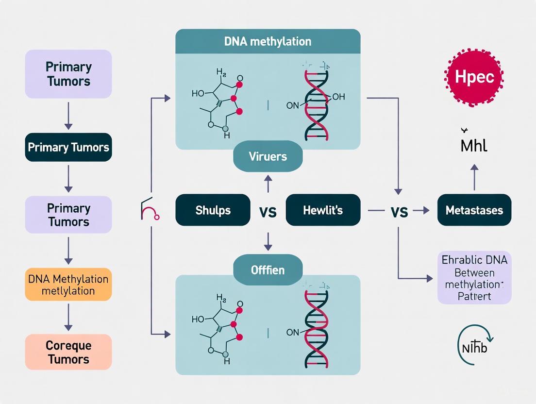
DNA Methylation Landscapes in Cancer Metastasis: From Primary Tumors to Disseminated Disease
This article synthesizes current research on DNA methylation patterns differentiating primary tumors from metastases, a critical area for understanding cancer progression.
DNA Methylation Landscapes in Cancer Metastasis: From Primary Tumors to Disseminated Disease
Abstract
This article synthesizes current research on DNA methylation patterns differentiating primary tumors from metastases, a critical area for understanding cancer progression. It explores the foundational concept that epigenetic profiles can trace tumor origin and reveal metastasis-specific alterations. The content details advanced methodological approaches for methylation analysis, from genome-wide sequencing to targeted validation, and addresses key challenges in sample handling and data interpretation. By comparing techniques and their clinical applications, particularly in cancer of unknown primary and liquid biopsy, this resource provides researchers and drug development professionals with a comprehensive framework for leveraging epigenetics in metastatic cancer research and therapeutic development.
Decoding the Metastatic Epigenome: Fundamental Shifts from Primary to Secondary Sites
A significant challenge in clinical oncology is determining the primary origin of metastatic tumors or classifying complex neoplasms with ambiguous morphology. This diagnostic precision is critical, as treatment strategies, including surgical approaches and systemic therapy regimens, are highly specific to the tumor's tissue of origin. Neuroendocrine neoplasms (NENs) exemplify this problem; they arise from diffuse neuroendocrine cells throughout the body, most commonly in the gastrointestinal tract and lungs, and often present with metastatic disease, particularly in the liver [1]. In 11-22% of patients with liver NENs, exhaustive clinical investigation fails to identify a primary tumor, leading to a diagnosis of 'liver metastatic NEN of unknown primary' or, in the current WHO classification, a primary hepatic neuroendocrine neoplasm [1]. DNA methylation profiling has emerged as a powerful solution to this diagnostic dilemma. DNA methylation involves the addition of a methyl group to a cytosine nucleotide, typically in a CpG dinucleotide context, and creates stable, tissue-specific epigenetic signatures that are maintained through cell divisions. These signatures act as a molecular fossil record, preserving clues about the original cell type from which the tumor arose, even in advanced metastatic deposits [1] [2] [3]. This guide synthesizes key evidence and experimental data demonstrating the utility of DNA methylation in tracing tumor origin, with a focus on neuroendocrine neoplasms.
Key Evidence: DNA Methylation Profiling in Neuroendocrine Neoplasms
Discriminating Primary Hepatic NENs from Metastases of Unknown Primary
A pivotal 2025 study published in Nature Communications undertook a comprehensive DNA methylation analysis of 212 NENs from two independent cohorts to address the diagnostic uncertainty surrounding hepatic NENs [1] [3]. The research yielded several critical findings:
- Distinct Organ-Specific Methylation Profiles: The DNA methylation profiles of NENs from different anatomical localizations (e.g., pancreas, ileum, appendix, colorectum, lung) differed significantly. Primary tumor and its metastasis pairs consistently clustered together in the analysis, demonstrating that the methylation signature of the primary site is retained in secondary deposits [1].
- Reclassification of Putative Primary Hepatic NENs: The subgroup of hepatic NENs that had been clinically classified as primary hepatic NENs (due to no extrahepatic primary being detected) did not form a distinct epigenetic cluster. Instead, these tumors colocalized with various subgroups of extrahepatic NENs, suggesting that a substantial proportion are in fact misclassified metastases from an unknown primary tumor [1].
- Development of a Predictive Classifier: The researchers developed a classifier based on the methylation profiles that could predict the origin of NENs with high accuracy. Specifically, organ-specific subtyping delineated a foregut-like epigenetic profile (including bronchopulmonary, stomach, duodenum, and pancreas) for the majority of hepatic NENs with an unknown primary [1].
This evidence underscores the clinical impact of methylation profiling, as it can redirect patient management towards the appropriate site-specific therapy.
Revealing Distinct Cellular Origins for Pancreatic NEN Subtypes
Beyond identifying the organ of origin, DNA methylation can trace the specific cell lineage within an organ. A 2022 study in Genome Medicine applied this principle to pancreatic neuroendocrine neoplasms (PanNENs), which are subdivided into well-differentiated neuroendocrine tumors (PanNETs) and poorly differentiated neuroendocrine carcinomas (PanNECs) [4] [5].
- Epigenetic Separation of PanNETs and PanNECs: DNA methylation analysis of 57 PanNEN samples revealed two major epigenetic groups that clearly distinguished high-grade PanNECs from all other PanNETs, including high-grade NET G3. This provides a robust molecular tool for a classification that is often histologically ambiguous [4] [5].
- Identifying the Cell of Origin: By comparing tumor methylation profiles to those of normal pancreatic cell types (alpha, beta, acinar, and ductal cells), the study found that PanNETs exhibit an endocrine cell lineage, while PanNECs share an exocrine cell (acinar/ductal) origin. This fundamental difference in cell lineage explains their distinct clinical behavior and mutational profiles (e.g., MEN1/ATRX/DAXX mutations in PanNETs vs. KRAS/TP53 in PanNECs) [4] [5].
The following table summarizes the core findings from these key studies on neuroendocrine neoplasms.
Table 1: Key Evidence from DNA Methylation Studies in Neuroendocrine Neoplasms
| Study Focus | Cohort Details | Key Methylation Finding | Biological & Clinical Impact |
|---|---|---|---|
| Origin of Hepatic NENs [1] | 212 NENs from two cohorts; included hepatic NENs with unknown primary. | Methylation profiles are organ-specific; primary-metastasis pairs cluster together. | Enabled high-accuracy classifier; suggested many "primary hepatic NENs" are metastases. |
| Classification of Pancreatic NENs [4] [5] | 57 PanNEN samples (PanNETs & PanNECs). | Two distinct epigenetic groups separate PanNECs from PanNETs. | Provides objective diagnostic tool; PanNECs show exocrine origin, distinct from endocrine PanNETs. |
Comparative Performance: Methylation vs. Other Modalities and Across Cancers
The utility of DNA methylation as a tracer extends beyond neuroendocrine tumors. When compared to other molecular and pathological techniques, it offers unique advantages.
Performance in Diagnosing Common Cancers and Metastases
A 2017 study evaluated a methylation-based classifier for four common cancers—lung, breast, colon, and liver—using data from The Cancer Genome Atlas (TCGA) and an independent Chinese cohort [6].
- High Diagnostic Accuracy: The classifier differentiated cancer tissue from normal tissue with >95% accuracy across both cohorts. This performance is comparable to standard diagnostic methods [6].
- Identification of Metastases: The signature correctly identified 29 out of 30 colorectal cancer metastases to the liver and 32 out of 34 colorectal cancer metastases to the lung, demonstrating its power to determine the origin of metastatic disease [6].
- Prognostic Value: The study also found that methylation patterns could predict patient prognosis and survival, with significant stratification of risk groups for breast and lung cancers [6].
Stability and Advantages Over Other Molecular Features
DNA methylation possesses several inherent properties that make it an excellent biomarker for tracing tumor origin [2]:
- Stability: Methylation marks are chemically stable, allowing profiling from formalin-fixed paraffin-embedded (FFPE) tissue, which is the standard in pathology departments.
- Pervasiveness and Consistency: Epigenetic changes in cancer are widespread and often occur early in tumorigenesis. They are highly pervasive across a tumor type, making them reliable markers.
- Tissue Specificity: Methylation patterns are inherently cell-type and tissue-type specific, providing a strong signal for cellular origin.
- Applicability to Liquid Biopsies: Cancer-associated methylation changes can be detected in cell-free DNA (cfDNA) from blood and other bodily fluids, enabling minimally invasive "liquid biopsies" for diagnosis and monitoring [2].
The following table compares DNA methylation profiling to other common approaches for determining tumor origin.
Table 2: Comparison of Methods for Tracing Tumor Origin
| Method | Key Principle | Advantages | Limitations |
|---|---|---|---|
| DNA Methylation Profiling | Detects tissue-specific epigenetic patterns. | High accuracy; reveals cell-of-origin; works on FFPE & liquid biopsies; stable markers. | Requires specialized bioinformatics. |
| Gene Expression Profiling | Identifies tissue-specific mRNA patterns. | Directly links to active biological processes; well-established protocols. | RNA is less stable; more sensitive to sample degradation. |
| Immunohistochemistry (IHC) | Detects tissue-specific protein markers via staining. | Low cost; widely available; integrates with histology. | Subjective interpretation; limited multiplexing; can be inconclusive. |
| Somatic Mutation Analysis | Identifies mutations in cancer driver genes. | Can reveal therapeutic targets (e.g., BRAF, EGFR). | Mutations are often not tissue-specific; tumor heterogeneity. |
Experimental Workflow: From Tissue to Diagnosis
Implementing DNA methylation analysis in a research or diagnostic setting involves a structured pipeline. The following diagram and description outline the standard workflow used in the cited studies.
Diagram 1: Workflow for DNA methylation-based tumor origin tracing.
Detailed Experimental Protocols
The key experimental and analytical steps are as follows:
Sample Acquisition and DNA Extraction:
- Tumor tissues, either fresh-frozen (FF) or formalin-fixed paraffin-embedded (FFPE), are used. A pathologist demarcates tumor-rich areas to ensure high tumor cell content (>75% is ideal) [1] [7].
- DNA is extracted using standard kits (e.g., GeneRead DNA FFPE kit from Qiagen), with quantity and quality assessed by methods like PicoGreen or agarose gel electrophoresis [5] [7].
Methylation Profiling:
- Bisulfite Conversion: Approximately 500 ng of genomic DNA is treated with sodium bisulfite using kits like the EZ DNA Methylation Kit (Zymo Research). This process converts unmethylated cytosines to uracils (which are read as thymines in subsequent steps), while methylated cytosines remain unchanged [7].
- Platform for Analysis:
- Methylation Microarrays: The most common platform in the cited studies is the Illumina Infinium MethylationEPIC BeadChip (850K array), which quantitatively assesses the methylation status of over 850,000 CpG sites across the genome [1] [5]. It provides a beta-value (β) for each site, representing the ratio of methylated signal intensity to the total intensity (β = 0-1, from completely unmethylated to fully methylated).
- Sequencing-Based Methods: For higher resolution, methods like Reduced Representation Bisulfite Sequencing (RRBS) can be used. RRBS uses restriction enzymes (e.g., MspI) to target CpG-rich regions, followed by bisulfite sequencing, providing base-precision methylation data across thousands of genomic fragments [8].
Bioinformatic and Statistical Analysis:
- Preprocessing: Raw data is normalized, and probes with low signal or those overlapping with known single-nucleotide polymorphisms are filtered out [7].
- Differential Methylation Analysis: Unsupervised analysis (e.g., hierarchical clustering) reveals natural groupings of samples. Supervised analysis (e.g., ANOVA, Mann-Whitney U tests) identifies specific CpG sites or regions that are differentially methylated between sample groups (e.g., primary vs. metastatic, or between different organs of origin) [1] [7].
- Classifier Training: Machine learning algorithms, such as the least absolute shrinkage and selection operator (lasso) under a multinomial distribution, are used to build a predictive model from a training cohort. The model selects a panel of the most informative methylation markers to create a classifier [6].
Validation: The classifier's performance is rigorously validated on an independent cohort of samples not used in the training phase to assess its real-world accuracy and robustness [1] [6].
The Scientist's Toolkit: Essential Research Reagents and Materials
Table 3: Essential Research Reagents for DNA Methylation Profiling of Tumor Origin
| Reagent / Kit | Specific Example(s) | Critical Function in Workflow |
|---|---|---|
| DNA Extraction Kit | GeneRead DNA FFPE Kit (Qiagen) | Isols high-quality genomic DNA from challenging FFPE tissue samples. |
| Bisulfite Conversion Kit | EZ DNA Methylation Kit (Zymo Research) | Chemically converts unmethylated cytosines, enabling methylation status detection. |
| Methylation Profiling Platform | Infinium MethylationEPIC BeadChip (Illumina) | Genome-wide quantitative analysis of >850,000 CpG sites; industry standard. |
| Library Prep Kit for Sequencing | Ion AmpliSeq Library Kit (Thermo Fisher) | Prepares sequencing libraries from bisulfite-converted DNA for targeted or RRBS approaches. |
| Bioinformatic Tools | R/Bioconductor packages (e.g., minfi, limma), Bismark (for sequencing) | Data normalization, quality control, differential methylation analysis, and visualization. |
Biological Pathways and Logical Relationships
The biological rationale for using DNA methylation lies in its role as a key regulator of cellular identity. The following diagram illustrates the logical pathway from an epigenetic defect to a diagnostic readout.
Diagram 2: Biological logic of methylation-based origin tracing.
The process is governed by several key biological principles:
- Developmental Programming: During normal cellular differentiation, a tissue-specific DNA methylation landscape is established. This "epigenetic blueprint" defines and maintains the identity and function of each cell type (e.g., a pancreatic beta cell vs. a pulmonary neuroendocrine cell) by locking in specific gene expression programs [2] [3].
- Retention in Oncogenesis: When a cell undergoes oncogenic transformation, this core tissue-specific methylation signature is largely retained, even as the cell acquires new cancer-specific methylation aberrations (e.g., global hypomethylation and focal hypermethylation of tumor suppressor genes). The tumor thus carries an epigenetic memory of its cell of origin [1] [2].
- Clonal Stability in Metastasis: During metastatic dissemination, the methylation signature of the primary tumor is stably inherited by daughter cells in secondary metastases. This fundamental principle—that a metastasis's methylation profile will most closely resemble its primary tumor of origin—is the foundation upon which this diagnostic approach is built [1] [6] [8].
The evidence from neuroendocrine and other solid tumors unequivocally demonstrates that DNA methylation profiling is a robust and accurate method for tracing tumor origin. Its ability to reclassify diagnostically challenging cases, such as hepatic NENs of unknown primary, and to reveal fundamental biological distinctions, as in pancreatic NEN subtypes, positions it as a powerful tool for precision pathology. The technology is mature, with standardized workflows and commercially available platforms.
Future directions in this field will focus on the widespread clinical implementation of methylation-based classifiers, potentially as part of routine diagnostic workups for cancers of unknown primary and metastatic disease. Furthermore, the integration of methylation data with other molecular modalities—such as genomic, transcriptomic, and proteomic profiles—in a multi-omics framework will yield even more comprehensive and powerful tumor characterization [3]. Finally, the development of highly sensitive liquid biopsy assays that detect tumor-derived methylation patterns in blood plasma promises to enable minimally invasive diagnosis, monitoring of treatment response, and detection of minimal residual disease, ultimately improving patient outcomes across the oncology spectrum [2].
Metastasis is a multi-step process responsible for the majority of cancer-related mortality, wherein cancer cells spread from a primary tumor to colonize distant organs [9]. While genetic mutations initiate oncogenesis, accumulating evidence demonstrates that epigenetic mechanisms, particularly DNA methylation, are fundamental regulators of metastatic progression [9]. DNA methylation involves the addition of a methyl group to the 5-carbon of cytosine within CpG dinucleotides, typically leading to gene silencing when it occurs in promoter regions [10]. The dynamic and reversible nature of epigenetic modifications allows cancer cells to rapidly adapt, survive in circulation, and establish secondary tumors [9]. This comparative guide analyzes pan-cancer studies to objectively distinguish conserved methylation patterns from tissue-specific alterations in metastasis, providing researchers with a synthesized overview of key epigenetic drivers and methodological approaches for investigating the metastatic methylome.
Comparative Analysis of Conserved versus Tissue-Specific Methylation Changes
Pan-cancer analyses reveal that metastatic progression is characterized by both universal epigenetic patterns that transcend tissue origins and distinct changes unique to specific cancer types. The table below summarizes the primary conserved and tissue-specific methylation alterations observed across multiple cancer studies.
Table 1: Conserved vs. Tissue-Specific DNA Methylation Changes in Metastasis
| Feature | Conserved Pan-Cancer Changes | Tissue-Specific Changes |
|---|---|---|
| Global Pattern | Global hypomethylation [8]; Focal hypermethylation [10] | Tissue-of-origin methylation signatures maintained in metastases [11] |
| Commonly Affected Genes | Metastasis-suppressor genes (CDH1, TIMP3, BRMS1) [9] | Subtype-specific methylation changes (e.g., FLNC in gastric cancer [12]) |
| Molecular Function | Silencing of cell-adhesion genes; Promotion of epithelial-mesenchymal transition (EMT) [13] | HER2 subtype switching in breast cancer [13] |
| Microenvironment Interaction | Immune evasion mechanisms (e.g., HLA hypermethylation) [13] | Stromal changes varying by subtype (e.g., ER+ breast cancer metastases show lower fibroblast content) [13] |
| Diagnostic Utility | Multi-omics classifiers for CUP origin prediction (>90% accuracy) [14] | Methylation markers for specific cancers (e.g., TDRD10, PRAC2, TMEM132C in breast cancer) [15] |
Conserved Methylation Drivers of Metastasis
Across multiple cancer types, certain DNA methylation alterations consistently appear in metastatic cells, forming a conserved epigenetic program that enables metastatic progression.
Global Hypomethylation with Focal Hypermethylation: Metastatic cells consistently exhibit global genomic hypomethylation, which promotes chromosomal instability and reactivates transposable elements, alongside focal hypermethylation at specific regulatory sites that silences tumor and metastasis suppressor genes [10] [8]. This paradoxical pattern is a hallmark of advanced tumors observed in melanoma, prostate cancer, and other malignancies.
Silencing of Metastasis Suppressors: Key genes governing cell adhesion and invasion are frequently hypermethylated in metastasis. Promoter hypermethylation of E-cadherin (CDH1), a critical cell-cell adhesion molecule, diminishes intercellular attachment and facilitates epithelial-mesenchymal transition (EMT) across multiple carcinomas [9]. Other consistently silenced genes include TIMP3, which regulates extracellular matrix remodeling, and BRMS1, a suppressor of metastasis [9].
Immune Evasion Mechanisms: A conserved mechanism of immune evasion in metastasis involves DNA hypermethylation and/or focal deletions near the HLA-A gene, which reduces HLA class I expression and cytotoxic T-cell recognition. This alteration was identified in breast cancer metastases, particularly in brain and liver lesions, suggesting a pan-cancer adaptation to evade immune surveillance [13].
Tissue and Lineage-Specific Methylation Patterns
Despite these common themes, DNA methylation patterns in metastasis also reflect the tissue of origin and molecular subtypes of the primary tumor.
Maintenance of Tissue-of-Origin Signature: Methylation profiling remains a powerful tool for identifying the origin of carcinomas of unknown primary (CUP). One pan-cancer methylation study developed a classifier (CACO) using six CpG sites located in five genes that accurately (>95%) traced metastatic tumors back to their primary site, demonstrating that lineage-specific methylation signatures are largely retained even after metastasis [14] [11].
Cancer-Type Specific Alterations: Specific methylation events are associated with metastasis in particular cancers. In gastric carcinoma, promoter methylation of the FLNC gene occurs more frequently in metastatic lesions compared to matched primary tumors [12]. In breast cancer, promoter hypermethylation contributes to the downregulation of estrogen receptor-mediated cell-cell adhesion genes in metastases [13].
Molecular Subtype Switching: Metastatic relapse can involve shifts in molecular subtypes dictated by epigenetic changes. In breast cancer, approximately 30% of metastases show PAM50 expression subtype switching compared to the primary tumor, often coinciding with DNA clonality shifts, particularly in HER2 status, which has significant therapeutic implications [13].
Key Methodologies for Metastatic Methylome Analysis
Investigating methylation changes in metastasis requires sophisticated multi-omics approaches. The following experimental protocols are central to generating high-quality, comparable data.
Genome-Wide Methylation Sequencing
Reduced Representation Bisulfite Sequencing (RRBS) is widely used for high-resolution methylation mapping. This method utilizes MspI restriction enzyme digestion to enrich for CpG-dense regions, followed by bisulfite conversion and next-generation sequencing. This protocol effectively covers promoter regions, CpG islands, and other regulatory elements with single-base-pair resolution, making it suitable for discovering novel metastasis-associated epigenetic alterations [8]. RRBS analysis of paired primary and metastatic melanoma cell lines revealed global hypomethylation and identified 75 shared differentially methylated fragments (DMFs) in metastases [8].
Multi-Omics Integration and Classifier Development
Advanced pan-cancer analysis involves integrating DNA methylation data with transcriptomic and genomic data from sources like The Cancer Genome Atlas (TCGA). A typical workflow includes:
- Differential Methylation Analysis: Identifying CpG sites with significant methylation differences (∆β > 0.4) between tumor and normal tissues [15].
- Methylation-Expression Correlation: Correlating methylation status (in promoters, gene bodies, intergenic regions) with gene expression data to identify functionally relevant epigenetic changes [14] [15].
- Machine Learning Classification: Using ensemble algorithms to construct a tissue-specific classifier based on a minimal set of highly informative CpG sites. This approach has achieved >90% accuracy in predicting the origin of metastatic cancers, including CUP [14].
Single-Cell Transcriptome Analysis
Single-cell RNA sequencing (scRNA-seq) of metastatic and non-metastatic tumors across multiple cancer types enables the deconvolution of tumor heterogeneity and identification of a core metastatic signature. The standard workflow involves:
- Data Integration: Curating and integrating scRNA-seq data from hundreds of patients and over a million cancer cells using tools like Seurat [16].
- Metastatic Archetype Identification: Applying multiresolution archetypal analysis (ACTIONet R package) to identify cell subpopulations with high metastatic potential based on expression of known metastasis-linked genes [16].
- Signature Refinement: Refining a core gene signature by selecting genes consistently expressed across multiple archetypes and patients, then further filtering for epithelial-specificity to isolate cancer-cell intrinsic signals [16].
Signaling Pathways and Regulatory Networks
The following diagram synthesizes key mechanistic relationships and regulatory networks derived from pan-cancer methylation studies, illustrating how conserved epigenetic drivers influence metastatic progression.
Figure 1: Conserved Epigenetic Pathways in Cancer Metastasis. This diagram illustrates the core regulatory networks through which conserved DNA methylation changes drive metastatic progression. Focal hypermethylation silences key tumor and metastasis suppressor genes, while global hypomethylation activates oncogenes and promotes genomic instability. Together, these alterations enable the hallmark capabilities of metastatic cells.
The Scientist's Toolkit: Essential Research Reagents and Platforms
Cutting-edge pan-cancer methylation research relies on specific reagents, computational tools, and experimental platforms. The following table details key resources for investigating metastatic methylation patterns.
Table 2: Essential Research Reagents and Platforms for Metastatic Methylation Analysis
| Category | Specific Tool/Reagent | Research Function | Example Use Case |
|---|---|---|---|
| Methylation Profiling | Illumina Infinium Methylation EPIC array [15] [13] | Genome-wide CpG methylation quantification (850,000+ sites) | Profiling primary and metastatic breast tumors in AURORA US study [13] |
| Methylation Profiling | Reduced Representation Bisulfite Sequencing (RRBS) [8] | High-resolution methylation sequencing of CpG-rich regions | Identifying 75 DMFs in paired primary/metastatic melanoma lines [8] |
| Bioinformatic Tools | Seurat [16] | Single-cell RNA-seq data integration and clustering | Analyzing 1.2 million cancer cells from 266 tumors for pan-cancer metastasis signatures [16] |
| Bioinformatic Tools | ACTIONet R package [16] | Multiresolution archetypal analysis of scRNA-seq data | Identifying metastatic cell subpopulations across cancer types [16] |
| Bioinformatic Tools | OncoScore [15] | Literature-based prioritization of cancer-associated genes | Selecting novel methylation markers in breast cancer [15] |
| Reference Data | The Cancer Genome Atlas (TCGA) [14] [15] [11] | Multi-omics data for 33 cancer types | Training pan-cancer methylation classifiers for tumor origin [14] [11] |
| Reference Data | Roadmap Epigenomics Consortium [15] | Reference epigenomes for diverse normal tissues | Annotating regulatory potential of differentially methylated regions [15] |
Pan-cancer analysis reveals that metastasis is governed by both a conserved epigenetic program and tissue-specific methylation alterations. The conserved program includes global hypomethylation, silencing of key metastasis suppressors like CDH1, and immune evasion through HLA methylation. Simultaneously, tissue-of-origin signatures persist in metastases, and specific methylation events drive subtype switching in cancers like breast and gastric carcinoma. Methodologically, the field is moving toward multi-omics integration and single-cell resolution to deconvolute metastatic heterogeneity. For drug development professionals, these findings highlight the potential of epigenetic biomarkers for diagnosing metastatic propensity and the promise of targeting the epigenetic machinery, such as DNMTs and TET enzymes, to reverse pro-metastatic phenotypes. Future research that further integrates spatial transcriptomics with methylation mapping will continue to refine our understanding of the metastatic landscape and guide the development of novel epigenetic therapies.
Metastatic progression represents the most profound challenge in clinical oncology, accounting for the vast majority of cancer-related mortality. While genetic mutations have long been studied as drivers of cancer progression, epigenetic alterations—particularly in DNA methylation patterns—are now recognized as equally critical determinants of metastatic behavior. The coexistence of global DNA hypomethylation alongside localized promoter hypermethylation creates an epigenetic landscape that facilitates cellular transformation, enhances migratory capacity, and enables metastatic dissemination across multiple cancer types. This paradoxical methylation pattern serves as a molecular signature of advanced disease, offering both prognostic value and potential therapeutic targets. Understanding how these opposing methylation states coordinately drive metastatic progression provides crucial insights into cancer biology and reveals novel avenues for diagnostic and therapeutic intervention in advanced stage malignancies.
Quantitative Evidence: Methylation Changes Across Cancer Types
Extensive profiling of primary and metastatic tumors across diverse cancer types has consistently demonstrated distinct methylation alterations associated with metastatic progression. The table below summarizes key quantitative findings from multiple studies investigating these epigenetic changes.
Table 1: Documented Methylation Changes in Human Cancers
| Cancer Type | Global Hypomethylation Evidence | Localized Hypermethylation Evidence | Functional Consequences |
|---|---|---|---|
| Thyroid Cancer | Distant metastatic DTC showed significantly increased Alu hypomethylation (median: 4.0, IQR: 3.1-6.2) vs. normal tissue (median: 2.75, IQR: 2.30-3.15); PDTC/ATC showed even higher hypomethylation (median: 9.3, IQR: 7.0-12.1) [17]. | Not specified in study | Associated with cancer-related and all-cause mortality; suggests role in thyroid cancer progression and dedifferentiation [17]. |
| Melanoma | Metastatic melanoma cell lines showed global hypomethylation compared to matched primary lines [8]. RRBS analysis identified 65 hypomethylated DMFs in metastatic vs. primary cell lines [8]. | 10 hypermethylated DMFs identified in metastatic vs. primary cell lines; EBF3 promoter hypermethylation associated with increased mRNA levels and aggressive phenotype [8]. | EBF3 knockdown decreased proliferation, migration and invasion; hypermethylation may drive metastasis [8]. |
| Breast Cancer | Not specifically highlighted | Downregulation of ER-mediated cell-cell adhesion genes through DNA methylation mechanisms in metastases; 17% of metastases showed DNA hypermethylation and/or focal deletions near HLA-A [13]. | HLA-A hypermethylation associated with reduced expression and lower immune cell infiltrates, particularly in brain and liver metastases [13]. |
| Prostate Cancer | Plasma DNA methylome analysis showed global hypermethylation in metastatic samples coupled with hypomethylation in pericentromeric regions [18]. | CpG island hypermethylation at MDR1 in 83.3%, EDNRB in 50%, RAR-β in 38.9% of metastatic cases; hypermethylation at NR3C1 promoter associated with decreased immune signature [19] [18]. | Hypermethylation profile distinguishes localized from metastatic disease with 0.989 prediction accuracy; may contribute to immune evasion [19] [18]. |
Table 2: Experimental Models of DNA Hypomethylation in Metastasis
| Experimental System | Intervention | Methylation Changes | Functional Outcomes |
|---|---|---|---|
| NIH3T3 cells (mouse fibroblast) | Enforced Stella (PGC7/Dppa3) expression | Induced global DNA demethylation [20] [21] [22] | Cellular transformation [20] [21] [22] |
| B16 melanoma cells | Stella overexpression | Global DNA demethylation [20] [21] [22] | Enhanced metastatic ability through induction of metastasis-related genes [20] [21] [22] |
Molecular Mechanisms and Pathways
Global Hypomethylation: Activating the Metastatic Program
Global DNA hypomethylation primarily affects repetitive genomic elements and gene-poor regions, leading to genomic instability and activation of normally silenced genes. In thyroid cancer, increasing hypomethylation of Alu repeats directly correlates with disease progression, with metastatic and dedifferentiated tumors showing the most pronounced hypomethylation [17]. This loss of methylation in repetitive elements can promote chromosomal rearrangements and activation of latent oncogenic pathways. In experimental models, enforced expression of Stella (PGC7/Dppa3) induces global DNA demethylation and directly transforms NIH3T3 cells while enhancing the metastatic capability of B16 melanoma cells [20] [21] [22]. The hypomethylated state appears to activate metastasis-related genes that facilitate invasion and colonization of distant sites, providing functional evidence that global hypomethylation can drive metastatic progression rather than merely correlating with it.
Focal Hypermethylation: Silencing Tumor Suppression
While global hypomethylation activates pro-metastatic genes, focal hypermethylation at specific promoter regions silences critical tumor suppressor genes and pathways that normally restrain metastasis. In breast cancer, downregulation of estrogen receptor-mediated cell-cell adhesion genes through DNA hypermethylation enables dissociation of tumor cells from the primary site [13]. Similarly, hypermethylation near HLA-A genes in breast cancer metastases reduces antigen presentation capability and immune cell infiltration, particularly in brain and liver metastases [13]. Melanoma metastases exhibit EBF3 promoter hypermethylation that surprisingly increases EBF3 expression, resulting in enhanced proliferation, migration, and invasion [8]. This paradoxical relationship between promoter hypermethylation and gene activation highlights the complexity of epigenetic regulation in metastasis and underscores the importance of considering genomic context when interpreting methylation patterns.
The Tumor Microenvironment: Epigenetic Crosstalk
The metastatic niche is shaped not only by tumor-intrinsic methylation changes but also by epigenetic alterations in the tumor microenvironment. In prostate cancer, the cell-free DNA methylome captures variations beyond the tumor itself, with lactate dehydrogenase (LDH) and alkaline phosphatase (ALP) levels explaining methylation variations in regions distinct from those associated with ctDNA fraction [18]. This suggests that methylation patterns in circulating DNA reflect contributions from both tumor cells and the surrounding microenvironmental components. Breast cancer metastases show subtype-specific microenvironment differences, with ER+/luminal metastases having lower fibroblast and endothelial content, while triple-negative/basal metastases show decreased B and T cells [13]. These microenvironment shifts are likely facilitated by epigenetic modifications that alter cell-cell communication and immune recognition.
Experimental Approaches and Methodologies
Genome-Wide Methylation Profiling Technologies
The comprehensive characterization of methylation patterns in metastasis relies on multiple high-throughput technologies, each with distinct strengths and applications.
Table 3: Methylation Analysis Techniques in Metastasis Research
| Methodology | Principle | Application in Metastasis Research | Example Studies |
|---|---|---|---|
| RRBS (Reduced Representation Bisulfite Sequencing) | Bisulfite treatment followed by sequencing of size-selected fragments covering CpG-rich regions | Genome-wide methylation comparison between primary and metastatic melanoma cell lines; identifies differentially methylated fragments (DMFs) [8] | Chatterjee et al. identified 75 shared DMFs (10 hyper- and 65 hypomethylated) in metastatic vs. primary melanoma [8]. |
| Infinium HumanMethylation450 BeadChip | Array-based profiling of ~450,000 CpG sites across the genome | Comprehensive DNA methylation analysis across progression stages (benign nevi, primary melanoma, metastases) [7] | Identification of HOXA9 and TBC1D16 as methylation biomarkers for melanoma development and progression [7]. |
| cfMeDIP-seq (Cell-free Methylated DNA Immunoprecipitation sequencing) | Immunoprecipitation of methylated DNA fragments from plasma followed by sequencing | Analysis of plasma DNA methylomes from localized and metastatic prostate cancer patients [18] | Distinguished localized from metastatic disease with 0.989 accuracy; captured fragmentation profiles [18]. |
| Quantification of Unmethylated Alu (QUAlu) | Measures global hypomethylation using Alu repeats as surrogate markers | Assessment of global DNA hypomethylation in thyroid cancer progression [17] | Revealed increasing hypomethylation in distant metastatic DTC and PDTC/ATC vs. normal tissue [17]. |
Functional Validation Approaches
Establishing causal relationships between methylation changes and metastatic phenotypes requires rigorous functional validation. In the melanoma metastasis study, researchers employed bisulfite sequencing to validate EBF3 promoter hypermethylation findings from RRBS in independent melanoma cohorts [8]. Critical functional evidence came from RNAi-mediated knockdown of EBF3, which decreased proliferation, migration, and invasion in both primary and metastatic melanoma cell lines [8]. In the Stella overexpression model, the direct induction of global demethylation followed by assessment of transformation and metastatic ability provided compelling evidence for the functional role of hypomethylation in driving malignant progression [20] [21] [22]. These functional studies transform correlative observations into mechanistic understanding, distinguishing driver epigenetic alterations from passenger events in metastatic progression.
Table 4: Key Research Reagents and Resources for Metastasis Methylation Studies
| Reagent/Resource | Function/Application | Examples from Literature |
|---|---|---|
| Patient-Derived Cell Lines | Paired primary and metastatic cell lines minimize heterogeneity for identifying driver epigenetic alterations | WM115 (primary) and WM266-4 (metastatic) melanoma cell lines; Hs688(A).T (primary) and Hs688(B).T (metastatic) [8] |
| Bisulfite Conversion Kits | Convert unmethylated cytosines to uracils while methylated cytosines remain unchanged, enabling methylation detection | EZ DNA methylation kit (Zymo Research) used in melanoma methylome study [7] |
| Methylation-Specific Restriction Enzymes | Digest unmethylated DNA sequences while leaving methylated sequences intact for methylation analysis | HpaII digestion used in quantitative real-time PCR for serum CpG island hypermethylation detection in prostate cancer [19] |
| Stella (PGC7/Dppa3) Expression Constructs | Experimentally induce global DNA demethylation to study functional consequences | Enforced Stella expression in NIH3T3 and B16 cells induced global demethylation and enhanced transformation/metastasis [20] [21] [22] |
| DNA Methylation Inhibitors | Demethylating agents to test functional consequences of methylation changes | 5-azacytidine treatment shown to enhance experimental metastatic capacity in tumor lines [20] |
Visualization of Metastatic Methylation Pathways
The following diagram illustrates the coordinated relationship between global hypomethylation and localized hypermethylation in driving metastatic progression:
Molecular Pathways Linking Methylation Changes to Metastasis
Clinical Implications and Therapeutic Opportunities
The distinct methylation patterns associated with metastatic progression offer significant clinical potential for diagnostics, prognostication, and therapeutic development. In prostate cancer, the cell-free DNA methylome distinguishes localized from metastatic disease with remarkable accuracy (0.989 prediction accuracy) [18], suggesting utility for non-invasive monitoring of disease progression. Similarly, in cutaneous melanoma, DNA methylation biomarkers such as PON3 methylation and OVOL1 expression provide prognostic information independent of traditional histological markers like tumor thickness and ulceration [7]. Therapeutically, the reversal of metastasis-associated methylation patterns represents an attractive strategy, with demethylating agents already showing potential to modulate metastatic behavior in experimental models [20]. Additionally, the identification of hypermethylation near HLA-A in breast cancer metastases [13] suggests possible combination strategies between epigenetic therapies and immunotherapies to enhance anti-tumor immune responses. As our understanding of the metastatic methylome deepens, these epigenetic insights will increasingly inform clinical decision-making and therapeutic development for advanced cancers.
The coordinated patterns of global hypomethylation and localized hypermethylation represent a fundamental hallmark of metastatic progression across diverse cancer types. These epigenetic alterations drive metastasis through complementary mechanisms: hypomethylation promotes genomic instability and activates metastasis-related genes, while hypermethylation silences tumor suppressors and immune recognition pathways. The consistency of these patterns—observed in thyroid, melanoma, breast, and prostate cancers—underscores their fundamental role in cancer progression. Advanced methylation profiling technologies now enable comprehensive characterization of these changes in both tissue and liquid biopsies, providing powerful tools for diagnosis, prognosis, and therapeutic monitoring. As functional studies continue to establish causal relationships between specific methylation events and metastatic phenotypes, the potential for targeting these epigenetic drivers therapeutically continues to grow. Ultimately, decoding the metastatic methylome offers promising avenues for addressing the greatest challenge in clinical oncology—preventing and treating metastatic disease.
Metastasis is responsible for the vast majority of cancer-related deaths, yet metastasis-specific genetic mutations have remained elusive [23]. This paradox has shifted research focus toward epigenetic alterations as potential drivers of metastatic progression. Among these, DNA methylation changes are increasingly implicated as "epi-drivers" that enable cancer cells to acquire metastatic capabilities without genetic mutations [23] [24]. This case study examines the evidence for EBF3 hypermethylation as a candidate epigenetic driver in melanoma metastasis, comparing its role across different cancer types and experimental models. We evaluate how this paradoxical phenomenon—promoter hypermethylation associated with gene activation—challenges conventional understanding of epigenetic regulation in cancer [25].
EBF3 Hypermethylation: Core Findings and Mechanistic Insights
Initial Discovery and Functional Validation
The role of EBF3 in melanoma metastasis was first identified through genome-wide DNA methylation analysis of paired primary and metastatic melanoma cell lines using reduced representation bisulfite sequencing (RRBS) [24] [8]. This approach revealed that metastatic melanoma cell lines exhibited promoter hypermethylation of EBF3 compared to matched primary melanoma cell lines [24] [8]. Functional validation demonstrated that this hypermethylation was associated with increased EBF3 mRNA levels in metastatic melanomas, and inhibition of DNA methylation reduced EBF3 expression [24]. Most importantly, RNAi-mediated knockdown of EBF3 decreased proliferation, migration, and invasion in primary and metastatic melanoma cell lines, establishing its functional role in aggressive phenotypic behavior [24] [8].
Paradoxical Gene Activation Mechanism
The EBF3 paradigm challenges the classical view that promoter hypermethylation universally silences gene expression. Contrary to this conventional understanding, EBF3 exhibits paradoxical activation through methylation-mediated mechanisms [25]. While the precise molecular mechanism continues to be investigated, this phenomenon may involve:
- Interference with repressive transcription factor binding
- Long-range enhancer-promoter interactions
- Expression of alternative gene isoforms [25]
Recent research using a CRISPR-SunTag All-in-One system for targeted methylation editing has established causality between EBF3 promoter hypermethylation and increased gene expression, confirming this paradoxical relationship [25].
Comparative Methylation Patterns Across Cancer Types
EBF3 methylation changes are not restricted to melanoma. Analysis of multiple tumor types reveals this as a common epigenetic event in cancer progression:
Table: EBF3 Methylation Patterns Across Cancer Types
| Cancer Type | Promoter Methylation | Gene Body Methylation | Functional Association |
|---|---|---|---|
| Melanoma | Hyper-methylated in metastases | Hypo-methylated in metastases | Metastasis progression [23] |
| Endometrial Cancer | Variable | Hyper-methylated in hyperplasia vs primary tumors | Tumourigenesis [23] |
| Prostate Cancer | Variable | Hyper-methylated in metastases | Metastasis progression [23] |
| Colorectal Cancer | Hyper-methylated in metastases | Not specified | Metastasis progression [23] |
The consistency of EBF3 methylation changes across diverse cancers suggests it may function as a generalized epi-driver of tumor progression rather than a cancer-type specific alteration [23].
Comparative Analysis: EBF3 Versus Other Methylation Drivers
EBF3 and TBC1D16 as Cooperative Epi-Drivers
Research has identified TBC1D16 as another significant epigenetic driver in melanoma metastasis, operating through distinct mechanisms:
Table: Comparison of EBF3 and TBC1D16 Methylation Alterations
| Feature | EBF3 | TBC1D16 |
|---|---|---|
| Methylation Change in Metastasis | Promoter hypermethylation | Gene body hypomethylation |
| Expression Change | Increased mRNA | Activation of cryptic transcript TBC1D16-47KD |
| Functional Role | Promotes proliferation, migration, invasion | Enhances melanoma proliferation, metastasis, and drug sensitivity |
| Mechanism | Paradoxical gene activation | Alternative promoter activation |
| Therapeutic Implications | Potential epigenetic therapy target | Predicts response to BRAF/MEK inhibitors |
Notably, these epigenetic alterations can occur simultaneously in metastatic tumors, suggesting they may act cooperatively to drive metastatic progression [23].
Broader DNA Methylation Landscape in Melanoma Progression
Beyond specific genes, melanoma progression involves genome-wide methylation restructuring:
- Global hypomethylation predominantly affects CpG-poor regions ("open seas" and shelves) [26]
- Focal hypermethylation occurs at CpG islands, particularly in advanced metastases [26]
- Melanoma brain metastases exhibit distinct methylation profiles with partially methylated domains (PMDs) affecting brain development genes [26]
Recent multi-omics profiling has classified metastatic melanomas into four methylation subsets with distinct clinical behaviors, where patients with low methylation tumors showed significantly longer survival compared to those with CpG island methylator phenotype (CIMP) tumors [27].
Experimental Models and Methodologies
Key Research Models for Studying Melanoma Methylation
Table: Research Models for Studying DNA Methylation in Melanoma
| Model System | Key Features | Applications | References |
|---|---|---|---|
| Paired primary/metastatic cell lines (WM115/WM266-4; Hs688(A).T/Hs688(B).T) | Minimal heterogeneity, controlled genetics | Identifying metastasis-specific methylation changes | [24] [8] |
| Mouse linear progression model (melan-a, 4C, 4C11-, 4C11+) | Represents distinct progression stages | Studying methylation changes during transformation | [28] |
| Patient-matched intra/extracranial metastases | Accounts for inter-patient heterogeneity | Identifying site-specific adaptation changes | [29] |
| CRISPR-epigenome editing systems | Locus-specific methylation manipulation | Establishing causality of methylation changes | [25] |
Core Methodological Approaches
Methylation Profiling Techniques
- Reduced Representation Bisulfite Sequencing (RRBS): Provides cost-effective, genome-wide methylation coverage with single-CpG resolution [24] [8]
- Enhanced RRBS (ERRBS): Offers expanded genomic coverage, particularly of CpG islands and shores [28]
- Infinium HumanMethylation450K BeadChip: Interrogates 485,512 methylation sites across genome; used for large cohort validation [26]
Functional Validation Methods
- Targeted epigenetic editing: CRISPR-dCas9 systems fused to DNMT3A (for methylation) or TET1 (for demethylation) establish causality [25]
- Pharmacological demethylation: DNMT inhibitors (5-aza-2'-deoxycytidine) assess methylation-dependent gene expression [28]
- Functional assays: Migration, invasion, and proliferation tests evaluate phenotypic consequences [24]
Research Reagent Solutions Toolkit
Table: Essential Research Reagents for Methylation Studies
| Reagent/Category | Specific Examples | Research Application | Key Features |
|---|---|---|---|
| Methylation Profiling | RRBS, ERRBS, Infinium HM450K | Genome-wide methylation analysis | Various coverage depths and resolutions |
| Targeted Editing | CRISPR-SunTag All-in-One, dCas9-DNMT3A, dCas9-TET1 | Locus-specific methylation manipulation | Precise epigenetic engineering |
| Cell Line Models | WM115/WM266-4, melan-a/4C/4C11-/4C11+ | Controlled progression studies | Paired primary-metastatic equivalents |
| Demethylating Agents | 5-aza-2'-deoxycytidine (decitabine) | Pharmacological demethylation | Global methylation inhibition |
| Analysis Tools | Bismark, RnBeads, methylKit | Bioinformatic processing | Differential methylation calling |
Signaling Pathways and Conceptual Framework
Diagram 1: EBF3 Paradoxical Activation Pathway. This workflow illustrates the unusual mechanism where promoter hypermethylation activates rather than silences EBF3 expression, leading to functional metastatic capabilities.
Clinical Implications and Therapeutic Opportunities
Diagnostic and Prognostic Applications
DNA methylation signatures, including EBF3 patterns, show significant clinical relevance:
- Methylation classification of melanomas into DEMethylated, LOW, INTermediate, and CIMP subsets correlates with patient survival [27]
- Patients with low methylation tumors exhibit longer survival and lower progression rates compared to CIMP patients [27]
- HOXD9 hypermethylation in lymph node metastases associates with poorer disease-free and overall survival [26]
Therapeutic Targeting Strategies
- DNMT inhibitors (e.g., decitabine, azacitidine) can reverse methylation-mediated gene silencing and induce viral mimicry through endogenous retrovirus reactivation [30]
- Combination therapies with immune checkpoint blockers may sensitize immunologically "cold" tumors [30] [27]
- Patient stratification based on methylation profiles could optimize therapy selection [27]
EBF3 hypermethylation represents a compelling example of an epigenetic driver in melanoma metastasis, challenging conventional paradigms through its paradoxical activation mechanism. The comparative analysis presented here demonstrates that EBF3 alterations occur across multiple cancer types alongside other epigenetic drivers like TBC1D16, suggesting common pathways in metastatic progression. Future research should focus on:
- Developing more specific epigenetic editors to precisely manipulate methylation states
- Exploring combination therapies that target both genetic and epigenetic vulnerabilities
- Validating methylation biomarkers for clinical stratification in prospective trials
- Investigating the immune-modulatory effects of epigenetic therapies in the tumor microenvironment
The evolving toolkit for methylation analysis and manipulation continues to provide unprecedented opportunities to understand and therapeutally target the epigenetic drivers of cancer metastasis.
The "seed and soil" hypothesis, first proposed by Stephen Paget in 1889, posits that metastatic cells ("seeds") preferentially grow in the microenvironments of specific organs ("soil") that support their growth [31] [32]. While this concept has been recognized for over a century, recent molecular evidence has revealed that epigenetic mechanisms, particularly DNA methylation, serve as critical mediators of this selective adaptation. DNA methylation patterns are established in a tissue-specific manner in both normal and tumor tissues, providing a molecular fingerprint of cellular origin [33]. This review synthesizes current evidence demonstrating how dynamic methylation changes enable metastatic cells to adapt to new microenvironments, compares methylation profiles between primary tumors and their metastases, and explores the clinical implications of these epigenetic adaptations for cancer diagnosis and treatment.
Historical Context and Modern Interpretation
The seed and soil hypothesis challenged the previously dominant anatomical-mechanical theory of metastasis, which attributed metastatic patterns solely to circulatory pathways [31]. Modern oncology recognizes that both mechanisms operate concurrently, with the extent of each varying by tumor type [31]. The hypothesis has evolved to incorporate contemporary understanding of tumor-stroma interactions, metastatic dormancy, and the formation of pre-metastatic niches [32]. Critical to this modern interpretation is the recognition that successful metastasis requires metastatic cells to dynamically adapt to new tissue environments, a process facilitated by epigenetic plasticity.
Fundamentals of DNA Methylation in Cancer
DNA methylation involves the addition of a methyl group to cytosine bases primarily at CpG dinucleotides, forming 5-methylcytosine (5mC) [34]. This epigenetic modification is mediated by DNA methyltransferases (DNMTs) and can be reversed by ten-eleven translocation (TET) enzymes that initiate demethylation [34]. In cancer, typical methylation patterns become disrupted, characterized by:
- Global hypomethylation leading to genomic instability
- Site-specific hypermethylation of tumor suppressor gene promoters
- Altered methylation at enhancer and super-enhancer regions affecting oncogene expression [35]
These changes facilitate the epigenetic plasticity that enables metastatic cells to adapt to new microenvironments, effectively preparing the "seed" for growth in foreign "soil."
Comparative Analysis: DNA Methylation in Primary Tumors vs. Metastases
Direct Comparisons in Paired Samples
Studies directly comparing primary tumors with their matched metastases have revealed consistent epigenetic differences. A multi-institutional study of breast cancer analyzing 51 primary cancers and 102 paired metastases found distinct molecular features in metastases, including immune-related methylation changes such as methylation of the HLA-A gene and focal deletions, resulting in reduced T-cell and B-cell infiltration in metastases compared to primary tumors [36]. The extent of these changes varied by metastatic site, with liver and brain metastases showing lower levels of immune cell infiltration compared to lung metastases [36].
Table 1: Key Methylation Differences Between Primary Tumors and Metastases
| Feature | Primary Tumors | Metastases | Functional Consequences |
|---|---|---|---|
| Immune-related methylation | Lower HLA-A methylation | Higher HLA-A methylation [36] | Reduced immune cell infiltration in metastases |
| T-cell/B-cell infiltration | Higher levels | Significantly lower levels [36] | Immune evasion in metastatic sites |
| Site-specific adaptation | Uniform pattern | Varies by metastatic organ [34] | Adaptation to local microenvironment |
| Super-enhancer methylation | Tissue-specific patterns | Reconfigured for new environment [35] | Altered oncogene expression programs |
| Metastasis prediction potential | Limited | Methylation changes can precede metastasis [37] | Potential for early detection |
Site-Specific Methylation Adaptation
Metastatic cells colonizing different organs exhibit distinct methylation patterns that reflect adaptation to specific microenvironments. Research on brain metastases from melanoma and non-small cell lung cancer (NSCLC) has revealed brain-specific methylation signatures, including ZNF154 promoter methylation associated with poor prognosis and a stem-like phenotype in metastatic cells [34]. Melanoma brain metastases show partially methylated domains associated with brain function and development, suggesting that melanoma cells undergo transcriptional reprogramming to a brain-like phenotype to facilitate survival in the cerebral environment [34].
In colorectal cancer, comprehensive methylation analysis has identified specific differentially methylated CpG sites (DMCs) that distinguish metastatic from non-metastatic cases [38]. These DMCs are enriched in promoter regions and transcription factor binding sites, influencing the regulation of genes critical for metastasis [38].
Table 2: Site-Specific Methylation Adaptations in Metastasis
| Metastatic Site | Cancer Origins | Key Methylation Changes | Functional Impact |
|---|---|---|---|
| Brain | Melanoma, NSCLC, Breast | ZNF154 promoter methylation; Brain function-related domains [34] | Stem-like phenotype; Adaptation to neural microenvironment |
| Liver | Gastrointestinal, Breast | Foregut-like epigenetic profile in NENs [39] | Metabolic adaptation; Immune evasion |
| Bone | Breast, Prostate | CXCL12/CXCR4 pathway methylation changes [31] | Enhanced homing and osteolytic activity |
| Lung | Multiple origins | Distinct from brain/liver metastases [36] | Tissue-specific growth programs |
Molecular Mechanisms: Methylation-Mediated Adaptation
Regulation of Super-Enhancers
Super-enhancers are large clusters of enhancer elements that drive high expression of genes critical for cell identity, including oncogenes in cancer cells [35]. These regulatory elements are particularly sensitive to methylation changes. In cancer, super-enhancer methylation alterations can lead to either silencing of tumor suppressor genes or activation of oncogenes, promoting metastatic progression [35].
Research across multiple cancer types has revealed that super-enhancers exhibit distinct methylation patterns in metastatic cells. Heyn et al. analyzed over 5,000 super-enhancers across normal tissues, primary tumors, and metastatic samples, finding distinct methylation alterations in cancer compared to healthy controls [35]. Reduced methylation in super-enhancers was consistently associated with increased gene expression, while increased methylation correlated with decreased expression levels [35]. This relationship between super-enhancer methylation and gene expression represents a key mechanism by which metastatic cells reprogram their transcriptome to adapt to new microenvironments.
Immune Evasion Mechanisms
Methylation changes in metastases facilitate immune evasion, a critical aspect of successful metastatic colonization. The AURORA study of metastatic breast cancer found that approximately 17% of metastatic tumors showed reduced expression of genes affecting cellular immunity due to methylation changes, particularly in the HLA-A gene [36]. This resulted in a reduced ability of immune cells to infiltrate the metastatic environment and eliminate cancer cells [36].
The same study revealed that the extent of immune-related methylation changes varied by metastatic site, with liver and brain metastases showing more pronounced reductions in immune cell infiltration compared to lung metastases [36]. This site-specific immune modulation through epigenetic mechanisms represents a sophisticated adaptation of metastatic cells to different organ microenvironments.
Field Effect and Metastatic Dormancy
Methylation changes can create a "field effect" in apparently normal tissue adjacent to tumors, predisposing these areas to metastatic colonization. In prostate cancer, DNA methylation analysis has revealed that methylation patterns associated with recurrence and metastasis can be found not only in cancer tissue but also in normal-appearing and cancer-adjacent normal tissue [37]. These methylation changes show low intrapatient heterogeneity between normal, normal adjacent, and cancer tissue, making them favorable as potential biomarkers for aggressive cancer [37].
This field effect may contribute to metastatic dormancy, where disseminated tumor cells remain quiescent for extended periods before reactivation. Methylation changes in the microenvironment may help maintain this dormant state and subsequently trigger reactivation through dynamic remodeling of the epigenetic landscape.
Experimental Approaches and Methodologies
Methylation Profiling Techniques
Current research on methylation in metastasis utilizes several established technological platforms:
Illumina Methylation BeadChips: Both the HumanMethylation27 (27K) and HumanMethylation450 (450K) platforms are widely used, with the EPIC array (850K) providing expanded coverage [33] [37]. These arrays quantify methylation levels at CpG sites across the genome using beta values ranging from 0 (unmethylated) to 1 (fully methylated) [33].
Whole-genome bisulfite sequencing (WGBS): Provides comprehensive base-resolution methylation maps but requires higher input DNA and more extensive bioinformatic analysis [35].
Methylation-specific multiplex-ligation probe amplification: Used in targeted approaches to examine specific genes of interest, as employed in studies of melanoma brain metastases [34].
Data Processing and Analytical Workflows
A standardized preprocessing pipeline is essential for robust methylation analysis. Key steps include:
- Quality control: Removal of probes with detection p-values > 0.05 or with low bead counts [33] [37]
- Normalization: Using methods such as functional normalization (funnorm) to remove technical variation [37]
- Probe filtering: Exclusion of probes containing single-nucleotide polymorphisms (SNPs), cross-reactive probes, and sex chromosome probes [33]
- Batch effect correction: Using packages like "SVA" in R to account for technical batches [38]
- Differential methylation analysis: Employing linear models with empirical Bayes moderation as implemented in the "limma" package [33] [37]
Machine Learning and Classification Approaches
Advanced computational methods have been developed to classify tumor origins and predict metastatic potential based on methylation patterns:
Random Forest classifiers have demonstrated exceptional performance in identifying tissue of origin, achieving up to 99% accuracy using methylation data [33].
Support Vector Machines (SVM) with recursive feature elimination (SVM-RFE) can identify optimal CpG panels for metastasis prediction, as demonstrated in colorectal cancer studies [38].
LASSO-Cox regression enables construction of prognostic models based on methylation signatures, identifying patients at high risk of metastasis [38].
Interpretable AI approaches like LIME (Local Interpretable Model-agnostic Explanations) help explain important methylation biomarkers for classification, addressing the "black box" problem in machine learning [33].
Research Reagent Solutions
Table 3: Essential Research Tools for Methylation Studies in Metastasis
| Category | Specific Products/Assays | Key Applications | Considerations |
|---|---|---|---|
| Methylation Profiling | Illumina Infinium MethylationEPIC BeadChip, Whole-genome bisulfite sequencing | Genome-wide methylation analysis, Discovery studies | EPIC covers >850,000 CpG sites; WGBS provides base resolution but higher cost |
| Data Analysis | R/Bioconductor packages (minfi, ChAMP, limma), MethAtlas, TIDE | Quality control, normalization, differential analysis, immune response prediction | minfi for preprocessing; limma for differential analysis; TIDE for immunotherapy response |
| Target Validation | Pyrosequencing, Methylation-specific PCR, Immunohistochemistry | Validation of candidate biomarkers, Tissue localization | IHC confirms protein expression changes resulting from methylation alterations |
| Cell Line Models | Patient-derived organoids, Metastatic cell lines | Functional validation of methylation changes | Organoids better preserve tumor microenvironment interactions |
| Epigenetic Editing | CRISPR-dCas9-DNMT3A/-TET1, Small molecule inhibitors (5-azacytidine) | Functional validation of causal methylation changes | CRISPR enables locus-specific methylation manipulation |
Clinical Implications and Therapeutic Opportunities
Diagnostic and Prognostic Applications
DNA methylation signatures have emerged as powerful tools for cancer diagnosis and prognosis:
Tissue of origin identification: Methylation classifiers can determine the origin of cancers of unknown primary with high accuracy, informing treatment selection [39] [33]. Studies of neuroendocrine neoplasms (NENs) demonstrate that methylation profiles of metastases cluster with their primary tumors, enabling identification of tumor origin even when clinically occult [39].
Early detection of metastasis: Methylation biomarkers can predict metastatic potential before clinical manifestation. In colorectal cancer, methylation-based models effectively predict both metastasis and progression-free survival [38].
Assessment of tumor heterogeneity: Methylation haplotype blocks (MHBs) capture local epigenetic concordance and serve as effective biomarkers for cancer detection, performing competitively with existing methods [40].
Therapeutic Targeting and Drug Development
The dynamic nature of epigenetic modifications presents unique therapeutic opportunities:
Demethylating agents: Drugs like 5-azacytidine and decitabine can reverse hypermethylation of tumor suppressor genes, potentially restoring anti-metastatic functions [35].
Combination therapies: Epigenetic drugs may enhance responses to immunotherapy, chemotherapy, and targeted therapies by modifying the epigenetic landscape of metastatic cells [35] [38].
Site-specific adaptation targeting: Understanding organ-specific methylation patterns may enable development of therapies that specifically disrupt metastatic adaptation mechanisms [34].
The integration of DNA methylation profiling with the classic seed-and-soil hypothesis provides a molecular framework for understanding metastatic organotropism. Methylation patterns serve as both mediators and markers of the adaptive process by which metastatic cells colonize specific organ environments. Current evidence demonstrates that primary tumors and their metastases exhibit consistent methylation differences that reflect both the origin of the cancer and its adaptation to new microenvironments.
The translational potential of these findings is substantial, with methylation biomarkers already demonstrating utility in tumor origin identification, metastasis prediction, and prognosis stratification. Future research directions should focus on single-cell methylation analyses to resolve intratumoral heterogeneity, longitudinal tracking of methylation dynamics during metastasis, and development of epigenetic therapies that specifically target the adaptation mechanisms of metastatic cells. As these advances mature, methylation-based approaches promise to significantly impact cancer diagnosis, prognosis, and therapeutic strategies for metastatic disease.
Advanced Profiling Techniques: From Genome-Wide Discovery to Clinical Application
DNA methylation, the addition of a methyl group to the fifth carbon of a cytosine base in a CpG dinucleotide, serves as a crucial epigenetic mechanism for regulating gene expression without altering the underlying DNA sequence [41] [42]. In cancer biology, profiling DNA methylation patterns has become indispensable for understanding tumor development and progression. The stability of DNA methylation marks and their reconstitution in dividing cells make them reliable biomarkers for tracing clonal evolution and metastatic spread [41] [43]. Within the specific context of comparing primary tumors and their metastases, DNA methylation analysis reveals critical insights into the molecular drivers of cancer dissemination, tumor heterogeneity, and the timing of metastatic spread [12] [7] [13]. This guide provides a comprehensive comparison of current DNA methylation analysis methods, with special emphasis on their application in metastasis research, to help researchers select the most appropriate methodological arsenal for their specific investigative needs.
Methodological Landscape: A Comparative Analysis
The selection of an appropriate DNA methylation analysis method depends on multiple factors including research aims, required resolution, DNA quantity/quality, budget, and bioinformatics capabilities [42]. The following sections and tables provide a detailed comparison of available platforms and techniques.
Table 1: Comparison of Genome-Wide DNA Methylation Analysis Platforms
| Method | CpG Coverage | Resolution | DNA Input | Best Applications in Metastasis Research | Key Advantages | Main Limitations |
|---|---|---|---|---|---|---|
| Infinium MethylationEPIC Array [41] [44] | ~850,000 CpG sites | Single CpG | 250-500 ng | High-throughput cohort screening; Classification models [41] | Cost-effective for large cohorts; Standardized workflow; Well-established analysis pipelines | Limited to pre-defined CpG sites; Cannot discover novel methylation sites |
| Whole-Genome Bisulfite Sequencing (WGBS) [45] [44] | ~28 million CpG sites (full methylome) | Single-base | 100 ng - 1 µg (lower with specialized protocols) | Discovery of novel metastasis-associated epigenetic alterations [7] | Comprehensive genome coverage; Single-base resolution | High cost; Computational demands; DNA degradation from bisulfite treatment |
| Methylation Capture Sequencing (MC-seq) [44] | 3-5 million CpG sites (targeted) | Single-base | 150-1000 ng | Focused analysis on regulatory regions; Enhanced coverage at lower cost than WGBS | Balances coverage and cost; Excellent for defined genomic regions | Requires probe design; Potential PCR amplification bias |
| Long-Read Sequencing (Nanopore/PacBio) [45] | Varies with coverage (theoretically full methylome) | Single-molecule | Varies | Simultaneous detection of genetic and epigenetic alterations; Haplotype resolution | Direct detection without bisulfite conversion; Detects 5mC and 5hmC; Long reads aid in phasing | Higher error rates; Specialized equipment; Evolving analysis tools |
Table 2: Comparison of Targeted DNA Methylation Validation Methods
| Method | Target Flexibility | Quantitative Capability | Throughput | Equipment Needs | Best Applications in Metastasis Research |
|---|---|---|---|---|---|
| Pyrosequencing [46] | Multiple CpGs in short regions (80-200 bp) | Excellent (quantitative for each CpG) | Medium | Specialized pyrosequencer | Validation of candidate metastasis biomarkers; Precise quantification of methylation levels |
| Methylation-Specific HRM [46] | Region-specific | Good (semi-quantitative) | High | Real-time PCR with HRM capability | Screening multiple samples for specific methylation events; When cost is a concern |
| qMSP [46] | Single CpG sites | Good (quantitative) | High | Standard real-time PCR | High-throughput clinical validation; Minimal DNA availability |
| MSRE Analysis [46] | Restriction enzyme recognition sites | Limited (site presence/absence) | Medium | Standard molecular biology equipment | Quick assessment without bisulfite conversion; When specific restriction sites are available |
DNA Methylation in Primary Tumors versus Metastases: Key Research Applications
Identifying Metastasis-Associated Methylation Changes
Comparative methylation studies between primary tumors and matched metastases have identified specific epigenetic alterations associated with metastatic progression. In gastric carcinoma, FLNC gene promoter methylation was significantly more frequent in metastatic lesions compared to primary tumors (p=0.004), suggesting its potential role in lymph node metastasis [12]. Similarly, metastatic melanoma samples showed distinct progression-associated methylation patterns in genes including TBC1D16, which demonstrated abnormal re-expression during tumor progression [7]. These metastasis-specific methylation signatures not only illuminate biological mechanisms but also represent potential diagnostic and prognostic biomarkers.
Tracking Tumor Evolution and Metastatic Timing
Passenger DNA methylation patterns serve as molecular clocks that can reconstruct tumor evolutionary history [43]. The diversity of these epigenetic patterns within and between tumor glands provides insights into the timing of metastatic spread. High intratumoral methylation diversity in both primary colorectal cancers and their metastases suggests early dissemination after transformation, rather than late metastatic spread following extensive clonal evolution [43]. This approach enables researchers to distinguish between linear progression models (where metastases arise from advanced primary tumor clones) and parallel progression models (where dissemination occurs early in tumor development).
Understanding Metastatic Heterogeneity
Multi-platform genomic studies of matched primary breast tumors and metastases have revealed significant methylation heterogeneity both between primary tumors and their metastases, and among different metastatic sites within the same patient [13]. Despite this heterogeneity, global methylation profiling shows that primary tumor-metastasis pairs generally maintain remarkable conservation of their overall methylation patterns, with 32 of 36 pairs showing highest correlation to each other compared to unrelated samples [13]. This conservation underscores the maintenance of core epigenetic programs despite metastatic progression and therapy exposure.
Experimental Protocols for Metastasis Research
DNA Methylation-Based Tumor Classification
Objective: To develop a robust classifier for central nervous system (CNS) tumor types using DNA methylation profiles [41].
Workflow:
- DNA Extraction: Isolate DNA from fresh-frozen or FFPE tumor tissues (minimum 75% tumor cells)
- Methylation Profiling: Process 500 ng DNA using Illumina Infinium MethylationEPIC BeadChip
- Data Preprocessing:
- Normalize raw fluorescence intensity values using minfi package in R
- Remove probes overlapping with SNPs and those with detection p-value > 0.01
- Annotate probes to CpG islands and genomic regions
- Classifier Training:
- Train multiple algorithms (neural network, k-nearest neighbor, random forest) on reference series
- Perform 1000 leave-out-25% cross-validations
- Evaluate using accuracy, precision, recall, and F1-score
- Validation: Test classifier performance on independent cohorts (n=2054 samples)
Key Findings: The neural network model demonstrated highest accuracy (99%) and was most resistant to performance reduction with decreasing tumor purity, maintaining good performance until tumor purity fell below 50% [41].
Comparative Analysis of Primary and Metastatic Tumors
Objective: To identify differential methylation between primary and metastatic gastric carcinomas [12].
Workflow:
- Sample Collection: Collect 74 matched sets of primary gastric carcinomas, lymph node metastases, non-neoplastic gastric mucosa, and uninvolved lymph node tissues
- Target Selection: Select 11 candidate genes (ADAM23, CDH1, FHIT, FLNC, GSTP1, ITGA4, LOX, RUNX3, THBS1, TIMP3, UCHL1) based on potential cancer relevance
- Methylation Analysis:
- Perform bisulfite conversion of DNA using Zymo EZ DNA Methylation Kit or equivalent
- Design methylation-specific PCR primers for each gene
- Run methylation-specific PCR with appropriate controls
- Validate results with bisulfite sequencing if needed
- Statistical Analysis:
- Compare methylation frequencies using Fisher's exact test or Chi-square test
- Calculate average number of methylated genes in primary vs. metastatic tumors
- Assess clinical correlations with methylation status
Key Findings: Seven genes showed cancer-specific methylation, with FLNC significantly more frequently methylated in metastases (p=0.004), and metastatic tumors had higher average number of methylated genes than primary tumors (p=0.004) [12].
Figure 1: Generalized Workflow for Comparative Methylation Analysis of Primary and Metastatic Tumors
The Scientist's Toolkit: Essential Research Reagent Solutions
Table 3: Essential Research Reagents and Kits for DNA Methylation Analysis
| Reagent/Kits | Primary Function | Key Features | Representative Providers |
|---|---|---|---|
| Bisulfite Conversion Kits | Chemical conversion of unmethylated cytosine to uracil | Preservation of DNA integrity; High conversion efficiency; Compatibility with FFPE DNA | Zymo Research (EZ DNA Methylation series); Qiagen (EpiTect series) |
| Methylation-Specific PCR Kits | Amplification of methylation-converted DNA | Optimized for bisulfite-converted templates; Specific primer design | Thermo Fisher Scientific; Bio-Rad |
| Methylation Arrays | Genome-wide methylation profiling at pre-defined sites | Standardized workflows; High reproducibility; Large reference databases | Illumina (Infinium MethylationEPIC) |
| Targeted Methylation Enrichment Kits | Capture of specific genomic regions for sequencing | Customizable target regions; High coverage of regulatory elements | Agilent (SureSelectXT Methyl-Seq); Illumina (TruSeq Methyl Capture) |
| Whole-Genome Bisulfite Sequencing Kits | Library preparation for comprehensive methylation analysis | Minimal bias; Compatibility with low-input samples | New England Biolabs; Diagenode |
| Methylation Validation Kits | Confirmatory analysis of specific CpG sites | Quantitative results; High sensitivity | Qiagen (PyroMark); Applied Biosystems |
Emerging Technologies and Future Directions
Long-read sequencing technologies from Oxford Nanopore Technologies (ONT) and Pacific Biosciences (PacBio) represent the frontier of methylation analysis, enabling direct detection of DNA modifications without bisulfite conversion [45]. Systematic comparisons show high correlation (r=0.9594) between nanopore sequencing and oxidative bisulfite sequencing (oxBS) for CpG methylation detection, with accuracy improving significantly at coverages above 12× and optimal performance at 20× or greater [45]. For metastasis research, these technologies offer the unique advantage of simultaneously detecting genetic mutations and epigenetic modifications in single molecules, providing a more comprehensive view of tumor evolution.
Machine learning approaches applied to methylation data are increasingly powerful for tumor classification and biomarker discovery. In CNS tumors, neural network models applied to methylation array data demonstrated 99% accuracy in classifying 91 methylation subclasses, outperforming both random forest and k-nearest neighbor models, particularly in maintaining performance with low tumor purity samples [41]. As methylation datasets grow in size and complexity, these computational approaches will become increasingly integral to extracting biologically and clinically meaningful insights from methylation patterns in primary and metastatic tumors.
The expanding methodological arsenal for DNA methylation analysis provides researchers with powerful tools to investigate the epigenetic dynamics of cancer progression and metastasis. The choice of method must align with specific research questions, sample characteristics, and resource constraints. For metastasis research, the integration of multiple platforms—from high-throughput arrays for classification to bisulfite sequencing for discovery and targeted methods for validation—offers the most comprehensive approach to understanding the epigenetic basis of metastatic spread. As technologies continue to evolve, particularly long-read sequencing and advanced computational methods, our ability to decipher the complex epigenetic landscape of cancer progression will further enhance our understanding of metastasis and identify new opportunities for therapeutic intervention.
The comparison of DNA methylation patterns between primary tumors and their metastatic counterparts is a cornerstone of cancer research, shedding light on the molecular drivers of disease progression. Two prominent genome-wide technologies for profiling these epigenetic changes are Reduced Representation Bisulfite Sequencing (RRBS) and Illumina's Infinium BeadChip arrays (e.g., MethylationEPIC). RRBS is a sequencing-based method that uses restriction enzymes to enrich for CpG-rich regions of the genome before bisulfite treatment and sequencing [47]. In contrast, the array-based approach hybridizes bisulfite-converted DNA to pre-designed probes on a chip to quantify methylation at specific, pre-defined sites [48]. This guide provides an objective, data-driven comparison of these two platforms, focusing on their performance in the context of paired sample analysis in cancer research, such as studies investigating primary tumors versus metastases.
The fundamental workflows for RRBS and array-based profiling involve distinct steps, from library preparation to data generation. The schematic diagrams below illustrate these core processes.
RRBS Workflow
Array-Based Profiling Workflow
Direct Performance Comparison
The choice between RRBS and array-based profiling involves trade-offs between coverage, resolution, cost, and practicality. The table below summarizes a direct, quantitative comparison based on empirical data.
Table 1: Head-to-Head Comparison of RRBS and Methylation Array Performance
| Feature | Reduced Representation Bisulfite Sequencing (RRBS) | Illumina MethylationEPIC Array |
|---|---|---|
| Genomic Coverage | ~1-4 million CpGs [49] | ~850,000 - 935,000 pre-defined CpGs [48] |
| Coverage Bias | Enriches for CpG-rich regions (islands, promoters); misses distal enhancers & low-CpG regions [47] [50] | Designed for RefSeq genes & promoter islands; EPIC v2 covers more enhancer regions [48] |
| Resolution | Single-base resolution [47] [51] | Single-CpG resolution, but limited to probe locations [48] |
| Input DNA | Low input (10-300 ng) [47], suitable for precious samples | Standard input (500 ng - 1 µg) [48] [49] |
| Reproducibility | High (Pearson's r = 0.96-0.98 for technical replicates) [8] | High reproducibility and reliability [49] |
| Multiplexing & Throughput | High (e.g., rmRRBS allows multiple libraries per lane) [49] | Fixed, high-throughput for large cohorts [50] |
| Cost & Infrastructure | Higher sequencing costs; requires bioinformatics expertise [48] | Lower per-sample cost; user-friendly analysis software [48] |
| Additional Capabilities | Can detect SNPs and allele-specific methylation [49] | Limited by probe design; SNPs can influence nearby probe measurements [49] |
Application in Primary Tumor vs. Metastasis Research
The comparative analysis of DNA methylation between primary and metastatic tumors aims to identify epigenetic drivers of cancer spread. Both RRBS and array-based methods have been successfully applied in this context, with performance characteristics directly impacting research outcomes.
Key Considerations for Paired Sample Studies
- Detection of Global Hypomethylation: Metastatic cell lines often display global hypomethylation compared to matched primary tumor lines [8]. RRBS, with its broad coverage of CpG-rich regions, is well-suited to capture this genome-wide trend.
- Identification of Specific Drivers: Hypermethylation of specific gene promoters in metastases can be identified by both platforms. For instance, hypermethylation of the EBF3 promoter in metastatic melanoma was discovered using RRBS and functionally validated as a candidate driver of aggressive phenotype [8].
- Concordance with Other Platforms: Studies show that results from RRBS and the Illumina Infinium methylation array are highly comparable, with a Pearson correlation of 0.92, allowing for cross-validation and data integration [47].
Case Study: RRBS in Melanoma Metastasis
A seminal study by Chatterjee et al. utilized RRBS to profile three matched pairs of primary and metastatic cutaneous melanoma cell lines [8]. The experimental protocol and key findings are summarized below, providing a real-world example of RRBS application.
Experimental Protocol:
- Cell Lines: Three primary melanoma cell lines (WM115, Hs688(A).T, WM75) and their metastatic derivatives (WM266-4, Hs688(B).T, WM373) from the same patients.
- RRBS Library Prep: Genomic DNA was digested with MspI, followed by end repair, adapter ligation, size selection (40-220 bp), bisulfite conversion, and PCR amplification [8].
- Sequencing & Analysis: Generated 172.5 million 100bp reads, aligned with Bismark. Differential methylation was analyzed for each cell line pair independently to account for distinct epigenomes [8].
Key Findings:
- Metastatic lines were globally hypomethylated compared to their primary counterparts [8].
- 75 shared differentially methylated fragments (DMFs) were identified, associated with 68 genes [8].
- EBF3 emerged as a top candidate with promoter hypermethylation in metastatic lines, which was paradoxically associated with increased mRNA expression. Functional knockdown of EBF3 decreased proliferation, migration, and invasion in melanoma cells [8].
Table 2: Key Research Reagent Solutions for DNA Methylation Profiling
| Reagent / Resource | Function | Application Notes |
|---|---|---|
| MspI Restriction Enzyme | Methylation-insensitive enzyme that cuts at CCGG sites, enriching for CpG-rich fragments. | Core component of the RRBS protocol; enables reduced representation [47]. |
| Methylated Adapters | Oligonucleotide adapters with methylated cytosines. | Prevents deamination of adapter cytosines during bisulfite conversion in RRBS, ensuring successful PCR amplification [47]. |
| Sodium Bisulfite | Chemical that deaminates unmethylated cytosine to uracil, while methylated cytosine remains unchanged. | Foundational for both RRBS and array methods; conversion efficiency is critical [48] [50]. |
| EZ DNA Methylation Kit (Zymo Research) | Commercial kit for efficient bisulfite conversion of DNA. | Commonly used for preparing samples for Illumina Infinium BeadChip arrays [48]. |
| Bismark / BS Seeker / BSMAP | Bioinformatics software packages. | Essential for aligning bisulfite-converted sequencing reads (RRBS) and calling methylated bases [47]. |
| minfi (R/Bioconductor) | Comprehensive R package for the analysis of Infinium DNA methylation arrays. | Standard for preprocessing, normalization, and quality control of array data [48]. |
The decision between RRBS and array-based profiling for comparing primary and metastatic tumors is not a matter of which technology is superior, but which is most appropriate for the specific research goals.
- Choose RRBS when your research requires maximum coverage of CpG-rich regions, single-base resolution, and the ability to detect novel methylation events or allele-specific methylation outside pre-defined probe sets. Its lower DNA input requirement is also a key advantage for precious biopsy samples. The trade-off is a higher cost and greater bioinformatic complexity [47] [49] [50].
- Choose MethylationEPIC arrays for large-scale cohort studies where cost-effectiveness, high throughput, and streamlined, standardized data analysis are prioritized. The platform provides excellent coverage of annotated genomic regions and is highly reproducible, making it ideal for biomarker discovery and validation in hundreds to thousands of samples [48] [49].
For a comprehensive research program, a synergistic approach is often highly effective: using RRBS for discovery in a subset of paired samples to identify candidate epigenetic drivers, followed by validation across a large patient cohort using the targeted and cost-efficient MethylationEPIC array.
In the field of cancer epigenetics, particularly in studies comparing DNA methylation patterns between primary tumors and their metastases, the selection of an appropriate validation methodology is paramount. DNA methylation, the covalent addition of a methyl group to the fifth carbon of cytosine in CpG dinucleotides, plays a fundamental role in gene regulation, and its aberration is a hallmark of cancer progression and metastasis [46] [52]. While genome-wide methylation screening approaches are invaluable for discovery, their findings require robust validation using targeted, quantitative methods [42]. Among the plethora of techniques available, bisulfite conversion of DNA remains the gold standard pre-treatment, chemically converting unmethylated cytosines to uracils while leaving methylated cytosines unchanged, thereby translating epigenetic information into sequence information [53].
This guide provides a practical, data-driven comparison of two prominent bisulfite-based methods for targeted DNA methylation validation: Pyrosequencing and Methylation-Sensitive High-Resolution Melting (MS-HRM). We focus on their performance within a research paradigm investigating epigenetic divergence between primary and metastatic lesions, providing experimental data, detailed protocols, and a clear framework for method selection to aid researchers, scientists, and drug development professionals.
The choice between Pyrosequencing and MS-HRM involves balancing factors such as throughput, cost, resolution, and accuracy. The table below summarizes a direct experimental comparison of these techniques from a controlled study.
Table 1: Practical Comparison of Pyrosequencing and MS-HRM for DNA Methylation Validation
| Feature | Pyrosequencing | MS-HRM |
|---|---|---|
| Principle | Sequential sequencing by synthesis; quantifies methylation via incorporation ratio [46] [52] | Post-PCR melting curve analysis; quantifies methylation via shift in melting profile [54] [55] |
| Throughput | High-throughput capable [42] | High-throughput capable [55] |
| Resolution | Quantitative, single-CpG site resolution [46] | Quantitative for a region; originally semi-quantitative (range), but can be made quantitative with standards [54] |
| Accuracy & Sensitivity | Highly accurate; can detect methylation differences as small as 5% [42]. | Highly sensitive; can detect as low as 0.1–1% methylated alleles in a background of unmethylated DNA [55]. |
| Primer Design | More complex; requires one biotinylated PCR primer and a sequencing primer [46]. | Simpler; requires a single pair of "methylation-independent" primers [54]. |
| Key Instrumentation | Pyrosequencer (e.g., Qiagen PyroMark) [46] | Real-time PCR instrument with high-resolution melting capability (e.g., Rotor-Gene) [55] |
| Cost & Feasibility | Higher instrument cost; higher per-reaction cost [46] | Lower instrument and per-reaction cost; "quick, cheap and very accurate" [46] |
| Best Suited For | Validation requiring precise methylation levels at specific CpG sites [46] | High-throughput screening of sample cohorts for specific loci; detecting low-level methylation [46] [55] |
Experimental Correlation Between Methods
The comparability of data generated by these two methods is a critical concern. A study directly comparing MS-HRM and Pyrosequencing for quantifying promoter methylation of the APC and CDKN2A genes in colorectal cancer tissues found a high correlation between the techniques. By developing an interpolation curve using standards of known methylation (0%, 12.5%, 25%, 50%, 75%, 100%), researchers enabled MS-HRM to provide single estimates of methylation percentage, which strongly correlated with Pyrosequencing data [54]. This demonstrates that with careful standardization, MS-HRM can yield quantitative results comparable to the gold-standard quantitative method of Pyrosequencing.
Experimental Protocols for Metastasis Research
The following section outlines standard protocols for applying Pyrosequencing and MS-HRM in a study comparing primary and metastatic tumors. A critical first step for both is bisulfite conversion, which can be performed using various commercial kits (e.g., EpiTect Bisulfite Kit [54], EZ DNA Methylation kit [56], MethylEasy [55]).
Pyrosequencing Protocol
Pyrosequencing is a sequencing-by-synthesis technique that provides quantitative methylation data for individual CpG sites within a short amplified sequence [46] [52].
Workflow Overview:
Step-by-Step Methodology:
PCR Amplification:
- Design primers flanking the region of interest using specialized software (e.g., PyroMark Assay Design, MethPrimer). Primers must be bisulfite-specific and avoid CpG sites to prevent amplification bias [46]. One primer (forward or reverse) must be 5'-biotinylated.
- Perform PCR using ~10-25 ng of bisulfite-converted DNA, a standard master mix (e.g., HotStarTaq, Qiagen), and optimized cycling conditions [54] [56]. A typical reaction for the APC gene uses an annealing temperature of 55°C for 50 cycles [54].
Single-Stranded Template Preparation:
Pyrosequencing Reaction:
- Hybridize a sequencing primer to the single-stranded template.
- Load the beads into a Pyrosequencer. The instrument sequentially dispenses nucleotides (dATPαS, dCTP, dGTP, dTTP). When a nucleotide is complementary to the template, it is incorporated by DNA polymerase, releasing pyrophosphate (PPi).
- A cascade of enzymatic reactions (ATP sulfurylase and luciferase) converts PPi into detectable light. The light signal is proportional to the number of nucleotides incorporated [46] [52].
Methylation Quantification:
- The resulting pyrogram displays peaks for each nucleotide incorporation. For each CpG site, the methylation percentage is calculated from the ratio of the cytosine peak height (methylated) to the sum of the cytosine and thymine peak heights (unmethylated) [46].
MS-HRM Protocol
MS-HRM is a closed-tube, post-PCR method that differentiates methylated and unmethylated alleles based on the different melting profiles of their PCR products, which have distinct GC contents after bisulfite conversion [54] [55].
Workflow Overview:
Step-by-Step Methodology:
PCR Amplification with Saturating Dye:
- Design "methylation-independent" primers that amplify both methylated and unmethylated sequences equally. The amplicon should cover several CpG sites of interest but the primers themselves should not contain CpG dinucleotides [54].
- Perform PCR in the presence of a saturating double-stranded DNA dye (e.g., Syto9) on a real-time PCR instrument. The reaction typically includes 1-10 ng of bisulfite-converted DNA. For example, a protocol for CDKN2A uses an annealing temperature of 62°C for 60 cycles [54].
High-Resolution Melting:
- After amplification, the instrument slowly increases temperature (e.g., 0.1°C/2 s increments) from a low (e.g., 65°C) to a high (e.g., 95°C) temperature while continuously monitoring fluorescence.
- As the DNA duplexes melt, the dye is released, causing a drop in fluorescence. The melting temperature of a PCR product is dependent on its GC content: methylated alleles (with more retained C's, thus higher GC content) melt at a higher temperature than unmethylated alleles [55].
Methylation Quantification:
- Normalized and shifted melting curves are compared to a standard curve created from DNA standards with known methylated/unmethylated ratios (e.g., 0%, 12.5%, 25%, 50%, 75%, 100%) [54].
- The methylation level of an unknown sample is estimated by determining which standard its melting profile most closely matches [54] [55].
The Scientist's Toolkit: Essential Research Reagents
Successful implementation of these techniques relies on specific reagents and kits. The following table lists essential solutions for setting up Pyrosequencing and MS-HRM workflows in a metastasis research context.
Table 2: Key Research Reagent Solutions for Bisulfite-Based Methylation Validation
| Reagent / Kit | Function | Application Notes |
|---|---|---|
| Bisulfite Conversion Kits (e.g., EZ DNA Methylation Kit [56], EpiTect Bisulfite Kit [54] [53], MethylEasy [55]) | Converts unmethylated cytosine to uracil; foundational first step for both methods. | Critical for high conversion efficiency (>99%). Kits minimize DNA fragmentation and loss, which is crucial for scarce metastatic samples [53]. |
| Pyrosequencing System (e.g., PyroMark Q24/48 [56]) | Instrumentation and software for performing pyrosequencing and analyzing pyrograms. | Requires specific consumables (e.g., reaction cartridges, beads). The initial instrument investment is significant [46]. |
| Real-time PCR System with HRM (e.g., Rotor-Gene 6000 [55]) | Instrumentation for performing PCR and high-resolution melting analysis. | Many standard real-time PCR instruments have HRM capabilities, making this accessible. |
| Methylated & Unmethylated Control DNA (e.g., EpiTect PCR Control DNA [54], CpGenome Universal Methylated DNA [55]) | Essential for generating standard curves for quantitative MS-HRM and validating pyrosequencing assays. | Must be bisulfite-converted alongside experimental samples. |
| Pyrosequencing Assay Design Software (e.g., PyroMark Assay Design [56]) | Designs PCR and sequencing primers optimized for pyrosequencing. | Ensures specific amplification and avoids sequence context issues that interfere with sequencing. |
| HotStarTaq DNA Polymerase [55] [56] | PCR enzyme for robust amplification of bisulfite-converted DNA. | The high sensitivity of MS-HRM to sequence composition makes a robust, unbiased polymerase essential. |
Application in Primary Tumor vs. Metastasis Research
The comparison of DNA methylation between primary and metastatic tumors is a key application for these validation methods. Research shows that while the hypermethylation phenotype is often conserved, specific epigenetic changes may drive metastasis.
Conserved Hypermethylation: A study on colorectal cancer liver metastases found that the hypermethylation phenotype in metastases largely resembled that of the primary tumor. Using MBD-seq, researchers showed that almost 80% of hypermethylated regions were common to both primary and metastatic lesions, suggesting a stable epigenetic program [57].
Metastasis-Associated Changes: Despite overall conservation, specific methylation alterations are linked to metastasis. In gastric carcinoma, the gene FLNC was significantly more frequently methylated in metastatic tumors than in their primary counterparts. Furthermore, metastatic tumors had a higher average number of methylated genes, indicating progressive epigenetic disruption [12].
Unique Methylation in Metastasis-Competent Cells: Epigenomic profiling of a metastasis-competent circulating tumor cell (CTC) line, CTC-MCC-41, revealed a globally distinct methylation profile compared to primary and metastatic CRC cell lines. This unique epigenomic landscape was associated with pathways like Wnt signaling, highlighting how methylation in rare CTCs may be critical for metastatic success [56]. Such findings are ideally validated in patient samples using targeted methods like Pyrosequencing or MS-HRM.
Both Pyrosequencing and MS-HRM are powerful techniques for the targeted validation of DNA methylation in the context of cancer metastasis research. The choice between them is not a matter of superiority, but of strategic alignment with research goals.
Pyrosequencing is the method of choice when the research question demands precise, quantitative data for individual CpG sites within a target region. Its higher resolution is ideal for validating subtle methylation differences between primary and metastatic lesions or for assessing methylation heterogeneity.
MS-HRM excels in scenarios requiring high sensitivity and higher throughput at a lower cost per sample. Its ability to detect very low levels of methylation makes it superb for screening clinical cohorts or for identifying rare methylated alleles in heterogeneous tumor samples.
For a comprehensive metastasis research pipeline, genome-wide discovery can first identify candidate differentially methylated regions between primary and metastatic tumors. These candidates can then be validated across a large patient cohort using the cost-effective and sensitive MS-HRM, followed by deep, single-CpG interrogation of key targets using Pyrosequencing. This combined approach leverages the strengths of both methods to build a robust and detailed epigenetic profile of cancer progression.
Liquid biopsy represents a transformative approach in oncology, enabling non-invasive detection and monitoring of cancer through the analysis of tumor-derived components in bodily fluids. Among these components, circulating tumor DNA (ctDNA) has emerged as a particularly promising biomarker. CtDNA consists of small DNA fragments shed into the bloodstream by apoptotic or necrotic tumor cells, preserving the genetic and epigenetic features of their tumor of origin [58] [59]. The epigenetic marker of DNA methylation has garnered significant attention due to its cancer-specific patterns, early onset in tumorigenesis, and biological stability in blood samples [58] [60]. DNA methylation involves the addition of a methyl group to the cytosine base in CpG dinucleotides, primarily catalyzed by DNA methyltransferase (DNMT) enzymes [58]. This modification can silence tumor suppressor genes or activate oncogenes, contributing directly to cancer development and progression [58].
The clinical significance of ctDNA methylation analysis is particularly evident when framed within research comparing DNA methylation patterns in primary tumors versus metastases. Understanding the epigenetic evolution during metastatic spread provides crucial insights into cancer biology and enables development of more effective diagnostic strategies. This guide systematically compares the performance of various technological platforms and analytical approaches for detecting methylation patterns in ctDNA, with supporting experimental data and methodologies relevant to researchers investigating tumor metastasis.
Biological Basis of ctDNA Methylation
DNA Methylation Mechanisms and Cancer Implications
DNA methylation is facilitated by DNA methyltransferase enzymes (DNMTs) that transfer methyl groups from S-adenosyl methionine to cytosine residues, forming 5-methylcytosine (5-mC) [58]. This process occurs through two main mechanisms: de novo methylation mediated by DNMT3A and DNMT3B, which methylates previously unmethylated DNA sequences, and maintenance methylation mediated by DNMT1, which preserves existing methylation patterns during DNA replication [58]. In normal cells, most CpG sites in the genome are methylated, while CpG islands (clusters of CpG sites) typically remain unmethylated. Cancer cells display a paradoxical pattern of global hypomethylation alongside CpG island hypermethylation [58]. The hypermethylation of promoter-associated CpG islands leads to transcriptional silencing of tumor suppressor genes, while hypomethylation can activate oncogenes and genomic instability [58].
When tumor cells undergo apoptosis or necrosis, they release DNA fragments into the circulation, creating a representative sample of the tumor's epigenetic landscape. CtDNA methylation patterns remarkably mirror those of the parent tumor tissue, enabling non-invasive access to critical diagnostic information [60] [61]. The stability and cancer-specific nature of DNA methylation markers make them particularly suitable for liquid biopsy applications, often outperforming mutation-based approaches in early cancer detection [58] [61].
Metastatic Evolution of DNA Methylation Patterns
Research comparing primary tumors and their matched metastases has revealed that DNA methylation patterns undergo specific changes during metastatic progression. Genome-wide methylation sequencing of paired primary and metastatic melanoma cell lines identified significant methylation alterations, with metastatic lines exhibiting global hypomethylation compared to their primary counterparts [8]. Similarly, a multiomics study of non-small cell lung cancer (NSCLC) pairs found that while the majority of somatic mutations were shared between primary tumors and metastases, epigenetic landscapes showed distinct alterations associated with metastatic spread [62].
Table 1: Characteristic Methylation Changes in Metastasis
| Feature | Primary Tumors | Metastases | Functional Impact |
|---|---|---|---|
| Global Methylation | Relatively higher | Hypomethylated [8] | Genomic instability, oncogene activation |
| Promoter Methylation | Variable | Specific hypermethylation events [8] | Silencing of metastasis suppressors |
| Gene Body Methylation | Maintained | Altered patterns [8] | Aberrant transcript processing |
| Metastasis-Associated Genes | Normal methylation | Hypermethylation (e.g., EBF3) [8] | Promoted migratory/invasive phenotype |
A comparative analysis of gastric carcinoma revealed that metastatic tumors frequently exhibit more extensive methylation than their primary counterparts, with specific genes like FLNC showing higher methylation frequencies in metastases [12]. These findings underscore the dynamic nature of the cancer epigenome during disease progression and highlight the importance of considering metastatic-specific methylation markers in liquid biopsy assay development.
Analytical Platforms for ctDNA Methylation Detection
Digital PCR Platforms
Digital PCR (dPCR) technologies enable absolute quantification of nucleic acids by partitioning samples into thousands of individual reactions, allowing for highly sensitive detection of rare methylation events in ctDNA. A recent comparative study evaluated two prominent dPCR platforms for detecting CDH13 gene methylation in 141 breast cancer tissue samples [63].
Table 2: Comparison of Digital PCR Platforms for Methylation Analysis
| Parameter | QIAcuity Digital PCR System | QX-200 Droplet Digital PCR System |
|---|---|---|
| Technology | Nanoplate-based (8,500 partitions/well) | Droplet-based (20,000 droplets/sample) |
| Specificity | 99.62% | 100% |
| Sensitivity | 99.08% | 98.03% |
| Correlation | Strong correlation between platforms (r = 0.954) | Strong correlation between platforms (r = 0.954) |
| Workflow | Integrated partitioning, PCR, and detection | Separate droplet generation and transfer steps |
| Throughput | Higher throughput with 24-well nanoplates | Lower throughput with manual droplet transfer |
| Sample Input | 12 μL reaction volume | 20 μL reaction volume |
Both platforms demonstrated excellent performance for methylation detection, with the main practical differences relating to workflow efficiency and throughput rather than analytical capabilities [63]. The strong correlation between platforms suggests that either system provides reliable methylation quantification, though the nanoplate-based system offers advantages in workflow simplicity.
Next-Generation Sequencing Approaches
Next-generation sequencing (NGS) enables comprehensive genome-wide methylation profiling, overcoming the limited multiplexing capacity of PCR-based methods. Different NGS approaches offer varying balances between coverage depth, resolution, and practical considerations:
Table 3: Sequencing-Based Methylation Detection Methods
| Method | Resolution | Advantages | Limitations |
|---|---|---|---|
| Whole Genome Bisulfite Sequencing (WGBS) | Single-base | Comprehensive genome-wide coverage [61] | High DNA input requirement; bisulfite-induced degradation |
| Reduced Representation Bisulfite Sequencing (RRBS) | Single-base for CpG-rich regions | Cost-effective; focuses on CpG-rich regions [8] | Limited genome coverage (primarily CpG islands) |
| Targeted Bisulfite Sequencing | Single-base for targeted regions | Cost-efficient for specific gene panels; high sensitivity | Restricted to pre-defined regions |
| Bisulfite-Free Methods (e.g., MeDIP-Seq) | ~100-500 bp | No DNA degradation; preserves DNA integrity [59] | Lower resolution than bisulfite-based methods |
The selection of an appropriate sequencing method depends on the specific research objectives, with targeted approaches offering greater sensitivity for monitoring known metastatic methylation markers, while genome-wide methods enable discovery of novel methylation alterations associated with metastatic progression.
Emerging Technologies: Nanopore Sequencing
Oxford Nanopore Technologies (ONT) sequencing represents a promising approach for direct detection of DNA modifications without bisulfite conversion. This method detects methylation through alterations in electrical current signals as DNA fragments pass through protein nanopores [64]. Several computational tools have been developed for methylation calling from nanopore data, with varying performance characteristics:
- Nanopolish: Uses a hidden Markov model to detect 5mC with moderate accuracy across genomic contexts
- Megalodon: Provides high basecalling accuracy with integrated modification detection
- DeepSignal: Employs a deep learning approach for methylation prediction
- Guppy: ONT's basecalling software with built-in modification detection
Performance evaluation reveals that these tools show variable accuracy in different genomic contexts, with particular challenges in regions of discordant methylation patterns, intergenic regions, and repetitive elements [64]. While nanopore sequencing shows promise for long-read epigenetic analysis, it currently faces limitations in detection accuracy compared to established bisulfite-based methods.
Experimental Protocols for Metastasis-Related Methylation Analysis
Genome-Wide Methylation Sequencing of Paired Primary-Metastatic Tumors
Objective: To identify metastasis-specific DNA methylation changes through comparative analysis of paired primary and metastatic tumor samples.
Sample Requirements:
- Matched primary tumor and metastatic tissue samples (fresh-frozen or FFPE)
- Germline DNA from blood or normal adjacent tissue as control
- Minimum DNA quantity: 100 ng for WGBS, 50 ng for RRBS
- DNA quality: A260/A280 ratio of 1.8-2.0, minimal degradation
Methodology:
- DNA Extraction: Use silica membrane-based kits (e.g., DNeasy Blood and Tissue Kit) for high-quality DNA isolation [8].
- Bisulfite Conversion: Treat DNA with sodium bisulfite using commercial kits (e.g., EpiTect Bisulfite Kit) to convert unmethylated cytosines to uracils while preserving methylated cytosines [8] [63].
- Library Preparation:
- For RRBS: Digest DNA with MspI restriction enzyme, followed by size selection, end repair, and adapter ligation [8].
- For WGBS: Fragment DNA by sonication, followed by end repair, A-tailing, and methylated adapter ligation.
- Bisulfite Treatment: Convert library DNA with bisulfite reagent.
- PCR Amplification: Amplify libraries with bisulfite-converted DNA-compatible polymerase.
- Sequencing: Perform high-throughput sequencing on Illumina platforms (minimum 30X coverage for WGBS, 10X for RRBS).
Data Analysis:
- Alignment to bisulfite-converted reference genome using tools like Bismark [8]
- Methylation calling at individual CpG sites
- Identification of differentially methylated regions (DMRs) between primary and metastatic pairs
- Validation of candidate DMRs using targeted approaches (dPCR, pyrosequencing)
This approach successfully identified 75 shared differentially methylated fragments in melanoma, including EBF3 as a candidate epigenetic driver of metastasis [8].
Targeted Methylation Detection in Liquid Biopsy Samples
Objective: Sensitive detection of metastasis-specific methylation markers in ctDNA from patient plasma.
Sample Requirements:
- Blood samples collected in cell-stabilizing tubes (e.g., PAXgene Blood ccfDNA tubes)
- Process within 6 hours of collection to prevent background DNA release
- Isolate plasma through double centrifugation (1,600×g for 10 min, then 16,000×g for 10 min)
- Extract cfDNA using silica membrane kits (elution volume: 20-50 μL)
Methodology:
- DNA Quantification: Measure cfDNA concentration using fluorescence-based methods (e.g., Qubit dsDNA HS Assay) [63].
- Bisulfite Conversion: Convert 5-20 ng cfDNA using commercial bisulfite kits [63].
- Assay Design: Design primers and probes targeting regions identified as differentially methylated in metastatic tumors.
- Probes should distinguish methylated (FAM-labeled) and unmethylated (HEX-labeled) alleles
- Primer binding sites should be free of CpG sites to ensure equal amplification
- Digital PCR Setup:
- Partition Generation and Amplification:
- Signal Detection and Analysis:
- Count positive partitions for methylated and unmethylated signals
- Calculate methylation ratio as methylated/(methylated + unmethylated) × 100%
This protocol achieved sensitivity of 98-99% and specificity of 99-100% for detecting CDH13 methylation in breast cancer samples [63], demonstrating the utility of targeted approaches for monitoring metastasis-associated methylation in liquid biopsies.
Research Reagent Solutions
Table 4: Essential Research Reagents for ctDNA Methylation Analysis
| Reagent/Category | Specific Examples | Function | Considerations |
|---|---|---|---|
| Blood Collection Tubes | PAXgene Blood ccfDNA Tubes, Cell-Free DNA BCT Tubes | Preserve blood cell integrity during storage/transport | Critical to prevent background DNA release; impacts sensitivity |
| DNA Extraction Kits | DNeasy Blood & Tissue Kit, QIAamp Circulating Nucleic Acid Kit | Isolation of high-quality cfDNA from plasma | Silica membrane-based methods preferred for cfDNA |
| Bisulfite Conversion Kits | EpiTect Bisulfite Kit, EZ DNA Methylation-Gold Kit | Convert unmethylated cytosine to uracil | Optimization needed for fragmented cfDNA inputs |
| PCR Master Mixes | QIAcuity PCR Master Mix, ddPCR Supermix for Probes | Enable amplification of bisulfite-converted DNA | Must be optimized for bisulfite-converted templates |
| Methylation-Specific Assays | Custom-designed primers/probes for metastatic markers | Detect specific methylation events | Should target regions with maximal differential methylation |
| Reference Materials | Fully methylated/unmethylated human DNA controls | Assay validation and quality control | Essential for establishing detection thresholds |
Signaling Pathways and Biological Mechanisms
The following diagram illustrates key methylation-regulated pathways involved in metastatic progression, integrating findings from primary-metastatic comparative studies:
This pathway highlights how specific methylation changes promote acquisition of metastatic capabilities. Global hypomethylation in metastatic cells promotes genomic instability and oncogene activation, while promoter hypermethylation silences tumor suppressor genes (e.g., DAPK, RASSF1A) that normally inhibit metastatic progression [58] [12]. Additionally, methylation alterations in gene bodies and regulatory elements disrupt normal gene expression patterns, facilitating epithelial-mesenchymal transition (EMT), enhanced migratory capacity, and immune evasion [58] [62] [8]. These molecular changes collectively enable tumor cells to disseminate, survive in circulation, and colonize distant organs. The detection of these metastasis-associated methylation patterns in ctDNA provides a window into these biological processes through non-invasive liquid biopsy.
The comparison of technological platforms for ctDNA methylation analysis reveals a trade-off between multiplexing capacity and detection sensitivity. Digital PCR platforms offer exceptional sensitivity for monitoring known metastasis-associated methylation markers, while sequencing-based approaches enable discovery of novel methylation alterations driving metastatic progression. The strong correlation between different dPCR platforms (r = 0.954) suggests methodological robustness, with selection criteria primarily revolving around workflow considerations rather than performance metrics [63].
Research comparing primary and metastatic tumors has identified characteristic methylation changes during metastatic progression, including global hypomethylation, specific promoter hypermethylation events, and altered gene body methylation patterns [12] [62] [8]. These findings provide a biological foundation for selecting target regions in liquid biopsy assays. The functional demonstration that methylation changes can drive aggressive phenotypes—such as EBF3 hypermethylation promoting migration and invasion in melanoma [8]—underscores the clinical relevance of monitoring these epigenetic alterations.
Future directions in ctDNA methylation analysis will likely involve standardized panels of metastasis-associated markers, integrated multi-analyte approaches combining methylation with fragmentomic patterns, and refined computational methods for deciphering complex epigenetic signatures. As technologies evolve and biological insights deepen, ctDNA methylation analysis promises to become an increasingly powerful tool for understanding metastatic biology and guiding clinical management throughout the cancer journey.
Cancer of unknown primary (CUP) constitutes a significant diagnostic challenge in oncology, representing between 2% and 5% of all human malignancies and ranking among the most common causes of cancer death in the United States [65] [66]. This heterogeneous group of metastatic tumors is defined by the presence of metastatic disease at diagnosis without an identifiable primary site despite thorough investigation, including extensive pathological and imaging studies [67]. The clinical significance of identifying the tissue of origin (TOO) for CUP patients cannot be overstated, as most conventional cancer treatment protocols are fundamentally based on the organ of origin [66]. Consequently, CUP is associated with poor prognosis, with a median survival of just 3 months and a one-year survival rate of less than 20% [67]. This grave prognosis underscores the critical need for advanced diagnostic approaches that can accurately identify the primary site to guide appropriate, targeted therapies.
The broader research context of comparing DNA methylation patterns in primary tumors versus metastases provides a crucial foundation for CUP diagnostics. Molecular studies have revealed that DNA methylation profiles serve as stable epigenetic fingerprints that reflect the cell of origin, even when other phenotypic markers are lost during metastatic progression [66]. These methylation patterns are preserved through cell divisions and remain largely stable even in metastatic lesions, making them ideal biomarkers for tracing the origin of CUP tumors [68] [69]. The AURORA US Metastasis Project, one of the most ambitious programs to improve molecular knowledge of metastatic breast cancer, has demonstrated the power of multiomic approaches, including DNA methylation analysis, in understanding metastatic processes and identifying potential therapeutic targets [13]. Similarly, studies comparing primary lung cancers and their distant metastases have revealed that while significant genetic and epigenetic changes occur during metastatic progression, the core tissue-specific methylation signature remains detectable [62]. This biological principle forms the foundation for using DNA methylation-based classifiers in CUP diagnostics.
Technological Landscape: Classification Platforms for CUP
The evolution of technologies for CUP classification has progressed from histopathological examination to sophisticated molecular profiling platforms. The current diagnostic landscape encompasses several technological approaches, each with distinct strengths and applications for tissue of origin identification.
Table 1: Comparison of Major Technological Platforms for CUP Classification
| Technology | Target Analytes | Key Features | Representative Tools | Best Application Context |
|---|---|---|---|---|
| DNA Methylation Microarrays | 850,000 CpG sites (EPIC array) | Genome-wide coverage, high reproducibility, FFPE-compatible | Deep learning classifiers [65], Random Forest [68] | Routine clinical diagnostics with FFPE samples |
| Whole-Genome Sequencing (WGS) | Simple/complex somatic mutations, structural variants | Comprehensive mutation profiling, captures driver events | CUPLR [70] | Cases where mutational signatures provide strong diagnostic clues |
| RNA Sequencing | Gene expression profiles | Direct measurement of transcriptional activity | 90-gene expression classifier [67] | When fresh frozen tissue is available |
| Nanopore Sequencing | Real-time methylation calling | Same-day results, long-read capabilities | Rapid-CNS2 [71] | Rapid diagnostic settings for CNS tumors |
| Targeted Methylation Panels | Organ-specific mQTLs | Reduced complexity, focused on informative sites | BLOCKT classifier [66] | Resource-limited settings or specific organ systems |
Among these platforms, DNA methylation microarrays have emerged as particularly powerful tools for CUP classification. The Illumina Infinium MethylationEPIC BeadChip array (EPIC array), which analyzes approximately 850,000 methylation loci, has become a cornerstone technology due to its comprehensive genome-wide coverage, compatibility with formalin-fixed paraffin-embedded (FFPE) tissues, and robust performance in clinical samples [65] [66] [69]. The technology's reliability stems from the inherent stability of DNA methylation patterns, which are reconstituted in dividing cells through the activity of DNA methyltransferases, particularly DNMT1, which maintains methylation patterns during DNA replication [69]. This stability ensures that methylation signatures remain consistent even in metastatic tumors, providing a reliable indicator of tissue origin.
Whole-genome sequencing approaches offer complementary advantages by capturing diverse genomic features beyond methylation, including single base substitutions, double base substitutions, indels, mutational signatures, regional mutational density, and structural variants [70]. The CUPLR classifier exemplifies this approach, utilizing 511 distinct features derived from simple and complex somatic mutations to distinguish 35 cancer (sub)types with approximately 90% recall and precision [70]. This platform is particularly valuable for classifying cancer types with distinctive mutational patterns or structural variants, such as liposarcomas that frequently harbor FUS-DDIT3 fusions and chromothripsis events [70].
Performance Comparison: Quantitative Analysis of Classifier Accuracy
Rigorous evaluation of classifier performance across multiple studies reveals distinct accuracy profiles for different technological approaches and algorithmic strategies. The quantitative comparison of these performance metrics provides crucial insights for selecting appropriate diagnostic tools for CUP.
Table 2: Performance Metrics of DNA Methylation-Based Classifiers
| Classifier | Algorithm | Cancer Types Covered | Accuracy | Precision | Recall | F1-Score | Reference |
|---|---|---|---|---|---|---|---|
| BLOCKT mQTL Classifier | Deep Neural Network | 6 organs (BLOCKT) | 93.12% | N/R | N/R | 93.04% | [65] |
| CNS Tumor NN Classifier | Neural Network | 75 methylation families | ~99% | ~99% | ~99.5% | ~99% | [68] |
| CNS Tumor RF Classifier | Random Forest | 75 methylation families | ~99% | ~98% | ~98% | ~98% | [68] |
| CNS Tumor kNN Classifier | k-Nearest Neighbors | 75 methylation families | ~96% | ~88% | ~93% | ~90% | [68] |
| CUPLR | Random Forest | 35 cancer types | ~90% | ~90% | ~90% | N/R | [70] |
The performance data demonstrates that neural network-based approaches generally achieve superior accuracy compared to traditional machine learning algorithms. The deep learning classifier developed for BLOCKT (breast, lung, ovarian/gynecologic, colon, kidney, testis) organs achieved 93.12% average accuracy and 93.04% F1-score across 10-fold validation [65]. Similarly, for CNS tumor classification, the neural network model (NNmod) outperformed both random forest (RFmod) and k-nearest neighbors (kNNmod) models, achieving approximately 99% accuracy, precision, and recall for methylation family prediction, compared to 99% accuracy, 98% precision, and 98% recall for RFmod and 96% accuracy, 88% precision, and 93% recall for kNNmod [68].
The impact of tumor purity on classification performance represents a critical consideration for clinical application. Studies have demonstrated that neural network models maintain robust performance until tumor purity falls below 50%, showing greater resistance to performance degradation compared to other algorithms [68]. This characteristic is particularly valuable for real-world clinical samples, which often exhibit varying degrees of stromal contamination and heterogeneous cellular composition. The ability to maintain accuracy in suboptimal samples significantly enhances the practical utility of these classifiers in routine diagnostic workflows.
Platform-specific performance characteristics also merit consideration. Comparative studies between Oxford Nanopore Technologies (ONT) and Illumina EPIC arrays for CNS tumor classification demonstrated strong concordance, with ONT enabling same-day, clinically reliable family-level classification with high concordance to arrays, while EPIC retained a modest advantage in class-level accuracy [71]. This trade-off between speed and ultimate resolution should be considered based on specific clinical needs and infrastructure capabilities.
Experimental Protocols: Methodologies for Classifier Development and Validation
DNA Methylation Workflow
The standard workflow for DNA methylation-based classifier development begins with DNA extraction from tumor samples, typically using automated nucleic acid purification platforms such as the Maxwell system [66]. For FFPE tissues, this involves processing tissue scrolls to recover DNA of sufficient quality and quantity for subsequent analysis. The extracted DNA then undergoes bisulfite conversion, a critical chemical treatment that converts unmethylated cytosines to uracils while leaving methylated cytosines unchanged, thereby introducing sequence differences based on methylation status [69].
The converted DNA is applied to Illumina Infinium MethylationEPIC BeadChip arrays following manufacturer protocols, with quality control measures implemented throughout the process [65] [66]. The resulting intensity data (IDAT files) are processed using bioinformatics packages such as minfi in R, with standard preprocessing including background correction, normalization, and removal of low-quality probes, sex chromosome probes, common SNPs, and poorly performing probes [66]. Batch effects, often introduced through sample processing over different time periods or multiple bead chip batches, are corrected using methods such as functional normalization ("funnorm") [66].
Organ-Specific mQTL Classifier Development
The development of organ-specific methylation quantitative trait loci (mQTL) classifiers represents an innovative approach that leverages biologically informed feature selection. This methodology begins with the identification of mQTLs specific to target organs through analysis of normal tissue methylation patterns, as demonstrated by Oliva et al. [66]. These organ-specific mQTLs are then cross-referenced with the EPIC array probe set to define a focused panel of informative CpG sites.
The classifier architecture typically employs a deep neural network with multiple fully connected layers. For the BLOCKT classifier, the input layer consisted of 20,000 nodes corresponding to selected mQTL probes, with subsequent layers decreasing to 1,024, 512, and finally 6 nodes for the output corresponding to the six target organs [66]. The network incorporates ReLU (rectified linear unit) activation functions and dropout layers (typically 0.5 dropout rate) between each fully connected layer to prevent overfitting and enhance model stability [66]. Training is performed with a batch size of 16 over 50 epochs, with robustness assessed through 10-fold cross-validation [66].
Whole-Genome Sequencing Classification Approach
The CUPLR classifier development follows a distinct workflow centered on whole-genome sequencing data. The process begins with the construction of a harmonized dataset from large pan-cancer WGS datasets, such as those from the Hartwig Medical Foundation and Pan-Cancer Analysis of Whole Genomes consortium, encompassing thousands of tumors across multiple cancer types [70]. Feature extraction encompasses a wide range of genomic characteristics, including: gain and loss of function events in cancer-associated genes; mutational load of single base substitutions, double base substitutions, and indels; contributions to mutational signatures from the COSMIC catalog; regional mutational density across 1 Mb genomic bins; copy number characteristics including ploidy and chromosome arm changes; and structural variant features including total load, type distribution, and specific events such as viral integrations [70].
The classifier employs an ensemble of binary random forest models, each discriminating one cancer type versus all others, followed by calibration of probabilities using isotonic regression to ensure interpretable and comparable prediction scores across cancer types [70]. This approach allows for cancer type-specific feature selection and handles the diverse data types inherent in WGS data without requiring extensive preprocessing or normalization.
Implementation Framework: Research Reagent Solutions for CUP Diagnostics
Successful implementation of CUP classification systems requires careful selection of reagents, platforms, and computational resources. The following table details essential research reagent solutions and their functions within the CUP diagnostic workflow.
Table 3: Essential Research Reagent Solutions for CUP Classification
| Category | Specific Product/Platform | Function | Implementation Considerations |
|---|---|---|---|
| DNA Extraction | Maxwell RSC DNA FFPE Kit (Promega) | Automated nucleic acid purification from FFPE tissue | Optimized for challenging FFPE samples; integrates with Maxwell RSC instruments |
| Bisulfite Conversion | EZ DNA Methylation Kit (Zymo Research) | Chemical conversion of unmethylated cytosines to uracils | Critical step for methylation analysis; conversion efficiency impacts data quality |
| Methylation Array | Infinium MethylationEPIC BeadChip (Illumina) | Genome-wide methylation profiling at 850,000 CpG sites | Industry standard; compatible with FFPE DNA; requires specialized equipment |
| Sequencing Platform | NovaSeq 6000 (Illumina) | Whole-genome sequencing for comprehensive mutation profiling | Higher cost but broader feature extraction; requires substantial bioinformatics infrastructure |
| Bioinformatics | Minfi R/Bioconductor Package | Processing and normalization of methylation array data | Open-source solution; extensive community support; continuous algorithm development |
| Computational Framework | PyTorch with Neptune.ai | Deep learning model development and experiment tracking | Facilitates reproducible model training; hyperparameter optimization essential |
The selection of appropriate reagent solutions must consider sample-specific characteristics, particularly the common use of FFPE tissue in CUP diagnostics. The quality and quantity of DNA extracted from FFPE samples can vary significantly based on fixation protocols, storage duration, and tissue age, necessitating robust extraction methods capable of handling degraded material [66]. The Maxwell RSC DNA FFPE Kit has demonstrated effectiveness in this context, providing sufficient DNA quality for subsequent EPIC array analysis [66].
For computational infrastructure, the development of deep learning classifiers typically utilizes Python-based frameworks such as PyTorch, with specialized packages for methylation data processing and model development [66]. The integration of experiment tracking platforms like Neptune.ai facilitates reproducible model development and hyperparameter optimization, which is crucial for achieving optimal classifier performance [66].
The evolution of CUP classifiers from research tools to clinical diagnostics represents a paradigm shift in cancer diagnosis. DNA methylation-based approaches, particularly those utilizing EPIC arrays and deep learning algorithms, have demonstrated sufficient accuracy and robustness for clinical implementation, with some classifiers achieving >93% accuracy in identifying the tissue of origin for metastatic carcinomas [65]. The integration of these molecular classifiers into diagnostic workflows has the potential to significantly improve patient outcomes by enabling targeted therapies based on the identified tissue of origin.
The future trajectory of CUP diagnostics points toward multi-platform integration, combining the strengths of methylation analysis, mutational signatures, and gene expression profiling to achieve even higher classification accuracy. Emerging technologies such as long-read sequencing with Oxford Nanopore platforms offer the promise of same-day methylation profiling, potentially revolutionizing diagnostic turnaround times [71]. Furthermore, the development of liquid biopsy approaches using circulating tumor DNA methylation patterns may eventually enable minimally invasive CUP diagnosis and monitoring [69].
As these technologies continue to mature, the translation of DNA methylation-based classifiers into routine clinical practice will require careful attention to validation standards, regulatory approval pathways, and integration with existing diagnostic workflows. The ongoing research comparing DNA methylation patterns in primary tumors and metastases will continue to refine our understanding of the biological foundations of these classifiers, ultimately enhancing their precision and clinical utility for patients with cancer of unknown primary.
Navigating Analytical Challenges: From Sample Limitations to Data Interpretation
Optimizing DNA Yield and Quality from FFPE and Liquid Biopsy Samples
The choice between Formalin-Fixed Paraffin-Embedded (FFPE) tissue and liquid biopsy for circulating tumor DNA (ctDNA) analysis presents a critical strategic decision in cancer research, particularly in the context of DNA methylation studies comparing primary tumors and metastases. Both sample types offer distinct advantages and challenges for investigating epigenetic changes during cancer progression. FFPE tissues provide a historically rich resource with morphological context but present significant DNA quality concerns, while liquid biopsies enable real-time monitoring of tumor dynamics but struggle with low analyte concentration. Understanding how to optimize DNA yield and quality from these sources is fundamental to generating reliable methylation data that can accurately reflect the biological differences between primary and metastatic lesions. This guide provides a comprehensive comparison of these approaches, supported by experimental data and methodological recommendations.
Comparative Analysis: FFPE versus Liquid Biopsy for DNA Analysis
The table below summarizes the fundamental characteristics, advantages, and challenges of FFPE and liquid biopsy samples for DNA extraction in research contexts, particularly for methylation studies.
Table 1: Core Characteristics of FFPE and Liquid Biopsy Samples for DNA Analysis
| Parameter | FFPE Tissue | Liquid Biopsy (cfDNA/ctDNA) |
|---|---|---|
| Sample Nature | Solid tissue, cross-linked | Cell-free DNA in blood/body fluids |
| Invasiveness | Invasive procedure [72] | Minimally invasive (blood draw) [72] |
| Tumor Heterogeneity | Limited; snapshot of a single biopsy site [72] | Representative of total tumor burden (including metastases) [73] [72] |
| Primary Challenge | DNA fragmentation and artifactual mutations (e.g., C>T transitions from cytosine deamination) [74] [72] | Low abundance of ctDNA amidst normal cfDNA; requires high-sensitivity detection [72] |
| Key Optimization Focus | DNA extraction method with repair enzymes and bioinformatics filtering [74] | Pre-analytical handling, plasma processing, and high-yield extraction kits [73] [72] |
| Suitability for Metastasis Research | Direct comparison of methylation in primary and metastatic tissues when both are available. | Monitoring clonal evolution and methylation changes in real-time, capturing heterogeneity [73]. |
Optimizing DNA from FFPE Tissues
Key Challenges and Solutions for FFPE-Derived DNA
FFPE tissues are invaluable for retrospective studies but present specific challenges for downstream molecular analysis, especially for techniques like next-generation sequencing (NGS).
- DNA Fragmentation and Cross-linking: Formalin fixation causes protein-DNA cross-links and fragments DNA. Optimization requires careful control of fixation time and the use of specialized extraction kits designed to reverse cross-links [72].
- Artifactual Mutations: A well-documented issue is cytosine deamination, which leads to false-positive C>T (and complementary G>A) transitions during sequencing [74] [72]. This is a critical confounder in methylation analysis, which also relies on C-to-T conversion after bisulfite treatment.
- Impact of Storage: Prolonged storage of FFPE blocks can further degrade nucleic acids, impacting both quality and yield [72].
Experimental Data: Comparison of FFPE DNA Extraction Methods
The choice of DNA extraction kit significantly impacts the quality of NGS data. A systematic study compared two extraction methods for FFPE breast tissue samples: the QIAamp DNA Mini Kit ("QA") and the GeneRead DNA FFPE Kit ("QGR"), with the latter incorporating a repair enzyme step using Uracil-DNA glycosylase (UDG) to address deamination artifacts [74].
Table 2: Performance Comparison of FFPE DNA Extraction Methods [74]
| Metric | QIAamp DNA Mini Kit (QA) | GeneRead DNA FFPE Kit (QGR) |
|---|---|---|
| Number of Variants Called | Highest number of variants vs. frozen reference | ~5x more variants than frozen reference (but fewer than QA) |
| False Discovery Rate (FDR) | 94.8% for variants with AAF < 1% | 69.8% for variants with AAF < 1% |
| C>T Transition Rate | 93-98% of called variants | 58-77% of called variants |
| Key Differentiator | Standard silica-column method | Includes enzymatic repair step (Uracil-DNA glycosylase) |
This data demonstrates that an optimized FFPE-specific extraction method with an enzymatic repair step is crucial for reducing false-positive variant calls, a finding that directly applies to obtaining accurate methylation data.
Optimized Protocol for DNA Extraction from FFPE Tissues
- Sectioning: Cut 4-10 μm sections using a microtome, employing a new blade for each block to avoid cross-contamination.
- Deparaffinization: Treat sections with xylene or a commercial deparaffinization solution, followed by ethanol washes.
- Lysis and Digestion: Incubate tissues in a lysis buffer containing proteinase K at 56°C for several hours (or overnight) to digest proteins and reverse cross-links.
- Enzymatic Repair (Critical Step): Use a kit that includes UDG or a similar repair enzyme to remove uracils resulting from cytosine deamination, thereby minimizing C>T artifacts [74] [72].
- Nucleic Acid Purification: Purify DNA using silica-membrane column technology or magnetic beads.
- Quality Control: Assess DNA concentration and size distribution using a fluorometer and fragment analyzer. A DV200 value (percentage of fragments >200 nucleotides) is a useful metric [72].
Workflow: Optimized DNA Extraction from FFPE
The following diagram illustrates the key steps in the optimized FFPE DNA extraction protocol, highlighting the critical repair stage.
Optimizing Cell-Free DNA from Liquid Biopsies
Key Challenges and Solutions for Liquid Biopsy
Liquid biopsy focuses on analyzing cell-free DNA (cfDNA), with the tumor-derived fraction (ctDNA) often making up only a small percentage (0.01% or less in early-stage cancer) of the total [72]. This presents unique challenges.
- Low Abundance and Short Half-Life: ctDNA has a short half-life (16 minutes to 2.5 hours), making pre-analytical handling critical [72]. It provides a real-time snapshot of tumor dynamics, which is highly valuable for monitoring metastasis [73] [72].
- Pre-analytical Variability: The time between blood draw and plasma processing, as well as the centrifugation steps, can significantly impact cfDNA yield and quality.
- Sensitivity Requirements: Detection of low-frequency variants and methylation patterns requires highly sensitive NGS methods and specialized panels [73].
Experimental Data: Liquid Biopsy Performance in Mutation Detection
A 2025 study on metastatic cancers demonstrated the utility of liquid biopsy for guiding therapy. The research showed that 88% of patients were eligible for treatment guidance using liquid biopsy, and somatic mutations were detected in 89% of the patients tested [73]. Furthermore, a dual NGS analysis study in NSCLC found that liquid biopsy could identify actionable mutations that were not detected in the tumor tissue itself, highlighting its ability to capture tumor heterogeneity missed by a single tissue biopsy [75].
Optimized Protocol for Cell-Free DNA Extraction from Plasma
- Blood Collection: Collect blood into dedicated stabilizing tubes (e.g., Streck Cell-Free DNA BCT) to prevent genomic DNA contamination and preserve cfDNA integrity [73] [75].
- Plasma Separation: Perform two-step centrifugation within a few hours of collection.
- First, low-speed centrifugation (e.g., 1000 × g for 10 minutes) to separate plasma from cells.
- Second, high-speed centrifugation (e.g., 16,000 × g for 10 minutes) of the transferred plasma to remove any remaining cellular debris [73].
- cfDNA Extraction: Use magnetic bead-based cfDNA extraction kits (e.g., QIAamp Circulating Nucleic Acid Kit, cfPure Kit) for high recovery and purity [73] [72]. These are scalable, provide high yields, and are automation-friendly [72].
- Quality Control: Quantify cfDNA using a fluorometer (Qubit). Due to the fragmented nature of cfDNA (~160-170 bp), a fragment analyzer is recommended for precise sizing.
Workflow: Optimized cfDNA Extraction from Liquid Biopsy
The following diagram outlines the critical steps for obtaining high-quality cfDNA from blood samples, emphasizing the importance of rapid and specialized processing.
The Scientist's Toolkit: Essential Reagents and Kits
The table below lists key reagents and solutions critical for success in optimizing DNA from FFPE and liquid biopsy samples.
Table 3: Essential Research Reagent Solutions for DNA Extraction
| Item | Function | Example Products/Brands |
|---|---|---|
| FFPE DNA Extraction Kit with Repair | Reverses cross-links, repairs deamination damage (C>T artifacts), and purifies DNA. | QIAGEN GeneRead DNA FFPE Kit [74] |
| cfDNA Stabilizing Blood Tubes | Prevents white blood cell lysis and preserves cfDNA profile post-phlebotomy. | Streck Cell-Free DNA BCT [73] [75] |
| Bead-Based cfDNA Extraction Kit | High-recovery, automation-friendly purification of short, fragmented cfDNA. | QIAamp Circulating Nucleic Acid Kit [73], cfPure Cell Free DNA Extraction Kit [72] |
| Automated Nucleic Acid Extraction System | Provides high-throughput, consistent DNA extraction from both FFPE and liquid samples. | Maxwell HT DNA FFPE Isolation System (can be automated on platforms like Hamilton STAR) [76] |
| Targeted NGS Panels | Sensitive detection of mutations and methylation patterns in degraded or low-input DNA. | Oncomine Pan-Cancer Cell-Free Assay [75], AVENIO Targeted Panels [77] |
Application in DNA Methylation Patterns: Primary Tumors vs. Metastases
Optimized DNA from both FFPE and liquid biopsies is paramount for reliable methylation studies. Research has shown that DNA methylation profiles can differentiate tumor tissue from normal tissue with >95% accuracy and can even identify the origin of metastases [6]. For instance, one study correctly classified 29 out of 30 colorectal cancer metastases to the liver using a methylation signature [6].
- Using FFPE Tissues: With optimized DNA, methylation analysis of multi-region FFPE samples from a primary tumor and its matched metastasis can reveal epigenetic evolution. However, the high intratumoral methylation diversity observed in some cancers suggests complex clonal dynamics, which a single biopsy might miss [43].
- Using Liquid Biopsies: The ability to serially monitor ctDNA methylation patterns provides an unparalleled opportunity to study the dynamics of metastasis without repeated invasive biopsies. Methylation patterns in ctDNA can reflect the dominant metastatic clone and have shown prognostic utility [6]. This approach can capture heterogeneity and track the emergence of metastatic subclones in real-time.
The optimal choice between FFPE and liquid biopsy for DNA methylation studies in primary and metastatic cancer is context-dependent. FFPE remains the gold standard for morphological correlation and retrospective studies, provided that extraction methods with enzymatic repair and careful bioinformatics are employed. Liquid biopsy offers a powerful, complementary tool for longitudinal monitoring of metastatic spread and tumor evolution, capturing heterogeneity that may be missed by tissue biopsy.
Future developments will likely focus on the integrated use of both sample types. Standardizing pre-analytical protocols for liquid biopsy [72] and improving bioinformatics pipelines to correct FFPE-specific artifacts [74] will be crucial. As methylation analysis techniques become more sensitive, the application of optimized DNA from both sources will be fundamental to unraveling the epigenetic drivers of metastasis and developing novel biomarkers and therapeutic targets.
Addressing Tumor Heterogeneity in Methylation Profiling Studies
Tumor heterogeneity presents a significant challenge in cancer research, particularly in studies aiming to compare epigenetic profiles between primary tumors and their metastatic lesions. DNA methylation profiling has emerged as a powerful tool for understanding the molecular evolution of cancer during metastatic progression. However, the accuracy of these comparisons depends heavily on the profiling technologies employed, experimental design considerations for heterogeneous samples, and appropriate computational correction methods. This guide provides an objective comparison of current DNA methylation profiling methods, with a specific focus on their application in studies addressing primary tumors versus metastases, to help researchers select the most appropriate methodologies for their investigative needs.
DNA Methylation Profiling Technologies: A Technical Comparison
Multiple technologies are available for genome-wide DNA methylation analysis, each with distinct strengths and limitations in coverage, resolution, and technical requirements.
Table 1: Comparison of DNA Methylation Profiling Technologies
| Method | Resolution | Genomic Coverage | DNA Input | DNA Degradation Concern | Key Strengths | Key Limitations |
|---|---|---|---|---|---|---|
| Illumina EPIC Array [48] | Single CpG | ~935,000 CpG sites | 500 ng [48] | Moderate (BS conversion) | Cost-effective, standardized analysis, high throughput | Limited to pre-defined CpG sites, lower genome coverage |
| Whole-Genome Bisulfite Sequencing (WGBS) [48] | Single-base | ~80% of CpGs (~28 million) | Varies | High (BS conversion causes fragmentation) | Most comprehensive coverage, absolute methylation levels | High cost, complex data analysis, DNA degradation |
| Enzymatic Methyl-Sequencing (EM-seq) [48] | Single-base | Comparable to WGBS | Lower than WGBS [48] | Low (enzymatic conversion preserves integrity) | High concordance with WGBS, superior DNA preservation, uniform coverage | Relatively newer method |
| Oxford Nanopore Technologies (ONT) [48] [45] | Single-base | Full genome with long reads | ~1 µg [48] | None (direct detection) | Long reads for phasing, detects modifications natively, access to complex regions | Higher error rate, requires high coverage (>20x) for accuracy [45] |
| Pacific Biosciences (SMRT Sequencing) [45] | Single-base | Full genome | Varies | None (direct detection) | Long reads, kinetic detection of modifications | Cost and throughput limitations |
The selection of a profiling method involves balancing practical considerations with research objectives. For large cohort studies where specific CpG sites are of interest, the Illumina EPIC array provides a cost-effective solution [48]. For discovery-oriented research requiring comprehensive coverage, WGBS and EM-seq offer single-base resolution, with EM-seq demonstrating superior DNA preservation [48]. Long-read sequencing technologies (ONT and PacBio) are particularly valuable for resolving allele-specific methylation and exploring methylation patterns in repetitive genomic regions that are challenging for short-read technologies [48] [45].
Experimental Design for Heterogeneity in Primary-Metastasis Studies
Robust experimental design is paramount for accurate methylation comparison between primary and metastatic tumors. Key considerations include sampling strategy, tumor purity assessment, and controlling for technical variability.
Multi-Region Sampling and Paired Study Design
Addressing spatial heterogeneity requires multi-region sampling within both primary and metastatic tumors. Studies on diffuse gliomas and meningiomas have demonstrated that higher-grade tumors exhibit greater intra-tumor methylation heterogeneity [78]. A paired design, where the metastasis is compared to its originating primary tumor from the same patient, controls for inter-individual variation. Multiomics studies in both lung and breast cancer have successfully employed this design, revealing that while global methylation profiles are largely conserved between primary and metastatic lesions, specific differentially methylated regions are consistently present [62] [13].
Tumor Purity Assessment and Correction
Tumor purity—the proportion of neoplastic cells in a sample—significantly impacts methylation measurements [78]. Several computational methods have been developed to estimate tumor purity from methylation array data [79] [78]. The performance of these methods depends on factors including inter-sample variation in cell-type proportions and the number of available samples [79]. It is critical to either macrodissect samples to enrich for tumor cells or computationally account for purity in downstream analyses. Samples with low tumor purity should be interpreted with caution or excluded, as they can lead to misinterpretation of methylation patterns [78].
Analytical Workflows for Methylation Data
The analysis of DNA methylation data involves several standardized steps to ensure robust and interpretable results, particularly in the context of tumor heterogeneity.
Figure 1: DNA Methylation Data Analysis Workflow. Key steps include quality control, normalization, and specialized analyses for tumor samples such as purity estimation.
Preprocessing and Tumor Purity Estimation
Raw methylation data (e.g., from EPIC arrays) undergoes quality control using packages like minfi in R to remove low-quality probes and samples [48]. Normalization methods like Beta-Mixture Quantile (BMIQ) are applied to correct for technical variation [48]. For tumor samples, estimating cellular heterogeneity is a critical next step. Reference-free deconvolution algorithms such as MeDeCom, EDec, and RefFreeEWAS can infer cell-type proportions from methylation data without requiring prior knowledge of pure cell-type profiles [79]. Performance evaluation shows that these methods yield more accurate results when inter-sample variation in cell-type proportions is large and when the number of available samples is sufficient [79]. Feature selection—removing probes correlated with confounders like age and sex—can reduce inference error by 30-35% [79].
Differential Methylation and Pathway Analysis
Differential methylation analysis identifies CpG sites with significant methylation level changes between primary and metastatic tumors. This is typically performed using linear modeling with packages like limma, which employs empirical Bayes moderation to improve power and stability [33]. The resulting differentially methylated positions (DMPs) or regions (DMRs) can then be annotated to genes and subjected to pathway enrichment analysis (e.g., using Gene Ontology or KEGG databases) to identify biological processes impacted by metastatic progression. For example, in metastatic breast cancer, downregulation of estrogen receptor-mediated cell-cell adhesion genes through DNA methylation mechanisms has been observed [13].
Research Reagent Solutions
Table 2: Essential Research Reagents and Kits for Methylation Profiling
| Item | Function | Example Product | Application Note |
|---|---|---|---|
| DNA Extraction Kit | High-quality, high-molecular-weight DNA isolation | Nanobind Tissue Big DNA Kit, DNeasy Blood & Tissue Kit [48] | Preserved DNA integrity is critical for long-read sequencing and EM-seq. |
| Bisulfite Conversion Kit | Chemical conversion of unmethylated cytosines to uracils | EZ DNA Methylation Kit (Zymo Research) [48] | Standard for WGBS and EPIC array; can cause DNA degradation. |
| Methylation Array | Interrogation of predefined CpG sites | Infinium MethylationEPIC BeadChip v2.0 [48] | Covers >935,000 CpG sites, including enhancer regions. |
| Enzymatic Conversion Kit | Enzyme-based conversion as an alternative to bisulfite | EM-seq Kit [48] | Reduces DNA damage, suitable for lower DNA input. |
| Library Prep Kit | Preparing sequencing libraries for NGS | Platform-specific kits (e.g., for ONT, PacBio) | Varies by sequencing technology; ONT requires ligation-based prep. |
Key Findings from Primary vs. Metastasis Methylation Studies
Application of these technologies and workflows to paired primary-metastasis samples has yielded critical insights into cancer progression.
Table 3: Select Findings from Multi-Omic Studies Comparing Primary and Metastatic Tumors
| Cancer Type | Key Methylation Findings | Associated Molecular Features | Reference |
|---|---|---|---|
| Non-Small Cell Lung Cancer (NSCLC) | Immunosuppression as a common characteristic of metastatic tumors. [62] | Lower infiltration of various immune cells (except CD4+ T cells and M2 macrophages) in metastases. [62] | [62] |
| Differentiated Thyroid Cancer (DTC) | Progressive increase in DNA hypomethylation from primary tumors to distant metastases. [80] | 156-CpG signature in primary tumors capable of distinguishing metastatic potential. [80] | [80] |
| Breast Cancer | Global methylation profiles largely conserved, but specific alterations (e.g., ER pathway genes, HLA hypermethylation) in metastases. [13] | HLA hypermethylation and/or focal deletions associated with reduced immune cell infiltrates, especially in brain/liver metastases. [13] | [13] |
| Diffuse Gliomas & Meningiomas | Intratumor methylation heterogeneity, particularly in higher-grade tumors. [78] | Heterogeneity in methylation-based classification and CDKN2A/B homozygous deletion in some cases. [78] | [78] |
These findings highlight that while metastatic lesions largely conserve the methylation landscape of their primary tumors, specific, recurrent epigenetic alterations are associated with the metastatic phenotype, often involving immune suppression and microenvironment remodeling.
Addressing tumor heterogeneity in methylation profiling requires careful integration of appropriate technologies, robust study design, and specialized computational methods. The choice of profiling platform should align with the specific research question, weighing the need for comprehensive coverage against throughput and cost. Future directions in this field will likely involve increased use of long-read sequencing to resolve methylation haplotypes in heterogeneous samples, multiomics integration to connect epigenetic changes with transcriptomic and genomic alterations, and the application of machine learning to complex methylation datasets for improved classification and prognostic biomarker discovery [33]. As these methodologies continue to mature, they will further elucidate the epigenetic drivers of metastasis and potentially identify new therapeutic targets for advanced cancer.
In the field of cancer epigenetics, the accurate profiling of DNA methylation patterns has become a cornerstone for understanding mechanisms of tumor progression and metastasis. Bisulfite conversion (BC) of DNA is a critical prerequisite for most methylation analysis methods, enabling the differentiation between methylated and unmethylated cytosines. The integrity of this process is paramount, as incomplete conversion can lead to the overestimation of methylation levels, directly impacting the validity of downstream biological interpretations. This is especially crucial in sensitive applications such as distinguishing subtle epigenetic alterations between primary tumors and their metastatic counterparts. This guide provides an objective comparison of DNA conversion methodologies, focusing on their performance characteristics, to inform best practices in metastatic cancer research.
DNA Conversion Methods: Bisulfite vs. Enzymatic
The gold standard for DNA methylation analysis has long been chemical bisulfite conversion (BC). This process involves treating DNA with sodium bisulfite under conditions of low pH and high temperature, which deaminates unmethylated cytosines to uracils, while methylated cytosines remain intact. The subsequent PCR amplification then reveals a C-to-T sequence change at unmethylated sites. Despite its widespread use, BC has significant drawbacks, including substantial DNA fragmentation and loss (often up to 90%), and the requirement for a relatively large input DNA amount, which can be prohibitive for precious samples like metastatic biopsies or cell-free DNA (cfDNA) [81].
Recently, a enzymatic conversion (EC) method has been developed as an alternative. This approach uses a series of enzymes—TET2, T4-BGT, and APOBEC—to first protect methylated cytosines with a glucose moiety and then deaminate unmethylated cytosines. This process achieves the same end result as BC but through gentler enzymatic steps, thereby causing significantly less DNA fragmentation [82] [83].
The fundamental workflow differences between these two methods are illustrated below.
Comparative Performance Evaluation
A recent independent benchmarking study (2025) provides a direct, quantitative comparison of a leading BC kit (EZ DNA Methylation kit, Zymo Research) and the primary commercially available EC kit (NEBNext Enzymatic Methyl-seq Conversion Module) [82] [83]. The evaluation used a multiplex qPCR assay (qBiCo) to assess key performance parameters.
Quantitative Performance Metrics
Table 1: Direct performance comparison of bisulfite and enzymatic conversion methods on key metrics. Data adapted from [82] [83].
| Performance Metric | Bisulfite Conversion (BC) | Enzymatic Conversion (EC) |
|---|---|---|
| Conversion Efficiency | 99.61% - 99.90% (High) [84] | ~94% (Moderately High) |
| Converted DNA Recovery | Structurally Overestimated (∼130%); Actual: 18-50% [84] [83] | Low (∼40%) |
| DNA Fragmentation | High (14.4 ± 1.2) | Low-Medium (3.3 ± 0.4) |
| Minimum Input for Reproducible Conversion | 5 ng | 10 ng |
| Protocol Duration | Long (Includes 16h incubation) | Shorter (4.5h total incubation) |
| Robustness on Degraded DNA | Poor | Good |
Impact on Methylation Analysis of Repetitive Elements
The DNA input amount used for bisulfite conversion is a critical, yet often overlooked, factor that can dramatically impact methylation quantification, particularly for highly abundant repetitive elements like LINE-1 and Alu. These elements constitute nearly a third of the human genome and are often used as surrogates for global methylation levels.
A 2024 study demonstrated that using excessive genomic DNA input (e.g., 500 ng-2 μg, as is commonly recommended) for bisulfite conversion led to measurement errors, falsely indicating LINE-1 and Alu hypomethylation in tumor samples. However, when the input was reduced to a more appropriate 0.5 ng, the same samples revealed significant hypermethylation in primary breast, colon, and lung cancers compared to matched normal tissues [85]. This finding is critical because it suggests that previously established hallmarks of cancer—global hypomethylation—may need re-evaluation in the context of technical artifacts. It underscores that optimal input DNA quantity is not a one-size-fits-all parameter and must be tuned for the specific genomic region being analyzed.
Experimental Protocols for Performance Validation
To ensure the reliability of DNA methylation data, especially in studies comparing primary and metastatic lesions, incorporating quality control protocols is essential. The following section outlines a standardized method for evaluating conversion efficiency.
The qBiCo Multiplex qPCR Assay for Conversion QC
The qBiCo (quantitative Bisulfite Conversion) assay is a multiplex TaqMan-based qPCR method designed to simultaneously assess three critical parameters of converted DNA: conversion efficiency, recovery, and fragmentation [82] [83].
Detailed Methodology:
- DNA Conversion: Convert the sample DNA using your standard BC or EC protocol.
- qPCR Setup: Perform a 5-plex qPCR reaction on the converted DNA. The assay targets:
- Genomic LINE-1 Assay: Detects the non-converted version of the LINE-1 repetitive element (~200 copies/genome). This signal should be minimal or absent in fully converted DNA.
- Converted LINE-1 Assay: Detects the successfully converted version of LINE-1.
- Short Amplicon Assay (hTERT): Amplifies a short fragment (~70-100 bp) from the converted, single-copy hTERT gene to quantify the total amount of converted DNA.
- Long Amplicon Assay (TPT1): Amplifies a longer fragment (>200 bp) from the converted, single-copy TPT1 gene.
- Data Analysis:
- Conversion Efficiency: Calculated using the formula:
[1 - (Quantity(Genomic LINE-1) / Quantity(Converted LINE-1))] * 100%. A efficiency of >99% is typically desired for BC. - Converted DNA Recovery: Determined by comparing the quantity from the Short Amplicon Assay to a standard curve of known genomic DNA quantities, providing an estimate of how much DNA was lost during conversion.
- Fragmentation Level: Calculated as the ratio of the long amplicon quantity to the short amplicon quantity. A lower ratio indicates higher levels of DNA fragmentation.
- Conversion Efficiency: Calculated using the formula:
This workflow is summarized in the diagram below.
The Scientist's Toolkit: Essential Reagents and Kits
Selecting the appropriate conversion kit is a fundamental decision. The table below catalogs key solutions used in the featured studies and their applications in methylation cancer research.
Table 2: Key research reagent solutions for DNA methylation analysis in cancer studies.
| Research Reagent / Kit | Function / Application | Relevance to Primary vs. Metastasis Research |
|---|---|---|
| EZ DNA Methylation-Gold Kit (Zymo Research) | Chemical bisulfite conversion. A popular, high-performance kit. | Used in studies profiling LINE-1/Alu in primary tumors [85]. Suitable for high-quality DNA from frozen tissues. |
| NEBNext Enzymatic Methyl-seq Kit (NEB) | Enzymatic conversion. Gentle on DNA, reduces fragmentation. | Ideal for low-quality/quantity samples (e.g., cfDNA, FFPE), common in metastatic patient monitoring [82] [83]. |
| MethylEdge Bisulfite Conversion System (Promega) | Chemical bisulfite conversion. Ranked high for performance in comparative studies. | Recommended for targeted bisulfite sequencing applications where high conversion efficiency is critical [81]. |
| Premium Bisulfite kit (Diagenode) | Chemical bisulfite conversion. Another high-performing alternative. | Validated for use with methylation microarrays (e.g., Illumina Infinium) for genome-wide discovery [81]. |
| QIAamp DNA FFPE Tissue Kit (Qiagen) | DNA extraction from formalin-fixed, paraffin-embedded (FFPE) tissues. | Essential for extracting DNA from archived primary and metastatic tumor samples [86]. |
| QIAamp Circulating Nucleic Acid Kit (Qiagen) | Cell-free DNA (cfDNA) extraction from plasma or CSF. | Critical for liquid biopsy approaches to study metastasis-specific methylation in blood or cerebrospinal fluid [87] [86]. |
The choice between bisulfite and enzymatic conversion methods is not a matter of declaring one universally superior, but rather of matching the method's strengths to the specific research question and sample type.
For discovery-phase research using high-quality DNA from primary and metastatic tissues (e.g., frozen biopsies), where maximizing conversion efficiency is the top priority, bisulfite conversion with optimized input DNA remains a robust choice. However, for translational applications involving liquid biopsies, FFPE samples, or other challenging samples where DNA integrity and quantity are limiting factors, enzymatic conversion offers a compelling advantage due to its minimal DNA damage.
Ultimately, rigorous quality control, such as with the qBiCo assay, and careful consideration of experimental parameters like DNA input, are non-negotiable for generating reliable and biologically meaningful DNA methylation data in the quest to understand cancer metastasis.
Overcoming Challenges in Analyzing Low-Input and Degraded DNA Samples
Understanding the DNA methylation (DNAm) differences between primary tumors and their metastases is crucial for uncovering the epigenetic drivers of cancer progression. However, this research is often hampered by the inherent limitations of clinical samples, which can be scarce, degraded, or of low quality. Metastasis research typically relies on formalin-fixed paraffin-embedded (FFPE) tissues or circulating free DNA (cfDNA), which are highly fragmented and available in minute quantities [88] [89]. Traditional bisulfite-based methods, while considered a gold standard, can exacerbate these challenges by causing further DNA damage, leading to unreliable data and failed experiments [88] [90]. This guide provides a objective comparison of current DNA methylation profiling technologies, focusing on their performance with low-input and degraded DNA, to empower researchers in selecting the most appropriate method for their metastasis studies.
Quantitative Comparison of DNA Methylation Profiling Technologies
The choice of DNA methylation sequencing method involves trade-offs between resolution, coverage, sensitivity to DNA quality, and cost. The table below summarizes the key characteristics of the leading technologies, with a specific focus on their applicability to challenging sample types common in metastasis research.
Table 1: Comparison of DNA Methylation Analysis Methods for Low-Input and Degraded Samples
| Method | Key Principle | Input DNA Recommendation | Performance with Degraded DNA | Pros | Cons |
|---|---|---|---|---|---|
| Enzymatic Methyl-Seq (EM-seq) [88] [90] | Enzymatic conversion; gentle on DNA | 1-25 ng (RREM-seq demonstrated success with 1 ng) [90] | Compatible with FFPE tissues; superior performance with fragmented DNA [88] | Base-pair resolution of 5mC and 5hmC; reduced DNA damage; better for low-input samples [88] | Relatively new, so fewer comparative studies [88] |
| Whole-Genome Bisulfite Seq (WGBS) [88] | Sodium bisulfite conversion | High-quality DNA recommended | Harsh chemical treatment degrades DNA; not ideal [88] | Gold standard; base-pair resolution; high coverage [88] | High DNA input; harsh treatment degrades DNA; resource-intensive [88] |
| Reduced Representation Bisulfite Seq (RRBS) [88] [8] | MSRE digestion + bisulfite conversion | <2 ng leads to unreliable libraries [90] | Limited data available | Cost-effective; focused on CpG islands/promoters [88] | Limited genome coverage (~5-10% of CpGs); failed library generation with <2 ng input [88] [90] |
| DNA Methylation Microarrays (EPIC v2.0) [88] [89] | Bisulfite conversion + hybridization | 250 ng (recommended); down to 20 ng possible [89] | Works down to 165 bp avg. fragment size; 95 bp fails QC [89] | Cost-effective for large sample sets; high-throughput; compatible with FFPE [88] | Limited to predefined CpG sites; no sequencing data [88] |
| meCUT&RUN [88] | Antibody-based enrichment | Robust down to 10,000 cells [88] | Not specified | Ultra-low sequencing (20-50 M reads); identifies patterns at key regulatory regions [88] | Nonquantitative; no percent DNA methylation analysis [88] |
Detailed Experimental Protocols and Performance Data
Protocol: Reduced Representation EM-seq (RREM-seq) for Low-Input Samples
A novel enzymatic-based reduced representation method was developed to enable DNA methylation profiling with single-nucleotide resolution from low-input samples, including clinical sources [90]. The following workflow and data outline the key steps and findings.
Key Experimental Findings [90]:
- Library Success: While RRBS failed to generate reliable DNA libraries with less than 2 ng of DNA, the RREM-seq method successfully generated reliable libraries from 1-25 ng of mouse and human DNA.
- Coverage Efficiency: Low-input (≤2 ng) RREM-seq libraries demonstrated superior coverage of regulatory genomic elements compared to RRBS libraries generated with over 10-fold higher DNA input.
- Biological Validation: RREM-seq successfully detected lineage-defining methylation differences between alveolar conventional T and regulatory T cells obtained from patients with severe SARS-CoV-2 pneumonia, confirming its utility for clinical samples.
Protocol: Evaluating Microarray Performance with Degraded DNA
A systematic 2025 study evaluated the performance of the Illumina EPIC v2.0 BeadChip using DNA samples with controlled combinations of fragment size and input amount [89]. The methodology and results provide a clear benchmark for researchers.
Table 2: EPIC v2.0 Microarray Performance with Low Quality/Quantity DNA [89]
| Average DNA Fragment Size | DNA Input Amount | Probe Detection Rate | Correlation (r) with Control | Median | Δβ | vs. Control |
|---|---|---|---|---|---|---|
| 350 bp | 100 ng | ~90% | 0.995 | 0.012 | ||
| 350 bp | 10 ng | ~85% | 0.992 | 0.016 | ||
| 230 bp | 100 ng | ~82% | 0.991 | 0.018 | ||
| 165 bp | 20 ng | ~65% | 0.975 | 0.027 | ||
| 95 bp | Any Input | Failed QC | Failed QC | Failed QC |
Experimental Workflow:
- DNA Fragmentation: Genomic DNA from peripheral blood was fragmented using a Covaris S220 instrument to achieve average sizes of ~350, ~230, ~165, and ~95 bp.
- Sample Preparation: From each fragmented extract, dilutions were prepared with DNA amounts of 100, 50, 20, and 10 ng in duplicate.
- Microarray Processing: Samples were bisulfite-converted and genome-wide DNAm was measured using the Infinium MethylationEPIC kit v2.0. Data was processed using the SeSAMe package in R.
- Quality Metrics: The study assessed probe detection rate, correlation with a high-quality control sample (250 ng, intact DNA), and the absolute difference in beta values (|Δβ|).
Key Conclusions [89]:
- Both DNA fragment size and input amount affect data quality, with fragment size having a greater impact than input amount.
- Usable DNAm measurements were achieved down to an average fragment size of 165 bp and 20 ng of DNA input.
- Highly fragmented DNA (95 bp average size) failed sample quality control and did not yield usable data, regardless of input amount.
- CpG sites with intermediate methylation levels (β = 0.1–0.9) were more affected by degradation and low input than sites with extreme methylation values.
The Scientist's Toolkit: Essential Research Reagent Solutions
Table 3: Key Reagents and Kits for DNA Methylation Analysis
| Reagent / Kit | Function | Application Note |
|---|---|---|
| Covaris S220 Instrument [89] | Reproducible DNA fragmentation for simulating degraded samples. | Critical for systematic studies of DNA quality impact. |
| QIAamp DNA Blood Mini Kit [89] | Genomic DNA extraction from blood. | Standard for obtaining high-quality starting material. |
| EZ DNA Methylation Kit (Zymo Research) [89] | Bisulfite conversion of DNA. | Standard for bisulfite-based methods (microarrays, WGBS, RRBS). |
| Infinium MethylationEPIC v2.0 Kit [89] | Genome-wide methylation profiling via microarray. | Contains the bead chip and all necessary reagents for the assay. |
| SeSAMe R Package [89] | Preprocessing and analysis of microarray IDAT files. | Includes quality control, background subtraction, and normalization. |
The comparative data reveals a clear hierarchy of methods suited for analyzing low-input and degraded DNA samples from metastatic cancer research:
- For Ultra-Low Input Samples (≤10 ng): Enzymatic conversion methods, particularly RREM-seq, are the superior choice, offering robust performance where RRBS and microarrays fail [90].
- For Moderately Degraded/Damaged DNA (e.g., FFPE): EM-seq and methylation microarrays (EPIC v2.0) are viable options. EM-seq provides base-resolution data, while microarrays offer a cost-effective solution for large cohort studies [88] [89].
- For Profiling with Minimal Sample: meCUT&RUN is a compelling alternative when cell numbers are limited (down to 10,000 cells) and base-pair resolution is not required, as it drastically reduces sequencing needs [88].
Selecting the appropriate technology is paramount for successfully mapping the epigenetic landscape of cancer metastasis. By aligning the specific challenges of your sample material with the demonstrated capabilities of these methods, researchers can reliably generate high-quality data to uncover critical epigenetic drivers of metastasis.
Bioinformatics Strategies for Differentiating Driver from Passenger Methylation Events
A fundamental challenge in modern cancer research lies in distinguishing driver DNA methylation events, which provide a selective growth advantage to cancer cells and actively contribute to tumorigenesis and metastasis, from passenger DNA methylation events, which represent stochastic epigenetic noise that accumulates during cancer progression but does not functionally contribute to the disease phenotype. This distinction is particularly crucial when comparing primary tumors and their metastases, as the metastatic process involves complex epigenetic reprogramming that enables cancer cells to survive, disseminate, and colonize distant sites. The ability to accurately identify driver methylation events has profound implications for understanding metastatic mechanisms, developing prognostic biomarkers, and identifying novel therapeutic targets.
Cancer genomes typically exhibit widespread methylation alterations, with only a small fraction constituting functionally important driver events. While driver mutations have been extensively studied, the role of epigenetic drivers—particularly DNA methylation changes that drive cancer progression and metastasis—has only recently gained significant attention. Emerging evidence suggests that metastatic dissemination relies heavily on epigenetic reprogramming, with specific methylation alterations enabling the adaptive plasticity required for successful metastasis. This comprehensive analysis examines current bioinformatics strategies, computational tools, and experimental frameworks for discriminating between driver and passenger methylation events in the context of cancer metastasis research.
Conceptual Framework: Defining Methylation Drivers in Metastasis
Characteristics of Driver versus Passenger Methylation
Driver DNA methylation events in cancer exhibit distinct characteristics that differentiate them from passenger events. Driver methylation is positively selected during tumor evolution and contributes directly to cancer hallmarks such as sustained proliferation, evasion of growth suppressors, tissue invasion, and metastasis. These events typically occur in specific genomic contexts and demonstrate recurrence across multiple samples or patients, affecting genes in key cancer-related pathways.
In contrast, passenger methylation events occur randomly throughout the genome due to increased stochastic methylation variation in cancer cells. They lack functional consequences for tumor growth or progression and do not exhibit patterns of positive selection. Passenger events can be considered the epigenetic equivalent of genetic "hitchhikers" that are carried along during clonal expansion but do not provide fitness advantages to cancer cells.
Cancer-associated DNA methylation changes originate from distinct biological sources, which can be categorized into four main types:
- Type I DMR (T1DMR): Cell-type-specific DNA methylation signatures inherited from the clonal ancestor of the cancer cell
- Type II DMR (T2DMR): De novo DNA methylation shifts occurring during oncogenesis that directly contribute to tumor development
- Type III DMR (T3DMR): Inherited cell-type-specific DNA methylation from non-cancer cells within the tumor microenvironment
- Type IV DMR (T4DMR): De novo DNA methylation shifts in non-cancer cell types during oncogenesis [91]
This classification is particularly relevant when studying primary tumors versus metastases, as the tumor microenvironment differs significantly between these contexts, potentially contributing to observed methylation differences.
Table 1: Types of Differentially Methylated Regions (DMRs) in Cancer
| DMR Type | Biological Source | Functional Significance | Example Contexts |
|---|---|---|---|
| Type I (T1DMR) | Inherited from normal cell ancestor | "Passenger" tissue-of-origin signature | Maintained in primary and metastatic sites |
| Type II (T2DMR) | De novo methylation during oncogenesis | "Driver" cancer-specific event | Metastasis-specific hypermethylation |
| Type III (T3DMR) | Inherited from non-cancer cells in TME | Microenvironment contamination | Immune cell infiltration patterns |
| Type IV (T4DMR) | De novo methylation in non-cancer cells | Stromal reprogramming | Cancer-associated fibroblasts |
Computational Methodologies for Driver Methylation Detection
Frequency-Based Statistical Approaches
Traditional approaches for identifying driver alterations in cancer have relied heavily on frequency-based statistical methods that identify genomic regions with recurrent alterations across patient cohorts. These methods assume that genomic regions exhibiting statistically significant recurrence are more likely to harbor driver events. For methylation analysis, this involves identifying consistently hypermethylated or hypomethylated regions across multiple samples of a specific cancer type.
The fundamental limitation of frequency-based approaches is their reduced sensitivity for detecting low-frequency drivers. As noted in the context of mutational drivers, "methods based on mutation frequency can only prioritize genes for further analysis but cannot unambiguously identify driver genes that are mutated at relatively low frequencies" [92]. This limitation applies equally to methylation analyses, as important but infrequent driver events may be missed when relying solely on recurrence-based statistics.
Advanced Bioinformatics Tools and Algorithms
MethSig: Bayesian Framework for Driver Inference
MethSig represents a significant advancement in driver methylation detection by employing a Bayesian hierarchical binomial model that accounts for the varying stochastic background methylation rates across different genomic regions and between samples. This approach addresses a critical limitation of conventional methods that assume a uniform background methylation rate, which does not reflect biological reality [93].
The key innovation of MethSig is its ability to distinguish locus-specific hypermethylation from the genome-wide stochastic hypermethylation that characterizes many cancer types. The model incorporates two essential components:
- Sample-specific baselines that account for variation in global methylation patterns between tumors
- Locus-specific baselines that address regional variation in methylation susceptibility across the genome
Application of MethSig to multiple cancer types, including chronic lymphocytic leukemia (CLL), multiple myeloma, ductal carcinoma in situ, and glioblastoma, has demonstrated improved calibration and reproducibility compared to conventional methods, with increased sensitivity and specificity for detecting likely DNA methylation drivers [93].
Network-Based and Functional Enrichment Approaches
Network-based methodologies offer an alternative approach that leverages functional relationships between genes to identify driver events. These methods are particularly valuable for detecting functional modules of co-regulated genes that may not reach individual significance in frequency-based analyses but collectively contribute to cancer phenotypes.
The core principle involves constructing functional networks where nodes represent genes and edges represent functional relationships, then identifying densely connected subnetworks that are enriched for methylation changes. As described for mutation analysis, such approaches can "probabilistically evaluate 1) functional network links between different mutations in the same genome and 2) links between individual mutations and known cancer pathways" [92]. When applied to methylation data, this strategy can reveal coordinated epigenetic silencing of functionally related genes.
Network enrichment analysis (NEA) represents a specific implementation of this approach, testing individual methylation events against functional gene sets (FGS) and known cancer pathways. This method can identify driver methylation events by examining relationships between individual epigenetic alterations in each somatic genome and other molecular events within the same genome [92].
Multi-Omics Integration Strategies
Integrative approaches that combine DNA methylation data with other molecular data types provide enhanced power for distinguishing driver from passenger events. By examining the functional consequences of methylation changes through associated transcriptional alterations or chromatin accessibility changes, these methods improve driver prediction accuracy.
Multi-omics integration typically involves:
- Correlation analysis between promoter methylation and gene expression changes
- Combined analysis of methylation and genetic alterations
- Pathway enrichment of genes showing coordinated epigenetic and transcriptional changes
- Chromatin state integration to prioritize methylation changes in active regulatory regions
The AURORA US Metastasis Project exemplifies this approach, integrating RNA sequencing, exome sequencing, low-pass whole-genome sequencing, and DNA methylation microarrays to study matched primary and metastatic breast tumors. This study identified metastasis-specific methylation changes associated with expression subtype switching and immune evasion mechanisms, including HLA-A hypermethylation in brain and liver metastases [13].
Comparative Analysis of Methodologies
Performance Metrics and Method Evaluation
Table 2: Comparison of Bioinformatics Methods for Driver Methylation Detection
| Method Category | Key Principles | Strengths | Limitations | Representative Tools |
|---|---|---|---|---|
| Frequency-Based | Statistical recurrence across samples | Simple implementation; Intuitive results | Poor sensitivity for low-frequency drivers; Requires large sample sizes | Wilcoxon rank-sum tests; Linear models |
| MethSig | Bayesian modeling of background rates | Accounts for genomic context; Well-calibrated p-values | Computational complexity; Requires bisulfite sequencing data | MethSig R package |
| Network-Based | Functional relationships between genes | Detects cooperative modules; Pathway-level insights | Dependent on quality of interaction networks | NEA; MEMo; DawnRank |
| Multi-Omics Integration | Concordance with expression/other data | Functional validation built-in; Mechanistic insights | Data availability; Integration challenges | MATCHER; MOFA; mixOmics |
Application to Primary Tumor-Metastasis Comparisons
When applying these methodologies to study methylation differences between primary tumors and metastases, several important considerations emerge:
- Paired sample designs significantly enhance detection power by controlling for inter-individual variation
- Cell-type deconvolution is essential to account for differences in tumor microenvironment composition
- Longitudinal analysis can help distinguish early versus late methylation events in metastatic progression
- Spatial heterogeneity must be considered, as multiple metastases from the same patient may exhibit distinct methylation profiles
Studies of breast cancer metastases have revealed that while overall methylation profiles are generally conserved between primary tumors and matched metastases, specific hypermethylation events in metastases can affect key tumor suppressor genes and immune recognition molecules. For example, hypermethylation near HLA-A has been associated with reduced immune cell infiltrates, particularly in brain and liver metastases [13] [94].
Experimental Validation Frameworks
Functional Validation of Candidate Driver Methylation
Computational prediction of driver methylation events requires rigorous experimental validation to establish functional significance. A comprehensive validation framework typically includes:
In Vitro Functional Assays
CRISPR/Cas9-mediated epigenetic editing enables targeted methylation or demethylation of specific genomic regions to directly test the functional impact of candidate driver events. As demonstrated in chronic lymphocytic leukemia, CRISPR/Cas9 knockout of candidate DNAme drivers identified by MethSig "provided a fitness advantage with and without therapeutic intervention" [93].
Additional in vitro approaches include:
- Demethylating agent treatment (e.g., 5-aza-2'-deoxycytidine) to assess gene re-expression
- RNAi-mediated knockdown of putative target genes to determine if they mimic methylation-induced silencing effects
- Phenotypic assays measuring proliferation, migration, invasion, and colony formation following epigenetic manipulation
In melanoma metastasis research, RNAi-mediated knockdown of EBF3—a gene exhibiting promoter hypermethylation in metastatic cell lines—demonstrated decreased proliferation, migration, and invasion in both primary and metastatic melanoma cell lines, supporting its role as a candidate epigenetic driver of metastasis [8].
In Vivo and Translational Validation
Advanced validation approaches include:
- Patient-derived xenografts with epigenetic manipulation to assess impact on tumor growth and metastasis
- Longitudinal clinical cohorts to evaluate association between methylation events and clinical outcomes
- Pharmacological inhibition of downstream pathways to assess therapeutic vulnerability
The clinical relevance of methylation drivers is supported by studies showing that DNAme driver risk scores are "closely associated with adverse outcome in independent CLL cohorts" [93].
Analytical Validation Methodologies
Methylation Sequencing Technologies
Table 3: Experimental Methods for DNA Methylation Analysis
| Method | Principle | Resolution | Advantages | Limitations |
|---|---|---|---|---|
| Whole Genome Bisulfite Sequencing (WGBS) | Bisulfite conversion of unmethylated cytosines | Single-base | Gold standard; Comprehensive | Expensive; Computational challenges |
| Reduced Representation Bisulfite Sequencing (RRBS) | MspI restriction enzyme + bisulfite sequencing | CpG-rich regions | Cost-effective; Good for promoters | Limited genomic coverage |
| Infinium Methylation BeadChip | Probe-based detection with bisulfite conversion | Pre-designed CpG sites | High-throughput; Cost-effective | Limited to pre-selected sites |
| Methylated DNA Immunoprecipitation (MeDIP) | Antibody-based enrichment + sequencing | ~100bp | No bisulfite conversion; Native DNA | Antibody specificity issues |
| Bisulfite Pyrosequencing | Bisulfite conversion + quantitative sequencing | Single-base | Quantitative; Highly accurate | Low-throughput; Limited targets |
Workflow for Integrated Methylation Analysis
The following diagram illustrates a comprehensive workflow for identifying and validating driver methylation events in primary tumor-metastasis pairs:
Case Studies in Metastasis Research
Melanoma Metastasis Methylation Drivers
Comprehensive DNA methylation analysis across melanoma progression stages has identified specific epigenetic drivers of metastasis. A study comparing primary and metastatic melanoma cell lines using reduced representation bisulfite sequencing (RRBS) identified 75 shared differentially methylated fragments associated with 68 genes. Functional validation revealed that hypermethylation of the EBF3 promoter—paradoxically associated with increased EBF3 mRNA expression—promoted aggressive phenotypes, suggesting EBF3 as a candidate epigenetic driver of melanoma metastasis [8].
Independent validation in clinical melanoma cohorts confirmed that a prognostic methylation signature could stratify patients based on survival outcomes. Key progression-related methylation biomarkers included TBC1D16 hypermethylation and PON3 DNA methylation, with OVOL1 protein expression providing additional independent prognostic information [7].
Breast Cancer Metastasis Methylation Drivers
Studies of matched primary breast cancers and metastases have revealed distinctive methylation patterns associated with metastatic progression. While one study found that "multigene promoter hypermethylation of RASSF1a, HIN1, cyclin D2, Twist, estrogen receptor alpha, APC1, and RARbeta was overall very similar in the primary breast carcinoma and all metastatic breast carcinomas in all cases," significant heterogeneity was observed for specific therapeutic targets such as COX-2, EGFR, MET, and mesothelin [95].
Another investigation of lymph node metastases from breast cancers found that "hypermethylation of tumor suppressor genes is extended from primary to metastatic tumors during tumor progression," with HIN-1 methylation showing significant association with hormone receptor status in metastatic lymph nodes [94]. These findings suggest both conservation and selective alteration of methylation patterns during metastatic progression.
Bladder Cancer Methylation Classification
In bladder cancer, comprehensive methylation profiling has enabled precise classification of tumor subtypes with clinical implications. A targeted sequencing approach for bladder cancer-specific methylation signatures accurately classified low-grade from high-grade tumors and distinguished muscle-invasive from non-muscle-invasive bladder cancers. Notably, "pre-surgery urine ctDNA methylation signature correlates with pathology and predicts recurrence and metastasis," outperforming fluorescence in situ hybridization (FISH) and DNA mutation analysis for tumor detection [91].
This approach demonstrates the clinical potential of methylation-based classification, with 100% sensitivity for detecting high-grade bladder cancer and 62% sensitivity for low-grade cases at 100% specificity in validation cohorts [91].
Table 4: Essential Research Reagents and Computational Resources
| Resource Category | Specific Tools/Reagents | Primary Application | Key Features |
|---|---|---|---|
| Methylation Profiling Technologies | Illumina Infinium MethylationEPIC; RRBS; WGBS | Genome-wide methylation analysis | EPIC: 850,000 CpG sites; RRBS: Cost-effective; WGBS: Comprehensive |
| Bioinformatics Software | MethSig R package; Minfi; Bismark | Data analysis and driver detection | MethSig: Bayesian driver inference; Minfi: Array processing; Bismark: BS-seq alignment |
| Functional Validation Reagents | CRISPR/dCas9-DNMT3A; CRISPR/dCas9-TET1; 5-aza-2'-deoxycytidine | Epigenetic editing and demethylation | Targeted methylation/demethylation; DNMT inhibition |
| Database Resources | TCGA; GEO; cBioPortal; ICGC | Data access and comparison | Multi-omics data; Clinical annotations; Cross-cancer comparisons |
| Pathway Analysis Tools | DAVID; GSEA; Cytoscape | Functional interpretation | Gene ontology; Pathway enrichment; Network visualization |
The discrimination between driver and passenger DNA methylation events represents a critical challenge in cancer epigenomics, with particular relevance for understanding metastatic progression. Current bioinformatics strategies have evolved from simple frequency-based approaches to sophisticated computational frameworks that account for genomic context, functional relationships, and multi-omics correlations. The integration of these computational predictions with rigorous experimental validation provides a powerful framework for identifying bona fide epigenetic drivers of metastasis.
Future advances in this field will likely come from several directions:
- Single-cell methylation profiling to resolve intra-tumor heterogeneity and identify rare metastatic subclones
- Longitudinal tracking of methylation dynamics during therapeutic intervention and disease progression
- Spatial epigenomics to characterize methylation patterns in geographical context within tumors
- Machine learning approaches that integrate multi-omics features for improved driver prediction
- Epigenetic therapeutic targeting of identified driver events for precision medicine approaches
As these methodologies continue to mature, the comprehensive identification of methylation drivers in cancer metastasis will enhance our understanding of metastatic biology and provide new opportunities for diagnostic, prognostic, and therapeutic development in oncology.
Benchmarking Performance: Validation Methods and Clinical Translation
The accurate assessment of DNA methylation patterns is a cornerstone of modern cancer research, providing critical insights into tumor development, progression, and metastatic behavior. DNA methylation, an epigenetic modification involving the addition of a methyl group to cytosine bases in CpG dinucleotides, regulates gene expression without altering the underlying DNA sequence [46]. In oncology, aberrant methylation patterns serve as crucial biomarkers for diagnosis, prognosis, and therapeutic decision-making, particularly in distinguishing between primary tumors and metastases [6] [96].
The reliability of methylation data heavily depends on the validation methodologies employed. This guide provides a comprehensive, objective comparison of three principal techniques: Pyrosequencing, Methylation-Sensitive High-Resolution Melting (MS-HRM), and quantitative Methylation-Specific PCR (qMSP). For researchers investigating the epigenetic differences between primary and metastatic tumors, selecting an appropriate validation method is paramount, as the choice impacts data accuracy, reproducibility, and clinical applicability [46] [97].
Methodological Principles
Pyrosequencing
Pyrosequencing is a sequencing-by-synthesis technique that provides quantitative, base-resolution methylation data for multiple CpG sites within a target region. After bisulfite conversion and PCR amplification using a biotinylated primer, the sequencing primer is hybridized to the single-stranded template. The sequential addition of nucleotides generates a light signal proportional to the number of incorporated bases, visualized on a pyrogram. The percentage of methylation at each CpG is calculated from the ratio of cytosine (methylated) to thymine (unmethylated) peaks [46]. Its ability to analyze individual CpG sites makes it exceptionally valuable for detecting heterogeneous methylation patterns common in cancer progression [97].
Methylation-Sensitive High-Resolution Melting (MS-HRM)
MS-HRM is a PCR-based method that analyzes the melting behavior of PCR products amplified from bisulfite-converted DNA. Methylated and unmethylated sequences exhibit different melting temperatures due to their varying GC content after conversion. The sample's melting profile is compared against standards of known methylation ratios, allowing for semi-quantitative estimation [46] [98]. Recent advancements have improved its quantitation, enabling it to provide single methylation estimates highly correlated with pyrosequencing data [98] [99].
Quantitative Methylation-Specific PCR (qMSP)
qMSP is a highly sensitive, PCR-based method that uses primers and probes specifically designed to amplify either methylated or unmethylated sequences following bisulfite conversion. The results are typically normalized to a reference gene and expressed as a relative ratio. While it excels in detecting low levels of methylation, its design means it only interrogates the specific CpG sites covered by the primers and probe, offering a more targeted rather than comprehensive view [46] [97].
Performance Comparison & Experimental Data
Direct comparative studies reveal significant differences in the performance characteristics of these three techniques, which are critical for experimental design and data interpretation.
Table 1: Overall Method Performance Comparison [46] [97] [98]
| Feature | Pyrosequencing | MS-HRM | qMSP |
|---|---|---|---|
| Quantitative Nature | Quantitative | Semi-quantitative (can be made quantitative) | Quantitative |
| Resolution | Single CpG site | Amplicon-wide | Specific CpG sites in primer/probe region |
| Accuracy | High | High (with calibration) | Lower, prone to bias |
| Primer Design | Moderately demanding | Moderately demanding | Highly demanding |
| Throughput | Medium | High | High |
| Cost per Sample | High (instrument cost significant) | Low | Medium |
| Bisulfite Conversion Required? | Yes | Yes | Yes |
| Best Suited For | Validation of heterogeneous methylation; clinical cut-offs | Screening; labs with budget constraints | Detecting low abundance methylation |
Table 2: Experimental Performance in Validation Studies [46] [97] [98]
| Parameter | Pyrosequencing | MS-HRM | qMSP |
|---|---|---|---|
| Correlation with Gold Standard | Considered a gold standard | High correlation with Pyrosequencing (r=0.98 for APC, CDKN2A) | Lower accuracy reported |
| Precision/Reproducibility | High | Good inter-assay reproducibility | Demanding optimization; can be variable |
| Clinical Predictive Power (e.g., in Glioblastoma) | Superior; 7% cut-off predicted survival (7.8 months longer) | Weak predictive value in some studies | Significantly less accurate than PSQ |
| Handling of Intermediate Methylation | Excellent | Accurate with calibrated standards | Less suitable |
Key Comparative Findings
- Pyrosequencing vs. MS-HRM: A direct comparison of APC and CDKN2A methylation in colorectal cancer tissues demonstrated that a quantitatively calibrated MS-HRM method could achieve a remarkably high correlation (r = 0.98) with pyrosequencing results [98] [99]. This indicates that MS-HRM is a robust and cost-effective alternative for many research applications.
- Pyrosequencing vs. qMSP: In a clinical study of glioblastoma patients, pyrosequencing was significantly more effective than qMSP at stratifying patients based on survival outcomes. Patients with an MGMT promoter methylation level above 7% (as determined by pyrosequencing) had a median survival that was 7.8 months longer than those below this cut-off. qMSP failed to separate the patient groups as effectively [97] [100].
- Multi-Method Comparison: A broader review identified pyrosequencing and MS-HRM as the most convenient methods, praising pyrosequencing for its detailed CpG resolution and MS-HRM for its speed and low cost. In contrast, qMSP was noted as the least accurate method with highly demanding primer design and optimization requirements [46] [101].
Detailed Experimental Protocols
To ensure reproducibility, below are detailed protocols for each method as implemented in the cited comparative studies.
- DNA Isolation & Bisulfite Conversion: Extract high-quality DNA from tumor tissue (e.g., FFPE sections). Convert 500 ng of DNA using a commercial bisulfite conversion kit (e.g., EpiTect Bisulfite Kit, Qiagen) on an automated system (e.g., QiaCube).
- PCR Amplification: Design primers to avoid CpG sites in their sequence. One PCR primer must be 5'-biotinylated. Perform PCR with ~50 ng of bisulfite-converted DNA.
- Sample Preparation for Sequencing: Bind the biotinylated PCR product to streptavidin-sepharose beads. Denature the double-stranded product and purify the single-stranded template using a vacuum prep tool.
- Pyrosequencing: Hybridize a sequencing primer to the template and run the reaction on a pyrosequencer (e.g., PyroMark Q96, Qiagen). The machine dispenses nucleotides sequentially according to a predefined order.
- Data Analysis: Use the instrument's software to generate a pyrogram. The methylation percentage at each CpG is calculated from the peak heights using the formula:
%5mC = C Peak Height / (C Peak Height + T Peak Height) * 100. The average methylation across all analyzed CpGs is often used for clinical interpretation.
- Bisulfite Conversion: Convert DNA using a standardized kit.
- Preparation of Methylation Standards: Create standards of known methylation ratio (0%, 12.5%, 25%, 50%, 75%, 100%) by mixing fully methylated and unmethylated control DNA.
- PCR and HRM: Perform PCR in a real-time PCR instrument capable of high-resolution melting. Use methylation-independent primers (MIP) that amplify both methylated and unmethylated alleles equally. The reaction mixture typically includes a master mix (e.g., from Qiagen), primers, and bisulfite-converted DNA.
- Melting Curve Generation: After amplification, slowly heat the PCR products from 65°C to 95°C while continuously monitoring fluorescence. The double-stranded DNA denatures, causing a sharp drop in fluorescence.
- Data Analysis: Generate normalized and temperature-shifted melting curves. Compare the melting profile of each sample to the standard curve. To obtain a single quantitative estimate, derive an interpolation curve from the fluorescence values of the standards at key temperatures and use it to calculate the methylation percentage of unknown samples.
- Bisulfite Conversion: As above.
- Primer and Probe Design: Design primers and probes that are fully specific to the methylated (or unmethylated) sequence after bisulfite conversion. The amplicon should be short.
- Quantitative PCR: Run the reaction on a real-time PCR system. The reaction contains the bisulfite-converted DNA, methylation-specific primers, and a fluorescent probe (e.g., TaqMan).
- Data Analysis: Determine the cycle threshold (Ct) for the target gene. Normalize this to the Ct of a reference gene (e.g., β-actin) that is amplified regardless of methylation status. The level of methylation is often calculated using the comparative Ct method (2^-ΔΔCt) and expressed relative to a calibrator sample.
Workflow Visualization
The following diagram illustrates the key procedural steps and decision points for each method, highlighting their operational differences.
Application in Primary Tumor vs. Metastasis Research
The choice of methylation validation method directly impacts the quality of insights gained in the critical area of tumor metastasis research.
- Detecting Epigenetic Heterogeneity: Pyrosequencing's single-CpG resolution is ideal for identifying minor methylation subpopulations within a tumor that may have enhanced metastatic potential [46]. This heterogeneity can be masked by methods like qMSP that provide a bulk measurement.
- Tracking Metastatic Evolution: Studies comparing primary colorectal cancers and their matched liver metastases have found that methylation profiles are largely conserved [102] [96]. Highly quantitative methods like pyrosequencing are essential to confidently detect the subtle changes that do occur during metastatic spread.
- Biomarker Discovery and Validation: The high accuracy and clinical predictive power of pyrosequencing make it the preferred method for validating novel methylation biomarkers identified through genome-wide screens. For instance, methylation markers like ZNF671 and ZNF132 in circulating tumor DNA (ctDNA) require robust validation before they can be used to predict recurrence and prognosis in late-stage CRC [102].
- Analysis of Limited Samples: MS-HRM offers a cost-effective solution for validating candidate biomarkers across large sample sets, such as in cohort studies designed to identify methylation differences between primary and metastatic lesions [98].
The Scientist's Toolkit: Essential Research Reagents
Successful methylation analysis relies on a foundation of specific reagents and kits. The following table details key solutions used in the featured protocols.
Table 3: Essential Research Reagents and Kits [97] [98]
| Reagent Solution | Function | Example Products / Components |
|---|---|---|
| Bisulfite Conversion Kit | Converts unmethylated cytosines to uracils while leaving methylated cytosines intact, enabling methylation detection. | EpiTect Bisulfite Kit (Qiagen) |
| Methylation Standards | Calibrate semi-quantitative assays (MS-HRM) and validate assay performance. Provide known methylation ratios. | Mixtures of 100% Methylated & 100% Unmethylated Control DNA (e.g., EpiTect PCR Control DNA Set, Qiagen) |
| Pyrosequencing Kit | Provides optimized reagents for sequencing-by-synthesis, including enzymes, substrate, and nucleotides. | PyroMark Q96 CpG MGMT Kit (Qiagen) |
| MS-HRM Master Mix | A specialized PCR mix that includes a saturating DNA dye compatible with high-resolution melting analysis. | Precision Melt Supermix (Bio-Rad) |
| Biotinylated Primers | Essential for pyrosequencing; the biotin tag allows for immobilization and purification of the PCR product. | HPLC-purified primers |
| Methylation-Independent Primers (MIP) | For MS-HRM; amplify both methylated and unmethylated alleles without sequence bias. | Designed to avoid CpG sites |
The head-to-head comparison of pyrosequencing, MS-HRM, and qMSP reveals a clear trade-off between analytical performance, practical feasibility, and cost.
- Pyrosequencing stands out as the gold standard for quantitative, single-base resolution analysis, offering superior clinical predictive power, as demonstrated in glioblastoma studies. Its main limitations are higher instrument costs and moderate throughput.
- MS-HRM is a highly cost-effective and rapid alternative for amplicon-wide methylation assessment. When calibrated with standard curves, its accuracy rivals that of pyrosequencing, making it excellent for screening and many research applications.
- qMSP, while highly sensitive and suitable for detecting rare methylated alleles, shows lower overall accuracy and more demanding assay optimization. It is less reliable for clinical stratification than pyrosequencing.
For research focused on the nuanced epigenetic differences between primary tumors and metastases, pyrosequencing is the recommended method for final validation due to its precision and resolution. MS-HRM serves as a powerful and reliable tool for high-throughput screening and studies with budget constraints. The choice ultimately depends on the specific research question, required resolution, and available laboratory resources.
Metastasis is the primary cause of cancer mortality, accounting for approximately 90% of cancer-related deaths [62]. The molecular mechanisms driving cancer progression from primary tumors to distant metastases involve complex genetic and epigenetic alterations. DNA methylation, an epigenetic modification involving the addition of a methyl group to cytosine nucleotides in CpG dinucleotides, has emerged as a critical regulator of gene expression in cancer development and progression [103] [33]. This chemical modification can silence tumor suppressor genes without changing the underlying DNA sequence, making it a key area of interest in cancer research [104].
The patterns of DNA methylation are established in a tissue-specific manner in both normal and tumor tissues, providing a unique opportunity for cancer classification and metastasis prediction [33]. Emerging evidence suggests that DNA methylation changes are important mediators in the transition to metastatic cancers, with distinct methylation profiles observed between primary tumors and their metastases [105] [33]. This review will explore the clinical validation of DNA methylation patterns as biomarkers for metastasis in colorectal and breast cancers, providing a comparative analysis of experimental approaches, key findings, and clinical applications.
Experimental Methodologies for DNA Methylation Analysis in Metastasis Research
Sample Processing and DNA Extraction
The foundation of reliable DNA methylation analysis begins with proper sample collection and processing. In metastasis research, this typically involves collecting paired primary tumor and metastatic tissues, often obtained through surgical resection or rapid autopsy protocols [104] [105]. Tissue samples are typically stored as frozen specimens at -80°C or as formalin-fixed paraffin-embedded (FFPE) blocks. For DNA extraction from frozen tissues, protocols using centrifugal adsorption columns with specialized buffer systems (e.g., TIANamp Genomic DNA Kit) are commonly employed. For FFPE tissues, additional steps including xylene deparaffinization and specialized lysis conditions are required to release DNA from tissue sections [104]. DNA concentration and purity are then quantified using UV spectrophotometry.
Bisulfite Conversion and Methylation-Specific Detection
The cornerstone of DNA methylation analysis is bisulfite conversion, where 500ng of extracted DNA is treated with sodium bisulfite, converting unmethylated cytosines to uracils while leaving methylated cytosines unchanged [104]. This sequence modification allows for the differentiation of methylated and unmethylated alleles in subsequent analyses.
The most widely used platforms for genome-wide methylation analysis are Illumina methylation arrays, including:
- Infinium HumanMethylation27 (27K): Interrogates 27,578 CpG sites
- HumanMethylation450 BeadChip (HM450K): Covers over 485,000 methylation sites
- MethylationEPIC BeadChip: Expands coverage to over 850,000 CpG sites
For these platforms, methylation levels are expressed as beta values (β), calculated using the formula: β = M/(M + U + 100), where M and U represent methylated and unmethylated probe intensities, respectively [33]. Beta values range from 0 (completely unmethylated) to 1 (fully methylated). Data preprocessing typically includes normalization using packages like limma, imputation of missing values using K-nearest neighbor approaches, and filtering of sex-chromosome probes and cross-reactive probes [33].
Downstream Analytical Approaches
Following data acquisition, several analytical methods are employed to identify methylation patterns associated with metastasis:
- Differential methylation analysis: Linear model fits using moderated t-statistics with empirical Bayes methods to identify CpG sites with significant methylation differences between primary tumors and metastases [33].
- Machine learning classification: Application of random forest, support vector machines, Naive Bayes, and XGBoost algorithms to classify cancer types and metastatic status based on methylation patterns [33].
- Pathway enrichment analysis: Identification of biological pathways enriched for genes with metastasis-associated methylation changes.
- Validation methods: Methylation-specific PCR, pyrosequencing, and immunohistochemical validation of protein expression changes resulting from methylation alterations.
Table 1: Key Experimental Platforms for DNA Methylation Analysis in Metastasis Research
| Platform/Method | Throughput | Coverage | Primary Applications | Limitations |
|---|---|---|---|---|
| Illumina 27K BeadChip | Medium | 27,578 CpG sites | Initial screening studies | Limited genome coverage |
| Illumina 450K BeadChip | High | ~485,000 CpG sites | Comprehensive methylation profiling | Incomplete coverage of regulatory regions |
| Illumina EPIC Array | High | ~850,000 CpG sites | Enhanced regulatory element coverage | Higher cost than earlier platforms |
| Whole-genome bisulfite sequencing | Very high | Genome-wide | Complete methylation mapping | Expensive; computationally intensive |
| Targeted bisulfite sequencing | Customizable | User-defined regions | Validation and focused studies | Limited to pre-selected regions |
DNA Methylation in Breast Cancer Metastasis
Septin9 Methylation as a Prognostic Biomarker
A 2024 study investigated the prognostic value of Septin9 DNA methylation in breast cancer, comparing patients with and without recurrence or metastasis [104]. The research employed bisulfite conversion and fluorescence quantitative methylation-specific PCR to detect Septin9 methylation status in breast tissue samples from 82 patients.
The study revealed that Septin9 DNA methylation was significantly more frequent in breast cancer tissues compared to non-cancerous tissues. Importantly, Septin9 methylation rates were higher in patients who experienced recurrence or metastasis (52.4% of all cases) [104]. Multivariate analysis demonstrated that Septin9 methylation, combined with traditional clinical factors including tumor size, lymph node status, and progesterone receptor expression, could significantly influence prognosis assessment.
Receiver operating characteristic curve analysis indicated that Septin9 methylation alone had good prognostic ability with an area under the curve value of 0.719. The predictive performance improved further when Septin9 methylation was combined with other clinicopathological factors, suggesting its utility as part of a comprehensive prognostic assessment tool [104].
Heterogeneity of Methylation Patterns in Primary Tumors and Metastases
A comprehensive rapid autopsy study of 10 patients who died from metastatic breast cancer provided remarkable insights into the heterogeneity of methylation patterns between primary tumors and their metastases [105]. Researchers constructed single-patient tissue microarrays from archived primary breast carcinomas and multiple different metastatic lesions harvested at autopsy.
This investigation examined promoter methylation of multiple genes including RASSF1a, HIN1, cyclin D2, Twist, estrogen receptor α, APC1, and RARβ. The study found that multigene promoter hypermethylation was generally very similar in primary tumors and all metastatic sites within each case [105]. However, significant heterogeneity was observed in protein expression patterns for therapeutic targets such as estrogen receptors, progesterone receptors, E-cadherin, cyclooxygenase-2, epidermal growth factor receptor, MET, and mesothelin.
This discordance between stable methylation patterns and variable protein expression highlights the complexity of regulatory mechanisms in metastatic progression and has important implications for therapeutic targeting. The findings suggest that therapeutic targets identified in primary tumors or even some metastases may not reflect targets present in all metastatic sites within the same patient [105].
Machine Learning Approaches for Metastasis Classification
Advancements in computational biology have enabled the development of machine learning classifiers to discriminate metastatic from primary breast cancers based on DNA methylation patterns. A 2021 study analyzed 9,303 methylome samples across 24 cancer types from TCGA and GEO databases [33]. The researchers applied support vector machines, Naive Bayes, extreme gradient boosting, and random forest models to classify cancer types based on their tissue of origin and metastatic status.
The random forest classifier outperformed other models, achieving an impressive 99% accuracy in classifying cancer types based on their tissue of origin [33]. The study also applied local interpretable model-agnostic explanations to identify important methylation biomarkers for cancer classification, enhancing the interpretability of the machine learning predictions.
Table 2: Key DNA Methylation Biomarkers in Breast Cancer Metastasis
| Biomarker | Methylation Status in Metastasis | Biological Function | Clinical Utility | Study |
|---|---|---|---|---|
| Septin9 | Hypermethylated | Cytoskeletal organization, cell migration | Prognostic indicator for recurrence/metastasis | [104] |
| RASSF1a | Consistent with primary tumor | Tumor suppressor, cell cycle regulation | Part of metastatic signature | [105] |
| HIN1 | Consistent with primary tumor | Tumor suppressor | Part of metastatic signature | [105] |
| Cyclin D2 | Consistent with primary tumor | Cell cycle regulation | Part of metastatic signature | [105] |
| ERα | Consistent with primary tumor | Estrogen receptor signaling | Part of metastatic signature | [105] |
DNA Methylation in Colorectal Cancer Metastasis
Methylation-Driven Pathways in Colorectal Cancer Progression
Colorectal cancer progression involves distinct molecular pathways characterized by specific methylation patterns. The two primary pathways are:
Chromosomal Instability Pathway: Characterized by mutations in APC, TP53, KRAS, and PIK3CA genes, with early event being mutational inactivation of APC [103]. This pathway demonstrates specific methylation changes that contribute to tumor progression.
CpG Island Methylator Phenotype Pathway: Exhibits regionalized hypermethylation, particularly of CpG islands in promoter regions [103]. CIMP-high cancers show distinct methylation profiles with implications for metastasis.
Research has revealed that DNA methylation changes in colorectal cancer affect key genes including APC, which regulates the Wnt signaling pathway; KRAS, involved in cell proliferation signaling; and COX-2, which converts arachidonic acid to prostaglandins and regulates CRC cell proliferation [103]. The methylation status of these genes significantly influences colorectal cancer behavior and metastatic potential.
Novel Methylation Biomarkers for Colorectal Cancer Classification
Genome-wide methylation analysis has identified novel methylation biomarkers that can improve diagnosis and management of colorectal cancer patients [103]. Studies have focused on identifying specific methylation patterns associated with metastatic behavior, which can help predict disease progression and guide treatment decisions.
The Consensual Molecular Subclassification of CRC alliance has identified four distinct molecular subtypes (CMS1-CMS4) with different methylation patterns and clinical behaviors [103]. CMS1 tumors are highly mutated with microsatellite instability and demonstrate high methylation status with BRAF mutations, associated with poor prognosis. CMS3 tumors show moderate hypermethylation levels but high KRAS mutation rates, and exhibit high mortality once recurrence occurs.
Machine learning approaches have successfully leveraged these methylation patterns to classify colorectal cancers and predict metastatic behavior. These computational methods can discriminate between primary and metastatic tumors with high accuracy based on their methylation profiles [33].
Comparative Analysis: Breast vs. Colorectal Cancer Methylation Patterns
Commonalities in Metastasis-Associated Methylation
Both breast and colorectal cancers demonstrate consistent methylation patterns between primary tumors and their matched metastases, suggesting that many methylation changes occur early in tumor development [105] [33]. This stability makes methylation biomarkers particularly valuable for cancer classification and origin identification, especially in cases of metastatic cancer of unknown primary origin.
In both cancer types, specific methylation signatures can predict metastatic potential and patient outcomes. For example, Septin9 methylation in breast cancer and CIMP status in colorectal cancer both provide prognostic information beyond standard clinicopathological factors [104] [103].
Divergent Methylation Patterns and Clinical Implications
While both cancer types show methylation stability between primary and metastatic lesions, they exhibit different specific genes affected by methylation changes and varying degrees of heterogeneity. Breast cancer metastases demonstrate more heterogeneity in protein expression despite stable methylation patterns [105], whereas colorectal cancer metastases show more consistent morphological and molecular features with their primary tumors.
The clinical applications of methylation biomarkers also differ between these cancer types. In breast cancer, Septin9 methylation shows promise as a prognostic biomarker for recurrence and metastasis [104]. In colorectal cancer, methylation biomarkers are increasingly used for early detection and classification into molecular subtypes with therapeutic implications [103].
Table 3: Comparison of Methylation Patterns in Breast and Colorectal Cancer Metastasis
| Characteristic | Breast Cancer | Colorectal Cancer |
|---|---|---|
| Stability of methylation patterns | High consistency between primary and metastases | High consistency between primary and metastases |
| Key methylated genes | Septin9, RASSF1a, HIN1 | APC, COX-2, genes in CIMP pathway |
| Heterogeneity | High protein expression heterogeneity despite stable methylation | More consistent molecular features |
| Clinical applications | Prognostication, recurrence prediction | Early detection, molecular subtyping, prognostication |
| Influence of molecular subtypes | Luminal A, Luminal B, HER2+, Basal-like with different methylation | CMS1, CMS2, CMS3, CMS4 with distinct methylation patterns |
Research Reagent Solutions for Metastasis Methylation Studies
Table 4: Essential Research Reagents for DNA Methylation Studies in Cancer Metastasis
| Reagent/Kit | Primary Function | Application Notes | Representative Studies |
|---|---|---|---|
| TIANamp Genomic DNA Kit | DNA extraction from frozen tissues | Uses centrifugal adsorption columns; effective for high-quality DNA | [104] |
| TIANamp FFPE DNA Kit | DNA extraction from FFPE tissues | Includes xylene deparaffinization; specialized lysis for cross-linked DNA | [104] |
| Sodium bisulfite | DNA conversion | Converts unmethylated C to U; critical for methylation detection | [104] [33] |
| Illumina Methylation BeadChips | Genome-wide methylation profiling | 27K, 450K, EPIC platforms with varying coverage | [33] |
| Methylation-specific PCR primers | Target amplification | Designed for bisulfite-converted DNA; specific to methylated/unmethylated sequences | [104] |
| Pyrosequencing kits | Quantitative methylation analysis | Provides precise methylation percentages at specific CpG sites | Validation studies |
The comprehensive analysis of DNA methylation patterns in primary tumors and their metastases provides valuable insights into the molecular mechanisms driving cancer progression. In both breast and colorectal cancers, methylation signatures demonstrate significant stability between primary and metastatic lesions, making them reliable biomarkers for cancer classification and origin identification [105] [33].
The clinical validation of methylation biomarkers like Septin9 in breast cancer and CIMP status in colorectal cancer highlights the translational potential of these epigenetic markers [104] [103]. Furthermore, the integration of machine learning approaches with methylation data has enabled highly accurate classification of cancer types and metastatic status, paving the way for more precise diagnostic tools [33].
Future research directions should focus on longitudinal studies tracking methylation changes throughout disease progression, the integration of multi-omics data for comprehensive metastatic profiling, and the development of targeted therapies that specifically address methylation-driven metastatic processes. As our understanding of metastasis-associated methylation patterns deepens, these epigenetic markers will increasingly guide clinical decision-making and therapeutic strategies for cancer patients.
The molecular characterization of cancer increasingly relies on robust DNA methylation analysis to discern critical patterns distinguishing primary tumors from metastatic lesions. Such differentiation is vital for accurate diagnosis, prognostication, and therapeutic decision-making in oncology [96] [106]. DNA methylation, an epigenetic mechanism involving the addition of a methyl group to cytosine bases in CpG dinucleotides, regulates gene expression without altering the DNA sequence [107]. In cancer, pervasive perturbations of DNA methylation patterns occur, including hypermethylation of tumor suppressor gene promoters and global hypomethylation, contributing to oncogenesis and metastatic progression [96] [106]. The stability and consistency of methylation patterns within tumor types make them particularly valuable biomarkers for detecting malignancy and determining the tissue of origin (TOO), especially in cases of metastatic disease or carcinoma of unknown primary (CUP) [96].
The assessment of DNA methylation patterns in primary tumors versus metastases presents unique technical challenges, requiring assays with high sensitivity, specificity, and reproducibility across different platforms and sample types [108]. Metastatic features may result from epigenetically regulated tumor cell gene expression, and methylation alterations can provide crucial diagnostic and prognostic information [96]. Furthermore, analyzing circulating tumor DNA (ctDNA) from liquid biopsies introduces additional complexity due to low abundance and high fragmentation of the target material, especially in early-stage disease [107]. This guide objectively compares the performance metrics of current DNA methylation analysis platforms, providing experimental data and methodologies relevant to researchers investigating tumor heterogeneity and metastatic progression.
Comparative Performance of Methylation Analysis Technologies
Multiple technological platforms are available for DNA methylation analysis, each with distinct strengths and limitations in sensitivity, specificity, reproducibility, and applicability to clinical samples. The choice of platform depends on the research question, sample type, required resolution, and available resources [108] [109].
Technology Categories and Key Characteristics
DNA methylation analysis methods can be broadly categorized into locus-specific techniques and genome-wide profiling approaches. Locus-specific methods, such as methylation-specific PCR (qMSP) and bisulfite pyrosequencing, target predefined CpG sites with high sensitivity and are suitable for validating candidate biomarkers [107]. Genome-wide methods, including microarrays and various sequencing-based approaches, provide comprehensive methylation profiling for discovery-based research [107] [109].
A community-wide benchmarking study comparing widely used methods for DNA methylation analysis found good agreement across all tested methods, with amplicon bisulfite sequencing and bisulfite pyrosequencing demonstrating the best all-round performance in terms of sensitivity on low-input samples and ability to discriminate between cell types [108]. This comparative evaluation highlighted the utility of such data for informing selection, optimization, and use of DNA methylation assays in biomarker development and clinical diagnostics.
Quantitative Performance Comparison Across Platforms
The table below summarizes the key performance metrics of major DNA methylation analysis platforms based on comparative studies:
Table 1: Performance Metrics of DNA Methylation Analysis Platforms
| Technology | Sensitivity | Specificity | Reproducibility | Genomic Coverage | Best Application Context |
|---|---|---|---|---|---|
| Amplicon Bisulfite Sequencing | High (detects low-abundance methylation) | High | High inter-laboratory concordance [108] | Targeted regions | Validated biomarker analysis; low-input samples [108] |
| Bisulfite Pyrosequencing | High quantitative accuracy | High | Robust intra- and inter-platform reproducibility [108] | Targeted regions | Validation studies; clinical diagnostics [108] |
| Infinium Methylation BeadChip (EPIC) | Moderate | Moderate | High for predefined CpGs [109] | ~935,000 CpG sites [109] | Large cohort studies; biomarker discovery [107] |
| Whole-Genome Bisulfite Sequencing (WGBS) | High (single-base resolution) | High (with complete conversion) | Robust but platform-dependent (NovaSeq > DNBSEQ for WGBS) [110] | ~80% of CpGs genome-wide [109] | Discovery research; comprehensive methylome profiling [109] |
| Enzymatic Methyl-Sequencing (EM-seq) | High (preserves DNA integrity) | High (avoids bisulfite artifacts) | Highest concordance with WGBS [109] | Comparable to WGBS with more uniform coverage [109] | Applications requiring high DNA integrity; low-input samples [109] |
| Oxford Nanopore Technologies (ONT) | Moderate (direct detection) | Moderate (developing) | Lower agreement with WGBS/EM-seq but captures unique loci [109] | Long-reads including challenging regions [109] | Long-range methylation phasing; structural variant analysis [109] |
Platform-Specific Performance in Metastasis Research
The ability to differentiate primary tumors from metastases using DNA methylation profiling has been demonstrated across multiple cancer types. A study evaluating methylation profiles for four common cancers (lung, breast, colon, and liver) found that methylation patterns could differentiate cancerous tissue from normal tissue with >95% accuracy. Notably, this signature correctly identified 29 of 30 colorectal cancer metastases to the liver and 32 of 34 colorectal cancer metastases to the lung, demonstrating utility for diagnosing metastatic disease [106].
In breast cancer, quantitative DNA methylation analysis of lymph node metastases compared to primary tumors revealed methylation heterogeneity between these sites. Specifically, APC, BMP6, BRCA1, and P16 displayed higher methylation proportions in matched lymph node metastases than in normal tissue, suggesting their potential as metastatic biomarkers [111].
For liquid biopsy applications, targeted methylation sequencing approaches have shown promising performance. The AnchorIRIS assay, which profiles tumor-derived methylation signatures from low-input cell-free DNA, achieved 89.37% sensitivity and 100% specificity in breast cancer detection [107]. Similarly, Enhanced Linear-Splinter Amplification Sequencing (ELSA-seq) improved early cancer detection by increasing methylation signal recovery, achieving 52–81% sensitivity and 96% specificity [107].
Experimental Protocols for Methylation Analysis
The reliability of methylation data depends critically on appropriate experimental design and execution. Below are detailed methodologies for key experiments cited in performance comparisons.
Whole-Genome Bisulfite Sequencing (WGBS)
Protocol Overview: WGBS remains the gold standard for comprehensive DNA methylation analysis, providing single-base resolution across the genome [109].
Detailed Methodology:
- DNA Input: 1 µg of high-molecular-weight DNA is typically used, though methods for low-input (1 ng ctDNA) have been developed [107].
- Bisulfite Conversion: DNA is treated with sodium bisulfite using kits such as the EZ DNA Methylation Kit (Zymo Research). This converts unmethylated cytosines to uracils while methylated cytosines remain unchanged.
- Library Preparation: Converted DNA is processed for sequencing with platform-specific library kits. For Illumina NovaSeq 6000 and MGI Tech DNBSEQ-T7 platforms, standard bisulfite sequencing libraries are constructed [110].
- Sequencing: Libraries are sequenced to sufficient depth (typically 20-30x genome coverage) to ensure accurate methylation calling.
- Quality Control: Assess raw read quality, alignment rates, and bisulfite conversion efficiency (>99% expected).
- Data Analysis: Alignment to reference genome, methylation calling at CpG sites, and differential methylation analysis.
Performance Notes: A comparative study of sequencing platforms revealed that NovaSeq performs better for WGBS than DNBSEQ, with better coverage uniformity in GC-rich regions [110].
MethylationEPIC BeadChip Array
Protocol Overview: The Illumina Infinium MethylationEPIC array provides a cost-effective solution for profiling over 935,000 CpG sites across the genome [109].
Detailed Methodology:
- DNA Input: 500 ng of DNA is standard, though lower inputs can be used with optimization.
- Bisulfite Conversion: DNA is bisulfite-treated using the EZ DNA Methylation Kit (Zymo Research) following manufacturer's recommendations for Infinium assays.
- Hybridization: 26 µl of processed sample is hybridized to the BeadChip microarray.
- Scanning and Imaging: The array is scanned using the Illumina iScan system.
- Data Processing:
- Initial quality checks and preprocessing using the minfi package (v1.48.0) in R [109].
- Calculation of β-values (ratio of methylated probe intensity to total intensity) using beta-mixture quantile normalization [109].
- Filtering of underperforming probes (detection p-value > 0.01), control probes, multihit probes, and probes with known SNPs using the ChAMP package (v2.12.2) [109].
Performance Notes: The EPIC array shows high reproducibility for predefined CpG sites and is particularly suitable for large-scale epidemiological studies [109].
Enzymatic Methyl-Sequencing (EM-seq)
Protocol Overview: EM-seq is an emerging bisulfite-free method that preserves DNA integrity while providing methylation data comparable to WGBS [109].
Detailed Methodology:
- DNA Conversion: Uses TET2 enzyme to convert 5-methylcytosine (5mC) to 5-carboxylcytosine (5caC). T4 β-glucosyltransferase (T4-BGT) glucosylates 5-hydroxymethylcytosine (5hmC) to protect it from deamination.
- Deamination: APOBEC enzyme selectively deaminates unmodified cytosines to uracils, while modified cytosines (5mC, 5hmC, 5caC, 5fC) are protected.
- Library Preparation: Standard NGS library construction without the DNA fragmentation associated with bisulfite treatment.
- Sequencing and Analysis: Similar to WGBS but with improved coverage in GC-rich regions.
Performance Notes: EM-seq shows the highest concordance with WGBS and demonstrates more uniform coverage across genomic regions [109].
Visualizing Methylation Analysis Workflows
The following diagram illustrates the key decision points and methodological pathways for selecting appropriate DNA methylation analysis platforms based on research goals and sample characteristics:
Diagram 1: Decision workflow for DNA methylation analysis platform selection based on research objectives and sample characteristics.
The Scientist's Toolkit: Essential Research Reagents and Materials
Successful DNA methylation analysis requires careful selection of reagents and materials optimized for specific platforms and sample types. The following table details essential solutions for methylation studies in primary tumor and metastasis research:
Table 2: Essential Research Reagents and Materials for DNA Methylation Analysis
| Category | Specific Product/Kit | Manufacturer/Provider | Primary Function | Considerations for Metastasis Research |
|---|---|---|---|---|
| Bisulfite Conversion Kits | EZ DNA Methylation Kit | Zymo Research | Converts unmethylated cytosines to uracils for downstream detection | Standard for Illumina BeadChip arrays; optimized for 500ng input [109] |
| DNA Extraction Kits | DNeasy Blood & Tissue Kit | Qiagen | High-quality DNA extraction from tissue samples | Suitable for cell lines and tissue specimens [109] |
| DNA Extraction Kits | Nanobind Tissue Big DNA Kit | Circulomics | High-molecular-weight DNA extraction from frozen tissue | Preserves DNA integrity for long-read sequencing [109] |
| Targeted Methylation PCR | MethyLight/qMSP reagents | Multiple vendors | Quantitative methylation-specific PCR for validated loci | High sensitivity for low-abundance ctDNA in liquid biopsies [107] |
| Pyrosequencing Systems | PyroMark Q系列 | Qiagen | Quantitative methylation analysis at single-base resolution | Validated for clinical biomarker assays; high reproducibility [108] |
| Methylation BeadChips | Infinium MethylationEPIC v2.0 | Illumina | Genome-wide methylation profiling of >935,000 CpG sites | Ideal for large cohort studies; covers enhancer regions [109] |
| Bisulfite-Free Conversion | EM-seq Kit | New England Biolabs | Enzymatic conversion preserving DNA integrity | Superior for GC-rich regions; avoids bisulfite artifacts [109] |
| Data Analysis Packages | minfi, ChAMP | Bioconductor | Preprocessing, normalization, and analysis of methylation data | Standardized pipelines for BeadChip data [109] |
The comparative analysis of DNA methylation platforms reveals a trade-off between comprehensive genome-wide coverage and targeted sensitivity that must be balanced against practical considerations of cost, throughput, and sample requirements. For metastasis research, the selection of an appropriate methylation analysis platform must align with the specific research question, whether investigating broad epigenetic alterations during metastatic progression or validating specific biomarker panels for clinical detection of metastases.
Bisulfite-based methods like amplicon bisulfite sequencing and pyrosequencing demonstrate robust performance for validated targets, while emerging technologies such as EM-seq offer promising alternatives that preserve DNA integrity [108] [109]. For liquid biopsy applications in metastasis detection, targeted methylation sequencing approaches show particular promise with enhanced sensitivity for low-abundance ctDNA [107].
The reproducibility of methylation data across platforms supports their utility in translational research, though platform-specific biases must be considered when integrating datasets. As methylation profiling continues to advance our understanding of metastatic processes, these performance metrics and methodological considerations will guide researchers in selecting optimal approaches for their specific applications in cancer research.
Integrating Methylation Data with Genetic and Transcriptomic Profiles
The integration of DNA methylation data with genetic and transcriptomic profiles represents a paradigm shift in cancer research, particularly in understanding metastatic progression. While genetic mutations provide the initial blueprint for cancer development, epigenetic alterations and gene expression changes often drive the complex process of metastasis. Multiomics approaches have revealed that DNA methylation serves as a critical regulatory layer that modulates transcriptional programs in metastatic cells, offering new insights into metastatic plasticity and potential therapeutic vulnerabilities. This guide objectively compares the leading methodologies for integrating these data types, supported by experimental evidence from recent cancer studies, with a specific focus on applications in primary tumor versus metastasis research.
Multiomics Insights into Metastatic Progression
Key Molecular Findings from Metastasis Studies
Comparative analyses of paired primary and metastatic tumors across multiple cancer types have yielded fundamental insights into the molecular drivers of metastasis:
Table 1: Key Multiomics Findings in Paired Primary-Metastasis Studies
| Cancer Type | Genetic Findings | Methylation Changes | Transcriptomic Alterations |
|---|---|---|---|
| Non-Small Cell Lung Cancer (NSCLC) [62] | 67-69% mutations shared; monoclonal seeding predominant; increased SCNA burden in metastases | Not specifically analyzed | Downregulation of immune-related pathways; decreased immune cell infiltration in metastases |
| Differentiated Thyroid Cancer [80] | Not primary focus | Progressive hypomethylation from primary to metastases; 156-CpG prognostic signature identified | Not primary focus |
| Breast Cancer [13] | TP53, FLG more frequently mutated in metastases; ESR1 mutations in ER+ metastases | Hypermethylation silencing ER-mediated cell-cell adhesion genes; HLA-A hypermethylation in 17% of metastases | Subtype switching in ~30%; decreased immunomodulatory signature in TNBC metastases |
| Melanoma [8] | Few metastasis-specific mutations | Global hypomethylation in metastases; EBF3 promoter hypermethylation associated with metastasis | EBF3 hypermethylation correlated with increased mRNA expression |
These consistent patterns across cancer types highlight the value of multiomics integration in uncovering metastasis-associated molecular events that would remain undetected in single-platform analyses.
Technological Platforms for Methylation Analysis
Table 2: Comparison of DNA Methylation Profiling Technologies
| Technology | Resolution | Coverage | Best Use Cases | Study Examples |
|---|---|---|---|---|
| Infinium MethylationEPIC BeadChip [13] [112] | Pre-defined CpG sites | 850,000 CpG sites | Large cohort studies; clinical biomarker validation | AURORA US breast cancer study [13]; Melanoma immunotherapy response [112] |
| Whole-Genome Bisulfite Sequencing (WGBS) [113] [114] | Single-base | Genome-wide | Comprehensive methylation mapping; novel region discovery | BLUEPRINT hematopoiesis atlas [113]; Grapevine epigenomics [114] |
| Reduced Representation Bisulfite Sequencing (RRBS) [113] [8] | Single-base | CpG-rich regions | Cost-effective targeted profiling; many samples | Melanoma cell line study [8] |
| Oxford Nanopore Sequencing [64] | Single-base | Genome-wide | Long-range epigenetic phasing; direct detection | Evaluation of methylation-calling tools [64] |
Computational Integration Methodologies
Comparison of Integration Approaches
Table 3: Performance Comparison of Multiomics Integration Tools
| Tool/Method | Integration Approach | Input Data Types | Performance Highlights | Limitations |
|---|---|---|---|---|
| iNETgrate [115] | Gene-centric network | Gene expression, DNA methylation | p-value 10⁻⁷ for survival stratification in LUSC vs. 10⁻⁴ for single-omics | ~6 hours computation time for full analysis |
| Similarity Network Fusion (SNFtool) [115] | Patient similarity network | Any omics data types | Fast computation (minutes) | Limited biological interpretability; non-significant p-value (0.819) in LUSC survival |
| RnBeads 2.0 [113] | Comprehensive methylation analysis suite | Methylation microarrays, bisulfite sequencing | Reference-based cell type decomposition; large dataset capability | Focused primarily on methylation analysis with correlation to other data |
| Msuite2 [116] | Methylation data processing | Bisulfite sequencing data | 5x faster than predecessor; balanced mapping efficiency | Specialized for methylation data preprocessing |
iNETgrate Integration Workflow
The iNETgrate package implements a sophisticated workflow for integrating DNA methylation and gene expression data into a unified gene co-expression network:
iNETgrate Workflow Diagram
The iNETgrate algorithm begins with preprocessing of both gene expression and DNA methylation data, where methylation values are summarized at the gene level using eigenloci (the first principal component of all CpG sites associated with a gene). The tool then computes pairwise gene correlations separately for expression and methylation data, combining them using an integration factor μ (where μ=0 uses only expression, μ=1 uses only methylation, and optimal values typically fall between 0.3-0.5). The resulting integrated network undergoes module detection, and eigengenes (first principal components of modules) are used for downstream analyses like survival prediction and pathway enrichment [115].
Experimental Protocol for Multiomics Integration
A standardized protocol for multiomics integration in metastasis research includes:
Sample Preparation and Data Generation:
- Collect paired primary and metastatic tissues with matched normal samples
- Extract high-quality DNA and RNA from the same tissue samples
- Perform whole exome sequencing (150bp paired-end, 100x coverage)
- Conduct RNA sequencing (poly-A selection, 50M reads per sample)
- Generate DNA methylation data (EPIC array or WGBS with 30x coverage)
- Include technical replicates for quality control [62] [13]
Data Processing:
- Align sequencing data using standardized pipelines (BWA for WES, STAR for RNA-seq, Bismark for WGBS)
- Perform quality control (FastQC, MultiQC) and normalization (SWAN for methylation arrays)
- Call somatic mutations (GATK best practices), expression values (HTSeq), and methylation levels (MethylKit)
- Annotate using latest genome builds (GRCh38) and gene annotations (GENCODE) [113]
Integration Analysis:
- Implement iNETgrate with optimization of integration factor μ
- Perform cross-omics validation (e.g., correlate promoter methylation with expression)
- Conduct survival analysis using Cox proportional hazards models
- Validate findings in independent cohorts when available [115]
Biological Validation and Functional Insights
Signaling Pathways in Metastasis
Multiomics integration has revealed several key pathways consistently altered in metastatic progression:
Metastasis-Associated Pathway Regulation
The integration of methylation and expression data has been particularly insightful for understanding immune evasion in metastases. In NSCLC, downregulated pathways in metastases were predominantly immune-related, with significantly lower infiltration of various immune cell types except for CD4+ T cells and M2 macrophages [62]. Similarly, in breast cancer, DNA hypermethylation and focal deletions near HLA-A were associated with reduced expression and lower immune cell infiltrates, particularly in brain and liver metastases [13]. These findings explain the immunosuppressive microenvironment of metastases and their potential resistance to immunotherapies.
Case Study: EBF3 as an Epigenetic Driver in Melanoma
A compelling example of multiomics integration revealing epigenetic drivers comes from melanoma research. RRBS analysis of paired primary and metastatic melanoma cell lines identified EBF3 promoter hypermethylation in metastatic lines. Surprisingly, this hypermethylation was associated with increased rather than decreased EBF3 expression. Functional validation through RNAi-mediated knockdown demonstrated that EBF3 reduction decreased proliferation, migration, and invasion in both primary and metastatic cell lines, establishing EBF3 as a candidate epigenetic driver of melanoma metastasis [8].
The Scientist's Toolkit
Table 4: Essential Research Reagents and Computational Tools
| Category | Item | Specific Example | Function/Purpose |
|---|---|---|---|
| Wet Lab Reagents | Bisulfite Conversion Kit | EZ DNA Methylation Kit | Converts unmethylated cytosines to uracil for methylation detection |
| DNA Methylation Array | Infinium MethylationEPIC | Profiles 850,000+ CpG sites across the genome | |
| Library Prep Kit | TruSeq DNA Methylation | Prepares libraries for bisulfite sequencing | |
| Nucleic Acid Extraction | AllPrep DNA/RNA | Co-extracts DNA and RNA from same sample | |
| Computational Tools | Methylation Analysis | RnBeads 2.0 [113] | Comprehensive DNA methylation analysis suite |
| Multiomics Integration | iNETgrate [115] | Integrates methylation and expression into gene networks | |
| Methylation Caller (Nanopore) | Megalodon, DeepSignal [64] | Detects methylation from nanopore sequencing | |
| Bisulfite Read Alignment | Bismark [114] | Aligns bisulfite-converted sequencing reads | |
| Differential Methylation | MethylKit | Identifies differentially methylated regions | |
| Reference Data | Cell Type Signatures | EPIC IDOL | Reference-based immune cell decomposition |
| Methylation Databases | NanoMe [64] | Cross-platform methylation database |
The integration of DNA methylation data with genetic and transcriptomic profiles provides a powerful approach for uncovering the molecular mechanisms driving metastatic progression. Among the available methodologies, gene-centric network approaches like iNETgrate demonstrate superior performance for biological discovery and prognostication compared to patient-similarity methods. The consistent findings across multiple cancer types—including immune pathway suppression, specific epigenetic drivers, and methylation-based biomarkers—highlight the value of integrated multiomics frameworks. As these technologies continue to evolve, particularly with long-read sequencing enabling phased epigenomic analysis, researchers will gain increasingly refined insights into metastatic biology, potentially identifying new therapeutic targets for preventing or treating metastatic disease.
DNA methylation is a stable epigenetic mark that is increasingly recognized for its utility in clinical diagnostics. Its patterns are robust, can be measured accurately from various sample types including Formalin-Fixed Paraffin-Embedded (FFPE) tissue, and provide crucial information about disease states [117] [118]. In oncology, characterizing the epigenetic differences between primary and metastatic tumors is critical for understanding tumor progression. Research has consistently demonstrated that DNA methylation profiles undergo significant remodeling during the transition from primary to metastatic disease, offering potential biomarkers for prognosis and treatment selection [12] [119]. For instance, studies have identified characteristic hypermethylation of the EBF3 promoter and hypomethylation of the TBC1D16 gene body in metastatic melanomas and other cancers compared to their primary counterparts [119]. The path to translating such research findings into clinically approved assays, however, is complex and requires navigating a rigorous regulatory landscape to ensure that tests are both clinically valid and analytically robust.
The Regulatory Pathway for IVD Test Approval
The journey from a research biomarker to a regulated In Vitro Diagnostic (IVD) test involves multiple defined stages. A methylation change is only one component of an IVD test; the technology required to detect it in clinical samples constitutes the other [118]. The development process demands thorough analytical validation of the test prototype and, most critically, well-designed clinical validation studies to demonstrate diagnostic accuracy [118].
Figure 1: The regulatory pathway for methylation-based IVD tests, from initial discovery to clinical approval.
Key Regulatory Requirements
- Intended Use and Target Population: A clear definition of the test's clinical purpose (e.g., "to diagnose metastatic origin of a tumor") and the specific patient population for its use is the foundational step [118].
- Analytical Validation: This pre-clinical stage assesses the test's technical performance, including its analytical sensitivity and specificity, accuracy, precision, and measuring range. This ensures the test reliably detects the methylation biomarker [118].
- Clinical Validation: This stage evaluates how accurately the test predicts the clinical condition. It is measured by diagnostic sensitivity (ability to correctly identify positive cases) and diagnostic specificity (ability to correctly identify negative cases). This study must generate high-quality data for regulatory bodies like the FDA or for CE marking under the In Vitro Diagnostic Regulation (IVDR) in the European Union [118].
Comparative Analysis of Methylation Analysis Technologies
Selecting the appropriate analytical method is a critical decision in the test development pipeline. The choice depends on the intended use, required throughput, and the necessary balance between scalability, resolution, and cost [117] [42].
Performance Benchmarking of Common Methods
A community-wide benchmarking study compared widely used methods for DNA methylation analysis compatible with routine clinical use. The evaluation focused on sensitivity, robustness, and the ability to discriminate between cell types using standardized reference samples [117].
Table 1: Quantitative comparison of DNA methylation assay performance based on a multi-laboratory benchmarking study [117].
| Technology | Category | Resolution | Sensitivity on Low-Input Samples | Key Strengths | Best Suited For |
|---|---|---|---|---|---|
| Amplicon Bisulfite Sequencing (AmpliconBS) | Absolute | Single CpG | High | Best all-round performance; quantitative | Discovery & validation; high accuracy applications |
| Bisulfite Pyrosequencing (Pyroseq) | Absolute | Single CpG | High | Best all-round performance; highly quantitative | Target validation; clinical diagnostics |
| MethyLight | Relative | Locus-specific | Moderate | High specificity for fully methylated DNA | Detecting methylated DNA in excess unmethylated DNA |
| Methylation-Specific PCR (MSP) | Relative | Locus-specific | High (to 0.1%) | Rapid, highly sensitive; ideal for FFPE DNA | Rapid assessment of known CpG sites; clinical screening |
| Infinium 450k/EPIC Array | Genome-wide | Single CpG | Moderate | Cost-effective for genome-wide screening; reproducible | Biomarker discovery; large cohort studies |
| ELISA-Based Global Kits | Global | Genome-wide | Low | Quick and easy; provides rough estimation | Identifying large global hypomethylation |
Method Selection Workflow
Figure 2: A simplified workflow for selecting the appropriate DNA methylation analysis method based on research or clinical objectives [117] [42].
Experimental Data: Methylation Changes in Primary vs. Metastatic Tumors
Key Findings from Comparative Studies
Research comparing primary and metastatic carcinomas has identified recurrent DNA methylation changes associated with tumor progression.
Table 2: Summary of key DNA methylation changes identified in studies comparing primary and metastatic tumors. [12] [119]
| Cancer Type | Gene/Region | Methylation Change in Metastasis | Functional Implication | Study Details |
|---|---|---|---|---|
| Gastric Carcinoma | FLNC | Significantly higher methylation (p=0.004) | Potential role in lymph node metastasis; one of 7 cancer-specific methylated genes | 74 matched primary/metastatic samples; MSP analysis |
| Gastric Carcinoma | Multiple Gene Panel | Higher avg. number of methylated genes in metastases (p=0.004) | High-methylation group correlated with diffuse type & female sex | 11-gene panel; MSP analysis |
| Melanoma | EBF3 | Promoter hypermethylation; Gene body hypomethylation | Oncogenic role; mRNA levels elevated in metastasis | Genome-wide analysis; validated in TCGA cohort |
| Melanoma, Breast | TBC1D16 | Gene body hypomethylation (cryptic promoter) | Activates cryptic transcript promoting proliferation & metastasis | Multi-cancer cell line analysis; functional validation |
| Endometrial Cancer | EBF3 & TBC1D16 | EBF3 hypermethylation & TBC1D16 hypomethylation in lymph node mets | Common event in multiple tumor types | Analysis of public 450k data |
Detailed Experimental Protocol: Methylation-Specific PCR (MSP)
MSP remains a widely used method for sensitive detection of methylation changes in defined genomic regions due to its compatibility with FFPE-derived DNA [120] [12].
1. Principle: The assay entails initial modification of DNA by sodium bisulfite, which converts unmethylated cytosines to uracil, while methylated cytosines remain unchanged. Subsequent PCR amplification is performed with two sets of primers: one set specific for the methylated sequence (where CpG cytosines are preserved) and another set specific for the unmethylated sequence (where CpGs are converted to UpG) [120].
2. Step-by-Step Protocol:
- DNA Extraction & Bisulfite Conversion: Extract genomic DNA from primary and metastatic tumor tissues (e.g., microdissected FFPE sections). Treat 1-2 μg of DNA with sodium bisulfite using a commercial kit (e.g., Zymo Research EZ DNA Methylation-Lightning Kit).
- Primer Design: Design primers that specifically anneal to the bisulfite-converted sequence of the target CpG island. Primers for the methylated reaction should end in a 3' base that corresponds to a CpG dinucleotide to maximize specificity.
- Example: For the FLNC gene promoter, as used in gastric carcinoma studies [12].
- PCR Amplification: Set up separate PCR reactions for the methylated (M) and unmethylated (U) primers. Use a hot-start Taq polymerase to enhance specificity. Always include controls: in vitro methylated DNA (positive control for M primers), water (negative control), and unconverted DNA (control for bisulfite conversion efficiency).
- Gel Electrophoresis: Resolve PCR products on a 2-3% agarose gel, visualize with ethidium bromide, and document. A sample is scored as methylated if a band is present in the M reaction.
3. Advantages and Limitations:
- Advantages: High sensitivity (can detect 0.1% methylated alleles); requires only small quantities of DNA; fast and cost-effective; eliminates false positives from incomplete restriction enzyme digestion [120].
- Limitations: Provides a qualitative or semi-quantitative result; design of specific primers can be challenging; risk of false positives if PCR conditions are not optimized.
The Scientist's Toolkit: Essential Research Reagent Solutions
Table 3: Key reagents and kits for DNA methylation analysis research and development. [120] [117] [42]
| Reagent/Kits | Function | Example Providers/Assays | Considerations for Clinical Development |
|---|---|---|---|
| Bisulfite Conversion Kits | Converts unmethylated C to U, distinguishing methylation status | Zymo Research, Qiagen, Millipore | Conversion efficiency and DNA degradation are critical validation parameters |
| MSP or qMSP Reagents | Sensitive detection of locus-specific methylation | Custom primers, Hot-Start Taq Polymerase | Must be optimized for analytical specificity and sensitivity |
| Pyrosequencing Kits & Systems | Quantitative locus-specific methylation analysis | Qiagen PyroMark, Diagenode | Excellent for quantitative results; requires specialized instrumentation |
| Global Methylation ELISA | Rough estimation of total 5mC content | Epigentek MethylFlash, Cell Biolabs | High variability; suitable only for large changes |
| LINE-1 Pyrosequencing Assay | Surrogate measure of global DNA methylation | EpigenDx, Active Motif | More accurate than ELISA; reflects genome-wide hypomethylation |
| Methylation Arrays | Genome-wide profiling at single-CpG resolution | Illumina Infinium MethylationEPIC | Discovery tool; reproducible; data analysis requires bioinformatics expertise |
The journey from a research finding, such as the hypermethylation of FLNC in metastatic gastric cancer or the hypomethylation of TBC1D16 in melanoma, to a clinically implemented IVD test is structured and rigorous [12] [119]. Success depends not only on the biological relevance of the methylation biomarker but also on the careful selection of a robust analytical method and the systematic generation of evidence for regulatory approval. As technologies like AmpliconBS and Pyrosequencing continue to demonstrate high performance, and as the regulatory framework becomes increasingly defined, the pipeline for translating epigenetic discoveries into tools that improve patient care in oncology is becoming more attainable. Future efforts must focus on conducting well-designed, large-scale clinical validation studies that unequivocally demonstrate the clinical utility of these promising methylation biomarkers.
Conclusion
The comparison of DNA methylation patterns between primary tumors and metastases reveals both conserved epigenetic memories of tissue origin and dynamic alterations that fuel metastatic progression. These patterns provide powerful tools for diagnosing cancers of unknown primary and understanding the molecular drivers of dissemination. Methodological advances now enable robust profiling from minimal samples, including liquid biopsies, opening avenues for non-invasive monitoring. Future research must focus on functional validation of metastasis-associated epigenetic changes, development of targeted epigenetic therapies, and the integration of methylation biomarkers into clinical trials. The translational potential of this field is substantial, promising to refine cancer classification, enable earlier detection of metastatic propensity, and ultimately inform more personalized treatment strategies for advanced cancer patients.
References
- 1. patterns facilitate tracing the DNA of... methylation origin [https://www.nature.com/articles/s41467-025-65227-8?error=cookies_not_supported&code=225b6c5c-da74-45d1-94a1-bc28b5c0e59c]
- 2. DNA Methylation Cancer Biomarkers: Translation to the Clinic - PMC [https://www.ncbi.nlm.nih.gov/pmc/articles/PMC6870840/]
- 3. DNA Methylation Traces Neuroendocrine Tumor Origins [https://bioengineer.org/dna-methylation-traces-neuroendocrine-tumor-origins/]
- 4. reveals distinct cells of DNA for pancreatic... methylation origin [https://pubmed.ncbi.nlm.nih.gov/35227293/]
- 5. reveals distinct cells of DNA for pancreatic... methylation origin [https://genomemedicine.biomedcentral.com/articles/10.1186/s13073-022-01018-w]
- 6. DNA methylation markers for diagnosis and prognosis of common cancers - PMC [https://pmc.ncbi.nlm.nih.gov/articles/PMC5514741/]
- 7. Comprehensive DNA study identifies novel... methylation [https://bmcmedicine.biomedcentral.com/articles/10.1186/s12916-017-0851-3]
- 8. Genome-wide methylation sequencing of paired primary ... | Oncotarget [https://www.oncotarget.com/article/14042/text/]
- 9. Rewriting the Fate of Cancer: Epigenetic and Epitranscriptomic Regulators in the Metastatic Cascade [https://www.mdpi.com/2218-273X/15/11/1573]
- 10. DNA Methylation, Aging, and Cancer - PMC [https://pmc.ncbi.nlm.nih.gov/articles/PMC12192155/]
- 11. - Pan methylome cancer for cancer diagnosis and... analysis [https://www.nature.com/articles/s41417-021-00401-w?error=cookies_not_supported&code=839184bd-0ba5-4f2b-8edb-aefa1f54f865]
- 12. analysis of Comparative between DNA and... methylation primary [https://pubmed.ncbi.nlm.nih.gov/19360301/]
- 13. Multiomics in primary and metastatic breast tumors... | Nature Cancer [https://www.nature.com/articles/s43018-022-00491-x?error=cookies_not_supported]
- 14. - Pan DNA Cancer and Tumor Origin... Methylation Analysis [https://pubmed.ncbi.nlm.nih.gov/35069697/]
- 15. Roadmap of DNA methylation in breast cancer identifies novel prognostic biomarkers | BMC Cancer | Full Text [https://bmccancer.biomedcentral.com/articles/10.1186/s12885-019-5403-0]
- 16. - Pan drivers of cancer | Molecular Cancer | Full Text metastasis [https://molecular-cancer.biomedcentral.com/articles/10.1186/s12943-024-02182-w]
- 17. Increased Global DNA Hypomethylation in Distant Metastatic and Dedifferentiated Thyroid Cancer - PubMed [https://pubmed.ncbi.nlm.nih.gov/29165662/]
- 18. The cell-free DNA methylome captures distinctions between localized and metastatic prostate tumors | Nature Communications [https://www.nature.com/articles/s41467-022-34012-2]
- 19. CpG Island Hypermethylation Profile in the Serum of Men With Clinically Localized and Hormone Refractory Metastatic Prostate Cancer - PMC [https://www.ncbi.nlm.nih.gov/pmc/articles/PMC2702714/]
- 20. (PDF) Global DNA hypomethylation coupled to cellular... [https://www.academia.edu/89948753/Global_DNA_hypomethylation_coupled_to_cellular_transformation_and_metastatic_ability]
- 21. DNA Global coupled to cellular transformation and... hypomethylation [https://pubmed.ncbi.nlm.nih.gov/26608031/]
- 22. Global DNA hypomethylation coupled to cellular transformation and metastatic ability - ScienceDirect [https://www.sciencedirect.com/science/article/pii/S0014579315010248]
- 23. Characterisation of DNA changes in methylation and TBC1D16... EBF 3 [https://clinicalepigeneticsjournal.biomedcentral.com/articles/10.1186/s13148-019-0710-5]
- 24. Genome-wide methylation sequencing of paired primary and... [https://pubmed.ncbi.nlm.nih.gov/28030832/]
- 25. Targeted DNA Methylation Editing Using an All-in-One System Establishes Paradoxical Activation of EBF3 [https://www.mdpi.com/2072-6694/16/5/898]
- 26. Epigenome-wide DNA methylation landscape of melanoma progression to brain metastasis reveals aberrations on homeobox D cluster associated with prognosis - PMC [https://pmc.ncbi.nlm.nih.gov/articles/PMC3857956/]
- 27. Integrated multi-omics profiling reveals the role of the DNA ... [https://jeccr.biomedcentral.com/articles/10.1186/s13046-025-03474-9]
- 28. Genes regulated by DNA methylation are involved in distinct phenotypes during melanoma progression and are prognostic factors for patients - PMC [https://www.ncbi.nlm.nih.gov/pmc/articles/PMC9067153/]
- 29. Patient-specific identification of genome-wide DNA - methylation ... [https://www.nature.com/articles/s41598-022-24940-w?error=cookies_not_supported]
- 30. DNA methylation in melanoma immunotherapy: mechanisms and therapeutic opportunities - PMC [https://pmc.ncbi.nlm.nih.gov/articles/PMC12044936/]
- 31. The seed and soil hypothesis revisited - the role of tumor ... [https://pmc.ncbi.nlm.nih.gov/articles/PMC3075088/]
- 32. Factors involved in cancer metastasis: a better understanding ... [https://molecular-cancer.biomedcentral.com/articles/10.1186/s12943-017-0742-4]
- 33. Machine Learning Approaches to Classify Primary and ... [https://pmc.ncbi.nlm.nih.gov/articles/PMC8345047/]
- 34. The epigenetic landscape of brain metastasis | Oncogene [https://www.nature.com/articles/s41388-025-03315-1]
- 35. Super-enhancer DNA methylation in cancer - PubMed Central [https://pmc.ncbi.nlm.nih.gov/articles/PMC12520912/]
- 36. Study identifies molecular differences between primary ... [https://unclineberger.org/news/study-identifies-molecular-differences-between-primary-breast-cancer-and-its-metastases/]
- 37. DNA methylation in normal-appearing tissue associated with ... [https://pmc.ncbi.nlm.nih.gov/articles/PMC12281782/]
- 38. Identification of DNA methylation characteristics associated ... [https://bmcmedgenomics.biomedcentral.com/articles/10.1186/s12920-024-01898-4]
- 39. DNA methylation patterns facilitate tracing the origin of ... [https://www.nature.com/articles/s41467-025-65227-8]
- 40. Toward the DNA methylation haplotype map of 11 common ... [https://www.sciencedirect.com/science/article/pii/S2211124725009684]
- 41. of Comparison based classification models for... DNA methylation [https://www.nature.com/articles/s41698-024-00718-3?error=cookies_not_supported&code=e7027aa1-aa1a-4d5a-9024-7592f46ee901]
- 42. DNA Methylation Analysis: Choosing the Right Method - PMC [https://pmc.ncbi.nlm.nih.gov/articles/PMC4810160/]
- 43. High DNA Methylation Pattern Intratumoral Diversity Implies Weak Selection in Many Human Colorectal Cancers - PMC [https://www.ncbi.nlm.nih.gov/pmc/articles/PMC3125304/]
- 44. of Comparison capture methylation and Infinium... sequencing [https://epigeneticsandchromatin.biomedcentral.com/articles/10.1186/s13072-020-00372-6]
- 45. A comparison of methods for detecting DNA from... methylation [https://genomebiology.biomedcentral.com/articles/10.1186/s13059-024-03207-9]
- 46. DNA Methylation Validation Methods: a Coherent Review with Practical Comparison | Biological Procedures Online | Full Text [https://biologicalproceduresonline.biomedcentral.com/articles/10.1186/s12575-019-0107-z]
- 47. Reduced representation bisulfite sequencing - Wikipedia [https://en.wikipedia.org/wiki/Reduced_representation_bisulfite_sequencing]
- 48. Comparison of current methods for genome-wide DNA methylation profiling - PMC [https://pmc.ncbi.nlm.nih.gov/articles/PMC12376410/]
- 49. Empirical comparison of reduced representation bisulfite sequencing and Infinium BeadChip reproducibility and coverage of DNA methylation in humans | npj Genomic Medicine [https://www.nature.com/articles/s41525-017-0012-9]
- 50. From DNA Methylation and Histone Modifications to Non-Coding RNAs: Evaluating Tools for Epigenetic Research [https://www.mdpi.com/2076-3417/15/18/9940]
- 51. MeDIP-seq vs . RRBS . WGBS - CD Genomics vs [https://www.cd-genomics.com/resource-medip-seq-vs-rrbs-vs-wgbs.html]
- 52. The common bisulfite - conversion - based techniques to analyze DNA... [https://pmc.ncbi.nlm.nih.gov/articles/PMC11234524/]
- 53. of Bisulfite : Performance Conversion ... | PLOS One DNA Comparison [https://journals.plos.org/plosone/article?id=10.1371/journal.pone.0135058]
- 54. Comparison Study of MS-HRM and Pyrosequencing Techniques for Quantification of APC and CDKN2A Gene Methylation - PMC [https://www.ncbi.nlm.nih.gov/pmc/articles/PMC3543439/]
- 55. Methylation-sensitive high resolution melting (MS-HRM): a new approach for sensitive and high-throughput assessment of methylation - PMC [https://pmc.ncbi.nlm.nih.gov/articles/PMC1874596/]
- 56. Epigenomic analysis reveals a unique DNA methylation program of metastasis-competent circulating tumor cells in colorectal cancer | Scientific Reports [https://www.nature.com/articles/s41598-023-42037-w]
- 57. The DNA hypermethylation phenotype of colorectal cancer liver metastases resembles that of the primary colorectal cancers - PMC [https://pmc.ncbi.nlm.nih.gov/articles/PMC7137338/]
- 58. Frontiers | The advances of DNA methylation in the liquid for... biopsy [https://www.frontiersin.org/journals/oncology/articles/10.3389/fonc.2025.1547797/full]
- 59. A Review of Circulating Tumor DNA ( ctDNA ) and the Liquid in... Biopsy [https://pmc.ncbi.nlm.nih.gov/articles/PMC12032849/]
- 60. Circulating tumor DNA methylation: a promising clinical tool for cancer diagnosis and management - PubMed [https://pubmed.ncbi.nlm.nih.gov/38443752/]
- 61. Circulating tumor DNA methylation detection as biomarker and its application in tumor liquid biopsy: advances and challenges - PMC [https://pmc.ncbi.nlm.nih.gov/articles/PMC11550092/]
- 62. Multiomics profiling of primary lung cancers and distant metastases ... [https://genomebiology.biomedcentral.com/articles/10.1186/s13059-020-02175-0]
- 63. Comparative Analysis of Two Digital PCR Platforms for Detecting DNA Methylation in Patient Samples - PMC [https://pmc.ncbi.nlm.nih.gov/articles/PMC12361875/]
- 64. DNA methylation-calling tools for Oxford Nanopore sequencing: a survey and human epigenome-wide evaluation | Genome Biology | Full Text [https://genomebiology.biomedcentral.com/articles/10.1186/s13059-021-02510-z]
- 65. Deep learning-based classifier for carcinoma of unknown primary using methylation quantitative trait loci - PubMed [https://pubmed.ncbi.nlm.nih.gov/39607989/?fc=None&ff=20241128195953&v=2.18.0.post9+e462414]
- 66. Deep learning-based classifier for carcinoma of ... unknown primary [https://pmc.ncbi.nlm.nih.gov/articles/PMC11747144/]
- 67. Frontiers | Case Report: Personalized diagnosis and treatment... [https://www.frontiersin.org/journals/oncology/articles/10.3389/fonc.2025.1505271/full]
- 68. Comparison of DNA methylation based classification models for precision diagnostics of central nervous system tumors | npj Precision Oncology [https://www.nature.com/articles/s41698-024-00718-3]
- 69. DNA methylation and machine learning: challenges and perspective toward enhanced clinical diagnostics | Clinical Epigenetics | Full Text [https://clinicalepigeneticsjournal.biomedcentral.com/articles/10.1186/s13148-025-01967-0]
- 70. Machine learning-based tissue of origin classification for cancer ... of [https://www.nature.com/articles/s41467-022-31666-w?error=cookies_not_supported&code=388e8723-660c-4b92-85cb-5fce848a8064]
- 71. DNA Methylation-Based Classification of CNS Tumors: Comparable Performance Between Nanopore and EPIC Technologies | medRxiv [https://www.medrxiv.org/content/10.1101/2025.11.02.25339324v1]
- 72. Pros and Cons of using FFPE vs Cell-free DNA in NGS applications | BioChain Institute Inc. [https://www.biochain.com/blog/pros-and-cons-of-using-ffpe-vs-cell-free-dna-in-ngs-applications/]
- 73. Mutational profiling using liquid biopsy to guide targeted therapy in patients with metastatic cancer | Scientific Reports [https://www.nature.com/articles/s41598-025-88094-1]
- 74. Bioinformatics and DNA-extraction strategies to reliably detect genetic variants from FFPE breast tissue samples | BMC Genomics | Full Text [https://bmcgenomics.biomedcentral.com/articles/10.1186/s12864-019-6056-8]
- 75. Dual NGS Comparative Analysis of Liquid Biopsy (LB) and Formalin-Fixed Paraffin-Embedded (FFPE) Samples of Non-Small Cell Lung Carcinoma (NSCLC) [https://www.mdpi.com/2072-6694/14/24/6084]
- 76. Improved FFPE Tissue Sample Processing with High-Throughput Automated DNA Extraction - Promega Connections [https://www.promegaconnections.com/improved-ffpe-tissue-sample-processing-with-high-throughput-automated-dna-extracti/]
- 77. Validation of a DNA -Based Next-Generation Sequencing Test for... [https://pubmed.ncbi.nlm.nih.gov/35787794/]
- 78. Spatial heterogeneity in DNA methylation and chromosomal... [https://www.nature.com/articles/s41379-022-01113-8?error=cookies_not_supported&code=97a92e54-1d14-4e37-bc39-ea5c9c7468ad]
- 79. Guidelines for cell-type heterogeneity quantification based on... [https://pmc.ncbi.nlm.nih.gov/articles/PMC6958785/]
- 80. Dynamics and Prognostic Implications in Metastatic... DNA Methylation [https://research.rug.nl/en/publications/dna-methylation-dynamics-and-prognostic-implications-in-metastati/]
- 81. Bisulfite Conversion of DNA: Performance Comparison of Different Kits and Methylation Quantitation of Epigenetic Biomarkers that Have the Potential to Be Used in Non-Invasive Prenatal Testing - PMC [https://pmc.ncbi.nlm.nih.gov/articles/PMC4527772/]
- 82. performance evaluation of Comparative - and enzyme-based... bisulfite [https://pubmed.ncbi.nlm.nih.gov/40181442/]
- 83. performance evaluation of Comparative - and enzyme-based... bisulfite [https://clinicalepigeneticsjournal.biomedcentral.com/articles/10.1186/s13148-025-01855-7]
- 84. Bisulfite-Converted DNA Quantity Evaluation: A Multiplex Quantitative Real-Time PCR System for Evaluation of Bisulfite Conversion - PubMed [https://pubmed.ncbi.nlm.nih.gov/33719336/]
- 85. An appropriate DNA input for bisulfite reveals LINE-1 and... conversion [https://pmc.ncbi.nlm.nih.gov/articles/PMC11684646/]
- 86. Integrated genomic and DNA methylation analysis of patients with advanced non-small cell lung cancer with brain metastases | Molecular Brain | Full Text [https://molecularbrain.biomedcentral.com/articles/10.1186/s13041-021-00886-4]
- 87. Whole-genome bisulfite sequencing of cell-free DNA identifies... [https://clinicalepigeneticsjournal.biomedcentral.com/articles/10.1186/s13148-015-0135-8]
- 88. A Comprehensive Guide to DNA Sequencing Methylation Methods [https://www.epicypher.com/resources/bloga-comprehensive-guide-to-dna-methylation-sequencing-methods/]
- 89. Evaluation of the HumanMethylationEPIC v2.0 Bead Chip Using Low ... [https://biologicalproceduresonline.biomedcentral.com/articles/10.1186/s12575-025-00292-3]
- 90. Novel enzyme-based reduced representation method for DNA ... [https://pubmed.ncbi.nlm.nih.gov/40586301/]
- 91. Non‐invasive diagnosis and surveillance of bladder cancer with driver ... [https://pmc.ncbi.nlm.nih.gov/articles/PMC9377153/]
- 92. Distinguishing between driver and passenger mutations in individual cancer genomes by network enrichment analysis | BMC Bioinformatics | Full Text [https://bmcbioinformatics.biomedcentral.com/articles/10.1186/1471-2105-15-308]
- 93. Discovery of Candidate DNA Methylation Cancer Driver Genes - PubMed [https://pubmed.ncbi.nlm.nih.gov/33972312/]
- 94. Tumor suppressor genes are frequently methylated in lymph node metastases of breast cancers - PubMed [https://pubmed.ncbi.nlm.nih.gov/20642860/]
- 95. Heterogeneity of breast cancer metastases: comparison of therapeutic target expression and promoter methylation between primary tumors and their multifocal metastases - PubMed [https://pubmed.ncbi.nlm.nih.gov/18381931/]
- 96. Circulating Tumor DNA Methylation Biomarkers for Characterization and Determination of the Cancer Origin in Malignant Liver Tumors - PMC [https://pmc.ncbi.nlm.nih.gov/articles/PMC9913861/]
- 97. MGMT Gene Promoter Methylation Status – Assessment of Two... [https://cgp.iiarjournals.org/content/15/6/437]
- 98. Comparison Study of MS-HRM and Pyrosequencing Techniques for Quantification of APC and CDKN2A Gene Methylation | PLOS One [https://journals.plos.org/plosone/article?id=10.1371/journal.pone.0052501]
- 99. (PDF) Comparison Study of MS - HRM and Pyrosequencing ... [https://www.academia.edu/11439831/Comparison_Study_of_MS_HRM_and_Pyrosequencing_Techniques_for_Quantification_of_APC_and_CDKN2A_Gene_Methylation]
- 100. MGMT Gene Promoter Methylation Status – Assessment of Two Pyrosequencing Kits and Three Methylation-specific PCR Methods for their Predictive Capacity in Glioblastomas - PMC [https://pmc.ncbi.nlm.nih.gov/articles/PMC6299788/]
- 101. DNA Methylation Validation Methods: a Coherent Review with Practical Comparison - PubMed [https://pubmed.ncbi.nlm.nih.gov/31582911/]
- 102. Frontiers | Combining the DNA methylation markers of circulating tumor cells with immune infiltrating cells to assess recurrence and prognosis and to suggest a therapeutic strategy in stage III-IV colorectal cancer [https://www.frontiersin.org/journals/immunology/articles/10.3389/fimmu.2025.1607548/full]
- 103. Research progress of DNA methylation in colorectal cancer (Review) - PMC [https://pmc.ncbi.nlm.nih.gov/articles/PMC11240861/]
- 104. Septin9 DNA methylation is associated with breast cancer recurrence or metastasis - PMC [https://pmc.ncbi.nlm.nih.gov/articles/PMC10771060/]
- 105. Heterogeneity of Breast Cancer Metastases : Comparison of... | Scilit [https://www.scilit.com/publications/6254cb7d23e76abbfdf644038d750220]
- 106. DNA methylation markers for diagnosis and prognosis of common cancers - PMC [https://www.ncbi.nlm.nih.gov/pmc/articles/PMC5514741/]
- 107. in breast cancer: early detection and biomarker... DNA methylation [https://translational-medicine.biomedcentral.com/articles/10.1186/s12967-025-06495-2]
- 108. Quantitative comparison of DNA for biomarker... methylation assays [https://pubmed.ncbi.nlm.nih.gov/27347756/]
- 109. Comparison of current methods for genome-wide DNA methylation profiling | Epigenetics & Chromatin | Full Text [https://epigeneticsandchromatin.biomedcentral.com/articles/10.1186/s13072-025-00616-3]
- 110. Beyond the base pairs: comparative genome-wide DNA methylation profiling across sequencing technologies - PubMed [https://pubmed.ncbi.nlm.nih.gov/39256199/]
- 111. Methylation signature of lymph node metastases in breast cancer patients - PubMed [https://pubmed.ncbi.nlm.nih.gov/22695536/]
- 112. Signatures Correlate with Response to Immune... DNA Methylation [https://link.springer.com/article/10.1007/s11523-024-01041-4]
- 113. RnBeads 2.0: comprehensive analysis of DNA methylation data | Genome Biology | Full Text [https://genomebiology.biomedcentral.com/articles/10.1186/s13059-019-1664-9]
- 114. Phased epigenomics and methylation inheritance in a historical Vitis vinifera hybrid | Genome Biology | Full Text [https://genomebiology.biomedcentral.com/articles/10.1186/s13059-025-03858-2]
- 115. Integrating DNA methylation and gene expression data in a single gene network using the iNETgrate package | Scientific Reports [https://www.nature.com/articles/s41598-023-48237-8]
- 116. Msuite2: All-in-one DNA methylation data analysis toolkit with enhanced usability and performance - ScienceDirect [https://www.sciencedirect.com/science/article/pii/S2001037022000800]
- 117. Quantitative comparison of DNA methylation assays for biomarker development and clinical applications | Nature Biotechnology [https://www.nature.com/articles/nbt.3605]
- 118. Current status of development of methylation biomarkers for in vitro... [https://clinicalepigeneticsjournal.biomedcentral.com/articles/10.1186/s13148-020-00886-6]
- 119. Characterisation of DNA changes in EBF3 and TBC1D16... methylation [https://link.springer.com/article/10.1186/s13148-019-0710-5]
- 120. -specific PCR: a novel PCR Methylation for assay status... methylation [https://pubmed.ncbi.nlm.nih.gov/8790415/]