Demethylation Dynamics: The Epigenetic Key to Cellular Reprogramming and Regenerative Medicine
This article provides a comprehensive review of the pivotal role of DNA demethylation in cellular reprogramming, targeting researchers and biotech professionals.
Demethylation Dynamics: The Epigenetic Key to Cellular Reprogramming and Regenerative Medicine
Abstract
This article provides a comprehensive review of the pivotal role of DNA demethylation in cellular reprogramming, targeting researchers and biotech professionals. It explores the fundamental mechanisms, including TET enzymes and passive demethylation, establishing the epigenetic landscape reset as essential for pluripotency. We detail current methodologies—from small molecule inhibitors to CRISPR/dCas9-TET1 fusions—for targeted demethylation in iPSC generation and transdifferentiation. The article addresses common experimental challenges, such as incomplete erasure and off-target effects, offering optimization strategies. Finally, we present comparative analyses of demethylation pathways and validation techniques, concluding with future implications for disease modeling and precision therapeutics.
Unlocking Potential: How DNA Demethylation Resets Epigenetic Memory for Reprogramming
Within the paradigm of cellular reprogramming research, a core thesis posits that successful reversion to pluripotency or direct conversion between somatic cell fates necessitates the dismantling of the epigenetic "barrier" that maintains cellular identity. DNA methylation, the covalent addition of a methyl group to the 5-carbon of cytosine primarily in CpG dinucleotides, constitutes a primary component of this barrier. This in-depth technical guide examines DNA methylation as a stable lock on transcriptional programs, detailing its role in repressing lineage-inappropriate genes and the experimental approaches used to study its removal in reprogramming contexts. Recent advances highlight the dynamic nature of this lock, with active and passive demethylation pathways serving as focal points for therapeutic intervention.
The Quantitative Landscape of DNA Methylation in Cellular States
Table 1: Comparative Global DNA Methylation Profiles Across Cell Types
| Cell Type/Tissue | Average % 5mC (Whole Genome) | CpG Island Methylation Level | Key Hypermethylated Loci in Stable State | Key Hypomethylated Loci in Stable State |
|---|---|---|---|---|
| Somatic Fibroblast | 70-80% | Low (~20%) | Developmental TF genes (e.g., OCT4, NANOG) | Lineage-specific genes (e.g., COL1A1) |
| Naive Pluripotent Stem Cell | ~20-30% | Very Low (<5%) | Imprinted control regions | Pluripotency network genes |
| Primed Pluripotent Stem Cell | ~50-70% | Low (~15%) | Germline-specific genes | Early differentiation genes |
| Differentiated Neuron | ~75-85% | Low (~25%) | Cell cycle promoters | Neuronal function genes (e.g., SYN1) |
| Cancer Cell (e.g., Glioblastoma) | Highly Variable (40-90%) | High (Frequent CGI Hypermethylation) | Tumor Suppressor Genes (e.g., MGMT) | Oncogene enhancers |
Table 2: Key Enzymes in Methylation/Demethylation and Their Knockout/Inhibition Phenotypes in Reprogramming
| Enzyme | Family/Function | Effect on Reprogramming Efficiency (KO/Inhibition) | Primary Readout |
|---|---|---|---|
| DNMT1 | Maintenance Methyltransferase | Increase (2-5 fold) | Increased expression of pluripotency genes; reduced global 5mC. |
| DNMT3A/3B | De Novo Methyltransferases | Moderate Increase (1.5-3 fold) | Demethylation of key pluripotency promoter regions. |
| TET1 | 5mC Dioxygenase (Active Demethylation) | Decrease (by 50-80%) | Impaired OCT4 and NANOG reactivation; hypermethylation at their promoters. |
| TET2 | 5mC Dioxygenase (Active Demethylation) | Mild Decrease or Context-Dependent | Altered hydroxymethylation (5hmC) dynamics during transition. |
| APOBEC3 | Cytidine Deaminase (AID/APOBEC Family) | Decrease in certain pathways | Interferes with iterative oxidation-deamination in reprogramming. |
Core Experimental Protocols
Protocol: Bisulfite Sequencing for Single-Locus Methylation Analysis During Reprogramming
Objective: To quantify methylation status at CpG sites within the promoter regions of core pluripotency genes (e.g., OCT4/POU5F1) at specific time points during induced pluripotent stem cell (iPSC) generation. Materials: Reprogramming cell samples (Days 0, 5, 10, 15, 20), EZ DNA Methylation-Lightning Kit, locus-specific primers designed for bisulfite-converted DNA, high-fidelity PCR mix, TOPO-TA Cloning Kit, Sanger sequencing reagents. Procedure:
- DNA Isolation & Bisulfite Conversion: Isolate genomic DNA using a column-based kit. Treat 500ng DNA with sodium bisulfite using the EZ DNA Methylation-Lightning Kit, converting unmethylated cytosines to uracil while leaving 5-methylcytosine unchanged.
- PCR Amplification: Design nested primers that exclude CpG sites to equally amplify converted DNA irrespective of methylation status. Perform PCR on bisulfite-converted DNA.
- Cloning & Sequencing: Purify PCR product, clone into a TA vector, and transform competent bacteria. Pick 10-20 individual bacterial colonies for plasmid purification and Sanger sequencing.
- Data Analysis: Use software like QUMA to align sequences to the reference, quantify methylation percentage at each CpG site per clone, and calculate average methylation per time point.
Protocol: Global 5hmC Quantification via hMeDIP-qPCR
Objective: To track global levels of the active demethylation intermediate 5-hydroxymethylcytosine (5hmC) during reprogramming. Materials: Fixed reprogramming cells, anti-5hmC antibody, protein A/G magnetic beads, sonicator, SYBR Green qPCR master mix, primers for positive/negative control genomic regions. Procedure:
- DNA Extraction & Shearing: Isolate genomic DNA and sonicate to ~200-500 bp fragments.
- Immunoprecipitation: Incubate 500ng sheared DNA with 1µg anti-5hmC antibody in IP buffer overnight at 4°C. Add protein A/G magnetic beads for 2 hours.
- Wash & Elution: Wash beads stringently, elute DNA, and purify.
- Quantitative PCR: Perform qPCR on immunoprecipitated DNA and input DNA using primers for known 5hmC-enriched regions (e.g., pluripotency gene enhancers) and control regions. Enrichment is calculated as % Input = 2^(Ct(Input) - Ct(IP)) * 100.
Signaling and Molecular Pathways in Methylation Dynamics
Title: DNA Demethylation Pathways in Reprogramming
Title: Temporal Workflow for Methylation Analysis in Reprogramming
The Scientist's Toolkit: Key Research Reagent Solutions
Table 3: Essential Reagents for Investigating DNA Methylation in Reprogramming
| Reagent/Category | Specific Example(s) | Function in Research | Key Application |
|---|---|---|---|
| DNMT Inhibitors | 5-Azacytidine (5-Aza-CR), Decitabine (5-Aza-dC), RG108 | Small molecule inhibitors that trap DNMTs, leading to their degradation and passive DNA demethylation. Used to lower the epigenetic barrier. | Enhancing reprogramming efficiency; studying passive demethylation dynamics. |
| TET Enzyme Cofactors | Ascorbic Acid (Vitamin C), α-Ketoglutarate (α-KG) | Promote TET enzyme activity, enhancing the oxidation of 5mC to 5hmC/5fC/5caC and facilitating active demethylation. | Boosting iPSC generation quality and efficiency; mechanistic studies of active demethylation. |
| Anti-5mC/5hmC Antibodies | Clone 33D3 (anti-5mC), Clone HMC-31 (anti-5hmC) | Specific detection of methylated or hydroxymethylated cytosine for techniques like immunofluorescence, dot blot, MeDIP, and hMeDIP. | Qualitative and quantitative assessment of global or locus-specific (via IP) methylation/hydroxymethylation status. |
| Bisulfite Conversion Kits | EZ DNA Methylation-Lightning Kit, MethylCode Kit | Chemical conversion of unmethylated C to U for downstream PCR-based sequencing, the gold standard for single-base resolution methylation mapping. | Locus-specific bisulfite sequencing, whole-genome bisulfite sequencing (WGBS), reduced representation bisulfite sequencing (RRBS). |
| Next-Gen Sequencing Kits | Illumina Methylation EPIC BeadChip, WGBS library prep kits (e.g., Accel-NGS Methyl-Seq) | Genome-wide profiling of methylation at >850,000 CpG sites (array) or at single-base resolution (WGBS). | Unbiased discovery of methylation changes during reprogramming; identifying barrier loci. |
| Reprogramming Factors | CytoTune-iPS Sendai Virus, Episomal Vectors (e.g., from Addgene), OSKM mRNA kits | Delivery of OCT4, SOX2, KLF4, MYC (OSKM) to initiate the reprogramming cascade and challenge the methylation barrier. | Standardized generation of iPSCs as a model system for studying epigenetic remodeling. |
Within the paradigm of cellular reprogramming, the erasure of epigenetic marks is as critical as the establishment of new ones. DNA methylation at cytosine residues (5-methylcytosine, 5mC) is a stable repressive mark, and its removal is essential for unlocking pluripotency and facilitating cell fate transitions. Passive dilution through replication is insufficient for rapid, locus-specific changes. This underscores the necessity for active, enzymatic demethylation pathways, centrally governed by the Ten-Eleven Translocation (TET) family of dioxygenases and the Base Excision Repair (BER) machinery. This whitepaper details the core enzymatic players and experimental approaches defining this field.
The TET Family: Initiating Oxidative Demethylation
The TET enzymes (TET1, TET2, TET3) are Fe(II)/α-Ketoglutarate-dependent dioxygenases that catalyze the sequential oxidation of 5mC to 5-hydroxymethylcytosine (5hmC), 5-formylcytosine (5fC), and 5-carboxylcytosine (5caC). This oxidation cascade serves as the committed step in active DNA demethylation.
Key Quantitative Data on TET Enzymes
Table 1: TET Family Enzymes and Catalytic Properties
| Enzyme | Primary Catalytic Products | Preferred Genomic Context | Knockout Phenotype in Reprogramming |
|---|---|---|---|
| TET1 | 5hmC, 5fC | Promoters, CpG Islands | Impaired iPSC generation, hypermethylation at pluripotency gene promoters |
| TET2 | 5hmC | Gene bodies, Enhancers | Reduced reprogramming efficiency, synergistic defect with Tet1 KO |
| TET3 | 5hmC, 5fC, 5caC | Zygotic paternal genome | Failure of paternal genome demethylation, embryonic lethality |
Base Excision Repair (BER): Completing Demethylation
The oxidized derivatives 5fC and 5caC are recognized as aberrant bases by DNA repair glycosylases, primarily Thymine DNA Glycosylase (TDG). TDG excises 5fC/5caC, creating an abasic site (AP site). The canonical BER pathway, involving APE1, DNA polymerase β, and DNA ligase, then restores an unmodified cytosine.
Table 2: Core BER Components in Active Demethylation
| Protein | Function in Demethylation | Substrate Specificity |
|---|---|---|
| TDG | Glycosylase | Excises 5fC and 5caC, weak activity on T:G mismatch |
| APE1 | AP Endonuclease | Cleaves backbone 5' to AP site |
| POLβ | DNA Polymerase | Inserts correct cytosine nucleotide |
| LIG3/XRCC1 | DNA Ligase Complex | Seals the nick |
Diagram 1: The Active DNA Demethylation Pathway
Key Experimental Protocols
Quantifying 5hmC/5fC/5caC via Dot Blot Assay
Purpose: Semi-quantitative assessment of global levels of oxidized 5mC derivatives. Protocol:
- DNA Preparation: Isolate genomic DNA (200 ng - 2 µg). Denature by heat (95°C, 10 min) and quick-chill on ice.
- Membrane Binding: Spot denatured DNA onto a nitrocellulose membrane using a vacuum manifold. Air-dry.
- Crosslinking: UV crosslink DNA to membrane (1200 J/m²).
- Blocking: Incubate membrane in 5% non-fat milk in TBST for 1 hour.
- Primary Antibody Incubation: Incubate with specific primary antibodies (e.g., anti-5hmC, Active Motif #39769, 1:10,000; anti-5fC, Merck MABE285, 1:5,000) in blocking buffer overnight at 4°C.
- Washing & Detection: Wash 3x with TBST. Incubate with HRP-conjugated secondary antibody (1:5000) for 1 hour. Develop using ECL reagent and image.
- Normalization: Stain membrane with Methylthiazolyldiphenyl-tetrazolium bromide (MTT) or re-probe with anti-ssDNA antibody to verify equal DNA loading.
Mapping TET Activity with hMeDIP-Seq (Hydroxymethylated DNA Immunoprecipitation Sequencing)
Purpose: Genome-wide profiling of 5hmC distribution. Protocol:
- DNA Sonication: Fragment genomic DNA to 100-500 bp using a sonicator.
- Immunoprecipitation: Denature 1 µg DNA (95°C, 10 min). Incubate with 2 µg of anti-5hmC antibody (e.g., Active Motif #39791) in IP buffer (10 mM Na-Phosphate, 140 mM NaCl, 0.05% Triton X-100) overnight at 4°C with rotation.
- Capture: Add pre-blocked Protein A/G magnetic beads, incubate 2 hours.
- Washing & Elution: Wash beads 5x with IP buffer. Elute DNA with Proteinase K digestion (50°C, 2 hours) followed by phenol-chloroform extraction.
- Library Prep & Sequencing: Construct sequencing libraries from Input and IP DNA using a standard kit (e.g., NEBNext Ultra II). Sequence on an Illumina platform.
- Bioinformatics: Align reads to reference genome. Call peaks (e.g., using MACS2) and compare enrichment between experimental conditions.
Assessing Demethylation Dynamics via Bisulfite Sequencing (BS-seq/oxBS-seq)
Purpose: Discriminating 5mC from 5hmC at single-base resolution. Protocol: Oxidative Bisulfite Sequencing (oxBS-seq)
- Chemical Oxidation: Treat 200 ng of genomic DNA with KRuO₄ (potassium perruthenate) to selectively convert 5hmC to 5fC, which subsequently reads as T after bisulfite treatment.
- Bisulfite Conversion: Treat oxidized DNA and a parallel unoxidized control with sodium bisulfite (using a kit, e.g., Zymo EZ DNA Methylation-Lightning). This converts unmodified C to U, while 5mC remains as C.
- Library Prep & Sequencing: Amplify and prepare sequencing libraries. Perform deep sequencing.
- Analysis: Align reads with a bisulfite-aware aligner (e.g., Bismark). 5mC levels = %C at CpGs in the oxidized sample. 5hmC levels = (%C in control - %C in oxidized) at each CpG.
The Scientist's Toolkit: Key Reagent Solutions
Table 3: Essential Research Reagents for Active Demethylation Studies
| Reagent / Kit | Vendor (Example) | Primary Function in Research |
|---|---|---|
| Anti-5hmC Antibody (clone 195.2) | Active Motif #39791 | Immunodetection and enrichment (hMeDIP) of 5hmC. |
| Anti-5caC Antibody | Diagenode C15200206 | Immunofluorescence and dot blot detection of 5caC. |
| TET1 Catalytic Domain (recombinant) | Active Motif #31478 | In vitro oxidation assays, substrate control. |
| TDG (recombinant human) | NEB M0282S | In vitro excision assays to validate 5fC/5caC generation. |
| EZ DNA Methylation-Lightning Kit | Zymo Research | Rapid, complete bisulfite conversion of DNA for sequencing. |
| oxBS-Seq Kit | Cambridge Epigenetix | Complete solution for chemical oxidation and bisulfite treatment. |
| α-Ketoglutarate (Cell-Permeable) | Sigma-Aldrich 349631 | Cell culture supplement to modulate endogenous TET enzyme activity. |
| Bobcat339 (TET Inhibitor) | Tocris 6050 | Small molecule inhibitor of TET1/2 for functional loss-of-function studies. |
Diagram 2: Experimental Workflow for Demethylation Studies
The TET-BER axis represents a master regulatory node for DNA methylation plasticity. In cellular reprogramming, its precise spatiotemporal control is paramount. For drug development, modulating this pathway offers tantalizing prospects: small molecule activators of TET enzymes could facilitate epigenetic resetting in degenerative diseases or enhance cellular reprogramming for regenerative therapies. Conversely, inhibitors might be useful in cancers driven by TET loss-of-function mutations. The continued refinement of the experimental toolkit—especially single-cell and multi-omics methods—will be essential to translate mechanistic understanding into targeted epigenetic therapeutics.
Within the field of cellular reprogramming, the erasure of DNA methylation marks is a critical step for resetting epigenetic memory and establishing pluripotency. This process occurs via two fundamental strategies: passive and active demethylation. This whitepaper provides a technical dissection of their molecular mechanisms, regulatory contexts, and experimental interrogation, framing the discussion within the broader thesis that the coordinated action of both pathways is essential for efficient epigenetic reprogramming.
Core Mechanisms
Passive DNA Demethylation
Passive demethylation refers to the dilution of 5-methylcytosine (5mC) marks across cell divisions due to the failure of maintenance methylation by DNA methyltransferase 1 (DNMT1) during DNA replication. Its efficiency is thus cell-cycle-dependent and non-targeted.
Key Regulators:
- DNMT1 Inhibition: Recruitment of mechanisms that prevent DNMT1 from accessing the replication fork (e.g., via UHRF1 disruption).
- Histone Modifications: Enrichment of H3K4me3 or H3K36me2/3 at gene bodies, which can inhibit DNMT3 binding and de novo methylation post-replication.
Active DNA Demethylation
Active demethylation involves the enzymatic removal of the methyl group from 5mC in a replication-independent manner. The predominant pathway in mammals proceeds through iterative oxidation by Ten-Eleven Translocation (TET) enzymes.
The TET-Oxidation Pathway:
- Oxidation: TET1/2/3 oxidize 5mC to 5-hydroxymethylcytosine (5hmC), then to 5-formylcytosine (5fC), and finally to 5-carboxylcytosine (5caC).
- Excision: Thymine DNA glycosylase (TDG) recognizes and excises 5fC and 5caC.
- Repair: The resulting abasic site is restored to unmethylated cytosine via the Base Excision Repair (BER) pathway, completed by AP endonuclease (APE1), DNA polymerase β (Pol β), and DNA ligase.
Contexts in Cellular Reprogramming
Primordial Germ Cell (PGC) Reprogramming
A paradigm for genome-wide epigenetic resetting, involving both passive and active mechanisms.
- Early Phase: Global loss of 5mC is initially passive, driven by downregulation of Uhrf1 and Dnmt1 mRNA and protein, and nuclear exclusion of DNMT1.
- Late Phase: TET1/2-mediated oxidation is critical for erasing methylation at specific loci (e.g., imprinted control regions).
Somatic Cell Nuclear Transfer (SCNT)
Inefficient demethylation is a major barrier to cloning efficiency.
- Active Demethylation: Rapid, TET3-dependent oxidation of the somatic genome occurs immediately post-fusion. This is essential for successful reprogramming.
- Passive Demethylation: Contributes to further demethylation in subsequent cleavages.
Induced Pluripotent Stem Cell (iPSC) Generation
- Early Events: The OCT4/SOX2/KLF4 (OSK) cocktail can induce a TET-dependent wave of active demethylation at pluripotency enhancers, crucial for factor binding and activation.
- Bulk Demethylation: Global methylation erasure occurs more gradually over multiple cell cycles, implicating a dominant passive mechanism facilitated by the repression of the de novo methyltransferases DNMT3A/B.
Table 1: Quantitative Comparison of Demethylation Pathways in Reprogramming
| Feature | Passive Demethylation | Active Demethylation (TET-dependent) |
|---|---|---|
| Cell Cycle Dependency | Strictly replication-dependent | Replication-independent |
| Primary Enzymatic Actors | DNMT1 (inhibition), DNA replication machinery | TET1/2/3, TDG, BER machinery |
| Key Intermediates | Hemimethylated DNA | 5hmC, 5fC, 5caC |
| Kinetics | Gradual (over several divisions) | Rapid (can occur within hours) |
| Locus Specificity | Global, non-targeted | Can be targeted (e.g., by transcription factors) |
| Role in PGCs | Major driver of global erasure | Essential for erasing imprints |
| Role in SCNT | Secondary role in later cleavages | Primary driver in zygote |
| Role in iPSCs | Major driver of global erasure | Critical for early enhancer demethylation |
| Pharmacological Inhibition | Aphidicolin (blocks replication) | Vitamin C (TET co-factor) augmentation, not inhibition |
Experimental Protocols for Investigation
Quantifying Global Demethylation Dynamics
Method: Liquid Chromatography-Tandem Mass Spectrometry (LC-MS/MS) for modified cytosines.
- DNA Extraction: Isolate genomic DNA from reprogramming time-course samples (e.g., days 0, 2, 5, 9, iPSC).
- Enzymatic Hydrolysis: Digest 500 ng DNA to individual nucleosides using nuclease P1, phosphodiesterase I, and alkaline phosphatase.
- LC-MS/MS Analysis: Separate nucleosides by reverse-phase chromatography. Quantify dC, 5mdC, 5hmdC, 5fdC, and 5cadC using multiple reaction monitoring (MRM). Express results as molar percentage of total cytosine.
- Data Interpretation: A drop in 5mdC% indicates demethylation. Rising 5hmdC% suggests active TET activity, while its absence suggests passive loss.
Assessing Locus-Specific Demethylation
Method: Bisulfite Sequencing (BS-seq) and Oxidative Bisulfite Sequencing (oxBS-seq).
- Bisulfite Conversion: Treat 1 µg DNA with sodium bisulfite, which deaminates unmethylated cytosine to uracil (read as thymine after PCR), while 5mC and 5hmC remain as cytosine.
- (For oxBS-seq): Split sample. Treat one aliquot with potassium perruthenate (KRuO₄) to selectively oxidize 5hmC to 5fC, which is then converted to uracil by bisulfite. This aliquot reveals 5mC only. The standard BS-seq aliquot reveals 5mC + 5hmC.
- PCR & Sequencing: Amplify target loci (e.g., pluripotency enhancers like OCT4 or NANOG) with primers designed for converted DNA. Clone PCR products and Sanger sequence, or perform targeted next-generation sequencing.
- Analysis: Use software (e.g., QUMA, Bismark) to map sequences and calculate methylation percentages. For oxBS-seq, subtract the 5mC-only signal from the total signal to quantify true 5hmC levels.
Functional Validation via Genetic Knockout
Protocol: CRISPR-Cas9 Mediated Knockout of Tet1/2 in Reprogramming.
- Design gRNAs: Design two single-guide RNAs (sgRNAs) targeting early exons of Tet1 and Tet2.
- Construct Delivery: Co-transfect fibroblasts with plasmids expressing Cas9, Tet1/2 sgRNAs, and the OSK reprogramming factors.
- Validation: Isolve clones or pooled populations. Confirm knockout via western blot (TET1/2 protein loss) and genomic DNA sequencing of target sites.
- Phenotyping: Assess reprogramming efficiency (alkaline phosphatase, Nanog-GFP+ colonies). Perform BS-seq/oxBS-seq on target enhancers to confirm loss of active demethylation.
Visualization of Pathways and Workflows
Mechanism of Passive DNA Demethylation
TET-Dependent Active Demethylation Pathway
Demethylation Dynamics During iPSC Reprogramming
The Scientist's Toolkit: Key Research Reagents
Table 2: Essential Reagents for Studying DNA Demethylation
| Reagent Category | Specific Example(s) | Function in Demethylation Research |
|---|---|---|
| Chemical Modulators | Vitamin C (L-ascorbic acid), 2-Hydroxyglutarate (2-HG), DMOG | Vitamin C is a co-factor for TET enzymes, enhancing active demethylation. 2-HG (an oncometabolite) and DMOG inhibit TETs/α-KG-dependent dioxygenases. |
| Nucleoside Analogs | 5-Aza-2'-deoxycytidine (5-Aza-dC, Decitabine) | DNMT inhibitor; incorporated into DNA, traps DNMT1, leading to its degradation and promoting passive demethylation. |
| Antibodies | Anti-5mC, Anti-5hmC, Anti-TET1/2/3, Anti-DNMT1 | Immunofluorescence, dot blot, or immunoprecipitation to localize and quantify proteins and epigenetic marks. |
| Bisulfite Kits | EZ DNA Methylation-Gold Kit (Zymo), EpiTect Bisulfite Kit (Qiagen) | High-efficiency conversion of unmethylated cytosine to uracil for downstream locus-specific or genome-wide sequencing. |
| Detection Kits | Quest 5hmC ELISA Kit, MethylFlash Global DNA Methylation (5-mC) ELISA Kit | Colorimetric or fluorometric quantification of global 5mC/5hmC levels without MS. |
| CRISPR Tools | Cas9/gRNA expression plasmids, TET catalytic domain (CD) overexpression constructs | Genetically manipulate demethylation pathways (KO TETs/DNMTs) or induce targeted demethylation (dCas9-TET1CD fusions). |
| Cell Lines | Tet1/2/3 TKO mouse ESCs, Dnmt1/3a/3b TKO ESCs | Defined genetic backgrounds to isolate the function of specific demethylation/methylation pathways. |
| Sequencing Services | Whole-Genome Bisulfite Sequencing (WGBS), TAB-Seq (for 5hmC), oxBS-Seq | Gold-standard methods for base-resolution mapping of 5mC, 5hmC, and other oxidative derivatives genome-wide. |
1. Introduction Within the paradigm of cellular reprogramming, the reactivation of the pluripotency network in somatic cells is a tightly orchestrated process. A critical, early epigenetic barrier is DNA methylation at the promoters and enhancers of pluripotency-associated genes, such as OCT4 (POU5F1) and NANOG. This article posits that active DNA demethylation is not merely a correlative event but a pioneer event, creating an epigenetically permissive landscape essential for the subsequent binding of pioneer transcription factors like OCT4. This process is central to the broader thesis that DNA demethylation is a deterministic, rate-limiting step in reprogramming, offering a tangible target for enhancing efficiency in regenerative medicine and drug discovery.
2. Quantitative Landscape of Demethylation in Key Loci Active demethylation, primarily mediated by Ten-Eleven Translocation (TET) enzymes oxidizing 5-methylcytosine (5mC) to 5-hydroxymethylcytosine (5hmC) and beyond, shows locus-specific dynamics. Key data from recent studies (2022-2024) are summarized below.
Table 1: Dynamics of DNA Demethylation at Core Pluripotency Loci During Early Reprogramming
| Genomic Locus | Initial 5mC% in Fibroblast | 5hmC% at 48-72h Post-OSKM | Final 5mC% in iPSC | Primary Demethylation Mechanism |
|---|---|---|---|---|
| OCT4 Proximal Enhancer | >80% | 15-25% | <10% | TET2/TET3-dependent, PARP1-assisted |
| NANOG Promoter | >75% | 10-20% | <5% | TET1/2-dependent, Recruitment by p53 |
| SOX2 Pluripotency Super-Enhancer | 60-70% | 8-12% | <8% | TET2-mediated, Requires histone acetylation |
| LIN28A Promoter | ~70% | 5-10% | <10% | Passive dilution post-TET initiation |
3. Experimental Protocols for Investigating Demethylation as a Pioneer Event
3.1. Protocol: Time-Resolved Analysis of Demethylation and Factor Binding Objective: To establish the temporal order of demethylation (5mC loss/5hmC gain) and OCT4 binding at endogenous loci. Methodology:
- Cell System: Use mouse embryonic fibroblasts (MEFs) carrying a doxycycline-inducible OSKM cassette.
- Time-Course Sampling: Harvest cells at 0, 24, 48, 72, and 96 hours post-induction.
- Bisulfite Amplicon Sequencing (BSAS):
- Isolate genomic DNA and treat with EZ DNA Methylation-Gold Kit.
- Design PCR primers for the OCT4 and NANOG enhancers.
- Amplify, barcode, and sequence on an Illumina MiSeq. Analyze with Bismark.
- hMeDIP-qPCR for 5hmC:
- Fragment DNA to 200-500bp via sonication.
- Immunoprecipitate with anti-5hmC antibody (Active Motif, 39769).
- Use qPCR with the same enhancer-specific primers to quantify 5hmC enrichment.
- CUT&RUN for OCT4 Binding:
- At matching time points, perform CUT&RUN using an anti-OCT4 antibody (Cell Signaling, 75463) and Protein A-MNase.
- Sequence libraries and map reads to the reference genome. Analysis: Overlay 5mC/5hmC levels with OCT4 binding signal at base-pair resolution. Pioneer status is supported if significant 5hmC increase precedes OCT4 peak appearance.
3.2. Protocol: Functional Validation via TET Inhibition Objective: To test the necessity of demethylation for pioneer factor binding. Methodology:
- Inhibition: Treat inducible MEFs with a potent TET inhibitor (e.g., Bobcat339 hydrochloride, 50µM) or vehicle at reprogramming initiation.
- Multiplexed Assay: At 72 hours, harvest one aliquot for hMeDIP-qPCR (as in 3.1) and another for CUT&RUN (as in 3.1).
- Phenotypic Readout: Continue parallel cultures until day 10, fix, and immunostain for nascent NANOG expression. Quantify colony formation. Analysis: Correlate the inhibitor-induced reduction in 5hmC at target enhancers with diminished OCT4 binding and reduced reprogramming efficiency.
4. Visualization of Pathways and Workflows
Diagram 1: Signaling and Molecular Cascade in Pioneer Demethylation (100 chars)
Diagram 2: Workflow for Time-Course Demethylation-Binding Analysis (99 chars)
5. The Scientist's Toolkit: Key Research Reagents
Table 2: Essential Reagents for Investigating Pioneer Demethylation
| Reagent/Catalog Number | Provider | Function in Protocol |
|---|---|---|
| TET Enzyme Inhibitor (Bobcat339) | Tocris (7233) | Selective chemical inhibition of TET1/2 catalytic activity to test functional necessity of demethylation. |
| Anti-5hmC Antibody (39769) | Active Motif | Specific immunoprecipitation or detection of 5-hydroxymethylcytosine for mapping oxidation dynamics. |
| Anti-OCT4 Antibody (75463) | Cell Signaling Technology | Target-specific antibody for CUT&RUN or ChIP to map pioneer factor binding sites. |
| EZ DNA Methylation-Gold Kit | Zymo Research (D5006) | High-efficiency bisulfite conversion of unmethylated cytosines for downstream sequencing. |
| Protein A-MNase Fusion Protein | Laboratory-made or commercial | Enzyme for targeted cleavage in CUT&RUN, enabling high-resolution mapping of protein-DNA interactions. |
| doxycycline-inducible OSKM lentivirus | Addgene (various) | Controllable, consistent delivery of reprogramming factors to somatic cell starting populations. |
| Illumina Methylation/Seq Kits | Illumina | Library preparation and sequencing for high-throughput bisulfite or oxidative bisulfite sequencing. |
Within the Context of DNA Demethylation in Cellular Reprogramming Research
Epigenetic reprogramming, particularly active DNA demethylation, is a cornerstone of induced pluripotent stem cell (iPSC) generation. A critical, nuanced aspect is the differential erasure of DNA methylation marks: global demethylation across the genome versus locus-specific targeting of key regulatory regions. This whitepaper dissects the dynamics of these processes at core pluripotency gene promoters (OCT4/POU5F1, NANOG) and developmental gene loci, a balance essential for achieving and stabilizing pluripotency while preventing aberrant differentiation.
Mechanisms of Demethylation: Pathways and Enzymes
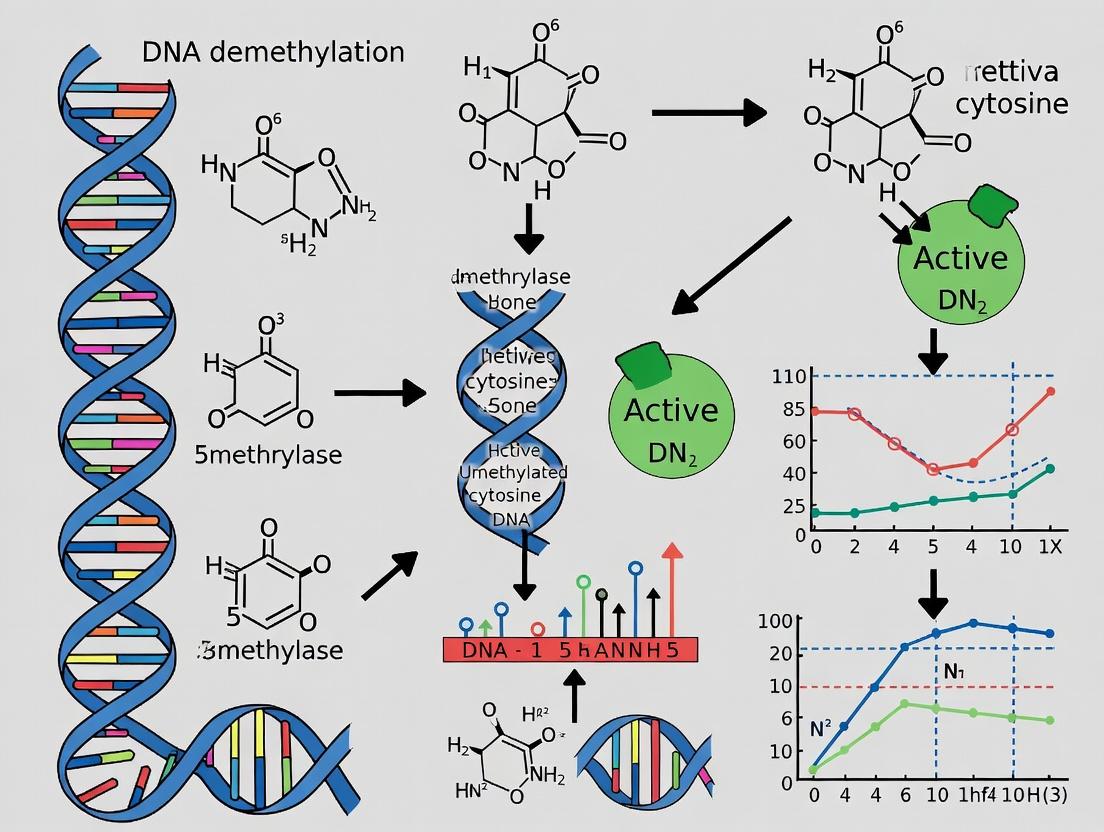
Active DNA demethylation is primarily mediated by the Ten-Eleven Translocation (TET) family of dioxygenases (TET1, TET2, TET3), which iteratively oxidize 5-methylcytosine (5mC) to 5-hydroxymethylcytosine (5hmC), 5-formylcytosine (5fC), and 5-carboxylcytosine (5caC). The latter intermediates are then excised by thymine DNA glycosylase (TDG) and repaired via the Base Excision Repair (BER) pathway, resulting in unmethylated cytosine.
The distinction between global and locus-specific erasure hinges on the recruitment mechanisms of these enzymes. Global demethylation often occurs passively over replication rounds or via widespread TET activation. In contrast, locus-specific targeting is directed by transcription factors (e.g., pioneer factors in reprogramming like OCT4 itself), histone modifications, or non-coding RNAs.
Diagram 1: Core Active DNA Demethylation Pathway
Quantitative Dynamics at Key Loci
The following table summarizes typical changes in 5mC/5hmC levels at critical loci during successful fibroblast-to-iPSC reprogramming (data derived from recent high-throughput sequencing studies).
Table 1: Methylation Dynamics During Reprogramming (Day 0-21)
| Locus/Gene | Starting 5mC% (Fibroblast) | Final 5mC% (iPSC) | Peak 5hmC% (During Reproming) | Recruitment Mechanism | Erase Type |
|---|---|---|---|---|---|
| OCT4 Proximal Promoter | >85% | <5% | ~15-20% (Day 10-12) | Pioneer factors (e.g., KLF4), histone acetylation | Locus-Specific |
| NANOG Proximal Promoter | >80% | <5% | ~12-18% (Day 12-15) | OCT4/SOX2 binding, H3K4me3 mark | Locus-Specific |
| Developmental Gene (e.g., PAX6) | 40-60% | 60-80% (maintained) | <2% | Polycomb repression (H3K27me3) blocks TET | Protected |
| Lineage-Specific Gene (e.g., COL1A1) | 20-40% | >80% (de novo methyl.) | <1% | DNMT3A/B recruitment for silencing | Global (Silencing) |
| Global Intergenic Repetitive (LINE-1) | ~75% | ~45% | ~5% | Passive loss & mild TET activity | Global/Passive |
Experimental Protocols for Assessing Demethylation Dynamics
Protocol 1: Locus-Specific 5hmC Quantification (Chemical Labeling & qPCR)
Purpose: To track active demethylation at specific promoters (e.g., OCT4) over a reprogramming time course.
- Cell Fixation & DNA Extraction: Harvest cells at defined days (D0, D7, D14, D21). Extract genomic DNA using a column-based kit.
- Chemical Labeling of 5hmC: Use the hydroxymethyl-sensitive selective chemical labeling (hme-Seal) method. Incubate 500 ng of DNA with 1.5 mM UDP-6-N3-Glucose and 2.5 µM T4 Phage β-glucosyltransferase (T4-BGT) in 1X NEBuffer 4 for 1h at 37°C.
- Biotin Conjugation & Pull-Down: Add 10 µM biotin linker (e.g., DBCO-PEG4-Biotin) to the reaction and incubate for 2h at 37°C. Bind biotinylated DNA to streptavidin-coated magnetic beads. Wash stringently.
- Elution & Quantification: Elute the 5hmC-enriched DNA. Analyze target loci (OCT4 promoter) and control regions (GAPDH exon, LINE-1) via quantitative PCR (qPCR). Calculate % enrichment relative to input DNA.
Protocol 2: Genome-Wide 5mC/5hmC Profiling (OxBS-seq)
Purpose: To distinguish 5mC from 5hmC at single-base resolution genome-wide.
- Bisulfite Conversion Duplicate Tubes: Split a single DNA sample (≥200 ng) into two tubes.
- Oxidative Treatment (Critical Step): Treat one tube with potassium perruthenate (KRuO₄) to oxidize 5hmC to 5fC, which subsequently reads as T after bisulfite treatment. The second tube is untreated.
- Standard Bisulfite Conversion: Subject both tubes to sodium bisulfite conversion, which deaminates unmethylated C (and 5fC) to U, but leaves 5mC unchanged.
- Library Prep & Sequencing: Prepare sequencing libraries from both samples using post-bisulfite adaptor tagging. Sequence on an Illumina platform.
- Bioinformatic Analysis: Align reads. 5mC is calculated from the oxidized sample. True 5hmC is inferred by subtracting the 5mC signal in the oxidized sample from the total "C" signal (5mC+5hmC) in the untreated sample.
Diagram 2: OxBS-Seq Workflow Logic
The Scientist's Toolkit: Key Research Reagents
Table 2: Essential Reagents for Demethylation Studies in Reprogramming
| Reagent / Kit | Provider Examples | Function in Experiment |
|---|---|---|
| T4 Phage β-Glucosyltransferase (T4-BGT) | NEB, Active Motif | Enzymatically labels 5hmC with a modified glucose for selective pull-down or detection (hme-Seal). |
| 5hmC Selective Chemical Labeling Kit | WiseGene, Merck | All-in-one kits for biotinylation and enrichment of 5hmC-containing DNA for locus-specific or seq. analysis. |
| TrueMethyl OxBS Kit | Cambridge Epigenetix | Streamlined kit for oxidative bisulfite conversion, enabling genome-wide 5mC/5hmC discrimination. |
| Anti-5hmC Antibody (monoclonal) | Diagenode, Active Motif | Immunoprecipitation of 5hmC-DNA (hMeDIP) or immunofluorescence staining. |
| DNMT & TET Enzyme Inhibitors | Sigma, Tocris, Cayman Chemical | Small molecules (e.g., 2-HG for TET, Decitabine for DNMT) to perturb methylation/demethylation dynamics. |
| Recombinant Human TET1 Catalytic Domain | Origene, BPS Bioscience | For in vitro assays to study enzyme kinetics or targeted demethylation experiments. |
| Methylation-Dependent Restriction Enzymes (e.g., GlaI) | NEB | Used in conjunction with methylation-sensitive enzymes for differential digestion assays of locus status. |
| Bisulfite Conversion Kit | Qiagen, Zymo Research | Standard for converting unmethylated cytosine to uracil prior to sequencing or PCR analysis of 5mC. |
The precise orchestration of global and locus-specific demethylation is fundamental to epigenetic resetting. Targeted erasure at pluripotency promoters, driven by transcription factor recruitment of TET enzymes, is a prerequisite for gene activation. Conversely, maintaining or establishing methylation at developmental loci ensures lineage commitment is suppressed. Advancing cellular reprogramming efficiency and fidelity hinges on deepening our understanding of these differential dynamics, offering potential targets for enhancing regenerative medicine and drug discovery platforms.
Tools of the Trade: Practical Approaches to Induce and Harness DNA Demethylation
Within the broader thesis on DNA demethylation in cellular reprogramming, pharmacological inducers represent a cornerstone for efficient epigenetic resetting. Small molecules, including Vitamin C (ascorbic acid), components of 2i/LIF culture media, and inhibitors targeting TET enzymes or DNA methyltransferases (DNMTs), enable precise control over the DNA methylation landscape. This guide details their mechanisms, quantitative impacts, and experimental applications in reprogramming somatic cells to pluripotency and beyond.
Mechanisms of Action
Vitamin C (Ascorbic Acid): Acts as a cofactor for the Ten-Eleven Translocation (TET) family of dioxygenases (TET1/2/3), which catalyze the iterative oxidation of 5-methylcytosine (5mC) to 5-hydroxymethylcytosine (5hmC) and further derivatives, leading to passive dilution or active replication-independent demethylation. Vitamin C enhances TET enzyme activity by promoting their folding and stability via its role as an electron donor, facilitating the Fe(II)/α-KG-dependent catalytic cycle.
2i/LIF Media Components: The "2i" cocktail typically consists of a MEK inhibitor (e.g., PD0325901) and a GSK3β inhibitor (e.g., CHIR99021), used alongside Leukemia Inhibitory Factor (LIF).
- MEK Inhibitor (PD0325901): Suppresses the FGF/ERK pathway, signaling that promotes differentiation and stabilizes a naive pluripotent ground state.
- GSK3β Inhibitor (CHIR99021): Activates Wnt/β-catenin signaling, which supports self-renewal and has complex, context-dependent interactions with epigenetic regulators, indirectly influencing the methylation landscape.
- LIF: Activates the JAK/STAT3 pathway, a key pro-pluripotency and self-renewal signal. Together, 2i/LIF creates a permissive environment for epigenetic remodeling by maintaining cells in a state prone to demethylation.
TET/DSG Inhibitors: This category includes molecules that either inhibit TET activity or target DNMTs (often referred to in the context of DNA methylation inhibition).
- TET Inhibitors (e.g., Bobcat339): Directly inhibit the catalytic activity of TET enzymes, used experimentally to probe the functional consequences of 5mC oxidation.
- DNMT Inhibitors: Divided into nucleoside analogs (e.g., 5-Azacytidine, AZA; Decitabine) that incorporate into DNA and trap DNMTs, and non-nucleoside inhibitors (e.g., RG108). They lead to passive DNA demethylation, synergizing with activators of the demethylation pathway.
Table 1: Impact of Pharmacological Inducers on DNA Demethylation Metrics
| Inducer Class | Example Compound | Typical Conc. (in vitro) | Key Readout | Observed Effect (Representative) | Reference System |
|---|---|---|---|---|---|
| Vitamin C | L-Ascorbic acid 2-phosphate | 50-200 µM | Global 5hmC levels | Increase of 2- to 5-fold | Mouse/human iPSC reprogramming |
| 2i/LIF Components | CHIR99021 (GSK3i) | 3 µM | Naive marker (e.g., Rex1) expression | >50-fold upregulation | Mouse Embryonic Stem Cells (mESCs) |
| PD0325901 (MEKi) | 1 µM | p-ERK levels | >80% reduction | mESCs | |
| LIF | 10-100 ng/mL | p-STAT3 levels | >10-fold increase | mESCs | |
| DNMT Inhibitors | 5-Azacytidine (AZA) | 0.5-2 µM | Global 5mC levels (by LC-MS/MS) | Reduction of 30-60% over 72h | Somatic cells (e.g., MEFs) |
| TET Inhibitors | Bobcat339 | 50-100 µM | TET-dependent 5hmC production | Inhibition of ~70-80% | In vitro TET activity assay |
Table 2: Synergistic Effects in Somatic Cell Reprogramming (Mouse MEFs to iPSCs)
| Inducer Combination | Reprogramming Efficiency (% AP+ Colonies) | Time to Fully Reprogrammed Colony (Days) | Global DNA Methylation State (vs. Somatic) |
|---|---|---|---|
| OSK (Baseline) | 0.1 - 0.5% | 25-30 | Partially hypomethylated |
| OSK + Vitamin C | 1 - 3% | 18-22 | Significantly hypomethylated |
| OSK + 2i/LIF | 3 - 10% | 14-18 | Naive pluripotency methylation pattern |
| OSK + Vitamin C + 2i/LIF | 10 - 20% | 12-16 | Near-complete demethylation |
Experimental Protocols
Protocol 1: Assessing TET Activity via 5hmC Quantification in Reprogramming Cells Objective: To measure the effect of Vitamin C on TET-mediated DNA demethylation during early iPSC induction.
- Cell Culture: Plate mouse embryonic fibroblasts (MEFs) carrying doxycycline-inducible OKSM factors. Culture in fibroblast medium.
- Reprogramming Induction: Switch to reprogramming medium (KnockOut DMEM, 10% KSR, L-glutamine, non-essential amino acids, β-mercaptoethanol) supplemented with doxycycline (2 µg/mL). For test groups, add Vitamin C (L-ascorbic acid 2-phosphate, 100 µM). Include a control without Vitamin C.
- Sample Harvest: Collect cells at days 0, 3, 7, and 10 post-induction using trypsin.
- Genomic DNA Extraction: Use a column-based gDNA extraction kit. Quantify DNA by Nanodrop.
- 5hmC Quantification: Use a commercially available ELISA-based 5hmC DNA Quantification Kit. Load 100 ng of denatured gDNA per well according to the manufacturer's instructions. Measure absorbance at 450 nm and calculate 5hmC percentage from the standard curve.
- Analysis: Normalize 5hmC levels to total input DNA. Compare temporal profiles between Vitamin C-treated and control samples.
Protocol 2: Evaluating Naive Pluripotency Establishment with 2i/LIF Objective: To convert primed human PSCs to a naive-like state using pharmacological inhibitors.
- Starting Cells: Culture primed human ESCs/iPSCs in mTeSR1 or equivalent on Matrigel-coated plates.
- Media Transition: Dissociate cells to single cells using Accutase. Seed onto pre-coated plates (Matrigel or Laminin-521) in naive conversion medium (e.g., N2B27 basal medium).
- 2i/LIF Supplementation: Add small molecules: CHIR99021 (3 µM), PD0325901 (1 µM), and human LIF (10 ng/mL). Refresh medium daily.
- Monitoring: Observe morphological shift from flattened to domed, compact colonies over 5-10 days.
- Validation: Harvest RNA and perform qRT-PCR for naive markers (e.g., KLF4, TFCP2L1, DNMT3L) and downregulation of primed markers (e.g., OTX2, ZIC2). Analyze by immunofluorescence for protein expression (e.g., NANOG, SSEA-4).
Diagrams
Title: Vitamin C Enhances TET-Mediated 5mC Oxidation
Title: 2i/LIF Mechanism in Naive Pluripotency
Title: Small Molecule Roles in Reprogramming Stages
The Scientist's Toolkit: Research Reagent Solutions
Table 3: Essential Materials for Demethylation & Reprogramming Studies
| Item | Example Product/Catalog # | Function in Experiment |
|---|---|---|
| L-Ascorbic Acid 2-phosphate | Sigma-Aldrich, A8960 | Stable form of Vitamin C; cofactor for TET enzymes in demethylation. |
| CHIR99021 (GSK3β inhibitor) | Tocris, 4423 | Component of 2i; activates Wnt signaling to promote naive pluripotency. |
| PD0325901 (MEK inhibitor) | Selleckchem, S1036 | Component of 2i; inhibits differentiation signaling via FGF/ERK. |
| Recombinant Human LIF | PeproTech, 300-05 | Cytokine for STAT3 pathway activation; essential for naive state maintenance. |
| 5-Azacytidine (AZA) | Sigma-Aldrich, A2385 | Nucleoside DNMT inhibitor; induces passive DNA demethylation. |
| Bobcat339 (TET inhibitor) | Cayman Chemical, 21873 | Selective, cell-permeable inhibitor of TET1/2 catalytic activity. |
| 5hmC DNA ELISA Kit | Zymo Research, D5425 | Quantifies global 5-hydroxymethylcytosine levels from genomic DNA. |
| EpiMark 5hmC/5mC Analysis Kit | NEB, E3317 | Enzymatic method to distinguish and analyze 5hmC and 5mC loci. |
| N2B27 Basal Medium | Home-made or commercial (e.g., StemMACS) | Defined, serum-free base medium for naive and primed PSC culture. |
| mTeSR1 / mTeSR Plus | STEMCELL Technologies, 85850/85875 | Feeder-free, defined medium for maintenance of primed human PSCs. |
| Matrigel / Laminin-521 | Corning, 354234 / Biolamina, LN521 | Extracellular matrix for coating culture vessels to support PSC attachment. |
| Doxycycline Hyclate | Sigma-Aldrich, D9891 | Inducer for tet-on reprogramming factor expression systems. |
| Accutase | Innovative Cell Tech., AT104 | Enzyme solution for gentle detachment of PSCs as single cells. |
1. Introduction and Thesis Context
Cellular reprogramming, the conversion of one somatic cell type into another, represents a paradigm shift in regenerative medicine and disease modeling. A central thesis in modern reprogramming research posits that targeted DNA demethylation at key genomic loci is not merely a correlative event but a critical driving force for cell fate conversion. This whitepaper provides an in-depth technical examination of two synergistic genetic engineering strategies rooted in this thesis: the overexpression of Ten-Eleven Translocation (TET) enzymes, the master facilitators of active DNA demethylation, and the canonical transcription factors used in direct reprogramming. We detail how their combined application can enhance the efficiency and fidelity of cell fate reprogramming.
2. Core Molecular Mechanisms
2.1 TET Enzymes: Catalysts of DNA Demethylation TET enzymes (TET1, TET2, TET3) are Fe(II)- and α-ketoglutarate-dependent dioxygenases that initiate active DNA demethylation by oxidizing 5-methylcytosine (5mC) to 5-hydroxymethylcytosine (5hmC) and further derivatives, ultimately leading to an unmodified cytosine base.
2.2 Direct Reprogramming Factors Direct reprogramming (or transdifferentiation) bypasses a pluripotent state by forced expression of lineage-specific transcription factors (TFs). These TFs bind to closed chromatin, but their ability to activate target genes is often hindered by repressive DNA methylation. This is where the synergy with TET enzymes is theorized: TET-mediated demethylation at TF binding sites can facilitate chromatin opening and enhance TF-driven gene regulatory network activation.
Table 1: Key TET Enzyme Isoforms and Properties
| Isoform | Catalytic Domain Preference | Key Role in Reprogramming | Associated Reprogramming Contexts |
|---|---|---|---|
| TET1 | High for 5mC->5hmC | Pioneer factor recruitment, enhancer demethylation | iPSC generation, neuronal reprogramming |
| TET2 | High for 5mC->5hmC | Global & locus-specific demethylation | Hematopoietic, cardiac reprogramming |
| TET3 | Oxidizes 5hmC further | Zygotic demethylation, terminal differentiation | Early development, specific neuronal subtypes |
Table 2: Common Direct Reprogramming Factor Cocktails
| Target Cell Type | Key Transcription Factors (Abbrev.) | Typical Delivery Method | Reported Efficiency (Baseline) |
|---|---|---|---|
| Induced Neurons (iNs) | ASCL1, BRN2, MYT1L (BAM) | Lentivirus | 2-20% (varies by source cell) |
| Induced Cardiomyocytes (iCMs) | GATA4, MEF2C, TBX5 (GMT) | Retrovirus/Lentivirus | ~1-15% (from fibroblasts) |
| Induced Hepatocytes (iHeps) | HNF4A, FOXA1, FOXA3 | Integrating Vectors | 10-30% (from fibroblasts) |
3. Experimental Protocols
Protocol 1: Co-Overexpression of TET and TF Constructs for Enhanced Fibroblast-to-Neuron Reprogramming Objective: To generate induced Neurons (iNs) from human dermal fibroblasts with increased efficiency and maturity via concurrent demethylation. Materials: See "Research Reagent Solutions" below. Procedure:
- Day 0: Cell Plating. Plate human fibroblasts at 50,000 cells/well in a Matrigel-coated 24-well plate in fibroblast growth medium.
- Day 1: Viral Transduction. Prepare a pooled viral supernatant containing: a) Lentivirus expressing ASCL1, BRN2, and MYT1L (polycistronic or mixed); b) Lentivirus expressing TET1 catalytic domain (TET1-CD); c) Optional: Lentivirus expressing a 5hmC reporter. Add polybrene (8 µg/mL). Replace medium with virus-containing medium for 24h.
- Day 2: Medium Change. Replace with fibroblast medium.
- Day 3: Switch to Neuronal Medium. Change to N2/B27-supplemented neuronal induction medium containing small molecules (e.g., BDNF, GDNF, cAMP, Dorsomorphin, SB431542).
- Days 4-28: Maintenance & Analysis. Perform half-medium changes every 2-3 days. Monitor morphological changes. At Day 14-21, fix cells for immunocytochemistry (β-III-Tubulin, MAP2) or harvest for 5hmC/5mC analysis via dot-blot or hMeDIP-seq.
Protocol 2: Quantitative Assessment of Demethylation at Target Loci Objective: To measure TET-induced demethylation at enhancers/promoters of neuronal genes. Procedure:
- Genomic DNA Extraction: Harvest cells at reprogramming timepoints (e.g., Day 0, 7, 14) using a column-based gDNA kit.
- Bisulfite Conversion: Treat 500 ng gDNA using a commercial bisulfite conversion kit, converting unmethylated cytosines to uracil (read as thymine post-PCR), while 5mC/5hmC remains as cytosine.
- Pyrosequencing or Bisulfite-Seq PCR: Design primers specific for bisulfite-converted DNA flanking loci of interest (e.g., NEUROD1 promoter, DCX enhancer). Perform PCR and analyze the ratio of C to T at individual CpG sites via pyrosequencing or next-generation sequencing.
- Data Analysis: Compare the percentage of methylation at each CpG site between control (TFs only) and experimental (TFs + TET) groups. A significant reduction indicates TET activity.
4. Visualization of Core Concepts
Title: Synergy of TET and TF Overexpression in Reprogramming
Title: TET+TF Reprogramming Experimental Timeline
5. The Scientist's Toolkit: Research Reagent Solutions
| Item | Function in TET/TF Reprogramming | Example/Note |
|---|---|---|
| Lentiviral Vectors | Stable delivery and integration of TET and TF genes into target cells. | Use inducible (doxycycline) or constitutive (EF1α) promoters. Biosafety Level 2 required. |
| TET Expression Constructs | Catalytic domain of TET1 (TET1-CD) is commonly used for potent, targeted demethylation without affecting endogenous regulation. | Add-on systems (e.g., SunTag) can recruit multiple TET1-CD molecules for enhanced localized activity. |
| Reprogramming Media Supplements | Support survival and maturation of target cell type while suppressing original cell identity. | N2/B27 for neurons; specific growth factors (VEGF, FGF) for cardiomyocytes. |
| Small Molecule Enhancers | Modulate signaling pathways to boost efficiency (e.g., inhibit TGF-β, GSK3β). | Valproic acid (HDACi), CHIR99021 (GSK3βi), RepSox (TGF-βRi). |
| 5hmC-Specific Antibodies | Detect and quantify the primary product of TET activity via immunostaining or hMeDIP. | Critical for validating TET enzyme functionality in situ. |
| Bisulfite Conversion Kit | Converts unmethylated cytosine to uracil for single-base resolution methylation analysis. | Gold-standard method. Distinguishes 5mC from 5hmC requires oxidative bisulfite sequencing (oxBS-seq). |
| Matrigel / Laminin | Provides an extracellular matrix coating to improve cell adhesion and support neuronal or epithelial morphology. | Essential for culturing sensitive reprogrammed cells. |
The pursuit of cellular reprogramming, from somatic cells to induced pluripotent stem cells (iPSCs) or to other differentiated lineages, requires precise control over the epigenetic landscape. DNA methylation, particularly at cytosine residues in CpG dinucleotides, is a key repressive mark that silences gene expression and maintains cellular identity. Targeted DNA demethylation is therefore a critical tool for reactivating pluripotency genes or unlocking developmental programs without altering the underlying DNA sequence. The fusion of catalytically dead Streptococcus pyogenes Cas9 (dCas9) to the catalytic domain of Ten-Eleven Translocation 1 (TET1) represents a breakthrough, enabling locus-specific conversion of 5-methylcytosine (5mC) to 5-hydroxymethylcytosine (5hmC) and further oxidized products, initiating the DNA demethylation pathway.
Core Mechanism and Pathway
The dCas9-TET1 system functions by recruiting TET1 enzymatic activity to specific genomic loci via a single-guide RNA (sgRNA). The TET1 catalytic domain (often referred to as the CD domain, comprising the cysteine-rich and double-stranded β-helix regions) is a Fe(II)- and α-ketoglutarate (α-KG)-dependent dioxygenase. It sequentially oxidizes 5mC to 5hmC, 5-formylcytosine (5fC), and 5-carboxylcytosine (5caC). These oxidized derivatives are then excised by thymine DNA glycosylase (TDG) in the base excision repair (BER) pathway, ultimately resulting in an unmodified cytosine.
Diagram: dCas9-TET1 Targeted Demethylation Pathway
Key Research Reagent Solutions
The following table details essential reagents and materials for implementing dCas9-TET1 epigenetic editing experiments.
Table 1: Research Reagent Toolkit for dCas9-TET1 Experiments
| Reagent / Material | Function / Role | Example or Key Consideration |
|---|---|---|
| dCas9-TET1 Fusion Construct | Core effector protein. Catalytically inactive dCas9 provides targeting; TET1 catalytic domain (CD) provides demethylase activity. | Common variants: dCas9-TET1(CD), SunTag-scFv-TET1(CD) for signal amplification. |
| sgRNA Expression Vector | Guides the fusion protein to the specific genomic locus of interest. | Requires careful design to avoid off-targets; typically expressed via U6 promoter. |
| Delivery System | Introduces constructs into target cells. | Lentivirus, adenovirus (AdV), or transfection (lipofection, electroporation) of plasmid/mRNA. |
| Target Cell Line | Cellular model for reprogramming or gene reactivation studies. | Human or mouse fibroblasts, iPSCs, primary cells. |
| α-Ketoglutarate (α-KG) | Essential metabolic cofactor for TET1 enzymatic activity. | Cell culture media supplementation may enhance efficiency. |
| Ascorbic Acid (Vitamin C) | Cofactor that promotes TET activity by maintaining Fe²⁺ in its reduced state. | Often added to culture media to boost demethylation. |
| Antibodies for Detection | Validate targeting and efficiency. | Anti-5mC, anti-5hmC for dot blot/immunofluorescence; anti-dCas9 for ChIP. |
| Bisulfite Sequencing Reagents | Gold standard for quantifying DNA methylation at single-base resolution. | Targeted bisulfite sequencing (e.g., Pyrosequencing, NGS) of the edited locus. |
| TDG Inhibitor | Optional tool to dissect mechanism. | Can be used to block BER and accumulate 5caC/5fC, confirming pathway. |
Detailed Experimental Protocols
Protocol A: Targeted Demethylation in Mammalian Cells
Objective: To achieve targeted CpG demethylation and gene reactivation in adherent mammalian cell lines (e.g., HEK293T, fibroblasts).
Materials:
- dCas9-TET1 expression plasmid (e.g., Addgene #84475).
- sgRNA expression plasmid (lentiGuide or similar).
- Lipofectamine 3000 or polyethylenimine (PEI).
- Opti-MEM Reduced Serum Medium.
- Cell culture medium with 10% FBS.
- Ascorbic acid (final concentration 50-100 µg/mL).
- QIAamp DNA Mini Kit.
- EZ DNA Methylation-Lightning Kit.
Procedure:
- Design & Cloning: Design two sgRNAs targeting ~100-200bp upstream of the transcription start site (TSS) of your gene of interest. Clone annealed oligonucleotides into the BsmBI site of your sgRNA expression vector.
- Cell Seeding: Seed 2e5 cells per well in a 12-well plate 24 hours before transfection to achieve 70-80% confluency.
- Transfection: For each well, prepare two tubes:
- Tube A (DNA): Dilute 1 µg of dCas9-TET1 plasmid + 1 µg of sgRNA plasmid in 125 µL Opti-MEM.
- Tube B (Reagent): Dilute 4 µL of Lipofectamine 3000 in 125 µL Opti-MEM. Combine tubes A and B, incubate 15 min at RT. Add dropwise to cells.
- Cofactor Supplementation: 6 hours post-transfection, replace medium with fresh medium containing 100 µg/mL ascorbic acid.
- Harvest & Analysis:
- 72 hrs post-transfection: Harvest cells for genomic DNA (gDNA) using the QIAamp kit.
- 5-7 days post-transfection: Harvest cells for RNA and downstream gene expression analysis (qRT-PCR).
- Methylation Analysis: Treat 500 ng of gDNA with the EZ Lightning Kit. Amplify the target region via PCR using primers designed for bisulfite-converted DNA. Submit products for Sanger sequencing or next-generation sequencing. Quantify methylation percentage at each CpG.
Protocol B: Validation by 5hmC Enrichment (hMeDIP-qPCR)
Objective: To confirm successful TET1 recruitment and activity by quantifying enrichment of 5hmC at the target locus.
Materials:
- Magnetic beads (Protein A/G).
- Anti-5hmC antibody.
- Normal rabbit IgG.
- Sonication device (Covaris or Bioruptor).
- DNA purification kits.
- SYBR Green qPCR Master Mix.
Procedure:
- DNA Extraction & Shearing: Isolate gDNA from edited and control cells. Sonicate to ~200-500 bp fragments.
- Immunoprecipitation: For each sample, set up two reactions: one with anti-5hmC antibody, one with normal IgG.
- Bind 2 µg of antibody to 50 µL magnetic beads for 1 hour at 4°C.
- Incubate antibody-bound beads with 2 µg of sheared DNA in IP buffer overnight at 4°C.
- Wash & Elute: Wash beads 5x with ice-cold IP buffer. Elute DNA in elution buffer with proteinase K at 55°C for 2 hours.
- Purification: Purify eluted DNA using a PCR purification kit.
- Quantitative PCR: Perform qPCR using primers specific for the target locus and a control non-target locus. Calculate % input and fold enrichment over IgG control.
Table 2: Efficacy Metrics of dCas9-TET1 Systems from Recent Studies
| Target Gene / Locus | Cell Type | Demethylation Efficiency (Max Reduction) | Gene Expression Fold-Change | Key Parameters | Citation (Year) |
|---|---|---|---|---|---|
| OCT4 promoter | Human fibroblasts | ~40-50% (at specific CpGs) | 10-100x (varies) | dCas9-TET1(CD), dual sgRNAs, Vit C supplement | Liu et al. (2016) |
| BACH2 promoter | HEK293T | ~30% (average across region) | 5x | SunTag-TET1(CD) system | Morita et al. (2016) |
| MASPIN/SERPINB5 | Breast cancer cells | ~35% (CpG island) | 8x | dCas9-TET1 catalytic domain fusion | Choudhury et al. (2016) |
| Imprinted H19/Igf2 DMR | Mouse embryonic stem cells | ~25-60% (allele-specific) | N/A | dCas9-TET1 with locus-specific sgRNA | Xu et al. (2016) |
| IL1RN promoter | Primary human T cells | ~20-30% | 4-6x | mRNA delivery of dCas9-TET1 | Rupp et al. (2017) |
Diagram: dCas9-TET1 Experimental Workflow
The dCas9-TET1 platform provides a precise, programmable tool for locus-specific DNA demethylation, directly addressing a key epigenetic barrier in cellular reprogramming. Its application has successfully reactivated silenced pluripotency genes (OCT4, NANOG), modified imprinting control regions, and unlocked developmental genes. Future optimization lies in improving delivery efficiency to primary cells, enhancing catalytic activity through engineered TET variants, and integrating with other epigenetic editors (e.g., histone demethylases) for synergistic effects. As a research tool, it is indispensable for causal epigenetic studies; for therapeutics, it holds promise for correcting disease-associated epigenetic silencing without double-strand DNA breaks.
Within the broader thesis of DNA demethylation's pivotal role in cellular reprogramming, this guide examines its application in induced pluripotent stem cell (iPSC) generation. The efficiency, speed, and epigenetic accuracy of reprogramming somatic cells to pluripotency are intrinsically linked to the erasure of somatic DNA methylation patterns and the establishment of a pluripotent epigenetic landscape. This document provides a technical overview of current strategies that target DNA demethylation pathways to enhance iPSC generation.
Core Mechanisms: DNA Demethylation in Reprogramming
Reprogramming requires genome-wide epigenetic remodeling. Active DNA demethylation, primarily mediated by the Ten-Eleven Translocation (TET) family of dioxygenases, which convert 5-methylcytosine (5mC) to 5-hydroxymethylcytosine (5hmC) and further oxidized derivatives, is a critical bottleneck. Passive dilution through DNA replication in the absence of maintenance methyltransferases (e.g., DNMT1) also contributes.
Key Signaling and Molecular Pathways
The core pathways regulating the epigenome during reprogramming involve pluripotency transcription factors (OCT4, SOX2, KLF4, c-MYC - OSKM) interacting with epigenetic modifiers.
Title: DNA Demethylation Pathways in OSKM Reprogramming
Experimental Protocols for Enhancing Reprogramming
Protocol: Small Molecule-Augmented Reprogramming for Enhanced Kinetics
This protocol uses vitamin C (ascorbic acid) and other small molecules to boost TET activity and demethylation.
Materials: Somatic cells (e.g., human dermal fibroblasts), OSKM expression vectors (Sendai virus or episomal), Essential 8 or mTeSR1 medium, VAL-853 (TET2 activator), Sodium ascorbate (vitamin C), PD0325901 (MEK inhibitor), Thiazovivin (ROCK inhibitor), 5-Azacytidine (DNMT inhibitor - low dose). Procedure:
- Seed somatic cells at 5x10^3 cells/cm².
- Transduce with OSKM factors on Day 0 and Day 1.
- From Day 2, switch to pluripotency maintenance medium supplemented with:
- Sodium ascorbate (50 µg/mL)
- VAL-853 (10 µM)
- PD0325901 (1 µM)
- Thiazovivin (0.5 µM)
- Refresh medium daily. For a low-dose epigenetic primer, include 0.5 µM 5-Azacytidine from Day 3 to Day 7 only.
- Monitor colony emergence from Day 10. Pick candidate iPSC colonies from Day 18-25 based on embryonic stem cell-like morphology.
- Validate through immunostaining (OCT4, NANOG), teratoma assay, and DNA methylation analysis at pluripotency gene promoters (e.g., OCT4, NANOG).
Protocol: CRISPR/dCas9-Targeted Epigenetic Editing for Fidelity
This protocol uses a catalytically dead Cas9 (dCas9) fused to the TET1 catalytic domain (dCas9-TET1) to direct demethylation to specific somatic loci that are resistant to reprogramming.
Materials: dCas9-TET1 expression plasmid, sgRNAs targeting somatic gene promoters or enhancers (e.g., MEF-specific genes), Lipofectamine Stem Transfection Reagent, Puromycin selection reagent. Procedure:
- Design and clone sgRNAs targeting hypermethylated somatic loci into the dCas9-TET1 system.
- Co-transfect somatic cells with the dCas9-TET1 construct and sgRNA plasmids 48 hours prior to OSKM transduction.
- Apply puromycin selection (1 µg/mL) for 72h to enrich transfected cells.
- Initiate standard OSKM reprogramming (as in 3.1).
- Analyze targeted loci via bisulfite sequencing post-transfection and post-reprogramming to confirm specific demethylation and assess its impact on iPSC colony purity and gene expression.
Table 1: Impact of Demethylation-Enhancing Strategies on iPSC Generation
| Strategy / Reagent | Target Mechanism | Reported Increase in Efficiency (vs. OSKM only) | Reprogramming Kinetics (Time to iPSC Colony) | Key Epigenetic Fidelity Metric (e.g., % Correctly Methylated Loci) | Reference Year (Post-2022) |
|---|---|---|---|---|---|
| High-dose Vitamin C (Ascorbic Acid) | TET enzyme co-factor | 5-15 fold increase | Reduced by 4-7 days | >80% similarity to hESC methylome | 2023 |
| VAL-853 (small molecule) | Direct TET2 activation | ~10 fold increase | Reduced by 5-10 days | Improved imprinting gene methylation patterns | 2023 |
| Low-dose 5-Azacytidine (pulsed) | DNMT1 inhibition | 3-8 fold increase | Reduced by 3-5 days | Moderate improvement; risk of global hypomethylation | 2022 |
| dCas9-TET1 Targeted Demethylation | Locus-specific 5mC removal | Colony yield increase variable (1-5 fold) | Minor reduction | >90% fidelity at targeted loci; reduces somatic memory | 2024 |
| TET1 or TET2 Overexpression | Global active demethylation | 20-30 fold increase in mouse; 5-10 fold in human | Significantly accelerated | High but can cause over-erosion of imprints | 2022 |
Table 2: Key Reagents for Demethylation-Enhanced iPSC Research
| Reagent / Solution | Function in Reprogramming | Example Product / Cat. No. |
|---|---|---|
| Sodium L-Ascorbate (Vitamin C) | Cofactor for Fe(II)/α-KG-dependent dioxygenases like TETs, promoting 5mC oxidation. Reduces replicative stress. | Sigma-Aldrich, A4034 |
| VAL-853 | Small molecule activator of TET2, enhances its catalytic activity. | MedChemExpress, HY-138395 |
| 5-Azacytidine | Nucleoside analog that inhibits DNA methyltransferases (DNMTs), leading to passive demethylation. | Sigma-Aldrich, A2385 |
| dCas9-TET1 Catalytic Domain Plasmid | Enables targeted DNA demethylation at specific genomic loci guided by sgRNAs. | Addgene, #84475 (pLV-dCas9-TET1CD) |
| PD0325901 | MEK/ERK pathway inhibitor; promotes ground-state pluripotency, synergizes with demethylation agents. | Stemgent, 04-0006 |
| StemMACS mRNA Reprogramming Kit | Non-integrating mRNA for OSKM delivery; allows precise timing with small molecules. | Miltenyi Biotec, 130-107-677 |
| EpiJET Bisulfite Conversion Kit | For high-efficiency conversion of unmethylated cytosines to uracil prior to methylation analysis. | Thermo Scientific, K1461 |
The Scientist's Toolkit: Research Reagent Solutions
Table 3: Essential Materials for Demethylation-Focused Reprogramming Experiments
| Item Category | Specific Reagent/Kit | Brief Function |
|---|---|---|
| Reprogramming Vectors | CytoTune-iPS 3.0 Sendai Virus (Thermo Fisher) | Non-integrating, high-efficiency delivery of human OSKML factors. |
| Episomal iPSC Reprogramming Vectors (Addgene) | Integration-free, plasmid-based OKSM delivery. | |
| Culture Media | Essential 8 Flex Medium (Thermo Fisher) | Xeno-free, feeder-free medium for human iPSC culture and reprogramming. |
| mTeSR Plus (StemCell Technologies) | Defined medium for maintenance of human pluripotent stem cells. | |
| Small Molecules | Thiazovivin (Tocris) | ROCK inhibitor; increases survival of reprogramming cells. |
| CHIR99021 (Tocris) | GSK3β inhibitor; activates Wnt signaling to enhance reprogramming. | |
| Trichostatin A (TSA) (Sigma) | HDAC inhibitor; opens chromatin structure, synergizes with demethylation. | |
| Analysis Kits | EZ DNA Methylation-Gold Kit (Zymo Research) | Reliable bisulfite conversion and clean-up for downstream methylation-specific PCR or sequencing. |
| Illumina EPIC Methylation BeadChip | Genome-wide profiling of DNA methylation at >850,000 CpG sites. | |
| SimpleChIP Kit (Cell Signaling Technology) | Chromatin immunoprecipitation to assess histone modifications (e.g., H3K4me3, H3K27me3) at pluripotency loci post-demethylation. |
Title: Workflow for Demethylation-Enhanced iPSC Generation
Strategically enhancing DNA demethylation represents a cornerstone for optimizing iPSC generation within modern reprogramming research. By leveraging small molecule activators of TET enzymes, transient DNMT inhibition, or targeted epigenetic editing, researchers can significantly improve the efficiency, speed, and epigenetic fidelity of the process. This directly addresses a core thesis in the field: that the controlled erasure of the somatic methylome is not merely a correlative event but a causal driver essential for achieving high-quality, clinically relevant induced pluripotency. Future directions will involve refining the temporal and locus-specific control of demethylation to generate iPSCs indistinguishable from their embryonic counterparts.
The broader thesis of DNA demethylation in cellular reprogramming posits that targeted epigenetic erasure, particularly of 5-methylcytosine (5mC), is not merely a permissive event but a critical driver for unlocking cellular plasticity and enabling direct lineage conversion. This whitepaper situates direct lineage conversion, or transdifferentiation, within this thesis, arguing that strategic demethylation of key developmental loci dismantles somatic cell epigenetic barriers, facilitating the action of lineage-specific transcription factors (TFs) and enabling the direct reprogramming of one somatic cell type into another (e.g., fibroblast to neuron) without passing through a pluripotent state. This approach offers potential advantages in speed, safety (reduced tumorigenicity), and preservation of epigenetic age for regenerative medicine and disease modeling.
Core Mechanisms: Demethylation Pathways in Transdifferentiation
Direct lineage conversion relies on the forced expression of master regulator TFs. However, their binding and transcriptional activation are often impeded by repressive DNA methylation at target enhancers and promoters. DNA demethylation facilitates this process primarily via two mechanisms:
- Active Demethylation via TET Enzymes: Ten-eleven translocation (TET1/2/3) dioxygenases catalyze the iterative oxidation of 5mC to 5-hydroxymethylcytosine (5hmC), 5-formylcytosine (5fC), and 5-carboxylcytosine (5caC), leading to eventual replication-dependent dilution or base excision repair (BER). Overexpression of TET enzymes or their recruitment to specific genomic loci can demethylate and activate silenced lineage-specifying genes.
- Passive Demethylation via DNMT Inhibition: Inhibition of DNA methyltransferases (DNMT1, DNMT3A/B) during cell division leads to the dilution of 5mC marks. Small molecule inhibitors like 5-Azacytidine (5-Aza) are used to promote a permissive epigenetic landscape for reprogramming factors.
Key Experimental Data & Applications
Recent studies demonstrate the efficacy of combining demethylation strategies with core transcription factor cocktails.
Table 1: Quantitative Impact of Demethylation on Transdifferentiation Efficiency
| Starting Cell Type | Target Cell Type | Key Transcription Factors | Demethylation Strategy | Reported Conversion Efficiency (vs. Control) | Key Demethylated Loci | Reference (Example) |
|---|---|---|---|---|---|---|
| Human Fibroblast | Induced Neuron (iN) | Ascl1, Brn2, Myt1l | TET1 co-expression | ~18% (Tau+ cells) vs. ~4% | Neurogenin2, Synapsin1 enhancers | Weng et al., 2022 |
| Mouse Fibroblast | Induced Cardiomyocyte (iCM) | Gata4, Mef2c, Tbx5 (GMT) | 5-Azacytidine (5-Aza) treatment | ~11% (cTnT+ cells) vs. ~2% | Nkx2-5, Myh6 promoters | Liu et al., 2020 |
| Human Fibroblast | Induced Hepatocyte (iHep) | Hnf4α, Foxa1, Foxa3 | shRNA knockdown of DNMT1 | ~35% (Albumin+ cells) vs. ~12% | Hnf4a, Albumin cis-regulatory regions | Liu et al., 2018 |
| Mouse Microglia | Induced Neuron | NeuroD1 | TET3 co-expression | ~85% (Map2+ cells) vs. ~60% | Neuron-specific gene promoters | Matsuda et al., 2019 |
| Human Fibroblast | Induced Dopaminergic Neuron | Ascl1, Lmx1a, Nurr1 | Vitamin C (TET co-factor) | ~15% (TH+ cells) vs. ~5% | Pitx3, Dat enhancers | Caiazzo et al., 2015 |
Detailed Experimental Protocols
Protocol 1: TET1-Facilitated Fibroblast-to-Neuron Conversion
Aim: Generate induced neurons (iNs) from human dermal fibroblasts (HDFs) by co-expressing neurogenic TFs with the catalytic domain of TET1 to enhance efficiency.
Materials: See "Scientist's Toolkit" below.
Method:
- Cell Culture: Maintain HDFs in DMEM + 10% FBS + 1% P/S. Plate at 50,000 cells/well in a 12-well plate 24h before transduction.
- Lentiviral Transduction:
- Prepare a viral cocktail containing pLVX vectors for: (i) TF Cocktail: Ascl1, Brn2, and Myt1l (ABM); (ii) Experimental: hTET1cd (catalytic domain); (iii) Control: empty vector.
- Add viral supernatant to cells in the presence of 8 µg/mL polybrene.
- Spinoculate at 1000 x g for 60 min at 32°C.
- Replace media with fresh fibroblast media after 12h.
- Media Switch & Demethylation Support:
- At 48h post-transduction, switch to neuronal induction media (NIM): Neurobasal-A, B-27, L-glutamine, BDNF, GDNF, NT-3, ascorbic acid.
- Supplement experimental group with Vitamin C (200 µM) to support endogenous TET activity.
- Analysis:
- Day 14-21: Fix cells and immunostain for neuronal markers (TUJ1, MAP2) and subtype markers (e.g., vGLUT1, GABA).
- Efficiency: Quantify percentage of TUJ1+ cells relative to total DAPI+ nuclei.
- Bisulfite Sequencing: Perform targeted BS-seq on purified iNs for loci like NGN2 to confirm demethylation.
Protocol 2: 5-Azacytidine Enhanced Fibroblast-to-Cardiomyocyte Reprogramming
Aim: Enhance iCM generation from mouse embryonic fibroblasts (MEFs) using transient DNMT inhibition.
Method:
- Retroviral Transduction:
- Transfect Plat-E cells with pMXs vectors for Gata4, Mef2c, Tbx5 (GMT).
- Collect viral supernatant at 48h and 72h.
- Transduce MEFs (passage 2) with GMT virus in the presence of 4 µg/mL polybrene for 24h.
- Small Molecule Treatment:
- After transduction, change to iCM media: DMEM + 10% FBS + 1% P/S.
- Treat cells with 5-Azacytidine (5-Aza, 1 µM) for the first 72 hours post-transduction only.
- Include a GMT-only control (no 5-Aza).
- Culture & Analysis:
- Change media every 3 days. Spontaneously beating areas typically appear by day 14.
- Day 21: Perform FACS analysis for cTnT+ cells.
- Functional Assay: Measure calcium transients using Fluo-4 AM dye.
- Methylation Analysis: Use Methylation-Specific PCR (MSP) for promoter regions of Nkx2-5 and α-MHC.
The Scientist's Toolkit: Key Research Reagent Solutions
Table 2: Essential Materials for Demethylation-Facilitated Transdifferentiation
| Item | Function/Description | Example Product/Catalog # (Illustrative) |
|---|---|---|
| TET Expression Vector | Delivers catalytic domain of TET1/2/3 to induce active DNA demethylation. | pLVX-hTET1cd (Addgene #143245) |
| DNMT Inhibitor | Small molecule to induce passive demethylation by inhibiting maintenance methylation. | 5-Azacytidine (5-Aza, Sigma A2385) |
| Vitamin C (Ascorbic Acid) | Essential co-factor for TET enzyme activity, enhances demethylation. | L-Ascorbic acid 2-phosphate (Sigma A8960) |
| Lineage-Specific TF Cocktail | Core transcription factors to drive target cell fate. | Ascl1, Brn2, Myt1l (ABM for neurons); Gata4, Mef2c, Tbx5 (GMT for cardiomyocytes) |
| Bisulfite Conversion Kit | For sequencing-based analysis of DNA methylation changes at single-base resolution. | EZ DNA Methylation-Lightning Kit (Zymo Research) |
| 5hmC/5mC Antibody | Immunodetection of DNA methylation/hydroxymethylation changes. | Anti-5hmC (Active Motif 39769) |
| Neuronal Induction Media | Chemically defined media supportive of neuronal survival and maturation. | Neurobasal-A + B-27 Supplement (Gibco) |
| Cardiomyocyte Maintenance Media | Media optimized for cardiac cell culture. | RPMI 1640 + B-27 Supplement (minus insulin) (Gibco) |
| Lentiviral/Retroviral Packaging System | For efficient, stable delivery of reprogramming factors. | Lenti-X/Plat-E Packaging System (Takara) |
| Epigenetic Modulator Panel | Library of small molecules targeting chromatin regulators for screening. | Stemolecule Epigenetic Compound Library (Reprocell) |
Workflow & Pathway Integration Diagram
Overcoming Hurdles: Solving Common Challenges in Demethylation-Driven Reprogramming
Within the paradigm of cellular reprogramming, the complete erasure of somatic epigenetic memory is paramount for generating bona fide induced pluripotent stem cells (iPSCs) or for directed transdifferentiation. A central tenet of this thesis is that DNA demethylation is not a uniform, global process, but is instead locus-specific and often incomplete. Residual methylation at critical regulatory loci—such as promoters and enhancers of developmentally essential genes—can impede full functional resetting, leading to partially reprogrammed cells with compromised differentiation potential or aberrant gene expression. This whitepaper provides a technical guide for identifying these stubborn epigenetic scars and outlines advanced strategies to achieve their complete erasure.
Identifying Residual Methylation: Critical Loci & Detection Methodologies
Residual methylation is frequently observed at:
- Imprinted Gene Control Regions (IGCRs): Gametic differentially methylated regions (gDMRs).
- Germline-Specific Genes: E.g., DAZL, SYCP1.
- Developmentally Regulated Enhancers: Particularly those active in the somatic cell of origin.
- Subtelomeric and Pericentromeric Repeats.
Core Detection Protocols:
A. Targeted Bisulfite Sequencing (Bisulfite-seq)
- Principle: Sodium bisulfite converts unmethylated cytosine to uracil (read as thymine after PCR), while methylated cytosine remains unchanged.
- Protocol: 1) Isolate genomic DNA (500ng-1μg). 2) Treat with EZ DNA Methylation-Gold Kit or equivalent (bisulfite conversion). 3) Design locus-specific primers (avoiding CpG sites). 4) Amplify and sequence via next-generation sequencing (NGS). 5) Analyze with tools like Bismark or QUMA for methylation percentage per CpG.
- Application: High-resolution, quantitative analysis of specific loci suspected of incomplete demethylation.
B. Whole-Genome Bisulfite Sequencing (WGBS)
- Principle: As above, applied to the entire genome.
- Protocol: 1) Fragment genomic DNA by sonication/covaris to ~300bp. 2) Perform bisulfite conversion on fragmented DNA. 3) Construct NGS libraries using methylated-adapter kits (e.g., TruSeq DNA Methylation). 4) Sequence deeply (>30x coverage). 5) Map reads and call methylation states.
- Application: Unbiased, genome-wide identification of residual methylation hotspots without prior locus knowledge.
C. Methylation-Sensitive Restriction Enzyme (MSRE)-qPCR
- Principle: Enzymes like HpaII (cuts CCGG only if internal C is unmethylated) or NotI (GCGGCCGC) fail to cut methylated sites, which is quantified by subsequent qPCR.
- Protocol: 1) Digest 200ng genomic DNA with a cocktail of MSREs and a reference enzyme (MspI, insensitive to methylation). 2) Purify DNA. 3) Perform qPCR for target and control loci using SYBR Green. 4) Calculate relative digestion (%) using ΔΔCt method.
- Application: Rapid, cost-effective screening of methylation status at specific restriction sites within candidate loci.
Table 1: Comparison of Methylation Detection Methods
| Method | Resolution | Throughput | Cost | Key Application |
|---|---|---|---|---|
| Targeted Bisulfite-seq | Single CpG | Low (targeted) | Medium | In-depth validation of specific loci |
| Whole-Genome Bisulfite Seq (WGBS) | Single CpG | Genome-wide | Very High | Discovery of novel residual methylated regions |
| MSRE-qPCR | Restriction Site | Low | Low | High-throughput screening of known sites |
Strategies for Complete Erasure
A. Pharmacological Inhibition Targeting maintenance (DNMT1) and de novo (DNMT3A/B) methyltransferases.
- Protocol for 5-Aza-2'-Deoxycytidine (5-Aza-dC, Decitabine) Treatment: Add 0.5μM 5-Aza-dC to reprogramming/media culture for 48-hour pulses. Refresh medium daily. Note: High concentrations/ prolonged exposure cause cytotoxicity and global hypomethylation.
- Next-Generation Inhibitors: GSK3685032 (DNMT1-selective, non-covalent) at 1μM shows improved specificity and reduced toxicity in recent studies.
B. Targeted Epigenetic Editing Utilizing CRISPR-dCas9 fused to catalytic domains of TET enzymes (TET1-CD) to induce active demethylation.
- Protocol: 1) Design and clone sgRNAs targeting specific residual methylated loci into a dCas9-TET1-CD expression vector. 2) Co-transfect with reprogramming factors into somatic cells. 3) After 72h, sort for transfection markers. 4) Culture and assess methylation at target sites via MSRE-qPCR or targeted Bisulfite-seq after 7-10 days.
C. Enhancing Passive Demethylation Promoting replication-dependent dilution of methylated cytosines by suppressing UHRF1 and DNMT1.
- siRNA Knockdown Protocol: Transfect cells with 50nM siRNAs targeting UHRF1 or DNMT1 using lipid-based transfection reagent at days 2 and 4 of reprogramming. Assess protein knockdown by Western blot at day 5 and methylation at day 7.
Table 2: Demethylation Strategy Comparison
| Strategy | Mechanism | Specificity | Potential Risk |
|---|---|---|---|
| 5-Aza-dC | DNMT1 trapping & degradation | Global | Genomic instability, cytotoxicity |
| GSK3685032 | Reversible DNMT1 inhibition | Global (but more specific) | Off-target hypomethylation |
| dCas9-TET1 | Active oxidation of 5mC to 5hmC/5caC | Locus-specific | Off-target editing, incomplete erasure |
| UHRF1 siRNA | Inhibition of DNMT1 recruitment | Global (but targeted by timing) | Impaired proliferation, pleiotropic effects |
The Scientist's Toolkit: Essential Research Reagents
Table 3: Key Research Reagent Solutions
| Reagent/Material | Function in Demethylation Research |
|---|---|
| EZ DNA Methylation-Gold Kit (Zymo Research) | Reliable bisulfite conversion of genomic DNA for downstream sequencing or qPCR. |
| TruSeq DNA Methylation Kit (Illumina) | Library preparation for whole-genome bisulfite sequencing, includes methylated adapters. |
| Decitabine (5-Aza-2'-Deoxycytidine) (Sigma) | Canonical DNMT inhibitor used to induce global demethylation. |
| dCas9-TET1-CD Plasmid (Addgene #83340) | All-in-one vector for targeted demethylation via CRISPR-guided TET1 catalytic domain. |
| Lipofectamine CRISPRMAX (Thermo Fisher) | High-efficiency transfection reagent for delivering RNP complexes or plasmids into hard-to-transfect primary cells. |
| Methylation-Sensitive Restriction Enzymes (HpaII, Acil) | Enzymes for MSRE-qPCR or MSRE-seq to assess methylation status at specific sequence motifs. |
| GSK3685032 (Cayman Chemical) | Potent, selective, non-covalent DNMT1 inhibitor for more refined demethylation studies. |
| Anti-5-Methylcytosine Antibody (Clone 33D3) | For immunofluorescence or MeDIP to visually assess global or focal methylation levels. |
Visualized Pathways and Workflows
Diagram Title: Strategies to Overcome Incomplete Demethylation in Reprogramming
Diagram Title: Targeted Demethylation with dCas9-TET1 Workflow
Diagram Title: DNMT1-UHRF1 Pathway and Intervention Points
Targeted DNA demethylation technologies, particularly those centered on TET enzymes and CRISPR/dCas9-TET fusion systems, are cornerstone methodologies in cellular reprogramming research. The goal is to achieve precise epigenetic reactivation of silenced genes. However, a significant and often underappreciated risk is the induction of off-target global hypomethylation. This widespread loss of 5-methylcytosine (5mC), particularly at repetitive elements and imprinted loci, is a well-documented driver of genomic instability, retrotransposon activation, and loss of cellular identity. This guide details the mechanisms, detection, and mitigation strategies for managing this critical safety concern in epigenetic editing.
Mechanisms Linking Global Hypomethylation to Genomic Instability
Off-target demethylation typically occurs via two primary mechanisms: 1) the non-specific binding or activity of demethylation effectors (e.g., dCas9-TET1 targeting spillover), and 2) the saturation and dysregulation of endogenous epigenetic maintenance machinery. The consequences are quantifiable and severe.
Key Pathogenic Consequences:
- Hypomethylation of Repetitive Elements: Loss of methylation at LINE-1, Alu, and satellite repeats leads to chromatin decondensation, transcriptional reactivation, and retrotransposition.
- Erosion of Imprinting Control Regions (ICRs): Disruption of allele-specific methylation causes aberrant expression of growth-regulatory genes (e.g., IGF2/H19).
- Centromeric/Pericentromeric Hypomethylation: Compromises chromatid cohesion and centromere function, promoting micronuclei formation and chromosome mis-segregation.
- Generalized DNA Hypomethylation: Correlates with increased DNA double-strand breaks (DSBs) and mutation rates, likely due to relaxed chromatin structure and replication stress.
The signaling pathways connecting hypomethylation to instability are summarized in the following diagram.
Diagram Title: Pathway from Off-Target Demethylation to Genomic Instability
Quantitative Assessment of Hypomethylation and Instability
Table 1: Key Metrics for Assessing Global Hypomethylation & Consequences
| Metric | Detection Method | Typical Baseline (Normal Cell) | Concerning Threshold (Post-Treatment) | Implication |
|---|---|---|---|---|
| Global 5mC Level | LC-MS/MS, ELISA | ~4% of total cytosine (cell-type dependent) | Reduction > 20-30% sustained | System-wide loss of methylation. |
| LINE-1 Methylation | Bisulfite Pyrosequencing (e.g., LINE-1 Met500 assay) | 70-85% (CpG methylation) | Reduction to < 50-60% | High risk of retrotransposition. |
| Satellite 2 (Juxtacentromeric) Methylation | LUMA, LINES PCR | High (>80%) | Reduction > 25% | Linked to micronuclei formation. |
| γH2AX Foci (per nucleus) | Immunofluorescence | 0-5 foci | > 10-15 foci | Elevated DNA double-strand breaks. |
| Micronuclei Frequency | Cytochalasin-B Block Micronucleus Assay | 1-5% of binucleated cells | > 10% of binucleated cells | Chromosome missegregation & breakage. |
Core Experimental Protocols for Monitoring Off-Target Effects
Protocol 1: Genome-Wide 5mC Quantification via LC-MS/MS
This gold-standard method provides absolute quantification of global methylation levels.
- DNA Isolation & Hydrolysis: Isolate high-molecular-weight DNA. Precipitate and hydrolyze 500 ng DNA to nucleosides using 10 U nuclease P1 (37°C, 2h), then 2 U alkaline phosphatase and 0.002 U phosphodiesterase I (37°C, 2h).
- LC-MS/MS Analysis: Inject hydrolyzed sample onto a reverse-phase C18 column. Use a water/acetonitrile gradient with 0.1% formic acid. Monitor mass transitions: 5mdC (m/z 242→126), dC (m/z 228→112).
- Quantification: Calculate %5mC = [5mdC peak area] / ([5mdC peak area] + [dC peak area]) x 100%. Compare to internal standard (e.g., 15N3-5mdC).
Protocol 2: Locus-Specific Methylation Analysis at Repetitive Elements
Bisulfite pyrosequencing provides quantitative, high-throughput data for specific repetitive families.
- Bisulfite Conversion: Treat 1 µg genomic DNA with sodium bisulfite (e.g., EZ DNA Methylation Kit) following manufacturer's protocol.
- PCR Amplification: Use primers specific for bisulfite-converted LINE-1 (e.g., L1Ta subset) or Alu elements. Perform PCR with hot-start Taq polymerase.
- Pyrosequencing: Immobilize PCR product on streptavidin sepharose beads, denature, and anneal sequencing primer. Analyze CpG sites sequentially on a Pyrosequencer (e.g., Qiagen PyroMark). Methylation percentage is calculated as C/(C+T) at each CpG.
Protocol 3: Integrated Genomic Instability Workflow
A multi-assay workflow to correlate hypomethylation with phenotypic instability.
Diagram Title: Integrated Genomic Instability Assessment Workflow
Mitigation Strategies and the Scientist's Toolkit
Table 2: Research Reagent Solutions for Managing Off-Target Effects
| Reagent / Material | Function / Purpose | Example Product / Method |
|---|---|---|
| High-Fidelity Demethylase Fusions | Engineered TET1/2 variants with reduced non-specific chromatin binding and increased on-target specificity. | SunTag-TET1CD; directed evolution-derived TET variants. |
| Transient, Self-Limiting Delivery Systems | Limits duration of demethylase expression to prevent saturation effects. | mRNA electroporation; protein-RNA complex delivery (e.g., dCas9-TET1 ribonucleoprotein). |
| Methylation-Sensitive Restriction Enzyme (MSRE)-qPCR Panels | Rapid, cost-effective screening for hypomethylation at specific vulnerable loci (e.g., ICRs, Sat2). | HpaII (cuts CCGG only when unmethylated) + qPCR at target loci. |
| Next-Gen Sequencing Controls | Spike-in controls for whole-genome bisulfite sequencing (WGBS) to control for technical bisulfite conversion bias. | EM-seq kits (enzymatic conversion); spike-in unmethylated λ phage and methylated pUC19 DNA. |
| Chemical Stabilizers of Methylation | Co-treatment agents to protect non-target regions. Low-dose 5-Azacytidine is NOT used; instead, S-adenosylmethionine (SAM) supplementation can support maintenance methylation. | |
| Single-Cell Multi-Omic Assays | Correlate methylation status, transcriptome, and karyotype in the same cell to identify rare, unstable clones. | scNMT-seq (single-cell nucleosome, methylation, transcription sequencing). |
In the pursuit of precise epigenetic reprogramming, managing off-target global hypomethylation is not optional—it is a critical safety requirement. A robust experimental framework must integrate quantitative global and locus-specific methylation tracking with direct assays for genomic instability. By employing high-specificity reagents, transient delivery, and integrative multi-omic analyses, researchers can advance DNA demethylation therapies while mitigating the significant risks of epigenetic and genomic catastrophe. The future of clinical epigenetic editing depends on the rigorous application of these monitoring and mitigation strategies.
Within the paradigm of cellular reprogramming, achieving a stable new cell identity requires more than just initiating a transcriptional program; it necessitates the durable rewriting of the epigenetic landscape. DNA demethylation, particularly at key lineage-specific loci, is a critical driver of this process. However, without the concomitant establishment of targeted re-methylation to silence previous cell identity genes and stabilize the new network, reprogrammed cells often face epigenetic drift, incomplete conversion, or reversion. This guide details the technical framework for balancing these two forces to ensure stable epigenetic reprogramming.
Core Mechanisms and Quantitative Data
The balance is mediated by the interplay between Ten-Eleven Translocation (TET) dioxygenases, which catalyze 5-methylcytosine (5mC) oxidation, and de novo DNA methyltransferases (DNMT3A/DNMT3B). Dysregulation leads to instability.
Table 1: Key Enzymes and Their Roles in Balancing Methylation States
| Enzyme/Factor | Primary Function | Consequence of Overexpression | Consequence of Knockdown |
|---|---|---|---|
| TET1 | Initiates active demethylation via 5hmC. | Erosion of methylation at pluripotency barriers, genomic instability. | Failure to activate pluripotency enhancers (e.g., OCT4, NANOG). |
| TET2 | Complementary role to TET1 in demethylation. | Similar to TET1. | Impaired differentiation capacity in reprogrammed cells. |
| DNMT3A | De novo methylation. | Hyper-methylation, silencing of newly activated genes. | Failure to silence somatic (e.g., MEF-specific) genes, chimeric identity. |
| DNMT3L | Stimulates DNMT3A/3B activity, targets methylation. | Off-target hyper-methylation. | Inefficient re-methylation of somatic loci. |
| UHRF1 | Recruits DNMT1 for maintenance methylation. | Locks in somatic methylation patterns. | Global hypomethylation, loss of imprinting, cell death. |
Table 2: Quantitative Outcomes from Balanced vs. Unbalanced Reprogramming (Representative Studies)
| Condition | Reprogramming Efficiency (%) | Methylation at Somatic Loci (e.g., Thy1)* | Methylation at Pluripotency Loci (e.g., Oct4 ESCRI)* | Stability Score (≥ 20 passages) |
|---|---|---|---|---|
| Standard OSKM Induction | 0.1 - 1.0 | ~80% | ~30% | Low |
| OSKM + TET1 Overexpression | 1.5 - 3.0 | ~40% | ~15% | Very Low (Reversion) |
| OSKM + DNMT3A Overexpression | 0.05 - 0.2 | ~95% | ~70% | Moderate (Silencing of new genes) |
| OSKM + Phased TET1 then DNMT3A/S3L | 4.0 - 8.0 | ~90% | ~20% | High |
| OSKM + DNMT3A Knockdown | < 0.01 | ~30% | ~10% | None |
*ESCRI: Embryonic Stem Cell-Related Region. *Data synthesized from recent (2022-2024) studies on mouse and human somatic cell reprogramming.
Experimental Protocols for Balance Assessment
Protocol 1: Time-Resolved Methylome Analysis for Defining the Demethylation/Re-Methylation Window
Objective: To identify the precise temporal windows when locus-specific demethylation and subsequent de novo methylation must occur for stable reprogramming.
- Cell Reprogramming: Perform doxycycline-inducible OSKM reprogramming of mouse embryonic fibroblasts (MEFs) carrying a secondary reprogrammable system.
- Time-Course Sampling: Harvest cells at days 0, 2, 4, 6, 8, 10, 12, 15, and 20 post-induction. FACS-sort for SSEA1+ (mouse) or TRA-1-60+ (human) intermediate populations at later time points.
- DNA Extraction & Library Prep: Isolate genomic DNA. Perform whole-genome bisulfite sequencing (WGBS) or targeted bisulfite sequencing (e.g., using SureSelectXT Methyl-Seq) for loci of interest.
- Bioinformatic Analysis: Align reads (using Bismark). Calculate methylation percentages per CpG. Cluster loci based on methylation dynamics: 1) Early demethylating (pluripotency genes), 2) Late demethylating, 3) Somatic loci undergoing late de novo methylation.
- Validation: Use locus-specific pyrosequencing or NuMA (nucleic acid-modifying enzyme-based methylation assay) to validate key loci from each cluster.
Protocol 2: Functional Perturbation of Balance via CRISPR/dCas9-Effector Systems
Objective: To test the necessity of precise methylation balance at specific loci for stable identity.
- Design: Select target loci identified in Protocol 1 (e.g., one pluripotency enhancer and one somatic gene promoter). Design sgRNAs for dCas9-TET1-CD (demethylation) and dCas9-DNMT3A (methylation) constructs.
- Transduction: Co-transduce reprogramming MEFs with OSKM, rtTA, and the inducible dCas9-effector/sgRNA constructs at day 0.
- Phased Induction:
- Group A (Early Demethylation): Induce dCas9-TET1 at target somatic locus from day 0-6. Wash out doxycycline.
- Group B (Late Re-Methylation): Induce dCas9-DNMT3A at target pluripotency locus from day 8-14.
- Group C (Balanced): Induce dCas9-TET1 (day 0-6) at somatic locus, then switch to dCas9-DNMT3A (day 8-14) at the same locus to test re-stabilization.
- Assessment: At day 20, quantify iPSC colonies (AP+), perform immunofluorescence for pluripotency markers, and conduct targeted bisulfite sequencing of the edited loci. Monitor stability over 10 passages.
Visualization of Core Concepts
Title: Two-Phase Model for Epigenetic Balance in Reprogramming
Title: Integrated Workflow for Studying Methylation Balance
The Scientist's Toolkit: Key Research Reagent Solutions
Table 3: Essential Reagents for Methylation Balance Studies
| Reagent / Kit | Vendor Examples | Function in Experiment |
|---|---|---|
| Doxycycline-Inducible OSKM Lentivirus | Addgene, Takara Bio, STEMCELL Tech. | Provides controlled, homogeneous induction of reprogramming factors. |
| dCas9-TET1CD & dCas9-DNMT3A Constructs | Addgene (Plasmid #83342, #110063) | Enables locus-specific targeted demethylation or re-methylation for functional tests. |
| Whole-Genome Bisulfite Sequencing Kit | Zymo Research (Pico Methyl-Seq), Qiagen (QIAseq Methyl) | Provides high-quality libraries from low-input DNA for comprehensive methylome analysis. |
| Targeted Bisulfite Sequencing Panel | Agilent (SureSelectXT Methyl-Seq), Twist Bioscience | Cost-effective, deep coverage methylation analysis of custom loci (e.g., pluripotency/somatic gene sets). |
| 5hmC/5mC DNA ELISA Kit | Zymo Research (5-hmC/5-mC ELISA Kit) | Rapid, quantitative assessment of global methylation/hydroxymethylation changes. |
| Locus-Specific Methylation Pyrosequencing Assays | Qiagen (PyroMark), Varionostic | Gold-standard validation of methylation percentages at single-CpG resolution. |
| DNMT/TET Activity Assays | Epigentek (Colorimetric Activity Kits) | Measures functional enzymatic activity in nuclear extracts during reprogramming time courses. |
| Maintenance Methylation Inhibitor (5-Azacytidine) | Sigma-Aldrich | Tool to perturb re-methylation, used as a control to induce instability. |
| Active Motif Antibodies (5mC, 5hmC) | Active Motif, Abcam | For immunofluorescence or dot blot to visualize global/nuclear methylation state changes. |
Within the broader thesis on DNA demethylation in cellular reprogramming research, the precise spatiotemporal control of demethylation agents is paramount. Inefficient delivery and unregulated activity of these agents can lead to incomplete reprogramming, off-target effects, and genomic instability. This whitepaper provides an in-depth technical guide on optimizing vector strategies and implementing temporal control systems to enhance the efficiency and safety of demethylation in reprogramming protocols.
Vector Strategies for Delivery of Demethylation Agents
Effective delivery is the first critical bottleneck. Strategies can be categorized into viral and non-viral systems, each with distinct kinetics and cargo capacities relevant to delivering demethylation enzymes (e.g., TET dioxygenases) or genetic circuits controlling their expression.
Viral Vector Systems
Title: Viral Vector Systems for Demethylation Agent Delivery
Table 1: Quantitative Comparison of Viral Vectors for Demethylation Cargo Delivery
| Vector | Max Cargo Capacity (kb) | Integration Profile | Expression Duration | Typical Titer (VG/mL) | Primary Use in Demethylation |
|---|---|---|---|---|---|
| Lentivirus (LV) | 8-10 | Integrative | Stable, Long-term | 1x10^8 - 1x10^9 | Delivery of large TET/tdg constructs for sustained demethylation. |
| Adeno-associated Virus (AAV) | ~4.7 | Predominantly Episomal | Long-term in vivo | 1x10^12 - 1x10^13 | In vivo delivery of smaller demethylation effectors (e.g., TET1 catalytic domain). |
| Adenovirus (AdV) | 8-36 (Gutless) | Episomal | Transient (weeks) | 1x10^11 - 1x10^12 | High-efficiency, transient delivery for rapid, pulsed demethylation. |
Non-Viral & Engineered Vector Systems
These include lipid nanoparticles (LNPs), electroporation of mRNA/protein, and engineered exosomes. Key advantages are reduced immunogenicity and the potential for repeated administration.
Table 2: Non-Viral Delivery Strategies for Demethylation Payloads
| Strategy | Typical Payload | Delivery Efficiency (in vitro) | Key Advantage for Timing Control |
|---|---|---|---|
| Lipid Nanoparticles (LNPs) | mRNA, siRNA, sgRNA | 70-95% (cell lines) | Enables precise, bolus delivery for acute, dose-controlled demethylation pulses. |
| Electroporation/Nucleofection | Protein (e.g., TET1), mRNA, RNP | 50-90% (primary cells) | Direct cytoplasmic delivery, immediate activity onset, no vector-driven persistence. |
| Engineered Exosomes | Protein, mRNA, miRNA | 10-40% (variable) | Cell-specific targeting via surface ligands; potential for endogenous, biocompatible delivery. |
Temporal Control Mechanisms
Once delivered, controlling when and how long the demethylation agent is active is crucial to mimic natural reprogramming kinetics and avoid over-editing.
Chemically Inducible Systems
These systems allow for external, small-molecule control of demethylase expression.
Title: Doxycycline-Inducible Expression System Workflow
Protocol 3.1: Implementing a Doxycycline-Inducible TET1 System
- Vector Construction: Clone the catalytic domain of human TET1 (e.g., aa 1419-2136) downstream of a tetracycline-responsive element (TRE) promoter in a lentiviral vector. A separate vector constitutively expresses the reverse tetracycline-controlled transactivator (rtTA).
- Cell Transduction: Co-transduce target fibroblasts with both lentiviral vectors at an MOI of 5-10 for each. Select stable polyclonal populations using appropriate antibiotics (e.g., puromycin for rtTA, hygromycin for TRE-TET1).
- Induction & Demethylation: Add doxycycline (1 µg/mL) to the culture medium to induce TET1 expression. The duration of DOX exposure defines the "on" window (e.g., 3, 7, or 14 days).
- Sampling & Analysis: Harvest cells at defined time points. Assess global 5hmC/5fC/5caC levels by dot-blot or LC-MS/MS and locus-specific demethylation by bisulfite pyrosequencing.
Optogenetic Control
Light-sensitive systems offer unparalleled temporal precision (minutes to hours).
Protocol 3.2: Light-Activated TET1 Recruitment with dCas9-CRY2/CIB1
- Construct Design:
- Component A: Fuse the catalytic domain of TET1 to the N-terminal fragment of CIB1 (CIBN). Express under a constitutive promoter.
- Component B: Fuse the C-terminal fragment of CRY2 (CRY2PHR) to a nuclease-dead Cas9 (dCas9). Express under a constitutive promoter. Co-express a sgRNA targeting a specific genomic locus of interest.
- Cell Transfection: Co-transfect HEK293T or target stem cells with both plasmid constructs and the sgRNA plasmid using a polyethylenimine (PEI) protocol.
- Optogenetic Stimulation: 48h post-transfection, expose cells to pulsed blue light (450-490 nm, 5-10 mW/cm², e.g., 30 sec on/30 sec off intervals) using a dedicated LED array. Control cells remain in darkness.
- Rapid Harvest: Harvest cells immediately after light pulses (e.g., at 1h, 6h, 24h). Analyze 5mC/5hmC changes at the sgRNA-targeted locus via bisulfite sequencing or TAB-seq.
Title: Optogenetic Locus-Specific Demethylation System
The Scientist's Toolkit: Research Reagent Solutions
Table 4: Essential Reagents for Delivery and Temporal Control Experiments
| Item | Function/Description | Example Supplier/Cat. No. (Illustrative) |
|---|---|---|
| Lentiviral Packaging Mix (3rd Gen.) | Essential for producing safe, high-titer LV particles for stable gene delivery. | Takara Bio, Lenti-X Packaging Single Shots (VSV-G). |
| Lipofectamine MessengerMAX | Optimized lipid nanoparticle reagent for high-efficiency mRNA delivery (e.g., TET1 mRNA). | Thermo Fisher Scientific, LMRNA001. |
| Doxycycline Hyclate | Small-molecule inducer for Tet-On/Tet-Off systems; enables chemical temporal control. | Sigma-Aldrich, D9891. |
| CRY2olig/CIBN Plasmids | Core optogenetic pair for blue-light-inducible heterodimerization. | Addgene, #26867 & #26868. |
| 5-Hydroxymethylcytosine (5hmC) ELISA Kit | Quantitative measurement of global 5hmC levels to assess demethylation activity. | Zymo Research, D5425. |
| EpiTect Fast DNA Bisulfite Kit | Converts unmethylated cytosine to uracil for downstream locus-specific methylation analysis. | Qiagen, 59824. |
| Anti-5mC / Anti-5hmC Antibodies | Critical for immunofluorescence, dot-blot, or enrichment-based assays (hMeDIP). | Diagenode, C15200006 (5hmC). |
| Polybrene (Hexadimethrine Bromide) | Cationic polymer used to enhance viral transduction efficiency. | Sigma-Aldrich, H9268. |
| Puromycin Dihydrochloride | Selection antibiotic for cells transduced with vectors containing puromycin resistance. | Gibco, A1113803. |
| Programmable LED Array (450nm) | Light source for precise, spatially controlled optogenetic activation in cell culture. | CoolLED, pE-4000. |
Within the broader thesis of DNA demethylation in cellular reprogramming research, the field has reached a critical juncture. While high-resolution methylation maps provide a foundational epigenetic blueprint, they are insufficient proxies for functional cellular identity. True assessment of reprogramming efficacy—whether for induced pluripotent stem cells (iPSCs), direct neuronal conversion, or cardiomyocyte maturation—demands a multidimensional analysis of cell function, electrophysiology, metabolism, and structural integration. This technical guide outlines the experimental paradigms and quantitative benchmarks necessary to move from correlative epigenetic states to causal functional outcomes.
From Demethylation to Functional Readouts: Core Principles
DNA demethylation, whether passive (replication-dependent) or active (enzymatically driven by TET proteins), initiates a permissive chromatin state. However, the subsequent recruitment of lineage-specific transcription factors and the establishment of functional gene networks are what ultimately define a cell’s phenotype. Key principles include:
- Epigenetic Permissiveness vs. Functional Commitment: Global hypomethylation may be necessary but is not sufficient for functional maturation.
- Temporal Decoupling: Demethylation events often precede functional maturation by days or weeks.
- Context-Dependent Outcomes: Identical demethylation patterns can yield divergent functional states depending on the cellular microenvironment (niche factors, mechanical cues, cell-cell interactions).
Quantitative Functional Assessment Frameworks
Table 1: Multidimensional Metrics for Functional Cell Assessment
| Assessment Dimension | Specific Metrics | Quantitative Tools/Assays | Key Benchmarks for Mature Cells |
|---|---|---|---|
| Electrophysiology | Action Potential Frequency, Resting Membrane Potential, Ion Channel Kinetics | Patch Clamp, Multielectrode Arrays (MEA) | Cardiomyocytes: Beating rate 60-100 bpm; Neurons: Defined firing patterns & synaptic currents |
| Metabolic Profile | Oxidative Phosphorylation vs. Glycolysis, ATP Production Rate | Seahorse XF Analyzer, Metabolic Flux Analysis | Mature cells typically show increased oxidative phosphorylation (OCR/ECAR ratio >2) |
| Contractility/Mechanics | Force Generation, Sarcomere Organization, Beating Synchrony | Traction Force Microscopy, Video-based Analysis | Cardiomyocyte sarcomere length: ~1.8-2.2 µm; Synchronized contraction in >80% of syncytium |
| Secretory Function | Peptide/Neurotransmitter Release, Quantified Secretome | ELISA, Mass Spectrometry, HPLC | Beta cells: Glucose-stimulated insulin secretion (>3-fold increase); Neurons: Quantified glutamate/GABA release |
| Morphological Complexity | Neurite Arborization, Synapse Density, Striation Pattern | High-Content Imaging, Sholl Analysis | Mature neurons: >5 branch points, synapse density >1 per 10 µm neurite |
| Transcriptomic & Proteomic | Lineage-Specific Gene & Protein Expression | scRNA-seq, CITE-seq, Western Blot | >80% expression of key maturity markers (e.g., MYH6/7 for CM, MAP2 for neurons) |
Table 2: Correlation Between Demethylation Levels and Functional Metrics (Representative Data)
| Target Gene Locus (Cell Type) | % Demethylation (Post-Reprogramming) | Time Lag to Function (Days) | Associated Functional Readout (Achieved % of Native) |
|---|---|---|---|
| MYH7 (Cardiomyocyte) | ~85% | 14-21 | Contractile Force (65-80%) |
| INS (Pancreatic Beta Cell) | ~90% | 10-15 | Glucose-Stimulated Insulin Secretion (70-75%) |
| SYN1 (Neuron) | ~78% | 7-14 | Evoked Neurotransmitter Release (60-70%) |
| CX43 (Cardiomyocyte) | ~70% | 21-28 | Conduction Velocity (50-60%) |
Experimental Protocols for Integrated Assessment
Protocol: Coupled Demethylation & Functional Maturation Analysis
Objective: To longitudinally track DNA demethylation at key loci alongside the acquisition of functional properties in reprogrammed cardiomyocytes. Materials: See "The Scientist's Toolkit" below. Workflow:
- Reprogramming Initiation: Transduce fibroblasts with Sendai virus vectors expressing OCT4, SOX2, KLF4, c-MYC (OSKM) or cardiac-specific factors (GMT).
- Time-Course Sampling: At days 0, 7, 14, 21, 28 post-induction, collect aliquots of cells.
- Epigenetic Analysis:
- Bisulfite Pyrosequencing: Design primers for CpG islands in promoters of TNNT2, MYH6, MYH7. Perform bisulfite conversion (EpiTect Kit), PCR, and pyrosequencing to calculate % methylation per locus.
- Alternative: Use targeted next-generation bisulfite sequencing for higher resolution.
- Parallel Functional Assays:
- Contractility: Record 60-second videos at each time point. Use software (e.g., SarcTrack) to analyze beating rate, amplitude, and synchronization.
- Electrophysiology: Perform whole-cell patch clamp on a subset of cells to measure action potential parameters.
- Data Integration: Correlate time-resolved demethylation data with functional metrics to establish temporal relationships.
Protocol: Assessing Neuronal Functional Maturation Post-Demethylation
Objective: Evaluate synaptic function in neurons derived via epigenetic reprogramming. Key Assay: Spontaneous Postsynaptic Current (sPSC) Recording. Methodology:
- Differentiate or transdifferentiate cells into neuronal lineage. Confirm initial identity via MAP2, TUJ1 immunostaining.
- At maturation timepoints (day 28+), plate cells on glass coverslips coated with poly-D-lysine/laminin.
- For patch clamp recording, use an internal solution containing (in mM): 135 CsCl, 10 HEPES, 1 EGTA, 4 Mg-ATP, 0.3 Na-GTP. Bath solution: 140 NaCl, 5 KCl, 2 CaCl2, 1 MgCl2, 10 HEPES, 10 Glucose.
- Voltage-clamp neurons at -70 mV (for excitatory currents) or 0 mV (for inhibitory currents) to isolate AMPA/kainate or GABAA receptors, respectively.
- Record sPSCs for 5-10 minutes per cell. Analyze frequency, amplitude, and decay kinetics using software (e.g., MiniAnalysis). Correlate with demethylation status of synaptic genes (GRIN1, GAD1) assessed via parallel bisulfite sequencing.
Signaling Pathways Integrating Epigenetic and Functional States
Pathway Integrating Demethylation and Functional Maturation
Integrated Experimental Workflow
Workflow for Integrated Epigenetic and Functional Assessment
The Scientist's Toolkit
| Research Reagent / Solution | Primary Function in Assessment |
|---|---|
| TET Enzyme Activators (e.g., Vitamin C, 2-HG inhibitors) | Enhance active DNA demethylation, promoting epigenetic reprogramming efficiency. |
| DNMT Inhibitors (e.g., 5-Azacytidine, RG108) | Induce global DNA hypomethylation, creating a permissive epigenetic landscape. |
| Lineage-Specific Reporter Constructs | Fluorescent (GFP/RFP) reporters under cell-specific promoters (e.g., MYH6-GFP for CMs) enable live tracking of fate commitment. |
| G-Seal or Perforated Patch Clamp Pipettes | High-resistance seals for stable, long-term electrophysiological recordings in fragile reprogrammed cells. |
| Seahorse XF Cell Mito Stress Test Kit | Standardized assay to measure mitochondrial respiration (OCR) and glycolysis (ECAR) in real-time. |
| Bisulfite Conversion Kits (e.g., EpiTect, EZ DNA Methylation) | Chemically convert unmethylated cytosines to uracil, allowing methylation quantification via sequencing/PCR. |
| Ion Channel & Receptor Modulators (e.g., TTX, Nifedipine, CNQX) | Pharmacological tools to dissect specific contributions of ion channels to functional activity. |
| Matrigel or Synthemax Substrates | Defined extracellular matrices that provide biomechanical and biochemical cues to support functional maturation. |
| Calcium & Voltage-Sensitive Dyes (e.g., Fluo-4 AM, Di-4-ANEPPS) | Fluorescent indicators for optical monitoring of action potentials and calcium transients. |
| scRNA-seq with Feature Barcoding (CITE-seq/REAP-seq) | Simultaneously profile transcriptome and surface protein expression to define functional cell states. |
Proof and Perspective: Validating Reprogramming Success and Comparing Demethylation Strategies
In the field of cellular reprogramming, the targeted erasure of DNA methylation marks is a critical step for reverting somatic cells to a pluripotent state. Validating the efficacy and specificity of demethylation—whether achieved via enzymatic mechanisms, small molecules, or novel editing technologies—requires robust, gold-standard methylation analysis. This guide details the core technologies for genome-wide and locus-specific methylation validation, framing them within the essential workflow of reprogramming research.
Core Technologies for Methylation Assessment
Whole-Genome Bisulfite Sequencing (WGBS)
Principle: Treatment of DNA with sodium bisulfite converts unmethylated cytosines to uracil, while 5-methylcytosines (5mC) remain unchanged. Subsequent high-throughput sequencing provides a single-base-resolution map of methylation across the entire genome. Role in Reprogramming: The gold standard for assessing global epigenetic remodeling, essential for confirming the genome-wide reset of methylation patterns akin to a naive pluripotent state.
Key Protocol:
- Input: High-quality genomic DNA (≥100 ng, from control or reprogrammed cells).
- Bisulfite Conversion: Use a commercial kit (e.g., EZ DNA Methylation-Lightning Kit). Incubate DNA in bisulfite reagent (e.g., 98°C for 8 minutes, 54°C for 60 minutes).
- Clean-up: Desalt and purify converted DNA.
- Library Prep & Sequencing: Perform bisulfite-converted DNA library preparation (typically involving adapter ligation and PCR amplification). Sequence on a platform like Illumina NovaSeq to a recommended depth of 20-30x coverage for mammalian genomes.
- Bioinformatics: Align reads to a bisulfite-converted reference genome using tools like Bismark or BSMAP. Calculate methylation percentage as (methylated reads / (methylated + unmethylated reads)) * 100 at each CpG.
Reduced Representation Bisulfite Sequencing (RRBS)
Principle: Genomic DNA is digested with a methylation-insensitive restriction enzyme (e.g., MspI, which cuts at CCGG sites). Size-selected fragments (enriched for CpG islands and promoters) undergo bisulfite conversion and sequencing. Role in Reprogramming: A cost-effective alternative to WGBS for focused analysis of CpG-rich regulatory regions, which are critical hotspots for methylation changes during reprogramming.
Key Protocol:
- Digestion: Digest 5-100 ng genomic DNA with MspI.
- End-Repair & Ligation: Repair ends and ligate methylated adapters.
- Size Selection: Gel-purify fragments in the 40-220 bp range (post-adapter ligation).
- Bisulfite Conversion & PCR: Convert purified DNA and perform PCR amplification.
- Sequencing & Analysis: Sequence to lower depth (~5-10x) than WGBS. Analyze similarly, focusing on captured CpG-dense regions.
Targeted Bisulfite Sequencing
Principle: Bisulfite-converted DNA is amplified via PCR using primers designed for specific genomic loci (e.g., pluripotency gene promoters, lineage-specific differentially methylated regions (DMRs)), followed by deep sequencing. Role in Reprogramming: Enables ultra-deep, quantitative validation of methylation changes at candidate loci identified from genome-wide screens or hypothesized to be critical for reprogramming efficiency.
Key Protocol:
- Design: Design bisulfite-specific PCR primers avoiding CpG sites.
- Amplification: Perform PCR on bisulfite-converted DNA.
- Library Prep: Barcode and pool amplicons for sequencing.
- Sequencing: Use high-depth amplicon sequencing (e.g., >500x coverage).
- Analysis: Utilize tools like QUMA for methylation quantification.
Methylation Microarrays
Principle: Bisulfite-converted DNA is hybridized to probes on a beadchip or array designed for hundreds of thousands of pre-selected CpG sites, predominantly in gene-associated regions. Role in Reprogramming: High-throughput, cost-effective screening tool for profiling large sample sets during reprogramming time courses or drug screens to identify DMRs.
Key Protocol:
- Input: 200-500 ng genomic DNA.
- Bisulfite Conversion: Use kit optimized for arrays (e.g., Infinium HD Assay Methylation Kit).
- Whole-Genome Amplification & Fragmentation.
- Hybridization: Apply to array (e.g., Illumina EPIC or MethylationEPIC v2.0 array, covering >935,000 CpG sites).
- Scanning & Analysis: Scan array and process data with software (e.g., GenomeStudio, RnBeads) to obtain beta values (β = intensity of methylated allele / total intensity).
Table 1: Technical Comparison of Gold-Standard Methylation Analysis Methods
| Feature | WGBS | RRBS | Targeted Bisulfite Seq | Methylation Array |
|---|---|---|---|---|
| Genome Coverage | ~95% of CpGs | ~3-5% of CpGs (CpG-rich regions) | User-defined loci | Pre-designed (~935k CpGs for EPIC) |
| Resolution | Single-base | Single-base | Single-base | Single CpG site |
| Typical Input DNA | 100 ng - 1 µg | 5-100 ng | 10-50 ng (post-conversion) | 200-500 ng |
| Cost per Sample | Very High | Medium | Low (per locus) | Low |
| Primary Application in Reprogramming | Global methylome reference; discovery of novel DMRs | Cost-effective profiling of regulatory regions | High-depth validation of candidate DMRs | High-throughput screening of sample cohorts |
Table 2: Example Quantitative Outcomes from a Hypothetical Demethylation Experiment
| Target Locus (Example) | Control Cell Methylation % | Reprogrammed Cell Methylation % (Post-Treatment) | Method Used | Validation Outcome |
|---|---|---|---|---|
| OCT4 Proximal Promoter | 95% | 15% | Targeted Bisulfite Seq (1000x) | Successful Demethylation |
| NANOG Enhancer | 90% | 8% | RRBS (20x genome coverage) | Successful Demethylation |
| LINE-1 Repetitive Element | 75% | 20% | WGBS (30x coverage) | Global Hypomethylation |
| Imprinted Gene DMR | 50% (Monoallelic) | 50% (Monoallelic) | Methylation Array (EPIC) | Specificity Confirmed (No off-target loss) |
Experimental Workflow in Reprogramming Research
Workflow for Validating DNA Demethylation in Reprogramming
The Scientist's Toolkit: Key Research Reagent Solutions
Table 3: Essential Materials for Bisulfite-Based Methylation Analysis
| Item | Function | Example Product |
|---|---|---|
| DNA Bisulfite Conversion Kit | Chemically converts unmethylated C to U while preserving 5mC and 5hmC. Critical first step. | EZ DNA Methylation-Lightning Kit (Zymo Research), MethylCode Kit (Thermo Fisher). |
| High-Fidelity Polymerase for Bisulfite PCR | Amplifies bisulfite-converted (A/T-rich) DNA with high accuracy and minimal bias. | KAPA HiFi HotStart Uracil+ ReadyMix (Roche), Platinum SuperFi II DNA Pol (Thermo Fisher). |
| Methylation-Specific Array BeadChip | High-density microarray for simultaneous interrogation of >935,000 CpG sites. | Infinium MethylationEPIC v2.0 BeadChip (Illumina). |
| Methylated & Unmethylated Control DNA | Positive controls for bisulfite conversion efficiency and assay specificity. | CpGenome Universal Methylated DNA (MilliporeSigma). |
| Post-Bisulfite Clean-Up Beads/Columns | Purifies bisulfite-converted DNA, removing salts and reagents that inhibit downstream steps. | AMPure XP Beads (Beckman Coulter), Zymo-Spin IC Columns. |
| Targeted Bisulfite Sequencing Panel | Pre-designed probe set for capturing and sequencing specific genomic regions of interest. | SureSelectXT Methyl-Seq (Agilent), xGen Methyl-Seq Panel (IDT). |
| Bioinformatics Software Suite | Aligns bisulfite-seq reads, calls methylation states, and identifies differentially methylated regions. | Bismark, BSMAP, MethylKit, SeSAMe. |
The advent of induced pluripotent stem cell (iPSC) technology revolutionized regenerative medicine and disease modeling. A central pillar of somatic cell reprogramming involves epigenetic remodeling, particularly DNA demethylation, to erase somatic memory and establish pluripotency. This process is often mediated by core factors like OCT4, SOX2, and KLF4, which recruit demethylases such as TET enzymes to activate pluripotency loci. The functional validation of resulting iPSCs, or directly reprogrammed cells like induced neurons (iNs), is paramount. This guide details three critical validation assays—teratoma formation, differentiation potential, and neuronal electrophysiology—framed within the necessity to confirm that epigenetic reprogramming, specifically DNA demethylation, has yielded cells with the intended functional maturity and stability.
Teratoma Formation Assay
Purpose: To confirm the in vivo pluripotency of iPSCs by demonstrating their ability to differentiate into derivatives of all three embryonic germ layers (ectoderm, mesoderm, endoderm). This assay is the gold standard for validating complete epigenetic reprogramming to a naive pluripotent state.
Protocol:
- Cell Preparation: Harvest 1-5 x 10^6 iPSCs (e.g., a well-characterized line like BJ-iPSCs) using gentle cell dissociation reagent. Ensure cells are at least 70-80% confluent and maintained under feeder-free conditions.
- Injection: Resuspend cells in 50-100 µL of a 1:1 mixture of cold Matrigel/Basement Membrane Matrix and sterile PBS. Using a sterile insulin syringe, perform an intramuscular or subcutaneous injection into an immunodeficient mouse (e.g., NOD/SCID or NSG). A contralateral injection of Matrigel/PBS only serves as a negative control.
- Monitoring: Monitor mice for 6-12 weeks for teratoma formation. Tumor growth is typically measured weekly using calipers. The endpoint is a tumor size not exceeding 1.5 cm in any dimension or at 12 weeks post-injection.
- Histopathological Analysis: Excise the teratoma, fix in 4% paraformaldehyde (PFA), and process for paraffin embedding. Section (5-7 µm thickness) and stain with Hematoxylin and Eosin (H&E). A validated teratoma must contain well-differentiated tissues from all three germ layers.
Key Data & Interpretation:
Table 1: Typical Teratoma Formation Data for Validated Human iPSC Lines
| iPSC Line | Injection Site | Time to Palpable Tumor (weeks) | Final Tumor Volume (mm³) | Germ Layers Confirmed (Y/N) | Key Histological Structures Identified |
|---|---|---|---|---|---|
| BJ-iPSC (Control) | Subcutaneous | 6-8 | 500-1000 | Y | Neural rosettes (ectoderm), cartilage (mesoderm), glandular epithelium (endoderm) |
| Experimental Line A | Intramuscular | 8-10 | 300-800 | Y | Pigmented epithelium, muscle, gut-like epithelium |
| Partially Reprogrammed Line | Subcutaneous | >12 or none | N/A | N | Undifferentiated cells only, cystic structure |
Diagram Title: Teratoma Formation Assay Workflow
Differentiation Potential: Directed Neuronal Differentiation
Purpose: To assess the in vitro differentiation capacity of pluripotent stem cells towards specific lineages, confirming successful epigenetic priming and lineage commitment. For neuronal lineages, this validates the demethylation and activation of key neuroectodermal genes.
Protocol (Dual-SMAD Inhibition for Cortical Neurons):
- Maintenance: Culture iPSCs to ~80% confluence in mTeSR1 or equivalent medium on Matrigel-coated plates.
- Neural Induction (Days 1-7): Switch to neural induction medium (NIM): DMEM/F-12, 1x N2 supplement, 1x Non-Essential Amino Acids (NEAA), 1 µg/mL laminin. Add SMAD inhibitors: 10 µM SB431542 (inhibits TGF-β/Activin/Nodal signaling) and 100 nM LDN-193189 (inhibits BMP signaling). Change medium daily.
- Neural Progenitor Expansion (Days 7-14): Mechanically dissect or enzymatically passage neural rosettes. Plate on poly-ornithine/laminin-coated dishes in neural progenitor medium (NPM): Neurobasal, 1x B-27 supplement, 20 ng/mL bFGF, 1x GlutaMAX.
- Terminal Differentiation (Day 14+): Withdraw bFGF and switch to neuronal differentiation medium: Neurobasal, 1x B-27, 20 ng/mL BDNF, 20 ng/mL GDNF, 1 mM dibutyryl-cAMP, 200 µM ascorbic acid. Culture for 4-8 weeks, with half-medium changes every 2-3 days.
Validation via Immunocytochemistry (ICC): Fix cells with 4% PFA, permeabilize with 0.1% Triton X-100, block with 5% normal serum. Incubate with primary antibodies overnight at 4°C, followed by fluorescent secondary antibodies. Image using confocal microscopy.
Table 2: Key Markers for Validating Neuronal Differentiation
| Stage | Marker | Protein Function | Expected Expression |
|---|---|---|---|
| Pluripotency | OCT4 | Transcription Factor | Positive in iPSCs, negative upon differentiation |
| Early Neural | PAX6, SOX1 | Transcription Factors | Positive in neural rosettes/progenitors (Day 7-14) |
| Neural Progenitor | Nestin | Intermediate Filament | Positive in neural progenitors |
| Mature Neuron | β-III-Tubulin (TUJ1) | Neuronal Cytoskeleton | Positive from Day 14+ in neurites |
| Cortical Neuron | CTIP2, TBR1 | Transcription Factors | Layer-specific, positive after Week 4 |
| Synaptic | Synapsin, PSD95 | Pre- & Post-Synaptic | Positive after Week 6, indicates synaptogenesis |
Diagram Title: Neuronal Differentiation Pathway with Key Markers
Electrophysiology for Functional Neuronal Validation
Purpose: To provide definitive functional validation of mature, reprogrammed neurons by measuring their intrinsic electrical properties and synaptic communication. This confirms that epigenetic reprogramming and differentiation have resulted in a functional neuronal phenotype.
Core Techniques:
- Patch-Clamp Electrophysiology: The gold standard for measuring ionic currents and membrane potential.
- Whole-Cell Configuration: Records from the entire cell membrane, allowing measurement of action potentials (APs) and postsynaptic currents.
- Protocol: Use differentiated neurons at 4-8 weeks. Maintain recording at 30-32°C in artificial cerebrospinal fluid (ACSF). Use borosilicate glass pipettes (5-7 MΩ resistance) filled with internal solution (e.g., containing K-gluconate, KCl, MgATP). Apply stepwise current injections to elicit APs.
- Multi-Electrode Arrays (MEAs): Enables long-term, non-invasive recording of network activity from multiple neurons simultaneously.
Key Electrophysiological Parameters:
Table 3: Quantitative Electrophysiological Metrics for Validated Human Neurons
| Parameter | Definition | Typical Value in Mature iNs (Mean ± SD) | Interpretation |
|---|---|---|---|
| Resting Membrane Potential (RMP) | Voltage difference across membrane at rest. | -50 to -65 mV | Healthy, polarized neuron. |
| Input Resistance (Rin) | Resistance to current flow across membrane. | 500 - 2000 MΩ | Indicator of cell size and channel density. |
| Action Potential Amplitude | Peak voltage of a single spike. | 80 - 100 mV | Robust voltage-gated Na⁺/K⁺ channel function. |
| Action Potential Threshold | Membrane potential that triggers an AP. | -40 to -35 mV | Excitability of the neuron. |
| Spontaneous Post-Synaptic Currents (sPSCs) | Miniature currents from neurotransmitter release. | Frequency: 0.1 - 5 Hz; Amplitude: 10-50 pA | Evidence of functional synaptogenesis. |
| Network Bursting (MEA) | Synchronized firing across electrode array. | Burst duration: 100-500 ms | Indicates mature, interconnected network. |
Protocol for Action Potential Recording (Whole-Cell Current Clamp):
- Solutions: Extracellular (ACSF): 125 mM NaCl, 2.5 mM KCl, 2 mM CaCl₂, 1 mM MgCl₂, 1.25 mM NaH₂PO₄, 26 mM NaHCO₃, 25 mM glucose (pH 7.4, bubbled with 95% O₂/5% CO₂). Internal: 130 mM K-gluconate, 10 mM KCl, 10 mM HEPES, 4 mM MgATP, 0.3 mM NaGTP, 0.5 mM EGTA (pH 7.3, ~290 mOsm).
- Recording: Target neurons with phase-bright somata and extensive neurites. Achieve GΩ seal, rupture membrane for whole-cell access. Compensate for capacitance and series resistance (target <20 MΩ). Bridge balance in current-clamp mode.
- Stimulation: Hold cell at -70 mV. Apply a series of 500ms current injections from -20 pA to +150 pA in 10 pA steps. Record membrane potential response.
- Analysis: Use Clampfit or analogous software to measure RMP, AP threshold, amplitude, and frequency.
Diagram Title: Electrophysiology Assays for Functional Neuronal Validation
The Scientist's Toolkit: Essential Research Reagents & Materials
Table 4: Key Reagent Solutions for Functional Validation Assays
| Item | Category | Function/Application | Example Product/Brand |
|---|---|---|---|
| Matrigel / Geltrex | Basement Membrane Matrix | Provides a biologically active substrate for pluripotent stem cell culture and teratoma formation assay. | Corning Matrigel hESC-Qualified |
| Dual-SMAD Inhibitors | Small Molecule Inhibitors | Drives efficient neural induction by inhibiting TGF-β/Activin (SB431542) and BMP (LDN-193189) pathways. | Tocris SB431542, Stemgent LDN-193189 |
| B-27 & N2 Supplements | Serum-Free Supplements | Chemically defined supplements essential for neuronal survival, growth, and differentiation. | Gibco B-27 Plus, Gibco N-2 Supplement |
| Recombinant Neurotrophins | Growth Factors | Support neuronal maturation, survival, and synaptic function (e.g., BDNF, GDNF, NT-3). | PeproTech Recombinant Human BDNF |
| Neuronal Lineage Antibodies | Immunocytochemistry | Validate differentiation stages (e.g., OCT4, PAX6, Nestin, TUJ1, MAP2, Synapsin). | Millipore Anti-TUJ1 (clone TU-20) |
| Patch-Clamp Pipettes | Electrophysiology Consumable | Borosilicate glass capillaries pulled to fine tips for recording ionic currents. | Sutter Instrument BF150-86-10 |
| Multi-Electrode Arrays | Electrophysiology Hardware | Non-invasive plates with embedded electrodes for network-level activity recording. | Axion Biosystems CytoView MEA 48 |
| Artificial Cerebrospinal Fluid | Electrophysiology Buffer | Ionic solution mimicking the extracellular environment of the brain for live cell recording. | Custom mix per protocol or commercial ACSF. |
Within the broader thesis on DNA demethylation in cellular reprogramming research, the strategic erasure of DNA methylation marks is a critical determinant of reprogramming efficiency and fidelity. This whitepaper provides a comparative technical analysis of three core intervention modalities: pharmacological inhibition, genetic overexpression, and CRISPR-based targeting. Each approach targets the epigenetic landscape with distinct mechanisms, efficiencies, and safety profiles, influencing their applicability in research and therapeutic development.
Core Mechanisms & Biological Targets
Pharmacological Demethylation
Pharmacological agents primarily inhibit DNA methyltransferases (DNMTs), the enzymes responsible for adding methyl groups to cytosine residues.
- 5-Azacytidine (5-Aza-CR) & Decitabine (5-Aza-dC): Cytidine analogs incorporated into DNA during replication. They form irreversible covalent complexes with DNMT1, trapping and depleting the enzyme, leading to passive demethylation.
- Zebularine: A stable cytidine analog that inhibits DNMT1 after incorporation into DNA, offering potential for oral administration.
- RG108: A non-nucleoside inhibitor that binds to the catalytic pocket of DNMT1, blocking its activity without incorporation into DNA.
Genetic Demethylation
This approach involves the overexpression of key enzymes or factors that drive active or passive DNA demethylation.
- TET Enzymes (TET1/2/3): Ten-eleven translocation dioxygenases catalyze the oxidation of 5-methylcytosine (5mC) to 5-hydroxymethylcytosine (5hmC) and further derivatives, initiating the active DNA demethylation pathway.
- Activation-Induced Deaminase (AID): In combination with DNA repair factors, can mediate demethylation via deamination of intermediates in the TET pathway.
- DNMT1 Knockdown: Using shRNA or siRNA to reduce maintenance methylation activity, leading to passive demethylation over cell divisions.
CRISPR-Based Demethylation
CRISPR systems are engineered to target specific genomic loci for demethylation without altering the DNA sequence.
- dCas9-TET1 Catalytic Domain Fusions: A nuclease-dead Cas9 (dCas9) is fused to the catalytic domain of TET1. Guided by a sgRNA, the complex localizes to a target sequence and catalyzes 5mC to 5hmC conversion.
- dCas9-SunTag/TET1: A recruitment system using the SunTag peptide array to recruit multiple copies of TET1 to a single dCas9-sgRNA complex, enhancing demethylation efficiency.
- CRISPR-Display with TET1 RNA: Utilizes a dCas9 bound to a long non-coding RNA scaffold that recruits endogenous TET1.
Quantitative Efficiency & Safety Comparison
Table 1: Comparative Efficiency Metrics in Cellular Reprogramming Models
| Parameter | Pharmacological (e.g., 5-Aza-dC) | Genetic (e.g., TET1 OE) | CRISPR-based (e.g., dCas9-TET1) |
|---|---|---|---|
| Global Demethylation Rate | High (>70% reduction in 5mC) | Moderate (30-50% increase in 5hmC) | Low, Locus-Specific (<20% reduction at target) |
| Onset of Action | Rapid (hours-days) | Moderate (days) | Slow (days-weeks, requires delivery) |
| Reprogramming Efficiency Increase | 2-5 fold (iPSC generation) | 1.5-3 fold (iPSC generation) | Up to 10-fold at specific loci (e.g., OCT4 promoter) |
| Duration of Effect | Transient (reversible upon withdrawal) | Sustained (stable expression) | Prolonged (epigenetic memory) |
| Specificity | Genome-wide, non-specific | Genome-wide, but enzyme has sequence context preference | High sequence-specific targeting |
Table 2: Safety & Risk Profile Assessment
| Parameter | Pharmacological | Genetic | CRISPR-based |
|---|---|---|---|
| Genomic Integrity Risk | High: Incorporation into DNA can cause DNA damage, mutations, and genomic instability. | Medium: Potential for insertional mutagenesis (viral delivery); off-target effects of TET activity. | Low-Medium: Risk of off-target binding and demethylation; minimal double-strand break risk with dCas9. |
| Cellular Toxicity | High: Cytotoxic at effective doses; affects all dividing cells. | Medium: Overexpression burden; potential immune response to viral vectors. | Low: Generally well-tolerated; depends on delivery method (e.g., lipofection, electroporation stress). |
| Target Specificity | Very Low: Global, non-discriminatory action. | Low: Modulated by endogenous enzyme targeting, but still broad. | Very High: Defined by sgRNA complementarity. |
| Controllability | Moderate: Dose and timing controlled. | Low: Difficult to reverse or modulate after delivery. | High: Can be modulated via sgRNA expression/design. |
Detailed Experimental Protocols
Protocol 1: Assessing Pharmacological Demethylation Efficiency
Aim: To measure global DNA methylation changes and reprogramming efficiency after 5-Aza-dC treatment.
- Treatment: Culture somatic cells (e.g., human dermal fibroblasts) in standard medium supplemented with 0.5-2.0 µM 5-Aza-dC for 48-72 hours. Include DMSO vehicle control.
- DNA Extraction & Analysis: Harvest cells. Extract genomic DNA using a silica-column based kit.
- Quantification of 5mC:
- Perform ELISA using a global 5-methylcytosine quantification kit.
- Alternatively, use Liquid Chromatography-Mass Spectrometry (LC-MS/MS) for absolute quantification of 5mC and 5hmC nucleosides.
- Reprogramming Assay: Post-treatment, transduce cells with OSKM (OCT4, SOX2, KLF4, c-MYC) lentiviruses. Count alkaline phosphatase-positive colonies at day 21.
Protocol 2: Genetic Demethylation via Lentiviral TET1 Overexpression
Aim: To induce targeted or global hydroxymethylation and assess its impact on gene reactivation.
- Vector Production: Package a lentiviral vector expressing full-length human TET1 (or catalytic domain) driven by a constitutive promoter (e.g., EF1α) into HEK293T cells.
- Transduction: Transduce target cells with TET1-lentivirus and a GFP-only control virus at an MOI of 10-20 in the presence of 8 µg/ml polybrene.
- Validation: After 96 hours, sort GFP+ cells via FACS.
- Downstream Analysis:
- hMeDIP-qPCR: Perform hydroxymethylated DNA immunoprecipitation using an anti-5hmC antibody, followed by qPCR at loci of interest (e.g., pluripotency gene promoters).
- RNA-seq: Analyze transcriptome changes to assess reactivation of silenced genes.
Protocol 3: Locus-Specific Demethylation with CRISPR-dCas9-TET1
Aim: To demethylate and activate a specific silenced promoter (e.g., OCT4 in fibroblasts).
- Design & Cloning: Design two sgRNAs targeting the proximal promoter region of the human OCT4 (POU5F1) gene. Clone them into a plasmid expressing dCas9 fused to the catalytic domain of human TET1 (Addgene #83340).
- Delivery: Co-transfect fibroblasts with the dCas9-TET1 plasmid and the sgRNA plasmid using a high-efficiency method (e.g., nucleofection).
- Validation:
- Bisulfite Sequencing (Targeted): Harvest genomic DNA 7 days post-transfection. Perform bisulfite conversion and PCR amplification of the OCT4 promoter target region. Clone PCR products and sequence 10-20 clones to calculate percentage methylation per CpG site.
- RT-qPCR: Isolate RNA and perform reverse transcription followed by qPCR for OCT4 mRNA expression.
Signaling Pathways & Workflow Visualizations
Diagram 1: Pharmacological DNMT Inhibition Mechanism.
Diagram 2: Genetic TET-Driven Active Demethylation Pathway.
Diagram 3: CRISPR-dCas9-TET1 Targeted Demethylation Workflow.
The Scientist's Toolkit: Key Research Reagent Solutions
Table 3: Essential Materials for DNA Demethylation Research
| Item | Function & Application | Example Vendor/Product |
|---|---|---|
| 5-Azacytidine / Decitabine | Nucleoside analog DNMT inhibitor for global, pharmacological demethylation studies. | Sigma-Aldrich, A2385 / Selleckchem, S1200 |
| Lentiviral TET1 Expression Vector | For stable, genetic overexpression of TET1 to study active demethylation. | Addgene, #49726 (human TET1) |
| dCas9-TET1 Fusion Plasmid | Core tool for CRISPR-based locus-specific targeting and demethylation. | Addgene, #83340 (pcDNA-dCas9-TET1CD) |
| Global 5-mC/5-hmC ELISA Kit | Colorimetric quantification of global methylation/hydroxymethylation levels. | Zymo Research, D5325 / Abcam, ab233486 |
| hMeDIP Kit | Antibody-based enrichment of 5hmC-containing DNA for locus-specific analysis. | Diagenode, C02010031 |
| EpiMark 5-hmC & 5-mC Analysis Kit | Enzymatic method to distinguish 5hmC from 5mC in PCR/qPCR applications. | NEB, E3317 |
| Bisulfite Conversion Kit | Gold-standard for single-base resolution DNA methylation mapping. | Qiagen, EpiTect Fast; Zymo Research, EZ DNA Methylation |
| Lipofectamine 3000 / Nucleofector | High-efficiency transfection systems for plasmid delivery, critical for CRISPR workflows. | Thermo Fisher Scientific |
The choice of demethylation strategy in cellular reprogramming research involves a direct trade-off between efficiency, specificity, and safety. Pharmacological methods offer potent, global demethylation but with high toxicity and off-target effects, suitable for initial bulk reprogramming studies. Genetic overexpression of TET enzymes provides a more sustained, naturalistic activation of the demethylation pathway but lacks locus control. CRISPR-dCas9-based systems represent a precision tool with unparalleled specificity for functional studies of individual loci, though with lower overall demethylation magnitude and delivery challenges. The optimal approach is contingent on the experimental goal: global epigenetic resetting, pathway dissection, or precise causal gene validation.
This whitepaper provides a technical analysis of how targeted DNA demethylation acts as a potent enhancer of the canonical Yamanaka factor (OSKM: OCT4, SOX2, KLF4, c-MYC) reprogramming paradigm. Framed within the broader thesis that epigenetic remodeling is the rate-limiting step in somatic cell reprogramming, we detail mechanistic insights, benchmark quantitative gains in efficiency and kinetics, and provide protocols for integrating demethylation strategies into reprogramming workflows. The convergence of small-molecule inhibitors and enzymatic tools to erase DNA methylation marks presents a transformative approach for generating induced pluripotent stem cells (iPSCs) with higher fidelity and reduced mutational burden.
The seminal Yamanaka method faces intrinsic limitations: low efficiency (<0.1% in fibroblasts), slow kinetics (2-3 weeks), and epigenetic aberrations in resultant iPSCs. A primary barrier is the densely methylated state of pluripotency-associated gene promoters (e.g., OCT4, NANOG) in somatic cells. This context underscores the broader thesis: active DNA demethylation is not merely辅助ary but a central driver for resetting the epigenetic landscape to a ground state of pluripotency. This guide benchmarks next-generation demethylation-augmented protocols against historical OSKM-only methods.
Core Mechanisms: How Demethylation Catalyzes Reprogramming
Demethylation enhances reprogramming through three primary axes:
- Direct Priming of Pluripotency Loci: Targeted erasure of 5-methylcytosine (5mC) at key promoter regions alleviates transcriptional repression, making genes like OCT4 and NANOG permissive for activation by exogenous factors.
- Dismantling Somatic Memory: Erasure of lineage-specific methylation patterns facilitates the exit from the somatic transcriptional program.
- Modulation of Signaling Pathways: Demethylation agents influence critical pathways such as TGF-β and MAPK, which are known to modulate mesenchymal-to-epithelial transition (MET), a crucial early reprogramming step.
Signaling Pathway Integration: Demethylation and MET
Diagram 1: Demethylation facilitates MET and pluripotency.
Quantitative Benchmarking: Demethylation vs. Historical Methods
The impact of demethylation agents is quantified across key metrics.
Table 1: Benchmarking Reprogramming Efficiency & Kinetics
| Metric | Traditional OSKM Only | OSKM + Demethylation (e.g., Vitamin C, 5-Azacytidine) | Enhancement Factor |
|---|---|---|---|
| Reprogramming Efficiency (Human Fibroblasts) | 0.01% - 0.1% | 1% - 4% | 50x - 100x |
| Time to iPSC Colony Emergence | 21 - 28 days | 10 - 14 days | ~2x faster |
| Colony Number (Mouse Embryonic Fibroblasts) | 100 - 200 (baseline) | 800 - 1200 | 6x - 8x |
| Alkaline Phosphatase+ Colonies | ~70% of colonies | >95% of colonies | ~1.35x |
| Global 5mC Reduction at Day 5 | 10-15% | 60-70% | 5x - 6x |
| Demethylation at OCT4 Proximal Promoter | <5% loci demethylated | >40% loci demethylated | >8x |
Table 2: Benchmarking iPSC Quality & Genomic Integrity
| Quality Marker | Traditional OSKM | OSKM + Demethylation | Interpretation |
|---|---|---|---|
| Expression Concordance with ESC (Transcriptome) | R² = 0.85 - 0.90 | R² = 0.93 - 0.97 | Higher fidelity |
| Residual Methylation at Somatic Loci | High | Significantly Reduced | Reduced epigenetic memory |
| Copy Number Variation (CNV) Burden | Higher incidence | Reduced incidence | Improved genomic stability |
| In Vitro Differentiation Potential | Variable, often biased | More robust, less biased | Enhanced functional pluripotency |
Experimental Protocols
Protocol A: Small-Molecule Enhanced Reprogramming (5-Azacytidine Pulse)
Objective: Boost early-phase epigenetic remodeling.
- Cell Seeding: Plate human dermal fibroblasts (HDFs) at 20,000 cells/cm² on Matrigel.
- Transduction: 24h post-seeding, transduce with OSKM lentivirus in the presence of 4 µg/mL polybrene.
- Demethylation Pulse: At 48h post-transduction, add 0.5 µM 5-Azacytidine (5-Aza) to the medium (DMEM/F12, 10% FBS, bFGF).
- Pulse Duration: Incubate for 48 hours, then replace with fresh iPSC medium without 5-Aza.
- Culture Maintenance: Feed daily with iPSC medium. Switch to feeder-free conditions upon colony emergence (Day 10-14).
- Analysis: Harvest cells at Day 5 for bisulfite sequencing (BS-seq) and Day 14 for immunostaining (OCT4, NANOG).
Protocol B: Enzymatic Demethylation via dCas9-TET1 Fusion
Objective: Targeted demethylation of specific pluripotency gene promoters.
- gRNA Design: Design 3-5 gRNAs targeting the proximal promoter region of human OCT4 (POU5F1) or NANOG.
- Vector Co-transfection: Co-transfect HDFs with plasmids expressing (a) dCas9-TET1 catalytic domain fusion and (b) the designed gRNAs, 24h before OSKM transduction. Use a 3:1 ratio (dCas9-TET1:gRNA pool).
- Transduction & Culture: Transduce with OSKM as in Protocol A. Maintain in iPSC medium.
- Validation: Perform targeted BS-seq or pyrosequencing on transfected cell pools at Day 3 to confirm locus-specific demethylation.
The Scientist's Toolkit: Key Reagent Solutions
| Reagent / Tool | Category | Function in Reprogramming | Example Product/Catalog # |
|---|---|---|---|
| 5-Azacytidine (5-Aza) | Small Molecule DNMT Inhibitor | Irreversibly inhibits DNA methyltransferases, causing passive demethylation. Used in a pulse. | Sigma-Aldrich, A2385 |
| Vitamin C (Ascorbic Acid) | Antioxidant & Cofactor | Enhances TET enzyme activity, promoting active 5mC to 5hmC oxidation. Sustained add. | STEMCELL Tech, 72132 |
| RG108 | Non-nucleoside DNMT Inhibitor | Directly binds DNMTs without incorporating into DNA; reduces toxicity vs. 5-Aza. | Tocris, 3837 |
| dCas9-TET1CD Plasmid | CRISPR-based Epigenetic Editor | Enables targeted demethylation of specific loci (e.g., OCT4 promoter) without DSBs. | Addgene, #84475 |
| Tranylcypromine (TCP) | LSD1/KDM1A Inhibitor | Inhibits H3K4me1/2 demethylation, synergizes with demethylation for gene activation. | Cayman Chemical, 14611 |
| Valproic Acid (VPA) | HDAC Inhibitor | Promotes open chromatin, works additively with demethylating agents. | Sigma-Aldrich, P4543 |
| EpiJET Bisulfite Conversion Kit | Analysis Kit | Converts unmethylated C to U for high-fidelity sequencing of methylation status. | Thermo Fisher, K1461 |
| Anti-5hmC Antibody | Detection Reagent | Immunostaining or dot-blot to quantify active demethylation intermediates. | Active Motif, 39769 |
Experimental Workflow for Demethylation-Enhanced Reprogramming
Diagram 2: Demethylation-enhanced reprogramming workflow.
Integrating DNA demethylation strategies with the Yamanaka protocol represents a definitive advance over historical methods. Quantitative benchmarking confirms order-of-magnitude improvements in speed, yield, and quality of iPSCs. This aligns with the central thesis that targeted epigenetic erasure is indispensable for achieving complete somatic cell reset. Future directions will involve temporally precise, locus-specific demethylation to further mimic developmental epigenetic resetting, thereby generating clinically relevant iPSCs for disease modeling and regenerative medicine.
This technical guide examines the distinct epigenetic barriers, particularly persistent DNA methylation, encountered when reprogramming somatic cells from aged individuals or those with age-related diseases into induced pluripotent stem cells (iPSCs). Framed within the broader thesis of DNA demethylation as a critical determinant of reprogramming efficiency and fidelity, this analysis details the molecular challenges and presents advanced experimental solutions for achieving complete epigenetic resetting.
Core Challenges in Aged/Diseased Cell Reprogramming
Reprogramming cells from aged or pathological sources faces unique hurdles that are less pronounced in young, healthy cells. The primary challenge is an entrenched epigenetic landscape resistant to standard reprogramming factors.
Table 1: Key Methylation Barriers in Aged/Diseased vs. Young Healthy Somatic Cells
| Epigenetic Feature | Young/Healthy Cell State | Aged/Diseased Cell State | Impact on Reprogramming |
|---|---|---|---|
| Global 5mC Level | Baseline, dynamic | Hyper/hypomethylated domains | Reduced plasticity, aberrant gene silencing |
| Specific Locus Methylation | Developmental genes poised | Developmental genes hypermethylated (e.g., OCT4, NANOG) | Blocks core pluripotency network activation |
| 5hmC Levels | Present, facilitates demethylation | Often significantly reduced | Impairs active demethylation pathways |
| Methylation Memory | Minimal | Strong, tissue-specific patterns persist | Incomplete resetting, lineage bias in iPSCs |
| Senescence-Associated Secretory Phenotype (SASP) | Absent | Present in aged/ stressed cells | Creates inhibitory microenvironment, alters signaling |
Detailed Experimental Protocols
Protocol 1: Assessing Age-Related Methylation Barriers
Objective: Map the DNA methylation landscape of somatic cells prior to reprogramming.
- Cell Isolation & Culture: Isolate primary human dermal fibroblasts (HDFs) from young (<30 yr) and aged (>65 yr) donors or disease-model mice. Culture in DMEM + 10% FBS.
- Genomic DNA Extraction: Use a column-based kit (e.g., DNeasy Blood & Tissue Kit) with RNAse A treatment.
- Methylation Analysis: Perform Whole-Genome Bisulfite Sequencing (WGBS). a. Treat 500ng DNA with EZ DNA Methylation-Gold Kit. b. Library preparation using a post-bisulfite adapter tagging method. c. Sequence on an Illumina platform for >30x coverage. d. Align reads using Bismark and calculate methylation percentages with MethylKit.
- Data Interpretation: Focus on differentially methylated regions (DMRs) at pluripotency gene promoters (OCT4, SOX2) and developmentally regulated CpG islands.
Protocol 2: Enhanced Reprogramming with Pharmacological Demethylation
Objective: Overcome hypermethylation blocks using small molecule inhibitors.
- Reprogramming Initiation: Transduce 1x10^5 aged HDFs with Sendai virus vectors carrying OSKM (OCT4, SOX2, KLF4, c-MYC) at an MOI of 5.
- Small Molecule Supplementation: From day 2 post-transduction, add compounds to the reprogramming medium (Essential 8):
- Group A: 1µM 5-Azacytidine (DNMT1 inhibitor).
- Group B: 1mM Vitamin C (enhances TET activity).
- Group C: Combined A+B.
- Control: DMSO vehicle.
- Culture & Monitoring: Feed cells every other day for 21 days. Monitor colony morphology.
- Validation: On day 21, pick colonies for alkaline phosphatase staining, immunocytochemistry for NANOG/TRA-1-60, and bisulfite pyrosequencing of the OCT4 promoter.
Protocol 3: Forced Demethylation via Targeted Epigenetic Editing
Objective: Directly demethylate specific pluripotency gene promoters.
- Design & Construction: Design sgRNAs targeting the hypermethylated regions of the human OCT4 proximal promoter. Clone into a dCas9-TET1 catalytic domain (CD) fusion expression plasmid.
- Co-transfection: Co-transfect aged HDFs with the dCas9-TET1-CD plasmid and sgRNA plasmid using nucleofection (Amaxa).
- Dual Reprogramming: 24h post-nucleofection, transduce cells with OSKM factors.
- Analysis: After 14 days, assess: a. Target Specificity: Perform targeted bisulfite sequencing of the edited OCT4 region. b. Efficiency: Quantify number of TRA-1-60 positive colonies vs. non-targeting sgRNA control.
Research Reagent Solutions Toolkit
Table 2: Essential Reagents for Demethylation-Focused Reprogramming
| Reagent Category | Specific Item/Kit | Function in Reprogramming Aged Cells |
|---|---|---|
| Reprogramming Vectors | CytoTune-iPS 2.0 Sendai Virus (OSKM) | Non-integrating, high-efficiency delivery of Yamanaka factors. |
| DNMT Inhibitors | 5-Azacytidine (AZA) | Nucleoside analog that incorporates into DNA, trapping DNMT1 and promoting passive demethylation. |
| TET Activity Enhancers | L-Ascorbic Acid 2-phosphate (Vitamin C) | Cofactor for Fe(II)/α-KG-dependent dioxygenases like TET, boosting 5mC to 5hmC conversion. |
| Epigenetic Editors | dCas9-TET1CD Plasmid System | Enables targeted, locus-specific demethylation without DNA cleavage. |
| Methylation Analysis | EZ DNA Methylation-Direct Kit | Bisulfite conversion of DNA directly from cells, ideal for low-input samples like picked colonies. |
| Senescence Modulators | ABT-263 (Navitoclax) | BCL-2 inhibitor; selectively eliminates senescent cells from the starting population. |
| Pluripotency Validation | Human Pluripotent Stem Cell Transcription Factor Analysis Kit (Flow Cytometry) | Multiplexed intracellular staining for OCT4, SOX2, NANOG to assess reprogramming quality. |
Visualizing Pathways and Workflows
Title: Epigenetic Barriers in Aged Cell Reprogramming
Title: Enhanced Reprogramming Protocol for Aged Cells
Title: Molecular Strategies to Overcome Promoter Hypermethylation
Successful reprogramming of somatic cells from aged or diseased models necessitates a targeted assault on the unique, reinforced methylation barriers that define these cells. Moving beyond standard OSKM delivery, integrating pharmacological demethylation agents or precise epigenetic editors is now essential for achieving a complete and faithful epigenetic reset. This focused approach validates the core thesis that controlled DNA demethylation is not merely a correlative event but a actionable, rate-limiting step in cellular reprogramming, with direct implications for generating high-quality iPSCs for disease modeling and regenerative therapies.
Conclusion
DNA demethylation is not merely a permissive event but an active and essential driver of cellular reprogramming, fundamentally reshaping the epigenetic landscape to enable fate change. Mastering the tools to control this process—from small molecules to precise epigenetic editors—has significantly advanced the generation of high-fidelity iPSCs and direct lineage conversions. However, challenges remain in achieving complete, locus-specific control and ensuring long-term stability. Future research must focus on refining the specificity and safety of these interventions, integrating multi-omics validation, and translating these insights into robust protocols for deriving clinically relevant cell types. The continued elucidation of demethylation mechanisms promises to unlock next-generation regenerative therapies, sophisticated disease models, and novel epigenetic therapeutics for cancer and age-related disorders.