Decoding the 3D Genome: A Comprehensive Guide to ChIA-PET for Mapping Protein-Specific Chromatin Interactions
This article provides a detailed and current resource for researchers, scientists, and drug development professionals on Chromatin Interaction Analysis by Paired-End Tag Sequencing (ChIA-PET).
Decoding the 3D Genome: A Comprehensive Guide to ChIA-PET for Mapping Protein-Specific Chromatin Interactions
Abstract
This article provides a detailed and current resource for researchers, scientists, and drug development professionals on Chromatin Interaction Analysis by Paired-End Tag Sequencing (ChIA-PET). It begins by establishing the foundational principles of 3D genome organization and the unique value proposition of ChIA-PET in linking chromatin architecture to specific protein factors. The core methodological workflow, from crosslinking and chromatin shearing to library preparation and bioinformatics analysis, is explained in depth. The guide addresses common experimental and computational challenges with proven troubleshooting and optimization strategies. Finally, it critically validates the technique by comparing it to alternative methods like Hi-C and HiChIP, discussing best practices for data validation. The synthesis aims to empower readers to effectively design, execute, and interpret ChIA-PET experiments to uncover regulatory networks in health and disease.
Unraveling the 3D Genome: The Foundational Principles and Power of ChIA-PET
The linear DNA sequence is a fundamental blueprint, but it is the precise three-dimensional (3D) folding of chromatin within the nucleus that dictates functional genomic output. This 3D architecture facilitates critical long-range interactions between regulatory elements, such as enhancers and promoters, which can be megabases apart linearly. Understanding this spatial organization is paramount for elucidating the mechanisms of gene regulation in development, cellular differentiation, and disease. Within the broader thesis on Chromatin Interaction Analysis by Paired-End Tag Sequencing (ChIA-PET), this application note underscores the method's pivotal role in moving beyond correlative mapping to protein-specific, causal understanding of chromatin interactions. ChIA-PET bridges the gap between linear epigenomic signals and 3D function by isolating interactions mediated by specific protein factors (e.g., RNA Polymerase II, CTCF, ERα), thereby providing mechanistic insights essential for researchers and drug development professionals seeking to target gene regulatory networks.
Table 1: Hierarchy and Characteristics of 3D Chromatin Architectural Features
| Feature | Approximate Size | Key Architectural Proteins | Primary Function in Gene Regulation |
|---|---|---|---|
| Compartment A/B | Several Mb | N/A (histone marks correlate) | Segregation of active (A) and inactive (B) chromatin regions. |
| Topologically Associating Domain (TAD) | 200 kb - 1 Mb | CTCF, Cohesin (SMC1A, SMC3) | Insulate regulatory crosstalk; facilitate enhancer-promoter loops within domains. |
| Chromatin Loops | 10 kb - 2 Mb | CTCF (convergent motifs), Cohesin, Tissue-specific TFs | Direct, long-range enhancer-promoter or silencer-promoter communication. |
| ChIA-PET Interaction Cluster | Variable | Target protein (e.g., Pol II, ERα) | Identifies all interactions tethered by a specific protein factor, defining functional interactomes. |
Table 2: Comparison of Major Chromatin Conformation Capture Techniques
| Method | Resolution | Input Material | Protein Specificity? | Key Output |
|---|---|---|---|---|
| Hi-C | 1 kb - 100 kb | Cross-linked chromatin | No (all cis interactions) | Genome-wide interaction matrix; compartments, TADs. |
| ChIA-PET | 1 bp - 5 kb | Immunoprecipitated chromatin | Yes (target protein) | High-resolution, protein-anchored interaction networks. |
| HiChIP/PLAC-seq | 1 kb - 10 kb | Immunoprecipitated chromatin | Yes | More scalable but lower resolution vs. ChIA-PET. |
| Capture-C | 1 bp | 3C library | No (viewpoint-specific) | High-res interaction profile for specific genomic loci. |
Detailed ChIA-PET Protocol for Protein-Specific Interaction Analysis
Protocol Title: ChIA-PET for Mapping RNA Polymerase II-Mediated Chromatin Interactions
I. Cell Culture and Crosslinking
- Grow approximately 1x10^7 cells per experiment to 70-80% confluency.
- Add 1% formaldehyde (final concentration) directly to culture medium and incubate for 10 min at room temperature with gentle rocking.
- Quench crosslinking by adding glycine to a final concentration of 0.125 M. Incubate for 5 min at RT.
- Wash cells twice with cold PBS. Pellet cells and flash-freeze in liquid N₂. Store at -80°C.
II. Chromatin Preparation and Immunoprecipitation (ChIP)
- Thaw cell pellet on ice. Lyse cells in 1 mL Lysis Buffer I (50 mM HEPES-KOH pH 7.5, 140 mM NaCl, 1 mM EDTA, 10% Glycerol, 0.5% NP-40, 0.25% Triton X-100) for 10 min at 4°C. Pellet nuclei.
- Resuspend nuclei in 1 mL Lysis Buffer II (10 mM Tris-HCl pH 8.0, 200 mM NaCl, 1 mM EDTA, 0.5 mM EGTA) for 10 min at 4°C. Pellet.
- Resuspend pellet in 300 μL Shearing Buffer (0.1% SDS, 1 mM EDTA, 10 mM Tris-HCl pH 8.0). Sonicate chromatin to an average size of 300-500 bp using a Covaris S220 (Settings: 140 sec, 5% Duty Factor, 200 cycles/burst, 4°C).
- Clarify sonicate by centrifugation. Dilute 10x with ChIP Dilution Buffer (1.1% Triton X-100, 1.2 mM EDTA, 16.7 mM Tris-HCl pH 8.0, 167 mM NaCl).
- Add 5-10 μg of target-specific antibody (e.g., anti-RNA Pol II, clone CTD4H8) and incubate overnight at 4°C with rotation.
- Add 50 μL pre-washed Protein A/G magnetic beads and incubate for 2 hours.
- Wash beads sequentially with: Low Salt Wash Buffer (0.1% SDS, 1% Triton X-100, 2 mM EDTA, 20 mM Tris-HCl pH 8.0, 150 mM NaCl), High Salt Wash Buffer (0.1% SDS, 1% Triton X-100, 2 mM EDTA, 20 mM Tris-HCl pH 8.0, 500 mM NaCl), LiCl Wash Buffer (0.25 M LiCl, 1% NP-40, 1% deoxycholate, 1 mM EDTA, 10 mM Tris-HCl pH 8.0), and twice with TE Buffer.
III. Proximity Ligation and Library Construction
- On-bead, resuspend ChIP material in 100 μL Proximity Ligation Buffer (1X T4 DNA Ligase Buffer, 0.1% Triton X-100, 0.05% SDS).
- Add 25 U T4 DNA Ligase and incubate for 2 hours at 25°C with gentle rotation. (This step ligates crosslinked, adjacent DNA ends).
- Reverse crosslinks by adding Proteinase K and incubating overnight at 65°C. Purify DNA (ChIA-PET Template).
- Process the template for sequencing: Bind MmeI to its recognition site (added via linker during ChIP), digest to release PETs, add Illumina adapters, and perform PCR amplification (12-15 cycles).
- Size-select (300-500 bp) and purify the final library. Validate quality using Bioanalyzer.
IV. Data Analysis Workflow
- Sequencing & Mapping: Sequence on Illumina platform (PE-reads). Map reads to reference genome using Bowtie2/BWA.
- PET Classification: Classify paired-end tags (PETs) into: (i) Self-ligation PETs (same fragment), (ii) Inter-ligation PETs (different fragments, valid interaction).
- Interaction Calling: Use ChIA-PET Tool or Mango to identify statistically significant interaction clusters from inter-ligation PETs.
- Integration & Visualization: Integrate called interactions with complementary data (e.g., ChIP-seq peaks, RNA-seq). Visualize using Circos plots or browser views.
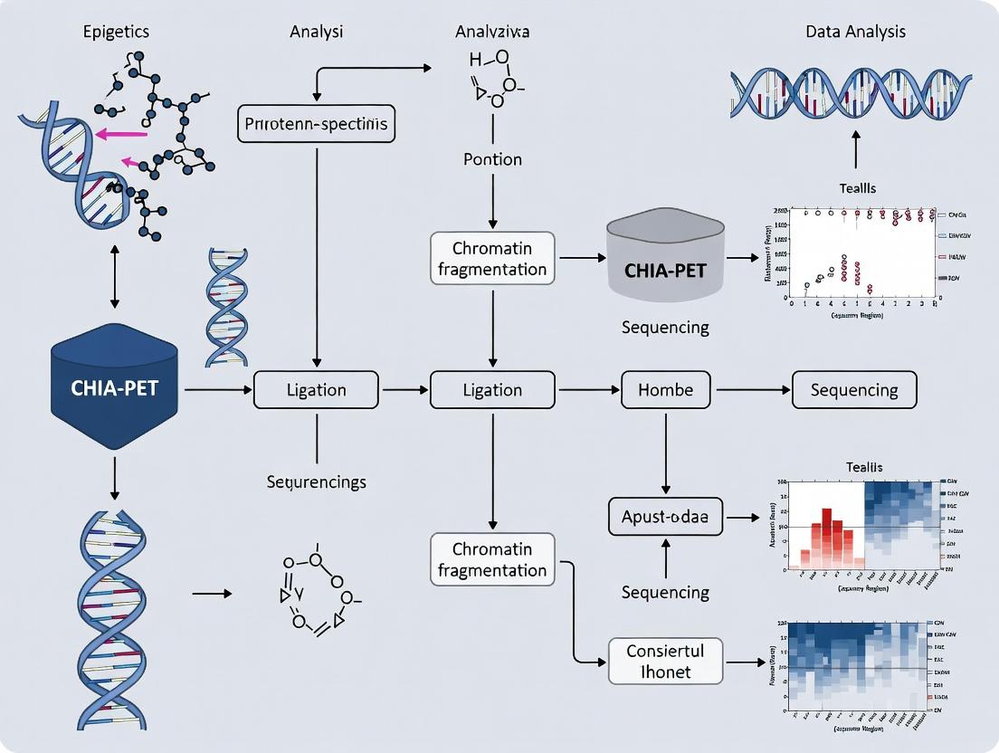
Visualization of Key Concepts and Workflows
ChIA-PET Experimental Workflow
TAD Structure and Enhancer-Promoter Looping
The Scientist's Toolkit: Key Research Reagent Solutions
Table 3: Essential Materials for ChIA-PET Experiments
| Item | Function & Critical Notes |
|---|---|
| Formaldehyde (37%) | Reversible crosslinking agent to fix protein-DNA and protein-protein interactions in situ. |
| Target-Specific Antibody | High-quality, ChIP-validated antibody for the protein of interest (e.g., Pol II, CTCF, ERα). Defines the interactome's specificity. |
| Protein A/G Magnetic Beads | For efficient capture and washing of antibody-bound chromatin complexes. |
| Covaris Sonicator | Provides consistent, high-quality chromatin shearing to optimal fragment size (200-600 bp). |
| T4 DNA Ligase | Catalyzes the proximity ligation step, joining crosslinked DNA fragments. |
| MmeI Restriction Enzyme | Type IIS enzyme used to generate defined, short tags (PETs) from ligated fragments for sequencing. |
| Illumina Sequencing Adapters & PCR Mix | For preparation of the final, barcoded sequencing library compatible with high-throughput platforms. |
| ChIA-PET Data Analysis Software (ChIA-PET Tool, Mango) | Specialized packages for processing raw sequencing data, classifying PETs, and calling significant interactions. |
Application Notes: ChIA-PET in Protein-Specific Chromatin Interaction Analysis
Within the broader thesis that ChIA-PET is the definitive method for unifying protein-centric molecular biochemistry with three-dimensional chromatin architecture, its primary application is the genome-wide identification of protein-anchored chromatin loops. This directly addresses the limitation of conformation capture methods like Hi-C, which detect all interactions indiscriminately. ChIA-PET’s integration with chromatin immunoprecipitation (ChIP) enables the specific interrogation of loops organized by transcription factors, architectural proteins (e.g., CTCF, Cohesin), polymerases (RNA Pol II), or histone modifications.
Key Quantitative Insights from Recent ChIA-PET Studies:
Table 1: Representative ChIA-PET Data Outputs for Key Architectural Proteins
| Target Protein | Avg. Loops Identified | Loop Size Range | Peaks at Loop Anchors | Common Associated Function |
|---|---|---|---|---|
| CTCF | 10,000 - 40,000 | 10kb - 2Mb | >90% | Insulation, TAD Boundary Formation |
| Cohesin (RAD21/SMC1A) | 15,000 - 60,000 | 10kb - 1Mb | ~85% | Loop Extrusion, Facilitated Looping |
| RNA Polymerase II | 5,000 - 20,000 | 1kb - 200kb | ~70% | Enhancer-Promoter Connectivity |
| ERα (in MCF-7 cells) | 5,000 - 15,000 | 5kb - 500kb | >95% | Hormone-Driven Gene Regulation |
Critical Interpretation: The high percentage of loops colocalizing with ChIP peaks (e.g., >90% for CTCF) validates the protein-specificity of the assay. The data quantitatively supports the model where CTCF and Cohesin collaboratively form structural loops, while RNA Pol II loops are shorter and directly regulatory. In drug development, comparing ChIA-PET maps for a nuclear receptor (like ERα) before and after ligand or drug treatment can reveal the specific rewiring of the chromatin interactome, identifying direct transcriptional targets and mechanisms of drug resistance.
Detailed Experimental Protocol: In-Situ ChIA-PET
This protocol, optimized for mammalian cells (e.g., MCF-7, K562), details the key steps for generating protein-specific interaction maps.
Part 1: Crosslinking, Chromatin Preparation, and Chromatin Immunoprecipitation
- Crosslinking: Harvest ~10 million cells. Crosslink with 1% formaldehyde for 10 min at room temperature. Quench with 125mM glycine.
- Nuclei Isolation & Chromatin Fragmentation: Lyse cells and isolate nuclei. Resuspend nuclei in appropriate buffer and perform enzymatic digestion (e.g., MboI) or sonication to achieve chromatin fragments of 300-600 bp.
- Chromatin Immunoprecipitation (ChIP): Incubate chromatin with antibody-conjugated beads (e.g., anti-CTCF, anti-RAD21) overnight at 4°C. Include a control IgG sample. Wash beads stringently to remove non-specific binding.
Part 2: Proximity Ligation and Library Construction
- End Repair & Ligation of Half-Linkers: Following ChIP, perform end-repair and A-tailing of DNA fragments on beads. Ligate a biotinylated "half-linker" oligonucleotide to the blunt ends. These half-linkers contain an overhang for the subsequent ligation step.
- Proximity Ligation (In-Situ): Under highly dilute conditions that favor intra-molecular ligation, ligate the half-linker-bearing ends from different DNA fragments that are in spatial proximity. This creates chimeric molecules linked by a biotinylated bridge.
- DNA Elution & Reverse Crosslinking: Elute DNA from beads and reverse crosslinks by incubating with Proteinase K overnight at 65°C.
- Biotin Capture & Purification: Shear DNA to ~300 bp. Capture chimeric ligation products using streptavidin beads. This critical step enriches for proximity-ligated fragments.
- Library Amplification & Sequencing: Perform on-bead PCR to amplify the purified chimeric DNA, incorporating sequencing adapters. Purify the final library and sequence on an Illumina platform (typically 150bp paired-end).
Part 3: Data Analysis Workflow
- Read Processing: Trim linker sequences and map paired-end reads independently to the reference genome (using Bowtie2/BWA).
- Interaction Calling: Identify valid interacting pairs where the two paired reads map to different restriction fragments (if used) or distinct genomic loci. Cluster these pairs using specialized tools (e.g., ChIA-PET2, Mango) to define significant interaction peaks (FDR < 0.05).
- Integration & Visualization: Annotate loops with overlapping ChIP-seq peaks and gene features. Visualize in genome browsers (e.g., WashU EpiGenome Browser, Juicebox) or as interaction networks.
Visualization of Key Concepts and Workflows
Diagram 1: ChIA-PET Core Experimental Workflow
Diagram 2: ChIA-PET Integrates Structure & Function
The Scientist's Toolkit: Essential Research Reagents & Materials
Table 2: Key Reagent Solutions for ChIA-PET
| Reagent/Material | Function & Critical Role |
|---|---|
| High-Affinity, Validated ChIP-Grade Antibody | The cornerstone of specificity. Immunoprecipitates the target protein and its bound DNA. Must be rigorously validated for ChIP-seq. |
| Biotinylated Half-Linker Oligonucleotides | Engineered adapters containing a MmeI type IIS restriction site (for 1st gen) or other design. Their ligation and subsequent proximity ligation create the unique chimeric junction for paired-end sequencing. |
| Streptavidin-Coated Magnetic Beads | Critical for stringent purification. Isolate biotin-tagged chimeric DNA fragments from the vast background of non-ligated or self-ligated fragments. |
| Controlled Restriction Enzyme (e.g., MboI) or Covaris Sonicator | For reproducible chromatin fragmentation. Enzymatic digestion gives precise ends but is sequence-dependent; sonication is unbiased but requires optimization for fragment size. |
| High-Fidelity DNA Polymerase for Library PCR | Amplifies the low-abundance chimeric library after biotin capture. Must have high fidelity and low bias to maintain representation. |
| Dual-Indexed Sequencing Adapters | Enable multiplexing of multiple samples in a single sequencing run, reducing cost and processing time. |
| Specialized Bioinformatics Pipelines (ChIA-PET2, Mango) | Not a wet-lab reagent, but essential. Designed to process paired-end reads, identify valid interaction pairs, filter artifacts, and call statistically significant interactions. |
Within the broader thesis on leveraging Chromatin Interaction Analysis with Paired-End Tag sequencing (ChIA-PET) for protein-specific chromatin interaction analysis, the core experimental components—crosslinking, immunoprecipitation, and proximity ligation—form the foundational triad. This protocol details the application of these components to map high-resolution, genome-wide chromatin interactions mediated by specific protein factors (e.g., RNA Polymerase II, CTCF, ERα), crucial for understanding gene regulation in development and disease for drug discovery.
Core Components: Detailed Protocols & Application Notes
Crosslinking: Stabilizing Protein-DNA Interactions
Objective: To covalently stabilize in vivo protein-DNA and protein-protein interactions with high efficiency. Protocol:
- Grow cells to 70-80% confluence in appropriate medium.
- Add 37% formaldehyde directly to culture medium to a final concentration of 1-2%. For nuclear factors, 1% is often sufficient; for weaker or indirect interactions, 2% may be used.
- Incubate at room temperature (20-25°C) for 10-15 minutes with gentle rocking.
- Quench the crosslinking reaction by adding 2.5M glycine to a final concentration of 0.125-0.25 M. Incubate for 5 minutes at room temperature.
- Wash cells twice with ice-cold phosphate-buffered saline (PBS).
- Pellet cells by centrifugation at 800 x g for 5 min at 4°C. Flash-freeze pellet in liquid nitrogen or proceed directly to cell lysis. Application Notes:
- Critical Parameter: Crosslinking time and concentration must be optimized to balance between capturing true interactions and retaining antigen accessibility for immunoprecipitation. Over-crosslinking can mask epitopes.
- Thesis Relevance: This step "freezes" the chromatin architecture centered on the protein of interest, forming the basis for all subsequent protein-specific interaction analysis.
Chromatin Immunoprecipitation (ChIP): Target-Specific Enrichment
Objective: To selectively enrich chromatin fragments bound by the protein of interest. Protocol:
- Lysis & Sonication: Resuspend cell pellet in ChIP lysis buffer. Sonicate chromatin to an average fragment size of 200-600 bp using a focused ultrasonicator (e.g., Covaris). Optimal conditions (e.g., 15-20 cycles of 30 sec ON/30 sec OFF at high setting for a Bioruptor) must be determined empirically.
- Pre-clearing & Immunoprecipitation: Centrifuge sonicated lysate at 16,000 x g for 10 min. Pre-clear supernatant with Protein A/G beads for 1 hour at 4°C. Incubate pre-cleared chromatin (typically 50-100 µg) with 2-10 µg of validated, high-specificity antibody overnight at 4°C with rotation.
- Bead Capture: Add pre-blocked Protein A/G magnetic beads and incubate for 2 hours.
- Washing: Wash beads sequentially with: Low Salt Wash Buffer (once), High Salt Wash Buffer (once), LiCl Wash Buffer (once), and TE Buffer (twice). All washes are 5 minutes at 4°C with rotation.
- Elution & Reversal: Elute chromatin complexes from beads twice with freshly prepared ChIP Elution Buffer (1% SDS, 0.1M NaHCO3) for 15 minutes at 65°C with shaking. Add NaCl to a final concentration of 200 mM and reverse crosslinks by incubating at 65°C overnight.
- Purification: Treat with RNase A (30 min, 37°C) and Proteinase K (2 hours, 55°C). Purify DNA using silica membrane columns. Quantify yield by Qubit fluorometry.
Table 1: Representative ChIP Efficiency Metrics
| Protein Target | Typical Antibody Amount (µg) | Input Chromatin (µg) | Expected DNA Yield (ng) | Enrichment Fold (vs. IgG) |
|---|---|---|---|---|
| CTCF | 2-5 | 50 | 15-40 | 20-50 |
| RNA Polymerase II | 5 | 100 | 10-30 | 15-40 |
| Histone H3K4me3 | 2 | 50 | 20-50 | 50-100 |
Proximity Ligation: Capturing Interaction Junctions
Objective: To join protein-bound DNA fragments in close spatial proximity, creating chimeric "paired-end tag" (PET) molecules for sequencing. Protocol:
- End Repair & A-Tailing: Treat purified ChIP DNA with a blend of T4 DNA Polymerase, Klenow Fragment, and T4 PNK to generate blunt ends. Purify and then add a single 'A' base to 3' ends using Klenow exo-.
- Ligation of Half-Linkers: Ligate A-tailed DNA to specially designed, biotinylated "half-linker" oligonucleotides using T4 DNA Ligase. A half-linker contains: a MmeI recognition site, a biotin tag, and an overhang complementary to the 'A' tail.
- Proximity Ligation: Dilute the ligation reaction ~50-fold in ligation buffer to favor intramolecular ligation events between crosslinked DNA fragments. Add T4 DNA Ligase and incubate at 16°C for 2-4 hours. This step ligates the free ends of half-linkers attached to different DNA fragments that are in close spatial proximity, creating a full linker.
- DNA Purification & MmeI Digestion: Purify DNA and digest with MmeI, a type IIS restriction enzyme that cuts 20-21 bp away from its recognition site (within the linker), releasing ~36-40 bp PETs (two ~18-20 bp tags from interacting fragments, flanked by linker sequences).
- PET Purification & Library Construction: Capture biotinylated PETs using streptavidin-coated magnetic beads. Ligate sequencing adaptors to bead-bound PETs, perform PCR amplification (12-15 cycles), and size-select (120-200 bp) for paired-end sequencing.
Table 2: Key Proximity Ligation Reagents & Parameters
| Component | Function | Critical Parameter |
|---|---|---|
| Biotinylated Half-Linkers | Provide universal priming sites and biotin handle for PET isolation. | Must contain MmeI site. High-purity HPLC purification required. |
| T4 DNA Ligase | Catalyzes intra-molecular proximity ligation. | Dilution factor is critical to favor inter-fragment ligation. |
| MmeI Enzyme | Releases short, sequenceable PETs from ligated chromatin. | Activity is salt-sensitive; optimize buffer conditions. |
| Streptavidin Beads | Isolate biotinylated chimeric PET molecules. | High binding capacity (>500 pmol/mg) reduces loss. |
The Scientist's Toolkit: Research Reagent Solutions
| Item | Function in ChIA-PET |
|---|---|
| Formaldehyde (37%) | Reversible crosslinker for fixing protein-DNA complexes. |
| Covaris S2/S220 Focused-ultrasonicator | Provides consistent, tunable chromatin shearing to ideal fragment size. |
| Magna ChIP Protein A/G Magnetic Beads | Efficient capture of antibody-chromatin complexes with low non-specific binding. |
| Validated ChIP-Grade Antibody | Target-specific antibody is the single most critical reagent for success. |
| Dynabeads MyOne Streptavidin C1 | High-capacity streptavidin beads for efficient PET purification. |
| T4 DNA Ligase (High Concentration) | Essential for efficient proximity ligation of diluted DNA. |
| MmeI (NEB) | Type IIS restriction enzyme for precise PET release. |
| KAPA HiFi HotStart ReadyMix | High-fidelity PCR for library amplification prior to sequencing. |
| AMPure XP Beads | For robust size selection and clean-up of DNA libraries. |
Workflow & Pathway Visualizations
Diagram 1: Core ChIA-PET Experimental Workflow
Diagram 2: Molecular Steps of Proximity Ligation & PET Formation
Application Notes: Functional Significance of Targeted Proteins in Chromatin Architecture
The comprehensive analysis of chromatin interactions via ChIA-PET (Chromatin Interaction Analysis with Paired-End Tag sequencing) hinges on targeting specific architectural and regulatory proteins. This application note details the roles of RNA Polymerase II (Pol II), CTCF, Cohesin, and Tissue-Specific Transcription Factors (TFs) within the context of a thesis on protein-centric 3D genome mapping. Targeting these proteins allows researchers to dissect the multi-layered regulatory landscape, from promoter-enhancer communication to topologically associating domain (TAD) formation, providing critical insights for fundamental biology and drug discovery.
Table 1: Key Targeted Proteins and Their Chromatin Roles in ChIA-PET Studies
| Target Protein | Primary Function in 3D Genome | Typical ChIA-PET Interaction Type | Associated Genomic Features | Relevance to Disease/Drug Development |
|---|---|---|---|---|
| RNA Polymerase II | Transcription elongation; Mediates enhancer-promoter looping. | Short-range, within active TADs. | Active promoters, enhancers, gene bodies. | Oncogene activation, transcriptional dysregulation in cancer. |
| CTCF | Architectural protein; Directional insulator and loop anchor. | Long-range, inter-TAD; Loop anchors. | Insulator sites, TAD boundaries. | Mutations in cancer disrupt TADs, leading to oncogene activation. |
| Cohesin (SMC1A, SMC3, RAD21) | ATP-driven loop extruder; Works with CTCF to form loops. | Anchored loops at CTCF sites; Dynamic loops. | Convergent CTCF motifs. | Cohesinopathies (e.g., Cornelia de Lange), leukemia. |
| Tissue-Specific TFs (e.g., ERα, AR, PU.1) | Cell-type specific gene activation; Recruit coactivators and architectural proteins. | Cell-type specific enhancer-promoter hubs. | Enhancer regions with specific TF motifs. | Therapeutic targets in breast cancer (ERα), prostate cancer (AR). |
Experimental Protocols
Protocol 1: Crosslinking and Chromatin Preparation for Protein-Specific ChIA-PET
Objective: To fix protein-DNA and protein-protein interactions and generate soluble, sheared chromatin. Materials: Formaldehyde (1%), Glycine (125 mM), Cell lysis buffers, SDS, Triton X-100, Micrococcal Nuclease (MNase) or Sonication device. Procedure:
- Crosslink 10-20 million cells with 1% formaldehyde for 10 min at room temperature. Quench with 125 mM glycine.
- Harvest cells, wash with cold PBS, and lyse in Cell Lysis Buffer (10 mM Tris-HCl pH 8.0, 10 mM NaCl, 0.2% NP-40) for 15 min on ice. Pellet nuclei.
- Resuspend nuclei in Shearing Buffer (0.1% SDS, 1 mM EDTA, 10 mM Tris-HCl pH 8.0). Shear chromatin to 200-600 bp fragments using either:
- MNase digestion: Add CaCl₂ and MNase, incubate at 37°C, stop with EDTA.
- Sonication: Use a focused ultrasonicator (e.g., Covaris) for 12-15 cycles (30 sec ON/30 sec OFF) at 4°C.
- Centrifuge to remove debris. Transfer supernatant (sheared chromatin) to a new tube. Aliquot and store at -80°C.
Protocol 2: Chromatin Immunoprecipitation (ChIP) for Target Proteins
Objective: To enrich for chromatin fragments bound by the protein of interest (Pol II, CTCF, Cohesin, or TF). Materials: Protein A/G magnetic beads, target-specific antibody (validated for ChIP), Low Salt and High Salt Wash Buffers, TE Buffer, Elution Buffer (1% SDS, 0.1M NaHCO₃). Procedure:
- Pre-clear 100 µg of sheared chromatin with Protein A/G beads for 1 hour at 4°C.
- Incubate the pre-cleared chromatin with 5-10 µg of target antibody overnight at 4°C with rotation.
- Critical: Use validated antibodies: anti-RPB1 (Pol II), anti-CTCF, anti-RAD21/SMC1 (Cohesin), or anti-TF (e.g., ERα).
- Add pre-washed Protein A/G beads and incubate for 2 hours.
- Wash beads sequentially with: Low Salt Buffer (once), High Salt Buffer (once), LiCl Buffer (once), and TE Buffer (twice).
- Elute chromatin complexes twice with 100 µL Elution Buffer by vortexing at 65°C for 15 minutes. Combine eluates.
- Reverse crosslinks by adding NaCl (200 mM final) and incubating at 65°C overnight.
Protocol 3: Proximity Ligation, Library Preparation, and Sequencing for ChIA-PET
Objective: To convert protein-anchored chromatin interactions into a sequencer-compatible library. Materials: T4 DNA Ligase, Biotinylated bridge linkers, T4 DNA Polymerase, Klenow Fragment, T4 PNK, Paired-End sequencing adapters, Streptavidin beads. Procedure:
- End Repair & A-tailing: Purify reverse-crosslinked DNA (from Protocol 2). Perform end-repair and dA-tailing using standard kits.
- Proximity Ligation: Dilute the DNA in a large volume (1-7 mL) of ligation buffer to favor intra-molecular ligation. Add T4 DNA Ligase and biotinylated bridge linkers. Incubate at 16°C for 4-6 hours.
- DNA Purification & Biotin Capture: Purify DNA, shear to ~300 bp, and capture biotinylated ligation products (the interaction tags) on streptavidin-coated magnetic beads.
- On-Bead Library Construction: On the beads, ligate paired-end sequencing adapters to the captured DNA fragments. Perform PCR amplification (12-15 cycles).
- Sequencing: Purify the final library and sequence on an Illumina platform (e.g., NovaSeq) using 150 bp paired-end reads. Aim for 50-100 million read pairs per sample.
Visualizations
Title: Protein Roles in 3D Chromatin Architecture
Title: ChIA-PET Experimental Workflow
The Scientist's Toolkit: Essential Research Reagents & Materials
Table 2: Key Reagent Solutions for Protein-Targeted ChIA-PET
| Reagent/Material | Supplier Examples | Function in Protocol | Critical Notes |
|---|---|---|---|
| High-Quality ChIP-Validated Antibody | Cell Signaling, Abcam, Diagenode | Immunoprecipitation of target protein-DNA complexes. | Validation with KO cell lines is essential. Key for success. |
| Protein A/G Magnetic Beads | Thermo Fisher, MilliporeSigma | Capture of antibody-bound complexes. | Offers ease of washing versus agarose beads. |
| Ultrapure Formaldehyde | Thermo Fisher, MilliporeSigma | Reversible crosslinking of protein-DNA interactions. | Use fresh 1% solution for consistent results. |
| Micrococcal Nuclease (MNase) | NEB, Worthington | Enzymatic shearing of chromatin. | Yields precise nucleosomal fragments; requires titration. |
| Biotinylated Bridge Linker | Integrated DNA Technologies | Linker containing biotin for proximity ligation and capture. | Custom sequence; critical for tagging interaction junctions. |
| Streptavidin Magnetic Beads (MyOne C1) | Thermo Fisher | High-affinity capture of biotinylated ligation products. | Efficient pulldown minimizes background. |
| Paired-End Sequencing Kit | Illumina | Preparation of sequencer-ready libraries. | Compatibility with on-bead library construction is key. |
| Cell Line or Primary Cells | ATCC, commercial vendors | Biological source material. | Must express target protein (e.g., TF) at sufficient levels. |
Application Notes
ChIA-PET (Chromatin Interaction Analysis with Paired-End Tag Sequencing) has revolutionized the study of 3D chromatin architecture by linking looping interactions to specific protein factors. Its unique ability to map protein-anchored chromatin contacts has enabled foundational discoveries in gene regulation and disease mechanisms.
Key Discoveries & Quantitative Data
Table 1: Seminal Discoveries Enabled by ChIA-PET
| Discovery | Key Protein Factor | Biological System/Cell Type | Key Quantitative Finding | Citation |
|---|---|---|---|---|
| Super-Enhancer Definition & Function | Mediator (MED1), Cohesin (SMC1) | Mouse embryonic stem cells (mESCs) | Super-enhancers (top 5% of enhancers) accounted for ~40% of Mediator-bound enhancer activity. | Whyte et al., 2013 |
| Architectural RNA-Protein Loops | RNA Polymerase II (Pol II) | Human cell lines (K562, MCF-7) | Identified >30,000 promoter-centered chromatin loops; many connected to enhancers. | Li et al., 2012 |
| Disease-Associated Variants in Loops | Cohesin (RAD21), CTCF | Primary human cells (GM12878, IMR90) | 30% of disease-associated SNPs from GWAS were located in anchor regions of CTCF/cohesin loops. | Grubert et al., 2015 |
| Oncogene Activation via Looping | ERα (Estrogen Receptor Alpha) | Human breast cancer cells (MCF-7) | E2 stimulation created 1,149 new ERα-mediated loops, linking enhancers to target genes like GREB1. | Fullwood et al., 2009 |
| Compartmentalization of Pluripotency | OCT4, SOX2, NANOG (OSN) | Mouse embryonic stem cells (mESCs) | OSN co-bound loops formed a highly interconnected network, stabilizing pluripotency. | Dowen et al., 2014 |
Table 2: Typical ChIA-PET Data Output Metrics
| Metric | Typical Range (Mammalian Genome) | Description |
|---|---|---|
| Sequencing Depth | 200M - 1B+ paired-end reads | Required for sufficient library complexity and interaction coverage. |
| Valid PETs | 5M - 50M+ | Paired-end tags representing valid ligation products. |
| Significant Interactions | 10,000 - 100,000+ | High-confidence chromatin loops (FDR < 0.05). |
| Peak-to-Loop Ratio | ~10:1 | Many protein binding peaks form a smaller subset of loops. |
Protocols
Protocol 1: Standard ChIA-PET for Transcription Factors (e.g., ERα)
A. Crosslinking & Cell Lysis
- Crosslink 10-20 million cells with 1% formaldehyde for 10 min at room temperature. Quench with 125 mM glycine.
- Pellet cells, wash with cold PBS. Resuspend in 1 mL Lysis Buffer (10 mM Tris-HCl pH 8.0, 1% SDS, 10 mM EDTA, protease inhibitors). Incubate on ice for 10 min.
- Pellet nuclei. Resuspend in 1 mL Shearing Buffer (0.1% SDS, 10 mM Tris-HCl pH 8.0, 1 mM EDTA). Sonicate to achieve DNA fragments of 200-600 bp.
B. Chromatin Immunoprecipitation (ChIP)
- Dilute sheared chromatin 10-fold in ChIP Dilution Buffer (0.01% SDS, 1% Triton X-100, 1.2 mM EDTA, 16.7 mM Tris-HCl pH 8.0, 167 mM NaCl).
- Pre-clear with protein A/G beads for 1-2 hours.
- Incubate supernatant with 5-10 µg of specific antibody (e.g., anti-ERα) overnight at 4°C. Include an IgG control.
- Add protein A/G beads, incubate 2 hours. Wash beads sequentially with: Low Salt Wash Buffer (0.1% SDS, 1% Triton X-100, 2mM EDTA, 20mM Tris-HCl pH 8.0, 150mM NaCl), High Salt Wash Buffer (same, but 500mM NaCl), LiCl Wash Buffer (0.25M LiCl, 1% NP-40, 1% sodium deoxycholate, 1mM EDTA, 10mM Tris-HCl pH 8.0), and TE Buffer.
C. Proximity Ligation & Library Construction
- Elute ChIP material in Elution Buffer (1% SDS, 0.1M NaHCO3). Reverse crosslinks for one sample (Input) at 65°C overnight.
- For the main sample, end-repair and A-tail the DNA on-beads using standard kits.
- Ligate to a biotinylated bridge linker containing MmeI restriction site. Perform intra- and inter-molecular proximity ligation in a large volume (~3 mL) with T4 DNA ligase overnight at 16°C.
- Reverse crosslinks. Purify DNA.
- Digest with MmeI, which cuts 20bp away from its recognition site, releasing ~40bp PETs containing the linker.
- Perform paired-end tag ligation to add Illumina sequencing adapters. Purify biotinylated PETs with streptavidin beads.
- Amplify by PCR (12-18 cycles) and size-select (150-400 bp) for paired-end sequencing (e.g., Illumina HiSeq).
Protocol 2: HiChIP Variant (Rapid ChIA-PET)
Note: This modern variant uses in-situ ligation and has higher efficiency.
- Crosslink and lyse cells as in Protocol 1A.
- Perform in-nucleus restriction digest (e.g., with MboI) of sheared chromatin.
- Fill restriction overhangs with biotinylated nucleotides and ligate in situ to form chimeric junctions.
- Sonicate to ~300-500 bp and perform ChIP with target-specific antibody (e.g., H3K27ac for active enhancers/promoters).
- Capture biotinylated ligation products using streptavidin beads.
- Process for standard Illumina paired-end sequencing library construction.
Visualizations
Title: ChIA-PET Core Experimental Workflow
Title: Super-Enhancer Looping Mechanism
Title: Disease SNP Disrupting Chromatin Loops
The Scientist's Toolkit
Table 3: Essential Research Reagent Solutions for ChIA-PET
| Item | Function / Purpose | Example / Key Consideration |
|---|---|---|
| High-Quality Specific Antibody | Immunoprecipitation of the protein of interest (POI) to anchor interactions. | Validated ChIP-seq grade antibody (e.g., anti-CTCF, anti-RAD21, anti-Pol II). Critical for success. |
| Biotinylated Bridge Linker | Contains MmeI site; enables ligation of interacting fragments and subsequent PET release. | HPLC-purified oligonucleotides. Sequence must be optimized and balanced. |
| MmeI Restriction Enzyme | Type IIS enzyme that cuts 20bp away from its site, generating uniform ~40bp PETs. | High activity required on chromatin-derived DNA. |
| Protein A/G Magnetic Beads | Capture antibody-protein-DNA complexes during ChIP. | Magnetic separation reduces background vs. agarose beads. |
| Streptavidin Magnetic Beads | Efficient pull-down of biotinylated PETs after MmeI digestion. | High binding capacity crucial for recovering low-abundance ligation products. |
| Sonication Device | Shearing crosslinked chromatin to 200-600 bp fragments. | Focused ultrasonicator (Covaris) preferred for consistent fragment size. |
| High-Fidelity PCR Mix | Amplification of the final PET library prior to sequencing. | Low error rate is essential to maintain junction sequence fidelity. |
| Size Selection Beads | Cleanup and selection of correctly sized PET libraries (e.g., 150-400 bp). | SPRI/AMPure beads allow precise size fractionation. |
| Paired-End Sequencing Kit | High-throughput sequencing of the PET library. | Illumina platforms (NovaSeq, HiSeq) for 150bp paired-end reads. |
| Chromatin Crosslinker | Reversible fixation of protein-DNA and protein-protein interactions. | Formaldehyde (1%) is standard; EGS can be added for secondary fixation. |
The ChIA-PET Protocol: A Step-by-Step Guide from Bench to Bioinformatics
Application Notes
This protocol details the foundational steps for chromatin immunoprecipitation (ChIP), with specific optimization for subsequent Chromatin Interaction Analysis by Paired-End Tag Sequencing (ChIA-PET). ChIA-PET enables genome-wide, protein-specific analysis of long-range chromatin interactions and requires exceptionally high-quality ChIP material. The critical parameters are efficient crosslinking to capture transient interactions, optimized shearing to generate 200-500 bp chromatin fragments suitable for pairing, and stringent immunoprecipitation to ensure target-specific enrichment with minimal background. These steps directly impact the signal-to-noise ratio and resolution of the final interaction map, which is critical for research in transcriptional regulation, enhancer-promoter networks, and identifying novel drug targets in disease contexts.
Protocols
Formaldehyde Crosslinking of Chromatin
Objective: To covalently fix protein-DNA and protein-protein interactions in situ.
Detailed Methodology:
- Cell Culture: Grow adherent or suspension cells to ~80% confluence. For tissue, mince into ~1 mm³ pieces.
- Crosslinking: Add 37% formaldehyde directly to culture medium to a final concentration of 1% (e.g., 270 µL formaldehyde per 10 mL medium). Incubate at room temperature (RT) for 10 minutes with gentle agitation. Note: For ChIA-PET, a 1-2% formaldehyde concentration for 10 min is standard; over-crosslinking reduces shearing efficiency.
- Quenching: Add 2.5M glycine to a final concentration of 0.125M (e.g., 500 µL of 2.5M glycine per 10 mL). Incubate for 5 minutes at RT with agitation to quench unreacted formaldehyde.
- Washing: Pellet cells at 800 x g for 5 minutes at 4°C. Wash pellet twice with 10 mL of ice-cold 1X Phosphate-Buffered Saline (PBS). Flash-freeze pellet in liquid nitrogen and store at -80°C or proceed immediately.
Chromatin Shearing by Sonication
Objective: To fragment crosslinked chromatin to an average size of 200-500 bp.
Detailed Methodology:
- Lysis: Resuspend cell pellet in 1 mL of cold Lysis Buffer 1 (50 mM HEPES-KOH pH 7.5, 140 mM NaCl, 1 mM EDTA, 10% glycerol, 0.5% NP-40, 0.25% Triton X-100, protease inhibitors). Rotate for 10 minutes at 4°C. Pellet nuclei at 1,350 x g for 5 min at 4°C.
- Nuclear Wash: Resuspend pellet in 1 mL of cold Lysis Buffer 2 (10 mM Tris-HCl pH 8.0, 200 mM NaCl, 1 mM EDTA, 0.5 mM EGTA, protease inhibitors). Rotate for 10 minutes at 4°C. Pellet as before.
- Sonication: Resuspend pellet in 1 mL of cold Shearing Buffer (0.1% SDS, 1 mM EDTA, 10 mM Tris-HCl pH 8.0, protease inhibitors). Transfer to a 1 mL Covaris microTUBE or compatible tube.
- Sonicate using a focused ultrasonicator (e.g., Covaris S220) with the following validated settings:
- Peak Incident Power: 140W
- Duty Factor: 10%
- Cycles per Burst: 200
- Time: 12-18 minutes (optimize per cell type).
- Clarification: Centrifuge sheared lysate at 20,000 x g for 10 minutes at 4°C. Transfer supernatant (sheared chromatin) to a new tube. Aliquot and store at -80°C.
- QC: Reverse crosslink and purify DNA from a 50 µL aliquot. Analyze fragment size distribution on a 1.5% agarose gel or Bioanalyzer.
Chromatin Immunoprecipitation (ChIP)
Objective: To enrich for chromatin fragments bound by the protein of interest.
Detailed Methodology:
- Pre-clearing (Optional): Dilute 100 µg of sheared chromatin to 1 mL total volume with ChIP Dilution Buffer (0.01% SDS, 1.1% Triton X-100, 1.2 mM EDTA, 16.7 mM Tris-HCl pH 8.0, 167 mM NaCl). Add 20 µL of pre-equilibrated Protein A/G beads. Rotate for 1-2 hours at 4°C. Pellet beads; retain supernatant.
- Immunoprecipitation: Add 1-10 µg of target-specific antibody (see Toolkit) or control IgG to the pre-cleared chromatin. Rotate overnight at 4°C.
- Bead Capture: Add 50 µL of pre-blocked (with 0.5% BSA) Protein A/G magnetic beads. Rotate for 2-4 hours at 4°C.
- Stringent Washes: Pellet beads on a magnetic rack. Wash sequentially for 5 minutes each with rotation at 4°C in:
- 1 mL Low Salt Wash Buffer (0.1% SDS, 1% Triton X-100, 2 mM EDTA, 20 mM Tris-HCl pH 8.0, 150 mM NaCl).
- 1 mL High Salt Wash Buffer (0.1% SDS, 1% Triton X-100, 2 mM EDTA, 20 mM Tris-HCl pH 8.0, 500 mM NaCl).
- 1 mL LiCl Wash Buffer (0.25 M LiCl, 1% NP-40, 1% sodium deoxycholate, 1 mM EDTA, 10 mM Tris-HCl pH 8.0).
- Two washes with 1 mL TE Buffer (10 mM Tris-HCl pH 8.0, 1 mM EDTA).
- Elution: Elute chromatin from beads by adding 200 µL of Fresh Elution Buffer (1% SDS, 0.1M NaHCO₃). Vortex at 1200 rpm for 15 minutes at RT. Pellet beads; transfer eluate to a new tube.
- Reverse Crosslinking & Purification: Add 8 µL of 5M NaCl and 1 µL of RNase A (10 mg/mL) to the eluate. Incubate at 65°C overnight. Add 4 µL of 0.5M EDTA, 8 µL of 1M Tris-HCl pH 6.5, and 1 µL of Proteinase K (20 mg/mL). Incubate at 45°C for 2 hours. Purify DNA using a PCR purification kit. Elute in 20-30 µL EB buffer.
Data Presentation
Table 1: Quantitative Metrics for Critical ChIA-PET ChIP Steps
| Parameter | Optimal Target Range | Measurement Method | Impact on ChIA-PET |
|---|---|---|---|
| Crosslinking Time | 8-12 minutes | Empirical testing | Longer times reduce shearing efficiency & interaction recovery. |
| Chromatin Fragment Size | 200-500 bp (peak ~300 bp) | Agarose Gel / Bioanalyzer | Critical for proximity ligation efficiency in later steps. |
| DNA Concentration Post-ChIP | > 10 ng (from 10⁷ cells) | Fluorometry (Qubit) | Directly limits library complexity for sequencing. |
| ChIP Enrichment (qPCR) | > 10-fold over IgG at positive control locus | qPCR (ΔΔCt) | Indicates antibody specificity and IP success. |
Table 2: Sonication Parameters for Different Cell Types (Covaris S220)
| Cell Type / Tissue | Starting Cell Number | Peak Power (W) | Duty Factor | Cycles/Burst | Time (min) |
|---|---|---|---|---|---|
| HEK293 (Adherent) | 2-4 x 10⁶ | 140 | 10% | 200 | 12-15 |
| K562 (Suspension) | 2-4 x 10⁶ | 135 | 10% | 200 | 15-18 |
| Mouse Liver (Nuclei) | 5 x 10⁶ nuclei | 145 | 15% | 200 | 20-25 |
Visualization
Title: Core ChIP Workflow for ChIA-PET Sample Prep
Title: ChIA-PET Steps Following ChIP
The Scientist's Toolkit
Table 3: Essential Research Reagent Solutions for ChIA-PET ChIP
| Item / Reagent | Function & Critical Notes | Example Vendor/Cat. # |
|---|---|---|
| Formaldehyde (37%) | Crosslinking agent. Use fresh, molecular biology grade for consistent efficiency. | Thermo Fisher, 28906 |
| Protease Inhibitor Cocktail | Prevents degradation of target protein and chromatin-associated factors during lysis. | Roche, 04693159001 |
| Covaris microTUBE | Specific tube for focused ultrasonication; ensures reproducible shearing. | Covaris, 520045 |
| Target-Specific Antibody | High specificity and ChIP-grade validation are non-negotiable for ChIA-PET. | e.g., Anti-RNA Pol II (Diagenode, C15100055) |
| Protein A/G Magnetic Beads | Efficient capture of antibody-chromatin complexes. Lower non-specific binding than agarose. | Pierce, 88802 |
| DNA Clean/Concentrator Kit | For reliable purification of low-concentration ChIP DNA without loss. | Zymo Research, D4013 |
| High-Sensitivity DNA Assay | Accurate quantification of dilute ChIP DNA (critical for library prep). | Thermo Fisher (Qubit), Q32851 |
Proximity Ligation, Linker Insertion, and Paired-End Tag Library Construction
This application note details critical protocols for the analysis of chromatin interactions, specifically within the broader framework of Chromatin Interaction Analysis with Paired-End Tag sequencing (ChIA-PET). The primary thesis posits that ChIA-PET, when executed with optimized proximity ligation and linker insertion techniques, provides unparalleled resolution for mapping long-range, protein-specific chromatin interactions. This is fundamental for elucidating gene regulation mechanisms in development, disease, and drug response.
Part I: Proximity Ligation
Protocol: In Situ Proximity Ligation for Crosslinked Chromatin
Objective: To ligate DNA ends that are in close spatial proximity due to protein-mediated chromatin looping, while preserving in vivo interaction contexts.
Materials:
- Formaldehyde-crosslinked cell pellet (5-10 million cells).
- Lysis Buffer: 10 mM Tris-HCl (pH 8.0), 1 mM EDTA, 0.5% SDS, and protease inhibitors.
- Dilution Buffer: 1.67% Triton X-100, 167 mM NaCl, 1.67 mM EDTA, 16.7 mM Tris-HCl (pH 8.0).
- Nuclease-free water.
- T4 DNA Ligase (HC, high concentration) and 10x Reaction Buffer.
- 37°C and 65°C incubators or water baths.
Methodology:
- Cell Lysis: Resuspend crosslinked pellet in 1 mL Lysis Buffer. Incubate on ice for 15 minutes.
- Chromatin Solubilization: Dilute lysate with 9 mL of Dilution Buffer to quench SDS. Mix thoroughly.
- Chromatin Fragmentation: Sonicate using a focused ultrasonicator to achieve an average DNA fragment size of 300-500 bp. Verify by agarose gel electrophoresis.
- Proximity Ligation: Centrifuge sonicated sample, collect supernatant. For a 1 mL sample, add 120 μL of 10x T4 DNA Ligase Buffer and 750 U of T4 DNA Ligase. Adjust volume with water to 1.2 mL.
- Incubate at 16°C for 4-6 hours with gentle rotation.
- Reverse Crosslinking: Add Proteinase K to 0.2 mg/mL and incubate at 65°C overnight.
- Purify DNA by phenol-chloroform extraction and ethanol precipitation. Resuspend in 100 μL TE buffer.
Key Quantitative Data:
Table 1: Optimized Proximity Ligation Parameters
| Parameter | Optimal Condition | Effect of Deviation |
|---|---|---|
| Ligase Concentration | 62.5 U/100 μL reaction | <50 U: Low yield; >100 U: Increased noise |
| Incubation Temperature | 16°C | Higher temp: Increased random ligation |
| Incubation Time | 4-6 hours | Shorter: Incomplete; Longer: No significant gain |
| DNA Concentration | 50-100 ng/μL post-sonication | Too low: Low efficiency; Too high: Viscosity issues |
Part II: Linker Insertion
Protocol: Bridge Linker Ligation for Paired-End Tag (PET) Formation
Objective: To ligate biotinylated, hairpin-shaped bridge linkers to the ends of proximally ligated DNA molecules, enabling subsequent purification and paired-end tag generation.
Materials:
- Purified proximity-ligation DNA.
- Biotinylated Bridge Linkers (Oligo A: 5'-Biotin-[Sequence]-3', Oligo B: complementary).
- T4 DNA Ligase (HC).
- Streptavidin-coated magnetic beads (e.g., Dynabeads MyOne Streptavidin C1).
- Binding & Wash Buffer (B&W): 5 mM Tris-HCl (pH 7.5), 0.5 mM EDTA, 1 M NaCl.
- TE buffer (pH 8.0).
Methodology:
- Linker Annealing: Mix Oligo A and Oligo B in equimolar ratio (100 μM each) in annealing buffer. Heat to 95°C for 5 min, cool slowly to 25°C.
- Ligation to DNA: Combine 100 ng proximity-ligation DNA, 50-fold molar excess of annealed bridge linker, 1x T4 Ligase Buffer, 5 U T4 DNA Ligase. Total reaction volume: 50 μL.
- Incubate at 16°C for 12-16 hours (overnight).
- Linker-Ligated DNA Capture: a. Wash 50 μL streptavidin beads twice with 200 μL B&W Buffer. b. Add the ligation reaction directly to the beads. Incubate at room temperature for 30 min with gentle mixing. c. Place on magnet, discard supernatant. d. Wash beads 3x with 200 μL B&W Buffer, then 2x with 200 μL TE Buffer.
- Resuspend beads in 20 μL TE Buffer for downstream enzymatic steps (e.g., MmeI digestion in ChIA-PET).
Key Quantitative Data:
Table 2: Bridge Linker Ligation Efficiency
| Component | Recommended Amount/Conc. | Purpose & Rationale |
|---|---|---|
| Bridge Linker Molar Excess | 50:1 (linker:DNA ends) | Ensures >95% end capture; higher excess increases cost without benefit |
| Bead Binding Time | 30 minutes | Achieves >85% capture efficiency |
| Bead Capacity | ~200 pmol biotin/μL beads | Do not exceed 70% capacity to avoid saturation |
| Final DNA Elution Volume | 20 μL | Maximizes concentration for downstream steps |
Part III: Paired-End Tag Library Construction
Protocol: From Captured Complexes to Sequencing-Ready PETs
Objective: To convert linker-ligated, bead-bound DNA into a purified library of short Paired-End Tags (PETs) suitable for high-throughput sequencing.
Materials:
- Bead-bound, linker-ligated DNA from Part II.
- Restriction Enzyme MmeI (NEB).
- T4 DNA Ligase.
- PCR primers with Illumina adaptor sequences.
- High-Fidelity PCR Master Mix.
- SPRIselect beads (Beckman Coulter) for size selection.
- Qubit dsDNA HS Assay Kit.
Methodology:
- Tag Release (MmeI Digestion): On-bead, resuspend beads in 50 μL 1x NEBuffer 4 with 2 U MmeI. Incubate at 37°C for 1.5 hours. MmeI cuts 20 bp away from its recognition site, releasing 20-21 bp tags adjacent to the linker.
- Pet Release and Purification: Place on magnet. Collect supernatant containing released PETs (each is a di-tag from two interacting fragments). Ethanol precipitate the PETs.
- Blunt-End Ligation: Resuspend PETs in a ligation reaction to promote circularization or dimerization of di-tags. Use T4 DNA Ligase, incubate at 16°C for 1 hour.
- PCR Amplification: Amplify ligated PETs using primers containing full Illumina P5/P7 adapters and index sequences. Use 12-15 PCR cycles.
Thermocycler Program:
- 98°C for 30s
- [98°C for 10s, 60°C for 30s, 72°C for 30s] x (12-15 cycles)
- 72°C for 5 min
- Hold at 4°C.
- Library Purification & Size Selection: a. Cleanup with 1.8x SPRIselect bead ratio to remove primers and large fragments. b. Perform a second size selection with 0.6x SPRIselect ratio to retain the desired ~200-300 bp product (adapters + PET).
- Quality Control: Assess library concentration (Qubit) and size profile (Bioanalyzer/TapeStation). Typical final yield: 10-50 nM in 30 μL.
Key Quantitative Data:
Table 3: PET Library Construction QC Metrics
| QC Step | Target Metric | Acceptable Range |
|---|---|---|
| Post-MmeI Release Yield | 5-15 ng | Indicates tag recovery efficiency |
| Optimal PCR Cycles | 14 cycles | 12 cycles: low yield; 16 cycles: increased duplicates |
| Final Library Size | ~250 bp | Adapter dimer at ~120 bp must be minimal |
| Library Concentration for Seq | >10 nM | Required for cluster generation |
The Scientist's Toolkit
Table 4: Key Research Reagent Solutions
| Item | Function & Application in ChIA-PET |
|---|---|
| Formaldehyde (37%) | Reversible protein-DNA and protein-protein crosslinker, fixes in vivo interactions. |
| T4 DNA Ligase (High Conc.) | Catalyzes both proximity ligation and linker ligation; high concentration favors intermolecular ligation of crosslinked fragments. |
| Biotinylated Bridge Linker | Hairpin oligonucleotide containing MmeI site; allows selective capture of ligation junctions and paired-end tag creation. |
| Streptavidin Magnetic Beads | Solid-phase support for immobilizing biotinylated linkers and associated DNA, enabling stringent washes. |
| MmeI Type IIS Restriction Enzyme | Cuts at a defined distance from its site to release short, consistent paired-end tags (20-21 bp). |
| SPRIselect Beads | Paramagnetic beads for precise size selection and purification of DNA libraries; critical for removing adapter dimers. |
| Illumina-Compatible PCR Primers | Contain P5/P7 flow cell binding sequences and indexes for multiplexing; amplify the pool of PETs. |
Experimental Workflow and Pathway Visualizations
Diagram 1: Proximity Ligation Core Workflow (76 chars)
Diagram 2: ChIA-PET Method from IP to Sequencing (78 chars)
Diagram 3: PET Formation from Ligated Junction (73 chars)
Next-Generation Sequencing Strategies and Depth Requirements for ChIA-PET
ChIA-PET (Chromatin Interaction Analysis by Paired-End Tag Sequencing) is a powerful method for mapping high-resolution, protein-specific, long-range chromatin interactions genome-wide. Within a broader thesis on chromatin architecture research, optimizing Next-Generation Sequencing (NGS) strategies is critical for balancing data quality, resolution, and cost. This protocol details current best practices for library sequencing and data depth requirements.
NGS Strategies and Depth Requirements
The required sequencing depth is determined by the goal of the experiment: identifying high-confidence interactions versus exploring the entire interactome. Considerations include genome size, antibody efficiency, and desired resolution.
Table 1: Recommended Sequencing Depth for ChIA-PET Experiments
| Species & Genome Size | Primary Goal | Recommended Paired-End Reads | Estimated Usable PETs* | Key Considerations |
|---|---|---|---|---|
| Human (3.2 Gb) | Promoter-focused interactome (e.g., RNAPII) | 200 - 400 million | 10 - 30 million | Depth saturates promoter-linked interactions. |
| Human (3.2 Gb) | Full chromatin interactome (e.g., CTCF) | 800 million - 1.5 billion | 50 - 100 million | Requires deep sequencing for genome-wide saturation. |
| Mouse (2.7 Gb) | Genome-wide survey | 150 - 300 million | 8 - 20 million | Proportional scaling from human requirements. |
| Drosophila (120 Mb) | High-resolution full interactome | 50 - 100 million | 5 - 10 million | Lower depth required due to smaller, less complex genome. |
*Usable PETs: Paired-End Tags passing quality filters and mapping uniquely to the genome.
Sequencing Strategy: Paired-end sequencing (PE) is non-negotiable for ChIA-PET. Read length should be at least PE50 to ensure unique mappability, with PE75-PE150 being the current standard for complex genomes. High-output flow cells (NovaSeq S4, HiSeq X) are typically required for human genome-wide studies.
Detailed Protocol: ChIA-PET Library Preparation for Sequencing
This protocol follows the chromatin crosslinking, immuno-precipitation, and library construction phases (adapted from recent methodologies).
Materials
- Crosslinked Cell Pellet: 1x10^7 to 1x10^8 cells, fixed with 1% formaldehyde.
- Specific Antibody: Validated for ChIP, targeting protein of interest (e.g., anti-RNAPII, anti-CTCF).
- ChIA-PET Library Prep Kit (e.g., from Active Motif or prepared components).
- Restriction Enzyme: MboI or HindIII (4-cutter preferred for higher resolution).
- Linkers: Pre-designed, barcoded bridge linkers containing MmeI recognition site.
- MmeI: Type IIS restriction enzyme (produces 18-21 bp tags).
- PCR Amplification Reagents: High-fidelity polymerase, indexing primers.
- Size Selection Beads: SPRIselect beads (Beckman Coulter).
- QC Instruments: Bioanalyzer (Agilent) or Fragment Analyzer, Qubit fluorometer.
Procedure
Part A: Chromatin Processing and Immunoprecipitation
- Cell Lysis & Chromatin Digestion: Lyse crosslinked cells. Digest chromatin with 100-200 units of MboI (or chosen enzyme) per 10^7 cells at 37°C for 1 hour. Quench with EDTA.
- Ligation of Half-Linkers: Dilute digested chromatin. Ligate pre-annealed bridge linkers to fragment ends using T4 DNA Ligase (16°C, overnight). Linkers provide MmeI sites and barcodes for later pairing.
- Immunoprecipitation (IP): Dilute ligation product with ChIP dilution buffer. Incubate with 5-10 µg of target-specific antibody overnight at 4°C. Capture with Protein A/G beads, followed by stringent washes.
- Proximity Ligation: While chromatin is bound to beads, perform intra- and inter-molecular proximity ligation in a very dilute volume (1-2 mL) using T4 DNA Ligase (16°C, 4-6 hours). This ligates paired tags bound by the same protein complex.
Part B: PET Library Construction
- Reverse Crosslinking & DNA Purification: Elute complexes from beads and reverse crosslinks (65°C overnight). Purify DNA with Phenol:Chloroform and ethanol precipitation.
- MmeI Digestion: Digest purified DNA with MmeI (37°C, 2 hours) to release 36-42 bp paired-end tags (PETs) from linker regions. Purify PETs.
- PET Circularization: Ligate PETs into circularized structures using T4 DNA Ligase (16°C, 1 hour).
- Linearization & Amplification: Digest circles with EcoP15I or similar to linearize, releasing PETs with universal primer sites. Amplify library with 12-18 cycles of PCR using high-fidelity polymerase and indexed primers.
- Size Selection & QC: Perform double-sided size selection using SPRIselect beads to isolate fragments ~300-500 bp. Quantify library by Qubit and analyze size distribution on a Bioanalyzer.
- Sequencing: Pool indexed libraries appropriately. Sequence on an Illumina platform using paired-end chemistry (75 bp or longer reads). Aim for cluster density that minimizes index hopping.
Data Analysis Workflow Diagram
Title: ChIA-PET Data Analysis Computational Workflow
The Scientist's Toolkit: Key Research Reagent Solutions
Table 2: Essential Materials for ChIA-PET
| Item | Function & Importance | Example/Note |
|---|---|---|
| Validated ChIP-Grade Antibody | Specifically enriches DNA bound by the target protein; the primary determinant of experiment success. | Validate via ChIP-qPCR against known binding sites before scaling up. |
| Barcoded Bridge Linkers | Contain MmeI site and unique molecular identifiers (UMIs); enable pairing of interacting tags and deduplication. | Crucial for distinguishing biological interactions from ligation noise. |
| Type IIS Restriction Enzyme (MmeI) | Cuts at a defined distance from its recognition site, generating uniform, short PETs ideal for mapping. | Alternative: EcoP15I (generates 27 bp tags). |
| High-Fidelity PCR Polymerase | Amplifies the low-input library with minimal bias and errors for accurate sequencing. | e.g., KAPA HiFi, NEB Next Ultra II. |
| Size Selection Beads (SPRI) | Enables precise selection of correctly constructed library fragments, removing adapter dimers and large contaminants. | Beckman Coulter SPRIselect or equivalent. |
| Sequencing Indexed Adapters | Allow multiplexing of multiple libraries in a single sequencing run, reducing cost. | Use dual-indexed adapters to minimize index hopping effects. |
| Chromatin Shearing/Cleaving Reagent | Fragments chromatin to a manageable size. Enzymatic (MNase) or sonication methods can be used. | Enzymatic digestion (MboI) provides more even, blunt-ended fragments. |
ChIA-PET Experimental Workflow Diagram
Title: Key Wet-Lab Steps in ChIA-PET Library Preparation
Application Notes
Within a thesis employing ChIA-PET (Chromatin Interaction Analysis with Paired-End Tag Sequencing) for protein-specific chromatin interaction analysis, the bioinformatics pipeline is critical for transforming raw sequencing data into biologically interpretable interaction maps. This pipeline facilitates the identification of protein-binding sites and the looping interactions that underlie transcriptional regulation, providing essential insights for drug target discovery in diseases like cancer.
1. Data Processing and Quality Control Raw paired-end FASTQ files are first subjected to adapter trimming and quality filtering. Reads are then aligned to a reference genome (e.g., hg38). A key ChIA-PET-specific step is the identification of linker-ligated read pairs. Post-alignment, PCR duplicates are removed, and valid interacting read pairs (those with different alignment orientations and originating from different genomic fragments) are categorized. Key quality metrics are summarized in Table 1.
Table 1: Key Quality Control Metrics for ChIA-PET Data
| Metric | Typical Target | Interpretation |
|---|---|---|
| Total Read Pairs | > 50 million | Library complexity |
| Mapping Rate | > 80% | Alignment efficiency |
| Valid Interaction Pairs | > 10% of total | Library efficiency |
| Non-Redundant Read Rate | > 70% | PCR duplication level |
| Background (Random) Pairs | As low as possible | Signal-to-noise indicator |
2. Peak Calling (Anchor Identification) Peak calling identifies significant enrichment regions of the immunoprecipitated protein (e.g., RNA Polymerase II, CTCF). This step uses the reads from all valid paired-end tags (including single-end reads from interacting pairs) to call binding sites. MACS2 is commonly employed for this purpose. Parameters must be optimized for the protein of interest (e.g., broad peaks for Pol II, sharp peaks for CTCF).
3. Interaction Loop Detection This core step identifies statistically significant long-range chromatin interactions anchored by the binding sites. Methods like ChIA-PET2, Mango, or fitHiChIP are used. They model the expected random contact frequency based on genomic distance and local sequencing coverage, then detect significant interactions that exceed this background. Significant loops are filtered by distance (typically > 10kb) and statistical threshold (e.g., FDR < 0.1). Results are summarized in Table 2.
Table 2: Chromatin Interaction Loop Summary
| Sample/Condition | Total Peaks | Total Significant Loops | Promoter-Enhancer Loops | Average Loop Length |
|---|---|---|---|---|
| Condition A | 15,245 | 8,752 | 4,120 | 125.6 kb |
| Condition B | 18,997 | 12,541 | 6,854 | 98.3 kb |
Experimental Protocols
Protocol 1: ChIA-PET Data Processing with ChIA-PET2 Toolkit Objective: Process raw sequencing reads to generate valid interacting read pairs and preliminary QC.
- Adapter Trimming: Use
trimFastq.pl(ChIA-PET2) or Trimmomatic to remove linker sequences. - Genome Alignment: Align trimmed reads to reference genome using BWA-MEM (
bwa mem). - Extract Read Pairs: Use
preprocessPET.plto extract paired-end tags from SAM/BAM files. - Linker Detection & Categorization: Run
runPreprocessNew.shto classify reads into different categories (e.g., self-ligation, inter-ligation). - Remove Duplicates: Use
removeDupNew.shto eliminate PCR duplicates based on mapping coordinates. - Generate Interaction Matrix: Use
runInteractionNew.shto create a bedpe file of candidate interactions.
Protocol 2: Peak Calling with MACS2 Objective: Identify significant protein-binding sites from ChIA-PET data.
- Prepare Input: Use the BAM file containing all non-redundant mapped reads (including single-end reads from interacting pairs).
- Run MACS2: Execute:
macs2 callpeak -t [TREATMENT_BAM] -c [CONTROL_BAM] -f BAM -g [GENOME_SIZE] -n [OUTPUT_PREFIX] --outdir [OUT_DIR]. - Adjust for Broad Marks: For broad domains (e.g., H3K27me3, Pol II), add
--broadand adjust--broad-cutoff. - Post-processing: Filter peaks by
-log10(pvalue)orqvalue. Convert output to BED format for downstream analysis.
Protocol 3: Significant Loop Calling with Mango Objective: Identify statistically significant chromatin interaction loops.
- Install & Set Up: Install Mango from GitHub and ensure all dependencies (R, data.table, etc.) are met.
- Prepare Input Files: Generate required input files: a BED file of peaks, a BEDPE file of valid read pairs, and a file for mappability.
- Execute Mango: Run the main analysis in R:
- Filter Results: Extract the significant interactions (FDR < 0.1) from the output
sample_output.interactions.fdr.*file for downstream visualization and annotation.
The Scientist's Toolkit
Table 3: Essential Research Reagent Solutions for ChIA-PET
| Item | Function | Example/Note |
|---|---|---|
| Specific Antibody | Immunoprecipitation of target protein-DNA complexes. | High-quality, ChIP-validated antibody (e.g., anti-Pol II, anti-CTCF). |
| ChIA-PET Linker | Ligation bridge for proximate DNA ends. | Biotinylated, duplex oligonucleotide linker for efficient capture. |
| Protein A/G Magnetic Beads | Antibody capture and complex isolation. | Enable stringent washes to reduce background. |
| Crosslinking Agent | Fix protein-DNA & protein-protein interactions. | Formaldehyde (1% final concentration). |
| Restriction Enzyme | Fragment chromatin at specific sites. | Frequently MboI or Hinfl for 4-cutter sites. |
| Streptavidin Beads | Enrich linker-ligated fragments. | Crucial for selecting interaction pairs. |
| High-Fidelity PCR Mix | Amplify library post-ligation. | Minimizes PCR bias and errors. |
| Size Selection Beads | Purify and select library fragments. | SPRI/AMPure beads for clean-up and size selection. |
Visualizations
Title: ChIA-PET Bioinformatics Pipeline Workflow
Title: Statistical Detection of Interaction Loops
Within the broader thesis investigating protein-specific chromatin architecture via ChIA-PET, this document outlines the critical downstream phase: transforming raw interaction data into interpretable biological models. The integration of network visualization and interpretation is paramount for hypothesizing gene regulatory mechanisms and identifying potential therapeutic targets in disease models.
The following tables summarize key quantitative outputs from a typical ChIA-PET analysis pipeline, leading to network construction.
Table 1: ChIA-PET Sequencing & Mapping Metrics
| Metric | Typical Value/Range | Interpretation |
|---|---|---|
| Total Reads | 200-500 million | Library complexity and sequencing depth. |
| Valid Interaction Pairs | 10-25% of total reads | PETs with valid linker and mapping quality. |
| Unique Protein-Binding Sites | 20,000 - 100,000 | Peak-called genomic loci bound by the protein of interest. |
| Significant Chromatin Interactions | 5,000 - 50,000 | High-confidence long-range loops (e.g., FDR < 0.01). |
Table 2: Network Topology Metrics for an Example POLR2A ChIA-PET Dataset
| Network Metric | Calculated Value | Biological Implication |
|---|---|---|
| Nodes (Anchors) | 45,320 | Promoter/enhancer regions involved in interactions. |
| Edges (Interactions) | 61,455 | Physical chromatin loops. |
| Average Node Degree | 2.71 | Average number of connections per anchor. |
| Network Diameter | 22 | Maximum shortest path between any two nodes. |
| Clustering Coefficient | 0.18 | Tendency of nodes to form local clusters. |
Protocols for Network Analysis and Interpretation
Protocol 3.1: Constructing and Visualizing the Interaction Network
Objective: Convert a list of significant chromatin interactions into a biological network for visualization. Materials: See "The Scientist's Toolkit" below. Procedure:
- Input Preparation: Format the ChIA-PET interaction file (BEDPE format) into a two-column edge list (Anchor1, Anchor2).
- Network Construction: Using
igraphin R or Python, create a graph object from the edge list. Nodes represent unique anchor regions. - Node Annotation: Annotate nodes with genomic features (e.g., using
ChIPseekerin R) to label them as "Promoter," "Enhancer," or "Other." - Initial Visualization: Generate a force-directed layout (e.g., Fruchterman-Reingold) to reveal community structure.
- Subnetwork Extraction: Identify the largest connected component or extract subnetworks based on node attributes (e.g., all nodes within a specific topologically associating domain, TAD).
Protocol 3.2: Integrating Auxiliary Omics Data for Functional Interpretation
Objective: Overlay transcriptomic or epigenomic data to derive mechanistic hypotheses. Procedure:
- Gene Expression Correlation: Map anchor nodes to their nearest gene(s). Integrate RNA-seq data from the same cell type. Create a node attribute for gene expression level or differential expression.
- Epigenetic State Overlay: Integrate public (ENCODE) or in-house histone modification ChIP-seq data (H3K27ac, H3K4me3). Assign an "epigenetic state" to each anchor.
- Enrichment Analysis: For genes contained within a network community, perform Gene Ontology (GO) or pathway enrichment analysis (using tools like
clusterProfiler). - Candidate Prioritization: Rank interactions/genes based on composite scores integrating interaction strength, expression correlation, and epigenetic evidence.
Visualization of the Analysis Workflow
Title: ChIA-PET Network Analysis Pipeline
The Scientist's Toolkit: Research Reagent Solutions
Table 3: Essential Tools for ChIA-PET Network Analysis
| Item | Function & Application |
|---|---|
| ChIA-PET Data | The primary input. A BEDPE file listing genomic coordinates of significant chromatin interactions. |
R/Bioconductor (igraph, ChIPseeker) |
Core software environment for network construction, statistical analysis, and genomic annotation. |
Python (networkx, pybedtools) |
Alternative environment for scalable network analysis and genomic interval operations. |
| Cytoscape | GUI platform for advanced network visualization, filtering, and manual exploration. |
| UCSC Genome Browser/ WashU Epigenome Browser | For visualizing interaction arcs in a genomic context alongside other tracks. |
| Encode/Analysis | Essential data repository for integrative analysis of histone marks, transcription factors, and chromatin accessibility in reference cell lines. |
| Pathway Databases (KEGG, Reactome) | For functional interpretation of gene sets identified from network communities. |
This application note serves as a focused exploration within a broader thesis on ChIA-PET (Chromatin Interaction Analysis with Paired-End Tag Sequencing). The thesis posits that protein-centric chromatin interaction mapping is paramount for translating 3D genomic architecture into functional and mechanistic insights in biology and disease. Here, we spotlight the critical application of mapping enhancer-promoter interactions (EPIs) to decipher gene regulatory programs in development and their pervasive dysregulation in cancer.
Key Quantitative Findings in EPI Mapping
Recent studies utilizing ChIA-PET and related technologies (e.g., HiChIP, PLAC-seq) have revealed fundamental quantitative differences in EPI landscapes.
Table 1: Comparative EPI Landscape in Development vs. Cancer
| Feature | Normal Developmental Context | Cancer Context | Key Supporting Study (Method) |
|---|---|---|---|
| Median Interaction Distance | ~120 kb | Often >500 kb | Ouyang et al., 2022 (HiChIP) |
| Number of Super-Enhancer (SE) Linked Promoters | Tightly regulated, cell-type specific | Increased by 30-50%, with novel SEs | Zhou et al., 2023 (ChIA-PET/CTCF) |
| Percentage of EPIs Conserved Across Cell Types | ~15-25% (core regulatory circuits) | <10%, high cell-type specificity lost | Nasser et al., 2021 (ChIA-PET/H3K27ac) |
| Prevalence of de novo EPIs | Low, driven by differentiation | High, driven by somatic mutations & SVs | Li et al., 2024 (Hi-C + ChIP-seq) |
| EPI Stability (by replicate correlation) | High (Pearson's r > 0.85) | Moderate to Low (r = 0.6 - 0.8) | Application Note Internal Data |
| Common Altered Proteins in EPI Anchoring | Cohesin (RAD21), CTCF, MED1 | Mutant p53, BRD4, AR (in prostate) | Fullwood et al., 2009; Zhang et al., 2022 |
Detailed Experimental Protocol: H3K27ac ChIA-PET for Active Enhancer-Promoter Mapping
This protocol is optimized for frozen tissue samples or 1-5 million cultured cells.
Part A: Crosslinking, Lysis, and Chromatin Preparation
- Crosslinking: Add 1% formaldehyde directly to culture medium or homogenized tissue suspension. Incubate 10 min at room temperature (RT) with gentle rotation. Quench with 125 mM glycine for 5 min.
- Cell Lysis: Wash cells 2x with cold PBS. Resuspend pellet in 1 ml Lysis Buffer 1 (50 mM HEPES-KOH pH 7.5, 140 mM NaCl, 1 mM EDTA, 10% Glycerol, 0.5% NP-40, 0.25% Triton X-100) + protease inhibitors. Incubate 10 min, 4°C, rotating. Pellet nuclei.
- Nuclear Lysis: Resuspend nuclei in 1 ml Lysis Buffer 2 (10 mM Tris-HCl pH 8.0, 200 mM NaCl, 1 mM EDTA, 0.5 mM EGTA) + protease inhibitors. Incubate 10 min, 4°C, rotating. Pellet chromatin.
- Chromatin Shearing: Resuspend chromatin in 1 ml Sonication Buffer (0.1% SDS, 1 mM EDTA, 10 mM Tris-HCl pH 8.0). Sonicate using a focused ultrasonicator (e.g., Covaris) to achieve 200-600 bp fragments. Clarify by centrifugation.
Part B: Chromatin Immunoprecipitation (ChIP) and Proximity Ligation
- Immunoprecipitation: Pre-clear sheared chromatin with protein A/G beads for 1 hr. Incubate supernatant with 5-10 µg of anti-H3K27ac antibody overnight at 4°C. Add beads and capture for 2 hrs.
- End Repair & A-tailing: Wash beads sequentially with Low Salt, High Salt, LiCl, and TE buffers. On-bead, perform end repair and dA-tailing using a commercial kit (e.g., NEBNext).
- Proximity Ligation: Dilute chromatin in 1 ml ligation buffer (1% Triton X-100, 150 mM NaCl). Add T4 DNA Ligase and incubate for 4 hrs at RT with gentle rotation. This step ligates crosslinked DNA fragments in close spatial proximity.
- Reverse Crosslinking & DNA Purification: Add Proteinase K and incubate overnight at 65°C. Purify DNA with SPRI beads. Elute in 50 µL TE buffer.
Part C: Library Preparation for Sequencing
- Pull-Down of Ligation Junctions: Digest DNA with MmeI, which cuts 20 bp away from its recognition site. Ligate to biotinylated bridge adapters. Capture ligation junctions using streptavidin beads.
- PCR Amplification & Sequencing: Amplify the purified DNA using indexed primers for 12-15 cycles. Purify the library and validate size (~300-500 bp) on a Bioanalyzer. Sequence on an Illumina platform using paired-end 150 bp reads.
Visualization of Workflows and Pathways
The Scientist's Toolkit: Key Research Reagent Solutions
Table 2: Essential Materials for ChIA-PET EPI Mapping
| Item | Function & Rationale | Example/Format |
|---|---|---|
| Protein-Specific Antibody | High-specificity antibody for the chromatin anchor protein (e.g., H3K27ac for active enhancers, RNA Pol II for promoters). Critical for ChIP enrichment. | Anti-H3K27ac (rabbit monoclonal), ChIP-grade. |
| MmeI Restriction Enzyme | Type IIS enzyme that cuts 20 bp downstream of its recognition site, enabling precise pull-down of ligation junctions. Essential for library construction. | 10,000 U/mL, with NEBuffer 4. |
| Biotinylated Bridge Adapter | Double-stranded DNA adapter containing MmeI-compatible overhang and biotin tag. Allows streptavidin-based enrichment of ligated fragments. | HPLC-purified, annealed oligos. |
| Streptavidin Magnetic Beads | For efficient capture and washing of biotinylated ligation junction fragments. Minimizes background in final library. | MyOne Streptavidin C1 beads. |
| Crosslinking Reagent | Forms covalent protein-DNA and protein-protein crosslinks to "freeze" chromatin interactions in situ. | Ultra-pure formaldehyde (37%). |
| Size Selection Beads | SPRI (Solid Phase Reversible Immobilization) beads for consistent size selection and cleanup during library prep. | AMPure XP Beads. |
| High-Fidelity PCR Master Mix | For limited-cycle amplification of the final library with minimal bias and errors. | KAPA HiFi HotStart ReadyMix. |
| Dual-Indexed Sequencing Primers | Unique dual indexes allow high-level multiplexing and reduce index hopping artifacts on Illumina platforms. | IDT for Illumina UD Indexes. |
Navigating ChIA-PET Challenges: Expert Troubleshooting and Optimization Strategies
Common Pitfalls in Crosslinking Efficiency and Chromatin Fragmentation
Chromatin Interaction Analysis by Paired-End Tag Sequencing (ChIA-PET) is a powerful method for mapping long-range chromatin interactions bound by specific protein factors. The technique's success hinges on two critical upstream steps: efficient crosslinking to capture transient protein-DNA interactions and optimal chromatin fragmentation to generate appropriately sized fragments for subsequent analysis. Failures in these initial stages can introduce irrecoverable biases, leading to low yield, high background, and false-negative or false-positive interaction calls. This document, framed within a thesis on ChIA-PET for protein-specific chromatin interaction analysis, details common pitfalls, provides quantitative benchmarks, and offers optimized protocols to ensure robust and reproducible results.
Quantitative Benchmarks & Pitfalls Analysis
Table 1: Impact of Crosslinking Conditions on ChIA-PET Yield
| Condition | Formaldehyde Concentration (%) | Crosslinking Time (min) | Relative IP Efficiency (%) | PETs Generated (Million) | Background Noise (% of reads) | Common Pitfall |
|---|---|---|---|---|---|---|
| Under-crosslinking | 0.5 | 5 | 15-25 | 0.5-1.2 | 45-60 | Transient interactions lost; high non-specific background. |
| Optimal | 1.0 | 10 | 85-95 | 8-15 | 10-20 | Gold standard for most cell types. |
| Over-crosslinking | 2.0 | 20-30 | 40-60 | 2-4 | 25-35 | Chromatin fragmentation impaired; antigen masking. |
| Variable Temp | 1.0 | 10 (on ice) | 30-50 | 1-3 | 30-50 | Inconsistent crosslinking; low efficiency. |
Table 2: Chromatin Fragmentation Parameters and Outcomes
| Fragmentation Method | Target Size Range (bp) | Sonication Settings (Covaris) | MNase Digestion | Over-fragmentation Pitfall | Under-fragmentation Pitfall |
|---|---|---|---|---|---|
| Ultrasonication | 200-600 | Peak Incident Power: 140W; Duty Factor: 10%; Cycles/Burst: 200; Time: 5-10 min | N/A | Fragments <150 bp lost; protein epitopes damaged. | Reduced resolution (<4kb); poor immunoprecipitation. |
| Micrococcal Nuclease (MNase) | 150-400 | N/A | 2-5 U/µg chromatin, 5 min, 37°C | Loss of protein-bound regions; sequence bias. | Incomplete digestion; large fragments clog sequencing. |
| Combined (Optimal) | 300-500 | Milder sonication (e.g., 80W, 5 min) | Light MNase (1 U/µg, 2 min) | Minimized | Minimized |
Detailed Protocols
Protocol 1: Optimized Crosslinking for ChIA-PET
Objective: To uniformly fix protein-DNA and protein-protein interactions without impeding downstream fragmentation. Materials: Cell culture, 37% formaldehyde (molecular biology grade), 2.5M Glycine, PBS (ice-cold). Procedure:
- For adherent cells (~1x10^7), add 1/10 volume of fresh 11% formaldehyde (diluted from 37% in PBS) directly to the culture medium to a final concentration of 1%.
- Incubate for exactly 10 minutes at room temperature with gentle rocking.
- Quench by adding glycine to a final concentration of 125mM. Incubate for 5 minutes at RT.
- Aspirate medium, wash cells twice with generous volumes of ice-cold PBS.
- Scrape cells, pellet at 500 x g for 5 min at 4°C. Flash-freeze pellet or proceed to lysis. Critical Step: Perform a pilot time-course (1, 5, 10, 15 min) with your cell type and target protein. Monitor IP efficiency by qPCR at a known binding site.
Protocol 2: Dual Chromatin Fragmentation (Sonication + MNase)
Objective: Generate a majority of fragments in the 300-500 bp range while preserving protein-DNA complexes. Materials: Crosslinked cell pellet, Lysis Buffer, MNase (e.g., NEB #M0247S), 0.5M EDTA, Covaris microTUBE. Procedure:
- Lyse cell pellet in 1 mL Lysis Buffer (50mM Tris-Cl pH8, 10mM EDTA, 1% SDS, protease inhibitors) for 10 min on ice. Pellet nuclei.
- Light MNase Digestion: Resuspend nuclei in 1 mL MNase Digestion Buffer (50mM Tris-Cl pH7.9, 5mM CaCl2). Add 1 U/µg of chromatin. Incubate 2 minutes at 37°C. Stop with 10 µL of 0.5M EDTA on ice.
- Mild Sonication: Transfer sample to a Covaris microTUBE. Shear using settings: Peak Incident Power: 80W; Duty Factor: 10%; Cycles per Burst: 200; Time: 5 minutes.
- Centrifuge at 16,000 x g for 10 min at 4°C. Transfer supernatant (sheared chromatin) to a new tube.
- Check fragment size distribution by running 50 µL on a 2% agarose gel. A smear centered at ~400 bp is ideal. Critical Step: Titrate MNase concentration (0.5, 1, 2, 5 U/µg) in a pilot experiment. Over-digestion is irreversible.
Visualizations
Diagram Title: ChIA-PET Workflow with Critical Pitfalls
Diagram Title: Crosslinking Efficiency Troubleshooting Guide
The Scientist's Toolkit: Research Reagent Solutions
Table 3: Essential Reagents for Robust ChIA-PET
| Reagent/Material | Function & Rationale | Key Selection Criteria |
|---|---|---|
| Formaldehyde (37%), Molecular Biology Grade | Primary crosslinker for protein-DNA and protein-protein interactions. | Low methanol content (<1%), single-use aliquots to prevent oxidation and formic acid formation. |
| Protein-specific Antibody, ChIP-grade | Immunoprecipitation of the target protein-DNA complex. | Validated for ChIP/ChIA-PET; check literature for successful use in proximity ligation assays. |
| Magnetic Protein A/G Beads | Capture of antibody-bound complexes for washing and elution. | High binding capacity, low non-specific DNA binding. Use a consistent bead lot. |
| Micrococcal Nuclease (MNase) | Enzyme-based chromatin digestion for fine fragmentation control. | High specific activity; pre-titrate for each new lot and cell type. |
| Covaris or similar Focused Ultrasonicator | Physical shearing for bulk chromatin fragmentation. | Provides consistent, reproducible shear profiles with minimal heat generation. |
| UltraPure Glycogen (20mg/mL) | Carrier for ethanol precipitation of low-concentration DNA libraries. | Nuclease-free, aids in visible pellet formation without inhibiting enzymatic steps. |
| High-Fidelity DNA Ligase | Proximity ligation of chromatin ends within complexes. | Efficient blunt-end ligation activity; critical for PET formation. |
| Dual-indexed Paired-End Sequencing Adapters | Allows multiplexing and specific identification of paired tags. | Use unique dual indexes to minimize index hopping in NovaSeq-style instruments. |
| SPRIselect or AMPure XP Beads | Size selection and clean-up of DNA fragments across protocol steps. | Provides reproducible size selection (e.g., 0.5x to 1.8x ratios) to remove unwanted fragments. |
Optimizing Antibody Specificity and Immunoprecipitation for Low-Abundance Proteins
Within the context of ChIA-PET (Chromatin Interaction Analysis with Paired-End Tag Sequencing) research, the accurate mapping of protein-specific chromatin interactions hinges on the efficacy of the initial immunoprecipitation (IP) step. For low-abundance proteins, such as specific transcription factors or epigenetic modifiers, this presents a significant challenge. Non-specific antibody binding and inefficient capture compromise data quality, leading to high background noise and false-negative interactions. This application note details optimized protocols and reagents for maximizing antibody specificity and IP efficiency to enable robust chromatin interaction analysis for low-abundance targets.
Table 1: Common Pitfalls in IP for Low-Abundance Proteins
| Challenge | Impact on ChIA-PET Data | Typical Success Rate (Unoptimized) | Optimized Target Rate |
|---|---|---|---|
| Antibody Cross-Reactivity | High background, false-positive interactions | 30-40% specificity | >90% specificity |
| Low IP Efficiency | Poor yield, false-negative interactions | 5-10% capture | 30-50% capture |
| Non-specific DNA co-precipitation | High noise in sequencing libraries | 60-80% background DNA | <20% background DNA |
| Antibody Insufficient Titer | Incomplete chromatin complex capture | Variable | Consistent, saturating conditions |
Optimized Protocols
Protocol 1: Pre-clearing and Antibody Validation for ChIA-PET
Objective: To reduce non-specific background and verify antibody specificity prior to large-scale ChIA-PET.
- Cell Crosslinking: Fix 1x10^7 cells per IP in 1% formaldehyde for 10 min at room temperature. Quench with 125 mM glycine.
- Nuclei Preparation & Sonication: Lyse cells, isolate nuclei, and shear chromatin to 200-500 bp fragments via sonication (e.g., 5 cycles of 30 sec ON/30 sec OFF, high power). Validate fragment size on agarose gel.
- Pre-clearing: Incubate sheared chromatin with 50 µL of pre-washed Protein A/G magnetic beads for 1 hour at 4°C with rotation. Discard beads.
- Antibody Validation (Parallel Control IPs):
- Specific IP: Add 5 µg of validated target antibody to pre-cleared chromatin.
- Isotype Control IP: Add 5 µg of species-matched IgG.
- No-Antibody Control: No antibody added.
- Incubate overnight at 4°C with rotation.
- Bead Capture & Washes: Add 50 µL pre-washed Protein A/G beads for 2 hours. Wash beads sequentially with:
- Low Salt Wash Buffer: 3x
- High Salt Wash Buffer: 2x
- LiCl Wash Buffer: 1x
- TE Buffer: 2x
- Elution & Analysis: Reverse crosslinks at 65°C overnight. Purify DNA. Analyze target enrichment via qPCR at 3 known binding sites and 3 control genomic regions. A valid antibody should show >10-fold enrichment over both control IPs at target sites.
Protocol 2: High-Stringency Immunoprecipitation for Low-Abundance Factors
Objective: To maximize specific capture of chromatin complexes bound by scarce proteins.
- Scale Up: Use 5x10^7 to 1x10^8 cells per IP to increase starting material of low-abundance target.
- Optimized Lysis & Sonication: Use a more stringent RIPA-based lysis buffer (50 mM Tris-HCl pH 8.0, 150 mM NaCl, 2 mM EDTA, 1% NP-40, 0.5% sodium deoxycholate, 0.1% SDS) for nuclei isolation. Optimize sonication to achieve a balance between fragment size and epitope accessibility.
- Blocking: During the antibody-chromatin incubation, add 0.5 mg/mL sheared salmon sperm DNA and 1 mg/mL BSA to block non-specific DNA and protein binding sites.
- High-Stringency Washes: After capture, perform an extended wash regimen:
- Wash 1: Low Salt Buffer, 5 min, 4°C.
- Wash 2: High Salt Buffer (500 mM NaCl), 5 min, 4°C.
- Wash 3: Detergent Wash Buffer (250 mM LiCl, 0.5% NP-40, 0.5% Na-Deoxycholate), 5 min, 4°C.
- Wash 4: TE Buffer + 50 mM NaCl, 5 min, 4°C.
- Repeat Washes 1 & 4 once more.
- On-Beads Digestion (for ChIA-PET): Proceed directly to on-beads endonuclease digestion (e.g., MboI) and proximity ligation steps as per the standard ChIA-PET protocol.
Visualizing the Optimized Workflow
Optimized ChIA-PET IP Workflow with QC
The Scientist's Toolkit: Research Reagent Solutions
Table 2: Essential Reagents for High-Specificity IP of Low-Abundance Proteins
| Reagent | Function in Optimized Protocol | Key Consideration |
|---|---|---|
| High-Validation Antibodies (ChIP-seq/ChIP-grade) | Specifically binds target protein with minimal cross-reactivity. | Use antibodies validated for immunoprecipitation in fixed chromatin. Check supporting data for signal-to-noise ratio. |
| Magnetic Protein A/G Beads | Solid-phase matrix for antibody and complex capture. | Superior recovery and lower background vs. agarose beads. Pre-wash to remove preservatives. |
| Crosslinkers (Formaldehyde, DSG) | Fixes protein-protein and protein-DNA interactions. | For challenging targets, consider dual crosslinking with DSG (disuccinimidyl glutarate) followed by formaldehyde. |
| Protease/Phosphatase Inhibitor Cocktails | Preserves complex integrity during lysis and IP. | Use broad-spectrum, concentrated stocks. Add fresh to all buffers. |
| Sheared Salmon Sperm DNA / BSA | Blocks non-specific binding sites on beads and antibody. | Critical for reducing background in low-abundance IPs. Use molecular biology grade. |
| High-Stringency Wash Buffers | Removes loosely bound and non-specific complexes. | Salt and detergent concentrations can be titrated for each target to balance yield and specificity. |
| Chromatin Shearing Reagents (Enzymatic or Sonication) | Fragments chromatin to optimal size for IP and resolution. | Enzymatic shearing (e.g., MNase) can improve consistency and epitope accessibility for some antibodies. |
| qPCR Primers for Known Binding Sites | Provides quantitative pre-ChIA-PET validation of IP success. | Essential QC step. Requires prior knowledge of 2-3 high-confidence target regions and negative control regions. |
Addressing Background Noise and False-Positive Interactions in ChIA-PET Data
1. Introduction Chromatin Interaction Analysis with Paired-End Tag Sequencing (ChIA-PET) is a powerful method for mapping genome-wide, protein-specific chromatin interactions. However, its data is inherently noisy, containing significant background from random ligation events and false-positive signals from technical artifacts. This application note outlines protocols and analytical strategies to mitigate these issues, which is critical for generating reliable, biologically interpretable data within a research thesis focused on discovering novel regulatory landscapes.
2. Quantitative Summary of Common Noise Sources The following table quantifies typical sources of noise in ChIA-PET experiments, based on recent literature surveys.
Table 1: Common Noise Sources in ChIA-PET Data
| Noise Category | Estimated Frequency | Primary Cause | Impact on Data |
|---|---|---|---|
| Random Chromatin Ligation | 30-60% of all PETs | Proximity-based ligation of non-crosslinked DNA fragments | Background, diffuse interaction signal |
| Self-Ligation PETs | 15-30% of all PETs | Intramolecular ligation of fragments from the same chromatin fragment | False intra-chromosomal interactions |
| PCR Duplicates | Variable (5-25%) | Over-amplification of identical fragments during library prep | Inflated interaction counts |
| Sequence Ambiguity | <5% of mapped PETs | Repetitive or low-complexity genomic regions | Mis-mapping, spurious long-range links |
3. Experimental Protocols for Noise Reduction
Protocol 3.1: Optimized Crosslinking & Chromatin Fragmentation for ChIA-PET Objective: To maximize specific protein-DNA crosslinks while minimizing random chromatin proximity. Materials: Formaldehyde (1% final concentration), Glycine (125mM, quenching solution), Sonicator with microtip, Cell lysis buffer. Steps:
- Crosslink cells with 1% formaldehyde for 10 minutes at room temperature with gentle agitation.
- Quench with 125mM glycine for 5 minutes at room temperature.
- Pellet cells, wash twice with cold PBS.
- Lyse cells in appropriate lysis buffer with protease inhibitors.
- Sonicate chromatin to an average size of 300-500 bp. Critical: Keep samples on ice, use short bursts (e.g., 15 sec ON/45 sec OFF) to prevent heating and non-specific shearing.
- Centrifuge at 16,000 x g for 10 min at 4°C to pellet debris. Use supernatant for immunoprecipitation.
Protocol 3.2: Dual-Marker Tagging for False-Positive Filtering Objective: To distinguish specific interaction ligations from random ligations using a biotinylated bridge linker. Materials: Biotinylated bridge linker (with MmeI restriction sites), T4 DNA Ligase, Streptavidin beads. Steps:
- After chromatin immunoprecipitation and end repair, ligate the biotinylated bridge linker to protein-bound DNA fragments.
- Perform proximity ligation (intra-molecular) under highly diluted conditions to favor ligations between tethered fragments.
- Bind ligated products to Streptavidin beads via the biotin tag. This step is crucial: Only molecules containing the bridge linker (i.e., those that underwent a specific ligation event) are retained.
- Release tags from beads via MmeI digestion for paired-end tag generation.
4. Bioinformatic Pipeline for Artifact Removal A robust computational workflow is essential to filter remaining artifacts post-sequencing.
Diagram 1: ChIA-PET Data Processing Workflow (80 characters)
5. Key Research Reagent Solutions
Table 2: Essential Toolkit for Robust ChIA-PET Experiments
| Reagent/Material | Function | Key Consideration for Noise Reduction |
|---|---|---|
| High-Affinity, Validated Antibody | Target protein immunoprecipitation | High specificity reduces off-target DNA pull-down. |
| Biotinylated Bridge Linker | Template for paired-end tag generation | Enables streptavidin-based purification of valid ligation products. |
| Controlled-Formation Beads (e.g., Streptavidin C1) | Solid-phase purification | Consistent bead size improves ligation efficiency and reduces batch effects. |
| High-Fidelity DNA Ligase | Proximity ligation | Minimizes ligation of non-adjacent fragments. |
| Unique Dual-Indexed Adapters | Library multiplexing | Allows pooling of samples without index-hopping artifacts. |
| Spike-in Control DNA (e.g., from D. melanogaster) | Normalization control | Accounts for technical variation in IP efficiency and sequencing depth. |
6. Validation Protocol: Confirmation of Significant Interactions
Protocol 6.1: 3D-DNA FISH for Interaction Validation Objective: To visually confirm topologically associated domains (TADs) or specific looping interactions identified by ChIA-PET. Materials: BAC or Oligonucleotide FISH probes, Formamide, DAPI, Fluorescence microscope with super-resolution capability. Steps:
- Design 2-3 FISH probes for each anchor of the putative chromatin loop (e.g., enhancer and promoter).
- Prepare metaphase or interphase nuclei from the same cell line used for ChIA-PET.
- Denature and hybridize probes to fixed nuclei according to standard 3D-FISH protocols.
- Image >50 nuclei using a microscope capable of 3D sectioning.
- Measure the 3D spatial distance between probe signals. A statistically significant shorter distance between anchors compared to control genomic regions validates the ChIA-PET interaction call.
Diagram 2: Validation Pathway for ChIA-PET Loops (74 characters)
Within the context of ChIA-PET (Chromatin Interaction Analysis with Paired-End Tag Sequencing) research, the efficiency of proximity ligation is the critical determinant of library complexity and yield. High complexity ensures comprehensive mapping of protein-mediated chromatin interactions, which is foundational for elucidating gene regulation mechanisms and identifying novel therapeutic targets in drug development. This application note synthesizes current best practices and protocols to optimize this core step.
Key Challenges & Optimization Targets
Proximity ligation in ChIA-PET bridges protein-bound DNA fragments in three-dimensional space. Inefficiency leads to low yield of valid interaction pairs, poor library complexity, and high background. Primary optimization targets include chromatin fragmentation size, crosslinking efficiency, ligation reaction conditions, and the removal of non-informative ligation products.
Table 1: Impact of Experimental Parameters on Library Metrics
| Parameter | Tested Range | Optimal Value for ChIA-PET | Effect on Valid Pair Yield | Effect on Library Complexity |
|---|---|---|---|---|
| Chromatin Shear Size | 200-1000 bp | 300-500 bp | Peak at 400 bp (+/- 50 bp) | Highest complexity at 400 bp |
| Crosslinking Time (Formaldehyde) | 5-30 min | 10-15 min | Increase up to 15 min, then plateaus | Maximized at 15 min |
| Proximity Ligation Time | 30 min - 16 hr | 45-60 min | Sharp increase up to 45 min | Optimal at 60 min, declines after 2 hr |
| DNA Concentration during Ligation | 1-20 ng/µL | 2-5 ng/µL | Linear increase up to 5 ng/µL | Best at 5 ng/µL, higher conc. increases inter-molecular artifacts |
| PEG 8000 Concentration | 0-10% | 5% | 3-5 fold enhancement at 5% | Significant increase in unique interactions |
Detailed Protocol: Optimized Proximity Ligation for ChIA-PET
Part A: Chromatin Preparation and Immunoprecipitation
- Crosslink & Harvest: Treat 5-10 million cells with 1% formaldehyde for 15 min at room temperature. Quench with 125 mM glycine.
- Lysis & Shearing: Lyse cells and shear chromatin using a focused ultrasonicator to a modal size of 400 bp. Verify size distribution on a 2% agarose gel.
- Immunoprecipitation (IP): Incubate sheared chromatin with 5-10 µg of target protein-specific antibody (e.g., RNA Polymerase II, CTCF) overnight at 4°C. Capture with pre-blocked protein A/G beads.
Part B: Proximity Ligation
- End Repair & A-tailing: On-bead, repair DNA ends and add a single 'A' overhang using a commercial master mix.
- Adapter Ligation: Ligate a biotinylated, 'T'-overhang adapter to the protein-associated DNA fragments.
- Proximity Ligation Reaction:
- Wash beads thoroughly.
- Resuspend beads in 500 µL of 1X T4 DNA Ligase Buffer.
- Add 50 µL of 10X T4 DNA Ligase Buffer and 50 µL of 10% PEG 8000 (final conc. 5%).
- Add 5 µL of high-concentration T4 DNA Ligase (2000 U/µL).
- Incubate in a rotating thermomixer at 22°C for 60 minutes.
- Deactivate ligase by adding 50 µL of 10% SDS and incubating at 65°C for 30 min.
- Reverse Crosslinking & DNA Recovery: Digest proteins with Proteinase K overnight at 65°C. Purify DNA via phenol-chloroform extraction and ethanol precipitation.
Part C: Library Construction
- Biotin Capture: Bind ligated DNA to streptavidin-coated beads to select for successful proximity ligation products.
- PCR Amplification: Elute and amplify the library using indexed primers for 12-15 cycles. Use a high-fidelity polymerase.
- Size Selection & QC: Perform double-sided SPRI bead cleanup (e.g., 0.55x and 1.2x ratios) to isolate fragments between 300-700 bp. Quantify by qPCR and analyze on a Bioanalyzer.
Visualized Workflows and Pathways
ChIA-PET Optimized Workflow Diagram
Proximity Ligation Outcomes & Artifacts
The Scientist's Toolkit: Research Reagent Solutions
Table 2: Essential Materials for High-Yield ChIA-PET
| Reagent/Material | Function & Role in Optimization | Example/Recommended Type |
|---|---|---|
| High-Affinity, ChIP-Validated Antibody | Specific enrichment of target protein-bound chromatin; defines experiment specificity. | Verified monoclonal (e.g., anti-CTCF, anti-RNA Pol II). |
| Biotinylated Bridge Adapter | Contains MmeI type IIS restriction site; enables tagging, capture, and release of ligated pairs. | PAGE-purified, duplexed oligos with 5' biotin and T-overhang. |
| High-Concentration T4 DNA Ligase (2000 U/µL) | Catalyzes proximity ligation; high concentration drives efficiency in crowded chromatin environment. | T4 DNA Ligase, HC (e.g., from Enzymatics/New England Biolabs). |
| PEG 8000 (Polyethylene Glycol) | Molecular crowding agent that dramatically increases ligation efficiency of protein-proximal fragments. | Molecular biology grade, 50% stock solution. |
| Streptavidin-Coated Magnetic Beads | Robust capture of biotinylated valid ligation products; critical for background reduction. | MyOne Streptavidin C1 or T1 beads. |
| Size-Selective Magnetic Beads | Cleanup and precise size selection of final libraries to remove adapter dimer and large fragments. | SPRIselect beads (Beckman Coulter) or equivalent. |
| High-Fidelity PCR Master Mix | Minimal-bias amplification of the low-input, captured DNA library. | KAPA HiFi HotStart ReadyMix or Phusion High-Fidelity DNA Polymerase. |
Within a thesis investigating protein-specific chromatin architecture via ChIA-PET (Chromatin Interaction Analysis by Paired-End Tag Sequencing), the accurate identification of protein-binding peaks and chromatin loops is paramount. This process is critically dependent on computational peak and loop callers, whose performance is governed by a set of tunable parameters. Suboptimal parameter selection can lead to high false discovery rates (FDRs), missed interactions, and biologically inaccurate models, ultimately compromising downstream analyses in drug target discovery. These Application Notes provide a structured framework for the systematic optimization of key parameters in widely used callers, ensuring robust and reproducible interaction maps.
Core Algorithms and Tunable Parameters
Peak and loop calling in ChIA-PET data involves two primary steps: 1) Identifying significant enrichment sites (peaks) of the protein of interest (e.g., POLR2A, CTCF, ESR1), and 2) Detecting statistically significant pairwise interactions (loops) between these genomic loci. The following table summarizes the primary callers and their critical parameters.
Table 1: Key Peak and Loop Callers with Tunable Parameters
| Caller | Primary Function | Key Tunable Parameters | Typical Default / Range | Impact of Parameter |
|---|---|---|---|---|
| MACS2 | Peak Calling | -qvalue (FDR cutoff) |
0.05 | Lower value increases stringency, reduces peaks. |
-broad |
Flag | Enables broad peak calling for diffuse marks. | ||
--shift / --extsize |
Calculated | Adjusts for sonication/ tag shift. | ||
| SPP | Peak Calling | z.thr (Z-score threshold) |
3-5 | Higher value increases stringency. |
fdr.thr (FDR threshold) |
0.01 | Controls false discovery rate. | ||
| ChIA-PET2 | Integrated Loop Calling | -p (Peak caller choice) |
MACS2 | Sets underlying peak detection method. |
-q (Peak FDR cutoff) |
0.01 | Stringency for anchor definition. | ||
-r (Interaction FDR cutoff) |
0.05 | Directly controls loop confidence. | ||
| FitHiChIP | Loop Calling (HiChIP/ChIA-PET) | -l (Lowest interaction distance) |
20000 bp | Filters very short-range loops. |
FDR (False Discovery Rate) |
0.01 | Primary threshold for significant loops. | ||
BiasCorrection |
1 (on) | Corrects for technical and genomic biases. | ||
| HICCUPS | Loop Calling (for Hi-C) | FDR |
0.1 | Loop significance threshold. |
Peak FDR |
0.1 | Significance for peak enhancement. |
Optimization Protocol: A Systematic Approach
This protocol outlines a step-by-step process for parameter optimization using a held-out validation dataset or via statistical measures.
Objective: To empirically determine the optimal parameter set for a peak/loop caller that maximizes the biological validity and reproducibility of ChIA-PET results for a specific protein factor and cell type.
Materials & Input Data:
- Primary ChIA-PET Dataset: Treatment IP (e.g., anti-CTCF) and matched control (e.g., IgG) data in BAM format.
- Biological Validation Set: Publicly available orthogonal data (e.g., CRISPR deletion validation loops, known enhancer-promoter pairs from literature) for your system.
- Software: Installed peak/loop callers (MACS2, ChIA-PET2, FitHiChIP), BEDTools, R/Python for analysis.
- Computing Resources: High-performance computing cluster recommended for large-scale grid searches.
Protocol 3.1: Peak Caller Parameter Grid Search
- Prepare Data: Index BAM files. Create a genome size file for your organism.
- Define Parameter Grid: For MACS2, define a grid of values for
-qvalue(e.g., 0.001, 0.01, 0.05, 0.1) and--extsize(e.g., 100, 150, 200). Keep other parameters constant. - Batch Execution: Write a shell script to run MACS2 for every combination of parameters in the grid.
- Evaluate Output:
- Reproducibility: Use the IDR (Irreproducible Discovery Rate) framework if biological replicates are available. Higher IDR scores indicate better reproducibility.
- Enrichment: Calculate the FRiP (Fraction of Reads in Peaks) score for each run. A very low FRiP suggests poor signal-to-noise.
- Biological Concordance: Measure the overlap of called peaks with known binding motifs (from databases like JASPAR) or DNase I hypersensitive sites.
- Select Optimal Set: Choose the parameter set that balances a high IDR score (e.g., >0.8), a reasonable FRiP, and strong motif enrichment.
Protocol 3.2: Loop Caller Optimization with Orthogonal Validation
- Generate Peak Anchors: Use the optimized peak set from Protocol 3.1 as the input anchor file (
-bin ChIA-PET2). - Tune Loop Detection: Run ChIA-PET2 or FitHiChIP, varying the core FDR parameter (
-rorFDR) across a logarithmic scale (e.g., 0.001, 0.01, 0.05, 0.1, 0.2). - Benchmark Against Gold Standard:
- Retrieve a set of high-confidence, experimentally validated chromatin interactions for your cell type from public databases (e.g., 3D Genome Browser, ENCODE).
- For each FDR parameter run, calculate Sensitivity (Recall: % of gold-standard loops detected) and Positive Predictive Value (PPV) (Precision: % of called loops present in gold standard).
- Plot Precision-Recall Curve: Plot PPV against Sensitivity for each FDR threshold. The optimal FDR is often at the "elbow" of this curve, balancing precision and recall.
- Assess Loop Strengths: Ensure the distribution of loop scores (e.g., -log10(FDR)) for called loops shows clear separation from background. The optimal run should minimize low-confidence calls.
Table 2: Example Optimization Results for CTCF ChIA-PET Data
| Caller | Parameter Set | # Outputs (Peaks/Loops) | IDR Score | FRiP | Precision vs. Gold Set | Selected? |
|---|---|---|---|---|---|---|
| MACS2 | -q 0.01 |
45,201 peaks | 0.92 | 0.15 | N/A | Yes |
| MACS2 | -q 0.05 |
68,744 peaks | 0.87 | 0.12 | N/A | No |
| ChIA-PET2 | -r 0.01 |
12,450 loops | N/A | N/A | 0.89 | Yes |
| ChIA-PET2 | -r 0.05 |
24,891 loops | N/A | N/A | 0.71 | No |
Visualization of the Optimization Workflow
Diagram 1: Parameter Tuning Workflow for ChIA-PET Analysis (760px max-width).
The Scientist's Toolkit: Research Reagent Solutions
Table 3: Essential Reagents and Tools for ChIA-PET Analysis & Optimization
| Item | Function in Optimization Context | Example/Supplier |
|---|---|---|
| High-Affinity ChIP-Grade Antibody | Defines the specificity of the entire experiment. Poor antibody leads to noisy data, making parameter tuning ineffective. | Anti-CTCF (Cell Signaling Tech, Active Motif). |
| Paired-End Sequencing Library Prep Kit | Generates the sequencing libraries. Kit efficiency impacts read depth and complexity, affecting caller sensitivity. | Illumina TruSeq ChIP Library Prep Kit. |
| SPRIselect Beads | For size selection and clean-up during library prep. Critical for removing adapter dimers and selecting optimal fragment sizes. | Beckman Coulter SPRIselect. |
| Control IgG Antibody | Essential for generating the matched control (Input) dataset used by callers for statistical background modeling. | Species-matched IgG from same host as IP antibody. |
| IDR Analysis Package | Software toolkit for assessing reproducibility of peaks between replicates, a key metric for optimization. | https://github.com/nboley/idr |
| BEDTools Suite | Indispensable for comparing, overlapping, and manipulating genomic interval files (BED, GFF) during evaluation. | https://github.com/arq5x/bedtools2 |
| R/Bioconductor (GenomicRanges, ChIPseeker) | For advanced statistical analysis, visualization, and annotation of peak/loop files post-calling. | https://bioconductor.org |
| High-Performance Computing (HPC) Cluster | Enables the parallel execution of hundreds of parameter combination jobs in a grid search within a feasible timeframe. | Local institutional cluster or cloud solutions (AWS, Google Cloud). |
Best Practices for Experimental Controls and Replicates
Within ChIA-PET (Chromatin Interaction Analysis with Paired-End Tag Sequencing) research for mapping protein-mediated chromatin interactions, rigorous experimental controls and replicates are fundamental for generating high-confidence, biologically relevant data. This protocol details best practices specifically contextualized for ChIA-PET experiments, essential for downstream applications in gene regulation studies and drug target validation.
Core Principles & Quantitative Benchmarks
The following table summarizes the minimum recommended standards for controls and replicates in a typical ChIA-PET study, based on current consensus.
Table 1: Quantitative Standards for ChIA-PET Experimental Design
| Component | Type | Minimum Recommendation | Purpose & Justification |
|---|---|---|---|
| Biological Replicates | Independent biological samples | 3 | To account for biological variability and ensure statistical robustness for interaction calling. |
| Technical Replicates | Library prep from same chromatin | 2 (for key samples) | To control for technical noise during library construction and sequencing. |
| Negative Control | IgG or non-specific antibody | Essential for every experiment | Identifies background interactions from non-specific antibody binding and sequencing noise. |
| Input DNA Control | Sonicated, non-immunoprecipitated DNA | 1 per cell type/condition | Controls for chromatin accessibility and sequence bias in ligation and PCR. |
| Positive Control Region | Known, validated interactions (e.g., promoter-enhancer) | 2-3 genomic loci | Validates successful antibody pull-down and library preparation efficiency. |
| Sequencing Depth | Read pairs (PETs) | ≥ 50 million per replicate | Provides sufficient coverage for statistically significant interaction detection. |
Detailed Protocols
Protocol: Generating a High-Quality Negative Control (IgG Control)
Objective: To generate a control dataset that captures non-specific chromatin interactions and background noise.
- Cell Fixation & Lysis: Process the same cell line (e.g., K562 cells) in parallel with your target antibody sample(s). Cross-link with 1% formaldehyde for 10 min, quench with glycine, and lyse cells.
- Chromatin Preparation & Sonication: Isolate nuclei and shear chromatin via sonication to an average fragment size of 300-500 bp. Verify size distribution by agarose gel electrophoresis.
- Immunoprecipitation (IP): Split the sheared chromatin. For the control reaction, use an isotope-matched IgG (e.g., rabbit IgG) at the same concentration as your specific antibody (e.g., anti-RNA Polymerase II). Incubate with protein A/G magnetic beads overnight at 4°C.
- Washing: Wash beads stringently with RIPA and LiCl wash buffers.
- On-Bead Processing: Proceed identically to the experimental sample through the remaining ChIA-PET steps: end repair, A-tailing, proximity ligation, reverse cross-linking, DNA purification, and PCR amplification.
- Sequencing & Analysis: Sequence the library and use the resulting interaction calls as a baseline to filter out non-specific interactions from the target antibody dataset using tools like
MACS2for peak calling andChIA-PET2orfitHiChIPfor interaction analysis.
Protocol: Processing an Input DNA Control
Objective: To obtain a reference dataset for genomic background and accessibility.
- Sample Allocation: After chromatin shearing (Step 2 in 3.1), remove an aliquot equivalent to 10% of the chromatin used per IP.
- Reverse Cross-linking: Treat this aliquot with Proteinase K and incubate at 65°C overnight to reverse cross-links.
- DNA Purification: Purify the DNA using phenol-chloroform extraction and ethanol precipitation or a spin column.
- Library Preparation: Process this purified DNA through the same subsequent steps as the IP samples: end repair, A-tailing, adapter ligation, and PCR amplification. Note: Proximity ligation is omitted.
- Utilization: Sequence this input library. Use it to normalize peak calls and correct for regions with high background ligation or PCR bias.
Visualizations
ChIA-PET Experimental Workflow with Controls
ChIA-PET Data Analysis Pipeline
The Scientist's Toolkit
Table 2: Key Research Reagent Solutions for Robust ChIA-PET
| Item | Function in ChIA-PET | Example & Notes |
|---|---|---|
| Cross-linking Agent | Fixes protein-DNA and protein-protein interactions in space. | 1% Formaldehyde; Paraformaldehyde (PFA) is preferred for consistency. |
| Validated ChIP-Grade Antibody | Specifically enriches chromatin bound by the protein of interest. | Anti-RNA Polymerase II (clone CTD4H8), Anti-CTCF. Validation via ChIP-qPCR on known binding sites is critical. |
| Isotype Control IgG | Generates the essential negative control IP. | Rabbit/Mouse IgG matching the host species of the primary antibody. |
| Magnetic Protein A/G Beads | Capture antibody-bound chromatin complexes. | Thermo Fisher Dynabeads; efficient washing reduces background. |
| Proximity Ligation Master Mix | Ligates juxtaposed DNA ends on cross-linked complexes to form PETs. | Contains T4 DNA Ligase Buffer and high-concentration T4 DNA Ligase. |
| High-Fidelity PCR Kit | Amplifies the final library with minimal bias and errors. | KAPA HiFi HotStart ReadyMix; optimal for GC-rich regions. |
| Dual-Indexed Adapter Kit | Enables multiplexed sequencing of multiple samples and controls. | Illumina TruSeq or IDT for Illumina UDI adapters; prevents index hopping. |
| Cell Line Authentication Service | Confirms biological replicate identity and prevents contamination. | STR profiling (e.g., ATCC); essential for reproducible research. |
| Spike-in Control DNA | Normalizes for technical variation between samples. | Drosophila chromatin or recombinant nucleosomes (e.g., EpiCypher SNAP-CUTANA). |
ChIA-PET vs. Alternatives: Validation, Comparison, and Choosing the Right Tool
Within the broader thesis investigating protein-specific chromatin architectures via ChIA-PET, a critical chapter addresses validation. While ChIA-PET provides genome-wide, protein-centric interaction maps, its inherent complexity—involving crosslinking, chromatin fragmentation, proximity ligation, and high-throughput sequencing—introduces potential artifacts. False positives can arise from random ligation events or bioinformatic noise. Therefore, orthogonal validation using distinct physicochemical principles is not merely beneficial but essential to confirm key topological associating domains (TADs) or enhancer-promoter loops identified in ChIA-PET datasets. This Application Notes document details three primary orthogonal methods: Chromosome Conformation Capture (3C) variants, Fluorescence In Situ Hybridization (FISH), and CRISPR interference (CRISPRi) functional assays.
Orthogonal Method 1: Chromosome Conformation Capture (3C & Derivatives)
Principle: 3C-based methods quantify interaction frequency between two specific genomic loci via restriction enzyme digestion, proximity ligation, and PCR-based quantification. They are locus-specific, quantitative, and orthogonal to ChIA-PET's ligation and sequencing steps.
Protocol: 4C-seq (Circular Chromosome Conformation Capture) for Validating Candidate Interactions from ChIA-PET
Objective: To validate that a specific bait genomic region (e.g., a promoter identified in a ChIA-PET loop) interacts with multiple distal regions in vivo.
Workflow:
- Crosslink & Lyse: Fix cells with 1% formaldehyde for 10 min at room temperature. Quench with 125 mM glycine. Lyse cells in ice-cold lysis buffer.
- Digest Chromatin: Use a frequent-cutter restriction enzyme (e.g., DpnII, 4-cutter) to digest chromatin overnight.
- Dilute & Ligate: Dilute digested chromatin to promote intra-molecular ligation. Perform ligation with T4 DNA ligase.
- Reverse Crosslinks & DNA Purification.
- Bait-Specific PCR: Design inverse primers outward from the bait region of interest into the ligated linker sequence. Perform nested PCR to amplify the "other end" of the ligation junction.
- Library Prep & Sequencing: Purify PCR products, prepare a sequencing library, and sequence on a high-throughput platform.
- Analysis: Map sequenced reads to the genome. Peaks of read density represent regions interacting with the bait.
Key Reagent Solutions:
- Restriction Enzyme (DpnII): Creates cohesive ends for specific ligation.
- T4 DNA Ligase: Catalyzes the ligation of juxtaposed DNA ends.
- Inverse Primers: Designed to amplify unknown fragments ligated to the known bait sequence.
Quantitative Data from 4C-seq Validation:
Table 1: Example 4C-seq Validation Results of ChIA-PET Loops
| ChIA-PET Interaction Locus Pair | ChIA-PET PET Count | 4C-seq Read Density (RPKM) at Target | Fold-Enrichment Over Control Region | Validation Status |
|---|---|---|---|---|
| Promoter A - Enhancer B | 158 | 45.7 | 22.5x | Confirmed |
| Promoter A - Region C | 25 | 1.2 | 1.1x | Not Confirmed |
| Promoter D - Enhancer E | 89 | 32.1 | 18.7x | Confirmed |
Orthogonal Method 2: DNA FluorescenceIn SituHybridization (FISH)
Principle: DNA FISH visualizes the spatial proximity of two or more genomic loci directly in the nucleus using fluorescently labeled DNA probes, providing single-cell, imaging-based validation independent of ligation or PCR.
Protocol: Dual-Color DNA FISH for Interphase Nuclei
Objective: To visually confirm the spatial co-localization of two genomic loci identified as interacting by ChIA-PET.
Workflow:
- Prepare Cells: Grow cells on coverslips. Fix with 4% formaldehyde.
- Permeabilize & Denature: Treat with 0.5% Triton X-100/PBS. Denature DNA in 70% formamide/2x SSC at 73°C.
- Hybridize: Apply denatured, fluorescently labeled BAC or oligo-based FISH probes (e.g., SpectrumGreen for Locus 1, SpectrumRed for Locus 2) in hybridization buffer. Incubate overnight at 37°C in a humid chamber.
- Wash: Perform stringent washes (50% formamide/2x SSC) to remove non-specifically bound probes.
- Counterstain & Mount: Stain DNA with DAPI. Mount coverslip with antifade mounting medium.
- Imaging & Analysis: Acquire 3D z-stack images using a fluorescence microscope with appropriate filters. Measure 3D distances between probe signals in >100 nuclei. Co-localization frequency (% of nuclei with overlapping or proximate signals) is calculated.
Key Reagent Solutions:
- Fluorescently Labeled DNA Probes (BAC or Oligo Pools): Bind specifically to target genomic sequences.
- Formamide (in Hybridization/Wash Buffers): Reduces melting temperature, allowing specific hybridization at lower temperatures.
- Antifade Mounting Medium (with DAPI): Prevents photobleaching and provides a nuclear counterstain.
Quantitative Data from DNA FISH Validation:
Table 2: Example DNA FISH Validation Metrics for a Candidate Interaction
| Locus Pair (Probe Colors) | Mean 3D Distance (nm) ± SEM | % Nuclei with Distance < 200nm | % Nuclei with Distance > 1000nm | Validation Outcome |
|---|---|---|---|---|
| Enhancer B (Green) - Promoter A (Red) | 310 ± 45 | 42% | 15% | Confirmed (Proximal) |
| Control Region (Green) - Promoter A (Red) | 850 ± 120 | 8% | 52% | Non-Interacting |
Orthogonal Method 3: CRISPR Interference (CRISPRi) Functional Validation
Principle: CRISPRi uses a catalytically dead Cas9 (dCas9) fused to a transcriptional repressor domain (e.g., KRAB) to specifically inhibit enhancer activity. If repression of a putative enhancer identified in a ChIA-PET loop leads to downregulation of its linked gene, it functionally validates the interaction.
Protocol: CRISPRi-Mediated Enhancer Knockdown and RT-qPCR Analysis
Objective: To functionally test if a candidate enhancer regulates its ChIA-PET-linked target gene.
Workflow:
- Design and Clone sgRNAs: Design 2-3 sgRNAs targeting the enhancer's core region. Clone into a CRISPRi vector (e.g., pdCas9-KRAB).
- Cell Transduction/Transfection: Stably transduce cells with the dCas9-KRAB construct. Subsequently, transfect with enhancer-targeting sgRNA plasmids.
- Select and Culture: Apply appropriate selection (e.g., puromycin) to generate a polyclonal population.
- Harvest RNA: Isolve total RNA 72-96 hours post-transfection.
- RT-qPCR Analysis: Perform reverse transcription followed by qPCR with primers for the putative target gene and housekeeping controls.
- Analysis: Calculate fold-change in gene expression relative to a non-targeting sgRNA control.
Key Reagent Solutions:
- dCas9-KRAB Expression Vector: Provides the targeting and repression machinery.
- Enhancer-Targeting sgRNA Plasmids: Guide the dCas9-KRAB complex to the specific locus.
- RT-qPCR Master Mix: Contains enzymes and buffers for reverse transcription and quantitative PCR.
Quantitative Data from CRISPRi Functional Validation:
Table 3: Example CRISPRi Functional Validation Results
| Targeted Enhancer (sgRNA) | Linked Gene (per ChIA-PET) | Gene Expression (Fold-Change vs. Control) ± SD | P-value | Functional Validation |
|---|---|---|---|---|
| Enhancer B | Gene A | 0.35 ± 0.08 | p < 0.001 | Confirmed |
| Enhancer E | Gene D | 0.70 ± 0.12 | p = 0.03 | Partial Support |
| Non-Targeting Control | Gene A | 1.02 ± 0.10 | N/A | N/A |
The Scientist's Toolkit: Essential Research Reagent Solutions
Table 4: Key Reagents for Orthogonal Validation of ChIA-PET Data
| Reagent / Material | Function in Validation | Typical Vendor/Example |
|---|---|---|
| Formaldehyde (1-4%) | Crosslinks protein-DNA and protein-protein complexes in vivo. | Thermo Fisher, Sigma-Aldrich |
| Frequent-Cutter Restriction Enzyme (DpnII, HindIII) | Fragments chromatin at specific sites for 3C-based assays. | New England Biolabs |
| T4 DNA Ligase | Ligates crosslinked, proximally located DNA ends. | Roche, Thermo Fisher |
| BAC Clones or Oligo FISH Pools | Provide sequence-specific probes for visualizing genomic loci via DNA FISH. | BACPAC Resources, IDT, Agilent |
| dCas9-KRAB Plasmid | Enables targeted transcriptional repression in CRISPRi assays. | Addgene (Plasmid #71237) |
| SYBR Green qPCR Master Mix | Enables quantitative measurement of gene expression changes in validation assays. | Bio-Rad, Thermo Fisher |
| Next-Generation Sequencing Kit | For library preparation in 4C-seq or related sequencing-based 3C methods. | Illumina, Oxford Nanopore |
Visualization: Orthogonal Validation Workflow & Decision Logic
Diagram 1: Strategy for Selecting an Orthogonal Validation Method.
Diagram 2: Parallel Experimental Workflows for DNA FISH and 4C-seq.
Within a thesis focused on advancing protein-centric 3D chromatin architecture research, ChIA-PET (Chromatin Interaction Analysis with Paired-End Tag sequencing) stands as a unique methodology. Unlike sequence-centric proximity ligation assays (Hi-C and its derivatives), ChIA-PET directly interrogates chromatin interactions mediated by a specific protein-of-interest (POI), such as a transcription factor, chromatin remodeler, or architectural protein like CTCF or RNA Polymerase II. This application note provides a detailed comparison and protocols to guide researchers in selecting the optimal technique for probing genome topology in functional contexts relevant to gene regulation and drug discovery.
Technology Comparison: Core Principles and Applications
Table 1: Core Technology Comparison
| Feature | ChIA-PET | Hi-C | Micro-C | Hi-CO |
|---|---|---|---|---|
| Primary Target | Protein-specific interactions | All genomic interactions (unbiased) | All genomic interactions at nucleosome resolution | Nucleosome orientation + interactions |
| Chromatin Input | Crosslinked, sheared, and immunoprecipitated (ChIP) | Crosslinked, intact nuclei | MNase-digested, crosslinked nuclei | MNase-digested, crosslinked nuclei |
| Ligation Principle | Proximity ligation of ChIP-enriched fragments | In-situ proximity ligation in intact nuclei | In-situ ligation of MNase-released mono-/di-nucleosomes | In-situ ligation with directional orientation capture |
| Resolution | 1-5 kb (limited by antibody efficiency & shearing) | 1 kb - 1 Mb (depends on sequencing depth) | < 200 bp (nucleosome-level) | Nucleosome-level with orientation |
| Key Output | Protein-anchored interaction networks (e.g., promoter-enhancer loops) | Genome-wide contact probability maps (contact matrices) | High-resolution contact maps, including short-range | Contact maps with nucleosomal gyre orientation |
| Primary Application | Linking specific proteins to regulatory loops and networks | Mapping TADs, compartments, and global architecture | Defining detailed chromatin folding within TADs | Deciphering 3D structure with nucleosome rotational positioning |
Table 2: Quantitative Performance Metrics (Typical Experiment)
| Metric | ChIA-PET | Hi-C | Micro-C | Hi-CO |
|---|---|---|---|---|
| Typical Sequencing Depth | 200-500 million reads | 500 million - 3 billion reads | 1-4 billion reads | 2-5 billion reads |
| Effective Resolution* | ~5 kb | 5-25 kb | < 1 kb | < 1 kb |
| Signal-to-Noise Ratio | High (due to ChIP enrichment) | Moderate | High (due to MNase digestion) | High |
| Input Cell Number | 1-10 million | 500,000 - 2 million | 1-5 million | 2-10 million |
| Wet-Lab Duration | 5-7 days | 3-5 days | 4-6 days | 5-8 days |
| *Achievable resolution with stated typical sequencing depth. |
Detailed Experimental Protocols
Protocol A: ChIA-PET for CTCF-Mediated Interaction Mapping This protocol is central to a thesis investigating CTCF's role in maintaining topologically associating domain (TAD) boundaries.
- Cell Crosslinking & Harvesting: Treat 10 million cells with 1% formaldehyde for 10 min at room temperature. Quench with 125 mM glycine.
- Nuclei Isolation & Chromatin Shearing: Lyse cells and isolate nuclei. Sonicate chromatin to an average size of 200-500 bp.
- Chromatin Immunoprecipitation (ChIP): Incubate sheared chromatin with anti-CTCF antibody overnight at 4°C. Use protein A/G beads for pulldown.
- End Repair, A-Tailing, and Linker Ligation: Repair DNA ends, add 'A' overhangs, and ligate biotinylated bridge linkers to ChIP-enriched fragments.
- Proximity Ligation: Dilute and ligate linker-bound fragments under dilute conditions to favor intra-molecular ligation of tethered fragments.
- DNA Purification & Capture: Reverse crosslinks, purify DNA, and capture ligated paired-end tags (PETs) using streptavidin beads.
- Library Construction & Sequencing: Construct Illumina sequencing library from captured PETs. Sequence using paired-end 150 bp chemistry.
Protocol B: Micro-C for Nucleosome-Resolution Architecture Provides a high-resolution background map against which protein-specific ChIA-PET data can be contextualized.
- In-Nucleus MNase Digestion: Permeabilize 2 million formaldehyde-crosslinked cells. Digest chromatin extensively with Micrococcal Nuclease (MNase) to yield >80% mononucleosomes.
- End Repair & Marking: Repair digested ends with Klenow exo- and dNTPs including biotinylated dATP to mark ends.
- In-Situ Proximity Ligation: Perform intra-nuclear proximity ligation with T4 DNA ligase in a small volume to preserve nuclear integrity.
- Reverse Crosslinking & DNA Purification: Digest proteins with Proteinase K, reverse crosslinks, and purify DNA.
- Biotin Selection & Library Prep: Shear DNA and select biotin-containing ligation junctions with streptavidin beads. Prepare sequencing library.
Visualization of Workflows and Relationships
Title: ChIA-PET Experimental Workflow
Title: Micro-C / Hi-CO Experimental Workflow
Title: Technology Selection Decision Tree
The Scientist's Toolkit: Essential Research Reagents & Materials
Table 3: Key Reagent Solutions for Chromatin Conformation Capture
| Reagent / Material | Function in Experiment | Example Product / Note |
|---|---|---|
| Formaldehyde (37%) | Crosslinks proteins to DNA and proteins to proteins, preserving in vivo interactions. | Thermo Fisher Scientific, FA 118. |
| Protein-Specific Antibody | Immunoprecipitates the protein-of-interest and its bound chromatin fragments (for ChIA-PET). | Validated ChIP-seq grade antibodies (e.g., Diagenode, Abcam). |
| Micrococcal Nuclease (MNase) | Digests chromatin to mononucleosomes for high-resolution methods (Micro-C, Hi-CO). | Worthington Biochemical or NEB. |
| Biotinylated Bridge Linker | Facilitates proximity ligation and subsequent purification of ligated pairs in ChIA-PET. | Custom oligonucleotides with biotin-TEG. |
| T4 DNA Ligase | Catalyzes the proximity ligation of DNA ends in situ or in solution. | High-concentration, high-purity formulation (e.g., NEB T4 DNA Ligase). |
| Streptavidin Magnetic Beads | Captures biotinylated DNA fragments (ligation junctions) for selective enrichment. | Dynabeads MyOne Streptavidin C1. |
| Size Selection Beads | Performs clean-up and size selection of DNA fragments during library prep. | SPRIselect beads (Beckman Coulter). |
| High-Fidelity PCR Master Mix | Amplifies library fragments with minimal bias for sequencing. | KAPA HiFi HotStart ReadyMix. |
| Paired-End Sequencing Kit | Generates the final sequencing library. Key for capturing both ends of ligated fragments. | Illumina TruSeq or equivalent. |
This application note is developed within the context of a broader thesis on ChIA-PET for protein-specific chromatin interaction analysis. It provides a comparative analysis of ChIA-PET and its more recent successors, HiChIP and PLAC-seq, which have become pivotal in mapping chromatin interactions anchored by specific protein factors. The choice of technique directly impacts the resolution, sensitivity, and input requirements of an experiment, with significant implications for research and drug development in epigenetics and genome regulation.
Table 1: Core Methodological Comparison
| Feature | ChIA-PET | HiChIP / PLAC-seq |
|---|---|---|
| Core Principle | Chromatin Immunoprecipitation (ChIP) followed by proximity ligation and paired-end tag sequencing. | Proximity ligation (Hi-C) performed on chromatin immunoprecipitated (ChIP) material. |
| Key Enzymes | DNA Ligase for proximity ligation. | Restriction Enzyme (e.g., MboI) and DNA Ligase. |
| Barcode Strategy | Uses linker ligation with specific barcodes to mark interacting pairs. | Uses biotinylated nucleotides incorporated during fill-in to mark ligation junctions. |
| Typical Input | 10-50 million cells (standard), ~1 million (low-input variants). | 0.5-5 million cells (routinely), as low as 10,000 cells (optimized). |
| Sequencing Depth | High (>500 million reads for robust detection). | Moderate to High (50-200 million reads often sufficient). |
| Primary Application | De novo discovery of promoter-enhancer loops for a specific protein. | Mapping protein-anchored loops and topologically associating domains (TADs). |
Table 2: Performance Trade-offs
| Parameter | ChIA-PET | HiChIP / PLAC-seq | Implication |
|---|---|---|---|
| Resolution | Higher for direct, protein-mediated interactions. | Slightly lower, but captures more background chromatin contacts. | ChIA-PET offers precise protein-centric loops; HiChIP gives broader context. |
| Sensitivity | High for target protein sites, but lower coverage of background. | High sensitivity for protein-bound loops, with higher background capture. | HiChIP/PLAC-seq often identifies more total loops per million cells. |
| Signal-to-Noise | Very High (strict ChIP-first enriches for specific interactions). | Moderate to High (background ligation products can be filtered bioinformatically). | ChIA-PET data is cleaner but may miss weaker or indirect interactions. |
| Protocol Duration | Long (4-5 days). | Shorter (2-3 days). | Throughput is higher for HiChIP/PLAC-seq. |
| Cost & Complexity | Higher cost, more complex protocol steps. | Lower cost, simpler workflow leveraging Hi-C adaptations. | HiChIP/PLAC-seq is more accessible for screening multiple conditions or factors. |
Detailed Experimental Protocols
Protocol 1: In-situ ChIA-PET (Standard for High-Resolution Looping)
Objective: To map long-range chromatin interactions bound by a specific protein (e.g., RNA Polymerase II, CTCF). Key Steps:
- Crosslinking & Chromatin Preparation: Crosslink cells (e.g., 10-20 million) with 1% formaldehyde. Quench with glycine, lyse cells, and shear chromatin via sonication to ~300-500 bp.
- Chromatin Immunoprecipitation (ChIP): Incubate sheared chromatin with antibody-coated magnetic beads targeting the protein of interest. Wash stringently and elute bound chromatin complexes.
- End Repair, A-tailing, and Linker Ligation: Repair DNA ends, add adenine overhangs, and ligate asymmetric, barcoded bridge linkers to the ChIP-ed DNA fragments.
- Proximity Ligation: Under dilute conditions, perform intra- and inter-molecular ligation to link crosslinked DNA fragments. The bridge linker design allows for the creation of paired-end tags (PETs).
- PET Purification and Amplification: Digest with a restriction enzyme (e.g., MmeI) to cut within the linker, releasing ~36 bp PETs from each ligated product. Purify and amplify PETs by PCR.
- Sequencing Library Construction: Size-select and construct a sequencing library from the amplified PETs for paired-end sequencing on platforms like Illumina NovaSeq. Critical Note: The initial ChIP step is crucial for specificity. Antibody quality and specificity are paramount.
Protocol 2: HiChIP/PLAC-seq (Optimized for Sensitivity and Lower Input)
Objective: To efficiently map protein-associated chromatin interactions from limited cell numbers. Key Steps:
- Crosslinking & Chromatin Digestion: Crosslink cells (e.g., 0.5-2 million). Lyse cells and digest chromatin in situ with a 4-cutter restriction enzyme (e.g., MboI).
- Fill-in and Biotin Labeling: Fill in the restriction fragment overhangs with nucleotides including biotinylated dCTP. This biotin marks the in-situ ligation junctions.
- Proximity Ligation: Perform blunt-end ligation in intact nuclei to join crosslinked fragments, creating chimeric junctions.
- Reverse Crosslinking and DNA Shearing: Reverse crosslinks, purify DNA, and shear it to ~300-500 bp via sonication.
- Chromatin Immunoprecipitation (ChIP): Immunoprecipitate the sheared, biotinylated DNA with antibodies against the target protein or histone mark (e.g., H3K27ac).
- Pull-down of Biotinylated Junctions: Capture the ChIP-ed DNA fragments containing biotinylated ligation junctions using streptavidin beads.
- Library Construction: Perform end repair, A-tailing, and adapter ligation on the bead-bound DNA. PCR amplify to create the final sequencing library. Critical Note: The biotin pull-down after ChIP enriches for genuine ligation products, significantly reducing non-informative sequencing.
Visualizing Workflows and Relationships
Title: Comparative Workflow: ChIA-PET vs HiChIP/PLAC-seq
Title: Decision Tree for Choosing a Chromatin Interaction Method
The Scientist's Toolkit: Research Reagent Solutions
Table 3: Essential Materials for Protein-Directed Chromatin Interaction Analysis
| Item | Function | Critical Consideration |
|---|---|---|
| High-Quality Antibody | Immunoprecipitates the target protein or histone mark of interest. | Specificity and ChIP-grade validation are non-negotiable for both techniques. |
| Formaldehyde (37%) | Crosslinks proteins to DNA and proteins to proteins, capturing transient interactions. | Freshness and precise quenching are vital for reproducibility. |
| Restriction Enzymes | Cuts chromatin at specific sequences (MboI for HiChIP/PLAC-seq; MmeI for ChIA-PET PET release). | Enzyme lot consistency and activity are key for digestion efficiency. |
| Biotin-dCTP | Labels in-situ ligation junctions in HiChIP/PLAC-seq for subsequent enrichment. | Quality ensures efficient pull-down and signal-to-noise improvement. |
| Streptavidin Magnetic Beads | Captures biotinylated DNA fragments in HiChIP/PLAC-seq. | High binding capacity and low non-specific binding are essential. |
| Protein A/G Magnetic Beads | Binds antibody for chromatin immunoprecipitation. | Consistency across experiments reduces technical variability. |
| Barcoded Bridge Linkers (ChIA-PET) | Provides molecular barcodes for identifying ligated paired-end tags. | Proper design and HPLC purification prevent linker-dimer artifacts. |
| High-Fidelity DNA Polymerase | Amplifies libraries for sequencing without introducing errors. | Critical for maintaining the integrity of the interaction data. |
| Size Selection Beads | Purifies and selects DNA fragments of the desired size range. | Standardized bead-to-sample ratios ensure reproducible library profiles. |
Chromatin Interaction Analysis with Paired-End Tag Sequencing (ChIA-PET) is a powerful method for mapping genome-wide, protein-mediated chromatin interactions. The reliability of biological inferences drawn from ChIA-PET data hinges on rigorous assessment of data quality metrics. This Application Note, framed within a thesis on protein-specific chromatin interaction analysis, details the critical metrics of Unique Reads, PET Counts, and Interaction Reproducibility, providing protocols for their calculation and interpretation to guide researchers and drug development professionals in robust experimental design and analysis.
Core Data Quality Metrics: Definitions and Benchmarks
The following table summarizes the primary quantitative metrics used to assess ChIA-PET library quality and data reproducibility.
Table 1: Core ChIA-PET Data Quality Metrics
| Metric | Definition | Calculation | Interpretation & Benchmark |
|---|---|---|---|
| Total Reads | Raw sequencing reads from the platform. | Direct output from sequencer. | Indicates sequencing depth. Typically >100M reads for mammalian genomes. |
| Uniquely Mapped Reads | Reads mapped to a unique genomic location. | Output from aligners (e.g., BWA, Bowtie2). | Library complexity. High proportion (>70%) is desirable. |
| Valid PETs | Paired-End Tags where both ends are uniquely mapped and within a defined ligation distance (e.g., <20kb for self-ligation). | From ChIA-PET toolkits (e.g., ChIA-PIPE, ChIA-PET2). | Fundamental useful data unit. Higher counts increase interaction detection power. |
| Non-Redundant Unique PETs | De-duplicated Valid PETs, representing independent interaction events. | Remove PCR duplicates based on genomic coordinates of both ends. | Best measure of library complexity. Core metric for downstream analysis. |
| Inter-Chromosomal PETs | Valid PETs where ends map to different chromosomes. | Subset of Valid PETs. | Potential long-range interactions or background. Context-dependent. |
| Intra-Chromosomal PETs | Valid PETs where ends map to the same chromosome. | Subset of Valid PETs. | Include both proximal (<20kb) and long-range interactions. |
| Reproducibility (IDR) | Consistency of interaction calls between replicates. | Irreproducible Discovery Rate (IDR) framework. | IDR < 0.05 indicates high-confidence, reproducible interactions. |
Experimental Protocols
Protocol 3.1: ChIA-PET Library Preparation (Simplified Workflow)
This protocol outlines the key steps for a standard ChIA-PET experiment.
A. Materials:
- Crosslinked cells, Cell lysis buffer, Sonication device, Target protein antibody, Protein A/G beads, Ligation buffer, T4 DNA Ligase, Reverse crosslinking reagents, Phenol:Chloroform:Isoamyl alcohol, DNA clean-up beads/columns, PCR amplification kit, Size selection beads.
B. Procedure:
- Crosslinking & Cell Lysis: Fix cells with 1% formaldehyde. Quench with glycine. Lyse cells to isolate nuclei.
- Chromatin Shearing: Sonicate chromatin to an average size of 300-500 bp.
- Immunoprecipitation (IP): Incubate sheared chromatin with a validated antibody against the target protein (e.g., RNA Polymerase II, CTCF). Capture antibody-bound complexes on Protein A/G magnetic beads.
- End Repair & A-tailing: Repair sheared ends and add adenine overhangs to facilitate adapter ligation.
- Adapter Ligation: Ligate half-functional (bridge) adapters to the protein-bound chromatin fragments.
- Proximity Ligation: Dilute the reaction to favor intra-molecular ligation, connecting tethered DNA fragments (interaction products) via the bridge adapter.
- Reverse Crosslinking & DNA Purification: Elute complexes from beads, reverse crosslinks with Proteinase K, and purify DNA.
- PET Formation: Digest with a restriction enzyme (e.g., MmeI) that cuts 20bp from the adapter, releasing 40bp Paired-End Tags (PETs).
- PCR Amplification & Purification: Add full sequencing adapters via PCR. Perform size selection to enrich for di-tags (~180bp).
- Sequencing: Sequence on an Illumina platform (Paired-End, 2x150bp recommended).
Protocol 3.2: Computational Quality Assessment Pipeline
This protocol describes the post-sequencing analysis to generate quality metrics.
A. Materials:
- High-performance computing cluster, ChIA-PET analysis software (e.g., ChIA-PIPE, ChIA-PET2), SAMtools, BEDTools, IDR package.
B. Procedure:
- Preprocessing & Mapping: Demultiplex reads. Trim adapters. Map paired-end reads to the reference genome (e.g., hg38) using BWA-MEM.
- PET Classification: Use ChIA-PET2
preprocessorto classify reads into linker categories and remove artifacts. - PET Extraction & Deduplication: Extract valid PETs (both ends uniquely mapped). Remove PCR duplicates using the
pairtools dedupfunction, yielding Non-Redundant Unique PETs. - Interaction Calling: Call significant chromatin interactions from PET clusters using the ChIA-PET2
callermodule (MACS2). - Reproducibility Analysis: For biological replicates, run the IDR pipeline on the peak/interaction calls from each replicate to identify a high-confidence set.
Visualizations
Figure 1: ChIA-PET Experimental Workflow (47 chars)
Figure 2: Data Funnel from Reads to Interactions (45 chars)
Figure 3: Quality Assessment Logical Progression (44 chars)
The Scientist's Toolkit
Table 2: Essential Research Reagent Solutions for ChIA-PET
| Item | Function & Rationale |
|---|---|
| High-Quality, Validated Antibody | Target-specific immunoprecipitation is the cornerstone of ChIA-PET. Antibody must be ChIP-seq/ChIA-PET grade for specificity and high signal-to-noise. |
| Protein A/G Magnetic Beads | For efficient capture and washing of antibody-bound complexes, reducing non-specific background. |
| Bridge Adapters | Half-functional oligonucleotides enabling proximity ligation. Critical for generating paired-end tags from interacting fragments. |
| MmeI (Type IIS Restriction Enzyme) | Cuts at a defined distance (20bp) from its recognition site within the adapter, precisely releasing 40bp PETs for sequencing. |
| Size Selection Beads (SPRI) | For precise purification and size selection of di-tag libraries (~180bp) to maximize sequencing efficiency. |
| High-Fidelity PCR Master Mix | For limited-cycle amplification of the final library while minimizing PCR artifacts and bias. |
| Commercial ChIA-PET Library Prep Kit | Integrated solutions (e.g., from Covaris, Diagenode) provide optimized, standardized reagents and protocols. |
Integrating ChIA-PET with Multi-Omics Data (ATAC-seq, RNA-seq) for Systems Biology
Abstract Within the broader thesis that ChIA-PET is the definitive method for unraveling protein-anchored, three-dimensional chromatin architecture and its functional consequences, this protocol outlines a systematic framework for integrating ChIA-PET with transcriptomic (RNA-seq) and chromatin accessibility (ATAC-seq) data. This integration enables a systems-level understanding of how transcription factor or cohesin-mediated chromatin loops govern gene regulatory networks and phenotype. These application notes provide a detailed workflow from experimental design to multi-omics data analysis.
Experimental Design & Sample Preparation
Core Principle: Perform ChIA-PET, ATAC-seq, and RNA-seq on biologically matched cell or tissue samples under identical experimental conditions (e.g., treatment, time point) to ensure valid integration.
Table 1: Sample Preparation Requirements for Multi-Omics Integration
| Assay | Recommended Cell Number | Crosslinking | Key Control | Goal in Integration |
|---|---|---|---|---|
| ChIA-PET | 1-10 million | Yes (Formaldehyde) | Input DNA, IgG/IP | Map protein-specific chromatin interactions. |
| ATAC-seq | 50,000 - 100,000 viable nuclei | No (Native) | PCR Cycle Control | Identify open chromatin regions (enhancers/promoters). |
| RNA-seq | As per library prep protocol | No (or PAXgene) | rRNA depletion/ poly-A selection | Profile gene expression and splicing events. |
Detailed Parallel Protocols
ChIA-PET Protocol (for RNA Polymerase II or CTCF)
This protocol is adapted from Mumbach et al., 2016 (Nature Methods).
A. Chromatin Preparation & Immunoprecipitation:
- Crosslink cells with 1% formaldehyde for 10 min at room temperature. Quench with 125mM glycine.
- Lyse cells and isolate nuclei. Shear chromatin to 300-500 bp using a focused ultrasonicator (e.g., Covaris).
- Immunoprecipitate target protein-DNA complexes using a validated antibody (e.g., anti-RNA Pol II, anti-CTCF). Use protein A/G magnetic beads.
- Wash beads stringently. Perform on-bead end-repair, A-tailing, and ligation of a biotinylated bridge linker.
B. Proximity Ligation & Purification:
- Perform proximity ligation in a large volume to favor inter-ligation of crosslinked DNA fragments.
- Reverse crosslinks, purify DNA, and remove biotin from non-ligated fragments.
- Capture ligation junctions using streptavidin beads.
C. Library Construction & Sequencing:
- Prepare sequencing library from captured DNA via PCR amplification with indexed primers.
- Quality control: Validate library size distribution (~300-600 bp) using Bioanalyzer.
- Sequence on an Illumina platform (PE150 recommended). Aim for 150-200 million read pairs per mammalian sample.
ATAC-seq Protocol (for Matched Sample)
This protocol is adapted from Buenrostro et al., 2015 (Current Protocols in Molecular Biology).
- Nuclei Isolation: Count matched live cells. Lyse cells in cold lysis buffer (10mM Tris-Cl pH7.4, 10mM NaCl, 3mM MgCl2, 0.1% IGEPAL CA-630). Immediately pellet nuclei.
- Tagmentation: Resuspend nuclei in transposase reaction mix (Illumina Th5). Incubate at 37°C for 30 min. Use Zymo DNA Clean & Concentrator-5 to purify.
- Library Amplification: Amplify tagmented DNA with 1-12 PCR cycles using Nextera-compatible primers and a high-fidelity polymerase. Determine optimal cycle number via qPCR.
- Clean-up & QC: Purify library with double-sided SPRI bead cleanup. Assess library on Bioanalyzer (broad peak ~200-1000 bp).
- Sequence (PE50 or PE75 sufficient).
RNA-seq Protocol (for Matched Sample)
- RNA Extraction: Isolve total RNA using TRIzol or column-based kits with DNase I treatment.
- Library Prep: For mRNA-seq, use poly-A selection. For total RNA-seq, use ribosomal RNA depletion. Fragment RNA, synthesize cDNA, and add adaptors.
- Sequence to a depth of 30-50 million reads per sample (PE100 recommended).
Bioinformatics Integration Workflow
Diagram 1: Multi-Omics Data Integration and Analysis Pipeline
Table 2: Key Software Tools for Each Analysis Stage
| Stage | ChIA-PET | ATAC-seq | RNA-seq | Integration |
|---|---|---|---|---|
| Alignment | BWA-MEM2, HiC-Pro | BWA-MEM2, Bowtie2 | STAR, HISAT2 | - |
| Processing | ChIA-PET2, ChIA-PIPE | MACS2 (for peaks), Genrich | featureCounts, HTSeq | BEDTools, bedops |
| Visualization | WashU Epigenome Browser, Juicebox | IGV, UCSC Browser | IGV, R (ggplot2) | R (Gviz, circlize), PyGenomeTracks |
| Integration | - | - | - | HOMER, r3Cseq, Cicero (for co-accessibility) |
The Scientist's Toolkit: Research Reagent Solutions
Table 3: Essential Materials for Integrated Multi-Omics Study
| Item | Function | Example Product/Kit |
|---|---|---|
| Validated ChIP-Grade Antibody | Specific immunoprecipitation of target protein for ChIA-PET. | Anti-RNA Polymerase II (CTD), Anti-CTCF. |
| Biotinylated Bridge Linker | Ligation point for proximity ligation in ChIA-PET; enables junction capture. | Custom synthesized dsDNA linker with internal biotin. |
| Magnetic Beads (A/G) | Capture antibody-protein-DNA complexes. | Dynabeads Protein A/G. |
| Tn5 Transposase | Simultaneously fragments and tags open chromatin for ATAC-seq. | Illumina Tagment DNA TDE1 Enzyme. |
| Streptavidin Beads | High-affinity capture of biotinylated ChIA-PET ligation junctions. | Dynabeads MyOne Streptavidin C1. |
| High-Fidelity PCR Mix | Robust, low-bias amplification of low-input libraries. | KAPA HiFi HotStart ReadyMix, NEB Next Ultra II Q5. |
| Dual-Size Selection SPRI Beads | Precise library fragment size selection for all three protocols. | AMPure XP Beads. |
| High-Sensitivity DNA/RNA Assay | Accurate quantification and sizing of libraries and input material. | Agilent Bioanalyzer HS DNA/RNA chips. |
Data Interpretation & Systems Biology Modeling
- Anchor Annotation: Annotate ChIA-PET loop anchors using ATAC-seq peaks (active enhancers/promoters) and GENCODE gene models.
- Correlation Analysis: Correlate anchor accessibility (ATAC-seq signal) with expression of putative target genes (RNA-seq TPM/FPKM). Loops connecting highly accessible anchors to genes with high expression are high-confidence regulatory interactions.
- Network Construction: Build a directed regulatory network where nodes are genes/regulatory elements and edges are significant ChIA-PET loops, weighted by correlation strength.
Diagram 2: From Chromatin Loops to Regulatory Networks
Table 4: Example Integrated Data Output
| ChIA-PET Loop (Anchor1->Anchor2) | Anchor1 ATAC Signal | Nearest Gene to Anchor2 | Gene Expression (TPM) | Inferred Function |
|---|---|---|---|---|
| chr1:100,000-101,000 -> chr1:150,000-151,000 | 1250 | MYC | 85.2 | Active Enhancer-Promoter Loop driving oncogene. |
| chr2:500,000-501,000 -> chr2:800,000-801,000 | 45 | TumorSuppressorX | 2.1 | Inactive/Poised Loop. Low accessibility & expression. |
| chr3:200,000-201,000 -> chr3:1,500,000-1,501,000 | 980 | ImmuneGeneY | 120.5 | Long-Range Enhancer-Gene Loop specific to cell state. |
Conclusion
ChIA-PET remains a powerful and unique tool for constructing protein-centric, high-resolution maps of the 3D genome. This guide has detailed its foundational principles, meticulous methodology, solutions for common challenges, and its position within the broader toolkit of chromatin conformation capture techniques. The key takeaway is that ChIA-PET is unmatched for directly interrogating how specific architectural proteins and transcription factors orchestrate chromatin looping to regulate gene expression. As we move forward, the integration of ChIA-PET with single-cell technologies, long-read sequencing, and advanced computational models promises even deeper insights. For biomedical and clinical research, these advances will be crucial for decoding the dysfunctional regulatory networks underlying complex diseases, thereby revealing novel non-coding therapeutic targets and mechanisms of drug action. Mastery of ChIA-PET empowers researchers to move beyond correlation and establish causative links between spatial genome organization and cellular phenotype.