CTCF vs BORIS (CTCFL): A Comprehensive Guide to Expression Patterns, Functional Divergence, and Clinical Implications in Cancer and Development
This article provides a detailed comparative analysis of the paralogous transcription factors CTCF and BORIS (CTCFL), addressing four core research intents.
CTCF vs BORIS (CTCFL): A Comprehensive Guide to Expression Patterns, Functional Divergence, and Clinical Implications in Cancer and Development
Abstract
This article provides a detailed comparative analysis of the paralogous transcription factors CTCF and BORIS (CTCFL), addressing four core research intents. It first explores their foundational biology, including gene structure, evolutionary origins, and tissue-specific expression patterns. It then details methodological approaches for detecting and manipulating their expression and function in research. The guide further addresses common experimental challenges and optimization strategies, followed by a critical validation and comparative analysis of their roles in chromatin organization, gene regulation, and disease. Aimed at researchers and drug development professionals, this synthesis highlights BORIS's re-expression in cancer as a promising epigenetic biomarker and therapeutic target.
Unveiling CTCF and BORIS: Gene Structure, Evolutionary Origins, and Expression Landscapes
The CTCF (CCCTC-binding factor) protein family, comprising the ubiquitously expressed CTCF and its testis-specific paralog CTCFL (BORIS), represents a master regulatory system governing chromatin architecture and gene expression. This guide is framed within the ongoing research thesis investigating the expression dynamics and divergent functional roles of CTCF versus CTCFL in development, cellular differentiation, and oncogenesis. While CTCF is a well-characterized architectural protein essential for insulator function, topologically associating domain (TAD) formation, and genomic imprinting, CTCFL/BORIS exhibits a restricted expression pattern but is aberrantly activated in numerous cancers, suggesting a complex, context-dependent interplay with profound implications for disease mechanisms and therapeutic targeting.
Core Functions and Molecular Mechanisms
CTCF: The Constitutive Architectural Regulator
CTCF, an 11-zinc finger (ZF) phosphoprotein, binds to a highly variable ~15-20bp motif via combinatorial use of its ZFs. It orchestrates long-range chromatin interactions by forming homodimers, facilitated by cohesin, to create chromatin loops. This activity is fundamental to the establishment of TAD boundaries, which constrain enhancer-promoter interactions and ensure proper gene regulation.
CTCFL/BORIS: The Epigenetic Rival
CTCFL shares ~75% amino acid similarity in the DNA-binding domain with CTCF, allowing it to bind similar, though not identical, genomic sequences. Its expression is normally limited to primordial germ cells and spermatocytes but is frequently reactivated in somatic cancers. CTCFL is hypothesized to act as a "molecular switch" that can displace CTCF or bind competitively, thereby reprogramming the cancer epigenome and chromatin topology to favor oncogenic expression programs.
Quantitative Comparison of CTCF and CTCFL Properties
Table 1: Core Comparison of CTCF and CTCFL (BORIS)
| Property | CTCF | CTCFL (BORIS) |
|---|---|---|
| Gene Locus | 16q22.1 | 20q13.31 |
| Expression | Ubiquitous, all somatic cells | Restricted: Primordial germ cells, spermatocytes; Reactivated in cancers |
| Protein Length | 727 aa (human) | 644 aa (human isoform 1) |
| Zinc Fingers | 11 | 11 (highly homologous DNA-binding domain) |
| Key Function | Chromatin insulator, loop formation, TAD boundary definition | Germline chromatin organization; oncogenic reprogramming in cancer |
| Binding Motif | Consensus: 5'-CCGCGNGGNGGCAG-3' (highly degenerate) | Similar core, but distinct preferences at variable positions |
| Interaction with Cohesin | Strong, essential for loop extrusion | Reported, but dynamics unclear and potentially competitive |
| Methylation Sensitivity | Binding blocked by CpG methylation at key positions | Binding often insensitive to CpG methylation, especially in cancer contexts |
Key Experimental Protocols
Chromatin Conformation Capture (3C and Hi-C) for Topology Analysis
Purpose: To identify chromatin loops and TADs mediated by CTCF/CTCFL. Protocol Summary:
- Cross-linking: Treat cells (e.g., cancer cell lines with induced CTCFL vs. normal) with 1-2% formaldehyde for 10 min at room temperature to fix protein-DNA interactions.
- Digestion: Lyse cells and digest chromatin with a frequent-cutter restriction enzyme (e.g., HindIII or MseI) overnight.
- Proximity Ligation: Dilute and add ligation reagents under conditions that favor intramolecular ligation of cross-linked fragments.
- Reverse Cross-linking & Purification: Degrade proteins and purify DNA.
- Quantification (3C): Design locus-specific primers for potential interaction anchors (e.g., CTCF sites) and perform quantitative PCR to measure interaction frequency.
- High-Throughput Sequencing (Hi-C): For genome-wide analysis, incorporate biotinylated nucleotides during ligation, pull down ligated junctions, and prepare a sequencing library. Analyze using tools like HiC-Pro or Juicer.
CUT&RUN or ChIP-seq for Binding Site Mapping
Purpose: To map genome-wide binding profiles of CTCF and CTCFL with high resolution. Protocol Summary (CUT&RUN):
- Permeabilization: Isolate nuclei from target cells. Bind to Concanavalin A-coated magnetic beads.
- Antibody Incubation: Incubate beads with primary antibody (anti-CTCF or anti-CTCFL) in a suitable buffer overnight at 4°C.
- pA-MNase Binding: Wash and incubate with Protein A-Micrococcal Nuclease (pA-MNase) fusion protein.
- Cleavage Activation: Add Ca²⁺ to activate MNase, which cleaves DNA around the antibody-bound site.
- DNA Extraction: Stop reaction, release fragments from the nuclei, and extract DNA.
- Library Prep and Sequencing: Construct a sequencing library from the fragmented DNA. Align reads to a reference genome (e.g., using Bowtie2) and call peaks (e.g., using SEACR or MACS2).
Functional Disruption via Degron or CRISPR/Cas9
Purpose: To assess the functional consequences of acute CTCF/CTCFL loss. Protocol Summary (Auxin-Inducible Degron - AID):
- Cell Line Engineering: Stably integrate a gene encoding for CTCF or CTCFL fused to an AID tag and the Oryza sativa Tir1 ubiquitin ligase into your cell line of interest.
- Degradation Induction: Treat cells with 500 µM auxin (IAA). The Tir1 protein recognizes the AID tag and targets the fusion protein for proteasomal degradation within hours.
- Phenotypic Analysis: Harvest cells at time points post-IAA addition (e.g., 0, 2, 6, 24h) and analyze by RNA-seq (transcriptome), Hi-C (topology), and western blot (protein depletion confirmation).
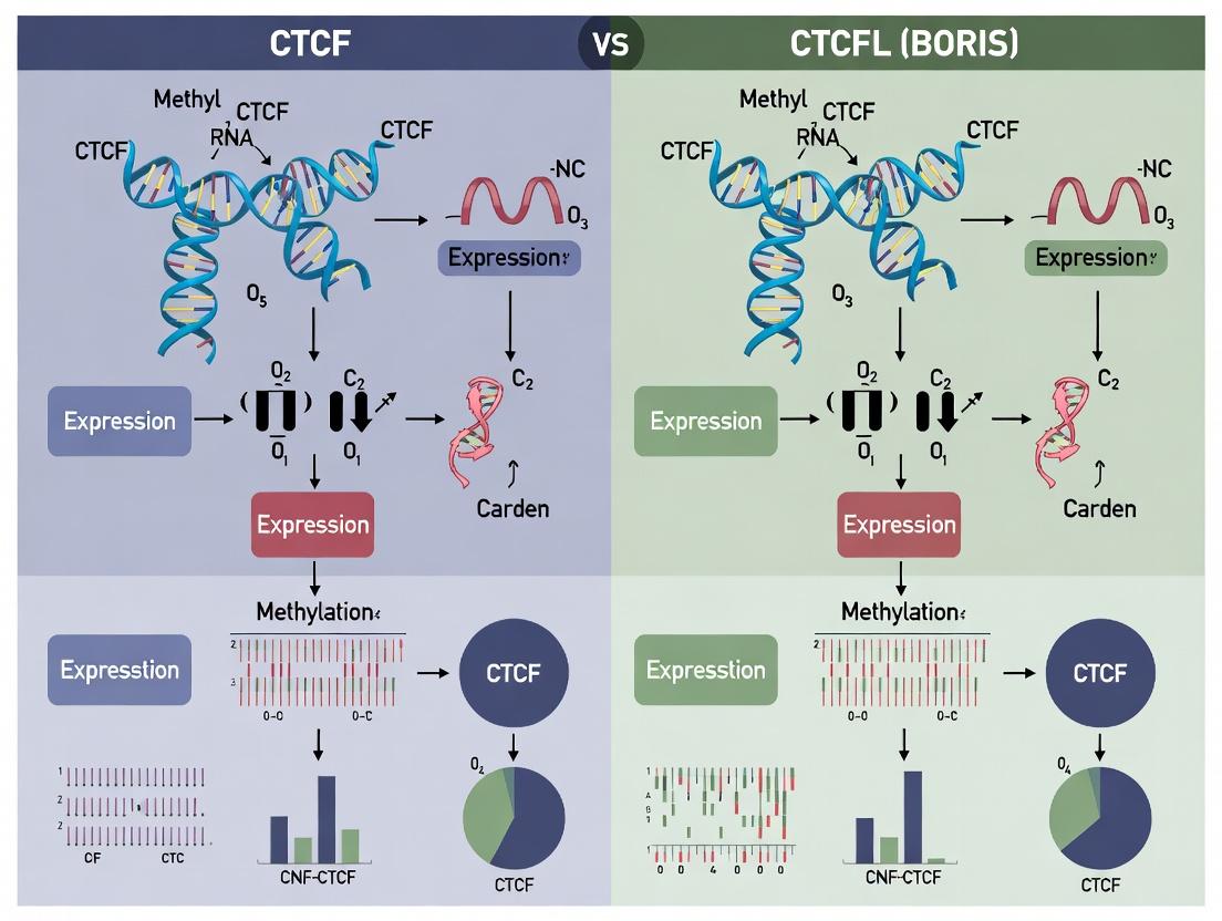
Visualizing Regulatory Pathways and Workflows
Title: CTCF vs. CTCFL Mechanism and Impact on Chromatin
Title: Integrated Experimental Workflow for CTCF/CTCFL Research
The Scientist's Toolkit: Key Research Reagent Solutions
Table 2: Essential Reagents and Tools for CTCF/CTCFL Research
| Reagent/Tool | Function/Application | Example & Notes |
|---|---|---|
| Validated Antibodies | For immunofluorescence, western blot, CUT&RUN, and ChIP. Critical for specificity given homology. | Anti-CTCF (D31H2) XP Rabbit mAb (Cell Signaling). Anti-CTCFL/BORIS (Abcam, Sigma). Validate with knockout cell lines. |
| ChIP-seq/CUT&RUN Grade Enzymes | For high-resolution mapping of protein-DNA interactions. | pA-MNase for CUT&RUN (available from commercial kits, e.g., EpiCypher). Micrococcal Nuclease or sonicator for ChIP. |
| CRISPR/Cas9 Systems | For generating knockout, knock-in (e.g., AID-tag), or transcriptional modulation cell lines. | Lentiviral sgRNA constructs (e.g., from Addgene). AID System plasmids (OsTir1, AID-tag vectors). |
| Auxin (IAA) | To induce rapid degradation of AID-tagged proteins for acute functional studies. | Dissolve in DMSO for a stock solution; use at 250-500 µM final concentration in media. |
| Next-Generation Sequencing Kits | For library preparation from ChIP, CUT&RUN, Hi-C, and RNA samples. | Illumina-compatible kits from NEBNext, KAPA, or Takara. Include spike-in controls (e.g., S. cerevisiae DNA) for normalization. |
| Bioinformatics Software | For analysis of binding, topology, and transcription data. | Peak calling: MACS2, SEACR. Hi-C analysis: HiC-Pro, Juicer, Cooler. Motif analysis: HOMER, MEME. Integration: Bedtools, R/Bioconductor (ChIPseeker, diffHiC). |
| Cell Lines | Models for CTCF (ubiquitous) and CTCFL (cancer/germ cell) studies. | CTCF essentiality: Use inducible knockout (e.g., Flp-In T-REx HeLa). CTCFL studies: Testicular germ cell tumor lines (e.g., NCCIT), breast/other cancer lines with BORIS reactivation. |
| Methylated/O-methylated DNA Probes | To test binding sensitivity to CpG methylation, a key differential feature. | Custom synthetic oligonucleotides with CpG methylation for EMSA or biotin pull-down assays. |
This technical guide explores the paralogous genes CTCF and CTCFL (BORIS), focusing on their genomic loci, protein domain architecture, and divergent biological functions. Framed within broader research on their expression and function, this whitepaper provides a comparative analysis crucial for understanding their roles in development, epigenetics, and oncogenesis.
CTCF (CCCTC-binding factor) is a ubiquitously expressed, multifunctional zinc-finger protein vital for chromatin architecture, insulation, and imprinting. Its paralog, CTCFL (CTCF-like), also known as BORIS (Brother of the Regulator of Imprinted Sites), exhibits testis-specific expression in normal somatic tissues but is aberrantly activated in numerous cancers. Derived from a common ancestral gene via duplication, their conserved domain architecture belies starkly divergent expression patterns and functional outcomes, making them a compelling model for studying paralog evolution and gene regulation.
Genomic Loci and Regulatory Context
Genomic Location and Structure
| Feature | CTCF (Gene ID: 10664) | CTCFL / BORIS (Gene ID: 140690) |
|---|---|---|
| Chromosomal Location | 16q22.1 | 20q13.31 |
| Gene Locus (GRCh38) | chr16:67,560,470-67,639,962 (reverse strand) | chr20:56,769,693-56,787,132 (forward strand) |
| Number of Exons | 15 | 16 |
| Transcript Length | ~5.8 kb (major transcript) | ~4.5 kb (major transcript) |
| CpG Island Status | Promoter associated | Promoter associated |
Epigenetic Regulation of Expression
CTCF is constitutively expressed across most cell types, maintaining essential 3D genome organization. In contrast, CTCFL expression is normally restricted to male germ cells (primarily pre-meiotic spermatogonia) due to tight epigenetic silencing in somatic tissues. This silencing involves promoter DNA methylation and repressive histone marks. In cancer, promoter hypomethylation leads to BORIS re-expression, contributing to oncogenic reprogramming.
Diagram: Epigenetic Silencing and Activation of CTCFL
Protein Domain Architecture: A Comparative Analysis
Quantitative Domain Comparison
| Protein Domain | CTCF (727 aa) | CTCFL / BORIS (644 aa) | Functional Implication |
|---|---|---|---|
| N-terminal Domain | Contains a poorly conserved region. | Unique, less conserved N-terminus. | Potential for distinct protein interactions. |
| Central 11-Zn Finger | 11 Zn fingers (ZF 1-11) highly conserved. | 11 Zn fingers with ~50-70% identity to CTCF. | Dictates DNA sequence recognition specificity. |
| Linker Regions | Specific sequences between ZFs. | Divergent linker sequences. | Influences 3D binding geometry and target selection. |
| Post-translational Modifications | Phosphorylation, PARylation, SUMOylation sites. | Different modification pattern predicted. | Alters protein stability, activity, and localization. |
| C-terminal Domain | Conserved, required for dimerization? | Truncated and divergent. | Loss of specific CTCF functions; novel functions. |
Functional Consequences of Domain Divergence
Despite similar DNA-binding capabilities, divergence in N- and C-terminal domains and linker sequences results in:
- Partial Overlap in Genomic Binding: BORIS can bind a subset of CTCF sites but also occupies unique loci, particularly hypomethylated sites in cancer.
- Altered Protein Interactome: BORIS fails to recruit key CTCF partners like cohesin, impacting its ability to form chromatin loops.
- Opposing Transcriptional Roles: CTCF often acts as an insulator; BORIS can function as a transcriptional activator of cancer-testis antigens (e.g., MAGE-A1) and oncogenes.
Diagram: Divergent Functional Outcomes of CTCF vs. BORIS Binding
Experimental Protocols for Comparative Analysis
Chromatin Immunoprecipitation Sequencing (ChIP-seq)
Purpose: To map genome-wide binding sites of CTCF and BORIS. Detailed Protocol:
- Crosslinking: Treat cells (e.g., somatic vs. cancer cell lines) with 1% formaldehyde for 10 min at room temperature. Quench with 125 mM glycine.
- Cell Lysis & Chromatin Shearing: Lyse cells and isolate nuclei. Sonicate chromatin to ~200-500 bp fragments using a Covaris sonicator (e.g., 10 cycles: 30 sec ON, 30 sec OFF, high power).
- Immunoprecipitation: Incubate sheared chromatin overnight at 4°C with:
- Anti-CTCF antibody (e.g., Millipore 07-729)
- Anti-BORIS antibody (e.g., Abcam ab56328)
- Species-matched IgG control.
- Use protein A/G magnetic beads for capture.
- Washing & Elution: Wash beads sequentially with Low Salt, High Salt, LiCl, and TE buffers. Elute complexes with elution buffer (1% SDS, 0.1M NaHCO3).
- Reverse Crosslinking & Purification: Incubate eluates with RNase A and Proteinase K. Purify DNA using a spin column kit.
- Library Prep & Sequencing: Prepare sequencing libraries using the NEBNext Ultra II DNA Library Prep Kit. Sequence on an Illumina platform (≥30 million reads/sample).
- Data Analysis: Align reads to reference genome (e.g., GRCh38). Call peaks using MACS2. Compare overlap and motif enrichment using tools like MEME-ChIP or HOMER.
Protein-Protein Interaction Analysis (Co-Immunoprecipitation)
Purpose: To identify differential interacting partners of CTCF and BORIS.
- Cell Transfection: Transfect HEK293T cells with expression vectors for FLAG-tagged CTCF and MYC-tagged BORIS.
- Cell Lysis: Harvest cells 48h post-transfection. Lyse in NP-40 lysis buffer with protease inhibitors.
- Immunoprecipitation: Incubate cleared lysate with anti-FLAG M2 magnetic beads for 2h at 4°C.
- Washing: Wash beads 3x with lysis buffer.
- Elution & Analysis: Elute proteins with 3X FLAG peptide or SDS sample buffer. Analyze by Western Blot using anti-MYC (for co-precipitated BORIS) and anti-cohesin subunit (e.g., SMC1A) antibodies.
Functional Assay: Reporter Gene Insulation Assay
Purpose: To test the insulator function of CTCF vs. BORIS.
- Reporter Construct: Use a vector with a strong enhancer (e.g., SV40) separated from a promoter (e.g., CMV) driving luciferase by a candidate CTCF/BORIS binding site.
- Transfection: Co-transfect the reporter construct with CTCF or BORIS expression plasmids (or empty vector control) into HeLa cells.
- Measurement: Harvest cells 36h later. Measure luciferase activity using a dual-luciferase reporter assay system. Normalize to Renilla luciferase control.
- Interpretation: Effective insulators (CTCF) will reduce enhancer-driven luciferase activity compared to control. BORIS is expected to show weak or no insulator activity.
The Scientist's Toolkit: Key Research Reagents
| Reagent / Material | Supplier Example (Catalog #) | Function in CTCF/CTCFL Research |
|---|---|---|
| Anti-CTCF Antibody (for ChIP) | Millipore (07-729) | Immunoprecipitates endogenous CTCF for genomic binding studies. |
| Anti-BORIS/CTCFL Antibody | Abcam (ab56328) | Detects endogenous or overexpressed BORIS in WB, IF, or ChIP. |
| Recombinant CTCF Protein | Active Motif (31489) | For in vitro EMSA to study DNA-binding specificity. |
| CTCFL (BORIS) Human cDNA ORF Clone | Origene (RC202677) | For mammalian expression and functional studies. |
| CpG Methyltransferase (M.SssI) | NEB (M0226S) | To methylate DNA probes in vitro for binding specificity assays. |
| Chromatin Shearing Enzymes (e.g., MNase) | Worthington Biochemical (LS004798) | For nucleosome mapping near CTCF/BORIS binding sites. |
| Bisulfite Conversion Kit | Zymo Research (EZ DNA Methylation-Lightning Kit) | To analyze methylation status of CTCFL promoter. |
| SMC1A Antibody | Bethyl Laboratories (A300-055A) | To probe for cohesin complex interaction in Co-IP experiments. |
Therapeutic Implications and Drug Development
The exclusive expression of BORIS in cancer and its role in epigenetic reprogramming make it a promising, albeit challenging, therapeutic target.
- Targeting Strategies: Include small molecule inhibitors of BORIS DNA-binding, degraders (PROTACs), or epigenetic drugs to re-silence its promoter.
- Immunotherapy: BORIS itself is a cancer-testis antigen, making it a potential target for cancer vaccines or adoptive T-cell therapies.
- Synthetic Lethality: Exploiting the functional antagonism between CTCF and BORIS could reveal context-dependent vulnerabilities in cancer cells.
The tale of the paralogs CTCF and CTCFL/BORIS illustrates how gene duplication, followed by divergence in regulatory loci and subtle changes in protein domain architecture, can lead to profoundly different biological functions—from guardian of the epigenome to a driver of oncogenesis. Continued comparative research is essential to unravel their complex interplay and unlock novel therapeutic avenues in cancer.
BORIS (Brother of the Regulator of Imprinted Sites, or CTCFL) is a paralog of the essential chromatin architectural protein CTCF. Its origin is traced to a gene duplication event in the ancestral therian mammal lineage. This whitepaper details the evolutionary genesis, molecular divergence, and functional divergence of CTCFL from CTCF, framing it within the critical context of their antagonistic expression and function in development and disease.
Evolutionary Genesis and Genomic Context
The CTCF gene, encoding a protein with 11 zinc fingers (ZFs), is highly conserved across bilaterian animals. BORIS/CTCFL arose from a retrotransposition-mediated duplication of the ancestral CTCF gene, followed by integration into a new genomic locus. This event is estimated to have occurred approximately 150-200 million years ago, coinciding with the divergence of therian mammals (marsupials and placentals).
Table 1: Key Genomic and Evolutionary Divergence Metrics
| Feature | CTCF | CTCFL/BORIS |
|---|---|---|
| Evolutionary Origin | Ancestral bilaterian gene | Therian-specific duplication (~150-200 MYA) |
| Conservation | Ultra-conserved across vertebrates | Rapid evolution, primate-specific isoforms |
| Genomic Locus (Human) | 16q22.1 | 20q13.31 |
| Exon Count | 10-12 (species-dependent) | 14 (human, with alternative promoters) |
| Expression Pattern | Ubiquitous in somatic cells | Normally restricted to male germline (pre-meiotic spermatocytes) |
| Epigenetic Regulation | Constitutively active promoter | Promoter regulated by dynamic DNA methylation |
Diagram 1: Evolutionary Timeline of CTCF/CTCFL Duplication
Molecular Divergence: Domain Architecture and Binding Specificity
Despite shared zinc finger domains, CTCF and BORIS exhibit critical differences in sequence and function. The central ZF domain (ZF4-7 in CTCF) responsible for core DNA binding is highly conserved, allowing recognition of similar DNA motifs. However, divergent N- and C-terminal domains mediate distinct protein partnerships.
Table 2: Quantitative Comparison of CTCF and BORIS Protein Features
| Domain/Feature | CTCF | BORIS | Functional Implication |
|---|---|---|---|
| Amino Acid Length (Human Canonical) | 727 aa | 644 aa | Altered protein interactome |
| N-terminal Domain | Acidic, phosphorylated | Glutamine-rich | Differential co-factor recruitment |
| Central Zinc Fingers (ZF) | 11 ZFs, conserved | 11 ZFs, ~70% identity | Partially overlapping DNA binding |
| Core Motif Binding (Affinity) | High (Kd ~10-50 nM) | Moderate (Kd ~2-5x higher) | Competitive displacement possible |
| C-terminal Domain | Conserved, interacts with cohesion | Highly divergent, intrinsically disordered | Loss of cohesion loading function in BORIS |
| Sumoylation Sites | Present (K74, K689) | Absent | Altered subnuclear localization & stability |
| Germline-Specific Expression | No | Yes (regulated by promoter hypomethylation) | BORIS establishes male germline epigenome |
Diagram 2: Domain Architecture and Interactome Comparison
The CTCF vs. BORIS Paradigm: Antagonistic Functions
The core thesis posits that CTCF and BORIS are antagonistic regulators of epigenetically controlled processes. CTCF maintains somatic insulator function, imprinting, and X-chromosome inactivation. BORIS, normally silenced in somatic cells, acts as a "wildcard" factor when aberrantly expressed (e.g., in cancer), outcompeting CTCF at shared targets and reprogramming the epigenome towards a germline-like, plastic state.
Key Experimental Protocol: Chromatin Immunoprecipitation Sequencing (ChIP-seq) for Binding Site Profiling
- Cell Cross-linking: Treat cells (e.g., cancer cell line with ectopic BORIS expression vs. normal somatic control) with 1% formaldehyde for 10 min at room temperature. Quench with 125 mM glycine.
- Chromatin Shearing: Sonicate lysed nuclei to yield DNA fragments of 200-500 bp. Verify fragment size by agarose gel electrophoresis.
- Immunoprecipitation: Incubate chromatin with validated, high-specificity antibodies: anti-CTCF (rabbit monoclonal, D31H2) and anti-BORIS (mouse monoclonal, 6C9). Use Protein A/G magnetic beads for capture.
- Library Prep & Sequencing: Reverse cross-links, purify DNA, and prepare sequencing libraries using a kit such as NEBNext Ultra II DNA Library Prep. Sequence on an Illumina platform to achieve >20 million aligned reads per sample.
- Bioinformatic Analysis: Align reads to reference genome (hg38). Call peaks using MACS2. Compare binding sites using diffBind to identify shared and unique loci. Annotate peaks relative to genomic features (promoters, enhancers, insulators).
Diagram 3: Antagonistic Regulation in Somatic vs. Cancer Cells
The Scientist's Toolkit: Essential Research Reagents
Table 3: Key Research Reagent Solutions for CTCF/BORIS Studies
| Reagent/Catalog # | Provider | Type | Key Application/Function |
|---|---|---|---|
| Anti-CTCF Antibody (D31H2) | Cell Signaling, #3418 | Rabbit monoclonal, validated for ChIP | Gold standard for CTCF chromatin binding studies. |
| Anti-BORIS/CTCFL Antibody (6C9) | Abcam, ab56328 | Mouse monoclonal | Most cited antibody for BORIS detection in IHC/WB. Specificity requires careful validation. |
| Recombinant Human BORIS Protein | Novus Biologicals, H00015469-P01 | Active protein | For in vitro DNA binding assays (EMSA) and competition studies with CTCF. |
| pCMV6-CTCFL (BORIS) Expression Vector | Origene, RC218017 | Mammalian expression plasmid | For ectopic BORIS overexpression in somatic cell lines. |
| CTCF & BORIS CRISPR/Cas9 Knockout Kits | Santa Cruz, sc-400776 & sc-400777 | Lentiviral particles | For complete gene knockout in cell lines to study loss-of-function phenotypes. |
| Methylated & Unmethylated BORIS Promoter Controls | MilliporeSigma, S7821 & S7822 | Bisulfite PCR controls | Essential for analyzing the methylation status of the BORIS promoter via bisulfite sequencing. |
| CTCF Motif DNA Array | Custom from ArrayIt | Spotted oligonucleotide array | High-throughput screening for binding specificity differences between CTCF and BORIS ZF domains. |
The evolutionary duplication that created BORIS yielded a potent antagonist to the conserved epigenetic guardian, CTCF. This duality is tightly regulated in normal biology but is catastrophically subverted in cancer. Understanding the precise molecular rules governing their competition—at the level of DNA binding, partner recruitment, and epigenetic regulation—offers a novel axis for therapeutic intervention. Strategies may include silencing BORIS expression, disrupting its specific protein interactions, or reinforcing CTCF function to re-establish epigenetic integrity in cancer cells.
This whitepaper provides an in-depth technical analysis of the expression and function of the paralogous proteins CTCF and BORIS (CTCFL). The central thesis posits that these proteins, despite sharing high sequence homology in their zinc finger DNA-binding domains and recognizing identical genomic sequences, exert opposing and mutually exclusive functions in cellular programming. This functional dichotomy is fundamentally established by their starkly divergent, almost antithetical, expression patterns: CTCF is ubiquitously expressed in somatic cells and is essential for viability, while BORIS expression is tightly restricted to the male germline but is aberrantly re-activated in a wide spectrum of cancers. Understanding this "yin-yang" relationship is critical for unraveling epigenetic reprogramming in gametogenesis and oncogenesis, presenting novel avenues for cancer biomarker and therapeutic development.
Quantitative Comparison of Expression Patterns
Table 1: Comparative Expression Profiles of CTCF and BORIS
| Characteristic | CTCF (CCCTC-Binding Factor) | BORIS (CTCFL; Brother of the Regulator of Imprinted Sites) |
|---|---|---|
| Primary Expression Domain | Ubiquitous in somatic nuclei; all cell lineages. | Restricted to pre-meiotic and meiotic male germ cells (spermatogonia, spermatocytes). |
| Expression in Adult Somatic Tissues | Constitutively high (Essential housekeeping function). | Undetectable (Epigenetically silenced). |
| Expression During Development | Essential; constitutive from zygote onward. Knockout is embryonic lethal. | Onset during embryonic gonad development; persists in adult testis. |
| Expression in Cancer | Often mutated or dysregulated, but rarely overexpressed. | Frequently aberrantly re-expressed (e.g., breast, lung, liver, ovarian carcinomas). |
| Regulatory Role | Master regulator of 3D chromatin architecture (insulators, TADs, loops). | Proposed role in epigenetic reprogramming, including erasure of somatic methylation marks in germ cells. |
| Cellular Essentiality | Essential for viability in somatic cells. | Non-essential for somatic cell viability; essential for normal spermatogenesis. |
Table 2: Key Molecular and Functional Distinctions
| Feature | CTCF | BORIS |
|---|---|---|
| Gene Location (Human) | 16q22.1 | 20q13.31 |
| Protein Isoforms | Multiple (∼11), varying in N- and C-termini. | Multiple, often testis-specific isoforms. |
| DNA Binding Motif | Identical core sequence to BORIS. | Identical core sequence to CTCF. |
| Binding Partners | Cohesin complex, CHD8, Nucleophosmin, YY1. | Testis-specific partners (e.g., MSH4, NSD1); can homodimerize or heterodimerize with CTCF. |
| Post-Translational Modifications | Phosphorylation, PARylation, Poly(ADP-ribosyl)ation regulate binding and insulator function. | Differential phosphorylation patterns in germ cells vs. cancer. |
| Impact on Methylation | Binds unmethylated motifs; binding can be blocked by CpG methylation. | Can bind methylated motifs; associated with loci undergoing demethylation. |
Detailed Experimental Protocols
Protocol 1: Chromatin Immunoprecipitation Sequencing (ChIP-seq) for CTCF/BORIS Binding Landscape Analysis
- Objective: To map genome-wide binding sites of CTCF and BORIS and assess overlap/competition.
- Key Reagents: Crosslinking Solution (1% formaldehyde), Cell Lysis Buffers, Magnetic Protein A/G Beads, Antibodies (Anti-CTCF [rabbit monoclonal, D31H2], Anti-BORIS [mouse monoclonal, 1G5]), RNase A, Proteinase K, PCR Purification Kit, Library Prep Kit (e.g., Illumina).
- Methodology:
- Crosslinking: Treat ~10^7 cells (somatic, germ, or cancer) with 1% formaldehyde for 10 min at RT. Quench with 125mM glycine.
- Sonication: Lyse cells and sonicate chromatin to 200-500 bp fragments using a focused ultrasonicator (e.g., Covaris).
- Immunoprecipitation: Incubate cleared chromatin with 2-5 µg of specific antibody or IgG control overnight at 4°C. Capture complexes with magnetic beads.
- Washing & Elution: Wash beads with low-salt, high-salt, LiCl, and TE buffers. Elute complexes with fresh elution buffer (1% SDS, 0.1M NaHCO3).
- Reverse Crosslinking & Purification: Incubate eluates with RNase A, then Proteinase K. Purify DNA using a spin column.
- Library Preparation & Sequencing: Prepare sequencing libraries and sequence on an Illumina platform (≥50M reads, 50bp SE).
- Analysis: Align reads to reference genome (e.g., hg38). Call peaks using MACS2. Compare peak locations, motif enrichment, and overlap using BEDTools.
Protocol 2: Quantitative Analysis of Expression by RT-qPCR and Western Blot
- Objective: To quantify mRNA and protein levels of CTCF and BORIS across cell types.
- Key Reagents: TRIzol Reagent, cDNA Synthesis Kit, SYBR Green qPCR Master Mix, Primers (CTCF: F-5’-CAGGTGGAGGAGTTTGTGCT-3’, R-5’-TTGCTGCTCCACCTTCTTCA-3’; BORIS: F-5’-AGCCACCTACAGCAACATCG-3’, R-5’-GCCTTCAGCTTGTAGGTGCT-3’; GAPDH reference), RIPA Lysis Buffer, Protease Inhibitors, SDS-PAGE Gel, Antibodies (as in Protocol 1), HRP-conjugated secondary antibodies.
- Methodology (RT-qPCR):
- Extract total RNA, treat with DNase I.
- Synthesize cDNA from 1 µg RNA using reverse transcriptase.
- Perform qPCR in triplicate with SYBR Green. Use ΔΔCt method for relative quantification against a housekeeping gene (e.g., GAPDH).
- Methodology (Western Blot):
- Lyse cells in RIPA buffer. Quantify protein via BCA assay.
- Separate 20-30 µg protein on a 4-12% Bis-Tris gel and transfer to PVDF membrane.
- Block with 5% BSA/TBST, incubate with primary antibody (CTCF 1:1000, BORIS 1:500) overnight at 4°C.
- Incubate with HRP-secondary antibody (1:5000) for 1h. Develop with ECL reagent.
Visualizations: Pathways and Experimental Workflows
Diagram Title: Normal vs. Cancer Expression & Function of CTCF/BORIS
Diagram Title: Core Experimental Workflow for CTCF/BORIS Research
The Scientist's Toolkit: Key Research Reagent Solutions
Table 3: Essential Reagents for CTCF/BORIS Functional Studies
| Reagent / Material | Provider Examples | Function in Research |
|---|---|---|
| Validated Anti-CTCF Antibody | Cell Signaling (D31H2), Abcam | Specific immunoprecipitation for ChIP, clear detection for Western Blot and IF. Crucial for distinguishing from BORIS. |
| Validated Anti-BORIS Antibody | Santa Cruz (1G5), Abcam | Detection of BORIS protein, which is often low-abundance. Specificity is paramount due to homology with CTCF. |
| CTCF/BORIS DNA Binding Motif Oligos | Custom synthesis (IDT) | For EMSA experiments to validate direct binding and study competition between paralogs. |
| Recombinant CTCF & BORIS Proteins | Active Motif, Abnova | For in vitro biochemical assays (EMSA, SELEX) to study DNA-binding specificity without cellular confounding factors. |
| Methylated & Unmethylated DNA Probes | Custom synthesis | To test the hypothesis that BORIS binds methylated motifs, while CTCF binding is methylation-sensitive. |
| CRISPR/Cas9 Knockout Kits (CTCF/BORIS) | Synthego, Horizon Discovery | To generate isogenic cell lines lacking either factor and study consequent transcriptional and architectural changes. |
| BORIS-Promoter Reporter Vectors | Addgene, custom construction | To study the epigenetic regulation (methylation status) of the BORIS gene itself in different cell types. |
| Chromatin Conformation Capture Kit (Hi-C) | Arima Genomics, Dovetail | To investigate how ectopic BORIS expression in cancer alters CTCF-mediated 3D genome organization (TADs, loops). |
The functional antagonism between CTCF and its paralog BORIS (CTCFL) is a pivotal axis in epigenetic regulation. CTCF, a ubiquitously expressed insulator protein, is crucial for genomic imprinting, chromatin looping, and transcriptional regulation. In contrast, BORIS expression is typically restricted to the male germline, where it is involved in epigenetic reprogramming. The broader thesis posits that the aberrant reactivation of BORIS in somatic tissues represents a fundamental epigenetic switch that drives oncogenesis by subverting normal CTCF-mediated genome organization and gene expression programs. This whitepaper details the mechanisms, consequences, and experimental investigation of this switch.
Mechanisms of BORIS Reactivation and Functional Impact
Epigenetic Derepression
BORIS reactivation in cancers is primarily driven by promoter demethylation. The germline-specific promoter of CTCFL is heavily methylated and silenced in somatic cells. In various carcinomas, this promoter undergoes specific demethylation, often linked to dysregulation of DNA methyltransferases (DNMTs) and Ten-Eleven Translocation (TET) enzymes.
Disruption of CTCF/Cohesin Biology
BORIS shares DNA binding specificity with CTCF, allowing it to occupy a subset of CTCF binding sites (CBS). However, BORIS lacks the N-terminal domain required for cohesin interaction. This results in:
- Competitive Binding: BORIS displaces CTCF at specific loci, dissolving insulator function and allowing aberrant enhancer-promoter interactions.
- Loss of Chromatin Looping: Failure to recruit cohesin leads to the collapse of Topologically Associating Domains (TADs), causing oncogene activation and tumor suppressor silencing.
Transcriptional and Phenotypic Consequences
BORIS acts as a transcriptional regulator, often activating cancer-testis antigens (CTAs) and proto-oncogenes (e.g., MYC). It promotes hallmark cancer phenotypes including stemness, proliferation, metastasis, and therapy resistance.
Table 1: Quantitative Data on BORIS Expression in Human Cancers
| Cancer Type | Frequency of BORIS Reactivation (%) | Primary Mechanism | Correlation with Prognosis | Key Co-activated Pathways |
|---|---|---|---|---|
| Non-Small Cell Lung Cancer (NSCLC) | 40-60% | Promoter hypomethylation | Poor overall survival | MYC, EZH2, EMT |
| Triple-Negative Breast Cancer (TNBC) | 50-70% | Histone modification (H3K4me3 gain) | Reduced disease-free survival | Stemness markers (OCT4, NANOG) |
| Hepatocellular Carcinoma (HCC) | ~55% | Promoter hypomethylation & LSH downregulation | Shorter recurrence time | IGF2, PI3K/AKT |
| Glioblastoma | 30-40% | Focal DNA hypomethylation | Advanced tumor grade | PDGFR, MGMT |
| Ovarian Cancer | ~45% | Unknown epigenetic driver | Chemoresistance | MAGE-A family, BCL2 |
Experimental Protocols for Key Investigations
Protocol: Assessing BORIS Promoter Methylation Status (Bisulfite Sequencing)
Objective: To quantify CpG methylation density in the CTCFL promoter.
- Genomic DNA Isolation: Extract DNA from tumor and matched normal tissue using a silica-column based kit.
- Bisulfite Conversion: Treat 500ng DNA with sodium bisulfite (e.g., EZ DNA Methylation-Lightning Kit). This converts unmethylated cytosine to uracil, while methylated cytosine remains unchanged.
- PCR Amplification: Design primers specific to the bisulfite-converted sequence of the CTCFL promoter core region. Perform touchdown PCR.
- Cloning & Sequencing: Purify PCR product, clone into a T-vector, and transform competent E. coli. Pick 10-15 colonies for Sanger sequencing.
- Analysis: Align sequences to reference. Calculate percentage methylation per CpG site across all clones. Compare tumor vs. normal.
Protocol: Chromatin Immunoprecipitation (ChIP) for BORIS/CTCF Binding Dynamics
Objective: To map genome-wide occupancy of BORIS and CTCF.
- Crosslinking & Cell Lysis: Crosslink cells (e.g., cancer cell line) with 1% formaldehyde for 10 min. Quench with glycine. Lyse cells and isolate nuclei.
- Chromatin Shearing: Sonicate chromatin to an average fragment size of 200-500 bp. Verify fragment size by agarose gel electrophoresis.
- Immunoprecipitation: Incubate chromatin aliquots overnight at 4°C with specific antibodies: anti-BORIS (e.g., Abcam ab121), anti-CTCF (positive control), and IgG (negative control). Use protein A/G magnetic beads for capture.
- Washing & Elution: Wash beads sequentially with low salt, high salt, LiCl, and TE buffers. Elute complexes in freshly prepared elution buffer (1% SDS, 0.1M NaHCO3).
- Reverse Crosslinking & DNA Purification: Add NaCl to eluates and incubate at 65°C overnight. Treat with Proteinase K, then purify DNA with spin columns.
- Analysis: Analyze enriched DNA by qPCR for specific loci or submit for next-generation sequencing (ChIP-seq). Use peak-calling software (e.g., MACS2) and compare binding profiles.
Protocol: Functional Assessment via siRNA Knockdown
Objective: To determine phenotypic consequences of BORIS loss.
- Cell Seeding: Seed cancer cells (confirmed BORIS+) in 6-well plates at 30-40% confluence 24h prior.
- Transfection: Transfect with 50nM ON-TARGETplus Human CTCFL siRNA SMARTpool or non-targeting siRNA control using lipid-based transfection reagent. Include a fluorescently-labeled siRNA to monitor efficiency.
- Harvesting: Harvest cells at 72h and 96h post-transfection.
- Validation & Assays:
- RNA: Extract total RNA, perform cDNA synthesis, and conduct qRT-PCR for CTCFL and target genes (e.g., MYC).
- Protein: Perform western blot with anti-BORIS antibody.
- Phenotype: Perform assays for proliferation (MTS), apoptosis (Annexin V), and invasion (Matrigel).
Visualization of Core Concepts
Title: The BORIS Reactivation Pathway in Oncogenesis
Title: Integrated Experimental Workflow for BORIS Research
The Scientist's Toolkit: Key Research Reagent Solutions
Table 2: Essential Reagents for BORIS/CTCFL Research
| Reagent Category | Specific Product/Assay | Function & Application in BORIS Research |
|---|---|---|
| Antibodies | Anti-BORIS/CTCFL (e.g., Rabbit monoclonal [EPR20029]) | Detection of BORIS protein in western blot, immunofluorescence, and Chromatin Immunoprecipitation (ChIP). |
| Antibodies | Anti-CTCF (e.g., Mouse monoclonal [E-8]) | Control for comparative binding studies and assessment of CTCF displacement. |
| DNA Methylation Analysis | EZ DNA Methylation-Lightning Kit | Rapid bisulfite conversion of genomic DNA for subsequent methylation-specific PCR (MSP) or bisulfite sequencing. |
| DNA Methylation Analysis | Methyl Primer Express Software | Design of primers for MSP and bisulfite sequencing assays targeting the CTCFL promoter CpG island. |
| Chromatin Analysis | Magna ChIP Protein A/G Magnetic Beads | Efficient pull-down of antibody-chromatin complexes for ChIP experiments. |
| Chromatin Analysis | SimpleChIP Enzymatic Chromatin IP Kit | Provides optimized reagents for chromatin preparation, digestion, and IP for BORIS/CTCF ChIP. |
| Gene Silencing | ON-TARGETplus Human CTCFL siRNA SMARTpool | A pool of 4 siRNAs for specific and effective knockdown of BORIS mRNA in functional studies. |
| Gene Silencing | DharmaFECT Transfection Reagents | High-efficiency delivery of siRNA into a wide range of cancer cell lines. |
| Expression Analysis | TaqMan Gene Expression Assay for CTCFL (Hs00218986_m1) | Precise, probe-based quantification of BORIS mRNA levels by qRT-PCR. |
| Expression Analysis | PrimeTime qPCR Assay for CTCFL | Customizable probe-based assay for BORIS expression quantification. |
| Cell Phenotyping Cell Counting Kit-8 (CCK-8) | Sensitive colorimetric assay to measure cell proliferation after BORIS modulation. | |
| Cell Phenotyping Corning Matrigel Invasion Chamber | Standardized assay to assess changes in invasive potential upon BORIS knockdown/overexpression. |
Within the broader thesis of CTCF versus CTCFL (BORIS) expression and function, a central focus is the intricate regulatory network governing these paralogous proteins. While CTCF is ubiquitously expressed and essential for chromatin architecture, CTCFL/BORIS expression is normally restricted to the male germline but is aberrantly reactivated in numerous cancers. This whitepaper provides an in-depth technical analysis of the transcriptional and post-translational mechanisms controlling their expression and activity, which represent critical points of divergence in their biological functions and potential therapeutic targeting.
Transcriptional Regulation
Promoter Architecture and Epigenetic Control
CTCF and BORIS promoters exhibit distinct epigenetic landscapes dictating their cell-type-specific expression.
Table 1: Core Promoter and Epigenetic Features of CTCF vs. BORIS
| Feature | CTCF (Ubiquitous Expression) | CTCFL/BORIS (Testis-Specific/Cancer) |
|---|---|---|
| CpG Island | Present, mostly unmethylated | Present, methylated in somatic cells |
| Histone Modifications (Somatic) | H3K4me3 (Active), H3K27ac (Active) | H3K9me3 (Repressive), H3K27me3 (Facultative heterochromatin) |
| Histone Modifications (Germline/Cancer) | Maintained active | H3K4me3, H3K27ac, loss of repressive marks |
| Primary Transcriptional Regulators | SP1, E2F, CREB, MYC (Maintainers) | MYC, CREB, E2F (in cancer); repression by PRC2 in somatic cells |
| Enhancer Engagement | Constitutive interaction with active enhancers | Germline/cancer-specific super-enhancer interaction |
The BORIS promoter is silenced in somatic cells via polycomb repressive complex 2 (PRC2)-mediated H3K27 trimethylation and DNA methyltransferase activity. In cancer, demethylation of specific CpG sites (e.g., -200 bp relative to TSS) correlates with reactivation. CTCF's promoter is protected from methylation, potentially by its own binding in an auto-regulatory loop.
Key Transcriptional Factors and Signaling Pathways
Pathways converging on the CTCF and CTCFL promoters integrate cellular state signals.
- MYC Oncogene: Binds and transactivates both CTCF and CTCFL promoters. In cancer, elevated MYC disproportionately upregulates BORIS, contributing to its ectopic expression.
- CREB Signaling: Activated by PKA, PKC, or MAPK pathways in response to growth signals. Phosphorylated CREB recruits CBP/p300 to cAMP response elements (CREs) in both promoters.
- E2F Family: E2F1, often released upon RB1 phosphorylation, binds and activates both genes, linking cell cycle progression to their expression.
- Epigenetic Modulators: The PRC2 component EZH2 directly represses CTCFL in somatic cells. Inhibitors of EZH2 or DNA methyltransferases (DNMTs) can de-repress BORIS.
Experimental Protocol: Chromatin Immunoprecipitation (ChIP) for Promoter Analysis
Aim: To assess transcription factor binding and histone modifications at CTCF/BORIS promoters.
Methodology:
- Cross-linking: Treat ~1x10^7 cells with 1% formaldehyde for 10 min at room temperature. Quench with 125mM glycine.
- Cell Lysis & Sonication: Lyse cells in SDS lysis buffer. Sonicate chromatin to 200-500 bp fragments. Confirm fragment size by agarose gel electrophoresis.
- Immunoprecipitation: Dilute sonicated lysate in ChIP dilution buffer. Pre-clear with protein A/G beads. Incubate overnight at 4°C with 2-5 µg of specific antibody (e.g., anti-MYC, anti-H3K27ac, anti-H3K27me3) or IgG control.
- Bead Capture & Washes: Add beads, incubate, then wash sequentially with low salt, high salt, LiCl, and TE buffers.
- Elution & Reverse Cross-link: Elute complexes in fresh elution buffer (1% SDS, 0.1M NaHCO3). Add NaCl to 200mM and reverse cross-link at 65°C for 4+ hours.
- DNA Purification: Treat with RNase A and Proteinase K. Purify DNA using phenol-chloroform extraction or spin columns.
- Analysis: Quantify target promoter regions via qPCR using specific primers. Express data as % input or fold enrichment over IgG.
Post-Translational Modifications (PTMs) and Protein Stability
PTMs provide rapid, dynamic control over protein function, stability, and localization.
Table 2: Documented Post-Translational Modifications of CTCF and BORIS
| Modification | Site (Example) | Enzyme (Putative) | Functional Consequence |
|---|---|---|---|
| Poly(ADP-ribosyl)ation | CTCF: Multiple | PARP1 | Chromatin dissociation, insulator function regulation. |
| Phosphorylation | CTCF: S224, S365, T374; BORIS: Similar motifs | CK2, PKC, PLK1 | Modulates DNA binding affinity, zinc finger occupancy, and protein-protein interactions. |
| Ubiquitination | CTCF: K74, K689; BORIS: Predicted sites | Unknown E3 Ligase (e.g., MDM2?) | Targets for proteasomal degradation. Regulated by phosphorylation state. |
| Sumoylation | CTCF: K74, K689 (same as Ub) | UBC9, PIAS family | Antagonizes ubiquitination, stabilizes protein, may alter transcriptional output. |
| O-GlcNAcylation | CTCF/BORIS: Ser/Thr residues | OGT | Nutrient-sensing modification; competes with phosphorylation; affects stability. |
Impact on Function and Stability
Phosphorylation at specific residues (e.g., by CK2) can enhance DNA binding, while other modifications promote degradation. The balance between ubiquitination (destructive) and sumoylation (protective) at shared lysine residues is a critical regulatory node. O-GlcNAcylation, responsive to cellular metabolism, provides a link between nutrient status and chromatin regulation.
Experimental Protocol: Co-Immunoprecipitation (Co-IP) to Detect PTM Enzymes
Aim: To identify interacting enzymes (kinases, ubiquitin ligases) that modify CTCF/BORIS.
Methodology:
- Cell Lysis: Lyse cells in non-denaturing IP lysis buffer (e.g., 25mM Tris pH 7.4, 150mM NaCl, 1% NP-40, plus protease/phosphatase inhibitors) for 30 min on ice.
- Pre-clearing: Centrifuge lysate. Pre-clear supernatant with species-matched control IgG and protein A/G beads for 1 hour at 4°C.
- Immunoprecipitation: Incubate pre-cleared lysate with antibody against CTCF or BORIS overnight at 4°C. Use IgG for control.
- Bead Capture: Add protein A/G beads for 2-4 hours at 4°C.
- Washes: Pellet beads and wash 3-5 times with ice-cold lysis buffer.
- Elution: Elute bound proteins by boiling beads in 2X Laemmli SDS-PAGE sample buffer.
- Analysis: Resolve proteins by SDS-PAGE. Perform western blotting with antibodies against suspected interactors (e.g., Anti-PARP1, Anti-CK2, Anti-Ubiquitin, Anti-SUMO1).
The Scientist's Toolkit: Key Research Reagent Solutions
Table 3: Essential Reagents for CTCF/BORIS Regulation Studies
| Reagent | Function/Application | Example/Supplier (Illustrative) |
|---|---|---|
| Anti-CTCF Antibody (ChIP-grade) | Chromatin immunoprecipitation, immunofluorescence, western blot. | MilliporeSigma 07-729, Abcam ab188408. |
| Anti-BORIS/CTCFL Antibody | Specific detection of BORIS protein; critical for distinguishing from CTCF. | Abcam ab167161, Active Motif 61375. |
| DNMT Inhibitor (5-Azacytidine) | DNA demethylating agent; used to probe epigenetic repression of CTCFL. | Sigma-Aldrich A2385. |
| EZH2 (PRC2) Inhibitor (GSK343, EPZ-6438) | Inhibits H3K27 methylation; tests PRC2-mediated repression of BORIS. | Selleckchem S7164, Selleckchem S7128. |
| PARP Inhibitor (Olaparib) | Inhibits poly(ADP-ribosyl)ation; used to study this PTM's role in CTCF function. | Selleckchem S1060. |
| Proteasome Inhibitor (MG-132) | Blocks degradation of ubiquitinated proteins; stabilizes CTCF/BORIS for PTM studies. | Sigma-Aldrich C2211. |
| Site-specific Phospho-CTCF Antibodies | Detects phosphorylation at specific residues (e.g., pS224, pT374). | In-house or custom from suppliers. |
| Recombinant CTCF Zinc Finger Array | For in vitro DNA binding assays (EMSA) to test impact of PTMs. | Active Motif 31497. |
| CTCF/BORIS Promoter Reporter Plasmids | Luciferase constructs to assay transcriptional activity of promoters. | Addgene, custom clones. |
| Bisulfite Conversion Kit | Analyzes DNA methylation status of CTCFL promoter CpG island. | Zymo Research EZ DNA Methylation kits. |
Visualization of Regulatory Networks
Title: Transcriptional Control of CTCF and BORIS in Different Cellular Contexts
Title: Post-Translational Modification Network Regulating CTCF/BORIS Stability and Function
Title: Chromatin Immunoprecipitation (ChIP) Experimental Workflow
Detecting and Manipulating CTCF/BORIS: Key Techniques and Research Applications
In the study of paralogous transcription factors CTCF and its testis-specific counterpart CTCFL (BORIS), precise nucleic acid-based detection is paramount. Discerning their expression patterns—where CTCF is broadly expressed and essential for chromatin architecture, while CTCFL is normally restricted to germ cells but aberrantly activated in cancers—requires robust, sensitive, and spatially resolved techniques. This guide details the core methodologies of quantitative PCR (qPCR), RNA Sequencing (RNA-Seq), and In Situ Hybridization (ISH) as applied to this critical research axis, providing a technical framework for investigating their distinct and overlapping functions in development and disease.
Quantitative PCR (qPCR)
qPCR remains the gold standard for quantifying gene expression levels of CTCF and CTCFL due to its sensitivity, specificity, and throughput.
Experimental Protocol: Two-Step RT-qPCR for CTCF/CTCFL Expression Profiling
- RNA Isolation & QC: Extract total RNA from cells/tissues (e.g., somatic cells, testis, cancer cell lines) using a column-based kit with DNase I treatment. Assess RNA integrity (RIN > 8.0) and purity (A260/A280 ~2.0) using a bioanalyzer or spectrophotometer.
- cDNA Synthesis: Using 1 µg of total RNA, perform reverse transcription with random hexamers and a high-fidelity reverse transcriptase. Include a no-reverse transcriptase (-RT) control for each sample to detect genomic DNA contamination.
- qPCR Assay Design: Design TaqMan probes or SYBR Green primers targeting unique genomic regions of CTCF (e.g., exon 3-4 junction) and CTCFL (e.g., a sequence within its exon 1 variant). Validate primer efficiency (90-110%) using a standard curve.
- qPCR Run: Prepare reactions in triplicate using a master mix containing cDNA, primers/probe, and enzyme. Run on a real-time cycler with standard cycling conditions (e.g., 95°C for 20 sec, followed by 40 cycles of 95°C for 1 sec and 60°C for 20 sec).
- Data Analysis: Calculate ∆Ct values relative to multiple reference genes (e.g., GAPDH, HPRT1). Use the comparative ∆∆Ct method to determine fold-change differences in expression between sample groups.
Table 1: Representative qPCR Data for CTCF vs. CTCFL Expression
| Sample Type | CTCF Mean Ct (±SD) | CTCFL Mean Ct (±SD) | ∆Ct (CTCFL-CTCF) | Relative CTCFL Abundance |
|---|---|---|---|---|
| Normal Somatic | 22.1 (±0.3) | Undetected (Ct > 35) | >12.9 | Negligible |
| Testis | 23.5 (±0.4) | 28.7 (±0.5) | 5.2 | ~3% of CTCF |
| Cancer Cell Line | 21.8 (±0.2) | 24.2 (±0.6) | 2.4 | ~20% of CTCF |
RNA Sequencing (RNA-Seq)
RNA-Seq provides an unbiased, genome-wide view of transcription, enabling the discovery of CTCFL-induced expression programs and alternative splicing events not detectable by qPCR.
Experimental Protocol: Stranded mRNA-Seq Workflow
- Library Preparation: Starting with 100-1000 ng of high-quality total RNA, enrich poly-A tailed mRNA using oligo-dT beads. Fragment the eluted mRNA and generate stranded cDNA libraries using dUTP-based second strand marking. Add unique dual-indexed adapters for multiplexing.
- Sequencing: Pool libraries and sequence on a platform such as Illumina NovaSeq to a depth of 25-40 million paired-end (150 bp) reads per sample for mammalian genomes.
- Bioinformatic Analysis:
- Alignment: Use a splice-aware aligner (e.g., STAR) to map reads to the human reference genome (GRCh38).
- Quantification: Count reads aligned to gene features (e.g., CTCF, CTCFL isoforms) using tools like featureCounts.
- Differential Expression: Use R/Bioconductor packages (DESeq2, edgeR) to statistically identify genes and isoforms differentially expressed upon CTCFL induction versus CTCF knockout.
- Pathway Analysis: Perform Gene Set Enrichment Analysis (GSEA) on ranked gene lists to identify pathways perturbed by CTCFL.
Table 2: Key RNA-Seq Metrics for CTCF/CTCFL Studies
| Metric | Typical Target Value | Relevance to CTCF/L Study |
|---|---|---|
| Total Reads per Sample | 25-40 million | Ensures detection of low-abundance transcripts like CTCFL. |
| % Aligned Reads | >90% | Indicates sample and library quality. |
| % Duplicate Reads | <20% (varies) | High duplication may indicate low input or PCR bias. |
| CTCF Isoform Diversity | 2-4 major isoforms | RNA-Seq reveals tissue-specific isoform usage. |
| CTCFL-Positive vs. Negative (DEGs) | Hundreds to thousands | Identifies the oncogenic gene network activated by CTCFL. |
In SituHybridization (ISH)
ISH, particularly RNAscope, provides spatial context, revealing CTCF and CTCFL mRNA localization within complex tissues like tumors or testis.
Experimental Protocol: RNAscope Assay on Formalin-Fixed Paraffin-Embedded (FFPE) Tissue
- Sample Preparation: Cut 5 µm sections from FFPE tissue blocks (e.g., normal testis, carcinoma). Bake slides at 60°C for 1 hour.
- Pretreatment: Deparaffinize in xylene, dehydrate in ethanol, and then perform target retrieval by heating in a specific buffer. Apply protease digest to permeabilize tissue.
- Hybridization: Apply target-specific probe pairs (ZZ probes) designed against CTCF or CTCFL mRNA. Incubate at 40°C in a hybridization oven for 2 hours.
- Signal Amplification: Perform a series of sequential amplifier applications (AMP1-AMP6) that build a branching structure only if the ZZ probes are bound. This provides high specificity and single-molecule sensitivity.
- Detection & Counterstaining: Apply chromogenic substrate (e.g., Fast Red) and counterstain with hematoxylin. Coverslip and image under a brightfield microscope.
- Analysis: Score signal as dots per cell within specific tissue compartments (e.g., seminiferous tubules, tumor nests, stroma).
The Scientist's Toolkit: Key Reagents & Materials
Table 3: Essential Research Reagents for CTCF/CTCFL Detection
| Item | Function & Application | Example/Note |
|---|---|---|
| High-Fidelity Reverse Transcriptase | Converts RNA to cDNA for qPCR/RNA-Seq with high accuracy and yield. | SuperScript IV |
| TaqMan Gene Expression Assays | Pre-validated primer/probe sets for specific, sensitive qPCR of CTCF or CTCFL. | Hs00914034_m1 (CTCF) |
| RNAscope Target Probe | Specially designed ZZ probe set for single-molecule RNA FISH; crucial for distinguishing highly homologous targets. | Probe-Hs-CTCFL-C1 |
| Ribonuclease Inhibitor | Protects RNA templates from degradation during all handling steps. | Recombinant RNase Inhibitor |
| Stranded mRNA-Seq Library Prep Kit | For generating sequencing libraries that preserve strand-of-origin information. | Illumina Stranded mRNA Prep |
| CTCF/CTCFL Specific Antibodies | For parallel protein-level validation (ChIP, Western, IF). Not for nucleic acid detection but essential for integrated studies. | Millipore 07-729 (CTCF), Abcam ab187143 (CTCFL/BORIS) |
| DNase I, RNase-free | Removes contaminating genomic DNA from RNA preps prior to sensitive assays. | |
| Dual-Indexing Adapter Kit | Allows multiplexing of many RNA-Seq libraries, critical for cohort studies. | IDT for Illumina UD Indexes |
Visualizing Experimental Workflows and Biological Relationships
Workflow for CTCF/L Expression Analysis
CTCF vs CTCFL Functional Competition Model
Within the expanding field of chromatin architecture and gene regulation, the functional antagonism between CTCF, the universal chromatin organizer, and its paralog CTCFL (BORIS), a testis-specific protein aberrantly expressed in cancers, is a critical area of investigation. This technical guide details the core protein-level methodologies essential for dissecting their expression dynamics, subcellular localization, and functional interplay in normal and pathological contexts.
Experimental Protocols for CTCF/CTCFL Analysis
Western Blot Protocol for Quantifying CTCF vs. CTCFL Expression
This protocol is optimized to distinguish the similar molecular weights of CTCF (~82 kDa) and CTCFL/BORIS (~75 kDa).
Detailed Methodology:
- Sample Preparation: Lyse cells or tissues in RIPA buffer supplemented with protease and phosphatase inhibitors. Quantify protein concentration using a BCA assay.
- Gel Electrophoresis: Load 20-30 µg of total protein per lane on a 4-12% Bis-Tris polyacrylamide gel. Include a pre-stained protein ladder. Run at 150V for ~60 minutes in 1X MOPS buffer.
- Transfer: Perform wet transfer to a PVDF membrane at 100V for 70 minutes at 4°C in Tris-Glycine buffer with 20% methanol.
- Blocking and Antibody Incubation: Block membrane in 5% non-fat milk in TBST for 1 hour. Incubate with primary antibody overnight at 4°C with gentle agitation.
- Primary Antibodies: Mouse anti-CTCF (1:1000, clone D31H2), Rabbit anti-CTCFL/BORIS (1:800, polyclonal), Mouse anti-β-Actin (1:5000, loading control).
- Washing and Detection: Wash 3x with TBST, incubate with appropriate HRP-conjugated secondary antibody (1:3000) for 1 hour at RT. Wash again and develop using enhanced chemiluminescence (ECL) substrate. Image on a chemiluminescent imager.
Immunofluorescence Protocol for Subcellular Localization
This protocol visualizes the nuclear distribution of CTCF and CTCFL, which may exhibit distinct speckling patterns.
Detailed Methodology:
- Cell Culture and Fixation: Seed cells on glass coverslips in a 24-well plate. At 70% confluence, wash with PBS and fix with 4% paraformaldehyde for 15 minutes at RT.
- Permeabilization and Blocking: Permeabilize with 0.2% Triton X-100 in PBS for 10 minutes. Block in 3% BSA in PBS for 1 hour at RT.
- Antibody Staining: Incubate with primary antibodies (CTCF and/or CTCFL, same as above) diluted in blocking buffer for 2 hours at RT or overnight at 4°C.
- Detection and Mounting: Wash 3x with PBS, incubate with fluorophore-conjugated secondary antibodies (e.g., Alexa Fluor 488 and 594) for 1 hour at RT in the dark. Stain nuclei with DAPI (300 nM) for 5 minutes. Mount coverslips using anti-fade mounting medium.
- Imaging: Acquire images using a confocal microscope with sequential laser scanning to avoid bleed-through. Analyze co-localization using software like ImageJ or Imaris.
Immunohistochemistry Protocol for Tissue-Level Expression
IHC is crucial for mapping CTCF and CTCFL expression in normal testis versus tumor tissue microarrays (TMAs).
Detailed Methodology (Automated Stainer):
- Tissue Sectioning and Deparaffinization: Cut formalin-fixed, paraffin-embedded (FFPE) tissue sections at 4µm. Bake slides at 60°C for 30 minutes. Deparaffinize in xylene and rehydrate through graded ethanol to water.
- Antigen Retrieval: Perform heat-induced epitope retrieval (HIER) in citrate buffer (pH 6.0) or Tris-EDTA buffer (pH 9.0) using a pressure cooker or steamer for 20 minutes.
- Endogenous Peroxidase Blocking: Quench endogenous peroxidase activity with 3% H₂O₂ in methanol for 10 minutes.
- Primary Antibody Incubation: Apply protein block (5% normal serum) for 10 minutes. Incubate with anti-CTCF or anti-CTCFL primary antibody for 60 minutes at RT.
- Detection: Apply a labeled polymer-HRP secondary antibody (e.g., EnVision+ system) for 30 minutes. Visualize with 3,3’-Diaminobenzidine (DAB) chromogen for 5-10 minutes.
- Counterstaining and Mounting: Counterstain with hematoxylin, dehydrate, clear, and mount with a permanent mounting medium. Score staining intensity (0-3+) and percentage of positive nuclei.
Table 1: Comparative Analysis of CTCF and CTCFL/BORIS Protein Characteristics & Detection
| Parameter | CTCF | CTCFL/BORIS | Analytical Implication |
|---|---|---|---|
| Molecular Weight | ~82 kDa | ~75 kDa | Requires high-resolution gels for separation. |
| Expression Pattern | Ubiquitous in somatic cells. | Restricted: normal testis, aberrant in cancers. | IHC requires distinct tissue controls. |
| Nuclear Localization | Diffuse/speckled pattern. | Often strong, focal nuclear speckles. | IF colocalization studies are complex. |
| Common Antibody Clones | D31H2 (C-terminal), 7C11C (N-terminal) | Polyclonal sera, EPR23177-78 (monoclonal) | Specificity validation via siRNA knockdown is critical. |
| Typical Band Intensity (WB) in Cancer Cell Lines | High, consistent. | Variable: absent to very high. | Normalize to a housekeeping protein (β-Actin, GAPDH). |
Table 2: Optimal Conditions for Key Antibodies in CTCF/CTCFL Protein Assays
| Assay | Target | Antibody (Example) | Dilution | Antigen Retrieval | Key Validation Step |
|---|---|---|---|---|---|
| Western Blot | CTCF | Mouse monoclonal [D31H2] | 1:1000 | N/A | Knockdown shows loss of ~82 kDa band. |
| Western Blot | CTCFL | Rabbit polyclonal [Abcam ab187143] | 1:800 | N/A | Express in CTCFL-negative cell line. |
| Immunofluorescence | CTCF | Same as WB | 1:250 | 0.1% Triton X-100 | Nuclear pattern lost with knockout. |
| Immunohistochemistry | CTCFL | Rabbit monoclonal [EPR23177-78] | 1:200 | Tris-EDTA, pH 9.0 | Staining only in testis germ cells (positive control). |
Visualizing CTCF/CTCFL Regulatory Pathways and Workflows
Protein Analysis Workflow for CTCF/CTCFL Research
CTCF/CTCFL Antagonism in Gene Regulation
The Scientist's Toolkit: Research Reagent Solutions
Table 3: Essential Materials for CTCF/CTCFL Protein Analysis
| Reagent/Material | Function & Specification | Example Product/Catalog # |
|---|---|---|
| Anti-CTCF Antibody | Detects the ubiquitous CTCF protein for WB, IF, ChIP. High specificity for C-terminus is crucial. | Cell Signaling Technology, #3418 (D31H2). |
| Anti-CTCFL/BORIS Antibody | Specifically detects the paralog CTCFL; validation in testis lysate is recommended. | Abcam, ab187143 (polyclonal). |
| Phosphatase/Protease Inhibitor Cocktail | Preserves post-translational modification states critical for CTCF function. | Sigma-Aldrich, PhosSTOP & cOmplete. |
| High-Sensitivity ECL Substrate | Detects low-abundance CTCFL protein in early-stage cancer cell lines. | Cytiva, Amersham ECL Prime. |
| Fluorophore-Conjugated Secondaries (IF) | For dual-color IF co-localization of CTCF and CTCFL. Minimal cross-reactivity. | Invitrogen, Alexa Fluor 488 and 594. |
| DAB Chromogen Kit (IHC) | For permanent, high-contrast visualization of protein in FFPE tissues. | Agilent, Dako DAB+ Substrate System. |
| CTCF/CTCFL Positive Control Lysates | Essential antibody validation. CTCF: HeLa cell lysate. CTCFL: Testis or NTERA-2 cell lysate. | Santa Cruz Biotechnology, sc-2477 (HeLa), sc-2478 (Testis). |
| BORIS/CTCFL siRNA | Functional validation of antibody specificity via knockdown in aberrant cells. | Horizon Discovery, SMARTpool ON-TARGETplus. |
The functional divergence between CTCF and its paralog, CTCFL (BORIS), represents a critical frontier in epigenetics and oncology. While CTCF is a ubiquitously expressed architectural protein essential for chromatin insulation and imprinting, CTCFL expression is normally restricted to the testis but is aberrantly activated in various cancers. Mapping their genome-wide binding landscapes via ChIP-Seq is fundamental to dissecting their overlapping and unique roles in gene regulation, chromatin organization, and oncogenesis. This guide details the technical application of ChIP-Seq within this specific research framework.
Core Principle and Workflow of ChIP-Seq
Chromatin Immunoprecipitation followed by sequencing (ChIP-Seq) isolates DNA fragments bound by a protein of interest (e.g., CTCF or CTCFL) and identifies their genomic locations via high-throughput sequencing.
Diagram 1: ChIP-Seq Core Workflow
Detailed Experimental Protocol for CTCF/CTCFL ChIP-Seq
Key Considerations: Use appropriate cell models (e.g., somatic cells for CTCF, cancer/testis cells for CTCFL). Employ highly specific, validated antibodies.
Protocol Steps:
- Cell Fixation: Treat ~10^7 cells with 1% formaldehyde for 10 min at room temperature. Quench with 125mM glycine.
- Chromatin Preparation: Lyse cells (SDS Lysis Buffer). Sonicate chromatin to 200-500 bp fragments (validated by gel electrophoresis).
- Immunoprecipitation: Pre-clear lysate with protein A/G beads. Incubate overnight at 4°C with:
- Experimental: 2-5 µg of specific antibody (anti-CTCF or anti-CTCFL).
- Control: Species-matched IgG or input DNA.
- Capture & Wash: Add beads, incubate, wash with low-salt, high-salt, LiCl, and TE buffers.
- Elution & Reverse Crosslinking: Elute in ChIP Elution Buffer (1% SDS, 0.1M NaHCO3). Add NaCl to 200mM and incubate at 65°C overnight.
- DNA Purification: Treat with RNase A and Proteinase K. Purify using phenol-chloroform or spin columns.
- Library Construction & Sequencing: Using a commercial kit (e.g., Illumina), perform end-repair, A-tailing, adapter ligation, size selection (~200-300 bp), and PCR amplification. Sequence on an Illumina platform (≥20 million reads/sample recommended).
The Scientist's Toolkit: Key Research Reagent Solutions
| Reagent / Material | Function in CTCF/CTCFL ChIP-Seq | Critical Specification |
|---|---|---|
| Formaldehyde | Crosslinks proteins to DNA, preserving in vivo interactions. | Molecular biology grade, 37% solution. |
| Specific Antibodies | Selective immunoprecipitation of target protein. | Anti-CTCF: Rabbit monoclonal [D31H2] (CST #3418). Anti-CTCFL/BORIS: Validated custom or commercial polyclonal (e.g., Abcam ab184381). |
| Protein A/G Magnetic Beads | Efficient capture of antibody-protein-DNA complexes. | High binding capacity, low non-specific DNA binding. |
| Sonicator (Covaris) | Reproducible chromatin shearing to optimal fragment size. | Focused ultrasonication for consistent results. |
| ChIP-Seq Library Prep Kit | Prepares immunoprecipitated DNA for sequencing. | Illumina TruSeq ChIP Library Preparation Kit. |
| SPRI Beads | Size selection and clean-up of DNA fragments. | AMPure XP beads for precise library size selection. |
Data Analysis and Key Bioinformatics Workflow
Diagram 2: ChIP-Seq Data Analysis Pipeline
Quantitative Insights: Comparative Analysis of CTCF vs. CTCFL
Table 1: Representative Genomic and Functional Features of CTCF vs. CTCFL Binding
| Feature | CTCF (Somatic/Cancer Cells) | CTCFL/BORIS (Cancer Cells) | Interpretation |
|---|---|---|---|
| Typical Peak Number | 40,000 - 80,000 | 15,000 - 40,000 | CTCFL binding is more cell-type specific and often a subset of CTCF sites. |
| Binding Motif | Consensus CTCF motif (20-bp) strongly enriched. | Identical or highly similar core motif to CTCF. | Shared DNA binding specificity, enabling competition for genomic sites. |
| Genomic Distribution | ~50% at TAD boundaries; promoters, intergenic. | Enriched at gene promoters and intronic regions; fewer at TAD boundaries. | CTCFL may differentially regulate transcription vs. chromatin architecture. |
| Co-occupancy | N/A | 30-60% of CTCFL sites co-bound by CTCF. | Indicates direct competition or cooperative regulation at shared loci. |
| Histone Modification Context | Associated with H3K4me3 (active) and H3K27me3 (poised) marks. | Stronger association with testis-specific histone variants (e.g., H3t). | Links CTCFL to a germline/epigenetic reprogramming signature in cancer. |
| Functional Outcome (Example Genes) | Insulates IGF2/H19 imprinting control region. | Binds and potentially dysregulates MYC and MAGE cancer-testis gene promoters. | CTCFL subverts normal CTCF-mediated regulation to activate oncogenic programs. |
Advanced Applications & Integrated Pathways in CTCF/CTCFL Research
Integrating ChIP-Seq with other assays is crucial for functional insight.
Diagram 3: Multi-Omics Integration to Decipher Function
Protocol Extension: ChIP-Seq Integration with Hi-C (HiChIP/PLAC-Seq) To directly assess how CTCF/CTCFL binding influences 3D contacts:
- Perform proximity ligation on crosslinked/sonicated chromatin before immunoprecipitation.
- Proceed with standard ChIP using CTCF/CTCFL antibody.
- Construct sequencing library capturing chimeric ligation junctions.
- Analyze using dedicated tools (e.g., HiC-Pro, fithichip) to generate protein-centric contact maps.
ChIP-Seq remains the cornerstone for defining the genome-wide occupancy of epigenetic regulators like CTCF and CTCFL. Precise execution of the protocol and rigorous bioinformatic analysis, as outlined, enables researchers to map binding sites, identify differential occupancy, and generate hypotheses about function. In the context of CTCF versus CTCFL research, these maps are the essential first step towards understanding how the aberrant recruitment of a germline factor to somatic chromatin drives oncogenic transcription and epigenetic dysregulation, offering potential novel therapeutic targets.
In the study of paralogous transcription factors CTCF and CTCFL (BORIS), loss-of-function (LOF) approaches are indispensable for delineating their unique and overlapping roles in gene regulation, chromatin architecture, and oncogenesis. CTCF is a ubiquitously expressed multifunctional protein, while BORIS is normally testis-specific but aberrantly expressed in various cancers. Precise LOF is required to dissect their isoform-specific functions, competitive binding at shared genomic sites, and impact on cellular phenotypes like proliferation and epithelial-mesenchymal transition. This guide details the core LOF technologies—siRNA, shRNA, and CRISPR-Cas9—within this specific research context.
Core Technologies: Mechanisms and Applications
siRNA (Small Interfering RNA)
Mechanism: Synthetic 21-23 bp double-stranded RNA duplexes are introduced via transfection. The RNA-induced silencing complex (RISC) incorporates one strand, guiding it to complementary mRNA for cleavage and degradation, causing transient knockdown (3-7 days).
Application in CTCF/BORIS Research: Ideal for rapid, acute knockdown to assess short-term phenotypic consequences and initial validation of gene function. Useful for distinguishing the roles of CTCF versus BORIS due to high sequence specificity, targeting unique 3' UTR regions.
shRNA (Short Hairpin RNA)
Mechanism: DNA vectors encode a stem-loop RNA transcript processed by cellular machinery into siRNA. Can be delivered via viral vectors (lentivirus, retrovirus) for stable integration and long-term knockdown.
Application in CTCF/BORIS Research: Enables selection of stably knockdown cell pools or inducible knockdown (e.g., via Tet-On systems) for studying long-term effects like changes in chromatin looping, sustained gene expression programs, and tumorigenesis in xenograft models.
CRISPR-Cas9 Knockdown/Knockout
Mechanism: The CRISPR-Cas9 system uses a guide RNA (gRNA) to direct the Cas9 nuclease to a specific genomic locus. A single gRNA creates a double-strand break (DSB), repaired by error-prone non-homologous end joining (NHEJ), often causing frameshift mutations and a complete knockout. For knockdown, catalytically dead Cas9 (dCas9) fused to transcriptional repressors (KRAB) can be used for CRISPR interference (CRISPRi) without altering the DNA sequence.
Application in CTCF/BORIS Research: Complete knockout is essential for studying essential genes like CTCF (where knockdown may be insufficient) or for creating clean, null backgrounds to study BORIS function in isolation. CRISPRi allows tunable, reversible repression.
Quantitative Comparison of LOF Approaches
The following table summarizes the key characteristics of each method for application in CTCF/BORIS studies.
Table 1: Comparison of Loss-of-Function Methodologies
| Feature | siRNA | shRNA (Lentiviral) | CRISPR-Cas9 (NHEJ Knockout) | CRISPRi (dCas9-KRAB) |
|---|---|---|---|---|
| Mechanism | mRNA degradation | mRNA degradation | DNA DSB, indel mutations | Transcriptional repression |
| Duration | Transient (3-7 days) | Stable/Long-term | Permanent | Stable but reversible |
| Delivery | Lipid transfection, electroporation | Viral transduction, transfection | Transfection, viral/non-viral delivery | Viral transduction |
| Genetic Change | None | None (unless random integration) | Permanent mutation | Epigenetic (no sequence change) |
| Off-Target Risk | Moderate (seed region effects) | Moderate (same as siRNA) | Low (but sequence-dependent) | Very Low (with high-fidelity Cas9) |
| Typical Efficiency | 70-90% knockdown | 70-95% knockdown | Variable, often >80% indels | 60-80% repression |
| Key Application in CTCF/BORIS | Acute functional assays, initial screening | Long-term chromatin/expression studies, in vivo models | Complete ablation of function, studying essential domains | Tunable repression, studying paralog competition |
| Time to Result | 2-3 days | Weeks (for stable line generation) | 2-4 weeks (for clonal isolation) | 1-2 weeks (for stable line generation) |
Experimental Protocols in CTCF/BORIS Research
Protocol 1: Acute siRNA Knockdown of BORIS in Cancer Cell Lines
Aim: To assess the immediate impact of BORIS loss on target gene expression (e.g., MYC) in a CTCF-positive cancer cell line.
- Design: Select 2-3 siRNA sequences targeting the unique exon 1 of human CTCFL (BORIS). Include a non-targeting (scramble) siRNA control and a positive control (e.g., siRNA against GAPDH).
- Reverse Transfection: Plate cells in a 12-well plate at 60% confluence. For each well, dilute 5 µL of 20 µM siRNA stock in 100 µL serum-free medium. Add 5 µL of lipid-based transfection reagent, incubate 20 min, then add mixture to cells with complete medium.
- Harvest: At 48-72 hours post-transfection, harvest cells for:
- RNA: Extract total RNA, perform RT-qPCR for CTCFL, CTCF, and putative target genes (e.g., MYC). Normalize to ACTB.
- Protein: Perform western blot using anti-BORIS and anti-CTCF antibodies to confirm specific knockdown.
- Phenotype: Conduct MTT assay for proliferation.
Protocol 2: Generation of Stable CTCF Knockdown Cell Line via Lentiviral shRNA
Aim: To create a model for studying long-term chromatin insulation defects.
- shRNA Clone: Obtain lentiviral shRNA plasmid (e.g., in pLKO.1) targeting CTCF (common to all isoforms) or specific 3' UTR sequences. Include a non-targeting shRNA control.
- Virus Production: Co-transfect HEK293T cells with the shRNA plasmid and packaging plasmids (psPAX2, pMD2.G) using PEI transfection reagent. Collect virus-containing supernatant at 48 and 72 hours.
- Transduction & Selection: Incubate target cells with viral supernatant plus 8 µg/mL polybrene for 24h. Replace with fresh medium containing 2 µg/mL puromycin. Maintain selection for 5-7 days to generate a stable polyclonal pool.
- Validation: Validate knockdown via western blot and functionally via ChIP-qPCR for known CTCF binding sites (e.g., at the IGF2/H19 imprinting control region).
Protocol 3: CRISPR-Cas9 Mediated Knockout ofCTCFExon 3
Aim: To generate a complete CTCF null clone to study BORIS function in the absence of CTCF.
- gRNA Design: Design two gRNAs flanking exon 3 (critical for zinc finger domain) using an online tool (e.g., Benchling). Clonal into pSpCas9(BB)-2A-Puro (PX459) plasmid.
- Transfection & Clonal Isolation: Transfect cells with the plasmid using a high-efficiency method (e.g., nucleofection). At 48h post-transfection, apply puromycin (1-2 µg/mL) for 72h for selection. Subsequently, dilute cells to ~1 cell/100 µL in a 96-well plate for clonal expansion.
- Screening: After 2-3 weeks, screen clones by:
- Genomic PCR: Amplify the targeted region. Clones with indels will show a shifted band on an agarose gel.
- Sanger Sequencing: Sequence the PCR product to confirm frameshift mutations.
- Western Blot: Confirm absence of CTCF protein.
- Functional Assay: Perform a Hi-C or 4C assay on the knockout clone versus wild-type to map changes in topologically associating domains (TADs).
Visualizing Experimental Workflows and Biological Context
The Scientist's Toolkit: Research Reagent Solutions
Table 2: Essential Reagents for CTCF/BORIS LOF Studies
| Reagent Category | Specific Item/Example | Function in CTCF/BORIS Research |
|---|---|---|
| Targeting Molecules | Silencer Select Pre-designed siRNAs (Thermo) | High-purity, chemically modified siRNAs for acute, specific knockdown of CTCF or CTCFL. |
| Delivery Vehicles | Lipofectamine RNAiMAX (Thermo) | Lipid-based transfection reagent optimized for high-efficiency siRNA delivery with low cytotoxicity. |
| Viral Systems | MISSION shRNA Plasmids & Lentiviral Particles (Sigma) | Pre-validated, TRC-based shRNA clones for generating stable, inducible knockdown cell lines. |
| CRISPR Tools | TrueGuide Synthetic gRNAs (Thermo) or pX459 V2.0 (Addgene #62988) | High-fidelity gRNAs or all-in-one Cas9/gRNA/selection plasmids for efficient knockout generation. |
| CRISPRi Systems | dCas9-KRAB Lentiviral System (Addgene #71237) | Enables reversible, transcriptional repression without DNA damage; ideal for competitive binding studies. |
| Validation - Antibodies | Anti-CTCF (D31H2, Cell Signaling), Anti-BORIS (ab56327, Abcam) | Essential for confirming protein-level knockdown/knockout via western blot or immunofluorescence. |
| Validation - Assay Kits | Chromatin Immunoprecipitation (ChIP) Kit (e.g., Cell Signaling #9005) | To validate functional loss by assessing depletion from known binding sites (e.g., MYC promoter). |
| Phenotypic Screening | CellTiter-Glo Luminescent Viability Assay (Promega) | Quantifies changes in cell proliferation/metabolic activity upon CTCF or BORIS loss. |
| Clonal Isolation | Puromycin Dihydrochloride (Gibco) | Selection antibiotic for cells transduced with puromycin-resistant shRNA or CRISPR plasmids. |
Gain-of-Function and Overexpression Models in Cell Lines
The functional dichotomy between CTCF, the ubiquitously expressed architectural protein, and its testis-specific paralog CTCFL (BORIS), is a pivotal area in epigenetics and oncology research. While CTCF is a well-characterized tumor suppressor and insulator protein, CTCFL's aberrant expression in cancers is oncogenic, subverting CTCF-mediated genomic organization and imprinting. To dissect their distinct and competing functions, gain-of-function (GOF) and overexpression models in engineered cell lines are indispensable. These models allow researchers to isolate the effects of CTCFL in a controlled cellular background, mimicking its pathological re-expression in somatic tissues, and to study dose-dependent effects of CTCF. This technical guide details the strategies, protocols, and analytical frameworks for implementing these critical models within a comprehensive CTCF/CTCFL research thesis.
Core Methodological Approaches
Vector Systems for Stable Overexpression
The choice of vector is critical for achieving physiological or supraphysiological expression levels.
| Vector Type | Promoter | Selection Marker | Key Feature for CTCF/CTCFL Studies | Best Use Case |
|---|---|---|---|---|
| Lentiviral | EF1α, CMV | Puromycin, Blasticidin | Stable integration in dividing & non-dividing cells; inducible versions available (Tet-On/Off) | Creating bulk polyclonal or clonal cell populations with consistent, long-term expression. |
| Retroviral | MSCV, LTR | Neomycin (G418) | Stable integration in dividing cells only. | Studies in rapidly dividing cell lines (e.g., HEK293, HeLa). |
| Episomal | EBV oriP, SV40 | Hygromycin | Maintains as an extrachromosomal plasmid; reduces position-effect variegation. | Transient, high-level expression without genomic integration side effects. |
| Inducible (Tet-On) | TREtight | Various | Doxycycline-dependent expression; allows study of acute effects and avoid clonal selection artifacts. | Studying time-sensitive functional consequences and toxicity of CTCFL. |
Critical Experimental Controls
To attribute phenotypes specifically to the transgene, the following controls are mandatory:
- Empty Vector Control (EV): Cells transduced with the vector backbone lacking the gene of interest.
- Wild-Type CTCF Overexpression: To compare and contrast effects with CTCFL.
- Domain-Mutant CTCFL: Overexpression of mutants (e.g., in the zinc finger domain) to link function to specific protein regions.
- Endogenous Expression Baseline: Unmodified parental cell line.
Detailed Experimental Protocols
Protocol: Generation of a Doxycycline-Inducible CTCFL-Expressing Cell Line using Lentivirus
Objective: To create a clonal breast cancer cell line (e.g., MCF-7) with tightly regulated, inducible expression of FLAG-tagged CTCFL for functional assays.
Materials:
- Plasmids: pLVX-TREtight-CTCFL-FLAG (transfer vector), psPAX2 (packaging plasmid), pMD2.G (envelope plasmid).
- Cells: MCF-7 cells, HEK293T packaging cells.
- Reagents: Lipofectamine 3000, Polybrene (8 µg/mL), Puromycin (for selection), Doxycycline hyclate (1 µg/mL working concentration), PEG-it virus concentration solution.
Procedure:
- Virus Production (in HEK293T cells): a. Co-transfect HEK293T cells at 70-80% confluence in a 10cm dish with 10 µg pLVX-TREtight-CTCFL-FLAG, 7.5 µg psPAX2, and 2.5 µg pMD2.G using Lipofectamine 3000. b. Replace medium 6 hours post-transfection with fresh complete medium. c. Harvest viral supernatant at 48 and 72 hours post-transfection. Pool, filter through a 0.45 µm filter, and concentrate using PEG-it solution per manufacturer's protocol. Aliquot and store at -80°C.
Target Cell Transduction and Selection: a. Plate MCF-7 cells at 30% confluence in a 6-well plate. b. Thaw virus aliquot. Replace medium with fresh medium containing 8 µg/mL Polybrene. Add concentrated virus (MOI ~5-10). c. Spinoculate by centrifuging plates at 800 x g for 30 min at 32°C. Then incubate at 37°C. d. 24 hours post-transduction, replace with fresh medium. e. 48 hours post-transduction, begin selection with puromycin (e.g., 2 µg/mL for MCF-7). Maintain selection for 5-7 days until all cells in an untransduced control well are dead.
Single-Cell Cloning: a. Trypsinize selected polyclonal population and serially dilute to ~1 cell/100 µL in conditioned medium. b. Seed 100 µL per well into a 96-well plate. Identify wells with single colonies. c. Expand clones and screen for low leakiness (-Dox) and high inducibility (+Dox 1 µg/mL, 24h) via western blot (anti-FLAG, anti-CTCFL).
Protocol: Validating Functional Impact by ChIP-qPCR
Objective: To confirm that overexpressed CTCFL is functionally recruited to known CTCF target sites (e.g., the H19/IGF2 Imprinting Control Region - ICR).
Procedure:
- Induce CTCFL expression in the engineered clone and controls with Dox for 48h.
- Crosslink cells with 1% formaldehyde for 10 min at room temperature. Quench with 125 mM glycine.
- Perform sonication to shear chromatin to 200-500 bp fragments.
- Immunoprecipitate 20-50 µg chromatin overnight at 4°C with 2-5 µg of antibody: Anti-FLAG M2 (for transgenic CTCFL), Anti-CTCF (C-terminal specific, does not cross-react with CTCFL), and Anti-CTCFL/BORIS (for total CTCFL). Include IgG control.
- Wash beads, reverse crosslinks, and purify DNA.
- Analyze by qPCR using primers flanking the H19/IGF2 ICR and a negative control region. Calculate % input enrichment.
Data Presentation: Quantitative Outcomes in CTCF/CTCFL Studies
Table 1: Example Phenotypic Data from CTCFL Overexpression in a Lung Adenocarcinoma Cell Line (A549)
| Cell Line & Condition | Doubling Time (hrs) | % Cells in S Phase | Invasion (Matrigel, Cells/Field) | MYC mRNA (RT-qPCR, Fold Change) | CTCF Occupancy at H19/IGF2 ICR (ChIP-qPCR, % Input) |
|---|---|---|---|---|---|
| A549 Parental | 24 ± 2 | 42 ± 3 | 55 ± 8 | 1.0 ± 0.2 | 2.5 ± 0.3 |
| A549 + EV | 25 ± 3 | 40 ± 4 | 58 ± 7 | 1.1 ± 0.3 | 2.4 ± 0.2 |
| A549 + CTCF-OE | 30 ± 2* | 35 ± 2* | 30 ± 5* | 0.6 ± 0.1* | 5.1 ± 0.4* |
| A549 + CTCFL-OE (Induced) | 18 ± 1 | 55 ± 4 | 120 ± 15 | 3.5 ± 0.5 | 0.8 ± 0.2 |
Data is representative (mean ± SD). *p<0.05 vs. EV; *p<0.01 vs. EV. OE = Overexpression.*
Visualizations
Pathway: CTCF vs. CTCFL Competition in Gene Regulation
Title: CTCF vs CTCFL Competition at Insulator Alters Gene Expression
Workflow: Creating Inducible Overexpression Cell Line Model
Title: Workflow for Inducible CTCFL Cell Line Generation
The Scientist's Toolkit: Key Research Reagent Solutions
Table 2: Essential Reagents for CTCF/CTCFL Overexpression Studies
| Reagent Category | Specific Item | Function & Rationale |
|---|---|---|
| Antibodies (Validation Critical) | Anti-CTCF (C-terminal specific) | For ChIP/WB detecting endogenous CTCF without cross-reacting with CTCFL. |
| Anti-CTCFL/BORIS (full length) | For detecting total (endo + transgene) CTCFL protein. | |
| Anti-FLAG M2 | For specific immunoprecipitation and detection of tagged transgenic protein. | |
| Critical Assay Kits | Chromatin IP (ChIP) Grade Kit | Optimized buffers for shearing and IP of chromatin-bound proteins like CTCF/CTCFL. |
| Lentiviral Packaging Mix (2nd/3rd Gen) | For producing high-titer, replication-incompetent virus with improved biosafety. | |
| Tet-Free FBS | Essential for inducible systems to prevent accidental transgene activation by tetracycline in standard FBS. | |
| Cell Lines | HEK293T/293FT | Standard for high-titer lentiviral production due to high transfectability. |
| Appropriate Disease Model | Select cell line with low/no endogenous CTCFL (e.g., MCF-7, A549) to study pure GOF effects. | |
| Specialized Vectors | pTREtight or equivalent | Very low basal leakiness is crucial for studying potentially toxic proteins like CTCFL. |
| LentiCas9-Blast + sgRNA vectors | For creating knockout backgrounds (e.g., CTCF haploinsufficient) to study CTCFL function in isolation. |
Chromatin architecture, governed by architectural proteins like CTCF and its testis-specific paralog CTCFL (BORIS), is a fundamental regulator of gene expression. Within a thesis investigating the divergent expression and functions of CTCF versus CTCFL, 3C-based technologies and transcriptome analysis are indispensable for linking molecular binding to functional outcomes. CTCF, ubiquitously expressed, is the canonical insulator protein and loop anchor. CTCFL, often aberrantly activated in cancers, may compete with CTCF, potentially rewiring chromatin loops and dysregulating oncogenic transcriptomes. This technical guide details the integration of chromatin conformation capture (3C/Hi-C) with RNA-seq to empirically test hypotheses on how differential CTCF/CTCFL occupancy translates into altered 3D genome organization and transcriptional programs, ultimately influencing cellular phenotype in health and disease.
Core Methodologies & Protocols
In-situ Hi-C for Genome-Wide Chromatin Interaction Profiling
This protocol maps all pairwise chromatin contacts in a nucleus.
Detailed Protocol:
- Cell Fixation: Crosslink cells (e.g., 10 million) with 1% formaldehyde for 10 min at room temperature. Quench with 0.125 M glycine.
- Nuclei Isolation & Lysis: Lyse cells in ice-cold lysis buffer. Pellet nuclei.
- Chromatin Digestion: Resuspend nuclei in restriction enzyme buffer. Digest chromatin with a frequent 6-cutter (e.g., MboI) overnight.
- Marking Digested Ends: Fill the 5'-overhangs with biotinylated nucleotides using Klenow fragment.
- Proximity Ligation: Dilute nuclei to favor intramolecular ligation. Perform blunt-end ligation with T4 DNA ligase to join crosslinked fragments.
- Reversal of Crosslinks & DNA Purification: Reverse crosslinks with Proteinase K, purify DNA, and remove biotin from unligated ends.
- Shearing & Size Selection: Shear DNA to ~300-500 bp using a sonicator. Size-select using SPRI beads.
- Pull-down of Biotinylated Ligated Fragments: Incubate with streptavidin beads to enrich for ligation junctions.
- Library Preparation & Sequencing: Prepare Illumina sequencing library from bead-bound DNA. Perform paired-end sequencing.
3C-qPCR for Targeted Loop Validation
This protocol validates specific chromatin loops quantified from Hi-C data.
Detailed Protocol:
- 3C Template Preparation: Follow steps 1-6 of the Hi-C protocol, but without biotinylation. Use a restriction enzyme relevant to the locus of interest.
- Quantitative PCR: Design primers anchored on the putative anchor region (e.g., a CTCF site). One primer is fixed; the other is designed across a series of potential interaction fragments.
- Interaction Frequency Calculation: Perform qPCR for all primer combinations. Generate a control template from a BAC clone or mixed genomic DNA to correct for primer efficiency. Calculate interaction frequency as the normalized qPCR efficiency-corrected value relative to a control genomic region.
RNA-seq for Transcriptome Analysis
This protocol profiles gene expression and splicing.
Detailed Protocol:
- RNA Extraction: Extract total RNA using TRIzol or column-based kits. Assess integrity (RIN > 8).
- Poly-A Selection: Isolate mRNA using oligo(dT) beads.
- Library Preparation: Fragment mRNA, synthesize cDNA, add adapters, and amplify.
- Sequencing: Sequence on an Illumina platform (≥ 30 million paired-end 150 bp reads per sample).
- Bioinformatic Analysis: Align reads to reference genome (STAR/HISAT2), quantify gene/isoform expression (featureCounts, StringTie), and perform differential expression analysis (DESeq2, edgeR).
Data Presentation: Quantitative Comparisons
Table 1: Representative Hi-C Data Metrics from CTCF vs. CTCFL-Expressing Cells
| Metric | CTCF-WT Cells (GM12878) | CTCFL-OE Cancer Cells | Experimental Implication | ||
|---|---|---|---|---|---|
| Valid Pairs (%) | ~90% | ~85% | Library complexity & quality. | ||
| Inter-chromosomal Contacts (%) | 8-10% | 12-15% (Increased) | Potential loss of compartmentalization. | ||
| Compartment Strength ( | PC1 | ) | 0.08 | 0.05 (Decreased) | Weakening of A/B compartments. |
| TAD Boundary Insulation Score | 1.0 (Reference) | 0.7 (Decreased) | Boundary erosion at shared sites. | ||
| Loop Calling (per 100Mb) | ~500 (e.g., at CTCF motifs) | ~450, with ~100 novel | Loop disruption and neo-loop formation. | ||
| Loop Anchor CTCF Motif Strength | Strong (High score) | Weakened at lost loops | Direct competition for motif binding. |
Table 2: Integrated RNA-seq & Hi-C Findings
| Gene Category | Expression Change (CTCFL vs. CTCF) | Associated Chromatin Change | Functional Hypothesis |
|---|---|---|---|
| Oncogenes (e.g., MYC) | Upregulated (Log2FC +2.5) | Gained new loop from enhancer via CTCFL-bound anchor | CTCFL-mediated neo-looping drives activation. |
| Tumor Suppressors (e.g., CDKN1A) | Downregulated (Log2FC -1.8) | Lost constitutive loop due to CTCF displacement | Loss of insulating loops leads to silencing. |
| Lineage-Specific Genes | Dysregulated | Shift from TAD A to TAD B | Altered compartment identity changes accessibility. |
Diagrams & Visualizations
Title: Integrated Hi-C & RNA-seq Experimental Workflow
Title: CTCF vs. CTCFL Competition Alters Chromatin Looping
The Scientist's Toolkit: Research Reagent Solutions
Table 3: Essential Reagents & Kits for 3C/Hi-C and Integrated Analysis
| Item/Category | Example Product/Code | Function in Context of CTCF/CTCFL Research |
|---|---|---|
| Crosslinking Reagent | Formaldehyde (16%, methanol-free) | Fixes protein-DNA and protein-protein interactions, capturing transient CTCF/CTCFL-mediated loops. |
| Restriction Enzyme | MboI (4-cutter), DpnII | Digests chromatin to generate fragments for ligation; choice influences resolution and mapping. |
| Biotinylated Nucleotide | Biotin-14-dATP | Marks restriction ends for selective pull-down of ligated junctions in Hi-C, reducing background. |
| Proximity Ligation Enzyme | T4 DNA Ligase (High-Concentration) | Catalyzes intra-molecular ligation of crosslinked fragments, the core step capturing spatial proximity. |
| Streptavidin Beads | Dynabeads MyOne Streptavidin C1 | Efficiently captures biotinylated ligation products for Hi-C library enrichment. |
| Hi-C Library Prep Kit | Arima-HiC Kit, Dovetail Hi-C Kit | Commercial, optimized workflows for robust, high-complexity library generation. |
| CTCF/CTCFL Antibodies | Anti-CTCF (D31H2), Anti-BORIS/CTCFL (E9Y6W) | For validating protein expression, localization, and performing complementary ChIP-seq experiments. |
| RNA-Seq Library Prep Kit | Illumina Stranded mRNA Prep | Generates strand-specific RNA-seq libraries from poly-A RNA for quantifying expression changes. |
| Analysis Pipeline | HiC-Pro, Juicer, fithic; DESeq2 | Essential software for processing raw Hi-C & RNA-seq data, calling features, and differential analysis. |
Overcoming Experimental Hurdles: Specificity, Cross-Reactivity, and Functional Redundancy
Within the broader thesis on CTCF versus CTCFL/BORIS expression and function, a primary experimental challenge is the discrimination between these paralogous proteins. CTCF (CCCTC-binding factor) and BORIS (Brother of the Regulator of Imprinted Sites, or CTCFL) share high sequence homology, particularly in their zinc finger DNA-binding domains. This homology confounds immunological detection, leading to potential cross-reactivity and misinterpretation of expression data. This whitepaper provides a technical guide for validating antibody specificity, a prerequisite for accurate research in embryonic programming, oncogenesis, and epigenetic regulation.
The Specificity Challenge: Homology and Isoform Complexity
The core challenge stems from significant amino acid sequence identity. The 11 zinc fingers (ZF) of BORIS share 65-80% identity with CTCF. The N- and C-terminal regions, however, are divergent. Both genes produce multiple splice isoforms, adding further complexity.
Table 1: Key Homology Regions Between Human CTCF and BORIS
| Protein Domain | Amino Acid Identity | Key Functional Note |
|---|---|---|
| Zinc Fingers 1-11 (Collective) | ~71% | DNA-binding domain; highest cross-reactivity risk. |
| Zinc Finger 1-2 | 80% | Often targeted by antibodies; high false-positive potential. |
| N-Terminal Region | <20% | Optimal for generating specific antibodies. |
| C-Terminal Region | <25% | Optimal for generating specific antibodies. |
| Central Linker Region | ~40% | Variable; requires validation for specific isoforms. |
Experimental Protocols for Validating Antibody Specificity
Protocol: Knockdown/Knockout Validation with Western Blot
Objective: Confirm antibody signal depletion upon targeted protein removal. Methodology:
- Cell Models: Use two cell lines: one expressing only CTCF (e.g., somatic cell line) and one expressing both (e.g., testis-derived or cancer cell line). Perform siRNA/shRNA-mediated knockdown or CRISPR-Cas9 knockout.
- Transfection: Transfect with CTCF-specific, BORIS-specific, or non-targeting siRNA pools.
- Lysis: Harvest cells 72h post-transfection. Use RIPA buffer with protease inhibitors.
- Electrophoresis: Load 20-30 µg protein on 4-12% Bis-Tris gels. Include molecular weight markers.
- Transfer: Perform wet transfer to PVDF membrane.
- Blocking: Block with 5% non-fat milk in TBST for 1h.
- Antibody Incubation: Incubate with primary antibody (e.g., anti-CTCF raised against N-terminus) overnight at 4°C. Dilution as per manufacturer.
- Detection: Use HRP-conjugated secondary antibody and chemiluminescence.
- Validation: The specific antibody should show signal loss only in its target knockdown, not in the other paralog's knockdown. Re-probe membrane with pan-reactive antibody (e.g., anti-ZF domain) as cross-reactivity control.
Protocol: Recombinant Protein ELISA for Cross-Reactivity Screening
Objective: Quantitatively measure antibody binding to purified paralog proteins. Methodology:
- Antigen Coating: Coat ELISA plate wells with 100 ng/well of recombinant full-length CTCF, full-length BORIS, and individual domain proteins (N-term, ZF cluster, C-term) in carbonate buffer, overnight at 4°C.
- Blocking: Block with 3% BSA in PBS for 2h.
- Primary Antibody: Add serially diluted test antibody (1:100 to 1:100,000) in blocking buffer for 2h.
- Secondary Antibody: Add HRP-conjugated anti-host IgG for 1h.
- Detection: Develop with TMB substrate, stop with H₂SO₄, read absorbance at 450nm.
- Analysis: Calculate EC₅₀ for each antigen. Specific antibodies should show at least a 100-fold lower EC₅₀ for their intended target versus the off-target paralog.
Protocol: Immunofluorescence with Isoform-Specific Overexpression
Objective: Visualize specificity in a cellular context. Methodology:
- Transfection: Seed HeLa cells (low endogenous BORIS) on glass coverslips. Transfect with plasmids expressing GFP-tagged CTCF, GFP-tagged BORIS, and untagged variants.
- Fixation: Fix with 4% PFA 24h post-transfection, permeabilize with 0.2% Triton X-100.
- Staining: Incubate with the test antibody (against endogenous protein) and a commercial anti-GFP antibody.
- Imaging: Acquire images using confocal microscopy. The test antibody signal should co-localize with the GFP signal only in cells expressing the untagged version of its target, confirming it does not bind the GFP-tagged paralog used as a decoy.
Visualizing the Validation Strategy
Diagram 1: Antibody Specificity Validation Workflow
Diagram 2: CTCF vs BORIS Domain Structure and Antibody Binding
The Scientist's Toolkit: Research Reagent Solutions
Table 2: Essential Reagents for CTCF/BORIS Specificity Research
| Reagent/Material | Supplier Examples | Function & Application Note |
|---|---|---|
| Validated CTCF-Specific Antibody (anti-N-term) | MilliporeSigma (07-729), Active Motif (61311) | Immunoprecipitation, ChIP-seq, IF. Confirm application-specific validation. |
| Validated BORIS-Specific Antibody | Abcam (ab56328), Sigma (HPA003023) | Detects BORIS in IHC/WB. Check reactivity against CTCF. |
| Pan-CTCF/BORIS Antibody (anti-ZF) | Santa Cruz (sc-271474) | Western blot control to detect total paralog presence; indicates cross-reactivity. |
| Recombinant Human CTCF Protein | Active Motif, Abnova | Positive control for ELISA, blocking assays, competition experiments. |
| Recombinant Human BORIS Protein | Novus Biologicals, MyBioSource | Essential for direct cross-reactivity testing in ELISA. |
| CTCF & BORIS siRNA Pools | Dharmacon, Santa Cruz | For knockdown validation experiments in relevant cell lines. |
| CRISPR/Cas9 Knockout Cell Lines | Synthego, Commercial KOs | Gold-standard control for antibody validation (complete signal ablation). |
| Plasmids: CTCF/BORIS Expression (Tagged/Untagged) | Addgene, Origene | For overexpression controls in IF and microscopy specificity tests. |
| Testis Tissue Lysate (Positive for BORIS) | Novus, Abcam | Essential positive control for BORIS detection in Western blot. |
| MCF-7 or HeLa Cell Lysate (CTCF only) | Cell Signaling, self-made | Standard negative control for BORIS antibody testing. |
Rigorous validation of antibody specificity is non-negotiable for credible research into the distinct and overlapping roles of CTCF and BORIS. The multi-pronged experimental approach outlined here—combining in silico epitope analysis with recombinant protein assays, genetic knockdowns, and cellular overexpression—provides a robust framework. Employing these protocols and the recommended toolkit will ensure data accuracy, a fundamental requirement for advancing the thesis on the dichotomous functions of these critical epigenetic regulators in development and disease.
This technical guide addresses the critical challenge of distinguishing overlapping from unique genomic binding sites for paralogous transcription factors, specifically within the context of CTCF and its testis-specific paralog, CTCFL (BORIS). Their highly similar zinc finger DNA-binding domains can recognize analogous sequences, yet their expression patterns and functional outcomes in development and disease, such as cancer, are profoundly divergent. Precise mapping and discrimination of their binding landscapes are fundamental to understanding their cooperative or antagonistic roles in gene regulation, chromatin architecture, and oncogenesis.
Core Concepts and Genomic Context
CTCF is a ubiquitous architectural protein essential for chromatin looping, insulation, and imprinting. CTCFL (BORIS) is normally restricted to the male germline but is aberrantly expressed in various cancers, often correlating with poor prognosis. The central hypothesis framing this research is that the oncogenic function of BORIS may stem from its ability to either co-opt canonical CTCF binding sites, disrupting normal chromatin organization, or to pioneer unique sites, activating a distinct oncogenic transcriptional program. Resolving their binding site occupancy—overlapping vs. unique—is thus a key mechanistic question.
Table 1: Comparative Overview of CTCF vs. CTCFL (BORIS)
| Feature | CTCF | CTCFL (BORIS) |
|---|---|---|
| Expression | Ubiquitous, somatic | Restricted (testis), aberrant in cancer |
| Protein Family | 11-Zinc Finger (ZF) protein | 11-ZF protein, ~70% ZF homology to CTCF |
| Core Consensus Motif | ~15-20 bp asymmetric (e.g., CCGCGNGGNGGCAG) | Highly similar, with subtle variations reported |
| Primary Function | Chromatin insulation, looping, transcriptional regulation | Gametogenesis; in cancer: epigenetic reprogramming |
| Cancer Role | Often a tumor suppressor | Putative oncogene, promoter demethylation |
Table 2: Representative ChIP-seq Binding Site Data
| Study Context (Cell Line) | Total CTCF Sites | Total BORIS Sites | Overlapping Sites (%) | Unique BORIS Sites (%) |
|---|---|---|---|---|
| Testis (normal) | ~40,000 | ~35,000 | ~85% | ~15% |
| Breast Cancer Cell | ~55,000 | ~20,000 | ~60-70% | ~30-40% |
| Lung Cancer Cell | ~48,000 | ~15,000 | ~50-60% | ~40-50% |
Note: Data is illustrative, synthesized from recent studies. Percentages highlight context-dependency.
Experimental Protocols for Discrimination
Parallel ChIP-seq with Isotype-Specific Antibodies
Objective: To map genome-wide binding profiles of CTCF and BORIS in the same cellular context.
- Cell Fixation: Crosslink cells with 1% formaldehyde for 10 min at room temperature. Quench with 125mM glycine.
- Chromatin Prep: Lyse cells, sonicate chromatin to 200-500 bp fragments using a Covaris sonicator.
- Immunoprecipitation: Use 10 µg of specific antibodies per reaction. Critical: Use validated, non-cross-reactive antibodies (e.g., anti-CTCF [D31H2] XP Rabbit mAb #3418 and anti-BORIS Rabbit pAb custom or commercial).
- Library Prep & Sequencing: Reverse crosslinks, purify DNA. Prepare sequencing libraries using the NEBNext Ultra II DNA Library Prep Kit. Sequence on an Illumina platform (≥ 30 million reads/sample, paired-end 50 bp).
- Bioinformatics Analysis: Map reads to reference genome (hg38). Call peaks using MACS2. Identify overlapping peaks (e.g., using BEDTools intersect with a minimum reciprocal overlap of 50%).
Competitive CUT&RUN/CUT&Tag for Sensitive Detection
Objective: High-resolution mapping with lower cell numbers and background, ideal for low-abundance BORIS.
- Cell Preparation: Harvest 100,000 cells. Permeabilize with Digitonin.
- Antibody Binding: Incubate with primary antibody (same as above) overnight at 4°C.
- pA-Tn5 Transposition: Use a hyperactive Tn5 transposase pre-loaded with sequencing adapters (e.g., from the CUT&Tag-IT Assay Kit) to cleave and tag DNA near the antibody-bound protein.
- DNA Extraction & Amplification: Extract DNA and amplify with indexed primers for multiplexing.
- Analysis: Similar to ChIP-seq but with inherently lower background, allowing clearer discrimination of weak/unique BORIS sites.
Motif Displacement Analysis by SELEX or Deep-Bind
Objective: Determine subtle differences in sequence preference that may dictate unique binding.
- Oligonucleotide Library: Create a randomized DNA library flanking a core CTCF motif.
- Protein Purification: Express and purify recombinant DNA-binding domains (DBDs) of CTCF and BORIS.
- Selection & Sequencing: Perform multiple rounds of SELEX (Systematic Evolution of Ligands by EXponential enrichment). Incubate protein with library, pull down protein-DNA complexes, and PCR-amplify bound sequences.
- High-Throughput Sequencing: Sequence final selected pools.
- Motif Discovery: Use MEME-ChIP or HOMER to generate and compare position weight matrices (PWMs) for each protein's optimal binding site.
Functional Validation by CRISPR/Cas9 Deletion
Objective: To test the functional consequence of unique vs. overlapping binding sites.
- sgRNA Design: Design two sgRNAs flanking a candidate unique BORIS site, an overlapping site, and a negative control region.
- Transfection: Co-transfect Cas9 and sgRNAs into relevant cancer cells.
- Phenotypic Assessment: Assess changes via:
- 3C or Hi-C: Changes in local chromatin interactions.
- RNA-seq: Alterations in expression of putative target genes.
- Proliferation/Assays: Impact on cell growth or drug resistance.
Diagrams
Diagram 1: CTCF vs. BORIS Binding Site Classification Logic
Diagram 2: ChIP-seq Workflow for Comparative Binding Analysis
The Scientist's Toolkit: Research Reagent Solutions
Table 3: Essential Reagents and Kits for Binding Site Discrimination
| Reagent/Kits | Function/Application | Key Considerations |
|---|---|---|
| Validated, Isoform-Specific Antibodies (e.g., Rabbit monoclonal anti-CTCF, Rabbit polyclonal anti-BORIS) | Critical for specific ChIP-seq/CUT&Tag. Prevents cross-reactivity and false overlap calls. | Validate via knockout/knockdown cell lines. Check for lot-to-lot consistency. |
| Magnetic Protein A/G Beads | Efficient capture of antibody-protein-DNA complexes during ChIP. | Use for low-abundance factors; reduces background vs. agarose beads. |
| CUT&Tag-IT Assay Kit (Active Motif) or similar | Provides a streamlined, high-signal-to-noise protocol for mapping low-input or challenging targets like BORIS. | Ideal for primary cells or rare cancer stem cell populations. |
| NEBNext Ultra II DNA Library Prep Kit | Robust, high-efficiency library preparation for Illumina sequencing from ChIP DNA. | Includes adapter and size selection steps optimized for low-input samples. |
| Recombinant CTCF & BORIS DBD Proteins | For in vitro binding assays (EMSA, SELEX) to define intrinsic sequence specificity. | Ensure proper folding of zinc finger domains; use HIS or GST tags for purification. |
| CRISPR/Cas9 Knockout Kits (e.g., synthetic sgRNAs, Cas9 expression vectors) | For generating isogenic cell lines lacking CTCF or BORIS to study dependency of binding sites. | Controls for antibody specificity and studies compensatory binding. |
| HOMER or MEME Suite Software | For de novo motif discovery and comparative motif analysis from peak datasets. | Essential for identifying subtle sequence divergences between overlapping and unique sites. |
This whitepaper addresses the critical challenge of functional complementation and redundancy, focusing on the paralogous chromatin organizers CTCF and CTCFL (BORIS). Within a broader thesis on their expression and function, this guide explores how knockout models reveal complex compensatory mechanisms, posing significant hurdles for target validation and therapeutic development.
Core Concepts: CTCF vs. CTCFL/BORIS
CTCF is a ubiquitously expressed, multifunctional zinc-finger protein essential for chromatin architecture, acting as a master regulator of 3D genome organization, insulator function, and imprinting control. CTCFL/BORIS is its testis-specific paralog, normally silenced in somatic cells but frequently aberrantly expressed in cancers, where it can occupy a subset of CTCF binding sites.
Table 1: Core Characteristics of CTCF and CTCFL/BORIS
| Feature | CTCF | CTCFL/BORIS |
|---|---|---|
| Expression Pattern | Ubiquitous, essential for somatic cells | Normally restricted to male germ cells; re-expressed in cancers |
| Knockout Phenotype (Somatic) | Embryonic lethal (E9.5-10.5); severe growth defects | Viable and fertile; no major somatic phenotype |
| Primary Molecular Function | Chromatin looping, insulation, transcriptional regulation | Can substitute for CTCF at some loci; may recruit distinct co-factors |
| Cancer Relevance | Often mutated or dysregulated | Acts as an oncofetal protein; promotes proliferation, drug resistance |
Quantitative Data from Knockout and Complementation Studies
Table 2: Key Findings from *In Vivo Knockout and Complementation Models*
| Study Model | Key Quantitative Result | Implication for Redundancy |
|---|---|---|
| CTCF Conditional KO (Somatic) | >90% loss of chromatin loops; ~30% change in gene expression in affected cells. | Little immediate compensation by CTCFL in somatic cells. |
| CTCFL/BORIS KO | No impact on somatic chromatin architecture; spermatogenesis defects observed. | CTCFL is non-essential for somatic CTCF function. |
| CTCF Haploinsufficiency + CTCFL Ectopic Expression | Rescue of ~15-20% of dysregulated genes in CTCF+/- models upon CTCFL overexpression. | Partial functional complementation in a dose-dependent manner. |
| Double KO (Cancer Cells) | Synthetic lethality in CTCF-low/CTCFL-high cancer cell lines; >70% reduction in colony formation. | Reveals context-dependent redundancy and co-dependency. |
Detailed Experimental Protocols
Protocol: Assessing Functional Complementation in CRISPR/Cas9 Knockout Cell Lines
- Cell Line Engineering: Generate isogenic cell lines using CRISPR/Cas9: (a) CTCF KO, (b) CTCFL KO, (c) Double KO, (d) CTCF KO with inducible CTCFL transgene.
- Validation: Confirm knockout via western blot (CTCF Ab: D31H2, Cell Signaling #3418; CTCFL Ab: EPR19128, Abcam ab245227) and Sanger sequencing of target loci.
- Phenotypic Assays:
- Proliferation: Perform Incucyte live-cell analysis over 96h. Calculate doubling time.
- Clonogenic Survival: Plate 500 cells/well in 6-well plates, stain with crystal violet after 10-14 days, and quantify colony area.
- 3D Chromatin Conformation: Perform in situ Hi-C (Lieberman-Aiden protocol) on each line. Process data using HiC-Pro and analyze TAD boundary strength with cooltools.
- Transcriptomic Analysis: RNA-seq (Illumina NovaSeq). Align reads (STAR), quantify gene expression (featureCounts), and perform differential expression analysis (DESeq2).
Protocol:In VivoTumor Xenograft Complementation Study
- Cell Preparation: Use engineered cell lines from Protocol 4.1.
- Xenograft Establishment: Subcutaneously inject 1x10^6 cells (Matrigel 1:1) into flanks of NSG mice (n=8 per group).
- Doxycycline Induction: For inducible lines, administer doxycycline (2 mg/mL in sucrose water) upon palpable tumor formation.
- Monitoring: Measure tumor volume (caliper, formula: (L x W^2)/2) bi-weekly for 6 weeks.
- Endpoint Analysis: Harvest tumors, weigh, and split for (i) flash-freezing (molecular analysis) and (ii) formalin-fixation/paraffin-embedding (IHC for CTCF/CTCFL, Ki67).
Visualization of Signaling and Logical Pathways
Diagram 1: CTCF-KO Complementation Logic Flow
Diagram 2: Redundancy Research Experimental Workflow
The Scientist's Toolkit: Research Reagent Solutions
Table 3: Essential Reagents for CTCF/CTCFL Functional Studies
| Reagent / Tool | Provider (Example) | Function in Experiment |
|---|---|---|
| Anti-CTCF Antibody (D31H2) | Cell Signaling Tech | Validating CTCF knockout and monitoring protein levels via Western Blot and ChIP. |
| Anti-CTCFL/BORIS Antibody | Abcam | Detecting aberrant CTCFL expression in knockout/complementation models. |
| CRISPR/Cas9 Knockout Kit | Synthego | Generating isogenic CTCF, CTCFL, and double knockout cell lines with high efficiency. |
| Doxycycline-Inducible Lentiviral System | Takara Bio | For controlled, inducible expression of CTCFL in complementation rescue experiments. |
| Incucyte Live-Cell Analysis System | Sartorius | Quantifying real-time proliferation kinetics in knockout vs. complemented cells. |
| Hi-C Kit (Ultra Deep) | Arima Genomics | Profiling genome-wide chromatin conformation changes resulting from knockout/complementation. |
| NSG (NOD-scid-gamma) Mice | The Jackson Laboratory | In vivo xenograft models for studying tumor growth dependency and complementation. |
| CTCF/CTCFL Motif Plasmid Library | Addgene | For screening binding specificity and functional outcomes in reporter assays. |
In the investigation of paralogous proteins like CTCF (CCCTC-binding factor) and its testis-specific counterpart CTCFL (BORIS), rigorous validation of genetic perturbation is paramount. CTCF is a ubiquitous chromatin architect, while CTCFL/BORIS exhibits restricted expression and is implicated in oncogenesis. Disentangling their unique and overlapping functions requires absolute confidence in the specificity and efficiency of knockdown (KD) or knockout (KO) models. This guide details a multi-assay strategy to robustly validate CTCF/CTCFL manipulation, critical for accurate functional genomics and therapeutic target identification.
The Imperative for Multi-Method Validation
Reliance on a single assay, such as qRT-PCR for mRNA, is insufficient. It fails to account for post-transcriptional regulation, protein half-life, and compensatory mechanisms. A tiered validation approach spanning genomic, transcriptomic, and proteomic levels is essential to confirm on-target efficacy and rule off-target effects, especially given the high sequence homology in the zinc finger domains of CTCF and CTCFL.
Core Validation Assays: A Tiered Approach
The following table summarizes the core assays recommended for comprehensive validation.
Table 1: Multi-Assay Validation Strategy for CTCF/CTCFL Perturbation
| Validation Tier | Assay Name | Target Level | Key Metric | Critical Insight Provided |
|---|---|---|---|---|
| Genomic | Sanger Sequencing / NGS | DNA | Indel frequency & pattern | Confirms CRISPR/Cas9-mediated genomic disruption. Essential for KO. |
| Transcriptomic | qRT-PCR | mRNA | % mRNA remaining vs. control | Quantifies transcript depletion. Must use primers specific to targeted exon/isoform. |
| Proteomic | Western Blot | Protein | % protein remaining vs. control | Gold standard for functional protein loss. Assesses cross-reactivity of antibodies. |
| Proteomic | Immunofluorescence (IF) | Protein & Subcellular Localization | Visual protein loss & localization | Confirms loss in relevant cell compartments (nuclear for CTCF/CTCFL). |
| Functional | ChIP-qPCR | Protein-DNA Binding | % loss of binding at known target sites (e.g., MYC insulator) | Ultimate functional validation for chromatin-binding factors. |
Detailed Experimental Protocols
1. Genomic Validation for CRISPR/Cas9 Knockout
- Goal: Confirm indel mutations at the targeted genomic locus.
- Protocol:
- Genomic DNA Extraction: Isolate gDNA from edited and control cells using a silica-column based kit.
- PCR Amplification: Design primers ~300-500 bp flanking the CRISPR target site. Amplify the region using a high-fidelity polymerase.
- Analysis: Purify PCR products and submit for Sanger sequencing. Analyze chromatograms using tools like TIDE (Tracking of Indels by DEcomposition) or ICE (Inference of CRISPR Edits) to quantify editing efficiency. For polyclonal populations, next-generation sequencing (NGS) of the amplicon is ideal.
2. Transcriptomic Validation by qRT-PCR
- Goal: Quantify reduction of CTCF or CTCFL (BORIS) mRNA.
- Protocol:
- RNA Extraction: Use TRIzol or column-based methods with DNase I treatment.
- cDNA Synthesis: Use 1 µg of total RNA with a reverse transcriptase and oligo(dT)/random hexamer primers.
- qPCR: Use SYBR Green or TaqMan chemistry. Primer Design is Critical: Design primers to span exon-exon junctions and, for CTCF vs. CTCFL, target regions of low homology (e.g., unique N-terminal). Normalize to at least two stable reference genes (e.g., GAPDH, HPRT1). Calculate % KD/KO using the 2^(-ΔΔCt) method.
3. Proteomic Validation by Western Blot
- Goal: Confirm reduction at the protein level.
- Protocol:
- Protein Lysate: Prepare RIPA buffer lysates with protease inhibitors.
- Electrophoresis & Transfer: Load 20-30 µg of protein per lane on an 8-12% SDS-PAGE gel. Transfer to PVDF membrane.
- Antibody Probing: Antibody Specificity is Paramount. Use validated antibodies: anti-CTCF (monoclonal, e.g., D31H2) and anti-CTCFL/BORIS (specific for the divergent N-terminus). Use β-Actin or Lamin B1 as a loading control. Perform densitometry analysis for quantification.
4. Functional Validation by Chromatin Immunoprecipitation (ChIP)-qPCR
- Goal: Demonstrate loss of DNA binding at canonical target sites.
- Protocol:
- Crosslinking & Sonication: Crosslink cells with 1% formaldehyde for 10 min. Quench with glycine. Sonicate chromatin to ~200-500 bp fragments.
- Immunoprecipitation: Incubate lysate with specific anti-CTCF or anti-CTCFL antibody (and IgG control) coupled to magnetic beads.
- Wash, Reverse Crosslink, & Purify: Follow standard ChIP wash series, reverse crosslinks, and purify DNA.
- qPCR Analysis: Amplify known high-affinity binding sites (e.g., the MYC promoter insulator or testis-specific PRM1 promoter for CTCFL). Express data as % input or fold enrichment over IgG. Successful KO should show >80% reduction in signal.
Visualization of Strategies and Pathways
The Scientist's Toolkit: Research Reagent Solutions
Table 2: Essential Reagents for CTCF/CTCFL Perturbation & Validation
| Reagent Category | Specific Item/Product | Function & Critical Consideration |
|---|---|---|
| Perturbation Tools | CRISPR/Cas9 sgRNA (e.g., from Synthego) | Targets specific exons in CTCF or CTCFL. Design to minimize off-target homology. |
| CTCF/CTCFL-specific siRNA pools (e.g., SMARTpools from Horizon) | For transient KD. Ensures multiple epitope targeting for robust knockdown. | |
| Validation Antibodies | Anti-CTCF, monoclonal (e.g., Cell Signaling #3418) | For WB, IF, ChIP. Must not cross-react with CTCFL. |
| Anti-CTCFL/BORIS, specific (e.g., Abcam ab56328) | For WB, IF, ChIP. Must be validated for specificity against CTCF. | |
| Molecular Assay Kits | High-Fidelity PCR Master Mix (e.g., NEB Q5) | For accurate amplification of genomic loci for sequencing. |
| SYBR Green qPCR Master Mix (e.g., Bio-Rad SSoAdvanced) | For sensitive and quantitative mRNA measurement. | |
| Magnetic ChIP Kit (e.g., Cell Signaling #9005) | Standardized protocol for efficient chromatin IP. | |
| Critical Controls | Non-Targeting Control siRNA/sgRNA | Essential negative control for perturbation experiments. |
| IgG Isotype Control Antibody | Critical negative control for ChIP and IF specificity. | |
| Primers for a Verified CTCF Binding Site (e.g., MYC insulator) | Positive control for ChIP-qPCR to confirm functional loss. |
1. Introduction: Framing the Approach within CTCF/CTCFL Research
The functional dichotomy between the ubiquitously expressed architectural protein CTCF and its testis-specific paralog, BORIS (CTCFL), presents a quintessential challenge in functional genomics. While they share an nearly identical zinc finger DNA-binding domain, their expression patterns and biological outcomes are starkly different. A core thesis in this field posits that context-specific functions arise not merely from sequence specificity, but from divergent protein-protein interactions and epigenetic contexts. Controlled ectopic expression—the precise induction of a gene in a heterologous cellular environment—emerges as a critical optimization strategy to isolate and dissect these unique functions. This guide details the technical application of this strategy to disentangle the unique roles of CTCF versus BORIS.
2. Quantitative Landscape: CTCF vs. BORIS Expression and Function
Table 1: Core Quantitative Differences Between CTCF and BORIS
| Parameter | CTCF | BORIS/CTCFL | Implication for Ectopic Studies |
|---|---|---|---|
| Normal Expression | Ubiquitous, all somatic cells. | Restricted: germ cells, some cancers. | Ectopic expression in somatic lines (e.g., HEK293) reveals gain-of-function. |
| mRNA Half-life | ~8-10 hours (stable). | ~2-4 hours (less stable). | Requires robust, inducible systems for sustained study. |
| Binding Sites (Genome-wide) | ~50,000-100,000 sites. | ~30,000-50,000 sites, ~70% overlap with CTCF. | Ectopic BORIS can outcompete CTCF at shared sites, revealing competition dynamics. |
| Methylation Sensitivity | Binds unmethylated motifs. | Binds both methylated & unmethylated motifs. | Ectopic expression in methylated genomic contexts uncooks unique targeting. |
| Associated Complexes | Cohesin, CHD8, SIN3A. | PRMT5, LSD1, MYC. | Proteomics post-ectopic expression identifies paralog-specific interactions. |
3. Experimental Protocols for Controlled Ectopic Expression
Protocol 3.1: Doxycycline-Inducible BORIS Expression in Somatic Cells Objective: To induce BORIS expression in a CTCF-expressing somatic cell line and assess early binding and transcriptional events.
- Cell Line: Use a Flp-In T-REx HEK293 cell line with a single genomic FRT site for isogenic integration.
- Vector Construction: Clone full-length human BORIS cDNA into pcDNA5/FRT/TO vector.
- Stable Generation: Co-transfect with pOG44 Flp-recombinase plasmid. Select with hygromycin (100 µg/mL) for 10-14 days.
- Induction: Treat cells with 1 µg/mL doxycycline for 6, 12, 24, 48 hours. Include uninduced control.
- Validation: Harvest cells for qRT-PCR (BORIS levels), western blot (anti-CTCFL antibody), and immunofluorescence.
- Downstream Analysis: Proceed to ChIP-seq (H3K27ac, CTCF) at 24h post-induction to map binding and chromatin effects.
Protocol 3.2: Competitive Binding Assay via Sequential ChIP Objective: To determine if ectopic BORIS displaces endogenous CTCF at shared genomic loci.
- Sample Preparation: Use cells from Protocol 3.1, induced for 24h.
- First Immunoprecipitation: Perform ChIP with anti-BORIS antibody. Elute beads with 10 mM DTT at 37°C for 30 min.
- Second Immunoprecipitation: Dilute eluate 50x and perform a second ChIP with anti-CTCF antibody.
- qPCR Analysis: Amplify known shared loci (e.g., H19/Igf2 ICR) and CTCF-only sites. Calculate % co-occupancy vs. displacement.
4. Visualizing the Experimental Strategy and Molecular Pathways
Title: Ectopic Expression Workflow for CTCF/L Research
Title: Molecular Competition Upon BORIS Ectopic Expression
5. The Scientist's Toolkit: Essential Research Reagents
Table 2: Key Reagent Solutions for Controlled Ectopic Expression Studies
| Reagent / Material | Provider Examples | Function in Experiment |
|---|---|---|
| Flp-In T-REx 293 Cell Line | Thermo Fisher Scientific | Provides a consistent, isogenic background for single-copy, inducible integration of gene of interest. |
| pcDNA5/FRT/TO Vector | Thermo Fisher Scientific | Doxycycline-inducible mammalian expression vector for targeted integration via Flp-recombinase. |
| Anti-CTCFL/BORIS Antibody (ChIP-grade) | Active Motif, Abcam | Specifically immunoprecipitates ectopic BORIS for chromatin binding studies; must not cross-react with CTCF. |
| Anti-CTCF Antibody | MilliporeSigma, Cell Signaling | Maps endogenous CTCF binding; used in competitive displacement assays. |
| Doxycycline Hyclate | MilliporeSigma, Takara | The inducing agent for the Tet-On system; precise concentration and timing are critical. |
| Hygromycin B | Thermo Fisher Scientific | Selection antibiotic for stable integration of the pcDNA5/FRT/TO construct. |
| Protease Inhibitor Cocktail (EDTA-free) | Roche, Thermo Fisher | Essential for preserving protein complexes and epigenetic marks during cell lysis for ChIP and Co-IP. |
| Magna ChIP Protein A/G Beads | MilliporeSigma | Magnetic beads for efficient chromatin immunoprecipitation, reducing background. |
| Tagment Enzyme (for ATAC-seq) | Illumina (Nextera) | Assess genome-wide chromatin accessibility changes following ectopic expression. |
Best Practices for Data Interpretation in Co-expressing Systems (e.g., Cancer Cells)
In cancer biology, the co-expression of paralogous transcription factors like CTCF and its testis-specific isoform, BORIS (CTCFL), presents a significant interpretative challenge. This guide outlines rigorous best practices for data interpretation in such systems, framed within ongoing research to delineate the dichotomous and overlapping functions of CTCF versus BORIS. Accurate interpretation is critical for understanding their role in epigenetic reprogramming, gene regulation, and oncogenesis.
Core Challenges in Co-expression Systems
- Cross-Reactivity: Antibodies and probes may not fully distinguish between highly similar proteins.
- Dynamic Ratios: The stoichiometric balance between CTCF and BORIS is cell-cycle and context-dependent.
- Competitive Binding: Both proteins share a large subset of genomic target sites, complicating the assignment of functional outcomes.
- Opposing Functions: At shared loci, they can act as antagonists (e.g., CTCF as an insulator, BORIS as a transcriptional activator).
Essential Methodological Controls & Protocols
1. Target-Specific Quantification Protocol:
- Method: Quantitative PCR (qPCR) with isoform-specific primers or Digital Droplet PCR (ddPCR).
- Protocol: Design primers spanning unique exons or the 5' UTR. For CTCF/BORIS, target the N-terminal region (divergent). Use cDNA from relevant cell lines (e.g., NCI-H1299 for BORIS+ lung cancer).
- RNA Extraction: Use TRIzol with DNase I treatment.
- Reverse Transcription: Use oligo(dT) or random hexamers.
- qPCR: Perform in triplicate with SYBR Green. Include no-template and no-RT controls.
- Normalization: Use multiple housekeeping genes (e.g., GAPDH, ACTB).
- Control: Validate primer specificity via melt-curve analysis and Sanger sequencing of amplicons.
2. Protein Discrimination Protocol:
- Method: Western Blot with validated antibodies.
- Protocol: Use high-percentage (8-10%) SDS-PAGE gels to separate full-length proteins (CTCF ~82 kDa, BORIS ~70-75 kDa).
- Antibodies: Mouse anti-CTCF (C-terminal specific, e.g., clone 3C8) and rabbit anti-BORIS (N-terminal specific).
- Key Control: Include a lysate from a BORIS-knockout cell line (e.g., using CRISPR-Cas9 in a BORIS+ line) to confirm antibody specificity.
- Quantification: Use fluorescent secondary antibodies and multiplexing to assess relative expression within the same lane.
3. Genomic Occupancy Deconvolution Protocol:
- Method: Chromatin Immunoprecipitation Sequencing (ChIP-seq) with careful data analysis.
- Protocol:
- Perform separate ChIP for CTCF and BORIS using specific antibodies.
- Spike-in Control: Use Drosophila chromatin and corresponding antibody as a normalization control.
- Bioinformatic Analysis:
- Call peaks separately for each factor.
- Identify "shared," "CTCF-only," and "BORIS-only" binding sites.
- Integrate with RNA-seq data to correlate binding with gene expression changes upon knockdown of each factor.
Table 1: Comparative Profile of CTCF vs. BORIS in Cancer Cells
| Feature | CTCF | BORIS (CTCFL) | Interpretation Caution |
|---|---|---|---|
| Expression in Somatic | Ubiquitous | Normally silenced (except testes) | BORIS re-expression is a cancer hallmark. Low-level detection requires sensitive assays. |
| Expression in Cancer | Consistently expressed | Reactivated in ~50% of cancers (e.g., breast, lung) | Co-expression ratio (BORIS:CTCF) may be more informative than absolute levels. |
| Primary Function | Chromatin insulator, organizer of TAD boundaries | Transcriptional activator, epigenetic reprogrammer | At shared sites, functional output depends on local complex composition and histone marks. |
| DNA Binding Motif | Identical 11-Zn finger domain; identical core motif | Identical 11-Zn finger domain; identical core motif | ChIP-seq peaks cannot be assigned by motif; require isoform-specific antibody. |
| Effect on Methylation | Binds to unmethylated sites, protects from methylation | Binds to methylated sites, can promote demethylation | Analysis requires bisulfite sequencing at binding sites to interpret competitive dynamics. |
Table 2: Key Experimental Outcomes from CTCF/BORIS Studies
| Experiment | Typical Outcome in Co-expressing Cells | Suggested Normalization |
|---|---|---|
| Co-IP of Partners | Identification of both shared and unique protein complexes. | Use isogenic cell lines differing only in BORIS expression. |
| Knockdown (siRNA) | BORIS KD may upregulate CTCF target genes and vice versa. | Use dual-luciferase reporters with specific binding sites. |
| Phenotypic Assay | BORIS overexpression correlates with increased proliferation/invasion. | Correlate with BORIS:CTCF mRNA ratio, not BORIS alone. |
| ChIP-seq Occupancy | Significant overlap (~30-40%) of topologically associated domain (TAD) boundaries. | Use differential peak calling and motif footprinting analysis. |
Visualization of Relationships and Workflows
Title: Analytical Framework for Co-expressing Factor Research
Title: Competitive Binding Drives Divergent Functional Outcomes
The Scientist's Toolkit: Research Reagent Solutions
Table 3: Essential Reagents for CTCF/BORIS Co-expression Studies
| Reagent / Material | Provider Examples | Function & Specificity Notes |
|---|---|---|
| Anti-CTCF Antibody (C-term) | Millipore (Clone 3C8) | Recognizes C-terminal epitope of CTCF; minimal cross-reactivity with BORIS. Essential for ChIP. |
| Anti-BORIS Antibody (N-term) | Abcam (ab56327) | Targets unique N-terminus of BORIS. Must be validated by knockout/knockdown. |
| Isoform-Specific qPCR Primers | Literature-designed | Amplify unique exons 1 of CTCF or BORIS. Verify specificity with plasmid controls. |
| CTCF/BORIS Expression Plasmids | Addgene | For ectopic expression with different tags (e.g., FLAG-CTCF, MYC-BORIS) in loss-of-function cells. |
| CRISPR Guide RNAs | Synthego, IDT | For generating isogenic knockout lines (e.g., BORIS KO in a co-expressing cell line). |
| Spike-in Chromatin & Antibody | Active Motif (Drosophila) | Normalizes ChIP-seq samples for technical variation, enabling quantitative comparisons. |
| Methyl-Sensitive Restriction Enzymes | NEB | Used in combination with PCR (MSRE-PCR) to assess methylation status at specific binding sites. |
Head-to-Head Comparison: Validating Divergent Roles in Development, Cancer, and Therapy
Within the broader investigation of CTCF versus CTCFL (BORIS) expression and function, comparative genomic binding analysis is foundational. CTCF, a ubiquitously expressed architectural protein, and its testis-specific paralog BORIS, share an identical zinc finger DNA-binding domain but exhibit largely mutually exclusive expression patterns. This parallelism suggests both overlapping and divergent genomic functions, critical in development, imprinting, and oncogenesis. Mapping their shared and unique target loci is essential for understanding their competitive and cooperative roles in gene regulation and chromatin topology.
Key Experimental Methodologies
2.1 Chromatin Immunoprecipitation Sequencing (ChIP-seq)
- Objective: Genome-wide identification of protein-DNA binding sites for CTCF and BORIS.
- Protocol:
- Crosslinking: Treat cells (e.g., somatic vs. germ/cancer cells) with 1% formaldehyde for 10 min.
- Cell Lysis & Chromatin Shearing: Lyse cells and sonicate chromatin to 200-500 bp fragments.
- Immunoprecipitation: Incubate chromatin with validated, specific antibodies against CTCF (e.g., rabbit monoclonal D31H2) or BORIS (e.g., mouse monoclonal 3B8). Use species-matched IgG as control.
- Washing & Elution: Wash beads stringently (e.g., high-salt wash) and elute immunocomplexes.
- Reverse Crosslinking & Purification: Incubate at 65°C with proteinase K, purify DNA.
- Library Prep & Sequencing: Prepare sequencing libraries from ChIP and input DNA for high-throughput sequencing (e.g., Illumina NovaSeq).
2.2 Comparative Bioinformatics Analysis
- Peak Calling: Use tools like MACS3 (Model-based Analysis of ChIP-Seq) to identify significant enrichment peaks (e.g., p-value < 1e-5) against the input control for each protein.
- Peak Overlap Analysis: Identify shared and unique loci using BEDTools intersect. A typical overlap threshold requires a minimum reciprocal overlap of 50% of peak width.
- Motif Analysis: Perform de novo and known motif discovery (with HOMER or MEME-ChIP) on shared and unique peak sets to identify binding sequence preferences and variants.
- Genomic Annotation: Annotate peaks to genomic features (promoters, enhancers, insulators) using tools like ChIPseeker.
Data Presentation: Comparative Binding Landscapes
Table 1: Representative Comparative ChIP-seq Data from Somatic vs. Cancer Cell Lines
| Metric | CTCF (Somatic Cell) | BORIS (Cancer Cell Ectopically Expressing BORIS) | Overlap (Shared Loci) |
|---|---|---|---|
| Total High-Confidence Peaks | ~40,000 - 80,000 | ~15,000 - 30,000 | ~5,000 - 12,000 |
| % Peaks at Promoters | 15-25% | 30-50% | 20-30% |
| % Peaks at Intergenic/Enhancer Regions | 50-70% | 40-55% | 50-65% |
| Canonical Motif Enrichment (p-value) | CTCF consensus motif (<1e-500) | CTCF/BORIS consensus motif (<1e-500) | CTCF/BORIS consensus motif (<1e-500) |
| Top De Novo Motif | Identical to consensus | Highly similar, minor variants | Identical to consensus |
Table 2: Functional Annotation of Unique vs. Shared Target Loci
| Loci Category | Associated Gene Function (GO Term Enrichment) | Epigenetic Context (Histone Mark) | Predicted Functional Outcome |
|---|---|---|---|
| CTCF-Unique | Chromatin organization, Insulator activity (p<1e-10) | H3K27me3 (Polycomb), H3K4me3 | Maintenance of TAD boundaries, Allelic silencing |
| BORIS-Unique | Germ cell development, Metabolic processes (p<1e-8) | H3K4me1 (Enhancer), H3K27ac (Active) | Ectopic activation of testis-specific genes in cancer |
| Shared Loci | Transcription regulation, DNA-templated (p<1e-12) | H3K4me3, H3K36me3 (Transcribed) | Competitive binding regulating alternative expression |
Visualizing the Analysis Workflow and Functional Relationships
Title: ChIP-seq & Analysis Workflow for CTCF/BORIS
Title: Functional Outcomes from Shared and Unique Binding
The Scientist's Toolkit: Key Research Reagent Solutions
Table 3: Essential Reagents for Comparative CTCF/BORIS Genomic Binding Studies
| Item | Function & Specification | Example Product/Cat. # (for informational purposes) |
|---|---|---|
| Validated CTCF Antibody | For specific ChIP of endogenous CTCF; must not cross-react with BORIS. | Cell Signaling Tech, D31H2 (Rabbit mAb) |
| Validated BORIS/CTCFL Antibody | For specific ChIP of BORIS; must not cross-react with CTCF. | Abcam, clone 3B8 (Mouse mAb) |
| Magnetic Protein A/G Beads | For efficient antibody-chromatin complex capture and low background. | Dynabeads Protein A/G |
| ChIP-seq Grade Proteinase K | For efficient reversal of crosslinks post-IP. | Thermo Scientific, EO0491 |
| High-Fidelity DNA Polymerase | For accurate amplification of low-input ChIP DNA during library prep. | KAPA HiFi HotStart ReadyMix |
| Dual-Indexed Adapter Kit | For multiplexed sequencing of multiple samples (CTCF, BORIS, inputs). | Illumina TruSeq ChIP Library Prep Kit |
| CTCF/BORIS Motif Arrays | For validation of binding specificity via electrophoretic mobility shift assay (EMSA). | Custom dsDNA oligos containing consensus/variant motifs |
| Positive Control Cell Lysate | For antibody validation (e.g., HEK293T overexpressing CTCF or BORIS). | Santa Cruz Biotechnology, sc-376235 |
Within the broader thesis of CTCF versus CTCFL/BORIS expression and function, this whitepaper examines the competitive displacement of the architectural protein CTCF by its paralog, BORIS (CTCFL). This antagonism is a cornerstone of oncogenic reprogramming, leading to widespread epigenetic dysregulation, altered 3D genome architecture, and aberrant gene expression in cancer.
Core Mechanism: A Battle for Genomic Insulators
CTCF is a ubiquitously expressed, multifunctional zinc-finger protein critical for genomic imprinting, X-chromosome inactivation, and the formation of topologically associating domains (TADs) via chromatin looping. BORIS, normally expressed only in the male germline, is aberrantly reactivated in numerous cancers. It shares high sequence homology with the DNA-binding domain of CTCF, allowing it to bind the same target motifs, but possesses divergent N- and C-terminal regions that recruit distinct protein complexes.
The functional antagonism arises from BORIS's ability to:
- Compete for Binding Sites: Displace CTCF from shared genomic insulators.
- Recruit Different Partners: Engage oncogenic co-factors (e.g., histone methyltransferases, demethylases) versus CTCF's typical partners (e.g., cohesin).
- Rewrite Epigenetic Marks: Establish a permissive chromatin state at loci normally silenced in somatic cells.
Table 1: Comparative Properties of CTCF and BORIS
| Property | CTCF (Somatic Guardian) | BORIS/CTCFL (Oncogenic Reprogrammer) |
|---|---|---|
| Expression Pattern | Ubiquitous in somatic cells | Restricted to testis (normal); Reactivated in cancers |
| Genomic Binding Sites | ~50,000-80,000 sites in somatic cells | Overlaps 30-70% of CTCF sites in cancer cells |
| Consensus Motif | 20-bp core motif (shared with BORIS) | Identical core motif recognition |
| Key Protein Partners | Cohesin (RAD21, SMC3), CHD8, nucleophosmin | LSD1, EZH2, PRMT5, specific kinase complexes |
| Primary Function | Insulator, enhancer-blocker, chromatin loop anchor | Epigenetic derepression, alternative looping, promoter activation |
| Impact on TADs | Maintains somatic TAD boundaries | Erodes or shifts boundaries, creates oncogenic neo-TADs |
| Associated Cancer Types | Tumor suppressor (frequently mutated/lost) | Oncogene; overexpressed in breast, lung, prostate, liver, ovarian |
Table 2: Experimental Data from Key Studies on BORIS-CTCF Antagonism
| Study (Key Finding) | System | Quantitative Outcome | Method |
|---|---|---|---|
| CTCF Displacement (BORIS overexpression displaces CTCF) | Breast cancer cell line (MCF-7) | ~40% of CTCF peaks showed reduced occupancy upon BORIS induction | ChIP-seq, CUT&RUN |
| Epigenetic Reprogramming (BORIS recruits PRMT5) | Lung adenocarcinoma | BORIS binding correlated with H3R2me2s mark at >2,000 promoter regions | ChIP-seq, Mass Spec |
| Transcriptional Derepression (Activation of Cancer-Testis Antigens) | Melanoma | Co-binding of BORIS and EZH2 loss activated MAGE-A1 expression by >50-fold | qRT-PCR, ChIP-qPCR |
| Altered 3D Genome (Neo-TAD formation) | Prostate cancer | BORIS-mediated loop created neo-TAD linking MYC enhancer to promoter, increasing expression 8-fold | Hi-C, 4C-seq |
| Clinical Correlation (High BORIS = Poor Prognosis) | TCGA Pan-Cancer Analysis | High BORIS mRNA associated with ~30% decrease in 5-year survival in 8 cancer types | Survival Analysis |
Detailed Experimental Protocols
Protocol 4.1: Chromatin Immunoprecipitation Sequencing (ChIP-seq) for CTCF/BORIS Binding Dynamics
Objective: To map genome-wide binding sites of CTCF and BORIS and assess competitive displacement. Steps:
- Crosslinking & Lysis: Treat cells (e.g., cancer cell line with inducible BORIS) with 1% formaldehyde for 10 min. Quench with 125mM glycine. Lyse cells in SDS Lysis Buffer.
- Chromatin Shearing: Sonicate chromatin to an average fragment size of 200-500 bp. Verify fragmentation by agarose gel electrophoresis.
- Immunoprecipitation: Pre-clear chromatin with Protein A/G beads. Incubate aliquots overnight at 4°C with specific antibodies: anti-CTCF, anti-BORIS, and IgG control. Capture immune complexes with beads.
- Washing & Elution: Wash beads sequentially with Low Salt, High Salt, LiCl, and TE buffers. Elute complexes in Elution Buffer (1% SDS, 0.1M NaHCO3). Reverse crosslinks at 65°C overnight.
- DNA Purification: Treat with RNase A and Proteinase K. Purify DNA using phenol-chloroform extraction and ethanol precipitation.
- Library Prep & Sequencing: Prepare sequencing libraries using a kit (e.g., NEBNext Ultra II). Sequence on an Illumina platform (≥ 40 million reads/sample).
- Bioinformatics Analysis: Align reads to reference genome (e.g., hg38). Call peaks using MACS2. Identify differential binding sites with tools like diffBind.
Protocol 4.2: Hi-C to Assess 3D Chromatin Architecture Changes
Objective: To characterize changes in TAD boundaries and chromatin loops upon BORIS expression. Steps:
- Crosslinking & Lysis: Crosslink cells with 2% formaldehyde. Lyse nuclei.
- Chromatin Digestion: Digest chromatin with a restriction enzyme (e.g., MboI or DpnII) overnight.
- Marking DNA Ends & Proximity Ligation: Fill in ends with biotinylated nucleotides. Perform proximity ligation in a large volume to favor intra-molecular ligation.
- Reverse Crosslinking & DNA Purification: Purify DNA and shear to ~400 bp. Pull down biotinylated ligation junctions with streptavidin beads.
- Library Preparation & Sequencing: Prepare a paired-end sequencing library and sequence deeply (~500 million read pairs per condition).
- Data Processing & Analysis: Process data with HiC-Pro or Juicer. Identify TADs with Arrowhead or Insulation Score methods. Call loops with HiCCUPS. Compare conditions to identify eroded boundaries and neo-loops.
Protocol 4.3: CRISPR/Cas9-Mediated Epigenetic Editing to Probe Function
Objective: To tether BORIS or CTCF to a specific locus and assess direct functional consequences. Steps:
- dCas9-Fusion Constructs: Clone plasmids expressing dCas9 fused to the functional domains of BORIS (e.g., N-terminal region) or full-length CTCF.
- sgRNA Design: Design and clone guide RNAs targeting a specific insulator element (e.g., near the MYC locus).
- Cell Transfection: Co-transfect dCas9-fusion and sgRNA plasmids into target cancer cells.
- Phenotypic Readouts:
- ChIP-qPCR: Validate recruitment at target site.
- 4C-seq: Assess changes in local chromatin interactions.
- RNA-seq/qRT-PCR: Measure expression changes of genes within the new interaction sphere.
- Proliferation/Colony Formation Assay: Assess oncogenic phenotype.
Visualization of Core Concepts
Title: CTCF vs. BORIS Competition Alters Chromatin Architecture
Title: Experimental Workflow for Studying BORIS Antagonism
Title: Oncogenic Signaling Pathway Driven by BORIS
The Scientist's Toolkit: Research Reagent Solutions
Table 3: Essential Reagents for Investigating CTCF/BORIS Antagonism
| Category | Item | Function & Explanation | Example Product/Provider |
|---|---|---|---|
| Antibodies | Anti-CTCF (ChIP-grade) | For mapping genomic occupancy and protein level detection via ChIP, CUT&RUN, Western Blot. Must not cross-react with BORIS. | Abcam ab128873, Cell Signaling 3418S |
| Anti-BORIS/CTCFL (ChIP-grade) | Specific detection of BORIS. Critical given high homology with CTCF. Validation in BORIS-KO cells is essential. | Active Motif 61356, Sigma HPA004871 | |
| Anti-H3K4me3 / H3K27me3 | Markers for active/repressive chromatin states to correlate with binding changes. | Millipore 07-473 / 07-449 | |
| Cell Lines & Models | Inducible BORIS/CTCFL | Enables controlled expression to study direct, acute effects without clonal selection bias. | Tet-On BORIS in MCF-7 (generated in-house) |
| CTCF/BORIS Knockout (KO) | CRISPR-generated KO lines to define essential functions and rescue requirements. | Available from academic collaborators or generated via CRISPR kits. | |
| Patient-Derived Xenografts (PDX) | Models retaining native tumor architecture and genetics for in vivo therapeutic studies. | Jackson Labs, Crown Bioscience | |
| Assay Kits | Chromatin IP (ChIP) Kit | Optimized buffers and beads for efficient, low-background chromatin immunoprecipitation. | Cell Signaling SimpleChIP, Diagenode iDeal ChIP-seq Kit |
| Hi-C Library Prep Kit | Streamlines the complex Hi-C protocol for reproducible 3D genome mapping. | Arima Hi-C Kit, Dovetail Genomics | |
| 4C-seq Library Prep Reagents | For targeted analysis of chromatin looping from a specific viewpoint. | Custom protocol; key enzymes: Csp6I, DpnII, T4 DNA Ligase | |
| Molecular Tools | dCas9-BORIS/CTCF Fusions | For locus-specific epigenetic editing to test sufficiency of recruitment. | Cloned in pLV-dCas9-P2A-BFP backbone. |
| sgRNA Libraries | For genome-wide screens targeting CTCF/BORIS binding sites or co-factor genes. | Synthego, Custom array-synthesized pools | |
| Chemical Inhibitors | PRMT5 Inhibitor (e.g., GSK3326595) | Disrupts BORIS-PRMT5 oncogenic axis; tests therapeutic vulnerability. | Cayman Chemical, MedChemExpress |
| EZH2 Inhibitor (e.g., GSK126) | Targets polycomb repression complex, which can collaborate with BORIS. | Selleckchem, Active Biochem |
This whitepaper provides a technical analysis of the contrasting developmental roles of CCCTC-binding factor (CTCF) and its paralog, Brother of the Regulator of Imprinted Sites (BORIS/CTCFL), within the broader thesis of their expression and functional divergence. CTCF is a ubiquitously expressed, multifunctional architectural protein essential for viability, while BORIS expression is predominantly restricted to the germline and is critical for gametogenesis. This guide details the molecular mechanisms, experimental methodologies, and research tools central to this field.
Gene Expression & Essentiality: Quantitative Comparison
Table 1: Expression Patterns and Knockout Phenotypes
| Parameter | CTCF | BORIS/CTCFL |
|---|---|---|
| Expression Profile | Ubiquitous in somatic cells; essential housekeeping function. | Restricted primarily to pre-meiotic and meiotic male germ cells; transiently in oogenesis. |
| Embryonic Lethality | Yes (knockout leads to early embryonic lethality in mice). | No (knockout mice are viable and fertile, though male subfertility is often reported). |
| Developmental Phenotype | Severe defects in gene imprinting, chromatin architecture, and cell differentiation. | Defects in spermatogenesis: impaired meiosis, synapsis anomalies, altered chromatin organization. |
| Molecular Essentiality | Critical for topologically associating domain (TAD) boundary formation, insulation, and enhancer-promoter regulation. | Involved in epigenetic reprogramming, including erasure of DNA methylation marks in germ cells. |
Table 2: Key Molecular and Functional Domains
| Feature | CTCF | BORIS/CTCFL |
|---|---|---|
| DNA-Binding Domain | Highly conserved 11-zinc finger domain; binds to a ~15bp consensus sequence. | Shares ~70% amino acid identity in zinc finger domain; binds similar motifs. |
| N- & C-Termini | Divergent from BORIS; interacts with cohesion, nucleophosmin, etc. | Divergent from CTCF; contains testis-specific serine-rich regions. |
| Regulation | Constitutively expressed; post-translational modifications (e.g., poly(ADP-ribosyl)ation) modulate function. | Expression driven by hypomethylated promoter in germ cells; silenced by methylation in soma. |
Experimental Protocols for Functional Analysis
Chromatin Immunoprecipitation Sequencing (ChIP-seq) for Binding Site Mapping
Objective: To identify genome-wide binding sites of CTCF and BORIS. Detailed Protocol:
- Crosslinking & Cell Harvesting: Treat cells (e.g., mouse testicular cells for BORIS, embryonic stem cells for CTCF) with 1% formaldehyde for 10 min at room temperature. Quench with 125mM glycine.
- Cell Lysis & Sonication: Lyse cells in SDS lysis buffer. Sonicate chromatin to fragments of 200-500 bp using a focused ultrasonicator (e.g., Covaris).
- Immunoprecipitation: Incubate sheared chromatin with 2-5 µg of specific antibody (anti-CTCF or anti-BORIS) overnight at 4°C. Use Protein A/G magnetic beads for capture.
- Washing & Elution: Wash beads sequentially with low salt, high salt, LiCl, and TE buffers. Elute complexes with freshly prepared elution buffer (1% SDS, 0.1M NaHCO3).
- Reverse Crosslinking & Purification: Incubate eluates at 65°C overnight with 200mM NaCl to reverse crosslinks. Treat with RNase A and Proteinase K. Purify DNA using SPRI beads.
- Library Prep & Sequencing: Prepare sequencing libraries using a commercial kit (e.g., NEBNext Ultra II DNA). Sequence on an Illumina platform.
Conditional Knockout in Mice
Objective: To assess developmental essentiality and tissue-specific function. Detailed Protocol (for CTCF):
- Targeting Vector Construction: Design a vector with loxP sites flanking critical exons of the Ctcf gene.
- ES Cell Electroporation & Screening: Electroporate embryonic stem (ES) cells with the targeting vector. Select with neomycin (G418). Screen clones via Southern blot or long-range PCR for correct homologous recombination.
- Generation of Floxed Mice: Inject positive ES clones into blastocysts to generate chimeras. Breed to obtain germline-transmitted Ctcfflox/flox mice.
- Crossing with Cre Drivers: Cross Ctcfflox/flox mice with tissue-specific (e.g., Sox2-Cre for early embryo) or inducible (e.g., Cre-ERT2) Cre-expressing lines.
- Phenotypic Analysis: Monitor embryonic lethality. Analyze tissue via histology, RNA-seq, and Hi-C for chromatin structural defects.
Visualization of Core Concepts
Diagram 1: Expression and Functional Divergence
Diagram 2: Experimental Workflow for Comparative Analysis
The Scientist's Toolkit: Key Research Reagents
Table 3: Essential Reagents for CTCF/BORIS Research
| Reagent / Solution | Provider Examples | Function in Research |
|---|---|---|
| Anti-CTCF Antibody | MilliporeSigma, Active Motif, Cell Signaling Technology | Immunoprecipitation for ChIP-seq; Western blot validation of protein expression and depletion. |
| Anti-BORIS/CTCFL Antibody | Abcam, Novus Biologicals | Detection of BORIS in germ cells via IF/IHC; specific immunoprecipitation for germline-specific ChIP. |
| Conditional Ctcf Knockout Mice | Jackson Laboratory, EMMA | In vivo model for studying essential roles in development and tissue-specific function. |
| BORIS Knockout Mice | Custom generated, KOMP | Model for studying male germ cell development and fertility without embryonic lethality. |
| Methylation-Sensitive Restriction Enzymes (e.g., HpaII) | NEB | Assay for methylation status of CTCF/BORIS binding sites and promoter regions. |
| Cre Recombinase (Cell lines/viral) | Addgene, Takara Bio | For inducing conditional knockout in Ctcfflox/flox cell lines or in vivo. |
| Next-Generation Sequencing Kits | Illumina, NEBNext | Library preparation for ChIP-seq, RNA-seq, and whole-genome bisulfite sequencing to assess epigenetic impact. |
This whitepaper, framed within a broader thesis on CTCF versus CTCFL/BORIS expression and function research, explores the antagonistic roles of these paralogous proteins in cancer. CTCF (CCCTC-binding factor) is a ubiquitously expressed architectural protein and tumor suppressor, while its testis-specific paralog, BORIS (Brother of the Regulator of Imprinted Sites, or CTCFL), is aberrantly re-expressed in cancers, functioning as an oncofetal driver. Their competition for shared genomic binding sites underpins a critical epigenetic switch in carcinogenesis.
Molecular Functions & Genomic Competition
Core Functions
CTCF acts as a multi-functional guardian: it insulates topologically associating domain (TAD) boundaries, mediates chromatin looping, enforces imprinting, and acts as a barrier to DNA methylation spread. BORIS shares high sequence homology in the DNA-binding zinc finger domain, allowing it to bind similar genomic targets, but lacks the conserved N- and C-terminal domains responsible for CTCF's architectural functions and partnerships with cohesion.
The Epigenetic Switch
In many cancers, promoter DNA demethylation leads to BORIS re-expression. BORIS can evict CTCF from its binding sites, disrupt normal chromatin architecture, and recruit novel co-factors to reprogram the epigenetic landscape, activating oncogenes and pluripotency networks.
Table 1: Expression Profile & Clinical Correlation
| Parameter | CTCF | BORIS (CTCFL) |
|---|---|---|
| Normal Tissue Expression | Ubiquitous (somatic cells) | Restricted (testis, fetal germ cells) |
| Cancer Association | Frequent haploinsufficiency/loss-of-function mutations; downregulated in some cancers | Frequent aberrant re-expression; promoter hypomethylation in ~20-30% of carcinomas |
| Prognostic Correlation | High expression often correlates with better survival (tissue-dependent) | High expression consistently correlates with poor prognosis, metastasis, and relapse |
| TCGA Pan-Cancer Analysis (approx.) | Mutated in ~5-10% of cases (e.g., endometrial, uterine) | mRNA upregulation in >15% of cases across major cancers (e.g., breast, lung, liver) |
Table 2: Functional & Biochemical Properties
| Property | CTCF | BORIS (CTCFL) |
|---|---|---|
| Primary Role | Chromatin insulator, architectural protein, tumor suppressor | Epigenetic reprogrammer, transcriptional activator, oncofetal driver |
| Interaction with Cohesin | Direct binding; essential for loop extrusion boundary function | No known interaction; disrupts CTCF-cohesin loops |
| Effect on DNA Methylation | Protects CpG islands from methylation | Recruits TET demethylases; associated with focal hypomethylation |
| Typical Target Gene Outcome | Maintenance of stable 3D genome; repression of oncogenes (e.g., MYC) | Activation of oncogenes (e.g., MAGE-A1), cancer-testis antigens, and stemness genes |
Key Experimental Protocols
Chromatin Immunoprecipitation Sequencing (ChIP-seq) for Binding Site Analysis
Purpose: To map genome-wide occupancy of CTCF and BORIS and identify competitive loci. Methodology:
- Cross-linking: Treat cells (e.g., cancer cell line expressing BORIS) with 1% formaldehyde for 10 min.
- Cell Lysis & Chromatin Shearing: Lyse cells and sonicate chromatin to 200-500 bp fragments.
- Immunoprecipitation: Incubate chromatin with specific antibodies (anti-CTCF, anti-BORIS, or IgG control). Use magnetic protein A/G beads for pull-down.
- Washing & Elution: Wash beads stringently. Reverse cross-links and purify DNA.
- Library Prep & Sequencing: Prepare sequencing libraries from IP and input control DNA. Perform high-throughput sequencing (e.g., Illumina).
- Data Analysis: Map reads to reference genome. Call peaks (e.g., using MACS2). Identify overlapping and unique binding sites.
Chromatin Conformation Capture (Hi-C) Upon BORIS Ectopic Expression
Purpose: To assess the impact of BORIS on 3D genome organization. Methodology:
- Cell Engineering: Create isogenic cell lines: control and with inducible BORIS expression.
- Cross-linking & Digestion: Cross-link cells with formaldehyde. Lyse and digest chromatin with a restriction enzyme (e.g., HindIII or MboI).
- Proximity Ligation: Dilute and ligate under conditions that favor intra-molecular ligation of cross-linked fragments.
- Reverse Cross-linking & Purification: Purify ligated DNA.
- Library Preparation & Sequencing: Process DNA into a sequencing library capturing chimeric ligation junctions.
- Analysis: Generate contact matrices. Identify changes in TAD boundaries and loop strength at CTCF/BORIS binding sites.
Methylated DNA Immunoprecipitation (MeDIP-seq) Analysis
Purpose: To evaluate DNA methylation changes upon BORIS expression. Methodology:
- DNA Extraction & Shearing: Extract genomic DNA from control and BORIS-expressing cells. Sonicate to ~300 bp.
- Immunoprecipitation: Denature DNA to produce single strands. Incubate with antibody specific to 5-methylcytosine (5mC). Capture with beads.
- Wash, Elute, and Purify: Recover methylated DNA fragments.
- Library Prep & Sequencing: Prepare and sequence libraries.
- Analysis: Map reads and quantify methylation enrichment, focusing on promoters and CTCF/BORIS binding sites.
Signaling and Regulatory Pathways
Diagram Title: The CTCF-BORIS Regulatory Switch in Carcinogenesis
The Scientist's Toolkit: Key Research Reagent Solutions
Table 3: Essential Reagents for CTCF/BORIS Research
| Reagent / Material | Function & Application | Example / Note |
|---|---|---|
| Validated Antibodies | For ChIP-seq, western blot, IF. Specificity is critical due to homology. | Anti-CTCF (Active Motif, 61311). Anti-BORIS/CTCFL (Abcam, ab129037). |
| BORIS-Expressing Cell Lines | Isogenic models to study gain-of-function. | Inducible lentiviral vectors (e.g., pLVX-TetOne) in relevant cancer lines. |
| CTCF Knockout/Knockdown Tools | Models to study loss-of-function. | CRISPR/Cas9 KO pools, siRNA/shRNA (e.g., Horizon, Sigma). |
| Methylation-Sensitive Restriction Enzymes | Assess methylation status at binding sites. | HpaII (sensitive) vs. MspI (insensitive) for Southern/PCR. |
| Recombinant Proteins | For EMSA (gel shift) to study DNA binding competition in vitro. | Purified full-length CTCF and BORIS protein. |
| Bisulfite Conversion Kit | Analyze promoter methylation status of CTCFL gene. | EZ DNA Methylation-Gold Kit (Zymo Research). |
| Proximity Ligation Assay (PLA) Kits | Detect direct protein competition or co-localization in situ. | Duolink PLA (Sigma-Aldrich). |
| TAD Boundary Reporter Assays | Functional test of insulator activity. | Plasmid-based reporter with candidate boundary sequence. |
1. Introduction: CTCF vs. BORIS in Oncogenesis CTCF (CCCTC-binding factor) is a ubiquitously expressed, multifunctional zinc-finger protein critical for genomic imprinting, chromatin insulation, and transcriptional regulation. Its paralogue, BORIS (Brother of the Regulator of Imprinted Sites, or CTCFL), is normally expressed only in the male germline. In the context of cancer, the aberrant, ectopic expression of BORIS is a hallmark of oncogenic transformation. This whitepaper frames the diagnostic and prognostic value of BORIS within the broader thesis of antagonistic duality: CTCF often acts as a tumor suppressor, maintaining genomic stability, while BORIS functions as an oncoprotein, promoting dedifferentiation, epigenetic reprogramming, and tumor progression. The specific expression profile of BORIS makes it a compelling candidate for a cancer-specific biomarker.
2. BORIS as a Diagnostic Biomarker: Expression Profiles The diagnostic utility of BORIS stems from its highly restricted expression. Its detection in somatic tissues is strongly indicative of malignancy. Quantitative data from recent studies (2022-2024) are summarized below.
Table 1: Diagnostic Sensitivity and Specificity of BORIS Detection in Human Cancers
| Cancer Type | Detection Method | Sample Type | Sensitivity (%) | Specificity (%) | Key Finding | Reference |
|---|---|---|---|---|---|---|
| Non-Small Cell Lung Cancer (NSCLC) | qRT-PCR | Tumor Tissue | 78.4 | 94.2 | High expression correlates with advanced stage. | Zhang et al., 2023 |
| Triple-Negative Breast Cancer (TNBC) | IHC | FFPE Tissue | 65.0 | 98.7 | BORIS+ tumors show poorer differentiation. | Chen et al., 2022 |
| Glioblastoma Multiforme (GBM) | RNA-Seq | Tumor Biopsy | 81.5 | 99.1 | Identifies a subset with stem-like properties. | Park et al., 2023 |
| Hepatocellular Carcinoma (HCC) | ddPCR | Plasma cfRNA | 62.3 | 96.5 | Detectable in liquid biopsy; correlates with tumor burden. | Li et al., 2024 |
| Ovarian Carcinoma | Methylation-Specific PCR | Tumor Tissue | 71.0 | 100 | Promoter hypomethylation drives expression. | Rodriguez et al., 2022 |
3. BORIS as a Prognostic Biomarker: Correlation with Clinical Outcomes BORIS expression is not merely a presence/absence marker but a quantitative indicator of disease aggressiveness. It is frequently associated with Epithelial-Mesenchymal Transition (EMT), stemness, therapy resistance, and metastatic potential.
Table 2: Prognostic Value of High BORIS Expression in Cancer Cohorts
| Cancer Type | Cohort Size (n) | Measured Endpoint | Hazard Ratio (HR) | 95% CI | p-value | Conclusion |
|---|---|---|---|---|---|---|
| Colorectal Cancer | 325 | Overall Survival (OS) | 2.45 | 1.78-3.38 | <0.001 | Independent prognostic factor. |
| Prostate Cancer | 187 | Biochemical Recurrence | 3.12 | 2.01-4.84 | <0.001 | Predicts early recurrence post-prostatectomy. |
| Esophageal SCC | 212 | Disease-Free Survival (DFS) | 2.18 | 1.52-3.13 | <0.001 | Linked to lymph node metastasis. |
| Pancreatic Ductal Adenocarcinoma | 145 | OS & Chemoresistance | 2.87 (OS) | 1.95-4.22 | <0.001 | Predicts poor response to gemcitabine. |
4. Core Experimental Protocols for BORIS Biomarker Research 4.1. Quantitative Detection of BORIS mRNA via RT-qPCR
- Principle: Measures CTCFL (BORIS) transcript levels relative to housekeeping genes.
- Protocol:
- RNA Extraction: Isolate total RNA from tissue or cell pellets using TRIzol or silica-membrane columns. Treat with DNase I.
- Reverse Transcription: Synthesize cDNA using 1 µg RNA, oligo(dT) or random primers, and a reverse transcriptase (e.g., M-MLV).
- qPCR: Prepare reactions with cDNA, gene-specific primers, and SYBR Green master mix.
- BORIS Forward: 5'-CAG CAG CCA AAG ACA ACC AG-3'
- BORIS Reverse: 5'-GTC TTG GTG GTG CTG GAA AC-3'
- Control (GAPDH) Forward: 5'-GAA GGT GAA GGT CGG AGT C-3'
- Control (GAPDH) Reverse: 5'-GAA GAT GGT GAT GGG ATT TC-3'
- Analysis: Calculate relative expression using the 2^(-ΔΔCt) method.
4.2. BORIS Protein Detection via Immunohistochemistry (IHC)
- Principle: Visualizes BORIS protein localization in formalin-fixed, paraffin-embedded (FFPE) tissue sections.
- Protocol:
- Deparaffinization & Antigen Retrieval: Bake slides, clear in xylene, rehydrate. Perform heat-induced epitope retrieval in citrate buffer (pH 6.0).
- Blocking & Incubation: Block endogenous peroxidase (3% H₂O₂) and non-specific sites (5% normal goat serum). Incubate with primary anti-BORIS antibody (e.g., Rabbit monoclonal [EPR20029]) at 4°C overnight.
- Detection: Apply HRP-conjugated secondary antibody, develop with DAB chromogen, and counterstain with hematoxylin.
- Scoring: Use a semi-quantitative H-score (intensity x percentage of positive nuclei).
5. The Scientist's Toolkit: Key Research Reagent Solutions Table 3: Essential Materials for BORIS Biomarker Research
| Reagent/Material | Function/Application | Example Product/Catalog # |
|---|---|---|
| Anti-BORIS/CTCFL Antibody | Primary antibody for IHC, Western Blot, and ChIP. Critical for protein detection. | Abcam, EP R20029; Active Motif, 61285 |
| BORIS (CTCFL) siRNA | Sequence-specific knockdown to study functional role in vitro. | Santa Cruz Biotechnology, sc-61410 |
| Methylated & Unmethylated CTCFL Promoter Controls | Standards for methylation-specific PCR to assess epigenetic regulation. | MilliporeSigma, MS-AR-10 |
| Recombinant Human BORIS Protein | Positive control for Western Blot, in vitro DNA binding assays. | Novus Biologicals, NBP2-59666 |
| CTCFL/BORIS qPCR Primer Assay | Validated primer-probe set for specific mRNA quantification. | Qiagen, GPH1010429 (-A) |
| BORIS Luciferase Reporter Plasmid | Assay for transcriptional activity and promoter studies. | Addgene, Plasmid #127229 |
6. Visualizing BORIS Pathways and Workflows
This whitepaper explores the therapeutic targeting of Brother of the Regulator of Imprinted Sites (BORIS, or CTCFL), framed within the critical research paradigm contrasting it with its paralog, CCCTC-binding factor (CTCF). CTCF is a ubiquitously expressed, essential architectural protein that orchestrates higher-order chromatin structure, including insulator function, transcriptional regulation, and 3D genome organization. In contrast, BORIS is normally restricted to the male germline, where it is involved in epigenetic reprogramming. The oncogenic premise lies in its aberrant re-expression across numerous cancer types, where it acts as a rival epigenetic reprogrammer, displacing CTCF, altering the cancer epigenome, and driving pro-proliferative gene expression. The core thesis posits that the CTCF vs. BORIS expression balance is a critical determinant of epigenetic integrity, and that selectively inhibiting BORIS represents a novel strategy to reverse oncogenic epigenetic states.
BORIS vs. CTCF: Functional Dichotomy and Oncogenic Role
BORIS shares high DNA-binding domain sequence homology with CTCF, allowing it to bind a substantial subset of CTCF target sites. However, its expression context and protein partners are distinct, leading to opposing functional outcomes.
Table 1: Core Functional Dichotomy of CTCF and BORIS
| Feature | CTCF (Guardian) | BORIS (Rival) |
|---|---|---|
| Normal Expression | Somatic cells, ubiquitous. | Male germ cells (spermatocytes). |
| Cancer Expression | Often mutated/lost; tumor suppressive. | Frequently re-expressed; oncogenic. |
| Primary Role | Maintains 3D chromatin architecture, insulators, imprinting. | Epigenetic reprogrammer; disrupts established architecture. |
| Binding Outcome | Stabilizes loops, enforces boundaries. | Competes for sites, creates aberrant loops. |
| Associated Partners | Cohesin, nucleophosmin, RNA Pol II. | PRC2, LSD1, various histone modifiers. |
| Effect on Methylation | Protects CpG islands from methylation. | Recruits DNMTs, promotes de novo methylation. |
The oncogenic mechanism is multifaceted: 1) Site Competition: BORIS displaces CTCF at key insulator sites, leading to loss of enhancer-blocking and aberrant gene activation (e.g., MYC). 2) Recruitment of Opposing Complexes: BORIS recruits polycomb repressive complex 2 (PRC2) and DNA methyltransferases (DNMTs) to tumor suppressor gene promoters (e.g., p16INK4a), inducing silencing. 3) Ectopic Loop Formation: It facilitates novel chromatin loops that juxtapose oncogenes with active enhancers.
Quantitative Evidence for BORIS as a Target
Table 2: Correlative and Functional Data Supporting BORIS Targeting
| Data Type | Cancer Type | Key Finding | Source/Reference (Example) |
|---|---|---|---|
| Expression Correlation | Breast, Lung, Liver, Ovarian, etc. | High BORIS mRNA correlates with advanced stage, metastasis, and poor survival (HR: 1.5-3.2). | TCGA Pan-Cancer Atlas |
| Genetic Dependency | Multiple (Cell Lines) | BORIS knockout/knockdown reduces clonogenic survival, invasion, and increases apoptosis (40-70% reduction). | CRISPR Screens (DepMap) |
| Tumorigenicity | In vivo Xenografts | BORIS-silenced cells show reduced tumor growth and metastasis (60-80% volume/weight reduction). | Preclinical studies |
| Epigenetic Impact | Glioblastoma, Melanoma | ChIP-seq shows BORIS binding at novel sites correlates with H3K27me3 gain and DNA hypermethylation. | Primary patient samples |
Experimental Protocols for BORIS Research
Protocol 4.1: Assessing CTCF/BORIS Expression Balance
Goal: Quantify mRNA and protein levels of CTCF and BORIS in cancer vs. normal tissue. Methodology:
- RNA Extraction & qRT-PCR: Use TRIzol reagent. Design primers for CTCF and BORIS (CTCFL). Normalize to GAPDH/ACTB. Calculate ∆∆Ct. Include a positive control (testis RNA).
- Western Blotting: Lyse tissue/cells in RIPA buffer. Use 30-50 µg protein. Antibodies: anti-CTCF (mouse monoclonal, 1:1000) and anti-BORIS (rabbit polyclonal, validated for specificity, 1:500). Critical: Include a germ cell line (e.g., NCCIT) as positive control for BORIS.
- Immunohistochemistry (IHC): Formalin-fixed, paraffin-embedded sections. Antigen retrieval (citrate buffer, pH 6.0). Primary antibodies as above, with appropriate detection systems. Score for nuclear staining intensity and percentage.
Protocol 4.2: Chromatin Immunoprecipitation Sequencing (ChIP-seq) for Binding Site Analysis
Goal: Map genome-wide occupancy of CTCF and BORIS to identify competitive/displacement events. Methodology:
- Crosslinking & Sonication: Crosslink 10^7 cells with 1% formaldehyde for 10 min. Quench with glycine. Sonicate chromatin to 200-500 bp fragments.
- Immunoprecipitation: Incubate chromatin with 2-5 µg of specific antibody (anti-CTCF, anti-BORIS, IgG control) overnight at 4°C. Use protein A/G magnetic beads for capture.
- Library Prep & Sequencing: Reverse crosslinks, purify DNA. Prepare sequencing library using NEBNext Ultra II DNA Library Prep Kit. Sequence on Illumina platform (>= 20 million reads/sample).
- Bioinformatics Analysis: Align reads (Bowtie2). Call peaks (MACS2). Identify differential/overlapping binding sites. Integrate with RNA-seq and histone mark ChIP-seq data.
Protocol 4.3: Functional Assay for BORIS Inhibition
Goal: Evaluate phenotypic consequences of BORIS inhibition via siRNA/shRNA or small molecules. Methodology:
- Genetic Knockdown: Transfect cells with 50 nM BORIS-specific siRNA or transduce with lentiviral shRNA. Include non-targeting (scramble) control. Confirm knockdown at 48-72h (qRT-PCR/WB).
- Phenotypic Assays:
- Clonogenic Assay: Seed 500-1000 cells/well in 6-well plate. Culture for 10-14 days, fix with methanol, stain with crystal violet (0.5%). Count colonies (>50 cells).
- Invasion Assay (Matrigel): Seed 5x10^4 serum-starved cells in top chamber of 8µm Transwell coated with Matrigel. Use 10% FBS as chemoattractant. After 24-48h, fix and stain cells on lower membrane. Count in 5 random fields.
- Apoptosis Assay: Use Annexin V-FITC/PI staining followed by flow cytometry at 72h post-knockdown.
Therapeutic Inhibition Strategies: Pathways and Workflow
The diagram below outlines the core oncogenic pathway of BORIS and the points of therapeutic intervention.
Title: BORIS Oncogenic Pathway and Therapeutic Intervention Points
The experimental workflow for validating a BORIS inhibitor is detailed below.
Title: BORIS Inhibitor Validation Workflow
The Scientist's Toolkit: Key Research Reagent Solutions
Table 3: Essential Reagents for BORIS/CTCF Research
| Reagent Category | Specific Item/Assay | Function & Rationale |
|---|---|---|
| Validated Antibodies | Anti-BORIS/CTCFL (Rabbit Polyclonal, e.g., Abcam ab56327) | Critical for specific detection in WB, IHC, ChIP. Must distinguish BORIS from CTCF. |
| Antibodies | Anti-CTCF (Mouse Monoclonal, e.g., Millipore 07-729) | Gold-standard for CTCF detection and ChIP. |
| Cell Lines | NCCIT (Embryonal Carcinoma) | Essential positive control for BORIS expression. |
| Assay Kits | EpiQuik BORIS/CTCFL DNA Binding Assay Kit (Fluorometric) | Quick in vitro assay to test inhibitor compounds for DNA-binding disruption. |
| Assay Kits | ChIP-Validated BORIS Antibody & Kit (e.g., Active Motif) | Optimized solution for successful ChIP-seq/qPCR experiments. |
| Gene Modulation | BORIS (CTCFL) Human siRNA Set (e.g., Dharmacon ON-TARGETplus) | Pooled, validated siRNAs for reliable knockdown phenotype studies. |
| Gene Modulation | Lentiviral BORIS shRNA Particles (e.g., Santa Cruz sc-61410-V) | For stable, long-term knockdown and in vivo studies. |
| Positive Control | Human Testis Total RNA (e.g., Clontech) | Critical positive control for BORIS mRNA detection via RT-PCR. |
| Bioinformatics | CTCF/BORIS Position-Specific Frequency Matrices (JASPAR) | For predicting binding sites and analyzing ChIP-seq data. |
Conclusion
The dichotomy between CTCF and BORIS represents a fundamental paradigm in epigenetic regulation, balancing maintenance of cellular identity with pathological reprogramming. While CTCF is an essential, ubiquitous architect of 3D genome organization, BORIS exhibits a tightly regulated oncofetal expression pattern, making it a compelling and specific target for intervention. Key takeaways include the necessity of highly specific tools for their study, the importance of context (normal vs. cancerous tissue) for interpreting function, and the clear therapeutic window offered by BORIS's cancer-specific expression. Future directions must focus on elucidating the precise molecular mechanisms by which BORIS subverts chromatin architecture to drive oncogenesis, developing high-affinity inhibitors or degradation agents, and advancing BORIS-targeted strategies into clinical trials for epigenetically defined cancers. This research holds significant promise for novel diagnostic and therapeutic avenues in oncology.