CRISPRa vs CRISPRi: Choosing the Right Strategy for Precision Transcriptional Control in Research and Therapeutics
This comprehensive guide for researchers and drug development professionals demystifies the competing CRISPR technologies for transcriptional modulation: CRISPR activation (CRISPRa) and CRISPR interference (CRISPRi).
CRISPRa vs CRISPRi: Choosing the Right Strategy for Precision Transcriptional Control in Research and Therapeutics
Abstract
This comprehensive guide for researchers and drug development professionals demystifies the competing CRISPR technologies for transcriptional modulation: CRISPR activation (CRISPRa) and CRISPR interference (CRISPRi). We explore the foundational molecular mechanisms of each system, detailing how dCas9 fusion proteins recruit effector domains to either activate (via VP64, p65, Rta) or repress (via KRAB, SID4x) gene expression. The article provides a practical methodology for system selection, design, and delivery, alongside troubleshooting common pitfalls in specificity and efficiency. A critical comparative analysis evaluates the performance of CRISPRa and CRISPRi across key metrics—dynamic range, specificity, multiplexing potential, and delivery challenges—informed by the latest literature and experimental data. The conclusion synthesizes these insights to inform strategic decisions for functional genomics screens, disease modeling, and the development of novel transcriptional therapeutics, outlining future directions in the field.
CRISPRa vs CRISPRi Demystified: Core Mechanisms and Key Components for Transcriptional Engineering
CRISPR-based transcriptional modulation, comprising activation (CRISPRa) and interference (CRISPRi), represents a paradigm shift in functional genomics and therapeutic development. Framed within the broader thesis of CRISPRa versus CRISPRi for transcriptional control research, this guide explores these technologies as precise, scalable, and reversible methods for gain- and loss-of-function studies without altering the underlying DNA sequence. This capability is crucial for modeling diseases, elucidating gene function, and developing novel therapeutic modalities.
Core Mechanisms and Systems
CRISPR Interference (CRISPRi)
CRISPRi utilizes a catalytically "dead" Cas9 (dCas9) protein, which retains its DNA-binding ability but lacks endonuclease activity. When fused to transcriptional repressor domains, dCas9 can be guided to specific genomic loci to inhibit transcription. The most common effector is the Kruppel-associated box (KRAB) domain, which recruits heterochromatin-forming complexes to silence gene expression. CRISPRi is highly specific, with minimal off-target effects compared to RNAi, and can target non-coding RNAs.
CRISPR Activation (CRISPRa)
CRISPRa also employs dCas9 but is fused to transcriptional activator domains. Early systems used single activators like VP64. Modern synergistic systems, such as VPR (VP64-p65-Rta) or SAM (SunTag-dCas9 with scFv-activator complexes), recruit multiple or arrays of activators, dramatically enhancing transcription. CRISPRa enables robust, multiplexed upregulation of endogenous genes, overcoming limitations of cDNA overexpression.
Quantitative Comparison: CRISPRa vs. CRISPRi
The following table summarizes key performance metrics for standard CRISPRa and CRISPRi systems based on recent literature.
Table 1: Performance Comparison of Standard CRISPRa and CRISPRi Systems
| Parameter | CRISPRi (dCas9-KRAB) | CRISPRa (dCas9-VPR/SAM) |
|---|---|---|
| Typical Repression/Activation Fold-Change | 10- to 100-fold repression (mRNA level) | 10- to 1,000-fold activation (mRNA level) |
| On-Target Efficacy Range | 80-95% repression for optimally designed sgRNAs | 50-500x activation varies by gene locus & system |
| Key Off-Target Effect | Minimal transcriptome-wide; some seed region binding | Potential "squelching" of endogenous factors |
| Optimal Targeting Region | -50 to +300 bp relative to TSS | -200 to -50 bp upstream of TSS |
| Multiplexing Capacity | High (pooled libraries >100,000 sgRNAs) | High, but may face activator resource competition |
| Common Delivery Method | Lentiviral vectors for stable integration | Lentiviral or Adeno-associated virus (AAV) |
Detailed Experimental Protocols
Protocol 1: CRISPRi Knockdown in Mammalian Cells Using Lentiviral dCas9-KRAB
Objective: Stable, inducible transcriptional repression of a target gene. Materials: See "The Scientist's Toolkit" below. Procedure:
- sgRNA Design & Cloning: Design two sgRNAs targeting -50 to +300 bp from the transcription start site (TSS) of your gene. Clone oligonucleotides into a lentiviral sgRNA expression vector (e.g., pLKO.5-sgRNA).
- Lentivirus Production: Co-transfect HEK293T cells with the sgRNA plasmid, a dCas9-KRAB expression plasmid (e.g., pLV-dCas9-KRAB-Puro), and packaging plasmids (psPAX2, pMD2.G) using PEI transfection reagent. Harvest viral supernatant at 48 and 72 hours post-transfection.
- Cell Transduction: Transduce target cells with filtered viral supernatant in the presence of 8 µg/mL polybrene. 24 hours later, replace with fresh medium.
- Selection & Induction: Begin selection with appropriate antibiotics (e.g., Puromycin for dCas9-KRAB, Blasticidin for sgRNA) 48 hours post-transduction. Maintain selection for 5-7 days. If using an inducible system (e.g., Tet-On), add doxycycline (1 µg/mL) to induce dCas9-KRAB expression.
- Validation: Harvest cells 5-7 days post-induction/selection. Assess knockdown efficiency via qRT-PCR (mRNA) and western blot (protein).
Protocol 2: CRISPRa Activation Using the SunTag System
Objective: Robust transcriptional activation of an endogenous gene. Procedure:
- sgRNA Design & Cloning: Design sgRNAs targeting regions -200 to -50 bp upstream of the TSS. Clone into an appropriate sgRNA vector.
- Cell Line Engineering: Create a stable cell line expressing the SunTag-dCas9 protein (e.g., pcDNA-dCas9-10xGCN4_v4) via lentiviral transduction or stable transfection, followed by antibiotic selection.
- Transient Activation: Transfect the stable dCas9-SunTag cell line with a plasmid expressing both the sgRNA and the scFv-VP64 activator fusion (e.g., pCMV-scFv-VP64-GB1-NLS). Alternatively, deliver the activator protein as mRNA.
- Analysis: 48-72 hours post-transfection, harvest cells and analyze gene activation via qRT-PCR and functional assays.
Visualizing Core Mechanisms
Diagram Title: Core Mechanism of CRISPRi and CRISPRa
Diagram Title: Typical CRISPRa/i Experimental Workflow
The Scientist's Toolkit: Essential Research Reagents
Table 2: Key Reagents for CRISPRa/i Experiments
| Reagent / Material | Function & Description |
|---|---|
| dCas9 Effector Plasmids | Express dCas9 fused to activator (VPR, SAM) or repressor (KRAB). Often include inducible systems (Tet-On) and selection markers (Puromycin, Blasticidin). |
| Lentiviral sgRNA Library Vectors | Allow cloning of target-specific sgRNA sequences for pooled or arrayed screens. Contain guides targeting promoter regions. |
| Lentiviral Packaging Plasmids | psPAX2 (gag/pol) and pMD2.G (VSV-G envelope) for producing replication-incompetent viral particles in HEK293T cells. |
| Polybrene (Hexadimethrine Bromide) | A cationic polymer that enhances viral transduction efficiency by neutralizing charge repulsion between virus and cell membrane. |
| Doxycycline Hyclate | Inducer for Tet-On systems. Binds to rtTA to initiate expression of dCas9-effector constructs, enabling temporal control. |
| Next-Generation Sequencing Kits | For library preparation and sequencing of sgRNA barcodes from pooled screens to quantify enrichment/depletion. |
| Validated Antibodies for dCas9 | Essential for confirming dCas9-effector fusion protein expression via western blot or immunofluorescence. |
| Guide RNA Design Software | (e.g., CRISPick, CHOPCHOP) Algorithms to predict on-target efficiency and minimize off-target binding for promoter-targeting sgRNAs. |
The strategic choice between CRISPRa and CRISPRi hinges on the research objective: elucidating the consequences of gene loss or gain. CRISPRi offers clean, specific knockdown, while CRISPRa enables physiological-level overexpression. Both integrate within a scalable screening framework, accelerating target discovery and validation in disease models. As delivery methods improve and effector domains diversify, these technologies will continue to refine our understanding of transcriptional networks and pave the way for novel "drugging the genome" therapeutic strategies.
Within the rapidly evolving field of transcriptional control research, the debate between CRISPR activation (CRISPRa) and CRISPR interference (CRISPRi) for precise gene regulation hinges on a foundational component: the deactivated Cas9 (dCas9) scaffold. This whitepaper provides an in-depth technical analysis of the dCas9 protein, elucidating its structural and functional properties that make it the indispensable core for both CRISPRa and CRISPRi systems. We detail its mechanism, key modifications, and present current experimental protocols and reagent solutions essential for researchers and drug development professionals.
CRISPRa and CRISPRi represent two sides of the same coin for programmable transcriptional control without altering the underlying DNA sequence. Both systems exclusively rely on a catalytically inactive Cas9 (dCas9). dCas9 is generated via point mutations (commonly D10A and H840A in Streptococcus pyogenes Cas9) that abolish its endonuclease activity while preserving its ability to bind single-guide RNA (sgRNA) and target specific genomic loci. In CRISPRi, dCas9 serves as a simple DNA-binding block, sterically hindering transcription initiation or elongation. For CRISPRa, dCas9 transforms into a programmable recruitment platform, fused to transcriptional activators (e.g., VP64, p65AD, Rta) to promote gene expression. The efficacy, specificity, and versatility of both approaches are intrinsically tied to the properties of the dCas9 scaffold.
Structural and Functional Analysis of the dCas9 Core
The dCas9 scaffold retains the bilobed architecture (nuclease and recognition lobes) of wild-type Cas9. Its inherent functions are:
- sgRNA Binding: The recognition lobe maintains high-affinity binding for the sgRNA scaffold.
- DNA Target Recognition: Formation of the sgRNA:DNA heteroduplex induces a conformational change, stabilizing DNA binding.
- PAM Recognition: The PAM-interacting domain remains active, ensuring target site specificity.
- Protein Fusion Tolerance: The N- and C-termini, as well as specific internal loops, serve as viable sites for effector domain fusion without compromising DNA binding.
Key Quantitative Properties of Common dCas9 Scaffolds: Table 1: Comparison of dCas9 Orthologs and Key Properties
| dCas9 Ortholog | PAM Sequence | Protein Size (aa) | Typical Fusion Sites | Binding Lifetime (approx.) | Notes |
|---|---|---|---|---|---|
| Sp-dCas9 (S. pyogenes) | 5'-NGG-3' | 1368 | N-term, C-term, Linker 713-717 | ~hours | Gold standard; well-characterized. |
| Sa-dCas9 (S. aureus) | 5'-NNGRRT-3' | 1053 | N-term, C-term | ~hours | Smaller size advantageous for delivery. |
| Nm-dCas9 (N. meningitidis) | 5'-NNNNGATT-3' | 1082 | C-term | ~hours | Longer PAM offers high specificity. |
| dCas12a (dCpf1) | 5'-TTTV-3' | 1307 | N-term, C-term | ~hours | Creates staggered DNA cut (inactive); uses a crRNA without tracrRNA. |
Experimental Protocols for dCas9-Based Systems
Protocol 3.1: Basal CRISPRi Knockdown in Mammalian Cells
Objective: To achieve targeted transcriptional repression using a dCas9-KRAB fusion protein. Materials: See "Research Reagent Solutions" below. Procedure:
- Design & Cloning: Design sgRNA targeting the promoter or early exon of the gene of interest (typically -50 to +300 bp relative to TSS). Clone into a U6-driven sgRNA expression vector.
- Cell Transfection: Co-transfect HEK293T cells (or cell line of interest) with:
- Plasmid expressing dCas9-KRAB (constitutive or inducible promoter).
- Plasmid expressing the target-specific sgRNA.
- Optional: Fluorescent marker plasmid for enrichment.
- Use a ratio of 1:3 (dCas9:sgRNA plasmid) using a PEI or lipid-based transfection reagent.
- Analysis (48-72h post-transfection):
- qRT-PCR: Measure mRNA levels of target gene versus control (non-targeting sgRNA).
- Flow Cytometry: If targeting a fluorescent reporter gene, measure fluorescence reduction.
- Western Blot: Assess protein level knockdown.
Protocol 3.2: Multiplexed CRISPRa Activation Screens
Objective: To perform a pooled genetic screen for genes that confer a phenotype when overexpressed using a dCas9-VPR activator. Materials: See "Research Reagent Solutions" below. Procedure:
- Library Design: Use a genome-wide sgRNA library designed to target transcriptional start sites (e.g., SAM or Calabrese libraries). Use a lentiviral vector containing both the sgRNA and the dCas9-VPR effector (all-in-one) or a two-vector system.
- Lentivirus Production: Generate lentivirus for the sgRNA library at low MOI (<0.3) to ensure one integration per cell.
- Cell Infection & Selection: Infect the target cell population (e.g., iPSCs, cancer cell lines) and select with puromycin (for the sgRNA) and potentially blasticidin (for dCas9-VPR) for 5-7 days.
- Phenotype Induction & Selection: Apply the selective pressure (e.g., drug treatment, nutrient deprivation) for 2-3 weeks.
- Genomic DNA Extraction & NGS: Harvest genomic DNA from pre-selection and post-selection populations. Amplify the integrated sgRNA region via PCR and subject to next-generation sequencing.
- Bioinformatic Analysis: Align sequences to the reference library. Use statistical packages (e.g., MAGeCK) to identify sgRNAs enriched or depleted in the post-selection population, indicating genes whose activation confers a survival advantage or disadvantage.
Visualizing dCas9 Mechanisms and Workflows
Diagram Title: dCas9 in CRISPRi vs. CRISPRa Transcriptional Control Pathways
Diagram Title: Generic dCas9 Experiment Workflow
The Scientist's Toolkit: Research Reagent Solutions
Table 2: Essential Reagents for dCas9-Mediated Transcriptional Control
| Reagent / Material | Function & Role | Example Product / Note |
|---|---|---|
| dCas9 Effector Plasmids | Express the core dCas9 fused to repressor (KRAB) or activator (VPR, SAM) domains. | Addgene: pLV-dCas9-KRAB, pHAGE-dCas9-VPR. Inducible versions (Tet-On) are critical for essential genes. |
| sgRNA Expression Vectors | Express the target-specific guide RNA, often under a U6 or H1 promoter. | Addgene: lentiGuide-Puro, pU6-sgRNA. For pooled screens, use barcoked lentiviral libraries. |
| Lentiviral Packaging System | For efficient, stable delivery of dCas9 and sgRNA constructs, especially in hard-to-transfect cells. | psPAX2 (packaging) and pMD2.G (VSV-G envelope) plasmids. Use 3rd gen systems for safety. |
| Cell Line Engineering Tools | To generate stable cell lines expressing dCas9-effectors for repeatable screening. | Selection antibiotics (Puromycin, Blasticidin). CRISPRa/i-ready cell lines are commercially available. |
| Next-Generation Sequencing Kits | For deep sequencing of sgRNA libraries in pooled screens to identify hits. | Illumina Nextera XT, NovaSeq kits. Adequate sequencing depth (>500x library coverage) is crucial. |
| qRT-PCR Reagents | For validation of transcriptional changes (knockdown or activation) of target genes. | SYBR Green or TaqMan assays. Always use multiple reference genes (GAPDH, ACTB, HPRT1). |
| Transfection Reagents | For plasmid delivery in amenable cell lines. | Lipofectamine 3000, PEI MAX. Optimize for each cell type. |
| dCas9-Specific Antibodies | For confirming dCas9 fusion protein expression via Western Blot or immunofluorescence. | Anti-Cas9 (7A9-3A3), Anti-FLAG (if tagged), Anti-HA (if tagged). |
Within the landscape of transcriptional control, CRISPR-Cas systems have evolved beyond genome editing into powerful tools for precise gene regulation without altering the DNA sequence. This whitepaper details CRISPR Activation (CRISPRa), a method for targeted gene upregulation, and frames it against its counterpart, CRISPR Interference (CRISPRi), for silencing. The core thesis differentiating these approaches lies in their mechanism: CRISPRa recruits transcriptional activators, while CRISPRi recruits transcriptional repressors to a target locus via a catalytically dead Cas9 (dCas9). CRISPRa is particularly valuable for gain-of-function studies, genetic screens for resistance or differentiation, and potential therapeutic applications requiring gene expression enhancement.
Core Molecular Mechanisms of CRISPRa
CRISPRa systems function by fusing dCas9 to transcriptional activation domains (ADs). The dCas9, guided by a single guide RNA (sgRNA), binds to DNA sequences upstream of the transcription start site (TSS) but does not cut. The tethered ADs then recruit co-activators and the basal transcriptional machinery to initiate transcription.
Key Synergistic Activation Systems:
- VP64-Based Systems: The first-generation activator, using four tandem copies of the Herpes Simplex Viral Protein 16 (VP16) AD.
- SunTag: A scaffold system where dCas9 binds a peptide array, which recruits multiple copies of antibody-fused ADs (e.g., scFv-VP64), enabling strong amplification.
- SAM (Synergistic Activation Mediator): A tripartite system where a modified sgRNA (with MS2 RNA aptamers) recruits MS2 coat protein (MCP) fused to p65 and HSF1 ADs, while dCas9 is fused to VP64. This creates a synergistic recruitment of multiple distinct ADs.
- VPR: A compact, potent activator where dCas9 is directly fused to a tripartite AD (VP64-p65-Rta).
Table 1: Comparison of Major CRISPRa Systems
| System | Core Components | Mechanism | Typical Fold Activation* | Key Advantage |
|---|---|---|---|---|
| dCas9-VP64 | dCas9-VP64 fusion | Direct recruitment of VP64 AD. | 2x - 10x | Simple, first-generation. |
| SunTag | dCas9-(GCN4)ₙ, scFv-VP64 | Scaffold recruits multiple VP64 proteins. | 10x - 100x+ | High amplification, modular. |
| SAM | dCas9-VP64, MS2-sgRNA, MCP-p65-HSF1 | sgRNA aptamers recruit additional ADs. | 10x - 100x+ | Highly synergistic, robust. |
| dCas9-VPR | dCas9-VP64-p65-Rta fusion | Three distinct ADs fused directly to dCas9. | 50x - 200x+ | Potent, single-vector delivery. |
*Fold activation is highly gene- and cell-type dependent.
Diagram 1: Core CRISPRa Systems Mechanism (Max 760px)
Detailed Experimental Protocol: A CRISPRa Activation Screen
This protocol outlines a pooled CRISPRa screen using the SAM system in mammalian cells to identify genes conferring resistance to a drug.
A. Library Design and Cloning:
- Design: Select sgRNAs targeting promoter regions (typically -200 to +50 bp from TSS) of genes of interest. Include multiple sgRNAs per gene (e.g., 3-10) and non-targeting control sgRNAs.
- Clone: Synthesize the oligo pool and clone it into the MS2-sgRNA backbone of the SAM lentiviral vector (e.g., lenti sgRNA-MS2-Puro).
B. Virus Production & Cell Transduction:
- Produce Lentivirus: Co-transfect HEK293T cells with the sgRNA library plasmid, the dCas9-VP64 plasmid, the MCP-p65-HSF1 plasmid, and packaging plasmids (psPAX2, pMD2.G). Collect supernatant at 48h and 72h.
- Titer Virus: Transduce target cells with serial dilutions to determine multiplicity of infection (MOI) for ~30% infection.
- Transduce at Scale: Transduce the target cell line (stably expressing dCas9-VP64 and MCP-p65-HSF1 or transduced sequentially) with the library virus at MOI~0.3 to ensure single integrations. Include a non-transduced control.
C. Selection and Screening:
- Selection: 24h post-transduction, add puromycin (for sgRNA selection) and maintain for 3-7 days.
- Challenge: Split cells into two arms: a treated arm (with the drug of interest) and an untreated control arm. Culture for 2-3 weeks, maintaining library representation (≥500 cells per sgRNA).
- Harvest Genomic DNA: Collect cells from both arms at end point. Extract gDNA using a mass-preparation kit.
D. Sequencing and Analysis:
- Amplify sgRNA inserts: Perform PCR on gDNA to amplify the integrated sgRNA sequences, adding Illumina adapter sequences and sample barcodes.
- High-Throughput Sequencing: Pool PCR products and sequence on an Illumina platform.
- Bioinformatic Analysis: Count sgRNA reads in treated vs. control samples. Use algorithms (e.g., MAGeCK, edgeR) to identify sgRNAs/genes significantly enriched in the treated condition, indicating they confer resistance upon activation.
The Scientist's Toolkit: Essential Research Reagent Solutions
Table 2: Key Reagents for CRISPRa Experiments
| Reagent / Solution | Function & Purpose | Example (Provider) |
|---|---|---|
| dCas9 Activator Plasmids | Express the core dCas9-activator fusion protein (e.g., VPR, VP64). | dCas9-VPR (Addgene #63798) |
| Modified sgRNA Expression Plasmids | Express sgRNAs, often with MS2 or other RNA aptamers for recruiter systems. | lenti sgRNA-MS2-Puro (Addgene #73797) |
| Recruiter Protein Plasmids | For scaffold systems, express proteins that bind to the sgRNA or dCas9 scaffold (e.g., MCP-fusions, scFv-VP64). | MCP-p65-HSF1 (Addgene #89308) |
| Lentiviral Packaging Mix | Essential for producing lentiviral particles to deliver CRISPRa components stably. | psPAX2, pMD2.G (Addgene) or commercial kits (e.g., Lenti-X, Takara) |
| Validated CRISPRa sgRNA Library | Pre-designed, cloned pools of sgRNAs targeting promoters of gene families or whole genomes. | Calabrese SAM Library (Addgene #1000000074) |
| Next-Generation Sequencing Kit | For preparing sgRNA amplicons from genomic DNA for deep sequencing. | NEBNext Ultra II DNA Library Prep Kit (NEB) |
| Transfection Reagent | For plasmid delivery in vitro, especially during virus production and stable line generation. | Lipofectamine 3000 (Thermo Fisher) or PEI (Polyethylenimine) |
| Selection Antibiotics | To select for cells successfully transduced with resistance gene-containing vectors (e.g., puromycin, blasticidin). | Puromycin Dihydrochloride (Thermo Fisher) |
Diagram 2: Pooled CRISPRa Screen Workflow (Max 760px)
Quantitative Data & Key Considerations
Table 3: Performance Metrics and Practical Considerations
| Parameter | Typical Range / Observation | Implication for Experimental Design |
|---|---|---|
| Optimal sgRNA Targeting Window | -200 to +50 bp from TSS, with -50 to -150 bp often most effective. | Requires prior knowledge of TSS; genome-wide screens need curated promoter annotations. |
| Transient vs. Stable Activation | Transient transfection: Peak at 48-72h. Stable integration: Sustained for weeks. | Choose based on required duration of phenotypic assay. |
| Multiplexing Capacity | Simultaneous activation of 2-5 genes is robust; more may dilute effect. | For combinatorial studies, use arrays of sgRNAs from a single transcript. |
| Off-Target Transcriptional Effects | Low sequence off-targets, but possible trans effects from binding/recruitment at non-promoter regions. | Include multiple targeting/non-targeting controls; validate hits with orthogonal methods. |
| Delivery Method Efficiency | Lentivirus: High efficiency, stable. Electroporation: High efficiency, transient. Lipofection: Variable, cell-type dependent. | Critical for achieving uniform activation in a population. |
| Fold Activation Variability | Highly gene- and locus-dependent (2x to >1000x). Chromatin state is a major determinant. | Epigenetic modifiers (e.g., dCas9-p300) can be co-delivered to open silent chromatin. |
Conclusion: CRISPRa provides a programmable, specific, and scalable platform for gene overexpression. When contrasted with CRISPRi in a transcriptional control thesis, the choice between activation and interference hinges on the biological question—whether probing gene necessity (CRISPRi) or sufficiency (CRISPRa). Continued optimization of activators, guide RNA design, and delivery methods will further solidify CRISPRa's role in functional genomics and therapeutic development.
The development of programmable CRISPR-Cas systems for transcriptional control has bifurcated into two principal strategies: CRISPR activation (CRISPRa) and CRISPR interference (CRISPRi). This whitepaper focuses on CRISPRi, a potent method for targeted gene repression. Within the broader thesis comparing CRISPRa and CRISPRi, CRISPRi represents the paradigm for precision gene silencing, offering high specificity and minimal off-target effects compared to traditional RNAi. Its utility spans functional genomics, pathway analysis, and therapeutic target validation in drug development.
Core Molecular Mechanism
CRISPRi in E. coli was pioneered using a catalytically "dead" Cas9 (dCas9), which lacks endonuclease activity but retains DNA-binding capability. When guided by a single-guide RNA (sgRNA) to a target sequence, dCas9 sterically blocks RNA polymerase (RNAP) traversal, leading to transcriptional repression. In eukaryotic cells, enhanced repression is achieved by fusing dCas9 to transcriptional repressor domains, such as the Krüppel-associated box (KRAB) from human Kox1. The KRAB domain recruits heterochromatin-forming factors, including SETDB1 (a histone methyltransferase), HP1 proteins, and chromatin remodelers, leading to histone H3 lysine 9 trimethylation (H3K9me3) and a locally repressive chromatin environment.
Diagram 1: Core CRISPRi Mechanism in Eukaryotes
Quantitative Data & Performance Metrics
Table 1: CRISPRi Repression Efficiency Across Systems
| Cell Type/Organism | dCas9 Fusion | Target Gene | Repression Efficiency (%) | Key Parameters |
|---|---|---|---|---|
| Human HEK293T | dCas9-KRAB | CXCR4 | 85-99 | sgRNA targeting -50 to +1 bp relative to TSS* |
| Mouse primary neurons | dCas9-KRAB | Fos | ~70 | AAV delivery, sgRNA at -120 bp |
| E. coli | dCas9 alone | yfp | ~300-fold (99.7%) | sgRNA targeting non-template strand |
| S. cerevisiae | dCas9-Mxi1 | ADH2 | 97 | Multiple sgRNAs per promoter |
| Human iPSCs | dCas9-KRAB | OCT4 | >95 | Stable dCas9-KRAB expression line |
TSS: Transcription Start Site. Data compiled from recent literature (2022-2024).
Table 2: Comparison of Key Characteristics: CRISPRi vs. RNAi
| Characteristic | CRISPRi | Traditional RNAi (shRNA/siRNA) |
|---|---|---|
| Target Specificity | DNA sequence (high) | mRNA sequence (moderate, seed-driven off-targets) |
| Mechanism | Transcriptional repression | Post-transcriptional mRNA degradation |
| Repression Kinetics | Slower (chromatin remodeling) | Faster (mRNA turnover) |
| Minimum Effective Dose | Low (catalytic binding) | High (stoichiometric) |
| Multiplexing Capacity | High (arrayed sgRNAs) | Moderate |
| Non-Specific Immune Response | Low | High (e.g., interferon response) |
Detailed Experimental Protocol: CRISPRi Knockdown in Mammalian Cells
A. sgRNA Design and Cloning
- Design: Select sgRNAs targeting the promoter region from -50 to +100 bp relative to the TSS. Avoid off-targets by using tools like CHOPCHOP or CRISPick.
- Cloning: Clone annealed oligos into a U6-driven sgRNA expression vector (e.g., Addgene #99373) via BsmBI restriction sites.
- Protocol: Phosphorylate and anneal oligos. Digest vector with BsmBI. Ligate using T4 DNA ligase. Transform into competent E. coli. Verify by Sanger sequencing.
B. Delivery and Transduction
- For transient expression: Co-transfect HEK293T cells with 500 ng dCas9-KRAB expression plasmid (e.g., Addgene #99370) and 250 ng sgRNA plasmid per well of a 24-well plate using a PEI or lipid-based transfection reagent.
- For stable lines: Generate lentivirus by co-transfecting dCas9-KRAB lentivector, psPAX2, and pMD2.G into Lenti-X 293T cells. Harvest supernatant at 48 and 72 hours. Transduce target cells with polybrene (8 µg/ml). Select with appropriate antibiotics (e.g., blasticidin for dCas9, puromycin for sgRNA) for 7-10 days.
C. Validation and Analysis
- Genomic DNA PCR & Surveyor Assay: Confirm on-target binding (optional, given dCas9 is catalytically dead).
- qRT-PCR: Harvest RNA 72-96 hrs post-transduction. Synthesize cDNA. Perform qPCR with gene-specific primers. Normalize to housekeeping genes (e.g., GAPDH, ACTB). Calculate repression as (1 - 2^(-ΔΔCt)) x 100%.
- Flow Cytometry: For fluorescent reporter genes, analyze fluorescence intensity 96-120 hrs post-transduction.
- Western Blot: Confirm knockdown at protein level 5-7 days post-transduction.
Diagram 2: CRISPRi Experimental Workflow
The Scientist's Toolkit: Essential Research Reagents
Table 3: Key Reagent Solutions for CRISPRi Experiments
| Reagent/Material | Supplier Examples | Function in CRISPRi Experiment |
|---|---|---|
| dCas9-KRAB Expression Plasmid | Addgene (#99370, #71236) | Source of the effector protein for targeted repression. |
| sgRNA Cloning Vector (U6 promoter) | Addgene (#99373, #53188) | Backbone for expressing custom sgRNAs. |
| Lentiviral Packaging Plasmids (psPAX2, pMD2.G) | Addgene (#12260, #12259) | Required for production of lentiviral particles for stable cell line generation. |
| Polyethylenimine (PEI) Transfection Reagent | Polysciences, Sigma-Aldrich | High-efficiency, low-cost chemical transfection for plasmid delivery. |
| Lipotransfectamine-based Reagents | Thermo Fisher Scientific | Lipid-based transfection for sensitive cell lines. |
| Selection Antibiotics (Puromycin, Blasticidin) | Invivogen, Thermo Fisher | For selecting and maintaining cells expressing CRISPRi components. |
| qRT-PCR Master Mix (One-Step or Two-Step) | Bio-Rad, Thermo Fisher | Quantify mRNA knockdown levels post-repression. |
| Next-Generation Sequencing Kit | Illumina, PacBio | For RNA-seq to assess genome-wide off-target effects and transcriptome changes. |
| Validated Antibodies for Target Protein | Cell Signaling, Abcam | Confirm knockdown efficacy at the protein level via Western blot. |
Advanced Considerations and Applications
CRISPRi's precision enables genome-scale knockout screens with fewer false positives from partial mRNA degradation (common in RNAi). In drug development, it is invaluable for synthetic lethality screens and identifying non-coding RNA functions. Recent advances include inducible CRISPRi systems (e.g., dCas9-KRAB fused to a dihydrofolate reductase (DHFR) degron for rapid, trimethoprim-controlled repression) and the use of smaller Cas variants (e.g., dead Cas12f) for improved viral packaging. A critical advantage in the CRISPRa vs. CRISPRi thesis is CRISPRi's generally lower off-target transcriptional perturbation, making it the preferred tool for definitive loss-of-function studies where complete and specific silencing is required.
This whitepaper provides a side-by-side technical comparison of CRISPR activation (CRISPRa) and CRISPR interference (CRISPRi) systems, two cornerstone technologies for programmable transcriptional control. Framed within the broader thesis of determining optimal strategies for gene regulation research, this guide details mechanisms, quantitative performance, experimental protocols, and essential toolkits for researchers and drug development professionals.
Core Mechanisms and Components
CRISPRa and CRISPRi repurpose the catalytically dead Streptococcus pyogenes Cas9 (dCas9) as a programmable DNA-binding scaffold. Transcriptional outcome is determined by the effector domain(s) fused to dCas9.
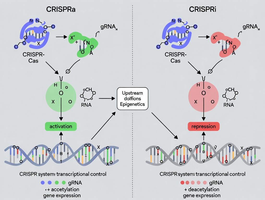
CRISPRa (Activation): Recruits transcriptional activators (e.g., VP64, p65, Rta) and histone acetyltransferases (e.g., p300) to gene promoters or enhancers, opening chromatin and recruiting RNA polymerase II to initiate transcription.
CRISPRi (Interference/Repression): Recruits transcriptional repressors (e.g., KRAB, SID4x) to gene promoters, facilitating histone methylation (H3K9me3) and chromatin condensation, which blocks RNA polymerase II binding or elongation.
Diagram 1: Core Transcriptional Mechanisms of CRISPRi and CRISPRa
Quantitative Performance Comparison
Table 1: Performance Characteristics of CRISPRa vs. CRISPRi Systems
| Parameter | CRISPRi (dCas9-KRAB) | CRISPRa (dCas9-VPR/SunTag) | Notes |
|---|---|---|---|
| Typical Repression Fold-Change | 5x - 100x (up to 99% knockdown) | 2x - 50x (up to 50-fold activation) | CRISPRi is generally more potent for gene silencing. |
| Activation Fold-Change | N/A | 10x - 1,000x+ (with synergistic systems) | Strongly dependent on target gene's baseline expression and chromatin state. |
| Time to Max Effect (Mammalian cells) | 24 - 72 hours | 24 - 96 hours | CRISPRa may require more time for chromatin remodeling. |
| Off-Target Transcriptional Effects | Low (primarily repression at intended sites) | Moderate (potential for off-target activation at enhancers) | Careful gRNA design is critical for CRISPRa specificity. |
| Optimal Targeting Region | -50 to +300 bp relative to TSS | -50 to -500 bp upstream of TSS (or enhancer regions) | CRISPRa has a more flexible but less defined optimal window. |
| Multiplexing Capacity | High (simultaneous repression of multiple genes) | Moderate (synergistic activators can be large) | CRISPRi is favored for genome-scale knockout screens. |
Table 2: Common Effector Domains and Their Properties
| System | Common Effector | Domain Origin | Mechanistic Action | Relative Size (kDa) |
|---|---|---|---|---|
| CRISPRi | KRAB | Human Kox1 | Recruits heterochromatin-inducing complexes (e.g., SETDB1) | ~45 (with dCas9) |
| SID4x | Engineered from MAD | Recruits transcriptional corepressors | ~40 (with dCas9) | |
| CRISPRa | VP64 | Herpes Simplex Virus | Minimal transcriptional activation domain | ~40 (with dCas9) |
| VPR (VP64-p65-Rta) | Engineered fusion | Strong synergistic activation | ~65 (with dCas9) | |
| SunTag System | Engineered peptide array | Recruits multiple copies of activator proteins (e.g., scFv-VP64) | >100 (complex) | |
| dCas9-p300 Core | Human p300 catalytic core | Catalyzes histone H3K27 acetylation | ~190 (with dCas9) |
Detailed Experimental Protocols
Protocol 3.1: Initial Vector Design and gRNA Cloning for Mammalian Cells
Objective: Clone target-specific gRNA(s) into a plasmid expressing dCas9-effector fusion.
- Design gRNAs: Using software (e.g., CRISPick, CHOPCHOP), design 3-5 gRNAs per target gene. For CRISPRi, select gRNAs targeting -50 to +300 bp from the transcription start site (TSS). For CRISPRa, select gRNAs targeting -50 to -500 bp upstream of the TSS or known enhancer regions.
- Oligo Annealing: Synthesize complementary oligos encoding the 20-nt guide sequence with appropriate 4-nt overhangs for your backbone (e.g., BsmBI site for lentiGuide). Resuspend oligos to 100 µM. Mix 1 µL of each oligo with 23 µL of annealing buffer (10 mM Tris, 50 mM NaCl, 1 mM EDTA, pH 7.5). Heat to 95°C for 5 min, then cool to 25°C at 1°C/min.
- Digestion & Ligation: Digest 2 µg of destination plasmid (e.g., lenti-dCas9-KRAB or lenti-dCas9-VPR) with BsmBI-v2 for 1 hour at 37°C. Gel-purify the linearized backbone. Dilute annealed oligos 1:250. Perform ligation with T4 DNA Ligase (50 ng backbone, 1 µL diluted oligos, 1x Ligase Buffer, 1 µL T4 Ligase) at 16°C for 16 hours.
- Transformation & Validation: Transform 2 µL ligation into competent E. coli, plate on ampicillin agar, and incubate overnight. Pick colonies for sequencing using a U6 promoter primer to confirm guide sequence insertion.
Protocol 3.2: Lentiviral Production and Cell Line Engineering
Objective: Generate stable cell lines expressing the dCas9-effector and target gRNA(s).
- Day 1 - Seeding: Plate HEK293T cells in a 6-well plate at 70% confluence in DMEM + 10% FBS (no antibiotics).
- Day 2 - Transfection: For one well, prepare two mixes:
- DNA Mix: 1.5 µg lentiviral packaging plasmid (psPAX2), 0.5 µg envelope plasmid (pMD2.G), and 2.0 µg of your transfer plasmid (lenti-dCas9-effector or lentiGuide-gRNA) in 250 µL Opti-MEM.
- Lipid Mix: 12 µL Lipofectamine 2000 in 250 µL Opti-MEM. Incubate 5 min. Combine mixes, incubate 20 min, then add dropwise to cells.
- Day 3/4 - Harvest: Replace media 6-8 hours post-transfection. Collect viral supernatant at 48 and 72 hours, filter through a 0.45 µm PVDF filter, and either use immediately or store at -80°C.
- Transduction: Plate target cells (e.g., HeLa, iPSCs) in a 24-well plate. Add viral supernatant containing dCas9-effector virus plus 8 µg/mL polybrene. Spinfect at 1000 × g for 1 hour at 32°C. Replace media after 24 hours.
- Selection: 48 hours post-transduction, add appropriate antibiotics (e.g., 2 µg/mL puromycin for lentiGuide, 5 µg/mL blasticidin for lenti-dCas9). Maintain selection for 5-7 days to generate a stable polyclonal population.
Protocol 3.3: Validation of Transcriptional Modulation by RT-qPCR
Objective: Quantify changes in target gene mRNA expression.
- RNA Extraction: Harvest 1x10^6 engineered cells 72-96 hours post-gRNA transduction (or induction). Lyse cells in TRIzol reagent, extract RNA with chloroform, and precipitate with isopropanol. Wash RNA pellet with 75% ethanol and resuspend in nuclease-free water.
- cDNA Synthesis: Treat 1 µg of total RNA with DNase I. Perform reverse transcription using a High-Capacity cDNA Reverse Transcription Kit with random hexamers (20 µL reaction: 10 µL RNA, 1x RT Buffer, 1 mM dNTPs, 1x Random Primers, 50 U Reverse Transcriptase). Incubate: 25°C for 10 min, 37°C for 120 min, 85°C for 5 min.
- qPCR: Prepare reactions in triplicate using SYBR Green Master Mix. Use 10 ng cDNA per 20 µL reaction with 500 nM gene-specific primers. Run on a real-time PCR system with the following program: 95°C for 10 min; 40 cycles of (95°C for 15 sec, 60°C for 60 sec). Include a melt curve analysis.
- Analysis: Calculate ∆∆Ct relative to a stable housekeeping gene (e.g., GAPDH, ACTB) and a control sample (e.g., non-targeting gRNA). Fold-change = 2^(-∆∆Ct).
Diagram 2: Workflow for Stable Cell Line Generation & Validation
The Scientist's Toolkit: Essential Research Reagent Solutions
Table 3: Key Reagents for CRISPRa/i Experiments
| Reagent / Material | Supplier Examples | Function in Experiment |
|---|---|---|
| dCas9-Effector Plasmids | Addgene (#114189 dCas9-KRAB, #114194 dCas9-VPR), Takara Bio | Backbone vectors for stable expression of dCas9 fused to transcriptional modulator. |
| gRNA Cloning Backbones | Addgene (#52961 lentiGuide-Puro, #99373 pl.entiCRISPR v2) | Vectors for expression of target-specific gRNA; contain selection markers. |
| Lentiviral Packaging Mix | OriGene, Sigma-Aldrich | Pre-mixed plasmids (psPAX2, pMD2.G) for simplified viral production. |
| Transfection Reagent (Lipofectamine 2000/3000) | Thermo Fisher Scientific | For transient plasmid delivery into HEK293T cells during lentivirus production. |
| Polybrene (Hexadimethrine bromide) | MilliporeSigma | Cationic polymer that enhances viral transduction efficiency. |
| Selection Antibiotics (Puromycin, Blasticidin) | Thermo Fisher Scientific, InvivoGen | For selecting cells successfully transduced with resistance gene-containing lentiviruses. |
| RNA Extraction Kit (TRIzol/Zymo) | Thermo Fisher Scientific, Zymo Research | For high-yield, high-purity total RNA isolation. |
| High-Capacity cDNA RT Kit | Applied Biosystems | For consistent, reliable generation of cDNA from mRNA templates. |
| SYBR Green qPCR Master Mix | Bio-Rad, Thermo Fisher Scientific | For sensitive and specific detection of amplified cDNA during qPCR. |
| Validated qPCR Primers | IDT, Sigma-Aldrich | Gene-specific primers for accurate quantification of target mRNA levels. |
| Control gRNA Libraries (Non-targeting, Targeting housekeeping genes) | Horizon Discovery, Sigma-Aldrich | Essential negative and positive controls for experimental validation. |
Pathway Diagrams: Key Regulatory Networks
Diagram 3: CRISPRi Transcriptional Repression Signaling Pathway
Diagram 4: CRISPRa Transcriptional Activation Signaling Pathway
Within the thesis of CRISPRa vs. CRISPRi for transcriptional control, the optimal system is application-dependent. CRISPRi is superior for loss-of-function studies, offering high-potency, specific, and consistent gene knockdown, making it ideal for functional genomics screens and modeling heterozygous disease states. CRISPRa excels in gain-of-function studies, allowing for tunable gene overexpression, endogenous pathway activation, and cellular reprogramming, but requires more optimization regarding gRNA positioning and effector choice. For drug discovery, CRISPRi can identify essential genes and validate therapeutic targets, while CRISPRa can be used to screen for gene products that confer therapeutic resistance or resilience. A combined approach often yields the most comprehensive mechanistic insights.
Designing and Implementing CRISPRa/i Experiments: A Step-by-Step Protocol Guide
Within the broader thesis on CRISPRa (CRISPR activation) versus CRISPRi (CRISPR interference) for transcriptional control research, this guide provides a structured decision framework. The selection between these two complementary technologies is not trivial and fundamentally shapes experimental outcomes, scalability, and biological interpretation. Both systems leverage a catalytically "dead" Cas9 (dCas9) to target specific genomic loci without inducing double-strand breaks, but diverge in their recruited effector domains to precisely upregulate (CRISPRa) or repress (CRISPRi) gene expression.
Core Technology Comparison
Molecular Architecture
CRISPRa systems fuse dCas9 to transcriptional activation domains (e.g., VP64, p65, Rta) or recruit synergistic activator complexes (e.g., SAM, SunTag). CRISPRi systems typically fuse dCas9 to repressive domains like the KRAB (Krüppel-associated box) or recruit chromatin modifiers that promote heterochromatin formation.
Quantitative Performance Profiles
The following table summarizes key performance metrics based on current literature and empirical data.
Table 1: Quantitative Performance Comparison of CRISPRa vs CRISPRi
| Parameter | CRISPRa (Typical Range) | CRISPRi (Typical Range) | Key Considerations |
|---|---|---|---|
| Max Fold Change | 10x - 1,000x+ activation | 5x - 100x+ repression (knockdown) | CRISPRa ceiling varies greatly by gene context; CRISPRi is more consistent. |
| On-Target Efficiency | 40-80% (varies by system) | 70-95% | CRISPRi generally more robust and predictable. |
| Multiplexing Capacity | High (but with additive size) | High | Both support multi-gene targeting; effector size can impact delivery. |
| Kinetics | Slower onset (hours to days) | Faster onset (hours) | Activation often requires chromatin remodeling. |
| Off-Target Effects | Low transcriptional off-targets | Low transcriptional off-targets | Both significantly cleaner than RNAi; comparable to each other. |
| Position Dependence | High (within -400 to -50 bp from TSS) | Moderate (within +50 bp downstream of TSS) | CRISPRa requires precise positioning upstream of TSS. |
| Delivery Challenge | Higher (large effector complexes) | Lower (compact repressors) | AAV packaging favors compact CRISPRi constructs. |
Decision Framework: Aligning System with Experimental Goals
Table 2: Decision Framework for Experimental Goals
| Experimental Goal | Recommended System | Rationale & Technical Notes |
|---|---|---|
| Functional Genomic Screens (Loss-of-Function) | CRISPRi | Superior consistency, deeper knockdown, lower false-negative rates compared to RNAi and CRISPR knockout (avoids confounders from DNA damage response). |
| Functional Genomic Screens (Gain-of-Function) | CRISPRa | Enables genome-wide overexpression screening; superior to cDNA libraries. Use synergistic systems (e.g., SAM) for robust activation. |
| Gene Network Mapping | Dual Use | Use CRISPRi for loss-of-function and CRISPRa for gain-of-function perturbations in parallel to map regulatory relationships. |
| Primary/Cell Therapy (Therapeutic Upregulation) | CRISPRa | Aimed at endogenous gene activation (e.g., fetal hemoglobin, tumor suppressors). Focus on compact, efficient activators (e.g., VP64-p65-Rta). |
| Target Validation (Drug Target) | CRISPRi | Mimics pharmacological inhibition more closely than knockout; reversible and titratable. |
| Studying Essential Genes | CRISPRi | Enables tunable, partial knockdown without killing cells, allowing study of gene function. |
| Synthetic Circuitry & Fine-Tuned Control | Dual Use | Combine CRISPRa and CRISPRi with orthogonal dCas proteins for independent, multi-gene regulation. |
Title: CRISPRa vs CRISPRi Decision Flowchart
Detailed Methodologies for Key Experiments
Protocol for a CRISPRi Knockdown and Rescue (Activation) Experiment
This protocol tests gene function by repression and subsequent targeted re-activation.
Day 1-2: Cell Seeding & Perturbation
- Seed HEK293T cells in a 24-well plate at 70,000 cells/well in DMEM + 10% FBS.
- Day 2: Transfect with CRISPRi construct. For each well, mix 500 ng of pLV-dCas9-KRAB-sgRNA (targeting your gene of interest) with 1.5 µL of Lipofectamine 3000 in Opti-MEM. Add complex to cells.
- Include controls: Non-targeting sgRNA and untransfected cells.
Day 5-7: Assay & Rescue
- Harvest a fraction of cells for qPCR/Western blot to confirm knockdown.
- For rescue arm: Transfect cells from the CRISPRi well with 400 ng of a CRISPRa construct (pLV-dCas9-VPR-sgRNA) designed to activate the same gene. Use a distinct antibiotic resistance for selection.
- Incubate for 72 hours.
Day 8-10: Functional Readout
- Perform functional assay (e.g., cell proliferation, migration, reporter assay) comparing:
- Untransfected control
- CRISPRi (knockdown)
- CRISPRi + CRISPRa (rescue)
- Validate: Confirm expression levels in all conditions via qPCR.
Protocol for a Dual CRISPRa/i Synthetic Circuit Characterization
This protocol tests a synthetic gene circuit where one gene is activated and another is repressed.
Day 1: Plasmid Preparation
- Clone two orthogonal sgRNA arrays into separate lentiviral vectors:
- Vector A (dCas9-VPR): Contains sgRNAs targeting Gene X promoter.
- Vector B (dCpf1-KRAB): Contains crRNAs targeting Gene Y promoter. (Note: dCpf1 is used for orthogonality to dCas9).
- Prepare high-quality plasmid DNA via endotoxin-free midiprep.
Day 2-3: Lentivirus Production
- Co-transfect Lenti-X 293T cells in a 6-well plate with:
- 1 µg of transfer vector (A or B)
- 0.75 µg of psPAX2 packaging plasmid
- 0.25 µg of pMD2.G envelope plasmid
- 6 µL of PEI transfection reagent.
- Collect viral supernatant at 48 and 72 hours post-transfection, pool, and concentrate using Lenti-X Concentrator.
Day 4: Cell Transduction
- Transduce target cells (e.g., HeLa) with:
- Virus A only
- Virus B only
- Viruses A & B (multiplicity of infection ~5 for each).
- Add polybrene (8 µg/mL) to enhance transduction.
Day 5-10: Selection & Analysis
- Begin antibiotic selection (e.g., Puromycin for A, Blasticidin for B) 48 hours post-transduction. Select for 5-7 days.
- Analyze populations via:
- RT-qPCR for Gene X and Gene Y mRNA levels.
- Flow cytometry if genes encode fluorescent proteins.
Title: Dual CRISPRa/i Circuit Workflow
The Scientist's Toolkit: Essential Reagents & Materials
Table 3: Key Research Reagent Solutions for CRISPRa/i Experiments
| Reagent / Material | Function in Experiment | Example Product/Catalog # (Illustrative) |
|---|---|---|
| dCas9-VPR Lentiviral Plasmid | Core CRISPRa vector. Fuses dCas9 to tripartite activator VPR (VP64-p65-Rta). | Addgene #63798 |
| dCas9-KRAB Lentiviral Plasmid | Core CRISPRi vector. Fuses dCas9 to the KRAB repressor domain. | Addgene #71237 |
| Lenti-Guide sgRNA Cloning Backbone | For high-throughput cloning of target-specific sgRNA sequences. | Addgene #52963 |
| Lentiviral Packaging Mix | For producing recombinant lentivirus (psPAX2 & pMD2.G plasmids or commercial kits). | Addgene #12260 & #12259 |
| Lenti-X Concentrator | To concentrate lentiviral supernatants, increasing titer for difficult-to-transduce cells. | Takara Bio #631232 |
| Polybrene (Hexadimethrine Bromide) | Enhances lentiviral transduction efficiency by neutralizing charge repulsion. | Sigma-Aldrich #H9268 |
| Validated sgRNA Libraries | Pre-designed, arrayed or pooled sgRNA libraries for genome-wide CRISPRa or CRISPRi screens. | Custom from suppliers like Synthego or Dharmacon |
| Puromycin Dihydrochloride | Selection antibiotic for cells transduced with puromycin-resistant vectors. | Thermo Fisher #A1113803 |
| RT-qPCR Kit with SYBR Green | For quantifying changes in mRNA expression of target genes post-perturbation. | Bio-Rad #1725121 |
| Next-Generation Sequencing Kit | For sequencing sgRNA representation in pooled screens (e.g., Illumina platforms). | Illumina #20020495 |
Title: Molecular Pathways of CRISPRa and CRISPRi
The choice between CRISPRa and CRISPRi is dictated by the specific experimental objective within a transcriptional control research thesis. CRISPRi offers robust, predictable knockdown ideal for loss-of-function studies and target validation. CRISPRa, while more variable, enables unique gain-of-function and rescue paradigms. Implementing them in tandem provides the most comprehensive approach to mapping gene function and regulatory networks, pushing forward both basic research and therapeutic development.
Within the strategic framework of CRISPR-based transcriptional regulation—encompassing CRISPR activation (CRISPRa) and CRISPR interference (CRISPRi)—the efficacy of any experiment is fundamentally determined by the design of the single guide RNA (gRNA). This guide provides an in-depth analysis of gRNA design principles specifically for targeting cis-regulatory elements: promoters and enhancers. The distinct architectures and functions of these regions necessitate tailored design rules to achieve optimal transcriptional control, a critical consideration for both basic research and therapeutic development.
Core Differences Between Promoter and Enhancer Targeting
Promoters are defined, typically nucleosome-depleted regions immediately upstream of the transcription start site (TSS), directly recruiting the basal transcriptional machinery. Enhancers are distal regulatory elements, often cell-type-specific, that loop to promoters via chromatin interactions to boost transcription. Targeting these elements with CRISPRa/i requires different strategic approaches.
Table 1: Key Characteristics of Promoters vs. Enhancers for gRNA Design
| Feature | Promoter | Enhancer |
|---|---|---|
| Genomic Location | Proximal to TSS (≈ -500 to +100 bp) | Distal (up to >1 Mb from TSS) |
| Chromatin State | Generally accessible | Variable accessibility; cell-type specific |
| Optimal Targeting Window for CRISPRa | Immediately upstream of TSS (-50 to -400 bp) | Across the entire accessible region |
| Optimal Targeting Window for CRISPRi | Overlaps TSS (+1 to -100 bp) or core promoter | Across the entire accessible region |
| gRNA Density Requirement | Lower (fewer, highly specific gRNAs often sufficient) | Higher (multiple gRNAs tiling the region recommended) |
| Primary Design Constraint | Avoid seed region within -10 to +10 bp of TSS for CRISPRi to prevent DNA cleavage by residual Cas9 nuclease activity. | Identify cell-type-specific accessible chromatin (via ATAC-seq/DNase-seq). |
| Effect Magnitude | Can induce strong, consistent activation/repression. | Can induce very strong activation (CRISPRa) or nuanced repression (CRISPRi); effects are more enhancer-context dependent. |
Foundational gRNA Design Rules (Universal)
Regardless of target, all effective gRNAs for transcriptional control (using catalytically dead dCas9 fused to effectors) should adhere to these core rules:
- Sequence Specificity & Off-Target Minimization: The 20-nt spacer sequence must be unique in the genome. Use algorithms (e.g., from ChopChop, CRISPOR) to quantify off-target potential via mismatch tolerance.
- On-Target Efficiency Prediction: Utilize empirical scoring models (e.g., Doench ‘16, Moreno-Mateos ‘17) trained on CRISPRa/i screens to predict on-target potency.
- Genomic Context: Avoid sequences with homopolymer runs (>4 bases) and extreme GC content (optimal 40-60%).
- Protospacer Adjacent Motif (PAM): For S. pyogenes Cas9 (SpCas9), the 5'-NGG-3' PAM must be present on the non-target strand. PAM availability dictates potential targeting sites.
Targeted Rules for Promoter gRNAs
For CRISPRi (dCas9-KRAB), the primary goal is to sterically block the binding of RNA Polymerase II or general transcription factors.
- Optimal Positioning: The most effective gRNAs for repression target the core promoter region, especially between -50 and +100 relative to the TSS, with the most potent sites often directly over the TSS itself.
- Critical Consideration: To eliminate confounding effects from DNA cleavage, ensure the gRNA seed sequence (positions 1-12 closest to PAM) does not directly overlap the TSS (-10 to +10). Use a nuclease-dead dCas9 (e.g., D10A, H840A for SpCas9).
For CRISPRa (e.g., dCas9-VPR), the goal is to recruit activators to nucleate transcription.
- Optimal Positioning: Target sites upstream of the TSS, typically between -50 and -400 bp. The effect diminishes sharply beyond -400 bp from the TSS for most promoters.
Table 2: Quantitative Outcomes from Promoter-Targeting Studies
| Study (System) | Effector | Target Position (vs. TSS) | Mean Transcriptional Change | Key Finding |
|---|---|---|---|---|
| Gilbert et al., 2014 (CRISPRi in human cells) | dCas9-KRAB | -50 to 0 bp | >90% repression (average) | Repression efficiency is highly sensitive to distance from TSS. |
| Horlbeck et al., 2016 (CRISPRi screen) | dCas9-KRAB | Over TSS | 94.5% median repression | Developed rules for highly effective "perfect match" gRNAs. |
| Konermann et al., 2015 (CRISPRa-VPR in human cells) | dCas9-VPR | -200 to -50 bp | Up to 25-fold activation | Activation domain identity and promoter context influence output. |
Targeted Rules for Enhancer gRNAs
Enhancer targeting requires prior identification of active, cell-type-specific elements. Efficacy is less about precise distance to TSS and more about targeting the accessible chromatin region of the enhancer itself.
- Identification is Key: Use ATAC-seq, DNase-seq, or histone modification ChIP-seq (H3K27ac, H3K4me1) to map active enhancers in your specific cell type.
- gRNA Tiling Strategy: Design 3-6 gRNAs tiling across the entire accessible region of the enhancer (often 200-500 bp). This accounts for uncertainty in the precise location of core regulatory sequences within the enhancer.
- CRISPRa vs. CRISPRi on Enhancers: CRISPRa is exceptionally potent on enhancers, often yielding stronger activation than promoter targeting. CRISPRi on enhancers can lead to specific gene repression but may require multiple gRNAs and can have subtler effects than promoter blockade.
Table 3: Quantitative Outcomes from Enhancer-Targeting Studies
| Study (System) | Effector | Targeting Strategy | Mean Transcriptional Change | Key Finding |
|---|---|---|---|---|
| Klamn et al., 2019 (CRISPRa in mouse neurons) | dCas9-p300Core | 5 gRNAs tiling a super-enhancer | Up to 80-fold activation | Activation via histone acetyltransferase recruitment is highly effective on enhancers. |
| Thakore et al., 2015 (CRISPRi in human cells) | dCas9-KRAB | 3 gRNAs tiling an enhancer | ~50-70% repression | Demonstrated specific gene repression via enhancer silencing. |
Experimental Protocol: Validating gRNA Efficacy for a Target Gene
A. Design & Cloning
- Identify Target Region: For promoters, extract sequence -500 to +100 bp from RefSeq TSS. For enhancers, extract the ATAC-seq peak interval (e.g., ±250 bp from peak summit).
- Design gRNAs: Using a tool like CHOPCHOP or CRISPOR, input the target sequence. Filter for high on-target and low off-target scores. Select top 3-4 gRNAs per region.
- Clone gRNAs: Clone oligos encoding the 20-nt spacer into your preferred dCas9-effector vector (e.g., lentiGuide-Puro for CRISPRi/VPR constructs) via BsmBI digestion and ligation.
- Sequence Verify: Confirm insert sequence.
B. Delivery & Expression (in vitro)
- Cell Seeding: Plate HEK293T or relevant cell line in 24-well plates.
- Transfection: Co-transfect 250 ng of dCas9-effector plasmid (e.g., dCas9-KRAB) and 250 ng of each individual gRNA plasmid using a polyethylenimine (PEI) or lipofectamine protocol.
- Control Conditions: Include a non-targeting control (NTC) gRNA and a gRNA targeting a known highly effective locus (e.g., promoter of EMX1 or AAVS1) as positive controls.
C. Readout & Analysis (48-72h post-transfection)
- RNA Extraction: Harvest cells using TRIzol reagent.
- cDNA Synthesis: Perform reverse transcription with random hexamers.
- Quantitative PCR (qPCR): Design TaqMan assays or SYBR Green primers for the target gene and 2-3 stable housekeeping genes (e.g., GAPDH, ACTB).
- Data Analysis: Calculate ΔΔCt values relative to the NTC gRNA condition. Express results as fold-change (2^-ΔΔCt) for CRISPRa or percent repression (1 - 2^-ΔΔCt) for CRISPRi.
Visualizing the Transcriptional Control Workflow
Title: gRNA Design and Validation Workflow for CRISPRa/i
Title: Promoter vs Enhancer Targeting Strategy
The Scientist's Toolkit: Essential Research Reagents
Table 4: Key Reagent Solutions for CRISPRa/i Experiments
| Reagent / Material | Function & Importance |
|---|---|
| dCas9-Effector Plasmids | Core expression vectors for dCas9 fused to transcriptional modulators (e.g., KRAB for repression; VPR, p300 for activation). |
| gRNA Cloning Backbone | Vector (e.g., lentiGuide, pU6-sgRNA) with U6 promoter for gRNA expression, often containing a selection marker (puromycin). |
| Validated Positive Control gRNAs | Pre-designed gRNAs targeting known effective loci (e.g., AAVS1 safe harbor promoter) essential for system calibration. |
| Non-Targeting Control (NTC) gRNA | A scrambled gRNA with no significant genomic target, critical for establishing baseline expression. |
| High-Efficiency Transfection Reagent | For plasmid delivery (e.g., PEI Max, Lipofectamine 3000). Viral packaging systems (lentiviral) are used for stable line generation. |
| qPCR Assay for Target Gene | Gene-specific TaqMan probes or SYBR Green primer sets, validated for efficiency, to quantify transcriptional changes. |
| ATAC-seq or DNase-seq Kit | Essential for de novo identification of cell-type-specific active enhancers prior to gRNA design. |
| Next-Generation Sequencing Library Prep Kit | For off-target assessment (e.g., GUIDE-seq, CIRCLE-seq) or pooled screen readout. |
Strategic gRNA design is the linchpin of successful CRISPR-mediated transcriptional control. Promoter targeting offers a direct, predictable route to modulation, with strict positional rules governing outcome magnitude. Enhancer targeting, while requiring prior epigenetic mapping, can yield exceptionally potent activation and allows for the interrogation of complex gene regulation networks. By adhering to the distinct design rules for each element class—and rigorously validating gRNAs within the specific cellular context—researchers can harness the full potential of CRISPRa and CRISPRi for precise genetic interrogation and therapeutic discovery.
The precise control of gene transcription is a cornerstone of functional genomics and therapeutic development. Within this landscape, CRISPR activation (CRISPRa) and CRISPR interference (CRISPRi) have emerged as powerful, programmable technologies for upregulating or suppressing gene expression without altering the underlying DNA sequence. The efficacy and specificity of these systems are fundamentally determined by the effector domains recruited to the target genomic locus by a catalytically dead Cas9 (dCas9) protein. This guide provides an in-depth technical analysis of four principal effector domain strategies for transcriptional control: the tripartite activator VP64-p65-Rta (VPR), the scaffold-recruiting SunTag system, the synergistic activator mediator (SAM), and the repressive Kruppel-associated box (KRAB) domain. The selection of an appropriate effector system is critical for optimizing dynamic range, specificity, and deliverability in both research and drug development applications.
Effector Domain Architectures: Mechanisms and Comparisons
VP64-p65-Rta (VPR)
VPR is a single, compact transcriptional activator created by fusing three potent activation domains: VP64 (from Herpes Simplex Virus), p65 (a subunit of NF-κB), and Rta (from Epstein-Barr virus). This chimeric protein is fused directly to dCas9, creating a potent, all-in-one CRISPRa complex. It recruits a broad suite of co-activators to efficiently drive gene expression.
SunTag
The SunTag system employs a modular scaffold approach. A dCas9 is fused to a chain of peptide epitopes (GCN4). Co-expressed single-chain variable fragment (scFv) antibodies, fused to a transcriptional activator domain (e.g., VP64), bind to these epitopes. This results in the recruitment of multiple activator proteins to a single dCas9, achieving strong signal amplification.
Synergistic Activation Mediator (SAM)
SAM is a three-component system that leverages cooperative recruitment. The core is dCas9 fused to VP64. A modified sgRNA contains two MS2 RNA aptamers in its tetraloop and stem-loop 2. MS2 coat proteins (MCP), fused to the activators p65 and HSF1, bind to these aptamers. This brings multiple distinct activation domains to the locus, creating a synergistic effect for robust gene activation.
KRAB Variants for CRISPRi
The Krüppel-associated box (KRAB) domain is the archetypal repressor for CRISPRi. Fused to dCas9, it recruits heterochromatin-forming complexes (via KAP1) to promote histone methylation (H3K9me3) and DNA methylation, leading to stable, long-term transcriptional repression. Engineered variants and fusions with other repressive domains (e.g., MeCP2, SID) enhance silencing potency.
Table 1: Quantitative Comparison of Effector Domain Systems
| System | Type | Typical Fold Activation (Range) | Key Components | Approx. Size (kDa) | Key Advantage | Key Limitation |
|---|---|---|---|---|---|---|
| VPR | CRISPRa | 100 - 1,000x | dCas9-VPR fusion, standard sgRNA | ~190 | Simple, all-in-one delivery; strong activation. | Potential for increased off-target effects; large fusion protein. |
| SunTag | CRISPRa | 1,000 - 10,000x | dCas9-GCN4, scFv-VP64, sgRNA | ~160 + ~30 per scFv | High amplification; modular. | Requires co-expression of two large proteins; potential for immunogenicity. |
| SAM | CRISPRa | 10,000 - 100,000x | dCas9-VP64, MCP-p65-HSF1, MS2-sgRNA | ~190 + ~55 | Extremely high activation; synergistic effect. | Complex 3-component delivery; large sgRNA may affect packaging. |
| dCas9-KRAB | CRISPRi | 5 - 20x knockdown (80-95% repression) | dCas9-KRAB fusion, standard sgRNA | ~190 | Potent, stable repression; well-characterized. | Can have variable efficiency across genomic contexts. |
Detailed Experimental Protocols
Protocol 3.1: Initial Validation of CRISPRa/i Systems in HEK293T Cells
Objective: To compare the transcriptional modulation efficacy of VPR, SAM, and KRAB on a stably integrated reporter gene (e.g., EGFP under a minimal promoter). Materials: See "Scientist's Toolkit" (Section 5). Procedure:
- Cell Seeding: Seed HEK293T cells in 24-well plates at 1x10^5 cells/well in DMEM + 10% FBS 24 hours before transfection.
- Plasmid Formulation: For each effector system, prepare plasmid mixtures in Opti-MEM (total DNA 500ng/well):
- VPR: dCas9-VPR (250ng), sgRNA targeting EGFP promoter (250ng).
- SAM: dCas9-VP64 (167ng), MCP-p65-HSF1 (167ng), MS2-sgRNA-EGFP (166ng).
- SunTag: dCas9-GCN4 (167ng), scFv-VP64 (167ng), sgRNA-EGFP (166ng).
- KRAB: dCas9-KRAB (250ng), sgRNA-EGFP (250ng).
- Control: dCas9-only (250ng), sgRNA-EGFP (250ng).
- Transfection: Use lipofectamine 3000 per manufacturer's protocol. Add 1μl of P3000 reagent per 1μg DNA.
- Incubation: Change media 6 hours post-transfection.
- Analysis: Harvest cells 72 hours post-transfection. Analyze EGFP mean fluorescence intensity (MFI) via flow cytometry. Calculate fold-change relative to dCas9-only control.
Protocol 3.2: Screening for Endogenous Gene Activation with SAM
Objective: To identify optimal sgRNAs for activating a lowly expressed endogenous gene (e.g., IL1RN) using the SAM system. Procedure:
- sgRNA Design: Design 5-10 MS2-sgRNAs targeting the region from -400 bp to +1 bp relative to the TSS of the target gene using established algorithms (e.g., CRISPick).
- Library Cloning: Clone pooled sgRNA sequences into the MS2-aptamer containing lentiviral sgRNA backbone via BsmBI Golden Gate assembly.
- Lentivirus Production: Co-transfect Lenti-dCas9-VP64, Lenti-MCP-p65-HSF1, and the pooled Lenti-MS2-sgRNA library into Lenti-X 293T cells using a 3rd generation packaging system. Harvest virus supernatant at 48 and 72 hours.
- Transduction & Selection: Transduce target cells (e.g., THP-1) at a low MOI (<0.3) to ensure single integration. Select with appropriate antibiotics (Blasticidin for dCas9, Puromycin for sgRNA) for 7 days.
- Phenotypic Readout: Harvest genomic DNA for sgRNA sequencing (NGS) to assess enrichment/depletion. In parallel, perform RT-qPCR on polyclonal populations for each sgRNA to measure mRNA levels of the target gene.
Signaling and Workflow Diagrams
Title: VPR CRISPRa Mechanism
Title: SAM System Experimental Workflow
Title: CRISPRa vs CRISPRi Core Pathways
The Scientist's Toolkit
Table 2: Essential Research Reagent Solutions for Effector Domain Studies
| Reagent / Material | Supplier Examples | Function in Experiments |
|---|---|---|
| dCas9-VPR Plasmid | Addgene #63798, Takara Bio | All-in-one CRISPRa effector for direct fusion activation studies. |
| SAM System Plasmids (3-plasmid set) | Addgene #1000000058 (MS2-sgRNA), #1000000056 (dCas9-VP64), #1000000057 (MCP-p65-HSF1) | Provides components for maximal synergistic gene activation. |
| SunTag System Plasmids | Addgene #60903 (dCas9-GCN4), #60904 (scFv-VP64) | Modular system for amplified activator recruitment. |
| dCas9-KRAB Plasmid | Addgene #110821 (dCas9-KRAB-MeCP2) | Potent, fused repressor for robust CRISPRi and epigenetic silencing. |
| LentiCRISPR v2 (with MS2 loops) | Addgene #98291 | Lentiviral backbone for stable delivery of MS2-sgRNA libraries. |
| Lipofectamine 3000 | Thermo Fisher Scientific | High-efficiency transfection reagent for plasmid delivery in adherent cells. |
| Lentiviral Packaging Mix (psPAX2, pMD2.G) | Addgene #12260, #12259 | Essential plasmids for producing replication-incompetent lentivirus. |
| Polybrene (Hexadimethrine bromide) | Sigma-Aldrich | Cationic polymer that enhances lentiviral transduction efficiency. |
| Puromycin Dihydrochloride | Thermo Fisher Scientific | Selection antibiotic for cells transduced with puromycin-resistant constructs (e.g., sgRNA vectors). |
| Blasticidin S HCl | Thermo Fisher Scientific | Selection antibiotic for cells expressing dCas9-effector fusions (common resistance marker). |
| RT-qPCR Master Mix (SYBR Green) | Bio-Rad, Thermo Fisher Scientific | For quantitative measurement of endogenous gene expression changes post-modulation. |
Within the ongoing thesis comparing CRISPR activation (CRISPRa) and CRISPR interference (CRISPRi) for transcriptional control, a critical determinant of experimental success is the delivery strategy. This guide details core delivery methodologies—viral vectors, transfection, and stable cell line generation—for deploying CRISPRa/i machinery, focusing on efficiency, scalability, and applicability for research and drug development.
Viral Vector Delivery
Viral vectors offer high delivery efficiency, especially in hard-to-transfect cells, and are essential for in vivo applications.
Key Vector Systems
Table 1: Comparison of Viral Vectors for CRISPRa/i Delivery
| Vector | Max. Capacity (kb) | Titer (TU/mL) | Integration | Pros for CRISPRa/i | Cons for CRISPRa/i |
|---|---|---|---|---|---|
| Lentivirus (LV) | ~8 kb | 1x10^8 - 1x10^9 | Stable (Random) | Sustained expression; broad tropism; good for stable lines. | Random integration risks insertional mutagenesis. |
| Adeno-associated Virus (AAV) | ~4.7 kb | 1x10^12 - 1x10^13 | Mostly Episomal | Low immunogenicity; excellent in vivo safety profile. | Limited cargo capacity; challenging to package SpCas9 + activators/repressors. |
| Adenovirus (AdV) | ~8 kb (High-capacity: ~36 kb) | 1x10^10 - 1x10^11 | Episomal | High titer; very large cargo capacity; efficient in vitro & in vivo. | High immunogenicity; transient expression. |
Protocol: Production of Lentiviral Vectors for CRISPRa
This protocol outlines the generation of VSV-G pseudotyped lentivirus encoding a CRISPRa system (e.g., dCas9-VPR).
Materials & Reagents:
- Plasmids: Transfer plasmid (e.g., lenti-dCas9-VPR), psPAX2 (packaging), pMD2.G (envelope).
- Cells: HEK293T cells (highly transferable).
- Media: DMEM + 10% FBS, antibiotics.
- Transfection Reagent: Polyethylenimine (PEI MAX, 40 kDa).
- Concentration: Lenti-X Concentrator (Takara Bio).
Procedure:
- Day 0: Seed HEK293T cells in a 10 cm dish to reach 70-80% confluence the next day.
- Day 1 (Transfection): a. Prepare DNA mix: 10 µg transfer plasmid, 7.5 µg psPAX2, 2.5 µg pMD2.G in 500 µL serum-free DMEM. b. Prepare PEI mix: 45 µL PEI (1 mg/mL) in 500 µL serum-free DMEM. Incubate 5 min. c. Combine DNA and PEI mixes, vortex, incubate 20 min at RT. d. Add dropwise to cells with fresh medium.
- Day 2 (Medium Change): Replace medium with 10 mL fresh, complete DMEM.
- Day 3 & 4 (Harvest): Collect supernatant (~48 and 72h post-transfection). Pool, filter through a 0.45 µm PES filter.
- Concentration (Optional): Mix 1 part Lenti-X Concentrator with 3 parts supernatant. Incubate overnight at 4°C, then centrifuge at 1500 × g for 45 min. Resuspend pellet in PBS/medium. Aliquot and store at -80°C.
- Titering: Use qPCR-based titering kit (e.g., Lenti-X qRT-PCR, Takara) or functional transduction assays.
Title: Lentiviral Production Workflow for CRISPRa/i
Transfection-Based Delivery
Transfection is a rapid, versatile method suitable for in vitro screening and initial functional validation.
Method Comparison
Table 2: Transfection Methods for CRISPRa/i RNP or Plasmid Delivery
| Method | Format | Max. Efficiency | Key Advantage | Best For |
|---|---|---|---|---|
| Lipid Nanoparticles (LNPs) | RNP, mRNA | >90% in easy lines | High efficiency, low toxicity. | Primary cells, sensitive cell types. |
| Electroporation (Nucleofection) | RNP, Plasmid | 70-95% (cell type dependent) | Best for hard-to-transfect cells (e.g., T cells, neurons). | Immune cells, stem cells, neurons. |
| Cationic Polymers (e.g., PEI) | Plasmid | 50-80% in HEK293 | Low cost, scalable for large DNA. | HEK293T production, large-scale screening. |
| Calcium Phosphate | Plasmid | 30-50% in adherent lines | Very low cost, established protocol. | Standard adherent lines (e.g., HeLa). |
Protocol: CRISPRa/i RNP Delivery via Electroporation
This protocol uses pre-assembled ribonucleoprotein (RNP) complexes for rapid, transient activity with minimal off-target effects.
Materials & Reagents:
- Components: Recombinant dCas9-VPR or dCas9-KRAB protein, synthetic sgRNA (with MS2 or other aptamers for CRISPRa).
- Buffer: Opti-MEM or specific Nucleofector Solution.
- Equipment: Neon (Thermo Fisher) or 4D-Nucleofector (Lonza) system.
- Cells: Target cells (e.g., Jurkat, iPSCs).
Procedure:
- RNP Complex Assembly: Incubate dCas9-effector protein (e.g., 5 µg) with sgRNA (molar ratio ~1:3) in Opti-MEM at RT for 10-20 min.
- Cell Preparation: Harvest and count cells. Wash with PBS. For 100 µL Neon tip, use 5x10^5 to 1x10^6 cells.
- Electroporation Setup: Resuspend cell pellet in R buffer (Neon) or specified Nucleofector solution. Mix with assembled RNP complexes.
- Electroporation: Transfer to electroporation cuvette or tip. Apply optimized pulse (e.g., 1400 V, 20 ms, 2 pulses for Neon with Jurkat).
- Recovery: Immediately transfer cells to pre-warmed, antibiotic-free complete medium. Plate in appropriate culture vessel.
- Analysis: Assess gene expression changes via qRT-PCR or RNA-seq 48-72 hours post-electroporation.
Title: RNP Complex Assembly & Electroporation Workflow
Stable Cell Line Generation
Generating stable cell lines ensures consistent, long-term expression of CRISPRa/i components, crucial for extended studies and screening.
Strategies and Timelines
Table 3: Strategies for Generating CRISPRa/i Stable Cell Lines
| Strategy | Method | Key Feature | Timeline to Clone | Consistency |
|---|---|---|---|---|
| Random Integration | Lentiviral transduction + antibiotic selection | Robust, but variable expression due to position effects. | 3-4 weeks | Moderate (requires screening) |
| Site-Specific Integration (e.g., FIp-In, Bxb1) | Recombinase-mediated cassette exchange (RMCE) | Uniform expression from a defined genomic locus. | 4-5 weeks | High |
| BAC Transgenesis | Introduction of a Bacterial Artificial Chromosome | Preserves genomic context and regulatory elements. | 8+ weeks | High (but complex) |
Protocol: Generation of a Doxycycline-Inducible CRISPRi Stable Line Using FIp-In
This protocol creates isogenic cells with a single-copy, genomically integrated dCas9-KRAB under inducible control.
Materials & Reagents:
- Parental Cell Line: FIp-In host cell line (e.g., FIp-In HEK293, Thermo Fisher).
- Plasmids: pcDNA5/FRT/TO-dCas9-KRAB, pOG44 (expressing FIp recombinase).
- Transfection Reagent: Lipofectamine 3000.
- Selection Antibiotics: Hygromycin B (for integration), Blasticidin (for FIp-In locus selection).
- Inducer: Doxycycline hyclate.
Procedure:
- Day 0: Seed FIp-In host cells in a 6-well plate to reach 70% confluence.
- Day 1 (Transfection): Co-transfect 0.9 µg pcDNA5/FRT/TO-dCas9-KRAB and 0.1 µg pOG44 using Lipofectamine 3000 per manufacturer's protocol.
- Day 2: Begin dual selection with 200 µg/mL Hygromycin B and 5-15 µg/mL Blasticidin. Maintain selection for 10-14 days, changing medium/antibiotics every 3-4 days.
- Day 14+: Isolate single clones using cloning rings or by limiting dilution. Expand clones.
- Validation: a. Genomic Integration: Perform PCR across the FRT site. b. Inducible Expression: Treat with doxycycline (1 µg/mL, 24-48h), assay dCas9-KRAB expression via western blot. c. Functional Test: Transfect with a target sgRNA and measure repression via qRT-PCR.
Title: Workflow for FIp-In Stable Cell Line Generation
The Scientist's Toolkit: Research Reagent Solutions
Table 4: Essential Reagents for CRISPRa/i Delivery Experiments
| Item | Function | Example Product/Brand |
|---|---|---|
| Lentiviral Packaging Plasmids | Provide viral structural proteins (gag/pol) and envelope (VSV-G) for virus production. | psPAX2, pMD2.G (Addgene). |
| Polyethylenimine (PEI MAX) | High-efficiency, low-cost cationic polymer for plasmid transfection, especially in HEK293T. | PEI MAX 40K (Polysciences). |
| Lenti-X Concentrator | Precipitation reagent for concentrating lentiviral supernatants, increasing functional titer. | Lenti-X Concentrator (Takara Bio). |
| sgRNA with Modified Backbones | Synthetic sgRNAs with chemical modifications (e.g., 2'-O-methyl) enhance stability for RNP delivery. | Synthego CRISPR sgRNA EZ. |
| Nucleofector Kits | Cell type-specific solutions and protocols for high-efficiency RNP or DNA electroporation. | 4D-Nucleofector X Kit (Lonza). |
| Lipid Nanoparticles (LNPs) | Formulated lipids for high-efficiency, low-toxicity delivery of RNPs or mRNA in vitro. | Lipofectamine CRISPRMAX (Thermo Fisher). |
| FIp-In System Components | Ensures single-copy, site-specific integration of dCas9 constructs for uniform stable lines. | FIp-In TREx Core Kit (Thermo Fisher). |
| Doxycycline Hyclate | Small molecule inducer for Tet-On systems, allowing tight temporal control of dCas9 expression. | Doxycycline hyclate (Sigma-Aldrich). |
| Hygromycin B & Blasticidin | Antibiotics for selection of successfully transfected/infected cells and maintenance of stable lines. | Hygromycin B (InvivoGen). |
| QuickTiter Lentivirus Titer Kit | Rapid quantification of lentiviral physical and infectious particles via ELISA. | QuickTiter Kit (Cell Biolabs). |
Within the ongoing debate on optimal transcriptional control strategies—CRISPR activation (CRISPRa) versus CRISPR interference (CRISPRi)—lies the practical application of these technologies. This guide details how CRISPRa and CRISPRi are leveraged for functional genomics screens, disease modeling, and synthetic biology circuits, providing a technical framework for researchers. The choice between a and i hinges on the biological question: gain-of-function versus loss-of-function perturbation.
Functional Genomics Screens
CRISPRa/i enable genome-wide interrogation of gene function by systematically modulating transcription.
Core Principles:
- CRISPRi: Utilizes a catalytically dead Cas9 (dCas9) fused to a transcriptional repressor domain (e.g., KRAB). Guide RNA (gRNA) targeting near the transcription start site (TSS) silences gene expression.
- CRISPRa: Employs dCas9 fused to transcriptional activator domains (e.g., VPR, SAM). gRNA targeting promoter or enhancer regions upregulates gene expression.
Quantitative Comparison of Major Systems: Table 1: Quantitative Comparison of CRISPRa and CRISPRi Systems for Functional Genomics
| Parameter | CRISPRi (dCas9-KRAB) | CRISPRa (dCas9-VPR) | CRISPRa (SAM) |
|---|---|---|---|
| Typical Fold Change | 5- to 20-fold knockdown | 10- to 100-fold activation | 100- to 1000-fold activation |
| Optimal Targeting | -50 to +300 bp from TSS | -400 to -50 bp from TSS | -400 to -50 bp from TSS |
| Library Size (Genome-wide) | ~5 guides/gene | ~5 guides/gene | ~5 guides/gene |
| Screen Noise (FDR) | Lower (strong repression) | Higher (variegated activation) | Moderate |
| Primary Application | Essential gene identification, vulnerability discovery | Resistance gene identification, suppressor screens | High-sensitivity activation screens |
Detailed Protocol: Genome-wide CRISPRa/i Positive Selection Screen
- 1. Library Design: Select a validated genome-wide sgRNA library (e.g., Calabrese, Brunello for CRISPRi; SAM, hCRISPRa-v2 for CRISPRa). Use 5-10 sgRNAs per gene and 1000 non-targeting controls.
- 2. Lentiviral Production: Generate lentivirus for the sgRNA library at low MOI (<0.3) to ensure single integration in HEK293T cells using third-generation packaging plasmids.
- 3. Cell Transduction: Transduce target cells (e.g., iPSCs, cancer cell lines) at a coverage of 500-1000 cells per sgRNA. Include puromycin selection (1-3 µg/mL, 3-7 days).
- 4. Selection Pressure: Apply phenotypic pressure (e.g., drug treatment, nutrient deprivation) for 14-21 population doublings. Maintain a minimum representation of 500x library coverage.
- 5. Genomic DNA Extraction & Sequencing: Harvest genomic DNA from pre-selection and post-selection pools (≥500x coverage). PCR amplify sgRNA cassettes with indexed primers for NGS.
- 6. Data Analysis: Align sequences to the reference library. Use MAGeCK or BAGEL2 algorithms to calculate sgRNA enrichment/depletion and identify significantly hit genes (FDR < 0.05).
Visualization: Functional Genomics Screen Workflow
(Diagram Title: CRISPR Screen Experimental Workflow)
Disease Modeling
CRISPRa/i facilitate precise, polygenic disease modeling in relevant cell types, surpassing knockout limitations.
Applications:
- CRISPRi: Model haploinsufficiency disorders by knocking down the functional allele. Suppress compensatory pathways to unmask phenotypes.
- CRISPRa: Overexpress risk variant alleles or dominant-negative proteins. Activate endogenous genes to mimic pathological overexpression states.
Detailed Protocol: Modeling Alzheimer's Risk with CRISPRa in iPSC-Derived Neurons
- 1. sgRNA Design: Design 3 sgRNAs targeting the promoter region of the APP gene (for overexpression) or the BACE1 gene. Include non-targeting control.
- 2. Stable Cell Line Generation: Co-transfect iPSCs with plasmids expressing dCas9-VPR and the APP-targeting sgRNA. Select with blasticidin (5 µg/mL) and puromycin (1 µg/mL) for 10 days. Pick single-cell clones and validate by qRT-PCR.
- 3. Neuronal Differentiation: Differentiate engineered iPSCs into cortical neurons using a validated dual-SMAD inhibition protocol (e.g., with Noggin and SB431542).
- 4. Phenotypic Validation (Day 60):
- Biochemical: Measure Aβ42/Aβ40 ratio in conditioned media via ELISA.
- Cellular: Immunocytochemistry for phospho-Tau (AT8 antibody) and synaptic markers (PSD95, Synapsin-1).
- Functional: Perform calcium imaging (Fluo-4 AM dye) to assess neuronal network hyperactivity.
Research Reagent Solutions:
- dCas9 Effector Plasmids: pLV-dCas9-KRAB-P2A-Puro (Addgene #89567); lenti-dCas9-VPR-blast (Addgene #114196).
- sgRNA Libraries: Human CRISPRi v2 library (Addgene #1000000132); hCRISPRa-v2 library (Addgene #1000000093).
- Cell Lines: Induced Pluripotent Stem Cells (iPSCs) from disease-relevant backgrounds.
- Differentiation Kits: Commercial cortical neuron differentiation kits (e.g., Stemcell Technologies #08500).
- Assay Kits: Aβ40/42 ELISA Kits (e.g., Invitrogen #KHB3481), Total Tau ELISA Kits.
Synthetic Biology Circuits
CRISPRa/i provide orthogonal, programmable components for constructing sophisticated genetic circuits in mammalian cells.
Core Logic: dCas9-effectors act as programmable transcription factors. Multiple sgRNAs can be used to implement Boolean logic gates (AND, OR, NOT).
Circuit Examples & Performance: Table 2: Performance Metrics of CRISPR-based Synthetic Circuits
| Circuit Type | Components | Logic | Response Time | Dynamic Range | Key Application |
|---|---|---|---|---|---|
| Transcriptional Cascade | dCas9-VPR -> sgRNA1 -> Gene A -> dCas9-VPR expression | Amplifier | 24-48 hrs | ~50-fold per stage | Signal amplification |
| AND Gate | dCas9-VPR + 2 sgRNAs targeting same promoter | INPUT1 AND INPUT2 | 12-24 hrs | Up to 100-fold | Cell-specific activation, safety switches |
| NOT Gate | dCas9-KRAB + sgRNA targeting gene promoter | NOT INPUT | 6-12 hrs | 5- to 20-fold repression | Feedback inhibition, toggle switches |
Detailed Protocol: Building a CRISPRa/i AND Gate for Cell-Selective Transgene Expression
- 1. Circuit Design: Clone two inducible promoters (e.g., Doxycycline-inducible, Cumate-inducible) each driving expression of a distinct sgRNA. Both sgRNAs must target the promoter of a reporter gene (e.g., GFP) in the presence of dCas9-VPR.
- 2. Plasmid Construction: Assemble components in a single lentiviral vector: [Prtet->sgRNA1] - [Pcum->sgRNA2] - [PminCMV->GFP] (with sgRNA target sites in promoter). A separate vector expresses dCas9-VPR constitutively.
- 3. Transfection & Testing: Co-transfect HEK293 cells with the circuit plasmid and the dCas9-VPR plasmid. Test conditions: No inducer, Dox only, Cumate only, Dox+Cumate.
- 4. Flow Cytometry Analysis: 48 hours post-transfection, analyze GFP expression. The circuit should only show high GFP fluorescence when both inducers are present.
Visualization: CRISPR-based AND Gate Logic Circuit
(Diagram Title: CRISPR Transcriptional AND Gate Schematic)
The selection between CRISPRa and CRISPRi is not trivial but dictated by the application's specific need for gene activation or repression. Functional genomics screens benefit from CRISPRi's precision for essential gene mapping and CRISPRa's power in suppressor screens. Disease modeling requires CRISPRa to overexpress genes and CRISPRi to model dosage sensitivity. Synthetic biology exploits both to create predictable, orthogonal logic gates. As effector domains and delivery methods advance, the precision and multiplexability of these transcriptional control tools will continue to redefine genetic research and therapeutic discovery.
Optimizing CRISPRa/i Performance: Solving Common Pitfalls in Efficiency and Specificity
Within the broader thesis of CRISPR activation (CRISPRa) versus CRISPR interference (CRISPRi) for transcriptional control, a critical challenge is diagnosing underwhelming phenotypic outcomes. Inefficient gene activation or repression can stem from three core technical pillars: guide RNA (gRNA) design and efficiency, effector expression and stability, and delivery efficacy. This guide provides a diagnostic framework and experimental protocols to identify and resolve these issues.
Table 1: Common Causes of Low CRISPRa/i Efficiency & Diagnostic Indicators
| Category | Specific Issue | Typical Impact on Efficiency | Key Diagnostic Assay |
|---|---|---|---|
| gRNA Efficiency | Suboptimal on-target binding (chromatin state, secondary structure) | Up to 100-fold variation in output | NGS-based chromatin accessibility (ATAC-seq), gRNA-seq enrichment |
| Off-target binding | Variable; can sequester effector | GUIDE-seq, CIRCLE-seq | |
| Effector Expression | Low dCas9-VP64/MS2-p65-HSF1 (CRISPRa) or dCas9-KRAB (CRISPRi) expression | >50% reduction vs. positive control | Western Blot (anti-Cas9, anti-epitope tag), Flow Cytometry (if fluorescently tagged) |
| Insufficient activator/repressor stoichiometry | Non-linear dose-response; plateau | Titration of effector plasmid/dose | |
| Delivery Issues | Low transfection/transduction efficiency | Directly proportional to cell modulation rate | Flow cytometry for reporter or effector |
| Inefficient nuclear localization (dCas9) | >70% reduction in activity | Immunofluorescence (Nuclear/Cytoplasmic fractionation) |
Table 2: Benchmark Values for Optimal CRISPRa/i Components (Mammalian Cells)
| Component | CRISPRa (e.g., SAM, VPR) | CRISPRi (e.g., dCas9-KRAB) | Measurement Method |
|---|---|---|---|
| dCas9-Effector Expression | >10^3 molecules/cell (estimated) | >10^3 molecules/cell (estimated) | Quantitative Western Blot |
| gRNA Expression | High (Pol III promoter, e.g., U6) | High (Pol III promoter, e.g., U6) | qRT-PCR (relative to housekeeping gRNA) |
| MOI (Lentiviral) | 0.3-1.0 (to avoid multi-copy integration) | 0.3-1.0 (to avoid multi-copy integration) | Genomic DNA qPCR (copy number assay) |
| Typical Activation/Repression Fold-Change | 10-1000x (mRNA) | 5-100x knockdown (mRNA) | RNA-seq, qRT-PCR |
Experimental Protocols for Diagnosis
Protocol 3.1: Assessing gRNA Efficiency via Targeted NGS (gRNA-seq)
Purpose: Quantify gRNA binding enrichment at the target locus. Materials: Genomic DNA isolation kit, PCR primers flanking target site, NGS library prep kit. Steps:
- Isolate genomic DNA from CRISPR-treated cells and a control (non-targeting gRNA) 72h post-delivery.
- Amplify the target genomic region (200-300 bp surrounding the gRNA site) by PCR. Use barcoded primers for multiplexing.
- Prepare NGS libraries and sequence on a MiSeq or similar platform.
- Align reads to the reference genome. Calculate enrichment as (reads in treated sample at target locus) / (reads in control sample at target locus). A value <5 suggests poor gRNA binding.
Protocol 3.2: Quantifying Effector Expression via Western Blot
Purpose: Confirm dCas9-effector fusion protein expression and approximate levels. Materials: RIPA buffer, anti-Cas9 primary antibody (or anti-FLAG/HA if tagged), HRP-conjugated secondary antibody. Steps:
- Lyse cells 96h post-transfection/transduction. Use ~1x10^6 cells per sample.
- Separate 30-50 µg of total protein on a 4-12% Bis-Tris gel. Transfer to PVDF membrane.
- Block membrane, incubate with anti-Cas9 (1:1000) overnight at 4°C.
- Incubate with HRP-secondary antibody (1:5000) for 1h. Develop with ECL and image. Compare band intensity to a titration of a known dCas9-expressing lysate control.
Protocol 3.3: Measuring Delivery Efficiency via Flow Cytometry
Purpose: Determine the percentage of cells successfully receiving CRISPR components. Materials: Cells with integrated reporter (if available), antibody for surface marker (if applicable), flow cytometer. Steps:
- For reporter lines: 72h post-delivery, analyze cells for reporter fluorescence (e.g., GFP activation). The percentage of GFP+ cells equals delivery/activation efficiency.
- For non-reporter lines: If effector is fluorescently tagged (e.g., dCas9-GFP), analyze directly. Alternatively, co-transfect/co-transduce with a traceable plasmid (e.g., encoding a surface marker like CD4) at a low, fixed ratio (1:10) to the CRISPR construct. Stain for the marker and analyze by flow cytometry.
Visualizations
Diagram Title: Systematic Diagnosis Workflow for Low CRISPRa/i Efficiency
Diagram Title: Core Mechanisms of CRISPRa vs. CRISPRi
The Scientist's Toolkit: Research Reagent Solutions
Table 3: Essential Reagents for Diagnosing CRISPRa/i Issues
| Reagent / Material | Function / Purpose | Example Product/Catalog |
|---|---|---|
| Validated Positive Control gRNA Plasmid | Controls for system functionality; targets a known high-expressibility locus (e.g., AAVS1 with a fluorescent reporter). | Addgene #92385 (sgRNA targeting AAVS1 with EF1a-GFP reporter) |
| Anti-Cas9 Antibody (for Western Blot) | Detects dCas9-effector fusion protein expression levels. | Cell Signaling Technology #14697 (Cas9 Antibody) |
| Anti-FLAG or Anti-HA Antibody | Alternative detection if dCas9 is epitope-tagged. | Sigma F1804 (Anti-FLAG M2); Roche 11867423001 (Anti-HA High Affinity) |
| Chromatin Accessibility Assay Kit | Assesses if target site is in open/closed chromatin (ATAC-seq). | Illumina/Swift Biosciences Accel-NGS 2S Plus DNA Library Kit |
| NGS-based Off-target Analysis Kit | Identifies potential off-target gRNA binding sites. | Takara Bio GUIDE-seq Detection Kit |
| Quantitative PCR (qPCR) Assay for gRNA | Measures gRNA expression levels from Pol III promoters. | Custom TaqMan assay for gRNA constant region. |
| Lentiviral Titering Kit | Accurately determines viral particle concentration (TU/mL) for consistent MOI. | Takara Bio Lenti-X qRT-PCR Titration Kit |
| Nuclear Localization Signal (NLS) Mutant Control | Control to confirm nuclear import is not limiting. | Addgene #85473 (dCas9-NLS mutant). |
| Fluorescent Protein Fused dCas9 Effector | Enables direct visualization of delivery and expression via flow cytometry/imaging. | Addgene #85474 (dCas9-VP64-GFP) & #127970 (dCas9-KRAB-mCherry) |
The advent of CRISPR-Cas systems for transcriptional modulation—CRISPR activation (CRISPRa) and CRISPR inhibition (CRISPRi)—has revolutionized functional genomics and therapeutic discovery. Both systems utilize a catalytically dead Cas9 (dCas9) fused to effector domains. CRISPRa recruits transcriptional activators (e.g., VPR, p65AD) to gene promoters, while CRISPRi employs repressors (e.g., KRAB, SID4x) to silence transcription. A central challenge plaguing both approaches is transcriptional off-target effects, where the dCas9-effector complex modulates genes other than the intended target, leading to confounded research data and potential therapeutic toxicity. This guide details technical strategies to enhance specificity within the comparative framework of CRISPRa and CRISPRi applications.
Quantitative Comparison of Off-Target Profiles
The off-target propensity varies between CRISPRa and CRISPRi due to differences in effector mechanism, chromatin state sensitivity, and sgRNA design constraints. The table below summarizes key quantitative findings from recent studies.
Table 1: Comparative Off-Target Profiles of CRISPRa vs. CRISPRi
| Aspect | CRISPRi (dCas9-KRAB) | CRISPRa (dCas9-VPR) | Notes & Key References |
|---|---|---|---|
| Primary Off-Target Source | Binding of dCas9 to mismatched genomic sites. | Binding of dCas9 + promiscuous activator recruitment. | CRISPRa effectors can activate nearby genes from off-target binding. |
| Typical Off-Target Rate (Genome-wide screens) | ~10-30% of sgRNAs show significant off-target effects. | ~20-40% of sgRNAs show significant off-target effects. | Measured by correlation between unrelated sgRNAs for the same gene. CRISPRa is generally more prone. |
| Effective Distance from TSS | Highly effective within -50 to +300 bp of TSS. | Requires precise positioning -50 to -150 bp upstream of TSS. | CRISPRa has a narrower window of effective activity, influencing sgRNA design. |
| Chromatin Sensitivity | High efficacy in heterochromatin; can spread repression. | Less effective in closed chromatin; requires accessible regions. | KRAB can spread H3K9me3 marks, potentially impacting neighboring genes. |
| Impact of sgRNA Truncation (tru-sgRNA) | Reduces off-target binding >50-fold with minimal on-target loss. | Often leads to significant reduction in on-target activation potency. | CRISPRa is more sensitive to perfect sgRNA-DNA complementarity. |
Core Strategies for Enhancing Specificity
sgRNA Engineering and Design
- Specificity-Enhanced sgRNAs: Using truncated sgRNAs (17-18 nt instead of 20 nt spacer) or adding 2-3 extra guanines at the 5' end (enhanced specificity sgRNAs) increases the energy threshold for DNA binding, dramatically reducing off-target binding for both CRISPRi and CRISPRa.
- Bioinformatic Prediction: Tools like CRISPOR, CHOPCHOP, and Elevation must be used to rank sgRNAs by off-target scores. For CRISPRa, additional filters for precise distance to the transcriptional start site (TSS) and chromatin accessibility data (ATAC-seq) are critical.
dCas9 Engineering
- High-Fidelity dCas9 Variants: Incorporating mutations from SpCas9-HF1 or eSpCas9 into dCas9 backbone reduces non-specific DNA contacts. These variants are compatible with both CRISPRi and CRISPRa fusions.
- Conditional and Split Systems: Utilizing chemically inducible dimerization systems (e.g., ABA, rapamycin) to control effector domain recruitment ensures temporal control and can limit sustained off-target exposure.
Effector Domain Optimization
- Minimized Effectors: Using smaller, more precise activator domains (e.g., SunTag-based systems with repeated epitopes for recruiting minimal antibodies) can reduce steric hindrance and non-specific recruitment compared to bulky VPR.
- Multipartite Recruitment: Systems like CRISPR-Act3.0 use engineered scaffold RNAs (scRNAs) that separate the dCas9 binding segment from multiple, distinct RNA aptamers that recruit activator proteins. This modularity can improve specificity by requiring co-localization.
Experimental and Protocol-Based Controls
- Essential Control Experiments: Always include multiple sgRNAs targeting the same gene to control for phenotype consistency. Use non-targeting sgRNA controls and target-negative control sgRNAs (e.g., targeting safe harbor loci or non-expressed genes).
- Orthogonal Validation: Confirm transcriptional phenotypes using independent methods (e.g., RNAi, small molecule inhibitors) where possible.
Detailed Experimental Protocols
Protocol 1: Genome-wide Specificity Assessment for CRISPRi/a Libraries Objective: To quantify off-target transcriptional effects using RNA-seq.
- Cell Line Preparation: Stably express dCas9-KRAB or dCas9-VPR in your target cell line (e.g., HEK293T, K562). Validate expression by Western blot.
- sgRNA Library Transduction: For a focused library (e.g., 5-10 sgRNAs per gene + 100 non-targeting controls), package lentiviral sgRNAs at a low MOI (<0.3) to ensure single integration. Include at least 500 cells per sgRNA representation.
- Selection and Harvest: Apply puromycin selection (2 µg/mL, 3-7 days). Harvest cells at a timepoint post-selection relevant to your study (e.g., day 7 and day 14). Split cells regularly to maintain representation.
- RNA-seq & Analysis: Extract total RNA, prepare libraries. Sequence to a depth of ~20-30 million reads per sample. Align reads to the reference genome. Quantify gene expression changes.
- Off-Target Analysis: Identify genes differentially expressed in non-targeting control cells vs. untreated. For each targeting sgRNA, identify genes beyond the intended target that are significantly dysregulated (FDR < 0.05, log2FC > |0.5|). Use correlation analysis between sgRNAs targeting the same gene to identify false positives.
Protocol 2: Validation of Candidate sgRNAs Using RT-qPCR Objective: To confirm on-target efficacy and check primary predicted off-targets.
- sgRNA Cloning: Clone candidate sgRNAs (including truncated and full-length versions) into a lentiviral sgRNA expression vector.
- Transduction: Transduce dCas9-effector cells in triplicate. Include a non-targeting sgRNA control.
- RNA Isolation: 72-96 hours post-transduction, harvest cells and isolate RNA. Treat with DNase I.
- cDNA Synthesis: Perform reverse transcription using a high-fidelity kit with random hexamers and oligo-dT primers.
- qPCR: Design primers for:
- The on-target gene.
- Top 3-5 predicted off-target genes from bioinformatic analysis.
- 2-3 stable housekeeping genes (e.g., GAPDH, ACTB).
- Data Analysis: Calculate ∆∆Cq values relative to the non-targeting control. On-target efficacy should show >5-fold change (activation or repression). Off-target hits are defined as significant changes (e.g., >2-fold) in genes with partial sgRNA complementarity.
Visualizations
Title: Workflow for Specific CRISPRa/i Experiment Design
Title: On vs. Off Target Mechanism & Mitigation
The Scientist's Toolkit: Research Reagent Solutions
Table 2: Essential Reagents for Specific Transcriptional Control Studies
| Reagent / Material | Function & Purpose | Example Source / Catalog # |
|---|---|---|
| High-Fidelity dCas9-Vector | Backbone for reduced off-target binding. Essential for both CRISPRi and CRISPRa. | Addgene: #72219 (pLV hUbC-dCas9-KRAB-HF1) or similar. |
| Modular Effector Plasmids | For flexible fusion of KRAB, VPR, or SunTag systems to dCas9 variants. | Addgene: #104174 (pcDNA-dCas9-SunTag-10x), #104174 (pHR-SFFV-dCas9-VPR). |
| Lentiviral sgRNA Library | Pre-designed, specificity-optimized libraries for genome-wide screens. | Custom designs from vendors like Twist Bioscience or Synthego. |
| Chromatin Accessibility Data | ATAC-seq or DNase-seq data for target cell type to inform CRISPRa sgRNA design. | Public repositories (ENCODE, GEO) or generated in-house. |
| Inducible Dimerization System | For temporal control of effector recruitment (e.g., ABA, rapamycin). | Takara Bio: #635056 (HyTrec System). |
| RT-qPCR Master Mix | For sensitive and quantitative validation of on/off-target transcriptional changes. | Thermo Fisher Scientific: #4369016 (TaqMan Fast). |
| Next-Generation Sequencing Kit | For RNA-seq library prep to assess genome-wide specificity. | Illumina: #20040529 (Stranded mRNA Prep). |
| Cell Line Engineering Service | For reliable generation of stable dCas9-expressing cell lines. | Various CROs (e.g., GenScript, Cellaria). |
Within the paradigm of programmable transcriptional regulation using CRISPR activation (CRISPRa) and CRISPR interference (CRISPRi), a fundamental challenge persists: the transition from binary, all-or-none responses to finely tunable, graded control of gene expression. The ability to precisely dial expression levels across a continuum is not merely a technical feat but a prerequisite for modeling complex biological processes, conducting sensitive functional genomics screens, and developing safe, effective gene-based therapies. This guide provides a technical framework for achieving graded transcriptional control, contrasting the mechanisms and optimization strategies for CRISPRa and CRISPRi systems.
Core Principles: Determinants of Tunability
The transition from digital to analog control hinges on several non-mutually exclusive parameters:
- Effector Dosage: The molar ratio of the guide RNA (gRNA) to the Cas9-effector fusion protein.
- Recruiter Strength: The potency and valency of transcriptional effector domains (e.g., VP64-p65-Rta (VPR) for activation; KRAB, SID4x for repression).
- gRNA Targeting: The epigenomic context, chromatin accessibility, and precise distance from the transcription start site (TSS).
- Regulatory Element: Targeting promoters versus distal enhancers yields different dose-response curves.
Quantitative Comparison: CRISPRa vs. CRISPRi for Tunable Control
Table 1: Key Characteristics for Graded Control in CRISPRa vs. CRISPRi
| Parameter | CRISPRa (for Activation) | CRISPRi (for Repression) | Implication for Tunability |
|---|---|---|---|
| Primary Effector | dCas9-VPR, dCas9-SunTag/VPR, dCas9-SAM | dCas9-KRAB, dCas9-SID4x, dCas9-MECP2 | CRISPRi effectors (KRAB) often achieve near-complete repression, making graded control more challenging at the high-repression end. |
| Typical Dynamic Range | 10- to 1000-fold induction over baseline. | Up to 10-fold (for moderate) to 100-fold (for strong) repression. | CRISPRa offers a wide "activation" range for tuning; CRISPRi's range is compressed, often requiring sensitive assays. |
| Kinetics of Response | Slower (hours) due to need for transcriptional initiation and mRNA accumulation. | Faster (hours) due to rapid chromatin silencing. | Graded CRISPRa responses require stable, equilibrated systems; CRISPRi changes can be observed more rapidly. |
| Common Tuning Lever | Varying gRNA:dCas9-effector plasmid transfection ratios; using weaker synthetic activators (e.g., dCas9-VP64). | Using attenuated repressors (e.g., dCas9-MECP2Δ), or targeting with lower-efficiency gRNAs. | Titrating input amounts is a direct method for both, but effector domain engineering is critical. |
| "Digital" Risk | High expression of strong activators can lead to saturated, all-or-none responses. | High expression of strong repressors (KRAB) easily achieves full, binary silencing. | Avoiding overexpression of the effector component is paramount for achieving gradation. |
Experimental Protocols for Achieving Graded Responses
Protocol 4.1: Titration by Transfection Ratio (CRISPRa)
Objective: To achieve graded gene activation by varying the relative amounts of gRNA and dCas9-activator plasmids.
- Design: Use a single, highly validated gRNA targeting a locus ~100 bp upstream of the TSS.
- Vector Prep: Use two separate plasmids: one expressing the gRNA, another expressing dCas9-VPR.
- Transfection Matrix: In a 24-well plate, maintain total transfected DNA constant (e.g., 500 ng). Create a matrix where the molar ratio of gRNA plasmid to dCas9-VPR plasmid varies (e.g., 10:1, 5:1, 1:1, 1:5, 1:10). Include empty vector controls.
- Delivery: Transfect HEK293T cells using a polyethylenimine (PEI)-based method.
- Analysis: Harvest cells 48-72 hours post-transfection. Quantify target mRNA levels via RT-qPCR normalized to housekeeping genes. Plot expression fold-change against the log of the gRNA:dCas9-VPR ratio.
Protocol 4.2: Attenuated Repressor Tuning (CRISPRi)
Objective: To achieve partial, graded repression by employing a panel of engineered repressor domains with varying strengths.
- Design: Use a single gRNA targeting the promoter region (within -50 to +300 bp of TSS).
- Effector Library: Clone dCas9 fused to different repressive domains: strong (KRAB), medium (SID4x), weak (MECP2Δ truncated), or a fusion of calibrated strength.
- Delivery: Co-transfect a constant amount of each dCas9-repressor plasmid (e.g., 400 ng) with a constant amount of the gRNA plasmid (100 ng) into the cell line of interest.
- Control: Include a dCas9-only (no effector) condition and a non-targeting gRNA control.
- Analysis: At 72 hours, measure mRNA levels via RT-qPCR or protein levels via flow cytometry (if a surface marker). The different effector domains will produce a staircase of repression levels.
Visualization of Key Concepts
Title: Determinants of Digital vs Analog Transcriptional Control
Title: Workflow for Characterizing Graded Expression Outputs
The Scientist's Toolkit: Key Research Reagents
Table 2: Essential Reagents for Tunable Transcriptional Control Experiments
| Reagent / Solution | Function / Purpose | Example (Vendor) |
|---|---|---|
| Modular dCas9-Effector Plasmids | Backbone for fusing dCas9 to activator (VPR, p65) or repressor (KRAB, SID) domains. Enables easy domain swapping. | Addgene: pHR-dCas9-VPR, pLV hU6-sgRNA hUbC-dCas9-KRAB. |
| gRNA Cloning Kit | For efficient insertion of target-specific sequences into gRNA expression vectors. | Takara Bio In-Fusion Snap Assembly Master Mix. |
| Lipid or Polymer Transfection Reagent | For delivering plasmid DNA into mammalian cells; efficiency critical for ratio experiments. | Lipofectamine 3000 (Thermo Fisher) or PEI MAX (Polysciences). |
| RT-qPCR Master Mix with SYBR Green | For sensitive, quantitative measurement of target mRNA levels post-perturbation. | Power SYBR Green RNA-to-Ct Kit (Thermo Fisher). |
| Flow Cytometry Antibodies | For quantifying protein-level changes of surface or intracellular targets at single-cell resolution. | Fluorochrome-conjugated antibodies specific to target protein. |
| NGS Library Prep Kit | For assessing transcriptome-wide effects and specificity of graded perturbations. | Illumina Stranded mRNA Prep. |
| Cell Line with Reporter | Stable cell line with a fluorescent (e.g., GFP) reporter under control of a tunable promoter. Enables rapid readout. | Custom-generated via lentiviral transduction. |
The dichotomy between CRISPR activation (CRISPRa) and CRISPR interference (CRISPRi) for precise transcriptional control is a cornerstone of modern functional genomics and therapeutic development. However, the efficacy of both systems is fundamentally constrained by the native epigenetic landscape of target loci. Dense heterochromatin, characterized by histone H3 lysine 9 trimethylation (H3K9me3) and heterochromatin protein 1 (HP1), and promoter CpG methylation create formidable barriers that prevent the recruitment and function of engineered transcriptional effectors. This whitepaper provides a technical guide to strategies for overcoming these epigenetic constraints, thereby unlocking the full potential of CRISPR-based transcriptional modulation.
Table 1: Impact of Epigenetic Features on CRISPRa/i Efficiency
| Epigenetic Feature | Effect on CRISPRa | Effect on CRISPRi | Typical Reduction in Efficacy |
|---|---|---|---|
| H3K9me3-marked Heterochromatin | Severe impairment of activator recruitment & function. | Moderate impairment; silencing can be reinforced. | 70-90% for CRISPRa |
| Promoter CpG Methylation | Strong repression; blocks activator binding and PIC formation. | Minimal direct impact; may synergize with repression. | 80-95% for CRISPRa |
| H3K27me3 (Facultative Heterochromatin) | Moderate impairment. | Moderate enhancement. | 40-60% for CRISPRa |
| Open Chromatin (DNase I hypersensitive) | High efficacy. | High efficacy. | Baseline (0% reduction) |
Table 2: Comparison of Epigenome-Modifying Technologies
| Technology | Core Component | Primary Target | Effect on CRISPRa/i | Persistence |
|---|---|---|---|---|
| Catalytically dead Cas9 (dCas9) fused to: | ||||
| - SunTag-scFv-TET1 | TET1 dioxygenase | CpG Methylation | Demethylation; Enables CRISPRa | Transient (days) |
| - scFv-TDG | Thymine DNA glycosylase | 5fC/5caC (TET product) | Active demethylation; Enhances CRISPRa | Transient (days) |
| - SunTag-scFv-LSD1 | LSD1/KDM1A | H3K4me1/2 & H3K9me2 | H3K9me2 demethylation; Reduces barrier | Transient (days) |
| dCas9 fused to: | ||||
| - KRAB (CRISPRi) | KRAB domain | Recruitment of H3K9me3 machinery | Creates heterochromatin; Potentiates CRISPRi | Stable (weeks+) |
| - DNMT3A | DNMT3A methyltransferase | De novo CpG Methylation | Creates methylation; Potentiates CRISPRi | Stable (weeks+) |
| Pre-treatment with: | ||||
| - HDAC Inhibitors (TSA) | Trichostatin A | Histone acetylation | Chromatin opening; Enhances CRISPRa | Transient (hours) |
| - DNMT Inhibitors (5-Aza) | 5-Azacytidine | DNA methylation | Genome-wide demethylation; Enables CRISPRa | Transient (cell divisions) |
Detailed Experimental Protocols
Protocol 3.1: Combined dCas9-TET1 and CRISPRa for Activating Methylated Promoters
Objective: To reverse CpG methylation at a specific promoter and subsequently activate gene expression.
- Design & Cloning: Design two sgRNAs targeting the CpG island of the silenced promoter. Clone them into a dCas9-TET1 catalytic domain (CD) fusion expression vector (e.g., px458-based backbone).
- Cell Transfection: Transfect target cells (e.g., HeLa with methylated MGMT promoter) with the dCas9-TET1-CD and sgRNA constructs using Lipofectamine 3000.
- Demethylation Phase: Culture cells for 72-96 hours to allow targeted demethylation.
- Validation of Demethylation: Harvest a subset of cells. Perform bisulfite sequencing (BS-seq) or methylation-specific PCR (MSP) on the targeted region.
- CRISPRa Transfection: Transfect cells with a CRISPRa system (e.g., dCas9-VPR) and promoter-specific sgRNAs.
- Analysis: After 48 hours, harvest cells. Assess gene activation via RT-qPCR (mRNA) and western blot (protein).
Protocol 3.2: Epigenetic Silencing Stabilization with dCas9-KRAB-MeCP2
Objective: To establish stable, heritable transcriptional silencing by coupling H3K9me3 deposition (KRAB) with methylation readout (MeCP2).
- Vector Assembly: Construct a vector expressing a dCas9 fused to KRAB and the transcriptional repression domain of MeCP2.
- sgRNA Design: Design sgRNAs targeting the promoter or enhancer region of the gene of interest.
- Lentiviral Production: Generate lentiviral particles for the dCas9-fusion and sgRNA constructs.
- Cell Infection & Selection: Infect target cells and select with puromycin (for dCas9) and blasticidin (for sgRNA) for 7 days.
- Long-term Culture: Passage cells for 3-4 weeks in the absence of selection.
- Phenotypic Assessment: At regular intervals, measure:
- Gene Expression: RT-qPCR.
- Epigenetic Marks: ChIP-qPCR for H3K9me3 and DNA methylation (e.g., MeDIP-qPCR).
- Persistence: Compare to dCas9-KRAB alone.
Visualizations
Title: Strategy to Overcome Epigenetic Barriers for CRISPRa/i
Title: Experimental Workflow for Epigenetic Priming and CRISPR Control
The Scientist's Toolkit: Research Reagent Solutions
Table 3: Essential Reagents for Epigenetic Bypass Experiments
| Reagent / Material | Function & Purpose | Example Product/Catalog # |
|---|---|---|
| dCas9-Epigenetic Editor Plasmids | Core tools for targeted epigenetic modification. | Addgene: #114027 (dCas9-TET1-CD), #177138 (dCas9-SunTag-TET1), #73497 (dCas9-KRAB) |
| CRISPRa/i Effector Plasmids | For transcriptional control post-priming. | Addgene: #63798 (dCas9-VPR), #71237 (dCas9-SunTag-VP64), #122209 (dCas9-KRAB-MeCP2) |
| Lentiviral Packaging Mix | For stable, efficient delivery of CRISPR systems. | Lenti-X Packaging Single Shots (Takara) or psPAX2/pMD2.G |
| Lipofectamine 3000 | High-efficiency transfection reagent for plasmid delivery. | Thermo Fisher Scientific, L3000015 |
| 5-Azacytidine (5-Aza) | DNMT inhibitor for global demethylation pre-treatment. | Sigma-Aldrich, A2385 |
| Trichostatin A (TSA) | HDAC inhibitor for global chromatin opening pre-treatment. | Sigma-Aldrich, T8552 |
| EpiTect Bisulfite Kit | For bisulfite conversion of DNA prior to methylation analysis. | Qiagen, 59104 |
| Magna ChIP Kit | For chromatin immunoprecipitation (ChIP) to assess histone marks. | Millipore Sigma, 17-10085 |
| Anti-5-Methylcytosine (5-mC) Antibody | For MeDIP or immunofluorescence to assess DNA methylation. | Diagenode, C15200081 |
| Anti-H3K9me3 Antibody | For ChIP to confirm heterochromatin removal/formation. | Cell Signaling Technology, 13969S |
Best Practices for Experimental Controls and Validation in CRISPRa/i Studies
Within the broader thesis comparing CRISPR activation (CRISPRa) and interference (CRISPRi) for transcriptional control, rigorous experimental design is paramount. Both technologies utilize a catalytically dead Cas9 (dCas9) fused to effector domains to modulate gene expression without altering the DNA sequence. The fundamental difference lies in the effector: CRISPRa recruits transcriptional activators (e.g., VP64, p65, Rta) to enhance gene expression, while CRISPRi recruits repressors (e.g., KRAB, SID4x) to silence it. This guide outlines critical controls and validation strategies to ensure the specificity, efficacy, and interpretability of CRISPRa/i experiments.
Core Principles of Control Design
Effective controls must account for both on-target efficacy and off-target effects. The following hierarchy is essential:
- Negative Controls: Cells treated with a non-targeting sgRNA (scrambled or targeting a safe genomic locus like AAVS1 or ROSA26).
- Targeting Controls: Use multiple, independent sgRNAs against the same genomic target to control for sgRNA-specific artifacts.
- Efficacy Controls: Include known positive control sgRNAs for a gene with a robust and measurable phenotype (e.g., CD46 for activation, CD81 for repression).
- System Controls:
- For CRISPRa: A dCas9-VP64-only construct (no sgRNA) to assess basal activity.
- For CRISPRi: A dCas9-KRAB-only construct.
- Expression Controls: Monitor expression of a housekeeping gene or a non-targetable control gene to normalize data and assess global transcriptional impact.
Essential Validation Methodologies
Quantifying On-Target Transcriptional Change
Protocol: RT-qPCR for Target Gene Expression
- Cell Harvest: 72-96 hours post-transfection/transduction, harvest cells.
- RNA Isolation: Use a column-based kit (e.g., RNeasy) with on-column DNase I treatment.
- cDNA Synthesis: Use 500 ng - 1 µg total RNA with a reverse transcription kit using random hexamers and oligo-dT primers.
- qPCR: Perform in triplicate using SYBR Green or TaqMan assays. Design primers spanning an exon-exon junction.
- Data Analysis: Calculate ΔΔCq relative to a housekeeping gene (e.g., GAPDH, ACTB) and the non-targeting sgRNA control.
Table 1: Expected Validation Outcomes for CRISPRa vs. CRISPRi
| Validation Method | CRISPRa Expected Outcome | CRISPRi Expected Outcome | Key Control |
|---|---|---|---|
| RT-qPCR (mRNA) | >2-10 fold increase (varies by gene) | >70-90% reduction | Non-targeting sgRNA |
| RNA-seq | Increased reads at target gene locus | Decreased reads at target gene locus | Multiple sgRNAs |
| Western Blot (Protein) | Increased protein abundance | Decreased protein abundance | Loading control (e.g., β-Actin) |
| Flow Cytometry (Surface Protein) | Shift in median fluorescence intensity (MFI) | Decrease in MFI | Isotype control |
| Phenotypic Assay | Gain-of-function phenotype | Loss-of-function phenotype | Wild-type/untransduced cells |
Assessing Specificity and Off-Target Effects
Protocol: RNA-seq for Transcriptome-Wide Profiling
- Library Preparation: 72 hours post-treatment, isolate high-quality total RNA (RIN > 8.5). Use stranded, poly-A-selection library prep kits.
- Sequencing: Aim for 30-40 million paired-end reads per sample.
- Bioinformatics Analysis: Align reads to the reference genome (e.g., GRCh38). Compare gene expression profiles between targeting sgRNA and non-targeting controls. Use tools like DESeq2 to identify differentially expressed genes (DEGs). Critical Analysis: Look for:
- Expected: DEGs at the targeted locus.
- Undesired: Widespread dysregulation (>100s of DEGs) suggests nonspecific effects.
- Off-target: Dysregulation of genes with partial sequence homology to the sgRNA.
The Scientist's Toolkit: Research Reagent Solutions
Table 2: Essential Materials for CRISPRa/i Experiments
| Item | Function | Example/Consideration |
|---|---|---|
| dCas9 Effector Plasmids/Lentivirus | Delivers the dCas9-activator or -repressor fusion. | pLV hU6-sgRNA hUbC-dCas9-VP64; lenti-dCas9-KRAB-blast. |
| sgRNA Expression Vector | Delivers the target-specific guide RNA. | Must be compatible with dCas9 effector (e.g., use MS2 aptamer loops for SunTag/VP64-p65-Rta systems). |
| Non-Targeting Control sgRNA | Controls for nonspecific effects of dCas9 and sgRNA expression. | Commercially available scrambled sgRNAs with no predicted genomic target. |
| Positive Control sgRNA | Validates system functionality. | e.g., sgRNA for CD46 (activation) or CD81 (repression). |
| Delivery Reagent | Introduces constructs into cells. | Lipofectamine 3000 (transient), Lentiviral particles (stable), Nucleofection (primary cells). |
| Selection Antibiotics | Enriches for successfully transduced cells. | Puromycin, Blasticidin, Hygromycin B. Concentration must be titrated per cell line. |
| RT-qPCR Master Mix | Quantifies mRNA expression changes. | SYBR Green or TaqMan probe-based kits. |
| RNA-seq Library Prep Kit | Assesses genome-wide transcriptional changes. | Illumina Stranded mRNA Prep. |
| Validated Antibodies | Confirms protein-level changes. | Target-specific for Western Blot or Flow Cytometry. |
Experimental Workflow and Pathway Diagrams
Diagram 1: CRISPRa/i Experimental Validation Workflow
Diagram 2: Molecular Mechanism of CRISPRa vs. CRISPRi
Robust experimental controls and multi-layered validation are non-negotiable for generating reliable data in CRISPRa/i studies. By systematically implementing the negative, targeting, and system controls outlined here, and validating results at the mRNA, protein, and functional levels, researchers can confidently attribute observed phenotypes to the specific transcriptional modulation of their target gene. This rigorous approach is fundamental to advancing the comparative thesis of CRISPRa versus CRISPRi, enabling precise dissection of gene function and accelerating therapeutic discovery.
CRISPRa vs CRISPRi Head-to-Head: A Data-Driven Comparison for Strategic Decision Making
CRISPR activation (CRISPRa) and interference (CRISPRi) have emerged as precise, programmable tools for transcriptional control, enabling targeted upregulation and silencing of endogenous genes without altering the DNA sequence. This whitepaper provides a quantitative framework for comparing the dynamic range and magnitude of these two orthogonal approaches, a critical consideration for functional genomics, pathway analysis, and therapeutic development.
Quantitative Performance Metrics: Core Data
Table 1: Comparative Performance of CRISPRa and CRISPRi Systems
| Metric | CRISPRa (dCas9-VPR, SAM) | CRISPRi (dCas9-KRAB) | Notes |
|---|---|---|---|
| Typical Max. Upregulation | 10x - 1,000x+ (fold-change) | N/A | Highly gene- and context-dependent. SAM systems can exceed 1,000x. |
| Typical Max. Silencing | N/A | 70% - 95% (knockdown) | Rarely achieves complete (100%) knockout like Cas9 nuclease. |
| Dynamic Range (Log Fold) | ~2 - 3+ logs | ~0.5 - 1.5 logs | Range between minimal leakiness and maximal effect. |
| Baseline Activity (Leakiness) | Low to moderate | Low | dCas9-KRAB can have minimal basal repression. |
| Onset Kinetics | Hours to days | Hours to days | Silencing often manifests faster than strong activation. |
| Duration | Transient (epigenetic memory possible) | Transient (epigenetic memory possible) | Dependent on delivery method and cell division rate. |
| Key Influencing Factors | Chromatin state, sgRNA target site (TSS-proximal), effector strength. | Chromatin state, sgRNA target site (TSS-proximal -50 to +300 bp), KRAB efficiency. |
Table 2: Quantitative Data from Representative Studies (2022-2024)
| Study (Focus) | System Used | Target Gene | Reported Efficacy | Method of Measurement |
|---|---|---|---|---|
| Gilbert et al., 2024 (Multiplexed Activation) | dCas9-p300Core + MS2-P65-HSF1 | IL1RN | ~200-fold increase | RNA-seq, RT-qPCR |
| Nuñez et al., 2023 (Tiling Screen) | dCas9-KRAB-MeCP2 | MYC | 94% knockdown (best sgRNA) | RT-qPCR, Flow Cytometry |
| Weissman Lab, 2023 (Benchmarking) | SAM (MS2-P65-HSF1) | CXCR4 | >1,000-fold increase | Flow Cytometry, scRNA-seq |
| Fulco et al., 2022 (Enhancer Screens) | CRISPRi (KRAB) | Multiple Enhancers | Median 75% knockdown of eRNA | RNA-seq |
Detailed Experimental Protocols
Protocol 1: Measuring CRISPRa/i Dynamic Range via RT-qPCR Objective: Quantify transcript-level changes with high sensitivity. Steps:
- Cell Preparation: Seed target cells (e.g., HEK293T, K562) in 24-well plates.
- Transfection/Transduction: Deliver dCas9-effector (VPR, KRAB) and gene-specific sgRNA plasmids via lentiviral transduction (for stable expression) or lipofection. Include non-targeting sgRNA and no-sgRNA controls.
- Harvest: 72 hours post-delivery, lyse cells directly in the well with TRIzol reagent.
- RNA Isolation: Chloroform extraction, isopropanol precipitation, and 75% ethanol wash.
- cDNA Synthesis: Use 1 µg total RNA with a high-capacity reverse transcription kit and random hexamers.
- qPCR: Perform triplicate reactions using SYBR Green master mix and primers specific to the target gene and housekeeping controls (e.g., GAPDH, ACTB).
- Analysis: Calculate ∆∆Cq. Fold-change = 2^(-∆∆Cq). Plot mean ± SD for each sgRNA.
Protocol 2: High-Throughput Tiling Screen for Optimal sgRNA Identification Objective: Systematically identify the most effective sgRNA binding sites within a target promoter region. Steps:
- Library Design: Synthesize an oligonucleotide pool covering sgRNAs tiling every ~50-100 bp within ±1 kb of the transcription start site (TSS).
- Library Cloning: Clone the sgRNA pool into a lentiviral CRISPRa or CRISPRi backbone (e.g., lenti-sgRNA(MS2)_zeo for SAM).
- Virus Production: Package lentivirus in Lenti-X 293T cells.
- Cell Infection: Infect cells expressing the dCas9-effector at a low MOI (<0.3) to ensure single sgRNA integration. Maintain representation >500x per sgRNA.
- Selection & Harvest: Apply antibiotic selection (zeocin, puromycin) for 7 days. Harvest genomic DNA for sgRNA sequencing.
- Sequencing & Analysis: Amplify the sgRNA region via PCR and sequence on an Illumina platform. Align reads, count sgRNA abundances. The most enriched (CRISPRa) or depleted (CRISPRi) sgRNAs indicate optimal target sites.
Pathway & Workflow Visualizations
Title: CRISPRa vs CRISPRi Experimental Workflow
Title: Molecular Mechanism of CRISPRa vs CRISPRi
The Scientist's Toolkit: Essential Research Reagents
Table 3: Key Reagent Solutions for CRISPRa/i Experiments
| Reagent / Material | Supplier Examples | Function & Critical Notes |
|---|---|---|
| dCas9-Effector Plasmids | Addgene (e.g., #104174 dCas9-KRAB, #104093 dCas9-VPR), Takara Bio | Source of the catalytically dead Cas9 fused to transcriptional activator or repressor domains. |
| sgRNA Cloning Backbone | Addgene (e.g., #104093 for SAM), Sigma-Aldrich | Plasmid for expressing sgRNA, often containing MS2 or other RNA aptamers for effector recruitment. |
| Lentiviral Packaging Mix | Takara Bio (Lenti-X), Thermo Fisher (Virapower), OriGene | 2nd/3rd generation packaging plasmids (psPAX2, pMD2.G) for producing replication-incompetent lentivirus. |
| RT-qPCR Master Mix | Bio-Rad (iTaq Universal SYBR), Thermo Fisher (PowerUp SYBR), Roche | For sensitive and quantitative measurement of transcript level changes. |
| Next-Gen Sequencing Library Prep Kit | Illumina (Nextera XT), New England Biolabs (NEBNext) | For preparing sgRNA or transcriptome (RNA-seq) libraries from screening experiments. |
| Chromatin Modification Antibodies | Cell Signaling (Anti-H3K9me3), Abcam (Anti-H3K27ac) | For validating epigenetic changes via ChIP-qPCR following CRISPRa/i perturbation. |
| Lipofectamine 3000 | Thermo Fisher Scientific | High-efficiency transfection reagent for plasmid delivery in amenable cell lines. |
| Polybrene / Hexadimethrine Bromide | Sigma-Aldrich | Enhances lentiviral transduction efficiency in many mammalian cell types. |
Within the broader thesis comparing CRISPR activation (CRISPRa) and CRISPR interference (CRISPRi) for precise transcriptional control in research and therapeutic development, assessing specificity is paramount. Both systems, built upon catalytically dead Cas9 (dCas9) fused to effector domains, aim to modulate gene expression without altering the DNA sequence. However, unintended, off-target transcriptional changes can confound results and pose risks for clinical translation. This whitepaper provides an in-depth technical guide to conducting comparative transcriptomic analyses (RNA-seq) to rigorously evaluate the specificity and off-target profiles of CRISPRa and CRISPRi systems.
Core Principles of Specificity Assessment
Specificity in this context has two key dimensions: on-target efficacy and off-target effects. Off-target effects can arise from:
- dCas9-mediated binding: The guide RNA (gRNA) may direct dCas9 to genomic loci with sequence complementarity, leading to unintended transcriptional modulation.
- Effector domain activity: The transcriptional activator or repressor domain may non-specifically influence transcription at the on-target site or elsewhere. RNA-seq provides a genome-wide, unbiased readout to quantify both intended on-target changes and unintended differential expression across the transcriptome.
Experimental Design & Workflow
Critical Experimental Cohorts
A robust comparative analysis requires the following samples for each condition (CRISPRa and CRISPRi):
- Test Group: Cells expressing dCas9-effector and an on-target gRNA.
- Control Group 1: Cells expressing dCas9-effector and a non-targeting gRNA (controls for non-specific effects of effector domain expression and gRNA delivery).
- Control Group 2: Untreated or wild-type cells (baseline transcriptome control).
- Multiple gRNAs: Testing ≥3 independent gRNAs per target gene controls for gRNA-specific artifacts.
- Multiple Target Genes: Targeting several distinct genes strengthens conclusions about system-wide specificity.
Detailed Protocol: RNA-seq for CRISPRa/i Specificity Analysis
1. Cell Culture & Transduction/Transfection:
- Use a consistent, well-characterized cell line (e.g., HEK293T, K562).
- Deliver dCas9-effector constructs (e.g., dCas9-VPR for CRISPRa; dCas9-KRAB for CRISPRi) and gRNA constructs via lentiviral transduction (for stable expression) or lipofection/nucleofection (for transient expression). Include appropriate selection markers (e.g., puromycin) if generating stable lines.
- Harvest cells at a consistent time point post-induction (typically 48-72 hours for transient, 5-7 days post-selection for stable).
2. RNA Extraction & QC:
- Extract total RNA using a column-based kit (e.g., RNeasy Plus) with on-column DNase I digestion to remove genomic DNA.
- Assess RNA integrity using a Bioanalyzer or TapeStation; only proceed with samples having an RNA Integrity Number (RIN) > 9.0.
3. RNA-seq Library Preparation & Sequencing:
- Deplete ribosomal RNA using kits like NEBNext rRNA Depletion Kit.
- Generate sequencing libraries with a stranded, poly-A selection protocol (e.g., NEBNext Ultra II Directional RNA Library Prep).
- Perform quality control on final libraries using qPCR and fragment analyzer.
- Sequence on an Illumina platform to a minimum depth of 30-40 million paired-end 150bp reads per sample.
4. Bioinformatic Analysis Pipeline:
- Quality Control & Alignment: Use FastQC for read QC. Trim adapters with Trimmomatic. Align reads to the human reference genome (GRCh38) using a splice-aware aligner like STAR.
- Quantification: Generate gene-level read counts using featureCounts (from Subread package) against the GENCODE annotation.
- Differential Expression (DE) Analysis: Perform analysis in R using DESeq2 or edgeR.
- Compare Test (on-target gRNA) vs. Control (non-targeting gRNA) for each system.
- Model formula should account for batch effects if present.
- Genes with an adjusted p-value (FDR) < 0.05 and absolute log2 fold change > 1 are typically considered differentially expressed.
- Off-Target Analysis:
- Primary Off-Targets: All significantly DE genes excluding the intended target gene.
- Pathway Analysis: Perform Gene Set Enrichment Analysis (GSEA) or over-representation analysis (using tools like clusterProfiler) on off-target gene lists to identify disturbed biological pathways.
- Predicted gRNA Binding Sites: Cross-reference off-target genes with in silico predicted off-target genomic loci for the gRNA (using tools like Cas-OFFinder) to determine if changes are likely dCas9-binding-mediated.
RNA-seq Analysis Workflow for CRISPRa/i
Comparative Data Presentation & Interpretation
Data is illustrative, based on a synthesis of current literature.
| Metric | CRISPRa (dCas9-VPR) | CRISPRi (dCas9-KRAB) | Notes |
|---|---|---|---|
| Median On-Target Log2FC | +3.5 to +5.0 | -2.5 to -4.0 | Demonstrates system efficacy. |
| Number of Off-Target DE Genes (FDR<0.05) | 15 - 50 | 5 - 25 | Typically 0.5-2% of expressed genes. Varies by gRNA. |
| Magnitude of Largest Off-Target Change | Log2FC ± 2.0 | Log2FC ± 1.5 | Off-targets are usually subtler than on-target. |
| Enrichment for Predicted gRNA Off-Target Loci | Low (<10% of off-target genes) | Low (<10% of off-target genes) | Suggests many off-targets are not due to dCas9 binding. |
| Commonly Perturbed Pathways in Off-Target Sets | Immune response, Cell cycle | Chromatin organization, Metabolic processes | May reflect non-specific effector domain activity. |
Key Interpretation Guidelines
- High on-target, low off-target count/magnitude indicates high specificity.
- A strong correlation between off-target genes across different gRNAs for the same system suggests effector-domain-driven non-specificity.
- gRNA-specific off-targets that correlate with in silico predictions suggest dCas9-binding-driven off-target effects.
- CRISPRi systems (e.g., dCas9-KRAB) often show marginally fewer off-target transcriptional changes than CRISPRa systems (e.g., dCas9-VPR), potentially due to the more localized and repressive nature of chromatin silencing versus the promiscuous recruiting nature of strong activators.
The Scientist's Toolkit: Research Reagent Solutions
| Item | Function & Rationale |
|---|---|
| dCas9-Effector Plasmids | Core constructs: e.g., pHAGE dCas9-VPR (CRISPRa) or lenti-dCas9-KRAB (CRISPRi). Provide the targeting and transcriptional modulation machinery. |
| gRNA Cloning Vector | e.g., lentiGuide-Puro. Allows for efficient cloning and expression of specific gRNA sequences. |
| Lentiviral Packaging Plasmids | psPAX2 and pMD2.G for producing lentiviral particles for stable cell line generation. |
| RNA Extraction Kit | RNeasy Plus Kit (Qiagen). Provides high-quality, gDNA-free RNA essential for accurate RNA-seq. |
| rRNA Depletion Kit | NEBNext rRNA Depletion Kit. Crucial for enriching mRNA and non-coding RNA in total RNA-seq. |
| Stranded RNA Library Prep Kit | NEBNext Ultra II Directional RNA Library Prep Kit. Maintains strand information, improving transcript annotation. |
| Bioanalyzer/TapeStation | Agilent 2100 Bioanalyzer. For precise assessment of RNA and library fragment size/integrity (RIN/DIN). |
| DESeq2 R Package | Statistical software for determining differential expression from count data, modeling biological variance robustly. |
Mechanisms of On-Target and Off-Target Effects
Comparative transcriptomic analysis via RNA-seq is the gold standard for evaluating the specificity of CRISPRa and CRISPRi systems. A rigorously controlled experimental design, coupled with a standardized bioinformatics pipeline, allows researchers to dissect the sources of off-target effects. The data consistently indicates that while both systems are highly specific, their off-target profiles differ, with CRISPRi generally exhibiting slightly fewer off-target transcriptional changes. This analysis is critical for selecting the optimal transcriptional control system for functional genomics and for de-risking future therapeutic applications, a core tenet of the broader thesis on CRISPRa versus CRISPRi.
Multiplexing Potential and Scalability for Genome-Wide Screens
The advent of CRISPR-Cas systems for transcriptional modulation—CRISPR activation (CRISPRa) and CRISPR interference (CRISPRi)—has revolutionized functional genomics. CRISPRa uses a catalytically dead Cas9 (dCas9) fused to transcriptional activators (e.g., VPR, SAM) to upregulate gene expression. CRISPRi employs dCas9 fused to repressive domains (e.g., KRAB) to downregulate transcription. The choice between these modalities is central to a research thesis, as each offers distinct biological insights: CRISPRa identifies genes whose overexpression drives a phenotype (e.g., drug resistance, cell differentiation), while CRISPRi identifies essential genes and loss-of-function phenotypes. The true power of these technologies is unlocked through multiplexed, genome-wide screening, enabling the systematic interrogation of gene function at scale. This guide details the technical considerations for designing and executing such screens.
Core Principles of Multiplexed Screening
Multiplexing in this context refers to the simultaneous delivery and analysis of thousands to hundreds of thousands of single guide RNAs (sgRNAs) to perturb a multitude of genomic targets in a single pooled experiment. Scalability is achieved through:
- Pooled Library Design: Complex libraries of sgRNAs are cloned into lentiviral vectors.
- Massively Parallel Delivery: Lentiviral transduction at a low Multiplicity of Infection (MOI) ensures each cell receives one or a few sgRNAs.
- Phenotypic Selection: Cells are subjected to a selective pressure (e.g., drug treatment, cell survival, FACS sorting based on a reporter).
- Deep Sequencing & Analysis: The relative abundance of each sgRNA before and after selection is quantified by next-generation sequencing (NGS) to identify hits.
Quantitative Comparison: CRISPRa vs. CRISPRi for Genome-Wide Screens
The following table summarizes the key parameters differentiating CRISPRa and CRISPRi in scalable screening contexts.
Table 1: Key Operational Parameters for CRISPRa vs. CRISPRi Screens
| Parameter | CRISPRi (dCas9-KRAB) | CRISPRa (dCas9-VPR/SAM) | Implication for Scalability |
|---|---|---|---|
| Targeting Region | Transcription Start Site (TSS), typically -50 to +300 bp relative to TSS. | Upstream of TSS, typically -50 to -500 bp relative to TSS. | Library design requires precise annotation of TSS for both. |
| Typical Efficacy | 70-95% knockdown for many genes. | 2- to 50-fold activation; varies more by genomic context. | CRISPRi offers more consistent and predictable perturbation levels. |
| Off-Target Effects | Primarily due to off-target DNA binding; repression can be leaky. | Can activate non-target genes via "enhancer hijacking" or off-target binding. | Both require careful sgRNA design and control sgRNAs. CRISPRa may have higher false-positive risk. |
| Library Size (Human) | ~5-10 sgRNAs/gene is often sufficient due to high efficacy. | Often requires >10 sgRNAs/gene to overcome variability in activation. | CRISPRa libraries are larger, increasing cost and sequencing depth requirements. |
| Optimal Screening Phenotype | Loss-of-function: essentiality, vulnerability, sensitization. | Gain-of-function: resistance, differentiation, synthetic viability. | Defines the core thesis question—probing pathway necessity vs. sufficiency. |
| Baseline Noise | Lower, as it suppresses inherent expression. | Higher, as it adds to inherent expression and can be context-dependent. | CRISPRi screens often have higher signal-to-noise ratios. |
Detailed Experimental Protocol for a Pooled Genome-Wide Screen
Protocol Title: Lentiviral Pooled CRISPRa/i Screen with Phenotypic Selection
I. sgRNA Library Design & Cloning
- Select Library: Choose a validated genome-wide library (e.g., Brunello for CRISPRi, Calabrese for CRISPRa). For a custom design, use algorithms like CRISPRko/a/i (from the Broad Institute) for sgRNA selection.
- Clone Library: The oligonucleotide pool is synthesized, amplified by PCR, and cloned into the lentiviral sgRNA expression plasmid (e.g., lentiGuide-Puro for CRISPRi, lentiSAMv2 for CRISPRa) via Golden Gate assembly.
- Quality Control: Transform the assembly reaction into highly competent E. coli and perform maxiprep plasmid DNA isolation. Ensure >200x representation of the library. Validate by sequencing a subset of colonies.
II. Lentivirus Production & Titering
- Transfection: In a HEK293T cell line, co-transfect the sgRNA library plasmid with packaging plasmids (psPAX2 and pMD2.G) using a transfection reagent like PEI.
- Harvest: Collect viral supernatant at 48 and 72 hours post-transfection. Concentrate via ultracentrifugation or PEG precipitation.
- Titer: Transduce target cells (expressing dCas9-activator or -repressor) with serial dilutions of virus, followed by puromycin selection. Count surviving colonies to calculate TU/mL. Aim for an MOI of ~0.3 to ensure most cells receive a single sgRNA.
III. Cell Line Engineering & Screening
- Generate Stable Cas9-Expressing Line: Create or obtain your target cell line stably expressing dCas9-KRAB (for CRISPRi) or dCas9-VPR (for CRISPRa) via lentiviral transduction and blasticidin/antibiotic selection.
- Library Transduction & Selection: Transduce the Cas9-expressing cells at an MOI of 0.3-0.4 and 500x library representation. Select transduced cells with puromycin (e.g., 2 µg/mL for 5-7 days).
- Harvest "Time Zero" Sample: Collect ~50 million cells (at 500x coverage) as a baseline reference. Pellet, freeze, and store at -80°C.
- Phenotypic Selection: Split the remaining population into experimental arms (e.g., drug-treated vs. vehicle control, or passage for a defined number of doublings). Maintain 500x coverage throughout. Harvest cells at the endpoint (e.g., after 14-21 days or after FACS sorting).
IV. Sequencing & Bioinformatics Analysis
- Genomic DNA Extraction & Amplification: Isolate gDNA from cell pellets (e.g., using Qiagen Maxi Prep). Perform a two-step PCR: (i) amplify the sgRNA region from genomic DNA, (ii) add Illumina adapters and sample barcodes.
- Next-Generation Sequencing: Pool PCR products and sequence on an Illumina NextSeq or HiSeq platform (minimum 75-100 reads per sgRNA).
- Analysis Pipeline: Use MAGeCK (Model-based Analysis of Genome-wide CRISPR-Cas9 Knockout) or similar tools. Steps include:
- Read alignment and sgRNA count quantification.
- Normalization of read counts across samples.
- Calculation of sgRNA fold-change and statistical significance (using a negative binomial model).
- Gene-level scoring (e.g., MAGeCK RRA algorithm) to identify significantly enriched or depleted genes.
Visualizing Screening Workflows and Pathways
Title: Pooled CRISPRa/i Screening Workflow
Title: CRISPRi vs. CRISPRa Mechanism
The Scientist's Toolkit: Essential Research Reagents & Materials
Table 2: Key Reagent Solutions for Genome-Wide CRISPR Screens
| Item | Function & Critical Notes |
|---|---|
| Validated sgRNA Library (Plasmid Pool) | Pre-designed, cloned libraries (e.g., Brunello for CRISPRi, Calabrese for CRISPRa) ensure comprehensive coverage and optimized sgRNA activity. Critical for reproducibility. |
| Lentiviral Packaging Plasmids (psPAX2, pMD2.G) | Second-generation packaging system for producing replication-incompetent, high-titer lentivirus. pMD2.G provides VSV-G envelope for broad tropism. |
| Stable dCas9 Cell Line | Target cell line engineered to stably express dCas9-KRAB (CRISPRi) or dCas9-VPR (CRISPRa). Basal expression level and functionality must be validated prior to screening. |
| Polybrene (or Hexadimethrine Bromide) | A cationic polymer that enhances viral transduction efficiency by neutralizing charge repulsion between virus and cell membrane. |
| Puromycin (or appropriate antibiotic) | Selects for cells that have successfully integrated the sgRNA-expressing lentivirus. Concentration and duration must be optimized for each cell line. |
| Next-Generation Sequencing Kit (Illumina) | Kits for preparing sequencing-ready amplicon libraries from genomic DNA (e.g., Nextera XT). Includes indexes for multiplexing multiple screens. |
| MAGeCK Software Package | The standard computational pipeline for analyzing CRISPR screen NGS data. Performs quality control, normalization, and robust rank aggregation for gene-level statistics. |
| High-Quality gDNA Extraction Kit | For reliable, high-yield genomic DNA extraction from large cell pellets (e.g., Qiagen Blood & Cell Culture Maxi Kit). Purity is essential for PCR amplification. |
CRISPR activation (CRISPRa) and interference (CRISPRi) represent powerful, complementary tools for programmable transcriptional control in research and therapeutic development. While CRISPRa recruits transcriptional activators to upregulate gene expression, CRISPRi utilizes repressive domains to downregulate target genes. The application of these systems induces significant perturbations in cellular physiology. This technical guide examines the consequential cellular impacts—spanning fitness costs, toxicity profiles, and emergent adaptive responses—within the comparative framework of CRISPRa versus CRISPRi. Understanding these physiological ramifications is critical for experimental design, data interpretation, and translational applications in drug development.
Table 1: Comparative Fitness & Toxicity Metrics of CRISPRa vs. CRISPRi
| Parameter | CRISPRa (dCas9-VPR) | CRISPRi (dCas9-KRAB) | Measurement Method | Typical Cell Model |
|---|---|---|---|---|
| Proliferation Rate Change | -15% to -40% | -5% to -20% | Live-cell imaging, population doubling time | HEK293T, K562 |
| Apoptosis Induction (Fold Increase) | 2.5 - 5.0 fold | 1.2 - 2.0 fold | Caspase-3/7 activity, Annexin V staining | Primary T-cells, iPSCs |
| Transcriptional Burst Noise | High (CV ~ 0.5) | Low (CV ~ 0.25) | Single-molecule RNA FISH | U2OS |
| Off-Target Transcriptional Changes (Genes) | 50 - 200 | 10 - 50 | RNA-seq (FDR < 0.1) | Multiple lines |
| Activation/Repression Duration (t1/2) | 24 - 72 hrs | 72 - 120+ hrs | Time-course RT-qPCR | HeLa |
| Persistent Phenotype after guide loss | Low (Rapid reversal) | High (Epigenetic memory) | Phenotypic tracking post-transfection | MCF-7 |
Table 2: Documented Adaptive Cellular Responses
| Response Pathway | Primary Trigger (CRISPRa/i) | Key Mediators | Physiological Consequence |
|---|---|---|---|
| Persistent DNA Damage Response | CRISPRa (high transcriptional load) | γH2AX, p53, ATM/ATR | Cell cycle arrest, senescence |
| Proteostatic Stress (ER Stress) | CRISPRa (protein overproduction) | BiP, CHOP, XBP1 splicing | UPR activation, apoptosis |
| Chromatin Remodeling & Insulation | CRISPRi (long-term repression) | HDACs, H3K9me3, CTCF | Stabilized silencing, topologically associating domain (TAD) boundary reinforcement |
| Immune Sensing & Inflammatory Response | Both (cytosolic DNA/RNA) | cGAS-STING, RIG-I, PKR | Interferon secretion, reduced viral transduction efficiency |
| Metabolic Reprogramming | Both (energetic cost of synthetic circuits) | AMPK, mTOR, HIF1α | Shift to glycolytic metabolism, reduced anabolic reserves |
Experimental Protocols for Assessing Physiological Impact
Protocol 3.1: Longitudinal Fitness Tracking via Competitive Proliferation Assay
Objective: Quantify the fitness cost of sustained CRISPRa/i perturbation relative to unperturbed cells.
- Cell Preparation: Generate two populations of the same cell line (e.g., K562). Label the experimental population (expressing dCas9-VPR or -KRAB + target guide) with a lentiviral vector encoding a cell-surface marker (e.g., CD4). Label the control population (expressing dCas9-only) with a different marker (e.g., CD8).
- Co-culture: Mix labeled experimental and control cells at a 1:1 ratio. Seed in triplicate in 6-well plates.
- Flow Cytometric Sampling: Every 48 hours for 14 days, sample cells from each well. Stain with fluorescent antibodies against CD4 and CD8.
- Data Analysis: Acquire data on a flow cytometer. Calculate the ratio of experimental to control cells (CD4+:CD8+) over time. The slope of the log2 ratio vs. time plot represents the fitness coefficient (s). A negative slope indicates a fitness cost.
Protocol 3.2: High-Content Imaging for Morphological Toxicity & Apoptosis
Objective: Assess multi-parametric toxicity (nuclear morphology, membrane integrity, caspase activation) in single cells.
- Cell Seeding & Transduction: Seed cells (e.g., iPSC-derived neurons) in a 96-well imaging plate. Transduce with CRISPRa/i constructs targeting genes of interest and necessary fluorescent reporters (e.g., H2B-mCherry for nuclei, Annexin V-EGFP, Caspase-3/7 NIR dye).
- Staining & Fixation: At desired timepoints (e.g., 72h post-induction), add Annexin V binding buffer containing the fluorescent conjugate and the caspase dye. Incubate protected from light. Optionally fix with 4% PFA.
- Image Acquisition: Use an automated high-content microscope (e.g., ImageXpress) to capture 9-16 sites per well in appropriate fluorescent channels (DAPI, FITC, TRITC, Cy5).
- Image Analysis: Use software (e.g., CellProfiler, Harmony). Identify nuclei (H2B-mCherry), segment cells, and measure: nuclear area/intensity (stress), Annexin V positivity (early apoptosis), Caspase-3/7 signal (late apoptosis), and cell count. Normalize to non-targeting guide controls.
Protocol 3.3: Transcriptomic Profiling of Adaptive Responses (Bulk RNA-seq)
Objective: Identify genome-wide transcriptional changes and pathway activation resulting from CRISPRa/i.
- Experimental Design: Set up conditions: a) Non-targeting guide, b) CRISPRa at target locus, c) CRISPRi at target locus. Include biological triplicates. Harvest cells at a critical timepoint (e.g., 96h).
- RNA Extraction & QC: Extract total RNA using a column-based kit with DNase I treatment. Assess RNA integrity (RIN > 9.0) via Bioanalyzer.
- Library Prep & Sequencing: Deplete ribosomal RNA. Generate cDNA libraries using a stranded protocol (e.g., Illumina TruSeq). Sequence on a platform like NovaSeq to a depth of ~30-40 million paired-end reads per sample.
- Bioinformatic Analysis: Align reads to the reference genome (STAR). Quantify gene counts (featureCounts). Perform differential expression analysis (DESeq2). Use gene set enrichment analysis (GSEA) to identify enriched pathways (e.g., KEGG, Hallmark) in CRISPRa/i versus control.
Visualization of Key Pathways and Workflows
The Scientist's Toolkit: Research Reagent Solutions
Table 3: Essential Reagents for Studying CRISPRa/i Physiology
| Reagent/Material | Provider Examples | Function in Assay |
|---|---|---|
| dCas9-VPR & dCas9-KRAB Lentivectors | Addgene, Sigma-Aldrich | Stable delivery of CRISPRa/i effector proteins. |
| Synergistic Activation Mediator (SAM) System | Horizon Discovery | Alternative, high-potency CRISPRa platform. |
| CellTrace Proliferation Dyes (CFSE, CTV) | Thermo Fisher | Label cells for tracking division history and fitness via flow cytometry. |
| RealTime-Glo MT Cell Viability Assay | Promega | Non-lytic, kinetic luminescent monitoring of cell health and cytotoxicity. |
| Annexin V Apoptosis Detection Kits (with 7-AAD/ PI) | BioLegend, BD Biosciences | Distinguish early apoptotic (Annexin V+/PI-) from late-stage cells. |
| CellEvent Caspase-3/7 Green Detection Reagent | Thermo Fisher | Fluorescent probe for live-cell imaging of caspase activation. |
| Seahorse XFp/XFe96 Analyzer & Kits | Agilent | Profile mitochondrial respiration and glycolysis in real-time (metabolic adaptation). |
| TruSeq Stranded Total RNA Library Prep Kit | Illumina | Preparation of RNA-seq libraries for transcriptomic analysis of adaptive responses. |
| Chromatin Immunoprecipitation (ChIP) Kit (anti-H3K9me3, H3K27ac) | Cell Signaling Technology, Abcam | Assess epigenetic changes at target loci post-CRISPRi/a. |
| cGAS-STING Pathway Inhibitor (e.g., H-151) | Cayman Chemical | Probe the role of innate immune sensing in observed toxicity. |
Within the broader thesis on CRISPRa (activation) versus CRISPRi (interference/repression) for transcriptional control, this guide provides a technical framework for evaluating their therapeutic translation. The core distinction lies in modulating endogenous gene expression without altering the DNA sequence, offering a reversible and multiplexable alternative to permanent nuclease-based editing. The suitability for a specific therapy is governed by disease etiology, required expression dynamics, and safety profiles.
CRISPRi utilizes a catalytically dead Cas9 (dCas9) fused to transcriptional repressor domains (e.g., KRAB, SID4x) to recruit chromatin-modifying complexes, leading to epigenetic silencing and reduced gene expression.
CRISPRa employs dCas9 fused to transcriptional activator domains (e.g., VP64, p65, Rta) or engineered scaffolding systems (e.g., SunTag, SAM, VPR) to recruit multi-component activators, driving targeted gene upregulation.
Quantitative Comparison of Key Properties
Table 1: Comparative Profile of CRISPRa and CRISPRi for Therapeutic Translation
| Property | CRISPRi (Repression) | CRISPRa (Activation) | Therapeutic Implication |
|---|---|---|---|
| Maximal Efficacy | Up to 99% knockdown | Typically 2-100x induction; highly gene/context dependent | CRISPRi more predictable for strong silencing; CRISPRa efficacy is variable. |
| Multiplexing Capacity | High; simultaneous repression of multiple genes or pathways. | Moderate; activator complex size can limit delivery. | CRISPRi favored for polygenic/complex pathway diseases (e.g., cancer). |
| Specificity & Off-Targets | High; primarily determined by gRNA specificity. Epigenetic spread possible. | Moderate; activators can perturb non-target genes via "enhancer adoption." | Both require careful gRNA design and validation. CRISPRa may have broader off-target transcriptional effects. |
| Reversibility | Highly reversible upon cessation. | Highly reversible upon cessation. | Advantage over permanent editing for non-chronic or adjustable therapies. |
| Delivery Challenge | Moderate (dCas9-KRAB is ~4.2 kb). | High (dCas9-activator fusions or systems like SAM can be >10 kb). | CRISPRi more compatible with standard viral vectors (AAV, lentivirus). |
| Primary Therapeutic Targets | Gain-of-function mutations, oncogenes, dominant-negative alleles, pathogenic repeats. | Haploinsufficiency, loss-of-function diseases, regenerative factors (e.g., VEGF, PCSK9). | Mode-of-action dictates choice: "Turn off" vs. "Turn on." |
| Key Safety Consideration | Risk of excessive repression of essential genes. | Risk of supra-physiological or sustained overexpression leading to oncogenesis. | CRISPRi may have a wider therapeutic window for many targets. |
Detailed Experimental Protocols
Protocol 1:In VitroScreening for gRNA Efficacy and Specificity
Objective: Identify optimal gRNAs for transcriptional modulation in target cell lines. Materials: Target cell line, plasmid constructs (dCas9-KRAB for i; dCas9-VPR or SAM for a), lipofectamine/electroporation kit, qPCR reagents, RNA-seq library prep kit. Method:
- Design: Using tools like CRISPick or CHOPCHOP, design 3-5 gRNAs per target gene targeting regions -50 to -400 bp upstream of TSS for CRISPRa and within -50 to +500 bp for CRISPRi. Include negative control (non-targeting) gRNAs.
- Delivery: Co-transfect cells with dCas9-effector plasmid and gRNA expression plasmid (or all-in-one vector) at a 1:2 mass ratio.
- Validation: Harvest cells 72h post-transfection.
- RNA Analysis: Extract total RNA, perform reverse transcription, and conduct qPCR for target gene and off-target candidate genes. Calculate fold-change relative to non-targeting gRNA control.
- Phenotypic Assay: Perform relevant functional assays (e.g., proliferation, differentiation, biomarker staining).
- Specificity Assessment: For lead gRNAs, perform RNA-seq to assess genome-wide transcriptional changes.
Protocol 2:In VivoEfficacy and Safety in a Mouse Model
Objective: Evaluate therapeutic potential and acute toxicity. Materials: Disease mouse model, AAV vectors (e.g., AAV9 for liver/heart, AAV-PHP.eB for CNS), control vectors, ELISA/Western blot kits, histology reagents, ALT/AST assay kit. Method:
- Vector Production: Package lead dCas9-effector and gRNA expression cassettes into AAV. Use a dual-vector system if payload is large.
- Administration: Inject mice systemically (e.g., retro-orbital) or locally with experimental or control AAV (n=8-10 per group).
- Longitudinal Monitoring: Track disease-relevant physiological parameters (e.g., serum protein levels, tumor volume, behavior).
- Terminal Analysis (4-8 weeks post-injection):
- Efficacy: Quantify target gene expression (mRNA/protein) in target tissue.
- Toxicity: Measure serum liver enzymes (ALT/AST). Perform H&E staining on target and off-target organs (liver, spleen, heart).
- Off-target Analysis: Isolate genomic DNA from target tissue. Perform GUIDE-seq or CIRCLE-seq on treated vs. control samples to identify potential genomic off-target sites.
Visualizations
Diagram 1: Core Mechanism of CRISPRi vs CRISPRa
Diagram 2: Therapy Suitability Decision Workflow
The Scientist's Toolkit: Research Reagent Solutions
Table 2: Essential Reagents for CRISPRa/i Therapeutic Development
| Reagent / Material | Supplier Examples | Function in Therapeutic Translation |
|---|---|---|
| dCas9-KRAB Expression Plasmid | Addgene (#71237), Sigma-Aldrich | Core vector for CRISPRi; provides repressor domain fused to dCas9. |
| dCas9-VPR or SAM System Plasmids | Addgene (#63798, #1000000078) | Core vectors for robust CRISPRa. SAM uses synergistic activation mediator complex. |
| AAVpro Helper Free System | Takara Bio | For high-titer, pure AAV vector production for in vivo delivery. |
| LentiCRISPR v2 (with dCas9) | Addgene (#52961) | Lentiviral system for stable integration and long-term modulation in cells or ex vivo. |
| Alt-R CRISPR-Cas9 gRNA Synthesis Kit | Integrated DNA Technologies (IDT) | For high-efficiency chemical synthesis of modified, nuclease-resistant gRNAs. |
| Human/Mouse Primary Cell Nucleofector Kit | Lonza | For efficient delivery of RNP complexes or plasmids into hard-to-transfect primary cells. |
| TruSeq Stranded mRNA Library Prep Kit | Illumina | For RNA-seq analysis of on-target efficacy and genome-wide off-target effects. |
| GUIDE-seq Kit | Custom or published protocol | For unbiased identification of DNA off-target cleavage events in cells. |
| Recombinant AAVR (AAVR-621) Fc Protein | R&D Systems | Enhances AAV transduction efficiency in certain refractory cell types. |
| Cas9 ELISA Kit | Cell Biolabs, Inc. | Quantifies Cas9/dCas9 protein expression in vivo for pharmacokinetic studies. |
Conclusion
CRISPRa and CRISPRi represent two powerful, complementary sides of the same coin for precise transcriptional control. CRISPRi offers robust, specific, and often more straightforward silencing, making it ideal for loss-of-function studies and suppressing pathogenic genes. CRISPRa, while potentially more context-dependent, unlocks unique potential for gain-of-function studies, gene network reactivation, and therapeutic upregulation of endogenous genes. The choice between them is not a matter of superiority but of strategic alignment with experimental or therapeutic goals, considering factors like dynamic range, specificity, and the target genomic context. Future directions will focus on engineering next-generation effectors with improved potency and specificity, developing inducible and orthogonal systems for dynamic control, and overcoming delivery barriers for in vivo therapeutic applications. As these technologies mature, their convergence will enable sophisticated bidirectional gene regulation, paving the way for unprecedented precision in both basic research and the development of novel transcriptional medicines.