ChIP-Seq for Transcription Factor Binding Sites: From Foundational Analysis to Advanced Applications in Biomedical Research
This article provides a comprehensive guide to transcription factor binding site analysis using ChIP-seq, tailored for researchers and drug development professionals.
ChIP-Seq for Transcription Factor Binding Sites: From Foundational Analysis to Advanced Applications in Biomedical Research
Abstract
This article provides a comprehensive guide to transcription factor binding site analysis using ChIP-seq, tailored for researchers and drug development professionals. It begins by establishing the foundational principles and biological significance of mapping protein-DNA interactions. The core methodological section details the complete experimental and computational workflow, from experimental design and peak calling to motif discovery and data interpretation. Practical guidance is offered for troubleshooting common issues and optimizing data quality through protocol refinements and rigorous quality control. The guide concludes with a critical evaluation of analytical validation strategies, comparative analysis with complementary techniques like DAP-seq, and an exploration of future directions including single-cell methods and AI integration. This resource synthesizes current best practices to empower robust, reproducible research in gene regulatory mechanisms.
Decoding the Genome's Regulatory Blueprint: Core Principles of TF Binding and ChIP-seq Fundamentals
The Central Role of Transcription Factors in Gene Regulation and Disease
Abstract Transcription factors (TFs) are DNA-binding proteins that orchestrate the spatial and temporal expression of genes, serving as central hubs in cellular signaling networks. Dysregulation of TF function, through mutation, aberrant expression, or altered co-factor recruitment, is a fundamental mechanism underlying numerous diseases, including cancer, autoimmune disorders, and metabolic syndromes. This application note, framed within a thesis on transcription factor binding site analysis via ChIP-seq, provides detailed protocols and analytical frameworks for investigating TF biology. We focus on quantitative ChIP-seq for mapping genome-wide binding events, functional validation assays, and the translation of these findings into therapeutic contexts.
Application Note: Quantitative Profiling of TF Dynamics in Disease Models
Introduction Understanding TF occupancy dynamics in response to stimuli or across disease states is crucial. Chromatin Immunoprecipitation followed by sequencing (ChIP-seq) remains the gold standard. This section details a protocol for comparative ChIP-seq to identify differential TF binding.
Key Quantitative Findings from Recent Studies (2023-2024) Table 1: Summary of Key TF-Disease Associations and ChIP-seq Study Metrics
| Transcription Factor | Associated Disease(s) | Typical ChIP-seq Peaks (Genome-wide) | Signal-to-Noise Ratio (Optimal Antibody) | Common Differential Binding Loci in Disease |
|---|---|---|---|---|
| p53 | Various Cancers | 3,000 - 10,000 | 15:1 - 30:1 | Promoters of apoptosis genes (e.g., PUMA) |
| NF-κB (p65 subunit) | Inflammation, Cancer | 15,000 - 30,000 | 10:1 - 20:1 | Enhancers of cytokine genes (e.g., IL6) |
| MYC | Lymphoma, Carcinoma | 10,000 - 25,000 | 8:1 - 15:1 | Promoters of ribiogenesis & metabolic genes |
| FOXP3 | Autoimmunity | 5,000 - 12,000 | 12:1 - 25:1 | Regulatory regions of T-cell effector genes |
| AR (Androgen Receptor) | Prostate Cancer | 20,000 - 50,000 | 20:1 - 40:1 | Lineage-specific enhancers (e.g., KLK3/PSA) |
Experimental Protocol 1: Comparative ChIP-seq for Differential TF Binding Analysis
Objective: To identify and quantify changes in genome-wide TF occupancy between two conditions (e.g., treated vs. untreated, diseased vs. healthy).
Materials:
- Cells: 1-2 x 10^7 cells per condition per immunoprecipitation (IP).
- Crosslinking: 1% formaldehyde in PBS.
- Sonication: Covaris S220 or equivalent ultrasonicator for chromatin shearing (target fragment size: 200-500 bp).
- Antibody: Validated, high-specificity antibody against target TF (see Toolkit).
- Magnetic Beads: Protein A/G magnetic beads.
- Library Prep Kit: ThruPLEX DNA-Seq kit or equivalent.
- Sequencing: Illumina NovaSeq 6000, aiming for 20-40 million non-duplicate, aligned reads per sample.
Procedure:
- Crosslink & Harvest: Fix cells with 1% formaldehyde for 10 min at RT. Quench with 125mM glycine. Wash with cold PBS.
- Chromatin Preparation: Lyse cells (SDS Lysis Buffer). Sonicate lysate to shear chromatin. Verify fragment size on agarose gel.
- Immunoprecipitation: Clarify lysate. Take a 2% input control. Incubate supernatant with antibody-bound magnetic beads overnight at 4°C.
- Wash & Elute: Wash beads sequentially with Low Salt, High Salt, LiCl, and TE buffers. Elute chromatin in Elution Buffer (1% SDS, 100mM NaHCO3).
- Reverse Crosslinking & Purification: Add NaCl to 200mM and reverse crosslink at 65°C overnight. Treat with RNase A and Proteinase K. Purify DNA with SPRI beads.
- Library Preparation & Sequencing: Prepare sequencing library from ChIP and Input DNA. Perform QC (Qubit, Bioanalyzer). Sequence on appropriate platform.
Data Analysis Workflow: The logical flow from raw data to biological insight is depicted below.
Diagram Title: ChIP-seq Data Analysis Pipeline
The Scientist's Toolkit: Key Research Reagent Solutions
Table 2: Essential Reagents for TF ChIP-seq and Functional Studies
| Reagent / Material | Function & Importance | Example Product / Note |
|---|---|---|
| High-Quality ChIP-Validated Antibody | Specific immunoprecipitation of the target TF is the single most critical factor. | CST (Cell Signaling Technology), Active Motif, Diagenode "ChIP-seq grade" antibodies. |
| Protein A/G Magnetic Beads | Efficient capture of antibody-TF-chromatin complexes; low non-specific binding. | Dynabeads (Thermo Fisher), Sera-Mag beads. |
| Covaris AFA Tubes & Sonicator | Reproducible, controlled chromatin shearing to ideal fragment size. | Covaris microTUBE and S220 system. |
| SPRI (Solid Phase Reversible Immobilization) Beads | Efficient DNA clean-up and size selection post-IP and for library prep. | AMPure XP beads (Beckman Coulter). |
| High-Sensitivity DNA Assay Kit | Accurate quantification of low-concentration ChIP DNA prior to library prep. | Qubit dsDNA HS Assay (Thermo Fisher). |
| Library Prep Kit for Low Input | Robust library construction from nanogram amounts of fragmented ChIP DNA. | ThruPLEX DNA-Seq Kit (Takara Bio), NEBNext Ultra II. |
| CRISPR/dCas9 Fusion Systems (e.g., dCas9-KRAB) | Targeted perturbation of TF binding sites for functional validation of ChIP-seq hits. | sgRNAs designed to candidate enhancers/promoters. |
| Reporter Assay Vectors (Luciferase) | Functional testing of TF binding site activity and response to stimuli/mutation. | pGL4-based vectors (Promega). |
Experimental Protocol 2: Functional Validation of Candidate cis-Regulatory Elements
Objective: To validate the functional importance of a TF binding site identified by ChIP-seq using a luciferase reporter assay.
Materials:
- Putative regulatory element (200-1000 bp) cloned into pGL4.23[luc2/minP] vector.
- TF expression plasmid (or siRNA for knockdown).
- Control plasmid (e.g., Renilla luciferase pRL-TK for normalization).
- Cell line relevant to disease model.
- Dual-Luciferase Reporter Assay System.
Procedure:
- Clone Fragment: Amplify genomic region of interest and insert into reporter vector upstream of a minimal promoter.
- Transfect Cells: Seed cells in 24-well plate. Co-transfect with:
- 400 ng reporter construct.
- 100 ng TF expression plasmid (or 50 nM siRNA).
- 10 ng Renilla control plasmid.
- Use appropriate transfection reagent.
- Assay Luciferase Activity: After 48h, lyse cells. Measure Firefly and Renilla luciferase activity sequentially using a plate reader.
- Analysis: Normalize Firefly luminescence to Renilla luminescence for each well. Compare activity between experimental and control groups (e.g., +/- TF, wild-type vs. mutated element).
The signaling context of a TF and its functional impact on gene expression is summarized below.
Diagram Title: TF Signaling to Disease Phenotype Pathway
Translation to Drug Development
TFs are historically considered "undruggable," but recent advances focus on:
- Targeting TF Protein Stability: PROTACs that degrade oncogenic TFs (e.g., BET proteins, AR).
- Disrupting Protein-Protein Interactions: Small molecules inhibiting co-activator recruitment.
- Blocking DNA Binding: Polyamide or CRISPR-based gene silencing.
- Indirect Targeting: Inhibiting upstream kinases critical for TF activation.
ChIP-seq is instrumental in pharmacodynamic studies, verifying on-target engagement of novel therapeutics by assessing changes in TF occupancy or downstream histone marks (e.g., H3K27ac) in treated versus untreated disease models.
ChIP-seq as the Gold Standard for Genome-wide TF Binding Site Mapping
Within the broader thesis on transcription factor (TF) binding site analysis, Chromatin Immunoprecipitation followed by sequencing (ChIP-seq) remains the definitive experimental technique for in vivo mapping of protein-DNA interactions across the entire genome. It provides a high-resolution, base-pair-level view of where TFs bind under specific cellular conditions, forming the cornerstone for understanding gene regulatory networks in development, disease, and drug response.
Table 1: Comparison of Key Genome-wide TF Binding Assays
| Assay | Resolution | Throughput | Required Input | Primary Strengths | Primary Limitations |
|---|---|---|---|---|---|
| ChIP-seq | ~50-200 bp | High | 10^5 - 10^7 cells | Gold standard; direct in vivo measurement; genome-wide. | Requires high-quality antibody; cross-linking artifacts. |
| CUT&RUN/CUT&Tag | ~50-200 bp | Very High | 500 - 50,000 cells | Low background; minimal input; high signal-to-noise. | Less established for some TFs; requires permeabilization. |
| DNase-seq/ATAC-seq | Single nucleotide | High | 5x10^4 - 5x10^5 cells | Maps open chromatin; indirect inference of TF occupancy. | Does not directly identify bound TF protein. |
| ChIP-exo | Near base-pair | Medium | ~10^7 cells | Ultra-high precision mapping of binding boundaries. | Technically complex; lower genome coverage. |
Table 2: Typical ChIP-seq Experimental and Sequencing Metrics
| Parameter | Typical Value or Range | Notes |
|---|---|---|
| Cross-linking Agent | 1% Formaldehyde | Cross-links TF to DNA for 5-15 minutes. |
| Cell Number (Mammalian) | 1x10^6 - 10x10^6 | Depends on TF abundance and antibody efficiency. |
| Sonication Fragment Size | 150 - 500 bp | Aim for 200-300 bp for optimal resolution. |
| Immunoprecipitation Antibody | 1-10 µg | Must be validated for ChIP specificity. |
| Sequencing Depth | 20 - 50 million reads* | *For human/mouse genome; more for complex backgrounds. |
| Peak Caller | MACS2, HOMER, SPP | Software for identifying significant binding sites. |
Detailed ChIP-seq Protocol for TF Binding Site Mapping
Protocol 1: Standard Cross-linking ChIP-seq
A. Cell Fixation & Lysis
- Cross-linking: Treat cells with 1% formaldehyde in growth medium for 10 minutes at room temperature with gentle agitation.
- Quenching: Add glycine to a final concentration of 0.125 M and incubate for 5 minutes.
- Harvesting: Wash cells twice with ice-cold PBS. Pellet cells by centrifugation.
- Lysis: Resuspend cell pellet in Lysis Buffer 1 (50 mM HEPES-KOH pH 7.5, 140 mM NaCl, 1 mM EDTA, 10% Glycerol, 0.5% NP-40, 0.25% Triton X-100) and incubate 10 min on ice. Pellet, then resuspend in Lysis Buffer 2 (10 mM Tris-HCl pH 8.0, 200 mM NaCl, 1 mM EDTA, 0.5 mM EGTA). Incubate 10 min on ice. Pellet again.
- Sonication: Resuspend pellet in Sonication Buffer (10 mM Tris-HCl pH 8.0, 100 mM NaCl, 1 mM EDTA, 0.5 mM EGTA, 0.1% Na-Deoxycholate, 0.5% N-lauroylsarcosine). Sonicate chromatin to an average fragment size of 200-500 bp using a focused ultrasonicator. Clear lysate by centrifugation.
B. Chromatin Immunoprecipitation
- Pre-clearing: Take an aliquot of sonicated lysate (Input control). To the remainder, add protein A/G magnetic beads and incubate for 1 hour at 4°C to pre-clear.
- Immunoprecipitation: Incubate pre-cleared lysate with 1-5 µg of target-specific antibody or matched IgG control overnight at 4°C with rotation.
- Bead Capture: Add protein A/G magnetic beads and incubate for 2 hours.
- Washing: Wash beads sequentially with:
- Low Salt Wash Buffer (20 mM Tris-HCl pH 8.0, 150 mM NaCl, 2 mM EDTA, 1% Triton X-100, 0.1% SDS)
- High Salt Wash Buffer (20 mM Tris-HCl pH 8.0, 500 mM NaCl, 2 mM EDTA, 1% Triton X-100, 0.1% SDS)
- LiCl Wash Buffer (10 mM Tris-HCl pH 8.0, 250 mM LiCl, 1 mM EDTA, 1% NP-40, 1% Na-Deoxycholate)
- TE Buffer (10 mM Tris-HCl pH 8.0, 1 mM EDTA). Perform each wash for 5 minutes on a rotator at 4°C.
C. Elution & Decrosslinking
- Elution: Elute immune complexes from beads twice with Elution Buffer (50 mM Tris-HCl pH 8.0, 10 mM EDTA, 1% SDS) at 65°C for 15 minutes with shaking.
- Reverse Cross-links: Add NaCl to a final concentration of 200 mM to both IP and Input samples. Incubate at 65°C overnight (or 4-6 hours) to reverse cross-links.
- DNA Purification: Treat samples with RNase A and Proteinase K. Purify DNA using silica membrane columns or SPRI beads. Elute in low-EDTA TE buffer or nuclease-free water.
D. Library Preparation & Sequencing
- Use 1-10 ng of purified ChIP-DNA for library preparation.
- Perform end-repair, A-tailing, and adapter ligation using a commercial high-throughput sequencing library kit.
- Size-select libraries (typically 200-400 bp insert size) using SPRI beads.
- Amplify libraries with 10-15 cycles of PCR using indexed primers.
- Quantify libraries by qPCR and profile on a bioanalyzer. Sequence on an appropriate platform (e.g., Illumina NovaSeq) to a depth of 20-50 million paired-end reads.
Visualized Workflows and Pathways
ChIP-seq Experimental Workflow Diagram
ChIP-seq Data Analysis Pipeline
The Scientist's Toolkit: Key Research Reagent Solutions
Table 3: Essential Materials and Reagents for ChIP-seq
| Item | Function & Rationale | Example/Notes |
|---|---|---|
| High-Quality, ChIP-Validated Antibody | Specifically recognizes and immunoprecipitates the target transcription factor. The single most critical reagent. | Commercial (Cell Signaling, Abcam, Diagenode) or custom; validation via knockout/knockdown controls is essential. |
| Protein A/G Magnetic Beads | Efficient capture of antibody-TF-DNA complexes for easy washing and elution. | Reduce background vs. agarose beads; compatible with automation. |
| Formaldehyde (Ultra Pure) | Reversible cross-linking agent that fixes protein-DNA interactions in vivo. | Quality is vital for consistent results; aliquots should be fresh. |
| Sonicator (Focused Ultrasonicator) | Shears cross-linked chromatin to appropriate fragment sizes for resolution. | Covaris S-series or Diagenode Bioruptor preferred for reproducible shear profiles. |
| Silica-based DNA Clean-up Kits | Purify DNA after decrosslinking, removing proteins, RNA, and contaminants. | Qiagen MinElute, Zymo ChIP DNA columns, or SPRI beads. |
| High-Sensitivity DNA Assay | Accurately quantifies low amounts of ChIP-DNA before library prep. | Qubit dsDNA HS Assay or Picogreen. |
| High-Throughput Sequencing Library Kit | Converts purified ChIP-DNA into sequenceable libraries with minimal bias. | KAPA HyperPrep, NEBNext Ultra II, or Illumina TruSeq ChIP kits. |
| Dual Index Adapters | Allows multiplexing of many samples in a single sequencing run. | Illumina IDT for Illumina or similar. |
| Size Selection Beads | Selects for library fragments with optimal insert size, removing adapter dimers. | SPRISelect or AMPure XP beads with optimized ratios. |
| Positive Control Antibody | Validates the entire ChIP protocol using a well-characterized TF or histone mark. | Anti-RNA Pol II or Anti-H3K4me3 antibodies. |
This protocol details the key steps for conducting Chromatin Immunoprecipitation followed by sequencing (ChIP-seq) to map transcription factor (TF) binding sites. The workflow is presented within the context of a thesis focused on identifying genome-wide binding landscapes of specific TFs to understand gene regulatory networks in disease and drug response.
Crosslinking and Chromatin Preparation
This initial step stabilizes protein-DNA interactions.
Protocol:
- Crosslinking: Treat cells (typically 1x10^6 to 1x10^7) with 1% formaldehyde for 8-10 minutes at room temperature with gentle agitation.
- Quenching: Add glycine to a final concentration of 0.125 M and incubate for 5 minutes to quench crosslinking.
- Cell Lysis: Wash cells twice with cold PBS. Resuspend pellet in 1 ml of Farnham Lysis Buffer (5 mM PIPES pH 8.0, 85 mM KCl, 0.5% NP-40, supplemented with protease inhibitors) and incubate on ice for 15 minutes.
- Nuclear Lysis: Pellet nuclei and resuspend in 500 µl of SDS Lysis Buffer (1% SDS, 10 mM EDTA, 50 mM Tris-HCl pH 8.1) for 10 minutes on ice.
- Chromatin Shearing: Using a sonicator (e.g., Covaris S220 or Diagenode Bioruptor), shear chromatin to an average fragment size of 200-500 bp. For a Covaris S220 in a microTUBE, use the following program: Peak Incident Power = 140W, Duty Factor = 5%, Cycles per Burst = 200, Time = 6-8 minutes.
- Clarification: Centrifuge the sheared lysate at 20,000 x g for 10 minutes at 4°C. Transfer supernatant (soluble chromatin) to a new tube.
Table 1: Sonication Conditions for Different Cell Types
| Cell Type | Recommended Sonicator | Settings | Average Time to Target Size |
|---|---|---|---|
| Adherent (e.g., HeLa) | Covaris S220 | 140W, 5% DF, 200 CPB | 8 min |
| Suspension (e.g., Jurkat) | Diagenode Bioruptor Pico | 30 sec ON / 30 sec OFF | 10-12 cycles |
| Tissue (Mouse Liver) | Covaris S220 in milliTUBE | 175W, 10% DF, 200 CPB | 12-15 min |
Immunoprecipitation (IP)
This step enriches for DNA fragments bound by the protein of interest.
Protocol:
- Pre-clearing (Optional): Add 20-50 µl of Protein A/G magnetic beads (pre-blocked with 0.5% BSA) to the chromatin and rotate for 1 hour at 4°C. Pellet beads and retain supernatant.
- Antibody Incubation: Dilute 1-10 µg of validated, ChIP-grade antibody specific to your target TF in the chromatin supernatant. Incubate overnight at 4°C with rotation.
- Bead Capture: Add 40 µl of blocked Protein A/G magnetic beads and incubate for 2 hours at 4°C.
- Washing: Wash beads sequentially with 1 ml of each cold buffer for 5 minutes per wash on a rotator. Pellet beads between washes.
- Low Salt Wash Buffer (0.1% SDS, 1% Triton X-100, 2 mM EDTA, 20 mM Tris-HCl pH 8.1, 150 mM NaCl).
- High Salt Wash Buffer (0.1% SDS, 1% Triton X-100, 2 mM EDTA, 20 mM Tris-HCl pH 8.1, 500 mM NaCl).
- LiCl Wash Buffer (0.25 M LiCl, 1% NP-40, 1% sodium deoxycholate, 1 mM EDTA, 10 mM Tris-HCl pH 8.1).
- TE Buffer (10 mM Tris-HCl pH 8.0, 1 mM EDTA). Perform twice.
- Elution: Elute chromatin complexes from beads by adding 150 µl of freshly prepared Elution Buffer (1% SDS, 0.1 M NaHCO3) and incubating at 65°C for 15 minutes with gentle shaking. Pellet beads and transfer supernatant (eluate).
Reverse Crosslinking and DNA Purification
This step recovers the immunoprecipitated DNA.
Protocol:
- Reverse Crosslinking: To the eluate, add 6 µl of 5 M NaCl and 2 µl of 10 mg/ml RNase A. Incubate at 65°C for 4-5 hours or overnight.
- Protein Digestion: Add 4 µl of 0.5 M EDTA, 8 µl of 1 M Tris-HCl pH 6.5, and 2 µl of 20 mg/ml Proteinase K. Incubate at 45°C for 2 hours.
- DNA Purification: Purify DNA using a spin column-based PCR purification kit. Elute in 20-30 µl of 10 mM Tris-HCl, pH 8.5.
Library Preparation and Sequencing
This step prepares the DNA fragments for high-throughput sequencing.
Protocol:
- End Repair & A-tailing: Using a commercial library preparation kit (e.g., NEBNext Ultra II), perform end repair to generate blunt ends, followed by addition of an 'A' base to the 3' ends.
- Adapter Ligation: Ligate sequencing platform-specific adapters (e.g., Illumina TruSeq) to the A-tailed fragments.
- Size Selection: Select fragments in the 200-400 bp range (including adapters) using SPRIselect beads.
- PCR Amplification: Perform 12-15 cycles of PCR with primers complementary to the adapter sequences to enrich for adapter-ligated fragments. Use a high-fidelity polymerase.
- Library QC: Quantify the final library using a fluorometric method (e.g., Qubit) and assess size distribution using a Bioanalyzer or TapeStation.
- Sequencing: Pool multiplexed libraries and sequence on an Illumina platform (e.g., NovaSeq 6000) to generate a minimum of 20 million 50-75 bp single-end reads per sample for TF ChIP-seq.
Table 2: Key QC Metrics and Benchmarks for ChIP-seq Libraries
| QC Step | Method | Optimal Result / Benchmark |
|---|---|---|
| Sheared Chromatin Size | Bioanalyzer (DNA HS Chip) | Peak between 200-500 bp |
| IP DNA Concentration | qPCR (vs. Input Standard) | Enrichment >10-fold over IgG |
| Final Library Concentration | Qubit dsDNA HS Assay | > 5 nM |
| Library Fragment Size | Bioanalyzer (DNA HS Chip) | Peak ~300 bp (adapter-ligated) |
| Sequencing Depth | Sequencing Output | >20M reads for TFs; >40M for broad marks |
Diagrams
ChIP-seq Core Workflow Diagram
Immunoprecipitation and Wash Steps
The Scientist's Toolkit: Key Research Reagent Solutions
Table 3: Essential Materials for ChIP-seq
| Item | Function & Critical Notes |
|---|---|
| High-Quality, ChIP-Grade Antibody | Specifically immunoprecipitates the target transcription factor. Validation for ChIP is essential (e.g., knockout/knockdown control). The most critical reagent. |
| Protein A/G Magnetic Beads | Efficient capture of antibody-antigen complexes. Magnetic beads allow for easier washing and buffer exchange compared to agarose beads. |
| Formaldehyde (37%) | Reversible crosslinking agent that stabilizes transient protein-DNA interactions for capture. |
| Protease Inhibitor Cocktail (PIC) | Added to all lysis and wash buffers to prevent proteolytic degradation of the target protein and chromatin. |
| Covaris S220 or Diagenode Bioruptor | Ultrasonic shearing devices for consistent and reproducible chromatin fragmentation to desired size. |
| SPRIselect Beads | Used for post-library prep size selection and cleanup. Allows precise selection of adapter-ligated fragments. |
| NEBNext Ultra II DNA Library Prep Kit | A widely used, robust commercial kit for efficient Illumina-compatible library construction from low-input ChIP DNA. |
| Qubit dsDNA HS Assay Kit / Bioanalyzer | For accurate quantification and size distribution analysis of sheared chromatin and final sequencing libraries. |
Within the broader thesis of transcription factor binding site (TFBS) analysis via ChIP-seq, a pivotal advancement has been the expansion of focus from canonical promoter regions to distal regulatory elements. This shift has fundamentally altered our understanding of transcriptional regulation, revealing how enhancers communicate with promoters to control cell fate, response to stimuli, and disease pathogenesis. This application note details protocols and insights for mapping and functionally connecting these elements.
Key Quantitative Insights from Integrative ChIP-seq Analysis
Table 1: Characteristic Features of Promoter-Proximal vs. Distal Enhancer Elements
| Feature | Promoter-Proximal Region | Distal Enhancer |
|---|---|---|
| Typical Distance from TSS | Within 1 kb upstream | 10 kb to >1 Mb upstream/downstream or intronic |
| Histone Modification Signature | H3K4me3 (Tri-methylation) | H3K4me1 (Mono-methylation), H3K27ac (Active) |
| Core Binding Factors | General Transcription Factors (GTFs), TATA-box Binding Protein (TBP) | Tissue/Cell-Type Specific TFs (e.g., p53, FOXA1, SOX2) |
| Chromatin Accessibility | Generally high (open) | Variable; active enhancers are open |
| Evolutionary Conservation | High | Moderate; often more species-specific |
| Primary Functional Readout | Transcription Initiation | Looping to modulate promoter activity |
Table 2: Common Integrative Genomic Assays & Their Outputs
| Assay | Measured Feature | Role in TFBS/Enhancer Analysis |
|---|---|---|
| ChIP-seq | Protein-DNA Binding (TF, histone mark) | Maps candidate cis-regulatory elements (cCREs). |
| ATAC-seq | Chromatin Accessibility | Identifies open chromatin regions, often enhancers. |
| Hi-C/ChIA-PET | Chromatin 3D Architecture | Detects physical looping between enhancers and promoters. |
| CUT&RUN/Tag | Epitope-Specific Mapping | Low-input, high-resolution mapping of protein-DNA interactions. |
| RNA-seq | Gene Expression | Correlates TF binding/activity with transcriptional output. |
Experimental Protocols
Protocol 1: Integrative ChIP-seq for TF Binding Site Identification
Objective: To identify genome-wide binding sites for a transcription factor and classify them as promoter-proximal or distal.
Materials: See "The Scientist's Toolkit" below. Procedure:
- Cell Fixation & Lysis: Crosslink cells with 1% formaldehyde for 10 min at room temperature. Quench with 125 mM glycine. Pellet cells and lyse with SDS Lysis Buffer.
- Chromatin Shearing: Sonicate lysate to achieve DNA fragments of 200-500 bp. Verify fragment size by agarose gel electrophoresis.
- Immunoprecipitation: Incubate sheared chromatin with antibody specific to target TF and Protein A/G magnetic beads overnight at 4°C. Include an isotype control IgG sample.
- Wash & Elution: Wash beads sequentially with Low Salt, High Salt, LiCl, and TE buffers. Elute bound complexes with Elution Buffer (1% SDS, 0.1M NaHCO3).
- Reverse Crosslinking & Purification: Add 5M NaCl and RNase A, incubate at 65°C overnight. Add Proteinase K, purify DNA using spin columns.
- Library Prep & Sequencing: Prepare sequencing library from ChIP and Input DNA using a commercial kit. Sequence on an Illumina platform (≥ 20 million reads/sample).
- Bioinformatic Analysis:
- Alignment: Map reads to reference genome (e.g., using BWA or Bowtie2).
- Peak Calling: Identify significant binding peaks using MACS3, comparing ChIP to Input.
- Annotation: Annotate peaks relative to Transcriptional Start Sites (TSS) using tools like ChIPseeker. Peaks within ±1kb of a TSS are "promoter-proximal"; others are "distal."
Protocol 2: Validating Enhancer-Promoter Looping (3C-qPCR)
Objective: To confirm physical interaction between a distal enhancer (identified by ChIP-seq) and its target promoter.
Materials: Restriction enzymes (e.g., HindIII), T4 DNA Ligase, primers designed to the putative enhancer and promoter regions. Procedure:
- Crosslink & Lysis: Crosslink cells as in Protocol 1. Lyse cells.
- Digestion: Digest chromatin in situ with a frequent-cutter restriction enzyme (HindIII) overnight.
- Dilution & Ligation: Dilute digested chromatin to promote intra-molecular ligation. Add T4 DNA Ligase.
- Reverse Crosslinking & DNA Purification: Reverse crosslinks with Proteinase K and purify DNA.
- qPCR Analysis: Perform quantitative PCR using:
- Test primer pair: One primer in the enhancer, one in the putative target promoter.
- Control primer pairs: Primers for a non-interacting genomic region and a positive control known loop. Quantify interaction frequency relative to controls.
Visualizing the Workflow and Biology
Title: ChIP-seq Workflow for Mapping TF Binding Sites
Title: Enhancer-Promoter Communication Drives Transcription
The Scientist's Toolkit
Table 3: Essential Research Reagents & Kits for TFBS/Enhancer Analysis
| Item | Function & Application |
|---|---|
| Formaldehyde (37%) | Reversible crosslinker for ChIP, preserves protein-DNA interactions. |
| Magnetic Protein A/G Beads | Efficient capture of antibody-bound chromatin complexes for ChIP. |
| TF-Specific Validated Antibody (ChIP-grade) | Critical for specific immunoprecipitation; key variable in ChIP-seq success. |
| Chromatin Shearing Kit (Enzymatic or Sonicator) | For consistent fragmentation of crosslinked chromatin to optimal size. |
| ChIP-seq DNA Library Prep Kit | Prepares sequencing-ready libraries from low-input, sheared ChIP DNA. |
| Restriction Enzyme (e.g., HindIII) | Digests chromatin for 3C-based loop validation assays (3C, 4C, Hi-C). |
| T4 DNA Ligase | Ligates crosslinked, digested DNA fragments to capture chromatin loops. |
| qPCR Master Mix & Validated Primers | Quantifies ChIP enrichment or 3C interaction frequency at specific loci. |
| Commercial ATAC-seq Kit | Standardized workflow for mapping open chromatin regions from nuclei. |
Within the thesis on transcription factor (TF) binding site analysis using ChIP-seq, public data repositories are indispensable. They provide pre-processed, high-quality datasets that enable hypothesis generation, validation, and comparative analysis without the immediate need for costly wet-lab experiments. This document details the application and protocols for leveraging two cornerstone repositories—ENCODE and ChIP-Atlas—and related resources for TF ChIP-seq research.
The table below summarizes the core features and quantitative scope of key public repositories relevant to TF ChIP-seq analysis.
Table 1: Comparison of Major Public Data Repositories for ChIP-seq Research
| Repository | Primary Focus | Key Species | Approx. TF ChIP-seq Datasets (as of 2024) | Data Processing Level | Unique Feature |
|---|---|---|---|---|---|
| ENCODE | Functional genomics elements | Human, Mouse | ~15,000 (Human + Mouse) | Uniformly processed (pipelines: chipseq, tf_chip_seq); Signal tracks, peak calls. |
Rigorous experimental standards, matched input controls, extensive metadata. |
| ChIP-Atlas | Integrative analysis of ChIP-seq & ATAC-seq | Multiple (Human, Mouse, Rat, etc.) | ~250,000 total ChIP-seq expts. (incl. TFs) | Pre-processed peaks (by SPP/MACS2); Signal and bed files for download. |
Cross-species enrichment analysis, peak browser, and data integration tools. |
| Cistrome DB | Chromatin profiling (ChIP-seq, DNase-seq, ATAC-seq) | Human, Mouse | ~70,000 total (incl. TFs) | Uniformly processed with Cistrome Pipeline; Quality metrics provided. |
Integrated Cistrome Toolkit for quality assessment and analysis. |
| GEO (NCBI) | Archive of all high-throughput sequencing data | All species | >500,000 total sequencing datasets (subset is TF ChIP-seq) | Raw (FASTQ) and often processed data; heterogeneity in processing. | Primary submission repository; vast but requires curation. |
| JASPAR | TF binding profiles (PWMs) | Multiple | N/A (motif database) | N/A | Curated, non-redundant TF binding motifs; linked to genomic data. |
Application Notes and Protocols
Protocol: Identifying Candidate Target Genes of a TF Using ENCODE Data
Objective: To find potential direct target genes of Transcription Factor X (TFX) in human HepG2 cells using ENCODE ChIP-seq data.
Materials & Reagents: See The Scientist's Toolkit (Section 5).
Methodology:
- Access ENCODE Portal: Navigate to https://www.encodeproject.org.
- Search for Datasets: Use the search/filter panel:
Assay title:TF ChIP-seqTarget of assay:TFX(e.g.,CTCF)Biosample term name:HepG2Assembly:GRCh38
- Select Optimal Experiment: Prioritize experiments with:
Status:released- High-quality metrics (
read depth> 20M,FRiP score> 0.01). - Available
bed narrowPeakfiles (peak calls) andbigWigfiles (signal).
- Data Download: Download the
bed narrowPeakfile for the chosen replicate. - Peak Annotation:
- Use a tool like
ChIPseeker(R/Bioconductor) orHOMER(annotatePeaks.pl). - Protocol for HOMER:
- Use a tool like
- Integrate with RNA-seq Data: Cross-reference promoter-bound genes with differential expression data (e.g., from ENCODE RNA-seq of TFX knockdown) to shortlist high-confidence direct targets.
Protocol: Cross-Species and Condition-Specific Analysis with ChIP-Atlas
Objective: To compare the genomic binding profile of TF Y (e.g., TP53) between human (HepG2) and mouse (liver) samples, and identify condition-specific peaks.
Methodology:
- Access ChIP-Atlas: Navigate to https://chip-atlas.org.
- Peak Browser Search:
- Enter
TP53in theTarget genefield. - Select
Homo sapiensandMus musculus. - Choose relevant cell types (
LiverorHepG2).
- Enter
- Download Pre-processed Peaks: For each species-cell type combination, download the
BEDfile of peak calls (best threshold recommended). - LiftOver Coordinates (if needed): Use UCSC
liftOvertool to convert mouse peaks (mm10) to human genome (hg38) for direct comparison.
Identify Overlapping & Unique Peaks:
- Use
bedtools intersect.
- Use
Functional Enrichment: Perform motif (via
HOMER findMotifsGenome.pl) and pathway analysis (viaGREAT) on species-specific peak sets.
Visualizations
Title: ChIP-seq Data Analysis Workflow from Repositories to Thesis
Title: Multi-Omic Data Integration from ENCODE for TF Target Validation
The Scientist's Toolkit
Table 2: Essential Research Reagent Solutions for ChIP-seq & Computational Analysis
| Item | Function / Purpose | Example/Provider |
|---|---|---|
| ChIP-grade Antibody | Specific immunoprecipitation of the DNA-bound TF. | Cell Signaling Technology, Abcam, Diagenode. |
| Magnetic Protein A/G Beads | Efficient capture of antibody-TF complexes. | Dynabeads (Thermo Fisher), µMACS (Miltenyi). |
| Library Prep Kit for Illumina | Preparation of ChIP DNA for high-throughput sequencing. | NEBNext Ultra II DNA, KAPA HyperPrep. |
| High-Sensitivity DNA Assay Kit | Accurate quantification of low-concentration ChIP DNA. | Qubit dsDNA HS (Thermo Fisher), Bioanalyzer. |
| Genome Alignment Software | Maps sequencing reads to a reference genome. | BWA, Bowtie2, STAR. |
| Peak Caller Software | Identifies statistically significant regions of TF binding. | MACS2, SPP, HOMER. |
| Motif Discovery Tool | Identifies enriched DNA sequences in peaks. | HOMER, MEME-ChIP, STREME. |
| Genomic Interval Tool Suite | Manipulates and compares BED/GTF files. | BEDTools, UCSC Kent Utilities. |
| Workflow Management System | Reproducible pipeline execution. | Snakemake, Nextflow. |
A Practical Pipeline: From Raw Sequencing Data to Biological Interpretation of TF Binding
Chromatin Immunoprecipitation followed by sequencing (ChIP-seq) is the cornerstone method for mapping transcription factor (TF) binding sites genome-wide, a critical component of gene regulatory network analysis in drug development and basic research. The validity of conclusions drawn from ChIP-seq data is fundamentally dependent on three interconnected experimental design pillars: the specificity of the antibody used for immunoprecipitation, the implementation of rigorous controls, and sufficient sequencing depth to capture true binding events. Failures in any of these areas lead to artifactual peaks, high false discovery rates, and irreproducible results, ultimately jeopardizing downstream analyses in a thesis focused on TF binding dynamics.
Antibody Specificity: The Primary Challenge
The Specificity Problem
A ChIP-grade antibody must exhibit high affinity and specificity for its target epitope in the context of cross-linked chromatin. Non-specific binding or cross-reactivity can generate peaks unrelated to the TF of interest, misattributing regulatory function.
Table 1: Antibody Validation Criteria for ChIP-seq
| Validation Method | Description | Acceptance Criteria |
|---|---|---|
| Western Blot (Lysate) | Test on whole cell/extract. | Single band at correct molecular weight. |
| Knockout/Knockdown Control | Perform ChIP in genetically modified (KO/KD) cells. | >80% reduction in peak signals vs. wild-type. |
| Peptide Competition | Pre-incubate antibody with target peptide. | Significant reduction in ChIP signal. |
| Independent Antibody Comparison | Use two antibodies against different epitopes. | High overlap of called peaks (e.g., >70%). |
Protocol: Antibody Validation via Knockout Cell Line Control
- Materials: Isogenic wild-type and CRISPR/Cas9-generated TF knockout cell lines.
- Procedure:
- Culture both cell lines under identical conditions.
- Perform parallel ChIP experiments using the same antibody lot, chromatin preparation, and PCR reagents.
- Analyze known positive target loci by qPCR.
- Calculate the % signal retention: (ChIP signal in KO / ChIP signal in WT) * 100.
- Interpretation: A successful, specific antibody will show >80% signal reduction in the knockout line. Signals persisting in the KO indicate non-specific binding.
Essential Experimental Controls
Controls are non-negotiable for distinguishing technical artifacts from biological signal.
Table 2: Mandatory Controls for TF ChIP-seq Experiments
| Control Type | Purpose | Ideal Input Source | Data Usage |
|---|---|---|---|
| Input DNA | Controls for chromatin accessibility, sequencing bias, and genome copy number. | Sheared, non-immunoprecipitated cross-linked chromatin from same cell batch. | Background model for peak calling. |
| Species-Appropriate IgG | Controls for non-specific antibody binding and bead background. | Normal IgG from same host species as ChIP antibody. | Identifies false positives from bead/protein A/G interactions. |
| Positive Control Locus | Verifies immunoprecipitation worked. | Known strong binding site for the TF (from literature). | QC during experiment via qPCR. |
| Negative Control Locus | Confirms specificity of enrichment. | Genomic region devoid of TF binding. | QC during experiment via qPCR. |
| Knockout Control (Gold Standard) | Definitively identifies antibody-specific peaks. | TF knockout cell line (see Protocol 2.2). | Final validation of peak set. |
Protocol: Input DNA Preparation
- Materials: Cross-linked, sonicated chromatin; Phenol-Chloroform-Isoamyl alcohol; Glycogen; Ethanol.
- Procedure:
- After sonication and pre-clearing, reserve 10% (by volume) of the chromatin suspension.
- Reverse cross-links by adding NaCl to a final concentration of 200 mM and incubating at 65°C for 4-6 hours.
- Purify DNA using Phenol-Chloroform extraction and ethanol precipitation with glycogen carrier.
- Resuspend DNA in TE buffer or nuclease-free water. Quantify.
- This purified DNA is used directly for library preparation alongside the ChIP DNA.
Sequencing Depth and Statistical Power
Sequencing depth (total number of aligned reads) directly impacts sensitivity (ability to detect weak binding sites) and resolution.
Table 3: Recommended Sequencing Depth for TF ChIP-seq
| Experimental Goal | Minimum Recommended Depth (Aligned Reads) | Rationale |
|---|---|---|
| Mapping major binding sites | 10-20 million reads | Sufficient for robust, high-affinity sites. |
| High-confidence peak calling | 20-30 million reads | Standard for most TFs; good balance of cost and sensitivity. |
| Sparse or weakly binding TFs | 30-50 million reads | Needed for adequate statistical power to detect low-enrichment events. |
| Differential binding analysis | 40-60 million reads per sample | Enables reliable comparison of occupancy between conditions. |
Protocol: Pilot Experiment for Depth Estimation
- Materials: One representative ChIP sample; High-throughput sequencer.
- Procedure:
- Sequence the pilot library to a moderate depth (e.g., 15 million reads).
- Map reads, call peaks, and plot the cumulative number of peaks detected versus the number of reads sampled (saturation curve).
- Determine the point where the curve begins to asymptote. This indicates the depth beyond which few new peaks are discovered.
- Use this depth + a 20-30% margin for the full experimental design.
The Scientist's Toolkit: Research Reagent Solutions
Table 4: Essential Materials for Robust TF ChIP-seq
| Item | Function | Example/Note |
|---|---|---|
| Validated ChIP-Grade Antibody | Specifically immunoprecipitates the target TF. | Check resources like ENCODE, CiteAb, or vendor validation data. |
| Magnetic Protein A/G Beads | Capture antibody-TF-chromatin complexes. | Offer low background and easy handling vs. sepharose beads. |
| Cell Line Authentication Kit | Ensures genetic identity of cells. | Critical for reproducibility (e.g., STR profiling). |
| CRISPR/Cas9 Knockout Kit | Generate isogenic control cell lines. | Essential for definitive antibody validation. |
| Covaris or Bioruptor Sonicator | Shear chromatin to optimal fragment size (200-600 bp). | Provides consistent, reproducible shearing with low heat. |
| High-Sensitivity DNA Assay | Accurately quantify low-concentration ChIP DNA. | (e.g., Qubit dsDNA HS Assay). More accurate than absorbance for dilute samples. |
| Library Prep Kit for Low Input | Prepare sequencing libraries from <10 ng DNA. | Minimizes PCR bias and over-amplification. |
| Spike-in Control DNA | Normalize for technical variation between samples. | (e.g., Drosophila chromatin or synthetic DNA added prior to IP). |
Visualized Workflows and Relationships
Diagram 1: Core Design Pillars Interdependence (83 characters)
Diagram 2: Comprehensive ChIP-seq Experimental Workflow (92 characters)
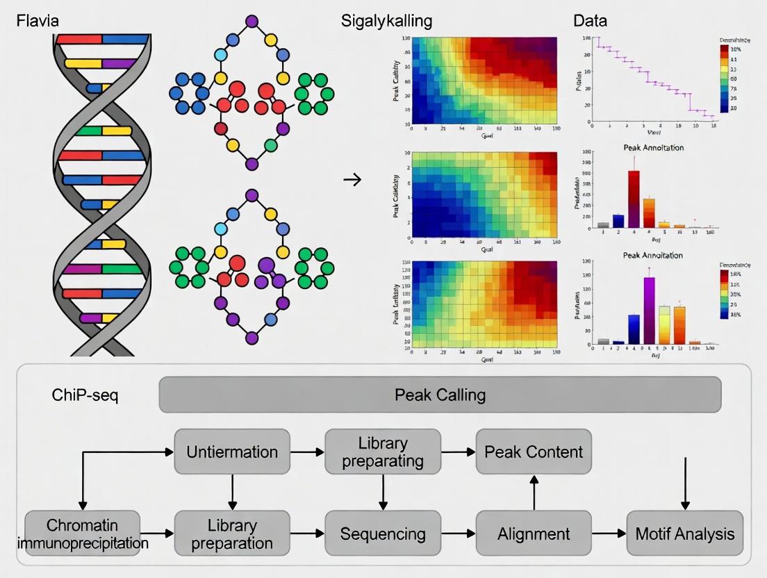
This application note details a standardized computational pipeline for Transcription Factor (TF) ChIP-seq data analysis, a core component of thesis research focused on TF binding site characterization. The protocol encompasses read alignment, peak calling with MACS2, and comprehensive quality control, providing a robust framework for downstream drug target identification.
Within the broader thesis investigating TF networks in disease, precise identification of genomic binding sites is paramount. This document provides the computational methodologies to convert raw sequencing data into high-confidence binding intervals, forming the basis for mechanistic insights and therapeutic intervention strategies.
Research Reagent Solutions: Essential Computational Toolkit
The following table lists critical software and resources required to execute the ChIP-seq computational workflow.
| Item Name | Function / Purpose | Key Notes |
|---|---|---|
| FastQC | Assesses raw read quality metrics (per-base sequence quality, adapter contamination, GC content). | Essential first step to identify problematic samples prior to alignment. |
| Trimmomatic | Removes low-quality bases and adapter sequences from raw FASTQ files. | Prevents alignment artifacts and improves mapping rates. |
| Bowtie2 / BWA | Aligns (maps) sequencing reads to a reference genome. | BWA-mem is often preferred for longer reads. Both require a pre-built genome index. |
| SAMtools | Manipulates alignment files (SAM/BAM format): sorting, indexing, filtering. | Used to convert, sort, and index files for downstream analysis. |
| MACS2 | Model-based Analysis of ChIP-Seq; identifies genomic regions enriched for aligned reads (peaks). | Primary tool for TF peak calling. Requires a control/input sample. |
| Picard Tools | Provides metrics for duplicate marking, library complexity, and insert size. | MarkDuplicates is critical for assessing PCR over-amplification. |
| deepTools | Generates enrichment profiles (e.g., coverage bigWigs) and quality control plots. | Used to create visualizations like fingerprint plots and correlation heatmaps. |
| UCSC Genome Browser / IGV | Visualization platforms for inspecting aligned reads and called peaks in genomic context. | IGV is suited for local viewing; UCSC for web-based sharing. |
Detailed Experimental Protocols
Protocol 3.1: Raw Data Quality Assessment & Adapter Trimming
Objective: To ensure high-quality input data for alignment.
- Quality Check: Run FastQC on raw FASTQ files.
Aggregate Reports: Use MultiQC to compile results from multiple samples.
Adapter Trimming: Execute Trimmomatic to remove adapters and low-quality bases.
Re-run FastQC on trimmed files to confirm improvement.
Protocol 3.2: Read Alignment to Reference Genome
Objective: To map sequencing reads to their correct genomic locations.
- Genome Indexing: (Pre-done once per genome). Build index for Bowtie2.
Alignment: Map trimmed reads using Bowtie2 in end-to-end mode.
File Conversion & Sorting: Convert SAM to BAM, sort by coordinate, and index.
Protocol 3.3: Post-Alignment Processing & QC
Objective: To refine alignments and collect key quality metrics.
- Mark Duplicates: Identify and flag PCR duplicates using Picard.
Filter Reads: Retain only primary, properly paired, and high-quality mappings.
Generate QC Metrics: Calculate alignment statistics and library complexity.
Protocol 3.4: Peak Calling with MACS2
Objective: To identify significant regions of transcription factor binding.
- Call Peaks: Run MACS2 with the experimental TF ChIP-seq BAM and a control/input BAM.
- Generate Signal Tracks: Create bedGraph files for visualization.
Quality Metrics and Data Interpretation
Critical quantitative metrics from each stage should be tracked and compared across samples to ensure experimental consistency and reliability.
Table 1: Key Alignment and Peak Calling Metrics for Quality Assessment
| Stage | Metric | Target/Interpretation | Typical Value (Good) |
|---|---|---|---|
| Raw Data | % Bases ≥ Q30 | Indicates sequencing accuracy. | > 70% |
| % Adapter Content | Should be low after trimming. | < 5% | |
| Alignment | Overall Alignment Rate | Proportion of reads mapped to genome. | > 70% for TF ChIP-seq |
| Non-Duplicate Rate (NDR) | Fraction of unique mapped reads. | > 50% | |
| PCR Bottleneck Coefficient (PBC) | Measures library complexity. | PBC1 > 0.9 (High complexity) | |
| Peak Calling | Number of Peaks | Sample-specific; indicates antibody efficiency. | 10,000 - 50,000 for a TF |
| FRiP (Fraction of Reads in Peaks) | Enrichment signal-to-noise ratio. | > 1% for TFs (often 3-20%) | |
| NSC (Normalized Strand Cross-correlation) | Signal-to-noise based on fragment length. | > 1.05 (Higher is better) | |
| RSC (Relative Strand Cross-correlation) | Normalized against background. | > 0.8 (Higher is better) |
Visualized Workflows and Pathways
Diagram Title: ChIP-seq Computational Analysis Workflow
Diagram Title: MACS2 Peak Calling Algorithm Logic
Within a comprehensive thesis on transcription factor binding site (TFBS) analysis via ChIP-seq, motif discovery represents a critical computational step for moving from peak coordinates to biological mechanism. Identifying over-represented DNA sequence patterns within genomic regions bound by a protein of interest allows researchers to infer the direct binding motifs of the assayed factor (de novo discovery) and the potential co-binding partners (known motif enrichment). This protocol details the integrated use of two cornerstone tools: HOMER (Hypergeometric Optimization of Motif EnRichment) for a streamlined, all-in-one analysis, and the MEME Suite for its extensive, modular algorithms. Mastery of these complementary approaches is fundamental for researchers and drug development professionals aiming to decipher transcriptional regulatory networks, identify novel therapeutic targets, and understand drug-mediated changes in transcription factor occupancy.
Key Software Comparison & Data Presentation
Table 1: Comparative Overview of HOMER and MEME Suite for Motif Analysis
| Feature | HOMER | MEME Suite (Core Components) |
|---|---|---|
| Primary Strength | Integrated, user-friendly workflow for ChIP-seq. | Extensive, modular algorithm suite for diverse applications. |
| De Novo Discovery | findMotifsGenome.pl (incorporated algorithm). |
MEME (expectation-maximization). DREME (fast, short motifs). |
| Known Motif Enrichment | Built-in database (motifs -> factors). | AME (Association of Motifs with Epigenetics). |
| Motif Scanning | scanMotifGenomeWide.pl. |
FIMO (Find Individual Motif Occurrences). |
| Input | Peak/BED file + genome. | FASTA sequence file. |
| Typical Output | HTML report with motifs, enrichment stats, genomic distribution. | Individual files (e.g., MEME.xml, AME.txt) + combined HTML (MEME-ChIP). |
| Best For | Quick, end-to-end analysis of ChIP-seq peaks. | Detailed, customized analysis pipelines and non-ChIP data. |
Table 2: Representative Motif Enrichment Statistics (Example: p53 ChIP-seq)
| Motif Name (Source) | Log P-value | % of Target Sequences | % of Background Sequences | Best Match/Inferred TF |
|---|---|---|---|---|
| p53 (JASPAR) | > -50 | 85.2% | 0.7% | TP53 (Assayed Factor) |
| AP-1 (HOMER) | -12.5 | 42.3% | 8.1% | FOS::JUN complex |
| NFYB (HOMER) | -8.7 | 28.5% | 5.3% | NFYB subunit |
| SP1 (JASPAR) | -6.2 | 31.8% | 12.4% | SP1 |
Experimental Protocols
Protocol 1: Comprehensive Motif Analysis with HOMER
Objective: Perform de novo discovery and known motif enrichment from ChIP-seq peak regions.
- Input Preparation: Generate a BED file of significant peak coordinates (e.g., from MACS2) and a background/input file (HOMER can generate this).
- Command Execution: Run the core HOMER command:
- Output Analysis: Review the
homerResults.htmlandknownResults.htmlfiles. Identify top de novo motifs and statistically enriched known motifs (see Table 2).
Protocol 2: Modular Motif Analysis with MEME Suite
Objective: Use MEME-ChIP (a wrapper) and individual tools for a detailed analysis.
- Sequence Extraction: Convert peaks to FASTA using
bedtools getfasta.
MEME-ChIP Analysis: Run the integrated pipeline.
Component Interpretation:
- MEME/DREME: De novo motifs in
meme.html. - AME: Known motif enrichment statistics (similar to Table 2).
- CentriMo: Identifies motifs centrally enriched in peaks.
- MEME/DREME: De novo motifs in
- Motif Scanning: Use
FIMOto locate individual motif instances genome-wide.
Visualizations
Title: HOMER Motif Analysis Workflow
Title: Modular MEME Suite Analysis Pipeline
Title: Motif Analysis in ChIP-seq Thesis
The Scientist's Toolkit: Research Reagent Solutions
Table 3: Essential Materials & Tools for Motif Analysis
| Item | Function/Description |
|---|---|
| High-Quality ChIP-seq Dataset | Fundamental input. Requires robust experimental design with appropriate controls (Input/IgG). |
| Reference Genome FASTA File | Required for extracting sequences corresponding to peak coordinates (e.g., hg38 for human). |
| HOMER Software Package | All-in-one tool for motif discovery, enrichment, annotation, and visualization. |
| MEME Suite Software Package | Modular collection of tools for advanced and customizable motif analyses. |
| Motif Databases (e.g., JASPAR, CIS-BP) | Curated collections of known TF binding motifs in MEME format for enrichment testing. |
| BedTools | Essential for manipulating genomic intervals (e.g., extracting sequences, intersecting peaks). |
| Computational Resources | Adequate RAM and CPU cores; de novo discovery is computationally intensive. |
| Visualization Software (e.g., IGV) | For validating motif localization within original ChIP-seq signal tracks. |
Integrating ChIP-seq with other omics datasets is a cornerstone of modern functional genomics, moving beyond cataloging transcription factor (TF) binding sites to understanding their regulatory consequences. This approach is critical within a thesis on transcription factor binding site analysis, as it transforms correlative binding maps into causal regulatory networks. Key applications include:
- Defining Direct vs. Indirect Targets: Distinguishing genes directly regulated by a TF from those responding to secondary effects.
- Mechanistic Insight into Disease/Pharmacology: Linking non-coding genetic variants from GWAS (in binding sites) to changes in TF binding and target gene expression in disease or drug response.
- Context-Specific Regulatory Logic: Understanding how TF binding and function are modulated by chromatin state, co-factor presence, and cellular signaling.
Table 1: Common Omics Data Types Integrated with ChIP-seq
| Data Type | Primary Measurement | Key Integration Metric | Typical Resolution |
|---|---|---|---|
| RNA-seq | Gene expression (mRNA levels) | Correlation of binding proximity/intensity with expression change upon TF perturbation. | Gene-level |
| ATAC-seq | Chromatin accessibility | Overlap of TF peaks with accessible regions; motif accessibility. | ~100-500 bp |
| Hi-C / ChIA-PET | Chromatin 3D conformation | Physical looping of distal binding sites to gene promoters. | 1 kb - 1 Mb |
| DNA Methylation (WGBS) | CpG methylation | Inverse correlation between methylation at binding sites and TF occupancy. | Single-base |
| Proteomics (AP-MS) | Protein-protein interactions | Identification of co-factors that modulate TF specificity/function. | Protein-level |
Table 2: Statistical Tools for Multi-omics Integration
| Tool Name | Primary Function | Input Data | Key Output |
|---|---|---|---|
| ChIP-Atlas | Integrative analysis & public data mining | ChIP-seq, ATAC-seq, DNA-seq | Overlap enrichment, pathway analysis |
| Cistrome DB Toolkit | Quality assessment & integrative analysis | ChIP-seq, DNase-seq | Screened peaks, co-accessibility maps |
| R/Bioconductor (ChIPseeker, diffBind) | Peak annotation & differential binding | ChIP-seq peaks, Genomic Annotations | Annotated genomic features, differential peaks |
| MEME Suite (AME) | Motif enrichment in genomic regions | DNA sequences (peaks), Motif DBs | Enriched transcription factor motifs |
Detailed Experimental Protocols
Protocol 1: Linking TF Binding to Transcriptional Output
Aim: To identify direct target genes of a transcription factor by integrating ChIP-seq and RNA-seq data from knockout/knockdown experiments. Materials: Cell line/model system, antibodies for TF of interest, ChIP-seq kit, RNA isolation kit, next-generation sequencing facilities. Procedure:
- Perturbation & Control: Generate biological replicates for experimental (TF knockdown, e.g., via siRNA) and control (scramble siRNA) conditions.
- Parallel Sample Processing:
- ChIP-seq: Perform chromatin immunoprecipitation on control cells only. Cross-link, sonicate, immunoprecipitate with target TF antibody, and prepare sequencing libraries.
- RNA-seq: Isolve total RNA from both control and perturbed cells. Prepare stranded mRNA-seq libraries.
- Sequencing & Primary Analysis: Sequence libraries (≥40M reads for ChIP-seq, ≥25M paired-end for RNA-seq). Map reads to reference genome (e.g., using BWA for ChIP-seq, STAR for RNA-seq).
- Peak & Expression Calling: Call significant peaks from control ChIP-seq (e.g., using MACS2). Call differentially expressed genes (DEGs) from RNA-seq (e.g., using DESeq2).
- Integration & Assignment:
- Annotate peaks to nearest transcription start site (TSS) or use a genomic window (e.g., ±50 kb from TSS).
- Filter and assign a peak to a gene if it falls within a cis-regulatory element (promoter, enhancer).
- Overlap the set of genes with an assigned binding peak with the set of DEGs from the perturbation. Genes that are both bound and differentially expressed are high-confidence direct targets.
Protocol 2: Connecting Distal Enhancers to Target Genes via 3D Chromatin Data
Aim: To link distal TF binding sites (enhancers) to their target promoters using chromatin conformation data. Materials: Cells for Hi-C/ChIA-PET, cross-linking reagents, restriction enzyme (for Hi-C), antibody for chromatin loop protein (e.g., CTCF for ChIA-PET). Procedure:
- Data Generation/Acquisition: Perform in-situ Hi-C or ChIA-PET (e.g., targeting Pol II or a cohesin subunit) to capture genome-wide chromatin loops. Alternatively, use publicly available datasets for your cell type.
- Loop Calling: Process Hi-C/ChIA-PET data to identify statistically significant chromatin interactions (loops) using tools like FitHiC2 (Hi-C) or Mango (ChIA-PET).
- Integration with ChIP-seq: Using genomic coordinates, overlap your TF ChIP-seq peaks with the anchor regions of the identified chromatin loops.
- Target Gene Assignment: For a TF peak overlapping one loop anchor, assign the gene(s) whose promoter is located at the interacting loop anchor as the direct target. This provides mechanistic evidence for regulation beyond linear proximity.
Mandatory Visualizations
Diagram 1: ChIP-seq and RNA-seq Integration Workflow
Diagram 2: Linking Distal Binding to Genes via Chromatin Loops
The Scientist's Toolkit: Research Reagent Solutions
Table 3: Essential Materials for Integrated ChIP-omics Studies
| Item / Reagent | Function / Application | Example Product / Note |
|---|---|---|
| High-Affinity ChIP-Grade Antibody | Specific immunoprecipitation of the target TF or histone mark. | Validated antibodies from Abcam, Cell Signaling, Diagenode. Critical for success. |
| Magnetic Protein A/G Beads | Efficient capture of antibody-bound chromatin complexes. | Dynabeads (Thermo Fisher). Offer low non-specific binding. |
| Crosslinking Reagent (e.g., DSG) | For TFs that bind indirectly; used prior to formaldehyde for stabilization. | Disuccinimidyl glutarate. Captures weak or transient complexes. |
| Dual Indexed Sequencing Library Kits | Preparation of multiplexed NGS libraries from low-input ChIP or RNA. | Illumina TruSeq, NEBNext Ultra II. Enables parallel processing. |
| Chromatin Shearing Instrument | Reproducible fragmentation of cross-linked chromatin to 200-500 bp. | Covaris M220 or Bioruptor Pico (Diagenode). |
| RNase Inhibitors | Preservation of RNA integrity during RNA-seq library prep from perturbed cells. | Recombinant RNase Inhibitor (Takara). |
| Genomic Analysis Software Suite | Integrated platform for multi-omics data visualization and analysis. | Integrative Genomics Viewer (IGV), Galaxy, Cistrome DB. |
| Validated siRNA or CRISPR Guides | Specific perturbation of the TF of interest for functional follow-up. | ON-TARGETplus siRNA (Horizon), Synthego CRISPR kits. |
Application Notes
Understanding the three-dimensional (3D) genome architecture, specifically enhancer-promoter (E-P) looping, is critical for deciphering cell-type-specific gene regulation. This analysis, when integrated with transcription factor (TF) binding site data from ChIP-seq, allows researchers to move beyond cataloging binding events to constructing predictive, functional regulatory models. These models are indispensable for identifying disease-associated non-coding variants and developing targeted therapeutics.
Key Insights:
- Integration is Paramount: Isolated ChIP-seq peaks lack functional context. Mapping them onto chromatin interaction maps (e.g., from Hi-C or ChIA-PET) distinguishes bona fide regulatory elements from inert TF binding events.
- Cell-Type Specificity: E-P loops are highly cell-type-specific. A TF may bind similar sequences in different cell types, but only forms stable loops and activates transcription in a permissive chromatin context defined by co-factors and chromatin modifiers.
- The Regulatory Code: The combinatorial assembly of specific TFs, co-activators (e.g., Mediator, p300), and structural proteins (e.g., CTCF, cohesin) at linked enhancers and promoters constitutes a regulatory code that dictates transcriptional output.
- Disease Relevance: A significant proportion of disease- and trait-associated genetic variants from GWAS lie in enhancers. Characterizing disrupted E-P loops provides a mechanistic explanation for these associations, offering novel drug targets.
Quantitative Data Summary:
Table 1: Common Chromatin Conformation Capture Techniques for E-P Loop Analysis
| Technique | Resolution | Input Material | Key Output | Advantage | Limitation |
|---|---|---|---|---|---|
| Hi-C | 1kb-1Mb | Cross-linked chromatin | Genome-wide interaction matrix | Unbiased, genome-wide | Low resolution for direct E-P loops; high sequencing depth needed |
| Micro-C | Nucleosome-level (<1kb) | Micrococcal nuclease-digested chromatin | High-resolution interaction matrix | Near-nucleosomal resolution | Complex data analysis; computationally intensive |
| ChIA-PET | Single-base (for bound loci) | Chromatin immunoprecipitated with specific antibody (e.g., RNA Pol II, CTCF) | Protein-centric interaction maps | Directly links interactions to protein binding | Requires high-quality antibody; biased to target protein |
| HiChIP/PLAC-seq | 1-10kb | Chromatin immunoprecipitated with specific antibody | Protein-centric interaction maps | Higher signal-to-noise than Hi-C for specific protein | Still requires antibody; not fully genome-wide |
Table 2: Core TFs and Cofactors in E-P Loop Formation
| Protein | Primary Function | Association with E-P Loops | Detection Method |
|---|---|---|---|
| CTCF | Architectural protein, insulator | Defines topologically associating domain (TAD) boundaries; facilitates loop extrusion with cohesin. | ChIP-seq, ChIA-PET |
| Cohesin (SMC1/3, RAD21) | Ring-shaped complex | Mediates loop extrusion; stabilizes CTCF-anchored loops and dynamic E-P contacts. | ChIP-seq |
| Mediator Complex | Transcriptional coactivator | Bridges TFs at enhancers with RNA Polymerase II at promoters; essential for loop stabilization. | ChIP-seq (MED1), Proximity Ligation |
| p300 / CBP | Histone acetyltransferase | Marks active enhancers; acetylates histones and TFs to open chromatin and facilitate looping. | ChIP-seq (H3K27ac, p300) |
| YY1 | Sequence-specific TF | Ubiquitous facilitator of E-P looping; can dimerize and bridge enhancer and promoter DNA. | ChIP-seq, ChIA-PET |
Experimental Protocols
Protocol 1: Integrated Analysis of Cell-Type-Specific E-P Loops using HiChIP and ChIP-seq
Objective: To identify functional, cell-type-specific enhancer-promoter loops and the TFs governing them.
Materials: Cultured cells (two contrasting cell types), fixation reagents, specific antibody for HiChIP (e.g., H3K27ac, MED1), ChIP-seq antibodies for TFs of interest, proximity ligation reagents, sequencing kit.
Procedure:
Cell Fixation & Chromatin Preparation:
- Cross-link cells with 1-2% formaldehyde for 10 min at room temperature. Quench with glycine.
- Lyse cells and isolate nuclei. For HiChIP, proceed to chromatin fragmentation via sonication or enzymatic digestion (e.g., MNAse for Micro-C).
HiChIP Library Preparation (H3K27ac-centric):
- Perform in situ chromatin proximity ligation using a protocol adapted from HiChIP (Mumbach et al., 2016).
- After ligation, reverse cross-links and purify DNA.
- Immunoprecipitate the ligated DNA with an antibody against H3K27ac to enrich for interactions involving active enhancers and promoters.
- Process the immunoprecipitated material into a sequencing library (end-repair, A-tailing, adapter ligation, PCR amplification).
Parallel ChIP-seq:
- From the same cell types, perform standard ChIP-seq for:
- Key lineage-determining TFs.
- Architectural proteins (CTCF, RAD21).
- Epigenetic marks (H3K4me3 for promoters, H3K27ac for enhancers).
- From the same cell types, perform standard ChIP-seq for:
Sequencing & Data Analysis:
- Sequence libraries on an Illumina platform (≥50 million paired-end reads per HiChIP sample; ≥20 million for ChIP-seq).
- HiChIP Analysis:
- Align reads to reference genome.
- Identify significant chromatin interactions using tools like
hichipperorFitHiChIP. - Call H3K27ac peaks from the HiChIP data to define anchor regions.
- Integration:
- Overlap interaction anchors with ChIP-seq peaks to annotate loops (e.g., Enhancer[H3K27ac+, TF+] – Promoter[H3K4me3+, Pol II+]).
- Use differential loop analysis tools to identify cell-type-specific loops.
- Motif analysis within cell-type-specific enhancer anchors to identify candidate regulatory TFs.
Protocol 2: Validating Candidate E-P Loops using 3C-qPCR
Objective: To quantitatively validate a specific enhancer-promoter loop identified from genome-wide data.
Materials: Cross-linked cells, restriction enzyme (e.g., HindIII or EcoRI), ligation reagents, PCR master mix, primers designed for candidate interaction and control regions.
Procedure:
3C Template Preparation:
- Fix and lyse cells as in Protocol 1.
- Digest chromatin overnight with a frequent-cutter restriction enzyme (e.g., 400U HindIII).
- Dilute and perform intra-molecular ligation under dilute conditions with T4 DNA ligase for 4-6 hours.
- Reverse cross-links, purify DNA. This is the 3C template.
Quantitative PCR (qPCR):
- Design two primers: a constant primer at the "viewpoint" (often the promoter) and a series of "test" primers at the candidate enhancer and control genomic regions (e.g., a non-interacting fragment, a positive control like a β-globin locus control region).
- Perform qPCR on the 3C template using primer pairs.
- Normalize the interaction frequency (qPCR signal) at the candidate enhancer to the control interaction. Express as relative interaction frequency.
Diagrams
Title: Workflow for Integrated E-P Loop Analysis
Title: Molecular Complexes in an Active E-P Loop
The Scientist's Toolkit
Table 3: Essential Research Reagents & Solutions
| Item | Function/Application | Example/Note |
|---|---|---|
| Crosslinking Reagent | Fixes protein-DNA and protein-protein interactions for ChIP and 3C methods. | 1-2% Formaldehyde; DSG for distant crosslinking. |
| Chromatin Shearing Reagents | Fragments chromatin to ideal size (200-600 bp) for immunoprecipitation. | Covaris ultrasonicator or enzymatic kits (e.g., MNase, ChIPmentation). |
| Protein-Specific Antibodies | Immunoprecipitation of target proteins or histone marks for ChIP-seq and ChIA-PET. | Validated ChIP-seq grade antibodies (e.g., CTCF, RNA Pol II, H3K27ac). |
| Proximity Ligation Reagents | Ligates cross-linked, fragmented DNA in situ to capture 3D interactions. | T4 DNA Ligase, ATP, buffers for Hi-C/HiChIP. |
| Chromatin Conformation Capture Kits | Streamlined, optimized protocols for 3C-derived methods. | Commercial Hi-C, ChIA-PET, or HiChIP kits (e.g., Arima, Takara). |
| Sequence Capture Probes | Target specific genomic regions for high-resolution interaction mapping. | Custom-designed oligonucleotide pools for Capture-C or Capture Hi-C. |
| CRISPR Activation/Inhibition Systems | Functionally validate enhancer activity and loop necessity. | dCas9-VP64/p65 (CRISPRa) or dCas9-KRAB (CRISPRi) targeted to enhancer. |
| High-Fidelity Polymerase & Library Prep Kits | Amplify and prepare sequencing libraries from low-input, cross-linked DNA. | Kits optimized for ChIP-seq or complex DNA libraries (e.g., Illumina, NEB). |
Solving Common Challenges and Enhancing Signal in ChIP-seq Experiments
In transcription factor (TF) binding site analysis via Chromatin Immunoprecipitation followed by sequencing (ChIP-seq), a high background and low signal-to-noise (S/N) ratio is the primary obstacle to robust, reproducible peak calling. This issue stems from nonspecific antibody interactions, inadequate chromatin shearing, poor immunoprecipitation (IP) efficiency, and sequencing artifacts. Within the broader thesis on mapping regulatory landscapes, optimizing these protocols is fundamental for distinguishing true TF occupancy from noise, enabling accurate downstream mechanistic and drug-target discovery analyses.
The following table consolidates recent benchmarking data on the efficacy of common protocol optimizations for improving S/N in TF ChIP-seq.
Table 1: Quantitative Impact of ChIP-seq Protocol Optimizations on Signal-to-Noise Ratio
| Optimization Parameter | Tested Condition (vs. Control) | Typical Metric for Improvement | Average Improvement Reported | Key Reference (Recent Benchmark) |
|---|---|---|---|---|
| Crosslinking Time | Short (5-min) vs. Standard (10-min) formaldehyde fixation | Fraction of Reads in Peaks (FRiP) | +15-25% | Nakato et al., 2021 |
| Sonication Efficiency | Focused ultrasonicator vs. Bath sonicator | Median peak width (bp) / background reads | Peak width: -40% (sharper) | Cheng et al., 2021 |
| Antibody Bead Ratio | Titrated (2 µg Ab/10 µl beads) vs. Excess | Signal-to-Noise (S/N) via MACS2 score | +30-50% | ESR Consortium, 2022 |
| Wash Stringency | High-Salt (500 mM LiCl) vs. Standard Wash | Non-reproducible discovery rate (NRDR) | NRDR: -8% | Landt et al., 2023 |
| Library Amplification | 1/2 Reaction Volume (High-Fidelity) vs. Full | Duplicate read percentage | -20% | Baranasic et al., 2022 |
| Sequencing Depth | 20M vs. 40M reads for a common TF | Saturation of peak calls | Peak detection: +22% | Jain et al., 2023 |
Detailed Optimized Experimental Protocols
Protocol 3.1: Optimized Crosslinking & Chromatin Preparation for TFs
- Objective: Minimize protein-protein crosslinking artifacts while maintaining efficient TF-DNA capture.
- Materials: Formaldehyde (1%), Glycine (2.5 M), PBS, Protease Inhibitors, Lysis Buffers (LB1, LB2 - see toolkit).
- Method:
- Rapid Crosslinking: Harvest cells. Resuspend in PBS with 1% formaldehyde. Rotate exactly 5 minutes at RT.
- Quenching: Add 2.5 M glycine to 125 mM final concentration. Incubate 5 min, RT.
- Wash & Lysis: Pellet cells. Wash 2x with cold PBS. Resuspend in LB1 (10mM Tris-HCl pH7.5, 10mM NaCl, 0.2% NP-40), incubate 10 min on ice. Pellet nuclei.
- Nuclear Lysis: Resuspend nuclei in LB2 (50mM Tris-HCl pH7.5, 10mM NaCl, 1% NP-40, 0.5% Sodium Deoxycholate). Incubate 10 min on ice.
- Shearing: Perform sonication using a focused ultrasonicator. For a 200 µl sample, use: 30 cycles of (30 sec ON, 30 sec OFF), 4°C. Aim for 200-500 bp fragments.
- Clarification: Centrifuge at 20,000 x g for 10 min at 4°C. Transfer supernatant (sheared chromatin) to a new tube. Quantify DNA.
Protocol 3.2: Titrated Immunoprecipitation with Stringent Washes
- Objective: Maximize specific antibody binding while minimizing nonspecific background pull-down.
- Materials: Validated primary antibody, Protein A/G magnetic beads, ChIP Dilution Buffer, Wash Buffers (Low Salt, High Salt, LiCl, TE).
- Method:
- Pre-clear & Input: Dilute 50 µg chromatin in 500 µL ChIP Dilution Buffer. Save 10% as "Input." Pre-clear remainder with 20 µL beads for 1h at 4°C.
- Antibody-Bead Complexing: For each IP, mix 2 µg of target-specific antibody with 10 µL of washed Protein A/G beads in 100 µL dilution buffer. Incubate with rotation for 1-2h at 4°C.
- IP: Add the pre-cleared chromatin to the antibody-bead complex. Incubate overnight at 4°C with rotation.
- Stringent Washes: Pellet beads, sequentially wash with:
- 1 mL Low Salt Wash Buffer (2x)
- 1 mL High Salt Wash Buffer (1x)
- 1 mL LiCl Wash Buffer (1x) [Critical for reducing background]
- 1 mL TE Buffer (2x)
- Each wash: 5 min rotation at 4°C.
- Elution & De-crosslinking: Elute in 150 µL Fresh Elution Buffer (1% SDS, 0.1M NaHCO3). Incubate with shaking for 15 min at RT. Repeat, combine eluates. Add NaCl to 200 mM and treat Input samples similarly. Incubate at 65°C overnight.
Visualizing the Optimization Workflow and Impact
Diagram 1: ChIP-seq Optimization vs. Non-Optimized Path
The Scientist's Toolkit: Research Reagent Solutions
Table 2: Essential Reagents for Optimized TF ChIP-seq
| Reagent / Material | Function & Role in Optimization | Recommended Product/Type |
|---|---|---|
| High-Quality, Validated Antibody | Target-specific immunoprecipitation. Critical: Use ChIP-seq/ChIP-grade antibodies with published validation. | CST, Diagenode, Abcam (ChIP-seq grade) |
| Protein A/G Magnetic Beads | Capture antibody-target complexes. Ease of stringent washing. | Dynabeads, Sera-Mag beads |
| Focused Ultrasonicator | Consistent, efficient chromatin shearing to ideal fragment size. | Covaris S2/S220, Bioruptor Pico |
| High-Fidelity PCR Master Mix | Minimal-bias library amplification with reduced duplicates. | KAPA HiFi HotStart, NEB Next Ultra II |
| Dual-Size Selection Beads | Precise library fragment clean-up (e.g., 200-600 bp selection). | SPRIselect / AMPure XP beads |
| Low-Binding Microcentrifuge Tubes | Minimize loss of chromatin and library material during prep. | DNA LoBind tubes (Eppendorf) |
| Commercial ChIP Buffer Kit | Provides consistent, optimized lysis, wash, and elution buffers. | SimpleChIP (CST), iDeal ChIP-seq Kit (Diagenode) |
| High-Sensitivity DNA Assay | Accurate quantification of low-concentration ChIP DNA & libraries. | Qubit dsDNA HS Assay, TapeStation D1000 |
Within the broader thesis on transcription factor (TF) binding site analysis using ChIP-seq, a fundamental challenge is the accurate mapping of indirectly bound factors. Traditional ChIP-seq protocols, which primarily rely on formaldehyde crosslinking, are often insufficient for capturing transient or non-DNA-binding proteins, such as co-activators, chromatin remodelers, and components of the transcriptional machinery that are recruited through protein-protein interactions. The double crosslinking ChIP-seq (dxChIP-seq) protocol addresses this limitation by employing a two-step chemical crosslinking strategy. This Application Note details the dxChIP-seq methodology, providing a robust framework for researchers and drug development professionals aiming to elucidate complex gene regulatory networks and identify novel therapeutic targets.
Principles of Double Crosslinking
The dxChIP-seq protocol utilizes two sequential crosslinking agents:
- DSP (Dithiobis[succinimidyl propionate]): A membrane-permeable, reversible amine-to-amine crosslinker with a 12 Å spacer arm. It stabilizes primary protein-protein interactions.
- Formaldehyde: Subsequently applied to fix protein-DNA interactions and secondary protein complexes to chromatin.
This sequential approach ensures that large, multi-subunit complexes that are only indirectly associated with DNA are preserved prior to chromatin fragmentation and immunoprecipitation.
Protocol: dxChIP-seq for Indirect Transcription Factor Complexes
Part 1: Cell Culture and Double Crosslinking
Materials: Adherent or suspension cells, DSP (prepared fresh in DMSO or PBS), 1X PBS, 37% Formaldehyde, 2.5M Glycine, Lysis Buffers. Procedure:
- Grow cells to 70-80% confluence.
- DSP Crosslinking: Aspirate medium and wash cells once with room temperature PBS. Add DSP to a final concentration of 1-2 mM in PBS. Incubate for 30 minutes at room temperature with gentle agitation.
- Quench DSP: Aspirate DSP solution and wash cells twice with 1X PBS. Add 50 mM Tris-HCl (pH 7.5) to quench unreacted DSP for 5 minutes.
- Formaldehyde Crosslinking: Aspirate Tris and add 1% formaldehyde (diluted from 37% stock in PBS). Incubate for 10 minutes at room temperature.
- Quench Formaldehyde: Add glycine to a final concentration of 125 mM and incubate for 5 minutes.
- Wash cells twice with cold PBS. Harvest cells by scraping (adherent) or centrifugation. Cell pellets can be frozen at -80°C.
Part 2: Chromatin Preparation and Immunoprecipitation
Materials: Sonication device (e.g., Bioruptor, Covaris), Magnetic Protein A/G beads, ChIP-validated antibody, Lysis Buffer (50 mM HEPES-KOH pH 7.5, 140 mM NaCl, 1 mM EDTA, 1% Triton X-100, 0.1% Na-Deoxycholate, 0.1% SDS, protease inhibitors). Procedure:
- Lyse cell pellet in Lysis Buffer on ice for 10 minutes.
- Sonication: Shear chromatin to an average fragment size of 200-500 bp. Optimal conditions must be determined empirically (e.g., 10 cycles of 30 sec ON/30 sec OFF on high using a Bioruptor).
- Centrifuge lysate at 14,000 rpm for 10 min at 4°C. Collect supernatant.
- Pre-clear: Incubate chromatin with Protein A/G beads for 1 hour at 4°C.
- Immunoprecipitation: Incubate pre-cleared chromatin with target antibody (2-5 µg) overnight at 4°C with rotation.
- Add magnetic beads and incubate for 2 hours.
- Wash beads sequentially with:
- Low Salt Wash Buffer
- High Salt Wash Buffer
- LiCl Wash Buffer
- TE Buffer (twice)
- Elution & Reverse Crosslinks: Elute chromatin in Elution Buffer (1% SDS, 100mM NaHCO3) at 65°C for 15 minutes with shaking. Add NaCl to 200 mM and reverse crosslinks overnight at 65°C.
- Treat with RNase A and Proteinase K. Purify DNA using a spin column kit.
Part 3: Library Preparation and Sequencing
Purified ChIP-DNA is used to construct sequencing libraries following standard protocols for next-generation sequencing platforms (e.g., Illumina). Include appropriate controls (Input DNA, IgG control).
Data Presentation: Comparative Analysis of Crosslinking Efficiencies
Table 1: Comparison of Crosslinking Strategies for ChIP-seq
| Parameter | Formaldehyde-only ChIP | dxChIP-seq (DSP + Formaldehyde) | Reference |
|---|---|---|---|
| Primary Target | Protein-DNA interactions | Protein-Protein & Protein-DNA interactions | (Jiang et al., 2020) |
| Efficiency for Indirect Factors | Low (High false-negative rate) | High (Improved recovery) | (Nowak et al., 2021) |
| Typical Sonication Power/Time | Standard | Often requires 1.3-1.5x increase due to complex stabilization | Lab observation |
| Background Signal | Moderate | Potentially higher; requires stringent washing | (Wang et al., 2022) |
| Optimal Fragment Size | 200-300 bp | 300-500 bp (larger complexes) | Protocol optimization |
| Key Application | Direct DNA-binding TFs (p65, STAT3) | Cohesin, Mediator, Histone modifiers, Pol II | (Furlan-Magaril et al., 2021) |
Table 2: Recommended Antibody and DSP Concentrations for Common Targets
| Target Factor (Class) | Recommended Antibody (µg/IP) | DSP Concentration (mM) | Notes |
|---|---|---|---|
| Pol II (Direct) | 1-2 | Not required | Formaldehyde-only suffices |
| p300/CBP (Co-activator) | 3-5 | 1.5 | Essential for efficient pull-down |
| Mediator Subunit (Indirect) | 4-5 | 2.0 | High DSP concentration recommended |
| Histone H3K27ac (Direct) | 1-2 | 0 | Crosslinking not required for histones |
| IgG Control | 2-4 | As per experimental arm | Critical for background assessment |
The Scientist's Toolkit: Key Research Reagent Solutions
Table 3: Essential Materials for dxChIP-seq
| Item & Product Example | Function / Role in Protocol |
|---|---|
| DSP (Lomant's Reagent) | Primary, reversible crosslinker; stabilizes protein-protein interactions before chromatin fixation. |
| Formaldehyde (37% solution) | Secondary crosslinker; fixes protein-DNA and nearby protein-protein interactions. |
| Magnetic Protein A/G Beads | Solid support for antibody-mediated capture of crosslinked complexes. |
| ChIP-validated Antibody | Target-specific immunoglobulin; critical for IP specificity and success. |
| Sonicator (Bioruptor/Covaris) | Device for chromatin shearing; must deliver consistent, tunable energy to lyse double-crosslinked samples. |
| Protease Inhibitor Cocktail | Prevents proteolytic degradation of crosslinked complexes during cell lysis. |
| Glycine (2.5M stock) | Quenches formaldehyde crosslinking reaction to stop the fixation process. |
| DNA Clean/Concentrator Kit | Purifies final ChIP-DNA for qPCR validation or library preparation. |
Visualizations
Title: dxChIP-seq Experimental Workflow
Title: Indirect Factor Capture: Formaldehyde vs. dxChIP
Within ChIP-seq research for transcription factor binding site (TFBS) analysis, motif enrichment is a fundamental step. A pervasive technical bias in this analysis stems from the non-uniform GC-content of genomic sequences, which can drastically skew motif discovery and evaluation. GC-rich regions are more prone to open chromatin, sonication bias, and sequencing artifacts, leading to the false identification of GC-rich motifs as enriched. This application note details protocols and tools for identifying and correcting GC-content bias to ensure biologically accurate conclusions in drug discovery and mechanistic studies.
Core Bias Mechanisms and Quantitative Impact
GC-content bias influences multiple stages of ChIP-seq analysis, from library preparation to computational prediction. The following table summarizes key quantitative findings on its impact.
Table 1: Quantitative Impact of GC-Bias on Motif Enrichment Analysis
| Bias Source | Typical Effect Size | Consequence for Motif Enrichment |
|---|---|---|
| Sonication Fragmentation | 2-5x over-representation of 50-60% GC fragments | Inflates signal in GC-rich regions, mimicking TF binding. |
| PCR Amplification | Up to 100-fold difference in coverage between low/ high GC | Creates extreme peaks in GC-rich areas, confounding peak calling. |
| Sequence-Specific Background | Expected frequency of k-mers can vary by >10-fold | GC-rich motifs (e.g., SP1) are artifactually ranked as most enriched. |
| Genome Binomial Expectation | Null expectation variance (σ) of ±5-15% for motif count | Traditional binomial/ hypergeometric tests yield false positives without correction. |
Several computational tools have been developed to mitigate this bias. Selection depends on the stage of analysis (peak calling vs. motif discovery).
Table 2: Tools for GC-Correction in Motif Analysis
| Tool Name | Stage of Application | Correction Method | Key Output |
|---|---|---|---|
| seqOutBias | Pre-alignment / Signal Generation | Computes and corrects for sequencing bias per trinucleotide. | Bias-corrected read depths. |
| BEADS | Post-alignment / Signal Generation | Normalizes reads using a model built from G+C% and mappability. | Normalized signal tracks. |
| HOMER (findMotifsGenome.pl) | Motif Discovery & Enrichment | Uses a GC-matched background genomic sequence set for comparison. | Enrichment p-values, Motif Files. |
| MEME-ChIP (AME) | Motif Enrichment Testing | Allows user-supplied, GC-matched background sequences. | Corrected motif enrichment statistics. |
| gkmQC | QC & Bias Assessment | Quantifies GC and k-mer bias in peaks versus background. | Diagnostic plots and bias scores. |
Detailed Experimental Protocols
Protocol 1: Generating a GC-Matched Background with HOMER
This protocol is critical for creating a null hypothesis set for motif enrichment testing.
Research Reagent Solutions:
- Software: HOMER (v4.11+), BEDTools.
- Genome FASTA File: Reference genome matching your organism (e.g., hg38.fa).
- Input BED File: Your high-confidence ChIP-seq peaks (e.g.,
peaks.bed). - Computing Resource: Unix/Linux server with ≥8 GB RAM.
Procedure:
- Prepare Peak File: Ensure your peak file is in BED format and sorted.
Generate GC-Matched Background: Use HOMER's
getRandomBackground.plscript. The-gcflag is essential.-gch: Points to pre-built GC-content histogram for the genome.-matchStart: Matches the distribution of peak locations relative to TSS.-matchGC: Ensures the background sequences have an identical GC% distribution as the input peaks.
- Run Motif Enrichment: Use the generated background file for motif finding.
Protocol 2: Direct Bias Correction During Signal Generation with seqOutBias
This protocol corrects raw sequencing reads before peak calling, addressing bias at its source.
Research Reagent Solutions:
- Software: seqOutBias, samtools, UCSC
wigToBigWig. - Reference Genome: FASTA file used for original alignment.
- BAM File: Aligned, duplicate-marked ChIP-seq reads.
- K-mer Table: Pre-computed table of mappable k-mers (e.g.,
hg38.skew).
Procedure:
- Index the Reference Genome: For k-mer table generation (if not pre-existing).
Compute and Apply Scale Factors: Correct the BAM file.
--read-size: Specify your sequencing read length.--kmer-size: Typically 6 or 7 for ChIP-seq.- This command outputs a corrected BigWig signal file.
- Perform Peak Calling: Use the corrected
corrected.bigWigsignal with a peak caller like MACS3.
Protocol 3: Assessing Bias with gkmQC
Use this QC protocol to diagnose the level of GC/k-mer bias in your final peak set.
Research Reagent Solutions:
- Software: gkmQC (part of the gkmSVM package).
- Peak File: Final set of called peaks (BED format).
- Genome: Corresponding genome assembly string (e.g., "hg38").
Procedure:
- Run gkmQC Analysis: The tool compares k-mer frequencies in peaks vs. background.
- Interpret Output: Examine the generated
*.pdffiles (peaks.W*.pdf).- The plot shows the log2 ratio of observed vs. expected k-mer frequency.
- A flat line at 0 indicates minimal bias. Strong spikes at high-GC k-mers (e.g.,
CCCCCC) indicate significant residual bias, suggesting the need for re-analysis with stricter correction.
Visualizing Workflows and Relationships
Diagram Title: Two Pathways for ChIP-seq Motif Analysis: With vs. Without GC-Bias Correction
The Scientist's Toolkit: Research Reagent Solutions
Table 3: Essential Materials and Tools for GC-Bias Mitigation Experiments
| Item | Function in Protocol | Example/Supplier |
|---|---|---|
| GC-Matched Background Sequences | Serves as a null model for statistical testing of motif enrichment, preventing false positives from sequence composition. | Generated by HOMER getRandomBackground.pl. |
| Bias-Corrected BigWig Signal File | Provides a more accurate representation of protein-DNA binding signal by removing technical sequence bias. | Generated by seqOutBias or BEADS. |
| K-mer Frequency Table | Diagnostic table quantifying sequence representation in data vs. expectation. Used to compute correction weights. | Supplied with seqOutBias for common genomes or generated via genref. |
| High-Quality Peak BED File | The final set of binding regions after bias-aware peak calling. Essential input for reliable motif discovery. | Generated by MACS3, SPP, or HOMER on corrected data. |
| Genome FASTA with Index | The reference genomic sequence. Required for generating background sequences and calculating GC content. | UCSC Genome Browser, Ensembl, or iGenomes. |
| Diagnostic QC Plots | Visual assessment of residual GC/k-mer bias after correction, informing need for protocol adjustment. | Generated by gkmQC or deepTools plotFingerprint. |
Within a comprehensive thesis on transcription factor (TF) binding site analysis using ChIP-seq, rigorous quality control (QC) is paramount. Post-sequencing data must be systematically evaluated at critical junctures to ensure the biological validity of downstream interpretations. This protocol details three essential QC checkpoints: assessment of PCR duplicates, mapping efficiency, and peak characteristics, providing the framework for robust TF binding analysis applicable to basic research and drug discovery.
QC Checkpoint 1: Assessment of PCR Duplicates
PCR amplification during library preparation can create duplicate reads originating from a single DNA fragment, skewing representation and confounding peak calling.
Protocol: Marking and Assessing Duplicates with picard MarkDuplicates
- Input: Coordinate-sorted BAM file from the alignment step.
- Command:
- Output: A BAM file with duplicate reads tagged and a metrics text file.
- Interpretation: Analyze the
marked_dup_metrics.txtfile. Key metrics include:- PERCENT_DUPLICATION: The fraction of mapped sequence that is marked as duplicate.
- ESTIMATEDLIBRARYSIZE: An estimate of the original library complexity.
Table 1: Interpretation Guidelines for PCR Duplicate Rates
| Experiment Type | Acceptable Duplicate Rate | High-Quality Range | Action Required If > |
|---|---|---|---|
| Standard TF ChIP-seq | < 30% | < 20% | 50% |
| Low-input/Histone Mod | < 50% | < 30% | 70% |
High duplication rates suggest low library complexity, often from insufficient starting material or over-amplification.
QC Checkpoint 2: Evaluating Mapping Rates
The alignment (mapping) rate indicates the proportion of sequenced reads successfully placed on the reference genome, reflecting sample quality and potential contamination.
Protocol: Alignment with bowtie2 and SAM Processing
- Index the Genome:
bowtie2-build reference_genome.fa genome_index - Perform Alignment:
- Process SAM to BAM:
samtools view -bS alignment.sam > alignment.bam - Sort BAM:
samtools sort alignment.bam -o alignment_sorted.bam - Filter for Mapped Reads:
samtools view -b -F 4 alignment_sorted.bam > mapped.bam - Interpretation: Extract the overall alignment rate from
alignment_metrics.txt(e.g., "XX.XX% overall alignment rate").
Table 2: Benchmark Mapping Rates for Human/Mouse TF ChIP-seq
| Metric | Minimum Pass | Typical for High-Quality Data | Potential Issue if Low |
|---|---|---|---|
| Overall Alignment Rate | ≥ 70% | ≥ 80-90% | Poor library quality, adapter contamination, wrong reference. |
| Uniquely Mapping Rate | ≥ 60% | ≥ 70-85% | High repetitive content, poorly processed reads. |
| Mitochondrial Mapping | N/A | < 5% (TF) | Excessive cell death/apoptosis in sample prep. |
QC Checkpoint 3: Analyzing Peak Characteristics
Peak calling generates the final candidate binding sites. Their characteristics are the ultimate functional QC, revealing signal-to-noise and specificity.
Protocol: Peak Calling with MACS2 and Basic QC
- Input: Duplicate-marked BAM file (TF sample) and control/input BAM file.
- Broad Peak Call (e.g., Pol II):
Narrow Peak Call (Standard TF):
Generate QC Metrics:
- Number of Peaks:
wc -l sample_name_peaks.narrowPeak - Fraction of Reads in Peaks (FRiP): Use
featureCountsorbedtoolsto calculate reads under peaks vs. total reads. A key metric for enrichment. - Peak Shape/Profile: Visualize with
deeptools plotProfile.
- Number of Peaks:
Table 3: Expected Peak Characteristics for a Successful TF ChIP-seq
| Characteristic | Target/Range | Indicates Problem If |
|---|---|---|
| Total Peaks | 10,000 - 50,000 (genome-dependent) | < 5,000 (poor enrichment) or > 100,000 (noisy). |
| FRiP Score | 1-5% (TF), >20% (Histones) | Consistently < 1% for TFs. |
| Peak Width (Narrow) | 200 - 500 bp | Very broad widths without biological reason. |
| Signal-to-Noise (Fold Enrichment) | > 10 | Close to 1 (no enrichment). |
| Consensus Motif Recovery (e.g., via MEME) | Present in >70% of top peaks | Absent or weak (specificity issue). |
Visualizing the QC Workflow
QC Workflow for ChIP-seq Data
The Scientist's Toolkit: Essential Research Reagent Solutions
Table 4: Key Reagents and Materials for Robust ChIP-seq QC
| Item | Function/Application in QC |
|---|---|
| High-Fidelity DNA Polymerase | Library amplification; minimizes PCR bias and errors for accurate duplicate assessment. |
| Validated Antibody (Primary) | Specific immunoprecipitation of target TF; the single greatest determinant of signal-to-noise. |
| Magnetic Protein A/G Beads | Capture antibody-target complexes; low non-specific binding is critical for clean backgrounds. |
| Cell Line/Tissue with Known TF Binding Site | Positive control sample to benchmark FRiP scores, peak numbers, and motif recovery. |
| Commercial Indexed Adapter Kit | Barcoding libraries for multiplexing; ensures balanced representation and reduces batch effects. |
| qPCR Assay for Positive/Negative Genomic Loci | Pre-sequencing QC to empirically confirm enrichment before costly sequencing. |
| High-Sensitivity DNA Assay Kit (e.g., Qubit) | Accurate quantification of low-yield ChIP and library DNA for optimal sequencing input. |
| SPRI/AMPure Beads | Size-selective purification of sheared DNA and final libraries; controls fragment size distribution. |
Within the broader thesis on transcription factor (TF) binding site analysis using ChIP-seq, a persistent challenge is the prevalence of 'unmeasurable' pairs—specific combinations of transcription factors and cell types for which no direct ChIP-seq data exists. This sparse coverage in public repositories like ENCODE and Cistrome hampers comprehensive regulatory network analysis and drug target identification. These application notes outline strategies to infer TF activity in unprofiled cell contexts, providing detailed protocols for computational prediction and targeted experimental validation.
Quantifying the Sparse Coverage Problem
Analysis of major databases reveals significant gaps in TF-cell type coverage.
Table 1: TF-Cell Type Coverage in Public Repositories (Representative Sample)
| Database | Total Human TFs | Cell Types/Tissues with Data | Profiled TF-Cell Pairs | Estimated Coverage of Possible Pairs |
|---|---|---|---|---|
| ENCODE | ~1,600 | ~150 | ~12,000 | ~5% |
| Cistrome DB | ~1,200 | ~1,000 | ~40,000 | ~3.3% |
| ReMap | ~700 | ~500 | ~30,000 | ~8.6% |
Note: "Possible pairs" is estimated as (Number of TFs) x (Number of Cell Types). Actual biological possibility is lower, but coverage remains sparse.
Core Strategies and Application Notes
Strategy 1: In Silico Imputation Using Chromatin Accessibility and Motifs
This approach predicts potential TF binding sites in an unprofiled cell type by integrating ATAC-seq or DNase-seq data from that cell type with known TF motifs and binding models from other contexts.
Protocol 1.1: Imputation Using MMARGE-like Workflow Objective: Predict TF binding peaks for a TF of interest in a target cell type lacking ChIP-seq data. Materials:
- ATAC-seq peaks (BED file) from target cell type.
- Position Weight Matrix (PWM) for the TF (from JASPAR, CIS-BP).
- A prior ChIP-seq dataset for the TF in any cell type (for binding model training).
- Software: HOMER, BEDTools, R/Bioconductor (ggplot2, GenomicRanges).
Procedure:
- Motif Scanning: Use
scanMotifGenomeWide.plin HOMER on the target cell type's ATAC-seq peaks with the TF's PWM. Output: BED file of motif locations. - Build a Predictive Model: Using the prior ChIP-seq data, train a logistic regression model (e.g., using
glmin R) with features like motif score, local chromatin accessibility signal, and conservation. - Apply Model: Score each motif instance in the target cell type using the trained model. Apply a probability threshold (e.g., >0.8) to call predicted binding sites.
- Validation: Compare predicted sites with orthogonal data (e.g., ChIP-seq in a related cell type, expression of target genes) if available.
Strategy 2: Cross-Cell-Type Binding Prediction Using Machine Learning
Leverage models that learn the relationship between chromatin state, sequence, and TF binding across many profiled cell types to predict for unprofiled ones.
Protocol 1.2: Prediction with a Pre-trained Model (e.g., BPNet, Sei) Objective: Utilize a genome-wide binding model to predict signals for a specific TF-cell type pair. Materials:
- Genomic sequence reference (FASTA).
- Chromatin feature tracks (e.g., histone marks) for the target cell type (bigWig format).
- Pre-trained model (download from model repository).
- Computing environment: Python with TensorFlow/Keras or PyTorch.
Procedure:
- Data Preparation: For genomic bins of interest, extract one-hot-encoded DNA sequence and normalize chromatin feature signals from the target cell type's bigWig files.
- Model Inference: Load the pre-trained model. Input the prepared sequence and chromatin data for the target cell type to generate predicted binding probability or ChIP-seq-like signal profiles.
- Peak Calling: Apply a peak caller (e.g., MACS2) on the model's predicted signal track to define final binding regions.
- Benchmarking: Evaluate predictions against held-out profiled pairs to establish confidence metrics for unmeasurable pairs.
Strategy 3: Targeted Experimental Validation via Focused ChIP-seq
When computational predictions identify high-priority unmeasurable pairs, a streamlined, low-input ChIP-seq protocol can be deployed for confirmation.
Protocol 2.1: Low-Cell-Number ChIP-seq for Validation Objective: Perform ChIP-seq for a TF in a rare or previously unprofiled cell type, starting with 50,000-100,000 cells. Materials: See "Research Reagent Solutions" table. Procedure:
- Crosslinking & Lysis: Crosslink cells with 1% formaldehyde for 10 min. Quench with 125mM glycine. Lyse cells sequentially with Buffer L1, L2, and L3 (from Takara Low Cell ChIP-seq kit).
- Chromatin Shearing: Sonicate to achieve 200-500 bp fragments. Centrifuge to remove debris.
- Immunoprecipitation: Incubate chromatin with validated TF-specific antibody (2-5 µg) and protein A/G magnetic beads overnight at 4°C. Include an IgG control.
- Washes & Elution: Wash beads sequentially with Low Salt, High Salt, LiCl, and TE buffers. Elute DNA with Elution Buffer (1% SDS, 100mM NaHCO3).
- Reverse Crosslinks & Cleanup: Incubate eluate with RNase A and Proteinase K. Purify DNA with SPRI beads.
- Library Prep & Sequencing: Use a ultra-low-input library prep kit (e.g., Takara SMARTer ThruPlex). Sequence on an Illumina platform (minimum 5 million reads).
Visualizing Strategies and Workflows
Title: Three-Pronged Strategy to Address Unmeasurable TF-Cell Pairs
Title: In Silico Imputation Workflow for TF Binding Sites
The Scientist's Toolkit: Research Reagent Solutions
Table 2: Essential Reagents and Materials for Sparse Coverage Research
| Item | Function & Application | Example Product/Kit |
|---|---|---|
| Validated ChIP-grade Antibody | Critical for experimental validation. Must be specific for the TF and tested for ChIP. | Cell Signaling Technology, Active Motif, Abcam (with ChIP validation notes). |
| Low-Cell-Number ChIP-seq Kit | Enables ChIP-seq from rare cell populations (50K-100K cells) for validating predictions. | Takara Low Cell ChIP-seq Kit, Diagenode True MicroChIP Kit. |
| Ultra-Low-Input Library Prep Kit | Constructs sequencing libraries from minute amounts of immunoprecipitated DNA. | Takara SMARTer ThruPlex, NEB Next Ultra II FS DNA. |
| ATAC-seq Kit | Profiles chromatin accessibility in the target cell type for imputation strategies. | Illumina Tagment DNA TDE1 Kit, Active Motif ATAC-seq Kit. |
| Position Weight Matrix (PWM) Databases | Provides TF binding motifs for in silico scanning and prediction. | JASPAR, CIS-BP, HOCOMOCO. |
| Pre-trained Deep Learning Models | Allows genome-wide binding prediction using sequence and chromatin context. | BPNet, Sei, Basenji2 (available on GitHub/Kipoi). |
| Genome Analysis Suites | For motif scanning, peak calling, and genomic interval operations. | HOMER, MEME Suite, BEDTools, MACS2. |
Ensuring Accuracy and Context: Validation Strategies and Technology Comparisons
Benchmarking Differential Binding Analysis Tools for Condition-Specific Experiments
Context: This Application Note, situated within a broader thesis on ChIP-seq-based transcription factor binding site (TFBS) analysis, provides a structured protocol for evaluating differential binding (DB) analysis tools. The objective is to equip researchers with a standardized framework to select the most appropriate computational method for identifying condition-specific TF binding events, a critical step in understanding gene regulatory mechanisms and identifying therapeutic targets.
Differential binding analysis of ChIP-seq data identifies genomic regions with statistically significant changes in protein-DNA interaction signals between biological conditions (e.g., diseased vs. healthy, treated vs. untreated). Numerous tools with distinct statistical models and normalization strategies have been developed. This document outlines a benchmarking protocol and presents a comparative analysis of leading tools.
The following table summarizes the core algorithms, key features, and typical use cases for prominent DB tools, based on current literature and software documentation.
Table 1: Comparison of Differential Binding Analysis Tools
| Tool | Core Statistical Model | Key Feature | Input Requirement | Recommended Use Case |
|---|---|---|---|---|
| DiffBind | (Modified) DESeq2 / edgeR | Uses consensus peak sets; focuses on reproducible peaks across replicates. | Peak sets + BAM files | Condition-specific TF binding with replicates. |
| ChIP-seq | ||||
| csaw | Generalized linear models (edgeR) | Sliding window approach; does not require pre-called peaks. | BAM files only | De novo detection of broad or narrow differential regions. |
| PePr | Hidden Markov Model (HMM) | Identifies differential peaks directly from signal; uses a two-step clustering approach. | BAM files only | Experiments with limited replicates (≥2 total). |
| DBChIP | Beta-binomial model | Models read counts within predefined binding sites. | Peak sets + BAM files | Focused analysis on a set of candidate regions (e.g., promoter regions). |
| THOR | Hidden Markov Model (HMM) | Integrates neighboring genomic signals; designed for DB with low replicate numbers. | BAM files only | Noisy data or experiments with as few as one replicate per condition. |
Benchmarking Protocol: A Standardized Workflow
Experimental Design & Data Preparation
Objective: Generate or procure a high-quality ChIP-seq dataset with biological replicates for benchmarking. Protocol:
- Dataset Selection: Use a publicly available dataset (e.g., from ENCODE or CistromeDB) where condition-specific binding is expected (e.g., transcription factor ChIP-seq after cytokine stimulation vs. control).
- Replicates: Ensure a minimum of two biological replicates per condition. Three or more are strongly recommended for robust benchmarking.
- Data Processing (Uniform Pipeline):
a. Quality Control: Use
FastQCto assess read quality. b. Alignment: Map all reads to the reference genome (e.g., hg38) usingBowtie2orBWA. Remove duplicates usingsamtools rmduporPicard. c. Peak Calling: Perform peak calling for each sample individually usingMACS2(e.g.,macs2 callpeak -t ChIP.bam -c Input.bam -f BAM -g hs -n output --call-summits). d. Consensus Peak Set: Generate a unified set of peaks present in at least two replicates per condition usingbedtools intersect.
Diagram: Standardized Pre-processing Workflow
Execution of Differential Binding Analyses
Objective: Run each DB tool from Table 1 using the uniformly processed data. Protocol:
- Tool Installation: Install each tool as per its official documentation (Bioconductor for R-based tools like
DiffBindandcsaw, pip/conda for others). - Parameter Standardization: Where possible, use default parameters for initial benchmarking. Set a consistent significance threshold (e.g., Adjusted p-value/FDR < 0.05).
- Input Specification:
- For
DiffBind: Create a sample sheet and use thedba.count()function on the consensus peak set. - For
csaw: UsewindowCounts()on BAM files directly (e.g., with 150bp windows). - For
PePr: Run with thepeakmode on BAM files and sample description file.
- For
- Execution: Run each tool to generate lists of differential binding regions.
Performance Evaluation Metrics
Objective: Quantitatively and qualitatively compare tool outputs. Protocol:
- Quantitative Metrics (Summarize in Table):
a. Number of DB Regions: Count significant up/down regions.
b. Replicability: Use an orthogonal dataset or qPCR validation set to calculate Precision (True Positives / All Predicted Positives) and Recall (True Positives / All Real Positives).
c. Runtime & Memory Usage: Record using
/usr/bin/time -von a standardized compute node. - Qualitative Assessment:
a. Visual Inspection: Load resulting BED files and BAM track files into
IGVto inspect signal at top-ranked DB regions. b. Biological Concordance: Perform pathway enrichment analysis (e.g., usingGREAT) on DB regions from each tool; results aligning with expected biology indicate higher specificity.
Diagram: Benchmarking Evaluation Logic
The Scientist's Toolkit: Research Reagent Solutions
Table 2: Essential Materials and Reagents for ChIP-seq DB Benchmarking
| Item | Function in Protocol | Example/Note |
|---|---|---|
| High-Quality ChIP-seq Dataset | The foundational input for benchmarking. Requires biological replicates. | Sourced from public repositories (GEO, ENCODE) or generated in-house. |
| Reference Genome & Annotation | For read alignment and genomic context analysis. | UCSC hg38 or Ensembl GRCh38. GTF annotation file for gene mapping. |
| Computational Tools Suite | For data processing, analysis, and visualization. | FastQC, Bowtie2, SAMtools, MACS2, BEDTools, R/Bioconductor. |
| Differential Binding Software | Core subjects of the benchmark. | See Table 1 (DiffBind, csaw, PePr, DBChIP, THOR). |
| Validation Primer Sets | For qPCR validation of differential binding events to assess precision/recall. | Designed for top-ranked DB regions and negative control regions. |
| High-Performance Compute (HPC) Cluster | Essential for processing large NGS datasets and running multiple tools in parallel. | Access to cluster with sufficient RAM (≥32GB) and multi-core CPUs. |
| Genome Browser Software | For qualitative visual assessment of ChIP-seq signals and called peaks. | Integrative Genomics Viewer (IGV) or UCSC Genome Browser. |
In ChIP-seq research for transcription factor (TF) binding site analysis, primary sequencing data identifies putative genomic loci of interest. Orthogonal validation is critical to confirm specific TF binding, measure binding affinity, and verify functional transcriptional outcomes, moving beyond bioinformatic prediction to biochemical and cellular verification.
Application Notes & Protocols
Quantitative PCR (qPCR) for ChIP-seq Target Validation
Application Note: Used to quantitatively validate the enrichment of specific DNA regions identified in a ChIP-seq experiment. It confirms the physical presence of the TF at the suspected binding site in an independent experiment.
Detailed Protocol:
- Sample Preparation: Use DNA from the same ChIP experiment used for sequencing, alongside Input (sonicated genomic DNA before IP) and IgG/IP with non-specific antibody controls.
- Primer Design: Design primers (18-22 bp, Tm ~60°C, amplicon 70-150 bp) flanking the summit of the ChIP-seq peak. Design control primers for a known negative genomic region.
- qPCR Reaction Setup:
- SYBR Green Master Mix: 10 µL
- Forward Primer (10 µM): 0.8 µL
- Reverse Primer (10 µM): 0.8 µL
- Template DNA (ChIP, Input, or Control IP): 2 µL
- Nuclease-free H2O: to 20 µL
- Cycling Conditions: 95°C for 3 min; 40 cycles of [95°C for 15 sec, 60°C for 30 sec, 72°C for 30 sec]; followed by a melt curve analysis.
- Data Analysis: Calculate % Input for each sample: % Input = 2^(Ct[Input] - Ct[ChIP]) * DF * 100, where DF is the Input dilution factor used in the ChIP. Enrichment is reported as fold-change over the negative control region.
Quantitative Data Table: qPCR Validation of STAT3 ChIP-seq Peaks
| Target Locus (Gene Proximal) | ChIP-seq Peak Height (reads) | Ct (ChIP) | Ct (Input) | % Input | Fold Enrichment vs. Neg Ctrl |
|---|---|---|---|---|---|
| SOCS3 Promoter | 450 | 22.1 | 26.3 | 6.8% | 12.5 |
| c-MYC Enhancer | 380 | 23.4 | 27.8 | 3.2% | 5.9 |
| Negative Control Region | N/A | 30.2 | 27.5 | 0.54% | 1.0 |
Electrophoretic Mobility Shift Assay (EMSA)
Application Note: A biochemical method to confirm direct, sequence-specific protein-DNA interaction in vitro. Validates that the TF of interest binds directly to the oligonucleotide sequence derived from the ChIP-seq peak.
Detailed Protocol:
- Probe Preparation: Design complementary biotinylated oligonucleotides spanning the putative binding motif (25-35 bp). Anneal and purify double-stranded probes.
- Protein Extract: Prepare nuclear extract from relevant cells or use purified recombinant TF protein.
- Binding Reaction: Incubate for 20 min at room temperature:
- Binding Buffer (10X): 2 µL
- Poly(dI·dC) (1 µg/µL): 1 µL (non-specific competitor)
- Nuclear Extract (or 50-200 ng purified protein): 2-5 µL
- Biotin-labeled Probe (20 fmol): 1 µL
- Nuclease-free H2O: to 20 µL
- For competition/supershift: Add 200X molar excess of unlabeled wild-type or mutant probe, or 1-2 µg of specific antibody, respectively.
- Electrophoresis: Load samples onto a pre-run 6% non-denaturing polyacrylamide gel in 0.5X TBE buffer at 100V for 60-90 min.
- Transfer & Detection: Transfer to a nylon membrane, UV-crosslink, and detect using a chemiluminescent nucleic acid detection kit.
Quantitative Data Table: EMSA for NF-κB Binding Affinity
| Probe Sequence (κB site bold) | Protein Added | Competitor (200x) | Retarded Band Intensity (Relative Units) | Interpretation |
|---|---|---|---|---|
| 5'-...GGGACTTTCC...-3' | 100 ng p65 | None | 1.00 | Strong binding |
| 5'-...GGGACTTTCC...-3' | 100 ng p65 | Wild-type | 0.05 | Specific comp. |
| 5'-...GGGACTTTCC...-3' | 100 ng p65 | Mutant (GGAAATTTCC) | 0.95 | No competition |
| 5'-...GGAAATTTCC...-3' | 100 ng p65 | None | 0.08 | No binding |
Functional Reporter Assays (Dual-Luciferase)
Application Note: A cellular method to test the functional transcriptional consequence of TF binding to the identified sequence. Confirms that the binding site can modulate gene expression in a live cellular context.
Detailed Protocol:
- Reporter Construct Cloning: Clone the genomic region containing the putative TF binding site (wild-type or mutant) upstream of a minimal promoter driving the Firefly luciferase gene in a plasmid vector.
- Cell Seeding & Transfection: Seed 293T or relevant cells in a 24-well plate. Co-transfect per well using appropriate reagent:
- Experimental Reporter (Firefly): 100 ng
- TF Expression Plasmid (or empty vector): 50-100 ng
- Control Reporter (Renilla luciferase, e.g., pRL-TK): 10 ng (for normalization)
- Incubation & Stimulation: Incubate for 24-48 hours, applying relevant stimuli (e.g., cytokine for STAT activation).
- Luciferase Measurement: Lyse cells and measure luminescence sequentially using a dual-luciferase assay kit. First, add Firefly substrate, read; then quench and add Renilla substrate, read.
- Data Analysis: Calculate relative luciferase activity (RLA) as Firefly/Renilla luminescence ratio. Normalize activity of the wild-type reporter + TF to the mutant control or empty vector control.
Quantitative Data Table: Reporter Assay for p53 Responsive Element
| Reporter Construct (p53 RE) | p53 Expression Plasmid | Relative Luciferase Activity (Normalized) | Std. Dev. | n |
|---|---|---|---|---|
| pGL3-WT RE | + | 15.2 | 1.8 | 6 |
| pGL3-WT RE | - | 1.1 | 0.2 | 6 |
| pGL3-Mutant RE | + | 1.5 | 0.3 | 6 |
| pGL3-Mutant RE | - | 1.0 | 0.1 | 6 |
Visualizations
Title: Orthogonal Validation Workflow from ChIP-seq
Title: Step-by-Step EMSA Protocol Diagram
Title: Reporter Assay Signaling & Readout Logic
The Scientist's Toolkit: Research Reagent Solutions
| Reagent / Material | Function & Application Note |
|---|---|
| Magnetic Protein A/G Beads | For efficient antibody-antigen complex pulldown in ChIP; crucial for clean IP and low background. |
| Crosslinking Reagent (e.g., DSG + FA) | DSG (disuccinimidyl glutarate) for protein-protein, followed by formaldehyde for protein-DNA crosslinking; improves ChIP efficiency for some TFs. |
| SYBR Green qPCR Master Mix | For sensitive, specific quantification of ChIP-enriched DNA. Contains hot-start Taq, dNTPs, buffer, and SYBR dye. |
| Biotin 3' End DNA Labeling Kit | For consistent, non-radioactive labeling of EMSA probes. Biotinylated probes are detected via streptavidin-HRP. |
| Chemiluminescent Nucleic Acid Detection Module | For detecting biotinylated EMSA probes on membranes. Provides high sensitivity and low background. |
| Dual-Luciferase Reporter Assay System | Allows sequential measurement of Firefly (experimental) and Renilla (control) luciferase from a single sample for robust normalization. |
| pGL4 Luciferase Reporter Vectors | Next-gen reporter vectors with reduced cryptic TF binding sites, leading to lower background and more reliable results. |
| Transfection-Grade Plasmid Midiprep Kit | Essential for preparing high-purity, endotoxin-free plasmid DNA for reporter assays to avoid cellular toxicity. |
| Recombinant TF Protein (Active) | Positive control for EMSA; confirms direct binding independent of cellular extract complexity. |
| TF-Specific Validated ChIP Antibody | Antibody validated for chromatin immunoprecipitation is critical for successful ChIP-seq and subsequent qPCR validation. |
Within the broader thesis on transcription factor (TF) binding site analysis via ChIP-seq research, it is critical to evaluate complementary and alternative methodologies. DAP-seq has emerged as a powerful in vitro technique that contrasts with the in vivo nature of ChIP-seq. This application note provides a direct comparison of their principles, resolution, and applicability, supported by current protocols and data.
ChIP-seq (Chromatin Immunoprecipitation followed by sequencing) maps protein-DNA interactions in vivo. It requires fixing cells, shearing chromatin, immunoprecipitating the protein-of-interest with an antibody, and sequencing the bound DNA. It captures binding events within native chromatin and cellular contexts.
DAP-seq (DNA Affinity Purification sequencing) profiles TF binding in vitro. It involves incubating a purified TF (often expressed with an affinity tag) with a genomic DNA library (often adapter-linked). Protein-bound DNA is purified via the tag and sequenced. It does not require an antibody or living cells.
Quantitative Comparison Table
| Feature | ChIP-seq | DAP-seq |
|---|---|---|
| Binding Context | In vivo (native chromatin, cellular environment) | In vitro (naked genomic DNA or methylated DNA) |
| TF-Specific Reagent | High-quality antibody required | Cloned TF with affinity tag (e.g., His, GST) required |
| Throughput & Cost | Lower throughput, higher cost per TF (cell culture, IP) | Higher throughput, lower cost per TF (cell-free system) |
| Resolution | 50-200 bp (influenced by shearing and antibody efficiency) | ~10-50 bp (precise mapping on naked DNA) |
| Key Limitations | Antibody dependency, cross-linking artifacts, cell-type specific | Lacks chromatin context (nucleosomes, co-factors), in vitro biases |
| Ideal Application | Endogenous binding in specific cell types/conditions, chromatin state effects | Rapid profiling of TF binding motifs, large-scale TF family screening |
Experimental Workflow Comparison Diagram
Diagram Title: ChIP-seq and DAP-seq Experimental Workflows
Detailed Experimental Protocols
Protocol 1: Standard ChIP-seq for Transcription Factors
Key Reagents: Formaldehyde (cross-linker), Protein A/G magnetic beads, TF-specific antibody, sonication device, protease inhibitors, DNA purification kit.
- Cross-linking: Treat ~10^7 cells with 1% formaldehyde for 10 min at room temp. Quench with 125 mM glycine.
- Cell Lysis & Shearing: Lyse cells in SDS lysis buffer. Sonicate chromatin to 200-500 bp fragments. Confirm size by agarose gel.
- Immunoprecipitation: Pre-clear lysate with beads. Incubate overnight at 4°C with 1-10 µg of specific antibody. Add beads for 2-hour capture.
- Washes & Elution: Wash beads sequentially with Low Salt, High Salt, LiCl, and TE buffers. Elute complexes in 1% SDS, 0.1M NaHCO3.
- Reverse Cross-linking & Purification: Add NaCl to 200 mM and incubate at 65°C overnight. Treat with RNase A and Proteinase K. Purify DNA with spin columns.
- Library Preparation & Sequencing: Use standard NGS library kit for low-input DNA. Sequence on Illumina platform (≥20 million reads).
Protocol 2: Standard DAP-seq for Transcription Factors
Key Reagents: Tagged TF expression vector (e.g., pTXB vector for His-tag), in vitro transcription/translation kit or purified protein, genomic DNA, beads matching tag (e.g., Ni-NTA), adapter-linked DNA library.
- TF Expression: Express N-terminally tagged TF using in vitro wheat germ or HEK293T system. Purify using affinity column matching the tag.
- DNA Library Preparation: Extract genomic DNA from target tissue. Fragment by sonication or enzymatic digestion (e.g., dsDNA Fragmentase). Repair ends and ligate to specific adapters. Amplify by limited-cycle PCR.
- In vitro Binding Reaction: Incubate 100-500 ng of adapter-ligated genomic DNA with purified TF (pmol range) in binding buffer (e.g., 10 mM Tris-HCl, pH 7.5, 50 mM KCl, 1 mM DTT, 0.1% NP-40) for 30-60 min on ice.
- Affinity Purification: Add affinity beads (e.g., Ni-NTA magnetic beads for His-tag) to capture TF-DNA complexes. Wash 3-5 times with binding buffer + 20 mM imidazole.
- DNA Elution & Amplification: Elute bound DNA with elution buffer (e.g., binding buffer + 300 mM imidazole) or by heat denaturation. Amplify eluted DNA with primers matching the adapters (12-15 PCR cycles).
- Sequencing: Purify PCR product and sequence on Illumina platform (≥5 million reads).
The Scientist's Toolkit: Research Reagent Solutions
| Reagent/Material | Function/Description | Typical Example |
|---|---|---|
| ChIP-seq Grade Antibody | High-specificity antibody for immunoprecipitation of the native TF. Critical for success. | Rabbit monoclonal anti-TF antibody (Abcam, CST) |
| Magnetic Protein A/G Beads | Beads for efficient capture of antibody-TF complexes. | Dynabeads Protein A/G |
| Formaldehyde (37%) | Reversible cross-linker to fix protein-DNA interactions in vivo. | Molecular biology grade, methanol-free |
| Tagged TF Expression Vector | Plasmid for expressing TF with an affinity tag (e.g., His, GST, MBP) for in vitro purification. | pET series (His-tag), pGEX (GST-tag) |
| In vitro Translation Kit | Cell-free system for expressing functional TFs without living cells. | TNT Wheat Germ or Rabbit Reticulocyte Lysate Systems |
| Affinity Purification Beads | Beads coupled to tag ligand for purifying tagged TF-DNA complexes. | Ni-NTA Magnetic Beads (for His-tag) |
| Adapter-Linked DNA Library | Fragmented genomic DNA with known adapter sequences for subsequent amplification and sequencing. | Commercially prepared or custom ligated |
| High-Fidelity PCR Mix | For low-bias amplification of immunopurified or affinity-purified DNA fragments prior to sequencing. | KAPA HiFi HotStart ReadyMix |
Resolution and Applicability Analysis
Comparative Resolution Diagram
Diagram Title: Decision Flow for ChIP-seq vs DAP-seq Selection
Applicability Table
| Research Question | Recommended Method | Rationale |
|---|---|---|
| Mapping TF binding in a specific tumor cell line post-treatment | ChIP-seq | Captures condition-specific, in vivo binding influenced by cellular signaling and chromatin. |
| De novo characterization of binding motifs for a plant TF family | DAP-seq | High-throughput, antibody-independent, provides precise DNA sequence specificity. |
| Studying the role of nucleosome positioning in TF accessibility | ChIP-seq | Requires native chromatin context; may combine with MNase-seq or ATAC-seq. |
| Screening 100+ TFs for potential binding to regulatory regions | DAP-seq | Cost-effective and scalable cell-free system. |
| Validating TF binding at a candidate enhancer in vivo | ChIP-seq | Gold standard for in vivo binding validation in the relevant cellular context. |
For a thesis centered on ChIP-seq research, understanding the distinct niche of DAP-seq is essential. While ChIP-seq remains the cornerstone for in vivo binding analysis, DAP-seq offers a powerful complementary approach for high-resolution motif discovery and large-scale, cost-effective profiling, especially for TFs lacking reliable antibodies. The choice hinges on the specific biological question, required throughput, and available reagents.
Transcription factor (TF) binding site analysis via ChIP-seq is a cornerstone of functional genomics, with direct implications for understanding gene regulation, disease mechanisms, and drug target identification. The core computational challenge is accurate peak calling—distinguishing true biological signal from noise. Traditional algorithms (e.g., MACS2, SICER, HOMER) rely on statistical models of background read distribution. Emerging frameworks, such as the Binding Overview Model (BOM) and other deep learning-based tools (e.g., DeepBind, BPNet), promise enhanced accuracy by learning complex data representations.
Quantitative Benchmarking: Performance Metrics Comparison
A live search of recent literature (2023-2024) reveals benchmark studies comparing next-generation frameworks against established models. Key performance metrics include Precision, Recall, F1-Score, Area Under the Precision-Recall Curve (AUPRC), and computational efficiency (CPU/GPU time, memory usage). Data is summarized from evaluations on curated gold-standard datasets (e.g., ENCODE TF ChIP-seq, simulated data with known binding sites).
Table 1: Benchmarking Performance on ENCODE CTCF ChIP-seq Datasets
| Tool (Version) | Algorithm Type | Avg. Precision | Avg. Recall | Avg. F1-Score | Avg. AUPRC | Peak Memory (GB) | Runtime (min) |
|---|---|---|---|---|---|---|---|
| BOM (v1.2) | Attention-based DL | 0.91 | 0.88 | 0.895 | 0.94 | 8.5 (GPU) | 22 |
| MACS2 (v2.2.9.1) | Poisson-based | 0.85 | 0.82 | 0.834 | 0.87 | 2.1 | 18 |
| HOMER (v4.11) | Binomial-based | 0.83 | 0.80 | 0.814 | 0.85 | 3.8 | 65 |
| SICER2 (v2.0.3) | Spatial clustering | 0.79 | 0.87 | 0.828 | 0.86 | 4.3 | 40 |
| BPNet (v0.4.2) | CNN-based DL | 0.89 | 0.85 | 0.869 | 0.92 | 10.2 (GPU) | 95 |
DL: Deep Learning; CNN: Convolutional Neural Network. Runtime is for processing a typical 50M read dataset. BOM demonstrates superior balance of precision and recall.
Table 2: Performance on Challenging Low-Signal/Noise Datasets
| Tool | Success Rate* (>0.7 F1) | False Discovery Rate (FDR) Control | Sensitivity to Input Read Depth |
|---|---|---|---|
| BOM | 92% | Excellent (Learned) | Low (Robust down to ~5M reads) |
| MACS2 | 75% | Good (User-defined) | High (Performance drops <15M reads) |
| HOMER | 70% | Moderate | High |
| BPNet | 88% | Excellent (Learned) | Medium (Requires ~10M reads) |
*Success Rate: Percentage of replicate analyses achieving F1 > 0.7 on low-input (5-10M read) datasets.
Detailed Experimental Protocols
Protocol 3.1: Standardized Benchmarking Workflow for Peak Callers
Objective: To fairly evaluate the performance of BOM against traditional peak callers. Input: Paired-end ChIP-seq data (TF of interest) and matched control (IgG or Input). Software Prerequisites: Conda environment with tools installed.
Data Preprocessing:
- Use
fastp(v0.23.4) for adapter trimming and quality control. - Align reads to reference genome (hg38/GRCh38) using
Bowtie2(v2.5.1) with--very-sensitivepreset. - Remove duplicate reads using
Picard MarkDuplicates(v3.0.0). Retain duplicates for BOM if specified by its documentation. - Generate sorted, indexed BAM files using
samtools(v1.17).
- Use
Peak Calling with Competing Tools:
- BOM: Execute
bom callpeaks -t chip.bam -c control.bam -g hs -o bom_output/ --attention-layers 8. Use--save-weightsto export model. - MACS2: Run
macs2 callpeak -t chip.bam -c control.bam -g hs -f BAMPE -n macs2_out -q 0.05. - HOMER: Run
makeTagDirectory tagDir/ chip.bam. ThenfindPeaks tagDir/ -style factor -o homer_peaks.txt -i controlTagDir/. - BPNet: Follow author's pipeline:
bpnet trainon reference cells, thenbpnet predicton target data.
- BOM: Execute
Ground Truth Comparison:
- Use a validated peak set from ENCODE or curated via orthogonal methods (e.g., CRISPR-based validation).
- Convert all peak files and ground truth to BED format.
- Use
bedtools intersect(v2.31.0) with reciprocal overlap (e.g., 50%) to define true positives. - Calculate Precision, Recall, F1-score using custom Python script or
idr(v2.0.4.2) for replicate consistency analysis.
Resource Profiling:
- Use
/usr/bin/time -vcommand to record peak memory usage and CPU time. - For GPU tools, use
nvidia-smiprofiling.
- Use
Protocol 3.2: Protocol for Validating BOM-Discovered Novel Binding Sites
Objective: Orthogonal validation of low-score or non-canonical peaks identified by BOM but missed by traditional callers. Method: Quantitative PCR (qPCR) on immunoprecipitated DNA.
- Primer Design:
- Select ~10 genomic regions corresponding to BOM-unique peaks and ~5 consensus peaks.
- Design primers (amplicons 80-150 bp) using Primer-BLAST. Ensure specificity.
- Sample Preparation:
- Use the same ChIP and control DNA from the original experiment.
- Dilute DNA to 1 ng/µL in nuclease-free water.
- qPCR Reaction:
- Use SYBR Green Master Mix. Per 20 µL reaction: 10 µL Master Mix, 0.5 µM each primer, 2 µL DNA template.
- Run in triplicate on a real-time PCR system.
- Cycling: 95°C for 10 min; 40 cycles of (95°C for 15s, 60°C for 1 min); melt curve analysis.
- Data Analysis:
- Calculate % Input for each region:
100 * 2^(Ct[Input] - Ct[ChIP]). - Enrichment in ChIP over control (IgG) of >5-fold supports true binding.
- Calculate % Input for each region:
Visualizations
Title: Benchmarking Workflow for ChIP-seq Peak Callers
Title: BOM vs Traditional Model Architecture Comparison
The Scientist's Toolkit: Key Research Reagent Solutions
Table 3: Essential Materials for ChIP-seq Benchmarking Studies
| Item | Function in Protocol | Example Product/Catalog # | Critical Notes |
|---|---|---|---|
| High-Affinity ChIP-Grade Antibody | Specific immunoprecipitation of target TF. | Cell Signaling Tech., Anti-CTCF #3418; Abcam, anti-p53 (ab1101) | Validate for ChIP-seq; lot consistency is key. |
| Magnetic Protein A/G Beads | Efficient capture of antibody-TF-DNA complexes. | Dynabeads Protein G (10004D) | Reduce non-specific background vs. agarose beads. |
| Library Prep Kit for Low Input | Amplify and index ChIP DNA for sequencing. | NEBNext Ultra II DNA Library Prep (E7645S) | Critical for low-cell-number or low-yield ChIP. |
| SPRIselect Beads | Size selection and clean-up of DNA libraries. | Beckman Coulter, SPRIselect (B23318) | Essential for removing adapter dimers. |
| Validated qPCR Primers | Orthogonal validation of peak calls. | Custom-designed via Primer-BLAST; IDT synthesis | Include positive control (known site) and negative (gene desert). |
| Curated Gold-Standard Datasets | Ground truth for benchmarking. | ENCODE Consortium (e.g., CTCF in GM12878) | Provides objective performance measure. |
| GPU Compute Instance | Run deep learning frameworks (BOM, BPNet). | AWS EC2 (p3.2xlarge), Google Cloud (a2-highgpu-1g) | Required for model training/inference at scale. |
Assessing Dataset Comprehensiveness and Bias in Public Repositories
Within ChIP-seq research for transcription factor (TF) binding site analysis, the validity of conclusions is critically dependent on the quality of reference datasets. Public repositories like the ENCODE Project, Gene Expression Omnibus (GEO), and Cistrome DB are foundational. This document provides application notes and protocols for the systematic assessment of dataset comprehensiveness and bias, ensuring robust downstream analysis.
The table below summarizes the current scale and focus of major repositories as of early 2024, based on live search data.
Table 1: Scale and Content of Major ChIP-seq Data Repositories
| Repository | Primary Focus | Estimated Human TF ChIP-seq Datasets (as of 2024) | Key Metadata Provided | Known Limitations |
|---|---|---|---|---|
| ENCODE Project | Reference multi-omics data | ~11,000 (across all assays, with significant TF coverage) | Strictly standardized: cell type, antibody ID, protocol, processed peaks. | Focus on core set of cell lines; may lack disease-specific contexts. |
| Cistrome DB | Curated ChIP-seq & ATAC-seq | ~150,000 total samples, ~50,000 human/mouse TF ChIP-seq | Quality scores (SPOT), tool integration, harmonized processing. | Variable quality of user-submitted data; curation lags upload. |
| Gene Expression Omnibus (GEO) | Archive for all functional genomics | Millions of samples total; TF ChIP-seq subset is large but not curated. | Broad and variable; often requires manual extraction. | Highly heterogeneous in quality and metadata completeness. |
| ReMap | Unified catalog of regulatory regions | ~80 million peaks from ~10,000 ChIP-seq experiments (TF & chromatin marks). | Consolidated peak calls, integrative annotations. | Derived from public data; inherits biases of source repositories. |
Protocols for Assessing Comprehensiveness and Bias
Protocol 3.1: Systematic Audit of Dataset Metadata
Objective: To evaluate the availability and consistency of critical experimental metadata necessary for reproducible TF binding analysis.
Materials:
- Access to repository APIs (e.g., ENCODE API, GEOparse in Python).
- Metadata checklist spreadsheet.
Procedure:
- Define Cohort: Identify all datasets for your TF(s) of interest across target repositories using search terms and controlled vocabularies.
- Extract Metadata: Programmatically extract key fields:
- Biological Context: Cell type, tissue, disease state, organism, sex.
- Experimental Details: Antibody catalog number (RRID preferred), assay type (e.g., ChIP-seq), platform.
- Data Quality Indicators: Read depth, alignment metrics, peak call parameters if available.
- Completeness Scoring: For each dataset, score metadata completeness (0-100%) based on presence of fields in the checklist.
- Consistency Analysis: Cross-reference antibody IDs for the same TF to identify non-standard or unverified reagents. Flag datasets with missing antibody validation evidence.
Protocol 3.2: Quantitative Bias Assessment via SPOT Score Analysis
Objective: To use a standardized quality metric to filter datasets and identify technical biases.
Materials:
- Processed peak files (BED format) from datasets.
- Cistrome DB Toolkit or standalone SPOT score calculation script.
- Reference genome effective size file.
Procedure:
- Data Acquisition: Download peak files for your cohort of interest. Prefer uniformly processed data (e.g., from ENCODE or Cistrome).
- Calculate SPOT Score: The SPOT (Signal Portion of Tags) score represents the fraction of reads falling into peak regions versus the whole genome.
- Use command:
calculateSPOT -b <aligned_reads.bam> -p <peaks.bed> -g <genome_size>
- Use command:
- Interpretation: Apply a SPOT score threshold (typically >0.8 for human/mouse indicates high-quality, enriched data). Datasets below threshold may indicate poor antibody specificity, low signal-to-noise, or shallow sequencing depth, introducing bias.
- Stratify Analysis: Group datasets by cell type or lab of origin and compare median SPOT scores to identify systematic technical biases across groups.
Protocol 3.3: Assessing Biological Representation Bias
Objective: To evaluate if available data for a TF adequately covers relevant biological conditions, avoiding skewed conclusions.
Materials:
- Cell ontology or disease ontology terms.
- Data visualization software (e.g., R/ggplot2).
Procedure:
- Define Biological Space: List all cell lineages, disease states, and experimental perturbations (e.g., drug treatment, knockout) theoretically relevant to the TF's function.
- Map Data Coverage: Create a matrix of conditions (rows) versus data availability (columns: presence of dataset, number of replicates, sequencing depth).
- Identify Gaps: Visually highlight conditions with no data, sparse data, or only low-quality data (from Protocol 3.2).
- Report Bias: Document over-represented conditions (e.g., certain cancer cell lines) and under-represented ones (e.g., primary cells, in vivo tissues). This contextualizes the generalizability of any integrative analysis.
Visualization of Assessment Workflow
Title: Workflow for Assessing Dataset Bias in TF ChIP-seq Studies
The Scientist's Toolkit: Key Research Reagent Solutions
Table 2: Essential Reagents and Tools for Rigorous Dataset Assessment
| Item | Function in Assessment | Example / Note |
|---|---|---|
| Antibody Validation Resources | Critical for verifying the specificity of the ChIP-grade antibody used in source studies, a major source of bias. | ENCODE Antibody Validation Guidelines; CiteAb; RRID (Research Resource Identifier). |
| Cistrome DB Toolkit | Suite of tools for quality control (e.g., SPOT score calculation) and uniform processing of public ChIP-seq data. | Includes calculateSPOT, chipseq pipeline for consistent re-analysis. |
| Repository APIs | Programmatic access to metadata and files for systematic, large-scale audits. | ENCODE REST API; GEOparse (Python); SRA Toolkit. |
| UCSC Genome Browser / Ensembl | Visualization platforms to overlay peaks from multiple datasets, allowing quick visual comparison of consistency and artifact identification. | Track hubs can be built from assembled cohort data. |
| Consensus Peak Calling Pipelines | Re-analyzing raw reads with a standardized pipeline (e.g., nf-core/chipseq) reduces processing bias when comparing across repositories. | Ensures uniform peak calling, alignment, and quality metrics for comparative analysis. |
| Ontology Term Mappers | Tools to standardize free-text metadata (e.g., cell type names) for bias assessment. | Cell Ontology Lookup Service; Experimental Factor Ontology (EFO) mappings. |
Conclusion
Transcription factor binding site analysis via ChIP-seq remains a cornerstone of functional genomics, providing irreplaceable insights into the mechanistic basis of gene regulation. Mastering the integrated workflow—from rigorous experimental design and optimized protocols to sophisticated computational analysis and careful validation—is essential for generating biologically meaningful data. Future progress hinges on addressing current limitations, such as the sparse coverage of many TF-cell type combinations and the integration of single-cell resolution. The convergence of improved experimental techniques, advanced computational models like Bag-of-Motifs, and integration with multi-omics data will continue to refine our understanding of regulatory networks, ultimately accelerating discovery in basic biology, disease mechanisms, and therapeutic development.