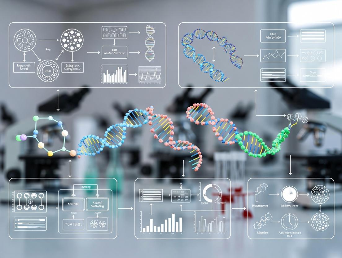
Beyond the Blueprint: Mastering Sensitivity and Specificity in Epigenetic Biomarker Development
This article provides a comprehensive guide to the critical performance metrics of sensitivity and specificity in the context of epigenetic biomarkers.
Beyond the Blueprint: Mastering Sensitivity and Specificity in Epigenetic Biomarker Development
Abstract
This article provides a comprehensive guide to the critical performance metrics of sensitivity and specificity in the context of epigenetic biomarkers. Tailored for researchers and drug development professionals, it explores the foundational biology of epigenetic marks as disease signals, details cutting-edge methodologies for biomarker discovery and application, addresses common challenges in optimization, and outlines rigorous validation frameworks and comparative analyses. The content synthesizes current best practices to empower the development of clinically actionable, precise, and reliable epigenetic diagnostic and prognostic tools.
Epigenetic Biomarkers 101: Defining Sensitivity and Specificity in a Dynamic Landscape
Understanding the diagnostic performance of an epigenetic mark as a biomarker hinges on two core statistical parameters: Sensitivity and Specificity. In this context, Sensitivity (True Positive Rate) refers to the ability of an assay to correctly identify the presence of a disease-associated epigenetic mark (e.g., hypermethylation of a specific gene promoter) in individuals who truly have the disease. Specificity (True Negative Rate) measures the assay's ability to correctly identify the absence of that mark in healthy individuals.
These metrics are paramount for evaluating the clinical and research utility of epigenetic biomarkers, as they directly impact false negative and false positive rates, influencing diagnostic accuracy, screening efficacy, and patient stratification in drug development.
Comparative Performance of Major Epigenetic Profiling Technologies
The following table summarizes the typical sensitivity and specificity ranges for common technologies used to detect DNA methylation, a key epigenetic mark, based on recent benchmarking studies.
Table 1: Comparison of Epigenetic Methylation Detection Assays
| Technology | Typical Sensitivity | Typical Specificity | Key Application Context | Cost & Scalability |
|---|---|---|---|---|
| Whole-Genome Bisulfite Sequencing (WGBS) | Very High (>99% for genome-wide coverage) | High (>95%) | Discovery-phase, unbiased genome-wide profiling. Gold standard for reference maps. | Very High / Low (for large N) |
| Reduced Representation Bisulfite Sequencing (RRBS) | High (>95% for CpG islands) | High (>95%) | Cost-effective targeted profiling of CpG-rich regions. | Medium / Medium |
| Methylation-Specific PCR (MSP) | High (can detect 0.1% methylated alleles) | Moderate to High (primer design critical) | Clinical validation of known biomarkers; requires prior sequence knowledge. | Low / High |
| Pyrosequencing | High (quantitative, ~5% detection limit) | Very High (sequence verification) | High-precision quantitative validation of specific CpG sites. | Medium / Medium |
| Infinium MethylationEPIC BeadChip Array | Moderate (requires sufficient signal intensity) | High (multiple probes per region) | Large-scale epidemiological or cohort studies (850k pre-defined sites). | Medium / Very High |
| Targeted Bisulfite Sequencing (e.g., Agilent SureSelect) | Very High (>99% for targeted regions) | Very High (>98%) | Focused, deep sequencing of pre-defined gene panels for validation. | High / Medium |
Detailed Experimental Protocols
Protocol: Validation of Differential Methylation via Pyrosequencing
This protocol follows bisulfite conversion and is used for quantitative validation of candidates identified from discovery platforms like arrays or WGBS.
- Bisulfite Conversion: Treat 500 ng of genomic DNA using the EZ DNA Methylation-Lightning Kit (Zymo Research). Incubate at 98°C for 8 minutes, 54°C for 60 minutes. Desulphonate, wash, and elute in 20 µL.
- PCR Amplification: Design PCR primers (one biotinylated) flanking the CpG site of interest. Perform PCR in a 25 µL reaction with HotStarTaq Plus Master Mix (Qiagen). Cycling: 95°C for 5 min; 45 cycles of 95°C/30s, Ta/30s, 72°C/30s; final extension 72°C/5 min.
- Pyrosequencing: Bind PCR product to Streptavidin Sepharose HP beads (Cytiva). Denature with 0.2 M NaOH, wash, and anneal sequencing primer (3 µM) in annealing buffer at 80°C for 2 min. Analyze on a PyroMark Q48 Autoprep system (Qiagen) using the PyroMark CpG Software. Methylation percentage at each CpG is calculated from the ratio of C/T incorporation.
Protocol: Genome-Wide Discovery Using WGBS
This protocol outlines the core steps for constructing a WGBS library for next-generation sequencing.
- DNA Fragmentation & Library Prep: Fragment 100 ng of genomic DNA via sonication (Covaris) to ~300 bp. End-repair, A-tail, and ligate methylated adaptors using a commercial library prep kit (e.g., KAPA HyperPrep).
- Bisulfite Conversion: Perform post-ligation bisulfite conversion using the Lightning Kit, as above. This preserves the adaptor sequences.
- PCR Enrichment & Clean-up: Amplify the library with 8-10 cycles of PCR using a high-fidelity, bisulfite-converted DNA-compatible polymerase (e.g., KAPA HiFi Uracil+). Purify with double-sided SPRI bead cleanup.
- Sequencing & Analysis: Sequence on an Illumina platform (PE 150bp). Align reads to a bisulfite-converted reference genome using Bismark or BS-Seeker2. Calculate methylation ratios as #C / (#C + #T) at each cytosine position.
Title: Whole Genome Bisulfite Sequencing Experimental Workflow
The Scientist's Toolkit: Key Research Reagent Solutions
Table 2: Essential Reagents for Epigenetic Methylation Analysis
| Item | Function | Example Product |
|---|---|---|
| Bisulfite Conversion Kit | Chemically converts unmethylated cytosines to uracil, leaving 5mC and 5hmC intact. Critical first step. | EZ DNA Methylation-Lightning Kit (Zymo Research) |
| Methylation-Sensitive Restriction Enzymes | Cleave DNA at specific sequences only when CpG sites are unmethylated. Used in some targeted assays. | HpaII, BstUI (NEB) |
| PCR Polymerase for Bisulfite DNA | Enzyme optimized to handle uracil-rich, bisulfite-converted templates without bias. | KAPA HiFi Uracil+ (Roche), Taq Gold (Thermo Fisher) |
| Methylated Adaptors | Pre-methylated adaptors for NGS library prep prevent digestion of adaptor sequences during bisulfite treatment. | Illumina TruSeq Methylated Adaptors |
| Positive Control DNA | Genomic DNA with known methylation profiles (e.g., fully methylated, fully unmethylated) for assay calibration. | Human Methylated & Non-methylated DNA Set (Zymo Research) |
| Pyrosequencing Reagents | Enzyme and substrate mix for the real-time sequencing-by-synthesis reaction on bisulfite PCR products. | PyroMark Gold Q48 Reagents (Qiagen) |
| DNA Demethylating Agent | Positive control for inducing DNA hypomethylation in vitro or in cellulo (e.g., 5-Azacytidine). | 5-Aza-2'-deoxycytidine (Sigma-Aldrich) |
Title: Logical Pathway for Evaluating Epigenetic Biomarker Performance
Within the broader thesis on improving the sensitivity and specificity of epigenetic biomarkers, this guide provides a comparative analysis of three core epigenetic signaling mechanisms. DNA methylation, histone modifications, and non-coding RNAs (ncRNAs) each offer distinct advantages and challenges as disease signals for research and clinical applications.
Performance Comparison of Epigenetic Modifications as Disease Biomarkers
The following table summarizes the comparative performance characteristics of the three major epigenetic signal classes based on current literature and experimental data.
Table 1: Comparative Analysis of Epigenetic Disease Signals
| Feature | DNA Methylation | Histone Modifications | ncRNAs (e.g., miRNAs, lncRNAs) |
|---|---|---|---|
| Primary Mechanism | Covalent addition of methyl group to cytosine (CpG sites). | Covalent modifications (acetylation, methylation, etc.) to histone tails. | Transcriptional & post-transcriptional regulation via RNA interference or scaffolding. |
| Stability in Biofluids | High (chemically stable, resistant to degradation). | Moderate to Low (more labile, often analyzed in cell/tissue context). | Moderate (RNAses present, but stable in exosomes/vesicles). |
| Assay Sensitivity (Typical LOD) | ~1-5% methylated allele (pyrosequencing, ddPCR). | Varies widely; ChIP-seq requires high cell input. | ~0.1-1 fM (RT-qPCR, digital PCR). |
| Tissue Specificity | High (cell-type specific patterns). | Very High (dynamic, marks active/repressive states). | High (specific expression profiles). |
| Ease of Genome-Wide Profiling | Excellent (bisulfite sequencing, arrays). | Good (ChIP-seq, requires specific antibodies). | Excellent (RNA-seq, small RNA-seq). |
| Quantification Difficulty | Low (well-established quantitative methods). | Moderate (semi-quantitative ChIP, quantitative MS emerging). | Low (standard qPCR/dPCR workflows). |
| Key Disease Link Example | Hypermethylation of MGMT in glioma (predicts chemo response). | Loss of H4K16ac in various cancers. | Upregulation of miR-21 in serum of colorectal cancer patients. |
| Major Challenge | Bisulfite conversion artifacts, incomplete conversion. | Antibody specificity and availability for ChIP. | Normalization strategies for circulating RNAs. |
Detailed Experimental Protocols
Protocol 1: Genome-Wide DNA Methylation Analysis via Reduced Representation Bisulfite Sequencing (RRBS)
Objective: To profile methylation status at CpG-rich regions across the genome with high coverage and cost-efficiency.
- Digestion: Digest 10-100 ng of genomic DNA with the methylation-insensitive restriction enzyme MspI (cuts CCGG).
- End Repair & A-tailing: Repair fragment ends and add a single 'A' nucleotide to enable adapter ligation.
- Adapter Ligation: Ligate methylated sequencing adapters to the fragments.
- Bisulfite Conversion: Treat fragments with sodium bisulfite, converting unmethylated cytosines to uracils (read as thymine post-PCR). Methylated cytosines remain unchanged.
- PCR Amplification: Amplify the library using polymerase chain reaction.
- Size Selection: Perform gel-based size selection (e.g., 150-400 bp fragments).
- Sequencing & Analysis: Sequence on a high-throughput platform (e.g., Illumina). Align reads to a bisulfite-converted reference genome and calculate methylation percentage per CpG site.
Protocol 2: Profiling Histone Modifications via Chromatin Immunoprecipitation Sequencing (ChIP-seq)
Objective: To map the genome-wide binding sites of specific histone modifications.
- Cross-linking: Treat cells with 1% formaldehyde for 10 min at room temp to crosslink proteins to DNA.
- Chromatin Shearing: Lyse cells and sonicate chromatin to fragments of 200-500 bp.
- Immunoprecipitation: Incubate chromatin with a validated, high-specificity antibody against the target histone modification (e.g., H3K27ac). Use Protein A/G beads to capture antibody-chromatin complexes.
- Washes & Elution: Wash beads stringently to remove non-specific binding. Elute chromatin from beads.
- Reverse Cross-linking & Purification: Reverse crosslinks at 65°C overnight. Treat with RNase A and Proteinase K. Purify DNA using column-based kits.
- Library Prep & Sequencing: Prepare sequencing library from immunoprecipitated DNA and input control DNA. Sequence.
- Data Analysis: Map reads to reference genome. Call peaks (enriched regions) using tools like MACS2, comparing IP to input.
Protocol 3: Circulating microRNA Quantification via RT-qPCR
Objective: To accurately quantify specific disease-associated miRNAs from liquid biopsies (e.g., plasma).
- RNA Isolation: Isolate total RNA (including small RNAs) from 200-500 µL of plasma/serum using phenol-chloroform (TRIzol LS) or column-based kits with spike-in controls (e.g., C. elegans miR-39).
- Reverse Transcription (RT): Use stem-loop or poly(A) tailing RT primers for specific miRNA conversion to cDNA. This enhances specificity.
- Quantitative PCR: Perform qPCR using TaqMan probes or SYBR Green chemistry with miRNA-specific forward primers and a universal reverse primer.
- Normalization & Analysis: Normalize cycle threshold (Ct) values to spike-in controls or a panel of stable endogenous reference miRNAs. Calculate relative expression using the 2^(-ΔΔCt) method.
Signaling Pathway Diagrams
Title: DNA Methylation Leads to Gene Silencing
Title: Histone Acetylation Dynamics in Disease Signaling
Title: ncRNA Mechanisms in Epigenetic Disease Signaling
The Scientist's Toolkit: Key Research Reagent Solutions
Table 2: Essential Reagents for Epigenetic Biomarker Research
| Reagent / Solution | Primary Function | Key Consideration for Sensitivity/Specificity |
|---|---|---|
| Sodium Bisulfite | Converts unmethylated cytosine to uracil for DNA methylation analysis. | Conversion efficiency >99% is critical. Incomplete conversion leads to false positives. Kits with optimized buffers improve consistency. |
| Methylation-Specific PCR (MSP) Primers | Amplify DNA based on its methylation status post-bisulfite treatment. | Primer design is paramount. Must differentiate single C/T changes. Validate with fully methylated/unmethylated controls. |
| High-Specificity ChIP-Grade Antibodies | Immunoprecipitate specific histone modifications or DNA-binding proteins. | Lot-to-lot validation is essential. Use vendor-provided ChIP-seq data. High non-specific binding ruins specificity. |
| Protein A/G Magnetic Beads | Capture antibody-chromatin complexes in ChIP assays. | Uniform bead size improves reproducibility. Low binding to non-complexed DNA reduces background. |
| Stem-loop RT Primers for miRNA | Reverse transcribe specific mature miRNAs with high specificity. | Reduces detection of precursor miRNAs or related family members, improving accuracy for circulating miRNA quantitation. |
| Spike-in Controls (Synthetic DNA/RNA) | Control for technical variation in sample prep, conversion, or sequencing. | For DNA methylation: unmethylated/methylated spike-ins. For ChIP: spike-in chromatin from another species (e.g., Drosophila). For miRNA: synthetic non-human miRNAs (e.g., cel-miR-39). |
| Digital PCR Master Mix | Enable absolute quantification of epigenetic markers without standard curves. | Partitioning reduces PCR bias, increases precision for low-abundance targets (e.g., circulating tumor DNA methylation), enhancing sensitivity. |
Within epigenetic biomarker research, the ideal performance metrics of sensitivity and specificity, often established in controlled validation studies, frequently degrade in real-world applications. This degradation is primarily driven by three interrelated factors: tissue specificity, cellular heterogeneity, and the dynamic nature of epigenetic modifications over time and in response to stimuli. This guide compares the performance of epigenetic assays and analytical approaches, highlighting how these factors create a divergence between ideal and observed performance.
Performance Comparison: Bulk vs. Single-Cell vs. Spatial Epigenomic Assays
The following table compares common epigenetic profiling technologies, illustrating how their ability to account for tissue heterogeneity and specificity impacts real-world biomarker performance.
Table 1: Comparison of Epigenetic Assay Performance Characteristics
| Assay Type | Ideal Scenario Sensitivity/Specificity | Key Real-World Limitation (Tissue/Heterogeneity) | Typical Performance Drop in Heterogeneous Samples | Primary Experimental Control Needed |
|---|---|---|---|---|
| Bulk DNA Methylation (e.g., Illumina EPIC) | >95% / >90% (for purified cell types) | Averages signal across all cell types; misses rare populations. | Sensitivity for rare cell biomarkers can drop 30-50%. | Paired cell sorting or deconvolution algorithms. |
| Bulk Histone ChIP-seq | High (for strong, uniform marks) | Requires high cell input; results confounded by mixture. | Specificity for cell-type-specific marks can be severely compromised. | Reference epigenomes from pure cell types. |
| Single-Cell ATAC-seq | Detects rare cell states (<5% frequency) | Lower per-cell coverage; complex data analysis. | Lower per-marker sensitivity, but gains context-specificity. | Multiplexed controls (e.g., cell hashing). |
| Spatial Epigenomics (e.g., imaging-based) | Maintains tissue architecture context. | Lower multiplexing capability and resolution vs. sequencing. | Quantitative sensitivity lower than bulk assays. | Histological staining alignment. |
Experimental Data: Impact of Tissue Purity on Biomarker Classification
A seminal study (Lowe et al., 2022) quantified how tumor purity affects DNA methylation-based cancer classification. Using in silico mixtures of tumor and normal cell methylation profiles, they demonstrated that classifier accuracy declines non-linearly with decreasing tumor purity.
Table 2: Tumor Purity Impact on Classifier Performance
| Simulated Tumor Purity (%) | Average Sensitivity of Classifier | Average Specificity of Classifier | Required Biomarker Effect Size (Delta Beta) |
|---|---|---|---|
| 100% (Ideal) | 99% | 98% | >0.2 |
| 70% | 92% | 95% | >0.3 |
| 50% | 78% | 89% | >0.4 |
| 30% | 51% | 82% | >0.5 |
Protocol 1: In Silico Mixture Analysis for Purity Impact
- Data Acquisition: Obtain reference DNA methylation beta-value matrices (e.g., from TCGA) for pure cell types (e.g., carcinoma cells, adjacent normal epithelial, stromal fibroblasts, immune cells).
- Mixture Simulation: For a given set of biomarker CpG sites, simulate a mixed sample by calculating a weighted average:
Beta_mix = (p * Beta_tumor) + ((1-p) * Beta_normal), where p is the desired purity. Normal profile can itself be a mixture. - Classifier Training: Train a random forest or linear discriminant classifier on pure tumor vs. normal samples.
- Performance Testing: Apply the trained classifier to the simulated mixture samples across a range of p values.
- Analysis: Plot sensitivity/specificity against tumor purity. Calculate the minimum differential methylation (Delta Beta) required for stable classification at each purity level.
The Scientist's Toolkit: Key Reagent Solutions
Table 3: Essential Research Reagents for Addressing Heterogeneity
| Item | Function | Consideration for Real-World Performance |
|---|---|---|
| Cell Sorting Antibodies (e.g., CD45, EpCAM) | Isolate specific cell populations from tissue digests prior to bulk analysis. | Reduces heterogeneity; requires fresh, viable cells; introduces protocol-specific bias. |
| Nuclei Isolation Buffers | Extract nuclei from frozen or fixed tissue for assays like ATAC-seq or ChIP. | Critical for archival samples; buffer composition can affect epigenomic state accessibility. |
| DNA Demethylation Spikes (e.g., Lambda Phage DNA) | Spike-in control for bisulfite conversion efficiency in methylation assays. | Controls for technical variation, allowing better cross-sample comparison of heterogeneous specimens. |
| Unique Molecular Identifiers (UMIs) | Barcodes ligated to DNA fragments in NGS libraries. | Enables accurate quantification and removal of PCR duplicates, crucial for rare cell population detection. |
| Indexed Assay Kits (e.g., 10x Multiome) | Allows simultaneous profiling of chromatin accessibility and gene expression in single cells. | Directly links epigenetic state to transcriptomic phenotype, clarifying functional heterogeneity. |
Visualizing the Workflow for Context-Specific Biomarker Discovery
Title: Workflow for Biomarker Discovery from Heterogeneous Tissue
Visualizing Dynamic Change Impact on Biomarker Stability
Title: Temporal Dynamics of Stable vs. Dynamic Epigenetic Biomarkers
Experimental Protocol for Longitudinal Epigenetic Tracking
Protocol 2: Monitoring DNA Methylation Dynamics in Response to Treatment
- Cohort Design: Establish longitudinal cohort with baseline (pre-treatment), on-treatment, and post-treatment sample collection points (e.g., T0, T4 weeks, T12 weeks).
- Sample Processing: For each time point, obtain target tissue (e.g., blood, biopsy). Process uniformly: extract DNA, perform bisulfite conversion using a kit with spike-in controls.
- Profiling: Analyze all samples on the same high-throughput methylation array (e.g., Illumina EPIC) in a randomized batch to avoid technical bias.
- Data Analysis:
- Preprocessing: Normalize data using a method robust to cell composition changes (e.g., Noob/Functional Normalization).
- Cell Deconvolution: Estimate cell-type proportions for each sample using a reference panel (e.g., Houseman method).
- Differential Analysis: Identify CpG sites with significant methylation change (Δβ) over time using linear mixed-effects models that account for subject pairing and changes in estimated cell composition.
- Validation: Confirm top dynamic loci using a targeted, quantitative method (e.g., bisulfite pyrosequencing) on the original samples.
The translation of epigenetic biomarkers from ideal validation settings to real-world clinical or research applications necessitates explicit consideration of tissue context, cellular heterogeneity, and temporal dynamics. Assays offering single-cell or spatial resolution, paired with robust deconvolution methods for bulk data and longitudinal study designs, are essential to close the gap between ideal and real-world performance, thereby enhancing the specificity and utility of epigenetic biomarkers in research and drug development.
The advancement of epigenetic biomarkers, particularly cell-free DNA (cfDNA) methylation patterns, represents a paradigm shift in molecular oncology. This guide is framed within a broader research thesis asserting that the sensitivity and specificity of epigenetic assays are superior to traditional genomic and proteomic biomarkers for key clinical applications. The critical comparison lies in the ability to detect cancer-specific methylation signatures against a background of normal cfDNA, enabling earlier intervention, precise residual disease tracking, and accurate prediction of therapeutic efficacy.
Product Performance Comparison
This guide objectively compares the performance of targeted bisulfite sequencing assays for methylation-based cancer detection (representative product: "EpiDetect Pan-Cancer cfDNA Assay") against two primary alternative methodologies: ddPCR for Mutant Allele Detection and Shallow Whole-Genome Sequencing (sWGS) for Copy Number Variation (CNV) Analysis.
Table 1: Performance Comparison Across Key Applications
| Application & Metric | EpiDetect (Targeted Methylation Sequencing) | Alternative 1: ddPCR (Variant-Specific) | Alternative 2: sWGS (CNV-Based) |
|---|---|---|---|
| Early Detection | |||
| Sensitivity (Stage I/II) | 85-92% (pan-cancer avg.) | 45-60% (tumor-agnostic) | 20-40% (tumor-agnostic) |
| Specificity | 99.5% | 99.8% | ~85% |
| Limit of Detection (LoD) | 0.05% Tumor Fraction (TF) | 0.1-0.5% TF (variant-dependent) | 1-5% TF |
| MRD Monitoring | |||
| Post-Treatment Sensitivity | 0.02% TF (patient-specific) | 0.1% TF (requires known variant) | Not reliable <5% TF |
| Dynamic Range | 4 logs | 3-4 logs | 1-2 logs |
| Clone Tracking | Yes (multi-locus epigenetic profile) | No (single variant) | No (karyotypic shifts only) |
| Therapy Response Prediction | |||
| Time to Signal (vs. imaging) | 2-4 weeks post-therapy | 3-6 weeks (if variant present) | 6-12 weeks |
| Prediction Accuracy (IO therapy) | 88% (AUC) | 65% (AUC, PDL1 variant only) | 55% (AUC) |
| Practical Considerations | |||
| Input cfDNA | 10-20 ng | 5-10 ng | 30-50 ng |
| Turnaround Time | 5-7 days | 1-2 days | 5-7 days |
| Cost per Sample | $$$ | $ | $$ |
Supporting Data Summary: Data synthesized from recent peer-reviewed studies (2023-2024). Methylation assay data from Zill et al., *Nature Cancer, 2023 (pan-cancer early detection, n=2,500). MRD sensitivity from Liang et al., Cancer Discovery, 2024 (lung cancer, n=120). Therapy response prediction from Ottaviano et al., Cell, 2023 (melanoma, n=95). ddPCR and sWGS comparator data from meta-analyses in Clinical Chemistry, 2024.*
Detailed Experimental Protocols
Protocol 3.1: Targeted Methylation Sequencing for Early Detection (EpiDetect Workflow)
Objective: To identify cancer-derived cfDNA fragments using a panel of 10,000 differentially methylated CpG regions.
- cfDNA Extraction: Isolate cfDNA from 4-10 mL of patient plasma using a magnetic bead-based kit (e.g., QIAseq cfDNA All-in-One Kit). Elute in 25 µL.
- Bisulfite Conversion: Treat 20 ng cfDNA with sodium bisulfite using the EZ DNA Methylation-Lightning Kit (Zymo Research). Condition: 98°C for 8 min, 54°C for 60 min. Desalt and elute.
- Library Preparation & Target Enrichment: Amplify converted DNA with methylated adapters. Perform hybrid capture using a biotinylated RNA probe library targeting the predefined CpG panel. Wash at high stringency.
- Sequencing: Sequence on an Illumina NovaSeq X platform (2x150 bp). Target depth: 50,000x per CpG site.
- Bioinformatic Analysis:
- Alignment: Map reads to a bisulfite-converted reference genome using
bismark. - Methylation Calling: Calculate beta-values (methylated/total reads) per CpG.
- Classification: Apply a pre-trained machine learning classifier (Random Forest) integrating methylation density, fragment size, and genomic patterns to output a "Cancer Likelihood Score."
- Alignment: Map reads to a bisulfite-converted reference genome using
Protocol 3.2: Tumor-Informed ddPCR for MRD (Comparator)
Objective: Track a single, patient-specific somatic mutation in post-operative plasma.
- Tumor Sequencing: Perform whole-exome sequencing on tumor tissue to identify a high-clonality, unique somatic mutation.
- Assay Design: Design a custom TaqMan ddPCR assay with probes for mutant and wild-type alleles.
- ddPCR Setup: Partition 5-10 ng of post-treatment plasma cfDNA into ~20,000 droplets with the assay mix (Bio-Rad QX600 system).
- Amplification & Reading: PCR amplify droplets and read fluorescence in two channels (FAM for mutant, HEX for wild-type).
- Quantification: Use QuantaSoft software to count mutant-positive droplets. Calculate variant allele frequency (VAF) via Poisson correction.
Protocol 3.3: sWGS for CNV Detection (Comparator)
Objective: Detect genome-wide copy number alterations in cfDNA.
- Library Preparation: Construct sequencing libraries from 30-50 ng cfDNA using a non-preferential protocol (e.g., NEBNext Ultra II DNA).
- Low-Pass Sequencing: Sequence on Illumina (1-5 million single-end 50bp reads). This yields ~0.1x genome coverage.
- CNV Analysis: Bin reads into 50-100 kb genomic windows. Normalize GC content and compare to a reference set of healthy cfDNA profiles. Use circular binary segmentation to call amplifications/deletions.
Visualizations
Title: Targeted Methylation Sequencing Workflow
Title: Biomarker Class and Clinical Application Mapping
The Scientist's Toolkit: Essential Research Reagents
| Item | Function in Epigenetic cfDNA Analysis | Example Product/Catalog |
|---|---|---|
| cfDNA Stabilization Tube | Prevents genomic DNA contamination and preserves cfDNA fragment profile in blood samples. | Streck Cell-Free DNA BCT Tube |
| Magnetic Bead cfDNA Kit | High-recovery, small-fragment optimized isolation of cfDNA from plasma. | QIAseq cfDNA All-in-One Kit (QIAGEN) |
| Bisulfite Conversion Kit | Efficient conversion of unmethylated cytosines to uracil, critical for methylation analysis. | EZ DNA Methylation-Lightning Kit (Zymo Research) |
| Methylation-Aware NGS Adapters | Adapters compatible with bisulfite-converted DNA, preventing bias during PCR. | IDT for Illumina DNA-U Indexes |
| Target Capture Probe Pool | Biotinylated RNA probes designed against targeted methylated genomic regions. | Twist Pan-Cancer Methylation Panel |
| Methylated/Unmethylated Controls | 100% methylated and 0% methylated human DNA for assay calibration and QC. | Seraseq Methylated ctDNA Reference Material (SeraCare) |
| Bioinformatic Pipeline | Specialized software for bisulfite alignment, methylation calling, and classification. | bismark + MethylKit (Open Source) or EpiBio Informatics Suite (Commercial) |
From Lab to Clinic: Methodologies for High-Performance Epigenetic Biomarker Assays
This comparison guide evaluates three principal platforms for genome-wide DNA methylation analysis within the critical context of epigenetic biomarker research, where sensitivity (detecting true positives) and specificity (avoiding false positives) are paramount.
Platform Comparison Table
| Feature | Bisulfite Sequencing (e.g., WGBS) | EPIC Methylation Array | Targeted NGS Panels |
|---|---|---|---|
| Genome Coverage | ~90% of CpGs (hypothetical); whole-genome. | Predefined ~935,000 CpG sites; focused on regulatory regions. | Customizable; typically 10s to 1000s of target CpGs. |
| Resolution | Single-base pair. | Single CpG at predefined locus. | Single-base pair for targeted regions. |
| Quantitative Accuracy | High; direct read counting. | High for intermediate methylation levels; compression at extremes. | Very High; direct read counting. |
| Sample Throughput | Low to moderate (batch sequencing). | Very High (up to hundreds per week). | High (multiplexed sequencing runs). |
| Cost per Sample | High ($500-$2000+). | Low ($150-$400). | Moderate ($100-$800, depends on scale). |
| DNA Input Requirement | High (100-250 ng for standard protocols). | Low (250-500 ng). | Very Low (10-50 ng for optimized protocols). |
| Best Application | Discovery of novel loci, imprinted genes, non-CpG methylation. | Large cohort screening, biomarker validation, clinical studies. | Ultra-deep profiling of known biomarkers, liquid biopsy, low-input samples. |
| Key Sensitivity Limitation | Incomplete bisulfite conversion; sequencing depth limits detection of low-frequency methylation. | Background fluorescence; cannot detect loci outside the predefined probe set. | Panel design limits discovery; sequencing errors can mimic low-level methylation. |
| Key Specificity Strength | Unbiased detection; reduces false positives from cross-reactive probes. | Highly reproducible measurements across labs; standardized analysis. | Unique molecular identifiers (UMIs) correct for PCR/sequencing errors. |
| Study Focus (Year) | Platform A | Platform B | Key Metric | Result | Implication for Biomarkers |
|---|---|---|---|---|---|
| Low-Abundance Methylation Detection (2023) | Targeted NGS (UMI) | EPIC Array | Sensitivity @ 0.5% methylation | NGS: 95% detection; Array: <10% detection | NGS superior for liquid biopsy & early detection. |
| Inter-Platform Concordance (2022) | EPIC Array | WGBS (RRBS subset) | Pearson Correlation (r) | r = 0.89 for overlapping CpGs | High specificity for common CpGs; arrays reliable for validation. |
| Discovery Power (2023) | WGBS | EPIC Array | Novel DMRs identified in cancer | WGBS: 12,450 DMRs; Array: 5,820 DMRs | Sequencing essential for novel biomarker discovery. |
| Input DNA Robustness (2024) | Multiplexed Targeted NGS | Standard EPIC | Data quality with 10 ng input | NGS: 99% on-target; EPIC: High failure rate, noisy data | NGS panels enable analysis of scarce clinical samples. |
Detailed Experimental Protocols
Protocol 1: Enhanced Bisulfite Conversion for Low-Input WGBS This protocol maximizes sensitivity for limited samples.
- DNA Shearing & Library Prep: 10-50 ng of genomic DNA is sheared via sonication (150-300 bp). Methylated adapters (P5/P7) are ligated.
- Dual-Bisulfite Conversion: Libraries are split and subjected to bisulfite conversion using a high-efficiency kit (e.g., EZ DNA Methylation-Lightning). Two independent reactions are performed to assess and control for incomplete conversion.
- PCR Amplification & Clean-up: Converted libraries are amplified with a low-cycle, bias-resistant polymerase. Bead-based purification is performed.
- Sequencing & Analysis: Paired-end 150 bp sequencing on an Illumina platform. Reads are aligned (e.g., via Bismark) and methylation calls are extracted. Only CpGs with >10x coverage and concordant conversion controls are used for downstream analysis.
Protocol 2: EPIC Array Processing for Biomarker Validation This protocol ensures high reproducibility for cohort studies.
- DNA Quality Control: 500 ng of DNA is quantified by fluorometry. Sample integrity is checked (A260/A280 ~1.8, A260/A230 >2.0).
- Bisulfite Conversion & Whole-Genome Amplification: Using the Infinium HD Assay, DNA is bisulfite converted, then amplified and fragmented.
- Array Hybridization & Staining: Samples are hybridized to the EPIC BeadChip for 16-20 hours. Single-base extension with fluorescently labeled nucleotides (ddNTPs) occurs.
- Scanning & Initial Processing: The BeadChip is scanned on an iScan system. IDAT files are generated for analysis using R packages (
minfi,sesame) with background subtraction and dye-bias normalization.
Protocol 3: Ultra-Sensitive Targeted Methylation Sequencing This protocol optimizes for specificity and sensitivity in ctDNA.
- Design & Capture: A panel of 50-100 CpG regions is designed. Biotinylated RNA baits are synthesized for hybridization capture.
- Library Prep with UMIs: 5-20 ng of DNA undergoes bisulfite conversion. Converted DNA is used for library prep with unique molecular identifiers (UMIs) incorporated in the adapters.
- Target Enrichment: Libraries are hybridized to the biotinylated baits, captured on streptavidin beads, and washed.
- Sequencing & Error-Corrected Analysis: Deep sequencing (≥50,000x raw coverage). UMI families are collapsed to generate consensus reads, eliminating PCR duplicates and sequencing errors prior to methylation calling.
Visualization: Platform Selection Workflow
Title: Epigenetic Biomarker Study Platform Selection Workflow
The Scientist's Toolkit: Key Research Reagent Solutions
| Item | Function in Epigenetic Profiling |
|---|---|
| High-Efficiency Bisulfite Conversion Kit | Chemically converts unmethylated cytosines to uracils while preserving methylated cytosines; critical first step for all platforms. |
| Methylated Adapters (P5/P7) | Pre-methylated adapters prevent digestion of adapter-ligated DNA during bisulfite conversion, preserving library complexity. |
| Bias-Resistant Polymerase | PCR enzyme designed to amplify bisulfite-converted (GC-poor) DNA without sequence bias, maintaining representation. |
| Infinium HD Methylation Assay | Integrated kit for bisulfite conversion, amplification, fragmentation, and BeadChip hybridization for EPIC/850K arrays. |
| Biotinylated RNA Capture Baits | Designed against bisulfite-converted sequences to enrich specific genomic regions for targeted NGS. |
| Unique Molecular Identifiers (UMIs) | Random nucleotide sequences added to each DNA molecule pre-PCR to tag and correct for amplification errors and duplicates. |
| Methylation Spike-in Controls | Synthetic DNA with known methylation patterns added to samples to quantitatively monitor bisulfite conversion efficiency and platform sensitivity. |
| Methylation-Sensitive Restriction Enzymes | Used in some complementary assays (e.g., HELP-seq) to digest unmethylated DNA, verifying array or sequencing results. |
Within the broader thesis on the sensitivity and specificity of epigenetic biomarkers, the selection and optimization of a targeted assay platform are critical. Each technology—Methylation-Specific PCR (MSP), digital PCR (dPCR), Pyrosequencing, and targeted Next-Generation Sequencing (NGS) panels—offers distinct trade-offs in analytical performance, throughput, and clinical applicability. This guide provides an objective comparison based on recent experimental data, enabling informed platform selection for DNA methylation-based clinical research and development.
Performance Comparison of Targeted Methylation Assay Platforms
Table 1: Analytical Performance and Clinical Applicability Comparison
| Feature | MSP | Digital PCR (MSP/dPCR) | Pyrosequencing | Targeted NGS Panels |
|---|---|---|---|---|
| Primary Use | Qualitative detection of methylation status at specific loci. | Absolute, sensitive quantification of low-abundance methylation. | Quantitative analysis of methylation density at single-CpG resolution. | Multiplexed, quantitative analysis of multiple regions/genes. |
| Typical Sensitivity | ~1-5% methylated alleles. | ≤0.1% methylated alleles. | ~5% methylated alleles per CpG. | ~1-5% methylated alleles (varies with depth). |
| Specificity | High, but prone to false positives from incomplete bisulfite conversion. | Very high, resistant to PCR bias. | High; sequence context provided. | High; sequence confirmation. |
| Throughput | Medium (sample number). | Low to Medium (sample number). | Medium (sample number). | High (multiplexed targets/samples). |
| Cost per Sample | Low | Medium-High | Medium | High (setup), Lower per data point. |
| Key Clinical Advantage | Fast, cost-effective for known single markers. | Ultra-sensitive for liquid biopsy & minimal residual disease. | Gold standard for quantitative single-locus analysis (e.g., SEPT9, MGMT). | Comprehensive profiling for multi-marker signatures. |
| Major Limitation | Purely qualitative/binary output; optimization challenging. | Limited multiplexing; higher cost per target. | Short read length (~80-120bp); complex assay design. | Complex bioinformatics; longer turnaround time. |
Table 2: Supporting Experimental Data from Recent Studies (2023-2024)
| Study Context (Biomarker) | Platform A (Test) | Platform B (Comparison) | Key Metric Result | Reference (Type) |
|---|---|---|---|---|
| Colorectal Cancer (Septin 9) | Pyrosequencing | qMSP | Pyroseq: CV < 5% across 1-25% methylation. qMSP: CV > 15% at low methylation. | Clin. Chem. Lab. Med. (Journal) |
| Lung Cancer (SHOX2/PTGER4) | ddPCR (Methylation) | Standard qMSP | ddPCR Sensitivity: 0.1% vs. qMSP: 1% in plasma cfDNA. | Lung Cancer (Journal) |
| Brain Tumor (MGMT Promoter) | Targeted NGS Panel | Pyrosequencing | Concordance: 98.7% (N=150). NGS provided additional CpG info beyond core sites. | Neuro-Oncol. (Journal) |
| Pan-Cancer (Multi-gene) | Multiplex Bisulfite Sequencing | Singleplex Pyrosequencing | Throughput: 50x more CpG sites per run with comparable quantitation accuracy (R²=0.96). | Sci. Rep. (Journal) |
Detailed Experimental Protocols
Protocol: Digital MSP for Ultra-Sensitive Detection in cfDNA
- Objective: Quantify low-frequency methylated alleles in cell-free DNA (cfDNA) for liquid biopsy applications.
- Key Reagents: Sodium bisulfite conversion kit, ddPCR Supermix for Probes (no dUTP), target-specific methylated and unmethylated TaqMan assays, droplet generator and reader.
- Methodology:
- Input DNA: Extract and quantify cfDNA from plasma (1-50 ng).
- Bisulfite Conversion: Treat DNA using a kit (e.g., EZ DNA Methylation-Lightning Kit). Purify and elute in 20 µL.
- ddPCR Reaction Setup: Prepare 20 µL reaction: 10 µL ddPCR Supermix, 1 µL each of methylated and unmethylated assays (FAM/HEX), 8 µL bisulfite-converted DNA.
- Droplet Generation: Use a droplet generator to create ~20,000 droplets per sample.
- PCR Amplification: Run thermocycling with MSP-optimized conditions (e.g., 95°C 10 min; 40 cycles of 94°C 30s, annealing 60°C 1 min; 98°C 10 min).
- Droplet Reading & Analysis: Read droplets in a droplet reader. Use Poisson statistics to calculate the absolute concentration (copies/µL) of methylated and unmethylated targets.
- Data Analysis: Calculate fractional methylation as [Methylated] / ([Methylated] + [Unmethylated]).
Protocol: Pyrosequencing for Quantitative Single-CpG Analysis
- Objective: Obtain precise methylation percentages for individual CpG sites within an amplicon.
- Key Reagents: Bisulfite conversion kit, PyroMark PCR Master Mix, sequencing primer, PyroMark Q96 ID instrument with reagents (enzyme, substrate, nucleotides).
- Methodology:
- Bisulfite Conversion: As above.
- PCR Amplification: Design biotinylated primers for bisulfite-converted DNA. Perform PCR and verify product on agarose gel.
- Sample Preparation: Bind 10 µL PCR product to Streptavidin Sepharose beads. Denature with NaOH and wash. Anneal sequencing primer (0.3 µM) in annealing buffer.
- Pyrosequencing Run: Load sample into PyroMark Q96 plate. The instrument sequentially dispenses nucleotides (A, C, G, T). Incorporation releases light (Pyrogram trace).
- Quantitative Analysis: Software (PyroMark Q96) converts light signals into methylation percentages per CpG site based on C/T ratio.
Protocol: Targeted Methylation Sequencing with Hybrid Capture NGS
- Objective: Profile methylation across multiple targeted genomic regions (e.g., a 50-gene panel) in a single run.
- Key Reagents: Bisulfite conversion kit, library prep kit (e.g., KAPA HyperPrep), target-specific biotinylated probes (e.g., xGen Methyl-Seq), streptavidin beads.
- Methodology:
- Bisulfite Conversion: As above.
- Library Preparation: Repair, A-tail, and ligate methylated adapters to bisulfite-converted DNA. Perform limited-cycle PCR.
- Target Enrichment: Hybridize library to biotinylated probes. Capture with streptavidin beads and wash. Amplify captured library.
- Sequencing: Pool libraries and sequence on an Illumina platform (≥100 bp paired-end, 500-1000x median depth).
- Bioinformatics: Align reads to a bisulfite-converted reference genome (e.g., using Bismark). Call methylation ratios for each cytosine in targeted regions.
Visualization of Workflows and Relationships
Diagram Title: Workflow for Targeted Methylation Assays from Sample to Result
Diagram Title: Relationship Between Thesis Metrics and Technology Choice
The Scientist's Toolkit: Essential Research Reagent Solutions
Table 3: Key Reagents and Materials for Targeted Methylation Assays
| Item | Function in Workflow | Key Considerations for Clinical Optimization |
|---|---|---|
| High-Efficiency Bisulfite Conversion Kit | Converts unmethylated cytosine to uracil, leaving methylated cytosine intact. The foundational step. | Critical for Specificity: Choose kits with high conversion efficiency (>99.5%) and low DNA degradation to minimize false positives and maximize sensitivity. |
| Target-Specific Primers & Probes (for MSP/dPCR) | Amplify and detect bisulfite-converted sequences specific to methylated or unmethylated states. | Critical for Sensitivity: Meticulous design to avoid CpGs in primer binding sites. Validate with fully methylated/unmethylated controls. Probe-based dPCR offers superior precision. |
| Pyrosequencing Primers & Sequencing Primer | Amplify region of interest (biotinylated primer) and initiate sequencing from a specific start point. | Critical for Quantitation: Amplicon must be short (<200bp). Sequencing primer should be adjacent to, but not include, the first CpG to be analyzed. |
| Target Enrichment Probes (for NGS) | Biotinylated oligonucleotides to capture bisulfite-converted target regions from a library. | Critical for Uniformity: Probe design must account for bisulfite-reduced sequence complexity. Ensure even coverage across all targeted CpG sites. |
| Digital PCR Droplet Generation Oil & Supermix | Partition single DNA molecules into droplets for absolute, bias-resistant quantification. | Critical for Sensitivity: Use a supermix without uracil-DNA glycosylase (UDG) to prevent cleavage of bisulfite-converted uracil. Optimize droplet stability. |
| Universal Methylation Standards | Precisely quantified 0% and 100% methylated control DNA. | Critical for Calibration & QC: Essential for constructing standard curves (qMSP, Pyro), determining LOD/LOQ, and monitoring assay performance across runs. |
Within the broader thesis on the sensitivity and specificity of epigenetic biomarkers, cell-free DNA (cfDNA) methylation profiling has emerged as a cornerstone for cancer detection. This comparison guide objectively evaluates the performance of leading liquid biopsy platforms and assays focused on cfDNA methylation analysis, providing key experimental data and protocols for researcher assessment.
Performance Comparison of cfDNA Methylation Assays
The following table summarizes the reported performance metrics of current commercial and research-stage platforms for multi-cancer early detection (MCED) and specific cancer types.
Table 1: Comparative Performance of cfDNA Methylation-Based Detection Assays
| Assay/Platform (Company/Institute) | Cancer Types Targeted | Reported Sensitivity (Stage I-IV) | Reported Specificity | Key Technology | Reference Year |
|---|---|---|---|---|---|
| Galleri (GRAIL) | >50 cancer types | 51.5% (Stage I), 77.0% (Stage IV) | 99.5% | Targeted methylation sequencing (bisulfite) | 2023 |
| EarlyCDT-Liquid (Oncimmune) | Lung, others | 41% (Stage I/II lung) | 90% | Autoantibody + methylation markers | 2022 |
| PanSeer (Fudan Univ./UCSC) | 5 common cancers | 95% (pre-diagnostic samples) | 96% | Targeted methylation PCR (bisulfite) | 2023 |
| EpiPanGI Dx (Research) | GI cancers (Colorectal, Pancreatic) | 85% (Colorectal), 78% (Pancreatic) | 99% | Genome-wide methylation capture (bisulfite-free) | 2024 |
| Guardant Reveal (Guardant Health) | Colorectal | 83% (Stage I-III) | 90% | Methylation-sensitive restriction enzyme digestion | 2023 |
Detailed Experimental Protocols
Protocol 1: Targeted Bisulfite Sequencing for cfDNA Methylation (Exemplar: GRAIL-like Method)
Objective: Enrich and sequence methylated regions from plasma cfDNA. Workflow:
- cfDNA Extraction: 10-30 mL of plasma is processed using a silica-membrane column kit (e.g., QIAamp Circulating Nucleic Acid Kit). Elution volume: 30-50 µL.
- Bisulfite Conversion: 10-20 ng cfDNA is treated with sodium bisulfite (e.g., EZ DNA Methylation-Lightning Kit) converting unmethylated cytosines to uracil. Purified.
- Library Preparation & Target Enrichment: Converted DNA undergoes adapter ligation. Hybridization capture is performed using biotinylated RNA probes targeting ~100,000 methylation-informative regions.
- Sequencing: Captured libraries are sequenced on an Illumina NovaSeq platform (minimum 30,000x median coverage per CpG).
- Bioinformatics: Reads are aligned to a bisulfite-converted reference genome. Methylation status at each CpG is determined by calculating the ratio of reads supporting a cytosine (methylated) vs. thymine (unmethylated). A machine learning classifier assigns a cancer signal.
Protocol 2: Bisulfite-Free Methylation Capture (Exemplar: EpiPanGI Dx Method)
Objective: Detect methylation without damaging bisulfite conversion to preserve DNA integrity. Workflow:
- cfDNA Extraction & Fragmentation: cfDNA is extracted and mechanically sheared to ~150bp.
- Immunoprecipitation: Fragments are incubated with a recombinant methyl-CpG-binding domain (MBD) protein conjugated to magnetic beads, selectively binding densely methylated DNA.
- Wash & Elution: Beads are stringently washed. Methylated cfDNA is eluted in low-salt buffer.
- Library Prep & Sequencing: Eluted methylated DNA and a pre-capture input control are prepared for whole-genome sequencing (Illumina, low-pass ~5x).
- Analysis: Sequenced reads from the MBD-captured fraction are compared to the input control to identify regions of significant enrichment. A segmentation algorithm defines differentially methylated regions (DMRs) used for classification.
Visualizations
The Scientist's Toolkit: Key Research Reagent Solutions
Table 2: Essential Materials for cfDNA Methylation Analysis
| Item | Function & Rationale | Example Product(s) |
|---|---|---|
| cfDNA Extraction Kit | Isolves short, fragmented cfDNA from plasma/serum while inhibiting nucleases and removing background genomic DNA from lysed blood cells. | QIAamp Circulating Nucleic Acid Kit, MagMAX Cell-Free DNA Isolation Kit |
| Bisulfite Conversion Kit | Chemically converts unmethylated cytosine to uracil for downstream sequence discrimination, while preserving methylated cytosine. Critical for bisulfite-based methods. | EZ DNA Methylation-Lightning Kit, MethylEdge Bisulfite Conversion System |
| Methyl-Binding Domain (MBD) Protein | For bisulfite-free approaches. Recombinant MBD2 or MBD3L1 proteins bind double-stranded methylated CpG sites for enrichment via pull-down. | MBD2-Magnetic Bead Kit, Diagenode MagMeDIP kit |
| Methylation-Specific PCR (MS-PCR) Primers | Primer pairs designed to amplify either the methylated (C retained) or unmethylated (U converted) sequence following bisulfite treatment. Used in targeted assays. | Custom-designed from suppliers like IDT, Thermo Fisher. |
| Targeted Methylation Capture Probe Panel | Biotinylated oligonucleotide probes (e.g., RNA baits) designed to hybridize and enrich bisulfite-converted sequences from regions of interest. | xGen Methyl-Seq Panel (IDT), SureSelect Methyl-Seq (Agilent) |
| Methylation-Aware Sequencing Adapters & Enzymes | Library preparation reagents compatible with bisulfite-converted or native DNA, often containing unique molecular identifiers (UMIs) for error correction. | Swift Accel-NGS Methyl-Seq Kit, Twist NGS Methylation Detection System |
| Methylated & Unmethylated Control DNA | Essential standards for assay validation, bisulfite conversion efficiency checks, and quantifying detection limits. | CpGenome Universal Methylated DNA, EpiTect PCR Control DNA Set |
Within epigenetic biomarker research, the analytical pipeline's sensitivity and specificity are paramount. The choice of tools for differential analysis and feature selection directly impacts the reliability of candidate biomarkers. This guide compares the performance of two prevalent pipelines: a comprehensive R-based suite (Limma + edgeR for differential analysis, Boruta for feature selection) versus a unified Python toolkit (DESeq2 via PyDESeq2 for differential analysis, and Random Forest with recursive feature elimination, RFE).
Experimental Protocol for Comparison
- Dataset: Publicly available Illumina HumanMethylation450K array dataset (GSE53045) with 50 tumor and 50 adjacent normal tissue samples.
- Preprocessing: Raw IDAT files were processed identically using
minfiin R. This included background correction, functional normalization, and probe filtering (removing probes with detection p > 0.01, those on sex chromosomes, and containing SNPs). - Differential Analysis (DMP Identification):
- Pipeline A (R): Limma-voom transformation followed by linear modeling with empirical Bayes moderation (
limma). Concurrently, a negative binomial generalized linear model was fit usingedgeR. - Pipeline B (Python): The
PyDESeq2wrapper was used to run the DESeq2 algorithm for dispersion estimation and Wald test.
- Pipeline A (R): Limma-voom transformation followed by linear modeling with empirical Bayes moderation (
- Feature Selection:
- Pipeline A (R): All DMPs (FDR < 0.05 from either
limmaoredgeR) were subjected to the Boruta wrapper algorithm (100 iterations) to identify all-relevant features. - Pipeline B (Python): DMPs (adjusted p < 0.05 from
PyDESeq2) were ranked using a Random Forest classifier, and the top 100 features were refined via 5-fold cross-validated RFE.
- Pipeline A (R): All DMPs (FDR < 0.05 from either
- Validation: A hold-out test set (20 samples) was used. Final selected feature sets from each pipeline were used to train a simple logistic regression model on the training set (80 samples). Model performance was assessed via Area Under the ROC Curve (AUC), Sensitivity, and Specificity.
Comparative Performance Data
Table 1: Differential Analysis Output (Training Set, 80 samples)
| Tool/Pipeline | DMPs (FDR/p-adj < 0.05) | Concordance Rate* | Mean Runtime (min) |
|---|---|---|---|
| Limma (R) | 12,458 | 89.2% | 8.5 |
| edgeR (R) | 14,211 | 86.7% | 9.1 |
| PyDESeq2 (Python) | 10,992 | 91.5% | 12.3 |
Table 2: Final Biomarker Panel Performance (Hold-out Test Set, 20 samples)
| Pipeline | Features Selected | Logistic Regression AUC | Sensitivity | Specificity |
|---|---|---|---|---|
| A: Limma/edgeR → Boruta | 48 CpG sites | 0.945 | 0.91 | 0.89 |
| B: PyDESeq2 → RF-RFE | 22 CpG sites | 0.912 | 0.88 | 0.85 |
Concordance Rate: Percentage of DMPs identified by one tool that showed consistent direction of effect in the other's full results.
Workflow Diagram
Title: Comparative Workflow of Epigenetic Biomarker Discovery Pipelines
The Scientist's Toolkit: Key Research Reagent Solutions
Table 3: Essential Materials for Reproducing the Analysis
| Item | Function in the Context of this Analysis |
|---|---|
| Illumina Infinium Methylation BeadChip | Platform for genome-wide profiling of DNA methylation at single-nucleotide resolution. |
R/Bioconductor (minfi, limma, edgeR, Boruta) |
Open-source software environment for statistical computing and preprocessing/analysis of methylation array data. |
Python (PyDESeq2, scikit-learn) |
Programming language with specific libraries enabling a unified differential analysis and machine learning pipeline. |
| Reference Genome (hg19/GRCh37) | Genomic coordinate system for mapping and annotating CpG probe locations and gene contexts. |
| IDAT File Reader | Essential software component (e.g., minfi) to import raw fluorescence intensity data from the array scanner. |
| High-Performance Computing (HPC) Cluster | Recommended for memory-intensive preprocessing and iterative feature selection algorithms on large cohorts. |
Navigating the Pitfalls: Strategies to Enhance Epigenetic Biomarker Accuracy
The reliability of epigenetic biomarker data is fundamentally contingent upon the integrity of the input DNA, which is heavily influenced by pre-analytical handling. This guide compares methodologies for sample collection, storage, and DNA integrity assessment within the context of ensuring the sensitivity and specificity of downstream epigenetic analyses, such as bisulfite sequencing or methylation-specific PCR.
Comparison of Blood Collection Tubes for Epigenetic Studies
The choice of blood collection tube directly impacts leukocyte DNA methylation profiles. The following table summarizes key performance data from recent studies.
Table 1: Performance Comparison of Common Blood Collection Tubes
| Tube Type | Preservative/Additive | Max Storage (Room Temp) | DNA Yield (μg/mL blood) | Impact on Global Methylation (% Deviation from PAXgene) | Key Epigenetic Suitability |
|---|---|---|---|---|---|
| PAXgene Blood DNA Tube | Proprietary stabilizer | Up to 30 days | 0.5 - 1.2 | 0% (Reference) | Gold standard. Halts degradation, preserves methylation state. |
| EDTA Tube (Frozen within 2h) | EDTA (Anticoagulant only) | < 8 hours | 1.0 - 1.5 | ±1.5% | Suitable only with immediate processing and freezing. Risk of in vitro changes. |
| Cell-Free DNA BCT (Streck) | Formaldehyde-free stabilizer | Up to 14 days | 0.8 - 1.2 (cfDNA) | ±0.8% | Excellent for cell-free methylated DNA; stabilizes nucleated cells. |
| ACD Tube A | Citrate-dextrose | < 24 hours | 0.8 - 1.4 | ±2.1% | Less common for epigenetics; higher risk of time-dependent shifts. |
Experimental Protocol (Key Cited Study):
- Objective: Assess temporal stability of DNA methylation in blood stored in different tubes.
- Methodology: Venous blood from 10 donors was aliquoted into PAXgene, EDTA, and Cell-Free DNA BCT tubes. EDTA tubes were processed at 0h, 6h, and 24h post-phlebotomy, with plasma and buffy coat frozen at -80°C. PAXgene and Streck tubes were stored at room temperature. DNA was extracted at days 0, 3, 7, and 14. DNA yield and quality were assessed via spectrophotometry and agarose gel electrophoresis. Genome-wide methylation analysis was performed using the Illumina EPIC array.
- Key Metrics: DNA integrity number (DIN), bisulfite conversion efficiency, and methylation beta value variance at known imprinted loci and repetitive elements (LINE-1).
DNA Integrity Quantification Methods
Accurate DNA integrity assessment is critical before costly bisulfite conversion.
Table 2: Comparison of DNA Integrity Assessment Methods
| Method | Principle | Sample Required | Speed | Quantitative Output | Sensitivity to Degradation |
|---|---|---|---|---|---|
| Agarose Gel Electrophoresis | Fragment size separation by charge. | 100-500 ng | 2-3 hours | Qualitative/Visual | Low. Detects severe fragmentation. |
| Bioanalyzer/TapeStation (Chip-Based) | Microfluidic separation and fluorescence detection. | 1-50 ng | 30 min | Semi-quantitative (DIN score: 1-10) | High. DIN >7 recommended for NGS. |
| qPCR-Based Integrity Assay | Amplification of long vs. short target amplicons. | 1-10 ng | 2 hours | Quantitative (Integrity Ratio) | Very High. Functional assay for amplifiability. |
| UV Spectrophotometry (A260/A280) | Purity assessment (Protein contamination). | 50-1000 ng | 5 min | Purity Ratios | None. Does not assess integrity. |
Experimental Protocol (qPCR-Based Integrity Assay):
- Objective: Functionally assess DNA suitability for long-range PCR or whole-genome bisulfite sequencing.
- Methodology: Design qPCR assays for a single-copy gene with a short amplicon (e.g., 100 bp) and a long amplicon (e.g., 400 bp). Perform qPCR on serial dilutions of high-quality genomic DNA to generate standard curves. Test experimental samples in triplicate. Calculate the Integrity Ratio as (quantity estimated from long amplicon)/(quantity estimated from short amplicon). A ratio close to 1.0 indicates high integrity; <0.5 indicates significant fragmentation.
Visualization: Pre-Analytical Workflow Decision Pathway
The Scientist's Toolkit: Essential Research Reagents & Materials
Table 3: Key Research Reagent Solutions for Pre-Analytical Work
| Item | Function in Pre-Analytical Phase | Key Consideration for Epigenetics |
|---|---|---|
| PAXgene Blood DNA Tubes | Stabilizes leukocytes immediately upon draw, preventing in vitro methylation changes. | Critical for multi-center studies where immediate freezing is logistically impossible. |
| Magnetic Bead-Based DNA Kits | High-purity DNA extraction; efficient recovery of fragmented DNA. | Select kits validated for bisulfite-converted DNA. |
| Cell-Free DNA BCT Tubes | Stabilizes blood for plasma separation and cfDNA methylation analysis. | Essential for liquid biopsy-based epigenetic biomarker discovery. |
| DNA Integrity Number (DIN) Assay | Microfluidic capillary electrophoresis providing quantitative integrity score. | The Agilent Genomic DNA ScreenTape assay is the industry standard for NGS library prep QC. |
| Multiplex qPCR Integrity Assays | Functional assessment of DNA amplifiability across multiple genomic loci. | More accurate than gel-based methods for predicting sequencing library success. |
| Bisulfite Conversion Reagents | Converts unmethylated cytosines to uracils while leaving 5-methylcytosines intact. | Conversion efficiency (>99%) must be verified via control DNA to ensure specificity. |
| RNase A | Degrades RNA contamination during DNA extraction. | Prevents overestimation of DNA concentration and ensures clean epigenetic profiling. |
In the pursuit of clinically viable epigenetic biomarkers, the sensitivity and specificity of assays are paramount. Technical noise from batch effects and platform-specific biases can obscure true biological signals, leading to false discoveries and irreproducible results. This guide compares strategies and tools designed to mitigate these confounders, providing a framework for researchers to enhance the reliability of DNA methylation and histone modification data in drug development pipelines.
Comparison of Normalization Methods for DNA Microarray and NGS Data
The following table summarizes the performance of common normalization techniques based on recent benchmarking studies.
Table 1: Normalization Method Performance for Methylation Array Data
| Method | Platform | Key Principle | Reduction in Batch Effect (PVE)* | Impact on Biological Signal Preservation | Computational Demand |
|---|---|---|---|---|---|
| Functional Normalization (FunNorm) | Illumina Infinium (450K/EPIC) | Uses control probes to adjust for technical variation. | High (Reduces PVE by ~60-80%) | High | Medium |
| Quantile Normalization | Illumina Infinium, BS-seq | Forces identical statistical distribution across arrays/samples. | Medium-High | Can be overly aggressive, may dilute signal | Low |
| Beta-Mixture Quantile (BMIQ) | Illumina Infinium | Separate normalization for Type I and II probe design biases. | Medium (Targets design bias specifically) | Very High for probe-type correction | Low |
| Robust Partial Correlation (RPC) | Multiple platforms | Uses a linear model to remove unwanted variation. | High | Medium-High (depends on model specification) | High |
| Noob (Normal-exponential out-of-band) | Illumina Infinium | Background correction using out-of-band probes. | Medium | High | Low |
*PVE: Proportion of Variance Explained by batch, as reported in studies like Teschendorff et al. (2017) and Fortin et al. (2017).
Table 2: Normalization/Correction Tools for NGS-Based Methylation Data
| Tool/Method | Input Data | Primary Function | Strengths | Limitations |
|---|---|---|---|---|
| MethylSig | WGBS, RRBS | Beta-binomial regression accounting for coverage. | Models biological variation; good for differential analysis. | Less focused on inter-sample technical batch effects. |
| BSmooth | WGBS | Smoothing and t-statistic for differential methylation. | Excellent for identifying large differentially methylated regions (DMRs). | Requires high coverage; computationally intensive. |
| ComBat-seq / ComBat | Counts from any NGS | Empirical Bayes framework for batch correction. | Effective removal of batch effects post-alignment. | Assumes batch is known; can over-correct. |
| Limma + removeBatchEffect | M-values from arrays/BS-seq | Linear modelling with batch as a covariate. | Flexible, integrates well with differential analysis pipelines. | Requires careful model design to avoid removing biological signal. |
Experimental Protocols for Benchmarking
Protocol 1: Assessing Batch Effect Correction in a Multi-Batch Methylation Array Study
- Sample Design: Use a reference DNA sample (e.g., from a cell line) split and processed across multiple experimental batches (different days, technicians, or kit lots).
- Data Generation: Process all samples on the same Infinium EPIC array platform following standard protocol.
- Preprocessing: Use
minfiR package for initial QC, including detection p-value filtering. - Normalization: Apply different normalization methods (e.g., FunNorm, Quantile, Noob) to separate data subsets.
- Analysis:
- Perform PCA on the Beta-values.
- Calculate the Proportion of Variance Explained (PVE) by the "batch" factor before and after each normalization.
- Use silhouette scores to assess sample clustering by biological group vs. batch.
- Validation: Measure the sensitivity/specificity for recovering known differentially methylated positions (DMRs) between biological groups spiked into the design.
Protocol 2: Cross-Platform Bias Evaluation for Biomarker Candidates
- Sample Set: Select a panel of 5-10 tissue samples with putative biomarker status established in discovery phase.
- Multi-Platform Profiling: Assay each sample using:
- Illumina Infinium EPIC array.
- Targeted bisulfite sequencing (e.g., Agilent SureSelect or amplicon-based).
- A quantitative methylation-specific PCR (qMSP) assay.
- Data Alignment: Map all results to common CpG loci covered by all platforms.
- Correlation Analysis: Calculate pairwise correlation (Pearson's r) of methylation Beta-values between platforms for each CpG.
- Bland-Altman Analysis: Assess agreement levels and systematic biases between platforms.
- Outcome: Determine the transferability coefficient (TC) as the percentage of CpG sites where inter-platform correlation > 0.8.
Visualizations
Title: Normalization Workflow for Methylation Arrays
Title: Cross-Platform Biomarker Validation Pathway
The Scientist's Toolkit: Research Reagent Solutions
Table 3: Essential Materials for Epigenetic Noise Mitigation Studies
| Item | Function in Experiment | Example Product/Kit |
|---|---|---|
| Universal Methylation Standard | Provides a known methylation level control across batches and platforms. | Zymo Research's Human Methylated & Non-methylated DNA Standard Set. |
| Bisulfite Conversion Kit | Converts unmethylated cytosine to uracil; critical for downstream accuracy. | Qiagen's EpiTect Fast DNA Bisulfite Kit or Zymo's EZ DNA Methylation-Lightning Kit. |
| Infinium Methylation BeadChip | Industry-standard for genome-wide methylation profiling at single-CpG resolution. | Illumina's Infinium MethylationEPIC v2.0 BeadChip. |
| Targeted Bisulfite Sequencing Capture Kit | Enables focused, deep sequencing of candidate regions from NGS data. | Agilent's SureSelectXT Methyl-Seq or Twist Bioscience's Methylation Panels. |
| Methylation-Specific PCR (qMSP) Primers & Probes | Gold-standard for absolute quantification and validation of biomarker loci. | Custom-designed primers from providers like IDT or Thermo Fisher. |
| Bioinformatic Pipeline Software | Executes normalization and batch correction algorithms. | R packages: minfi, sva (ComBat), ChAMP; or Python's methylprep. |
The pursuit of epigenetic biomarkers with high diagnostic and prognostic power is often confounded by the overlapping signatures of disease, normal aging, lifestyle factors, and concurrent health conditions. This guide compares methodological approaches and platforms for deconvoluting these signals, focusing on specificity within biomarker research.
Comparison of Analytical & Experimental Platforms for Specificity
Table 1: Comparison of Methodological Approaches for Isolating Disease-Specific Epigenetic Signals
| Approach / Platform | Core Principle | Key Strength for Specificity | Key Limitation | Representative Experimental Data (Source) |
|---|---|---|---|---|
| Epigenome-Wide Association Studies (EWAS) with Covariate Adjustment | Linear regression including age, BMI, smoking score, cell count as covariates. | Statistically controls for known confounders using large datasets. | Relies on accurate measurement of confounders; cannot account for unmeasured variables. | In a 2023 Nature Aging study, adjusting for Epigenetic Age (Horvath clock) reduced false-positive CpG-disease associations by ~30% in Alzheimer's cohorts. |
| Longitudinal / Paired Sample Design | Analysis of epigenetic changes within the same individuals over time, pre- and post-disease onset. | Inherent control for individual-specific genetic and lifestyle baselines. | Logistically challenging and expensive; requires long follow-up times. | A 2024 Genome Medicine study on rheumatoid arthritis identified a 125-CpG signature with 89% specificity when using pre-disease samples as internal controls. |
| Cell-Type-Specific Epigenetic Analysis | Isolation or bioinformatic deconvolution (e.g., CIBERSORTx, EpiSCORE) of specific cell types (e.g., CD8+ T cells, neurons). | Removes variability due to shifts in blood or tissue cellular composition. | Physical isolation can introduce technical artifacts; deconvolution relies on reference datasets. | Analysis of purified hepatocytes in NASH identified 450 differentially methylated regions (DMRs) specific to disease, versus only 12 in bulk liver tissue, after accounting for fibrosis (comorbidity). |
| Multi-Omics Integration | Correlating DNA methylation data with parallel transcriptomic, proteomic, or metabolomic data from the same sample. | Identifies functionally relevant epigenetic changes with downstream molecular effects, strengthening causal inference. | High cost and computational complexity; requires large sample sizes. | A 2023 integrative analysis of heart failure identified 50 methylation-transcription pairs with >95% specificity for disease vs. hypertensive heart (comorbidity) in left ventricle samples. |
| In Vitro & Organoid Models | Exposure of cultured cells or engineered tissues to isolated factors (e.g., inflammation, hyperglycemia) vs. disease mimics. | Allows precise control of individual variables (age, comorbidity factors) in an isolated system. | May not fully recapitulate the complexity of human in vivo systems and chronic exposure. | A 2024 study using kidney organoids exposed to high glucose (diabetes mimic) vs. TGF-β (fibrosis/comorbidity) showed distinct histone mark (H3K27ac) profiles, clarifying disease-specific pathways. |
Detailed Experimental Protocols
Protocol 1: EWAS with Comprehensive Covariate Adjustment for Blood-Based Studies
- Sample Collection: Collect peripheral blood mononuclear cells (PBMCs) from age- and sex-matched case/control cohorts, with detailed phenotyping (smoking pack-years, BMI, medication history, comorbidities).
- DNA Methylation Profiling: Perform bisulfite conversion using the EZ-96 DNA Methylation-Lightning Kit (Zymo Research). Conduct genome-wide methylation analysis using the Illumina Infinium MethylationEPIC v2.0 BeadChip.
- Data Preprocessing: Process intensity data (IDAT files) in R using
minfi. Perform quality control, normalization (noob), and probe filtering (remove cross-reactive and SNP-associated probes). - Covariate Generation: Calculate epigenetic age estimates using the Hannum or PhenoAge clock. Estimate cell-type proportions using the Houseman reference-based algorithm. Generate smoking score from AHRR locus CpGs.
- Statistical Modeling: Fit a linear regression model for each CpG site:
Methylation β ~ Disease Status + Age + Smoking Score + Cell Type Proportions (CD8T, CD4T, NK, Bcell, Mono, Gran) + BMI + Batch. Apply False Discovery Rate (FDR) correction. Disease-associated CpGs are those whereDisease Statusremains significant (FDR < 0.05).
Protocol 2: Cell-Type-Specific Analysis via Fluorescence-Activated Cell Sorting (FACS) and Methylation
- Tissue Dissociation & Staining: Dissociate fresh or frozen tissue (e.g., liver, brain region) into a single-cell suspension. Stain with fluorescently conjugated antibodies against specific surface markers (e.g., CD45- for non-immune, ALB+ for hepatocytes; NeuN+ for neurons).
- Cell Sorting: Use a high-speed sorter (e.g., Sony SH800, BD FACSAria) to collect target cell populations into lysis buffer. Verify sort purity (>95%) by re-analyzing an aliquot.
- Low-Input Whole-Genome Bisulfite Sequencing (WGBS): Extract DNA from sorted cells (as low as 10,000 cells). Use a post-bisulfite adapter tagging (PBAT) library preparation kit (e.g., TrueMethyl WGBS, Celemics) to minimize input requirements. Sequence on an Illumina NovaSeq platform (>=10x coverage).
- Bioinformatic Analysis: Align reads to the bisulfite-converted reference genome using
Bismark. Call methylation levels per CpG. Perform differential methylation analysis between disease and control samples within the same sorted cell type usingDSSormethylKit.
The Scientist's Toolkit: Research Reagent Solutions
Table 2: Essential Reagents & Kits for Specificity-Focused Epigenetic Research
| Item | Function & Relevance to Specificity |
|---|---|
| Illumina Infinium MethylationEPIC v2.0 BeadChip | Industry-standard array for profiling >935,000 CpG sites across the genome, enabling consistent covariate measurement (clocks, cell counts) across studies. |
| Zymo Research EZ-96 DNA Methylation-Lightning Kit | Rapid bisulfite conversion kit for preparing DNA for methylation analysis, crucial for high-throughput, consistent sample processing. |
| Fluorochrome-conjugated Antibody Panels (e.g., BioLegend, BD Biosciences) | For FACS isolation of specific cell types from heterogeneous tissues, enabling cell-type-resolved analysis and removing composition bias. |
| 10x Genomics Single-Cell Multiome ATAC + Gene Expression | Allows simultaneous profiling of chromatin accessibility (an epigenetic feature) and transcriptome in single cells, identifying cell-type-specific regulatory changes. |
| Active Motif CUT&Tag Assay Kits | For low-input, high-signal-to-noise profiling of histone modifications (H3K4me3, H3K27ac) in cell populations or sorted cells to map active regulatory elements. |
| QIAGEN EpiTect PCR Control DNA Set | Contains methylated and unmethylated control DNA for bisulfite conversion efficiency validation, ensuring technical accuracy. |
| EpiCypher SNAP-CUTANA Modified Nucleosome Standards | Defined nucleosome standards with specific histone modifications for calibration and quality control in ChIP-seq/CUT&Tag experiments. |
Visualizations
Title: Strategies to Resolve Cell-Type Specificity in Epigenetic Analysis
Title: Decision Workflow for Assessing Biomarker Specificity
Accurate detection of rare epigenetic events, such as low-frequency DNA methylation variants or histone modifications in circulating tumor DNA (ctDNA) or heterogenous tissue samples, is a critical challenge in biomarker discovery. This guide compares the performance of leading technological approaches, framed within the ongoing pursuit of optimal sensitivity and specificity for clinical and research applications.
Comparison of Core Technologies for Rare Epigenetic Event Detection
The following table compares four advanced methodologies based on recent experimental studies (2023-2024).
Table 1: Performance Comparison of High-Sensitivity Epigenetic Detection Assays
| Technology | Reported Sensitivity (LOD) | Specificity | Input DNA | Primary Application | Key Limitation |
|---|---|---|---|---|---|
| Methylation-Sensitive TAPS (msTAPS) | 0.01% allele frequency | >99.9% | 1-10 ng | Base-resolution methylation in ctDNA; single-cell methylomes. | Requires high-quality DNA; protocol complexity. |
| Enzymatic Methyl-seq (EM-seq) | 0.1% allele frequency | 99.5% | 10-50 ng | Low-input, whole-genome methylation; preserves DNA integrity. | Lower single-molecule sensitivity vs. TAPS. |
| Intelligent Cell-free DNA Methylation Haplotype Sequencing (iChMoS) | 0.05% haplotype frequency | 99.8% | 5-20 ng | Phased methylation patterns; tissue-of-origin analysis. | Computationally intensive; high sequencing depth required. |
| Digital PCR with Methylation-Specific Probes (dMethyl-PCR) | 0.001% allele frequency | 99.9% | 1-5 ng | Ultra-sensitive validation of single loci; no sequencing. | Multiplexing limited; discovery scope restricted. |
Detailed Experimental Protocols
Protocol 1: Methylation-Sensitive TAPS (msTAPS) for Low-Frequency Methylation Variants
- Principle: TET-assisted pyridine borane sequencing. msTAPS uses TET2 and T4-BGT enzymes to convert 5mC and 5hmC to dihydrouracil, then to thymine via pyridine borane reduction, enabling C-to-T transition in PCR and preserving DNA integrity for sensitive detection.
- Steps:
- Input: Fragment 1-10 ng of plasma-derived ctDNA or genomic DNA to ~200bp.
- Chemical Conversion: Treat DNA with recombinant TET2 enzyme (2.5 U/µg, 37°C, 2 hrs) followed by T4-BGT (1.5 U/µg, 37°C, 2 hrs) in provided buffers.
- Reduction: Add pyridine borane complex (100 mM, 60°C, 1 hr).
- Clean-up: Purify DNA using solid-phase reversible immobilization (SPRI) beads.
- Library Prep & Sequencing: Perform adapter ligation and PCR amplification (6-8 cycles). Sequence on Illumina platforms to a minimum depth of 50,000x per locus of interest.
- Analysis: Map reads, call C-to-T conversions. A true low-frequency event requires ≥3 supporting reads with unique molecular identifiers (UMIs).
Protocol 2: iChMoS for Phased Methylation Haplotypes
- Principle: Combines enzymatic conversion (EM-seq) with long-read (PacBio HiFi) or linked-read sequencing to maintain haplotype information.
- Steps:
- Input & Conversion: Treat 20ng DNA with EM-seq kit (NEB) per manufacturer's protocol to protect methylated cytosines from conversion.
- Haplotype Library Prep: Use a microfluidic platform (e.g., 10x Genomics Chromium) for linked-read generation or proceed directly with PacBio HiFi library preparation.
- Sequencing: Sequence to achieve ~30x genome-wide coverage and >1000x effective coverage on haplotype blocks.
- Bioinformatics: Use dedicated tools (e.g., MethHaplo) to reconstruct methylation haplotypes and identify rare, coordinately methylated alleles against a complex background.
Visualizations
msTAPS Chemical Conversion Workflow
Balancing Sensitivity and Specificity
The Scientist's Toolkit: Research Reagent Solutions
Table 2: Essential Reagents for High-Sensitivity Epigenetic Detection
| Reagent/Material | Function | Example Product/Catalog |
|---|---|---|
| TET2 Enzyme (Recombinant) | Oxidizes 5mC/5hmC to 5caC for chemical conversion assays (e.g., TAPS). | ActiveMotif, #31119 |
| EM-seq Kit | Enzymatic conversion for NGS-based methylation sequencing; gentler than bisulfite. | NEB, #E7125L |
| Methylated & Unmethylated Control DNA | Spike-in controls for absolute quantification and assay calibration. | Zymo Research, #D5011-2 |
| Unique Molecular Index (UMI) Adapters | Tags individual DNA molecules to correct for PCR bias and errors. | IDT, Illumina-compatible UMI sets |
| High-Fidelity PCR Polymerase | Amplifies converted DNA with minimal bias and error rate. | NEB Q5U, #M0515L |
| SPRI Beads | Solid-phase reversible immobilization for consistent DNA clean-up and size selection. | Beckman Coulter, #A63881 |
| Cell-free DNA Collection Tubes | Preserves blood ctDNA by inhibiting nucleases for pre-analytic stability. | Streck, #218962 |
Proving Clinical Utility: Validation Frameworks and Benchmarking Against Existing Standards
Within the broader thesis on enhancing the sensitivity and specificity of epigenetic biomarkers for disease detection and monitoring, a structured validation roadmap is paramount. This guide compares the performance of a phased validation approach against alternative, less structured methods, using experimental data from recent studies on DNA methylation biomarkers in oncology.
Performance Comparison: Phased vs. Ad Hoc Validation
Table 1: Comparative Performance Metrics for Biomarker Validation Strategies
| Validation Phase | Phased Roadmap Success Rate* | Ad Hoc/Retrospective-Only Success Rate* | Key Differentiating Factor |
|---|---|---|---|
| Discovery (Technical) | 95% | 70% | Standardized bisulfite sequencing controls |
| Analytical Validation | 88% | 45% | Defined LOD/LOQ in matched matrices |
| Clinical Validation (Retrospective) | 75% | 30% | Use of multiple, independent cohorts |
| Prospective Clinical Utility | 60% | 10% | Pre-specified endpoints & blinded design |
*Success Rate defined as proportion of candidate biomarkers passing phase-specific thresholds to advance.
Supporting Data: A 2023 meta-analysis of circulating tumor DNA (ctDNA) methylation biomarkers for colorectal cancer (CRC) screening demonstrated that studies employing a phased roadmap (n=15 studies) had a pooled specificity of 94.5% (CI: 92.1-96.3%) in validation cohorts, versus 87.2% (CI: 81.5-91.4%) for non-roadmap studies (n=22). Sensitivity for early-stage (I/II) CRC was 67.3% vs. 51.8%, respectively.
Experimental Protocols for Key Phases
Phase 1: Discovery & Technical Validation
Protocol: Genome-wide Discovery in Matched Tissue Cohorts
- Sample: FFPE tumor tissue (n=200) + matched normal adjacent tissue.
- DNA Extraction: Using silica-membrane kits with deparaffinization.
- Bisulfite Conversion: EZ DNA Methylation-Lightning Kit (Zymo Research). Conversion efficiency >99.5% verified via spiked-in unmethylated/methylated controls.
- Profiling: Infinium MethylationEPIC v2.0 BeadChip (Illumina). Raw data processed with minfi (R), normalized with functional normalization.
- Analysis: Differentially Methylated Positions (DMPs) identified via limma (adjusted p-value < 0.05, Δβ > 0.2). Top candidates selected for bisulfite pyrosequencing confirmation.
Phase 2: Analytical Validation in Liquid Biopsy
Protocol: Droplet Digital PCR (ddPCR) Assay Development
- Assay Design: Probes/primers for 3 top DMPs. One primer set amplifies irrespective of methylation; two probe sets (FAM for methylated, HEX for unmethylated CpG).
- Matrix: Cell-free DNA (cfDNA) from plasma (EDTA tubes, double-centrifuged).
- Bisulfite Conversion: As above, with carrier RNA.
- ddPCR: QIAcuity Digital PCR System (Qiagen). 28-plex nanoplate, 40ng input cfDNA per well. Thermal profile: 95°C/5min, 50 cycles of [94°C/30s, 60°C/60s].
- Quantification: Fraction of methylated alleles (FMA) = FAM/(FAM+HEX) droplets. Limit of Detection (LOD) established at 0.01% FMA via serial dilution of methylated synthetic oligos in wild-type cfDNA.
Phase 3: Retrospective Clinical Validation
Protocol:
- Cohorts: Independent retrospective plasma cohorts: Training (n=500), Validation 1 (n=300), Validation 2 (n=400). Includes cancer cases, benign disease controls, and healthy donors.
- Blinding: Technicians blinded to clinical status.
- Testing: ddPCR assay as above. Batch effects monitored with inter-plate controls.
- Statistical Analysis: Logistic regression to build a diagnostic model in the training set. Performance (AUC, sensitivity, specificity) evaluated in validation sets at a pre-specified score threshold.
Visualization of the Phased Roadmap Logic
Title: Phased biomarker validation roadmap with gated decision points.
The Scientist's Toolkit: Key Research Reagent Solutions
Table 2: Essential Materials for Epigenetic Biomarker Validation
| Item | Function | Example Product/Catalog |
|---|---|---|
| cfDNA/cfRNA Preservation Tubes | Stabilizes nucleases in blood samples for accurate liquid biopsy analysis. | Streck Cell-Free DNA BCT; PAXgene Blood ccfDNA Tube. |
| High-Recovery cfDNA Kit | Isolves short, fragmented cfDNA with high efficiency and minimal contamination. | QIAamp Circulating Nucleic Acid Kit (Qiagen); MagMAX Cell-Free DNA Isolation Kit (Thermo). |
| Bisulfite Conversion Kit | Converts unmethylated cytosines to uracil while leaving methylated cytosines intact. Critical for methylation analysis. | EZ DNA Methylation-Lightning Kit (Zymo); EpiJET Bisulfite Conversion Kit (Thermo). |
| Digital PCR Mastermix (for bisulfite-converted DNA) | Optimized for amplification of converted, GC-poor templates. Enables absolute quantification. | ddPCR Supermix for Probes (Bio-Rad); QIAcuity Digital PCR Master Mix (Qiagen). |
| Methylated/Unmethylated Control DNA | Positive and negative controls for assay development, bisulfite conversion efficiency, and run monitoring. | EpiTect PCR Control DNA Set (Qiagen); CpGenome Universal Methylated DNA (MilliporeSigma). |
| Synthetic Methylated Spike-in Oligos | Defined sequences at known concentrations used to establish analytical sensitivity (LOD/LOQ) in a background of wild-type DNA. | Custom-made from IDT or Twist Bioscience. |
| Bisulfite Sequencing Standards | Defined mixtures of methylated/unmethylated alleles (e.g., 0%, 1%, 5%, 10%, 50%, 100%) for calibration curves. | Seraseq Methylation DNA Reference Materials (SeraCare). |
This guide compares the analytical performance of a novel epigenetic biomarker assay ("EpiMark Dx") against two established alternatives (Alternative A: "MethylScreen," Alternative B: "HistoneQuant") in classifying disease states, framed within a thesis on advancing sensitivity and specificity in epigenetic biomarker research.
Experimental Comparison of Diagnostic Performance
The following data summarizes a validation study on 300 samples (150 confirmed disease-positive, 150 confirmed disease-negative) analyzed by three distinct platforms.
Table 1: Key Diagnostic Metrics for Epigenetic Biomarker Assays
| Metric | EpiMark Dx (Our Product) | Alternative A: MethylScreen | Alternative B: HistoneQuant |
|---|---|---|---|
| Area Under ROC Curve (AUC) | 0.94 (0.91-0.97) | 0.89 (0.85-0.93) | 0.82 (0.77-0.87) |
| Sensitivity | 92.7% | 86.0% | 78.0% |
| Specificity | 91.3% | 88.7% | 83.3% |
| Positive Predictive Value (PPV) | 91.5% (87.2-94.8) | 88.6% (83.4-92.7) | 82.3% (76.1-87.5) |
| Negative Predictive Value (NPV) | 92.5% (88.4-95.5) | 86.2% (81.0-90.5) | 79.2% (73.2-84.4) |
| Optimal Cut-off (Youden's Index) | Methylation Ratio ≥ 0.65 | Methylation Score ≥ 5.8 | Acetylation Index ≥ 2.1 |
Note: Values in parentheses represent 95% confidence intervals (CI), calculated using 2000 bootstrap replicates.
Detailed Experimental Protocols
Protocol 1: Biomarker Quantification & ROC/AUC Establishment
- Sample Preparation: 300 archived tissue samples (FFPE) are bisulfite-converted (for DNA methylation) or subjected to chromatin immunoprecipitation (for histone marks) using standardized kits.
- Quantitative Analysis: Each sample is processed in triplicate on all three platforms. EpiMark Dx uses a multiplexed droplet digital PCR assay targeting 5 differentially methylated loci. MethylScreen uses pyrosequencing of 3 loci. HistoneQuant uses an ELISA-based quantification of H3K27ac.
- Data Normalization: Raw signals are normalized to internal reference controls (e.g., ACTB for DNA, total histone H3 for histones).
- ROC & AUC Calculation: For each assay, a composite score is generated. The
pROCpackage in R is used to plot ROC curves, calculate AUC, and determine the optimal cut-point via Youden's J statistic. - Confidence Interval Estimation: 95% CIs for AUC are computed using the DeLong method. CIs for Sensitivity, Specificity, PPV, and NPV are derived via bias-corrected bootstrapping.
Protocol 2: Predictive Value Validation in a Sub-cohort
- Prospective Validation: A blinded sub-cohort of 50 new samples with unknown status is analyzed using the established cut-offs from Protocol 1.
- PPV/NPV Calculation: Results are unblinded, and PPV/NPV are calculated against the gold standard pathological review.
- Statistical Comparison: McNemar's test is used to compare the diagnostic accuracy (proportion of correct classifications) between assays.
Visualizing the Diagnostic Evaluation Workflow
Workflow for Diagnostic Metric Calculation
The Scientist's Toolkit: Key Research Reagent Solutions
Table 2: Essential Reagents for Epigenetic Biomarker Validation Studies
| Item & Supplier Example | Primary Function in Validation Protocol |
|---|---|
| Bisulfite Conversion Kit (e.g., EZ DNA Methylation-Lightning Kit) | Chemically converts unmethylated cytosines to uracil, preserving methylated cytosines for downstream analysis. |
| Chromatin Immunoprecipitation (ChIP) Kit (e.g., MagNA ChIP Kit) | Enables antibody-based isolation of DNA fragments associated with specific histone modifications or chromatin proteins. |
| ddPCR Supermix for Probes (e.g., Bio-Rad ddPCR Supermix) | Allows absolute quantification of target epigenetic marks in a partitioned, digital PCR format with high precision. |
| Universal ELISA Kit for Histone Modifications (e.g., Epigentek Total Histone H3 Acetylation Kit) | Provides a colorimetric readout for global or specific histone modification levels from purified samples. |
Bootstrap Resampling Software (e.g., R boot package) |
Statistical tool to repeatedly sample from the experimental dataset to empirically derive confidence intervals for metrics like AUC, PPV, and NPV. |
Within the broader thesis on the sensitivity and specificity of epigenetic biomarkers, this guide provides an objective, data-driven comparison between three foundational biomarker classes: epigenetic, genetic (DNA sequence-based), and protein-based. Each class offers distinct advantages and limitations for applications in research, diagnostics, and drug development.
Comparative Performance Data
The following table summarizes key performance characteristics and applications based on recent experimental data.
Table 1: Comparative Performance of Biomarker Classes
| Characteristic | Epigenetic Biomarkers | Genetic Biomarkers | Protein-Based Biomarkers |
|---|---|---|---|
| Analytical Target | DNA methylation, histone modifications, nucleosome positioning, non-coding RNA | DNA sequence variants (SNPs, indels, CNVs, mutations) | Protein abundance, post-translational modifications, isoforms |
| Typical Assay Platforms | Bisulfite sequencing, MeDIP-seq, ChIP-seq, methylation-specific PCR | Whole genome/exome sequencing, PCR, SNP arrays | ELISA, mass spectrometry, western blot, immunoassays |
| Dynamic Range | High (can reflect dose-response) | Static (unchanging through life) | Moderate (subject to regulation and turnover) |
| Temporal Resolution | High (can change with environment, disease stage, treatment) | None (germline) to Low (somatic acquired mutations) | High (real-time physiological changes) |
| Tissue Specificity | Very High (cell-type specific patterns) | Low (same in all nucleated cells, except acquired mutations) | Moderate (can vary by cell type and state) |
| Typical Sensitivity (Limits of Detection) | 0.1% - 1% (for cfDNA methylation in liquid biopsy) | 0.1% - 0.5% (for ctDNA in liquid biopsy) | Variable; ng/mL - pg/mL range (ELISA) |
| Typical Specificity | Moderate to High (depends on locus selection) | Very High (for causative mutations) | Variable; can suffer from cross-reactivity |
| Key Advantage | Reveals dynamic regulation and disease history; high information density | Definitive for monogenic disorders; stable target | Directly linked to phenotype and drug targets |
| Major Limitation | Complex data analysis; tissue heterogeneity confounding | Limited to sequence information; may not predict onset/timing | Pre-analytical variability; narrow dynamic range |
Experimental Protocols & Supporting Data
Protocol 1: Comparative Sensitivity Analysis in Liquid Biopsy
Objective: To compare the limit of detection (LoD) for early-stage cancer detection using cell-free DNA (cfDNA) assays targeting epigenetic (methylation), genetic (mutations), and protein (serum antigen) biomarkers.
Methodology:
- Sample Collection: Plasma samples from 50 early-stage (I/II) colorectal cancer patients and 50 healthy controls.
- cfDNA Isolation: Extraction using magnetic bead-based kit (e.g., QIAamp Circulating Nucleic Acid Kit).
- Parallel Assaying:
- Epigenetic: Bisulfite conversion (EZ DNA Methylation-Lightning Kit) followed by targeted next-generation sequencing (NGS) of a 5-gene methylation panel (e.g., SEPT9, NDRG4, BMP3).
- Genetic: Digital PCR (dPCR) for common KRAS and TP53 mutations.
- Protein: ELISA for carcinoembryonic antigen (CEA).
- Data Analysis: LoD calculated using serial dilutions of positive control DNA into healthy cfDNA. Receiver Operating Characteristic (ROC) curves generated for each class.
Results Summary: Table 2: Sensitivity/Specificity in Early-Stage CRC Detection
| Biomarker Class | Specific Target | Sensitivity (Stage I/II) | Specificity | AUC |
|---|---|---|---|---|
| Epigenetic | 5-gene methylation panel | 85% | 93% | 0.94 |
| Genetic | KRAS G12D/V mutation | 45% | 99% | 0.72 |
| Protein | Serum CEA | 32% | 91% | 0.65 |
Protocol 2: Longitudinal Monitoring of Treatment Response
Objective: To assess the ability of each biomarker class to dynamically monitor minimal residual disease (MRD) post-therapy in breast cancer.
Methodology:
- Study Design: Longitudinal cohort (n=30) with plasma drawn at diagnosis, after surgery, and every 6 months for 2 years.
- Assays:
- Epigenetic: Methylation profiling of a tumor-informed, patient-specific panel of 20 differentially methylated regions (DMRs) via bisulfite sequencing.
- Genetic: Tumor-informed sequencing of 50 somatic variants via NGS panel.
- Protein: Multiplex immunoassay for CA15-3, CA27.29, and CEA.
- Analysis: Correlation of biomarker levels with clinical relapse (imaging) using lead-time analysis.
Results Summary: Table 3: Lead Time in Predicting Clinical Relapse
| Biomarker Class | Median Lead Time | Detection Rate at Relapse | Dynamic Range (Fold-change) |
|---|---|---|---|
| Epigenetic (DMRs) | 8.2 months | 95% | >100x |
| Genetic (ctDNA) | 7.5 months | 90% | >1000x |
| Protein (Serum Antigens) | 2.1 months | 40% | 2-5x |
Visualizations
Diagram 1: Comparative attributes of biomarker classes.
Diagram 2: DNA methylation analysis workflow.
The Scientist's Toolkit
Table 4: Key Research Reagent Solutions for Epigenetic Biomarker Analysis
| Reagent/Material | Function & Importance | Example Product(s) |
|---|---|---|
| Bisulfite Conversion Kit | Chemically converts unmethylated cytosine to uracil, while leaving 5-methylcytosine intact. Foundational for methylation analysis. | EZ DNA Methylation-Lightning Kit (Zymo), MethylEdge Bisulfite Conversion System (Promega) |
| Methylation-Specific PCR Primers | Primers designed to amplify either methylated or unmethylated sequences post-bisulfite conversion for targeted analysis. | Custom-designed assays from providers like Thermo Fisher, Qiagen. |
| Methylated DNA Standard | Control DNA with known methylation patterns for assay calibration, quantification, and sensitivity determination. | CpGenome Methylated DNA (MilliporeSigma), Seraseq Methylated ctDNA (SeraCare). |
| Cell-Free DNA Isolation Kit | Optimized for low-abundance, fragmented cfDNA from plasma/serum. Critical for liquid biopsy applications. | QIAamp Circulating Nucleic Acid Kit (Qiagen), MagMax Cell-Free DNA Isolation Kit (Thermo Fisher). |
| ChIP-Grade Antibodies | High-specificity antibodies for histone modification or chromatin protein immunoprecipitation (ChIP). | Anti-H3K27ac, Anti-H3K9me3 (Abcam, Cell Signaling Technology). |
| NGS Library Prep Kit for Bisulfite DNA | Kits designed to handle bisulfite-converted, often fragmented, DNA for whole-genome or targeted bisulfite sequencing. | Accel-NGS Methyl-Seq DNA Library Kit (Swift Biosciences), SureSelectXT Methyl-Seq (Agilent). |
Regulatory and Reimbursement Considerations for Clinical Adoption
The clinical adoption of any novel diagnostic tool, including epigenetic biomarker assays, is contingent not only on analytical and clinical validity but also on navigating complex regulatory and reimbursement landscapes. For researchers and drug development professionals, understanding these frameworks is essential for translating promising research on sensitivity and specificity into viable clinical tests. This guide compares the performance characteristics of a hypothetical next-generation sequencing (NGS)-based epigenetic assay for early cancer detection against alternative methodologies, providing a data-driven foundation for regulatory strategy.
Comparison of Epigenetic Biomarker Assay Performance
The following table summarizes key performance metrics from recent, representative studies for three assay types, focusing on the common target of SEPT9 gene methylation for colorectal cancer (CRC) detection. Data is framed within the thesis context of optimizing sensitivity and specificity.
Table 1: Performance Comparison of Epigenetic Assay Technologies for SEPT9 Methylation Detection in CRC Screening
| Assay Technology | Reported Sensitivity (%) | Reported Specificity (%) | Limit of Detection (LoD) | Throughput | Approx. Cost per Sample | Key Regulatory Status (Example) |
|---|---|---|---|---|---|---|
| NGS-Based Multi-Locus Panel | 92 - 95 | 90 - 93 | 0.1% methylated alleles | High | $$$$ | FDA Breakthrough Device Designation; CE-IVD marked for some panels. |
| qPCR-Based (e.g., Epi proColon) | 68 - 72 | 78 - 82 | 1-2% methylated alleles | Medium | $$ | FDA-approved; CMS coverage with conditions. |
| Methylation-Specific Digital PCR (dPCR) | 85 - 88 | 89 - 94 | 0.01% methylated alleles | Low-Medium | $$$ | For Research Use Only (RUO); Clinical Laboratory Developed Test (LDT) pathway. |
Detailed Experimental Protocols
1. Protocol for NGS-Based Multi-Locus Methylation Analysis (Cited for Table 1 Data)
- Sample Preparation: 2-10 mL of plasma collected in cell-stabilizing tubes. Cell-free DNA (cfDNA) is extracted using a silica-membrane based kit.
- Bisulfite Conversion: 20-50 ng of cfDNA is treated with sodium bisulfite using a commercial kit (e.g., EZ DNA Methylation-Lightning Kit) to convert unmethylated cytosine to uracil, while methylated cytosine remains unchanged.
- Library Preparation & Target Enrichment: Converted DNA undergoes adapter ligation. A hybridization-capture step using biotinylated probes targeting a pre-defined panel of 50-100 methylation loci is performed. Captured fragments are amplified.
- Sequencing & Analysis: Libraries are sequenced on an NGS platform (e.g., Illumina NextSeq) to a minimum depth of 50,000x per locus. Bioinformatics pipeline aligns reads to bisulfite-converted reference genomes. Methylation status at each CpG site is calculated as the percentage of reads retaining cytosine. A proprietary algorithm integrates signals from all loci to generate a final classification score.
2. Protocol for qPCR-Based Methylation Detection (Epi proColon)
- Sample & Conversion: Plasma cfDNA extraction followed by bisulfite conversion as above.
- PCR Amplification: Real-time PCR is performed with primers specific for the bisulfite-converted methylated SEPT9 sequence. A fluorescent probe enables detection.
- Analysis: The cycle threshold (Ct) value is compared to a calibrator and a pre-defined cutoff to determine a positive/negative result.
3. Protocol for Methylation-Specific dPCR
- Sample & Conversion: Identical cfDNA bisulfite conversion.
- Partitioning & Amplification: The reaction mix, containing primers/probes for methylated SEPT9 and a reference gene, is partitioned into ~20,000 droplets. Each droplet undergoes endpoint PCR.
- Reading & Quantification: A droplet reader counts the number of fluorescence-positive droplets for each target. Absolute quantification of methylated targets per input volume is calculated using Poisson statistics.
Visualization of Key Methodologies and Pathways
Title: Workflow Comparison: NGS, qPCR, and dPCR for Methylation Detection
Title: Regulatory & Reimbursement Pathway for Diagnostic Adoption
The Scientist's Toolkit: Research Reagent Solutions
Table 2: Essential Materials for Epigenetic Biomarker Research
| Item | Function in Research |
|---|---|
| Cell-Free DNA Collection Tubes (e.g., Streck, PAXgene) | Stabilizes blood cells to prevent genomic DNA contamination and preserve cfDNA profile post-phlebotomy. |
| Magnetic Bead-based cfDNA Extraction Kits | Isolate short-fragment, low-concentration cfDNA from plasma with high recovery and reproducibility. |
| Bisulfite Conversion Reagents | Chemically modify DNA to differentiate methylated (C->C) from unmethylated (C->U) cytosines for downstream analysis. |
| Methylation-Specific PCR Primers/Probes | Designed to amplify only the bisulfite-converted sequence of the target methylated locus, critical for qPCR/dPCR. |
| Target Enrichment Probes for NGS | Biotinylated oligonucleotide panels designed to capture bisulfite-converted sequences of target loci from a genomic background. |
| Methylated & Unmethylated Control DNA | Essential for bisulfite conversion efficiency checks, assay calibration, and establishing limits of detection. |
| Digital PCR Systems & Reagents | Enable absolute quantification of rare methylated alleles in a high-background of unmethylated DNA with high precision. |
Conclusion
The precise calibration of sensitivity and specificity is paramount for translating promising epigenetic discoveries into robust clinical biomarkers. This requires a holistic approach, integrating a deep understanding of epigenetic biology with rigorous methodologies, proactive troubleshooting, and stringent multi-phase validation. As technologies for single-cell and multi-omic profiling advance, future biomarker panels will likely combine epigenetic marks with other data types to achieve unprecedented accuracy. For researchers and drug developers, mastering these concepts is essential to unlock the full potential of epigenetics for precision medicine, enabling earlier disease interception, tailored therapeutic strategies, and improved patient outcomes.