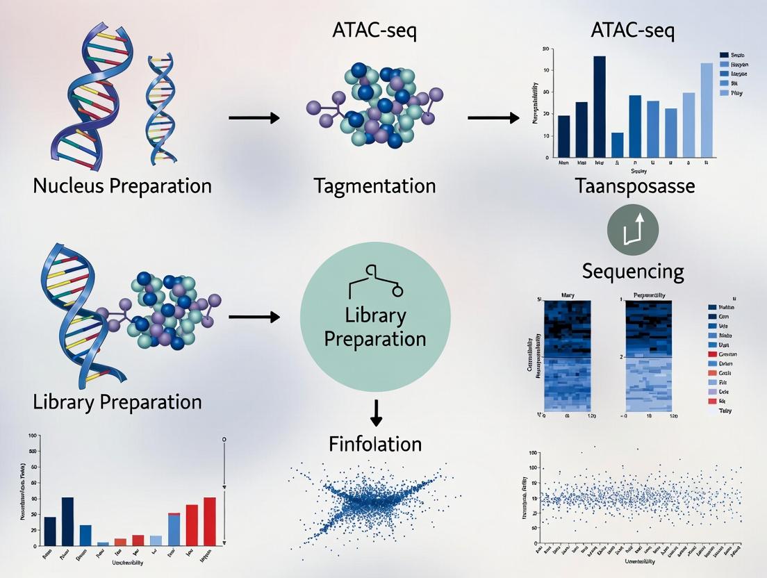
ATAC-seq Mastery 2024: Unlocking Chromatin Accessibility for Drug Discovery & Disease Research
This comprehensive guide provides researchers and drug development professionals with an in-depth exploration of Assay for Transposase-Accessible Chromatin using sequencing (ATAC-seq).
ATAC-seq Mastery 2024: Unlocking Chromatin Accessibility for Drug Discovery & Disease Research
Abstract
This comprehensive guide provides researchers and drug development professionals with an in-depth exploration of Assay for Transposase-Accessible Chromatin using sequencing (ATAC-seq). Covering foundational principles, cutting-edge methodologies, practical troubleshooting, and rigorous validation strategies, the article synthesizes the latest (2024) advancements and best practices. Readers will gain actionable insights for experimental design, data analysis, and interpretation, enabling them to effectively profile the epigenetic landscape to identify regulatory elements, understand disease mechanisms, and discover novel therapeutic targets.
What is ATAC-seq? Demystifying the Core Principles of Chromatin Accessibility Profiling
Application Note: Integrating ATAC-seq into Epigenetic Research Workflows
ATAC-seq (Assay for Transposase-Accessible Chromatin with high-throughput sequencing) is a pivotal methodology for probing chromatin accessibility, a fundamental component of the epigenetic landscape. This application note details its role in elucidating the central dogma of epigenetics—where heritable changes in gene expression occur without altering the underlying DNA sequence, governed by mechanisms such as DNA packaging into nucleosomes and higher-order structures.
Key Insights:
- Nucleosome Positioning & Occupancy: ATAC-seq fragment size distribution quantitatively maps nucleosome-free regions (NFRs) and nucleosome-occupied sequences, correlating accessibility with transcriptional regulatory elements.
- Transcription Factor (TF) Footprinting: The precise pattern of Tn5 insertion allows for the identification of TF binding sites within accessible chromatin, revealing transient, sequence-specific protein-DNA interactions.
- Multi-omics Integration: Combining ATAC-seq data with RNA-seq (gene expression) and ChIP-seq (histone modifications, TF binding) provides a systems-level view of epigenetic regulation driving phenotypic outcomes.
Table 1: Quantitative Metrics from a Representative ATAC-seq Experiment in Human Cell Lines
| Metric | HeLa Cells (Value) | HEK293T Cells (Value) | Interpretation |
|---|---|---|---|
| Total Fragments | 45,000,000 | 52,000,000 | Library complexity & sequencing depth. |
| Fraction of Reads in Peaks (FRiP) | 32% | 28% | Proportion of signal in accessible regions; >20% is good. |
| Peaks Called | 78,500 | 82,300 | Total identified regions of significant accessibility. |
| Promoter-associated Peaks | 38% | 35% | Indicates accessibility at canonical gene regulatory regions. |
| Enhancer-associated Peaks | 41% | 43% | Suggests prevalence of distal regulatory elements. |
| TSS Enrichment Score | 18.5 | 16.8 | Measures signal at transcription start sites; >10 indicates high quality. |
Detailed Protocol: ATAC-seq for Chromatin Accessibility Profiling
Principle: A hyperactive Tn5 transposase simultaneously fragments and tags accessible genomic DNA with sequencing adapters. Regions of tightly packed nucleosomes are less susceptible to Tn5 insertion, yielding a genome-wide map of open chromatin.
Protocol Part A: Cell Lysis and Tagmentation
Materials: Nuclei isolation buffer (10 mM Tris-HCl pH 7.5, 10 mM NaCl, 3 mM MgCl2, 0.1% IGEPAL CA-630), ATAC-seq Tagmentation buffer (Illumina or equivalent), Tn5 Transposase, Purification beads (SPRI beads).
Procedure:
- Nuclei Preparation: Harvest 50,000 - 100,000 viable cells. Wash with cold PBS. Lyse cells in 50 µL ice-cold nuclei isolation buffer for 10 minutes on ice. Pellet nuclei at 500 RCF for 10 minutes at 4°C. Resuspend pellet in 50 µL of Tagmentation buffer.
- Tagmentation Reaction: Add pre-loaded Tn5 transposase (2.5 µL) to the nuclei suspension. Mix gently and incubate at 37°C for 30 minutes in a thermomixer with shaking (1000 rpm).
- Clean-up: Immediately purify tagmented DNA using SPRI beads (1.8x ratio). Elute in 20 µL of Elution Buffer (10 mM Tris-HCl, pH 8.0).
Protocol Part B: Library Amplification and Sequencing
Materials: NEBNext High-Fidelity 2X PCR Master Mix, Custom Indexed PCR Primers, Size Selection Beads (e.g., SPRIselect).
- PCR Amplification: To the purified tagmented DNA, add 2.5 µL of each forward and reverse indexed primer (1.25 µM final) and 25 µL of 2X PCR Master Mix. Amplify using the following program:
- 72°C for 5 minutes (gap filling)
- 98°C for 30 seconds
- Cycle 5-12x: 98°C for 10 seconds, 63°C for 30 seconds, 72°C for 1 minute.
- Hold at 4°C. Determine optimal cycle number via qPCR side reaction to avoid over-amplification.
- Double-Sided Size Selection: Perform sequential SPRI bead cleanup.
- First (Remove Large Fragments): Add beads at 0.5x ratio to raw PCR product. Keep supernatant.
- Second (Remove Primer Dimers): Add beads to supernatant at 1.2x ratio. Discard supernatant, wash beads, and elute in 20 µL EB.
- Quality Control & Sequencing: Assess library concentration (Qubit) and profile (Bioanalyzer/TapeStation; expect a nucleosome periodicity pattern). Pool libraries and sequence on an Illumina platform (typically 2x75 bp paired-end, aiming for 50-100M reads per sample).
Visualizing the Epigenetic Workflow & Logic
Diagram 1: The Epigenetic Regulation Cascade.
Diagram 2: ATAC-seq Experimental Workflow.
The Scientist's Toolkit: Key Research Reagent Solutions
Table 2: Essential Materials for ATAC-seq and Epigenetic Analysis
| Item | Function & Rationale |
|---|---|
| Hyperactive Tn5 Transposase | Engineered enzyme for simultaneous fragmentation and adapter tagging of accessible DNA. Core reagent defining ATAC-seq. |
| Nuclei Isolation Buffer (with Detergent) | Gently lyses the plasma membrane while keeping nuclear membrane intact, preventing cytoplasmic contamination. |
| SPRI/Sera-Mag Beads | Magnetic beads for consistent post-tagmentation and post-PCR clean-up and size selection. |
| Indexed PCR Primers (i5/i7) | Adds unique dual indices to each library for sample multiplexing during sequencing. |
| High-Fidelity PCR Master Mix | Amplifies tagmented DNA with low error rates and high yield, crucial for low-input samples. |
| Cell Viability Stain (e.g., Trypan Blue) | Accurate counting of viable cells is critical, as dead cells have aberrant chromatin accessibility. |
| DNA High-Sensitivity Assay Kit (Qubit/Bioanalyzer) | Precisely quantifies low-concentration DNA libraries and assesses fragment size distribution profile. |
| Chromatin Analysis Software (e.g., ENCODE Pipelines) | Standardized bioinformatics tools for alignment (Bowtie2), peak calling (MACS2), and footprinting (HINT-ATAC). |
Within the context of a comprehensive thesis on chromatin accessibility profiling, Assay for Transposase-Accessible Chromatin using sequencing (ATAC-seq) stands out for its simplicity, sensitivity, and speed. The core innovation is the repurposing of a hyperactive Tn5 transposase, which simultaneously fragments and tags open chromatin regions with sequencing adapters. This protocol-centric application note details the methodology, key applications, and reagent toolkit essential for implementing ATAC-seq in drug discovery and basic research.
Key Quantitative Metrics and Comparisons
Table 1: Comparison of Chromatin Accessibility Profiling Methods
| Method | Key Reagent | Cell Number Input (Typical) | Assay Time (Active Hands-on) | Resolution | Primary Advantage |
|---|---|---|---|---|---|
| ATAC-seq | Hyperactive Tn5 Transposase | 50,000 - 500 (Nuclei) | 3-4 hours | Single Nucleosome (~200 bp) | Speed, simplicity, low cell input |
| DNase-seq | DNase I Enzyme | 1,000,000+ | 2-3 days | ~150 bp | Historical gold standard, well-validated |
| MNase-seq | Micrococcal Nuclease | 1,000,000+ | 2-3 days | Single Nucleosome (~147 bp) | Maps nucleosome positions precisely |
| FAIRE-seq | Phenol-Chloroform Extraction | 1,000,000+ | 2-3 days | ~200 bp | No enzyme bias, simple in principle |
Table 2: Recommended ATAC-seq Sequencing Parameters
| Library Type | Recommended Read Length | Paired-End? | Recommended Sequencing Depth (per sample) | Key Quality Metric (Post-processing) |
|---|---|---|---|---|
| Standard (Bulk) ATAC-seq | 75 bp - 150 bp | Yes | 50 - 100 million aligned reads | Fragment Size Periodicity (e.g., ~200bp nucleosome-free) |
| Single-Cell ATAC-seq (scATAC) | 50 bp - 100 bp | Yes (Dual Indexing Critical) | 25,000 - 100,000 reads per cell | Transcription Start Site (TSS) Enrichment Score > 10 |
Detailed Protocol: Bulk ATAC-seq from Cultured Cells
I. Cell Harvesting and Nuclei Preparation
- Harvest up to 50,000 viable cells. Wash once with 1x PBS.
- Lyse cells in Cold Lysis Buffer (10 mM Tris-HCl pH 7.4, 10 mM NaCl, 3 mM MgCl2, 0.1% IGEPAL CA-630) for 3-10 minutes on ice.
- Immediately pellet nuclei at 500 x g for 10 minutes at 4°C. Resuspend pellet in 50 μL of Transposition Mix.
II. Transposition Reaction
- Prepare the 50 μL transposition reaction on ice:
- 25 μL 2x TD Buffer (Illumina)
- 2.5 μL Tn5 Transposase (Illumina, Tagment DNA TDE1)
- 22.5 μL Nuclease-free water
- 50 μL Total (including nuclei suspension)
- Mix gently by pipetting. Incubate at 37°C for 30 minutes in a thermomixer with agitation (1000 rpm).
- Immediately purify DNA using a MinElute PCR Purification Kit (Qiagen) or SPRI beads. Elute in 21 μL Elution Buffer.
III. Library Amplification and Clean-up
- Amplify the transposed DNA using a limited-cycle PCR program:
- PCR Mix: 21 μL purified DNA, 2.5 μL Custom PCR Primer 1 (25 μM), 2.5 μL Custom PCR Primer 2 (25 μM), 25 μL NEBNext High-Fidelity 2X PCR Master Mix.
- Cycling Conditions: 72°C for 5 min; 98°C for 30 sec; then cycle (98°C for 10 sec, 63°C for 30 sec, 72°C for 1 min). Determine optimal cycle number (N) using a qPCR side reaction to avoid over-amplification (typically 8-12 cycles).
- Purify the final library using double-sided SPRI bead selection (e.g., 0.5X then 1.5X ratios) to remove primer dimers and select for optimal fragment size.
- Quantify using a fluorometric assay (Qubit) and assess fragment distribution (Bioanalyzer/TapeStation). Sequence on an Illumina platform using paired-end reads.
Visualization of Workflows and Pathways
ATAC-seq Experimental Workflow
Tn5 Mechanism in Open Chromatin
The Scientist's Toolkit: Essential Research Reagent Solutions
Table 3: Key Reagents and Materials for ATAC-seq
| Item | Function & Critical Notes |
|---|---|
| Hyperactive Tn5 Transposase (e.g., Illumina Tagment DNA TDE1) | Engineered enzyme that cuts DNA and ligates sequencing adapters in a single step. Activity lot consistency is critical. |
| 2x TD Buffer (Illumina) | Optimized reaction buffer for Tn5 transposition. Contains Mg2+ which catalyzes the transposition reaction. |
| Cell Permeabilization/Lysis Buffer | Mild detergent-based buffer (e.g., with IGEPAL CA-630) to lyse the plasma membrane while keeping nuclei intact. |
| Dual-Indexed i5/i7 PCR Primers | Contains Illumina P5/P7 flow cell binding sites and unique index sequences for sample multiplexing. |
| SPRI (Solid Phase Reversible Immobilization) Beads | Magnetic beads for size-selective purification of DNA fragments before and after PCR. |
| High-Fidelity PCR Master Mix (e.g., NEBNext) | Minimizes PCR bias and errors during the limited-cycle library amplification step. |
| Nuclei Counter (e.g., Trypan Blue/Countess II) | Accurate quantification of nuclei concentration after lysis is essential for optimal transposition. |
| Fluorometric DNA Quantification Kit (Qubit dsDNA HS) | Accurately measures low-concentration, adapter-ligated libraries. Avoid spectrophotometry. |
ATAC-seq (Assay for Transposase-Accessible Chromatin using sequencing) has become a cornerstone method for profiling chromatin accessibility, a key determinant of gene regulatory potential. Within the broader thesis of utilizing ATAC-seq for deciphering regulatory genomics, the primary analytical outputs—Peaks, Signals, and the annotation of Accessible Regions—form the fundamental language for biological interpretation. These outputs enable researchers to identify putative enhancers, promoters, insulators, and other cis-regulatory elements, thereby linking chromatin state to cellular function, development, and disease mechanisms critical for drug discovery.
Core Data Outputs: Definitions and Quantitative Benchmarks
Table 1: Key ATAC-seq Outputs and Their Characteristics
| Output Type | Description | Typical Scale/Units | Primary Biological Interpretation | Common Tool for Generation |
|---|---|---|---|---|
| Peak Calls | Discrete genomic intervals identified as significantly enriched for Tn5 insertion events. | Genomic coordinates (e.g., chr1:10,000-10,500). Number of peaks per sample varies. | Putative open chromatin regions, including promoters, enhancers, insulators. | MACS2, Genrich, HMMRATAC |
| Insertion Signal | The raw or smoothed count of Tn5 insertion sites per base pair. | Reads per million per bp (RPM/bp) or similar. | Quantitative measure of accessibility intensity. Can indicate activity level of a regulatory element. | DeepTools bamCoverage, IGV |
| Footprint Signal | A local depletion of insertions within an accessible region, indicating transcription factor binding. | Depth-normalized read count profiles. | Inferred protein-DNA binding events and transcription factor occupancy. | TOBIAS, HINT-ATAC, pyDNase |
| Peak Annotation | Genomic context assignment of called peaks relative to genes and other features. | Percentage of peaks in promoter (± 3kb TSS), intron, intergenic, etc. | Links accessible regions to potential target genes and functional categories. | ChIPseeker, HOMER annotatePeaks.pl |
| Differential Accessibility | Statistically significant change in signal or peak presence between conditions. | Log2 fold-change, p-value, FDR. | Regulatory elements potentially driving phenotypic differences (e.g., disease vs. healthy). | DESeq2 (on counts), edgeR, diffBind |
Quantitative Note: A typical mammalian ATAC-seq experiment yields 50,000-150,000 high-confidence peaks per sample, with 15-40% located in promoter-proximal regions. Differential analysis typically focuses on regions with |log2FC| > 1 and FDR < 0.05.
Detailed Experimental Protocols
Protocol 3.1: Standard ATAC-seq Library Preparation (50k-100k Cells)
Objective: To fragment accessible chromatin with Tn5 transposase and generate sequencing-ready libraries. Reagents: Cell lysis buffer, Tn5 transposase (commercial kit, e.g., Illumina Nextera or similar), PCR reagents, SPRI beads. Procedure:
- Cell Preparation: Harvest and wash 50,000 viable cells in cold PBS. Pellet at 500 x g for 5 minutes at 4°C.
- Cell Lysis: Resuspend pellet in 50 µL of cold lysis buffer (10 mM Tris-HCl pH 7.4, 10 mM NaCl, 3 mM MgCl2, 0.1% IGEPAL CA-630). Incubate on ice for 3 minutes.
- Nuclei Wash: Immediately add 1 mL of cold wash buffer (same as lysis buffer without IGEPAL). Pellet nuclei at 500 x g for 10 minutes at 4°C. Carefully remove supernatant.
- Tagmentation: Prepare 25 µL tagmentation mix: 12.5 µL 2x Tagmentation Buffer, 11.5 µL nuclease-free water, 1 µL Tn5 enzyme. Add directly to the pelleted nuclei. Mix gently and incubate at 37°C for 30 minutes in a thermomixer (300 rpm).
- DNA Cleanup: Immediately add 250 µL of DNA Binding Buffer (from a miniprep kit) with 2% SDS to stop reaction. Purify DNA using a commercial silica column kit. Elute in 21 µL elution buffer.
- Library Amplification: Perform PCR in a 50 µL reaction: 21 µL tagmented DNA, 2.5 µL of each barcoded primer (25 µM), 25 µL 2x NEB Next High-Fidelity PCR Master Mix. Cycle: 72°C for 5 min (gap filling); 98°C for 30 sec; then 5-12 cycles of (98°C 10 sec, 63°C 30 sec, 72°C 1 min). Determine optimal cycle number via qPCR side reaction.
- Size Selection & Cleanup: Purify final PCR product with double-sided SPRI bead selection (e.g., 0.5x then 1.2x ratios) to remove primer dimers and large fragments. Elute in 20 µL. Assess library quality on a Bioanalyzer (peak ~200-600 bp).
Protocol 3.2: Bioinformatic Processing for Peak Calling & Signal Generation
Objective: Process raw sequencing reads to generate consensus peak sets and normalized signal tracks. Tools: Trimmomatic, BWA-MEM or Bowtie2, SAMtools, PICARD, MACS2, DeepTools. Procedure:
- Quality Control & Trimming: Use FastQC for initial QC. Trim adapters and low-quality bases with Trimmomatic:
java -jar trimmomatic.jar PE -phred33 R1.fastq.gz R2.fastq.gz ... LEADING:3 TRAILING:3 SLIDINGWINDOW:4:15 MINLEN:36. - Alignment: Align to reference genome (e.g., hg38) using BWA-MEM:
bwa mem -t 8 genome.fa R1_paired.fq R2_paired.fq | samtools view -bS - > aligned.bam. - Post-Alignment Processing:
- Sort and index:
samtools sort -@ 4 -o sorted.bam aligned.bam; samtools index sorted.bam. - Filter for properly paired, uniquely mapped, non-mitochondrial reads:
samtools view -b -q 30 -f 2 -F 1804 sorted.bam chr1 chr2 ... > filtered.bam. - Remove PCR duplicates using PICARD MarkDuplicates.
- Shift aligned reads to account for 9-bp Tn5 offset: Use
alignmentSieve --ATACshiftfrom DeepTools.
- Sort and index:
- Peak Calling: Call peaks using MACS2:
macs2 callpeak -t shifted_reads.bam -f BAMPE -g hs -n output --keep-dup all -q 0.05 --nomodel --shift -100 --extsize 200. - Signal Track Generation: Create a normalized bigWig file for visualization:
bamCoverage -b filtered_shifted.bam -o signal.bw --binSize 10 --normalizeUsing RPGC --effectiveGenomeSize 2913022398 --smoothLength 50. - Consensus Peak Set: For multi-sample projects, merge replicate peak calls using
bedtools mergeor create an overlap-based consensus set.
Visualizing Pathways and Workflows
Diagram 1: ATAC-seq Data Generation & Processing Workflow
Diagram 2: From Accessible Regions to Biological Insight
The Scientist's Toolkit: Research Reagent Solutions
Table 2: Essential Reagents and Materials for ATAC-seq
| Item | Function/Benefit | Example Product/Catalog # | Notes |
|---|---|---|---|
| Tn5 Transposase | Enzyme that simultaneously fragments and tags accessible chromatin with sequencing adapters. | Illumina Tagment DNA TDE1 Enzyme (20034197), or homemade. | Critical for reaction efficiency. Commercial kits ensure reproducibility. |
| Nuclei Isolation & Lysis Buffer | Gently lyses cell membrane while keeping nuclear membrane intact for clean tagmentation. | 10x Genomics Nuclei Buffer (2000153), or homemade (see Protocol 3.1). | Avoid over-lysis, which releases genomic DNA and causes background. |
| SPRI Beads | Magnetic beads for size selection and clean-up of libraries, removing primer dimers and large fragments. | Beckman Coulter AMPure XP (A63880). | Double-sided size selection (e.g., 0.5x & 1.2x ratios) is standard for ATAC-seq. |
| High-Fidelity PCR Master Mix | Amplifies tagmented DNA with low error rate and high yield. Minimal bias is crucial. | NEB Next High-Fidelity 2x PCR Master Mix (M0541). | Cycle number optimization (5-12 cycles) is essential to prevent over-amplification. |
| Dual Indexed PCR Primers | Adds unique sample barcodes and full Illumina adapters during PCR. | Nextera CD Indexes, or IDT for Illumina UD Indexes. | Enables multiplexing. Unique dual indexes (UDI) are recommended for demultiplexing accuracy. |
| Cell Viability Stain | Assesses viability before assay; dead cells release chromatin, creating background. | Trypan Blue, AO/PI stain on cell counter. | >90% viability is strongly recommended. |
| QC Instrument | Assesses final library fragment size distribution and concentration. | Agilent Bioanalyzer/TapeStation, or Fragment Analyzer. | Characteristic nucleosomal ladder pattern (e.g., ~200, 400, 600 bp) indicates success. |
Chromatin accessibility, governed by the dynamic interplay of nucleosome positioning and transcription factor binding, is a fundamental regulator of gene expression. Profiling this accessibility, primarily through the Assay for Transposase-Accessible Chromatin using sequencing (ATAC-seq), provides a critical window into cellular state, lineage commitment, disease pathogenesis, and therapeutic response. Within the broader thesis of ATAC-seq for chromatin accessibility profiling, this document details application notes and protocols for leveraging this technology to dissect mechanisms in development, disease, and pharmacology.
Application Notes & Key Findings
Developmental Biology: Mapping Cell Fate Decisions
ATAC-seq enables the identification of cell-type-specific regulatory elements and transcription factor networks driving differentiation.
Table 1: ATAC-seq Insights into Developmental Trajectories
| Study System | Key Accessible Regions Identified | Associated Regulatory Factor | Functional Outcome | Citation (Year) |
|---|---|---|---|---|
| Mouse Embryonic Stem Cell to Neural Progenitor | ~5,000 new open chromatin regions | SOX2, POU3F2 (Brn2) | Activation of neural tube development genes | Trevino et al., Nat. Methods, 2021 |
| Human Hematopoiesis | 2,152 differential peaks between HSCs and myeloid progenitors | C/EBPα, PU.1 | Commitment to granulocyte-macrophage lineage | Corces et al., Nat. Genet., 2016 |
| Drosophila Embryogenesis | 84,000 accessible regions across 24 time points | Temporal cascade of Zelda, GAGA, Trl | Zygotic genome activation patterning | Blythe & Wieschaus, Dev. Cell, 2016 |
Disease Pathogenesis: Uncovering Dysregulated Epigenomes
Aberrant chromatin accessibility is a hallmark of cancer, autoimmune disorders, and neurodegeneration.
Table 2: Chromatin Accessibility Alterations in Disease States
| Disease | Sample Comparison | Quantitative Change | Dysregulated Pathway | Therapeutic Implication |
|---|---|---|---|---|
| Acute Myeloid Leukemia (AML) | Primary patient blasts vs. normal CD34+ | 12,849 gained, 8,732 lost accessibility regions | MYB, RUNX1 complexes | Vulnerability to BRD4 inhibition (BET inhibitors) |
| Rheumatoid Arthritis | Synovial fibroblast subsets | 31% of open sites unique to pathogenic THY1+ subset | AP-1 (FOS/JUN) driven inflammatory program | JAK/STAT inhibitor sensitivity prediction |
| Alzheimer's Disease | Post-mortem prefrontal cortex | ~3,000 hyper-accessible sites near synaptic genes | Microglial activation (SPI1/PU.1 binding) | Novel targets for neuroinflammation modulation |
Drug Response & Resistance: Profiling Epigenetic Adaptations
ATAC-seq can track dynamic chromatin remodeling in response to therapeutic agents, identifying mechanisms of sensitivity and resistance.
Table 3: Chromatin Dynamics in Drug Response Profiling
| Drug Class | Cell/Model System | Time Point | Key Accessibility Shift | Linked Outcome |
|---|---|---|---|---|
| BET Inhibitor (JQ1) | Triple-Negative Breast Cancer | 72h post-treatment | Loss of accessibility at super-enhancers of MYC and FOSL1 | Cytostatic response; residual cells show regained access at RTK genes |
| HDAC Inhibitor (Panobinostat) | Multiple Myeloma | 24h post-treatment | Increased accessibility at interferon response genes (ISRE motifs) | Priming for immune checkpoint therapy |
| Androgen Receptor Antagonist (Enzalutamide) | Prostate Cancer | Chronic exposure (4 weeks) | De novo accessibility at glucocorticoid receptor (GR) binding sites | GR-driven resistance bypassing AR blockade |
Detailed Protocols
Protocol 1: High-Throughput ATAC-seq on Primary Patient Samples (e.g., Cancer Biopsies)
Goal: Generate chromatin accessibility profiles from low-input, frozen clinical specimens.
Materials:
- Cryopreserved tissue sample or cell suspension
- Nuclei Extraction Buffer (10 mM Tris-HCl pH 7.5, 10 mM NaCl, 3 mM MgCl2, 0.1% IGEPAL CA-630, 0.1% Tween-20, 0.01% Digitonin)
- ATAC-seq Kit (e.g., Illumina Tagmentase TDE1, Buffer TD)
- SPRIselect beads (Beckman Coulter)
- Qubit dsDNA HS Assay Kit
Procedure:
- Nuclei Isolation: Thaw sample on ice. Homogenize in 1 mL cold Nuclei Extraction Buffer with Dounce homogenizer (15 strokes). Filter through a 40-μm strainer. Centrifuge at 500 rcf for 5 min at 4°C. Carefully aspirate supernatant.
- Nuclei Count & Tagmentation: Resuspend pellet in 50 μL of cold PBS. Count using Trypan Blue in a hemocytometer. Adjust concentration to 5,000-10,000 nuclei in 50 μL. Add Tagmentation Mix (25 μL 2x TD Buffer, 2.5 μL TDE1, 22.5 μL nuclease-free water). Incubate at 37°C for 30 min in a thermomixer.
- DNA Clean-up: Immediately add 250 μL of Buffer PB and mix. Bind to a MinElute column, wash with PE buffer, and elute in 21 μL EB buffer.
- Library Amplification: Amplify tagmented DNA using 1x NEBnext PCR master mix and custom Ad1_noMX and Ad2.xx barcoded primers. Determine cycle number via qPCR side reaction (usually 10-13 cycles).
- Size Selection & QC: Perform double-sided SPRI bead cleanup (0.55x and 1.5x ratios) to isolate fragments primarily between 150-800 bp. Quantify with Qubit and analyze fragment distribution (e.g., TapeStation). Pool and sequence on Illumina NovaSeq (Paired-end, 50 bp).
Protocol 2: Integrative ATAC-seq + Transcriptomics (Multiome) for Target Discovery
Goal: Simultaneously capture chromatin accessibility and gene expression from the same single cell.
Materials:
- Chromium Controller & Chip G (10x Genomics)
- Chromium Next GEM Single Cell Multiome ATAC + Gene Expression Kit
- Dual Index Kit TT Set A
- Bioanalyzer High Sensitivity DNA kit
Procedure:
- Nuclei Preparation: Prepare nuclei as in Protocol 1, aiming for viability >90% and concentration ~1,000 nuclei/μL. Keep nuclei cold and process within 1 hour.
- GEM Generation & Barcoding: Follow 10x Genomics User Guide (CG000338). Combine nuclei, Transposition Mix, and Master Mix with gel beads in the chip. Run on Chromium Controller to generate single-cell GEMs where transposition and lysis occur.
- Post GEM-RT Cleanup & Library Construction: Break emulsions, recover barcoded DNA. Perform split of material for ATAC library and cDNA synthesis for gene expression.
- ATAC Library Amplification: Amplify with sample index PCR using Dual Index Kit. Include size selection steps (SPRIselect 0.6x right-sided).
- Sequencing: Pool libraries. Recommended sequencing depth: 25,000 paired-end reads per nucleus for ATAC, 20,000 reads per nucleus for Gene Expression.
Protocol 3: ATAC-seq for Pharmacodynamic Monitoring inIn VivoModels
Goal: Assess chromatin remodeling in tumor or tissue after drug treatment in mouse models.
Materials:
- Drug- or vehicle-treated mouse tissues (e.g., tumor allografts)
- GentleMACS Dissociator (Miltenyi)
- Debris Removal Solution (Miltenyi)
- Sucrose-based Nuclei Cushion Buffer (0.9 M Sucrose, 5 mM MgCl2, 1x PBS)
Procedure:
- Rapid Tissue Harvest & Dissociation: Euthanize mouse at predetermined time point. Excise target tissue, mince in 1 mL cold Nuclei Extraction Buffer. Process using GentleMACS "soft" program. Filter through 70-μm then 40-μm strainers.
- Nuclei Purification: Layer filtrate over 1 mL of cold Sucrose Cushion Buffer in a 2 mL tube. Centrifuge at 13,000 rcf for 10 min at 4°C. Pellet contains clean nuclei. Aspirate supernatant completely.
- Tagmentation & Downstream Processing: Proceed with tagmentation (as in Protocol 1, Step 2) using 10,000-50,000 nuclei. Complete library preparation.
- Bioinformatic Analysis: Align to mm10 genome. Call peaks per sample. Perform differential accessibility analysis (e.g., using DESeq2 on count matrices) between treatment and vehicle groups to identify pharmacodynamic regulatory changes.
The Scientist's Toolkit: Research Reagent Solutions
Table 4: Essential Reagents for ATAC-seq Research
| Item | Supplier/Example | Function |
|---|---|---|
| Tn5 Transposase | Illumina Tagmentase TDE1, Diagenode Hyperactive Tn5 | Enzyme that simultaneously fragments and tags accessible chromatin with sequencing adapters. |
| Nuclei Extraction Buffer with Digitonin | 10x Genomics Nuclei Buffer, Homebrew (see Protocol 1) | Gently lyses plasma membrane while keeping nuclear membrane intact for clean tagmentation. |
| SPRIselect Beads | Beckman Coulter, Sigma | Magnetic beads for size selection and cleanup of DNA libraries, critical for removing adapter dimers. |
| Single-Cell Multiome Kit | 10x Genomics Chromium Single Cell Multiome ATAC + Gene Expression | Enables simultaneous profiling of chromatin accessibility and transcriptome from the same cell. |
| Indexed PCR Primers | Illumina Indexing Primers, IDT for Illumina | Unique dual indices allow multiplexing of many samples in a single sequencing run. |
| High-Sensitivity DNA Assay | Agilent Bioanalyzer/TapeStation, Qubit dsDNA HS | Accurate quantification and quality control of final libraries prior to sequencing. |
Visualizations
Title: ATAC-seq Data Analysis Workflow
Title: Drug Response Mediated by Chromatin Dynamics
Title: From Chromatin Dysregulation to Disease Insights
1. Introduction and Thesis Context Within the broader thesis on ATAC-seq for chromatin accessibility profiling, this document details the evolution of the Assay for Transposase-Accessible Chromatin using sequencing. The original protocol, a pivotal innovation, has been iteratively optimized to enable high-throughput, multi-modal analyses, fundamentally accelerating epigenomic research and drug target discovery.
2. Quantitative Evolution: Key Parameters Table 1: Evolution of ATAC-seq Core Parameters
| Parameter | Original Protocol (2013) | Modern High-Throughput Applications (2024) |
|---|---|---|
| Starting Cells/Nuclei | 50,000 - 500,000 | 500 - 100,000 (Single-cell) |
| Handling Time | ~3 hours hands-on | <1 hour (with automation) |
| Library Prep Time | ~3 hours | ~1.5 hours (commercial kits) |
| Multiplexing Capacity | Low (sample pooling) | High (96+ samples, indexed transposomes) |
| Data Yield per Sample | ~50 million reads | 25,000 - 50,000 reads/cell (scATAC) |
| Primary Output | Bulk chromatin accessibility | Single-cell accessibility, multi-omics (paired w/ RNA, protein) |
3. Detailed Protocols
Protocol A: Original Bulk ATAC-seq (Adapted from Buenrostro et al., 2013)
- Cell Lysis & Transposition: Resuspend 50,000 viable cells in 50 µL cold lysis buffer (10 mM Tris-HCl pH 7.4, 10 mM NaCl, 3 mM MgCl2, 0.1% IGEPAL CA-630). Incubate on ice for 3 min. Centrifuge (500 RCF, 10 min, 4°C). Immediately resuspend pellet in 50 µL transposition mix (25 µL 2x TD Buffer, 2.5 µL Tn5 Transposase (Illumina), 22.5 µL nuclease-free water). Incubate at 37°C for 30 min.
- DNA Clean-up: Purify transposed DNA using a MinElute PCR Purification Kit (Qiagen). Elute in 21 µL Elution Buffer.
- Library Amplification: Amplify using 1x NEBnext PCR master mix, 1.25 µM custom Ad1_noMX and Ad2.x primers (see Reagent Table). Cycle: 72°C 5 min; 98°C 30 sec; then [98°C 10 sec, 63°C 30 sec, 72°C 1 min] for 5-10 cycles (qPCR-guided).
- Size Selection & Clean-up: Clean PCR reaction with a MinElute kit. Optional: size select for fragments < 1,000 bp using SPRI beads (0.5x ratio). Sequence on Illumina platforms (paired-end).
Protocol B: Modern High-Throughput Single-Cell ATAC-seq (10x Genomics Workflow)
- Nuclei Isolation & Transposition: Isolate nuclei from fresh/frozen tissue using a Dounce homogenizer in chilled lysis buffer. Filter (40 µm strainer). Count nuclei. For 10,000 nuclei, combine with transposition mix (from Chromium Next GEM ATAC Kit) and loaded into a Chromium Next GEM Chip G.
- Partitioning & Barcoding: The Chromium Controller partitions nuclei, transposed fragments, and Gel Beads containing unique barcodes into nanoliter-scale droplets. Within each droplet, barcoded primers anneal to transposed fragments.
- Post-GEM Clean-up & Amplification: Break droplets, pool barcoded DNA. Perform a post-EM clean-up with Silane magnetic beads. Amplify libraries via PCR (13 cycles).
- Library Construction: Fragments undergo size selection (SPRIselect beads) and are converted into sequencing-ready libraries via a second, short PCR (5 cycles) to add sample indexes and P5/P7 flow cell adapters.
- Sequencing: Sequence on Illumina NovaSeq (typically 25-50k read pairs per cell).
4. Visualized Workflows and Pathways
Title: Original Bulk ATAC-seq Protocol Flow
Title: Modern Single-Cell ATAC-seq Workflow
5. The Scientist's Toolkit: Essential Research Reagents & Materials Table 2: Key Reagent Solutions for ATAC-seq
| Item | Function & Role | Example (Vendor) |
|---|---|---|
| Hyperactive Tn5 Transposase | Enzyme that simultaneously fragments and tags accessible DNA with sequencing adapters. Core of the assay. | Tn5 Transposase (Illumina), Tagmentase (Diagenode) |
| 2x TD Buffer | Optimized buffer providing Mg2+ for Tn5 activity, enabling efficient transposition. | Illumina Tagment DNA Buffer |
| Nuclei Isolation Buffer | Gently lyses plasma membrane while keeping nuclear membrane intact. Critical for clean signal. | 10mM Tris-HCl, 10mM NaCl, 3mM MgCl2, 0.1% IGEPAL CA-630 |
| SPRI/Silane Magnetic Beads | For size selection and clean-up of DNA libraries. Enables removal of short fragments and reaction cleanup. | SPRIselect / AMPure XP Beads (Beckman Coulter) |
| Unique Dual Index Primers | Primers containing i5 and i7 indexes for multiplexing many samples in one sequencing run. | Nextera DNA CD Indexes (Illumina), IDT for Illumina Tagment Indexes |
| Chromium Next GEM Chip G | Microfluidic chip for partitioning single nuclei into droplets with barcoded gel beads. | 10x Genomics Chromium Chip G |
| Cell Ranger ATAC | Primary software pipeline for processing raw scATAC-seq data into count matrices and basic analysis. | 10x Genomics Cell Ranger ATAC |
| Chromatin Accessibility Signal Peaks | Primary output data type, representing genomic regions of open chromatin, used for downstream analysis. | N/A |
ATAC-seq in Action: A Step-by-Step Protocol from Cell to Data (2024 Best Practices)
Within the context of an ATAC-seq (Assay for Transposase-Accessible Chromatin using sequencing) thesis, robust experimental design is the cornerstone of generating reproducible and biologically meaningful chromatin accessibility profiles. This application note details the critical considerations for cell number, replicates, and controls to ensure statistical power and valid interpretation in drug development and basic research.
Quantitative Parameters for Experimental Design
Table 1: Recommended Cell Numbers and Replicates for ATAC-seq Experiments
| Experimental Condition | Minimum Viable Cells per Reaction (Fresh) | Minimum Viable Cells per Reaction (Frozen) | Recommended Biological Replicates | Notes |
|---|---|---|---|---|
| Standard Human/Mouse Cell Lines | 50,000 | 75,000 | 3-4 | For homogeneous populations. |
| Primary Cells (e.g., PBMCs) | 50,000 - 100,000 | 100,000 - 200,000 | 4-5 | Higher numbers account for viability loss and heterogeneity. |
| Rare Cell Populations (Sorted) | 10,000 - 50,000 | Not Recommended | 3 (if feasible) | Amplification cycles may increase; use stringent QC. |
| Tissue Samples (Nuclei Isolation) | 50,000 nuclei | 100,000 nuclei | 3-4 (pool from multiple organisms if needed) | Tissue dissociation efficiency is key. |
| Drug Treatment Studies | 100,000 per condition | 150,000 per condition | ≥4 | Essential for capturing subtle chromatin remodeling. |
Sources: Current protocols emphasize that 50,000 fresh cells is a robust starting point, but requirements can vary by transposase batch and cell type. For frozen cells or nuclei, a 1.5-2x increase compensates for increased fragmentation. In drug development, increased replicates are non-negotiable to achieve power for differential accessibility analysis.
Table 2: Essential Control Experiments for ATAC-seq
| Control Type | Purpose | Recommended Specs | When to Include |
|---|---|---|---|
| Negative Control (No Transposase) | Detects background DNA contamination & endogenous nucleases. | Use same cell/nuclei input as main assay. Process identically. | In every experiment. |
| Positive Control (Known Accessible Cell Line) | Benchmarks tagmentation efficiency and library complexity across runs. | e.g., K562 or GM12878 cells. Include in each sequencing batch. | In every experimental batch. |
| Genomic DNA Control (gDNA + Transposase) | Assesses sequence bias of the transposase enzyme batch. | 100 ng gDNA. Tagment alongside samples. | With new enzyme lot. |
| Mitochondrial DNA Depletion Assessment | QC step to evaluate nuclear isolation/tagmentation specificity. | Calculate % mtDNA reads in FASTQ files. | For every sample. |
| Technical Replicate | Assesses library prep variability. | Split a single sample preps into multiple libraries. | During protocol establishment. |
| Biological Replicate | Captures biological variability; essential for statistics. | Independently derived samples from different cultures/animals. | Always. Non-negotiable for thesis research. |
| Process Control (Spike-in Nuclei) | Normalizes for technical variation in tagmentation between samples. | e.g., D. melanogaster nuclei added to mammalian samples pre-tagmentation. | For complex multi-condition or time-course drug studies. |
Detailed Protocols
Protocol 1: Determining Optimal Cell Number for a New Cell Type
Objective: To empirically determine the minimum number of cells yielding a high-complexity ATAC-seq library for a novel primary cell or cell line. Materials: Single-cell suspension of interest, Trypan Blue or AO/PI stain, hemocytometer or automated cell counter, ATAC-seq buffers, purified transposase (e.g., Illumina Tagment DNA TDE1), PCR reagents, bioanalyzer/tapestation. Procedure:
- Prepare a Viability-Adjusted Series: Count cells and assess viability. Prepare four aliquots with viable cell counts: 5,000, 25,000, 50,000, and 100,000 cells in 50 µL of cold PBS.
- Cell Lysis & Tagmentation: Pellet cells (500 x g, 5 min, 4°C). Resuspend pellet in 50 µL of ATAC lysis buffer (10 mM Tris-HCl pH 7.4, 10 mM NaCl, 3 mM MgCl2, 0.1% IGEPAL CA-630). Incubate on ice for 3 min. Immediately add 1 mL of wash buffer (10 mM Tris-HCl pH 7.4, 10 mM NaCl, 3 mM MgCl2), invert to mix, and pellet nuclei (500 x g, 10 min, 4°C). Carefully aspirate supernatant.
- Transposition Reaction: Resuspend nuclei in 50 µL of transposition mix (25 µL 2x Tagmentation Buffer, 2.5 µL Transposase, 22.5 µL nuclease-free water). Incubate at 37°C for 30 min in a thermomixer with shaking.
- DNA Purification: Immediately purify tagmented DNA using a MinElute PCR Purification Kit. Elute in 21 µL of Elution Buffer.
- Library Amplification & QC: Amplify 20 µL of eluate in a 50 µL PCR reaction using indexed primers and NEBNext High-Fidelity 2X PCR Master Mix. Use 5 cycles, then perform a qPCR side reaction to determine additional cycles needed (cycle where SYBR Green signal is 1/3 max). Amplify the main reaction for the total calculated cycles. Purify final library with SPRI beads.
- Analysis: Assess library fragment distribution on a High Sensitivity DNA chip. The optimal cell input will yield the characteristic nucleosomal ladder pattern with minimal adapter dimer peak (~100 bp) and high library concentration. Select the lowest cell number yielding this profile.
Protocol 2: Implementing a Spike-in Control for Normalization
Objective: To use Drosophila melanogaster nuclei as a process control to normalize for technical variation in tagmentation efficiency across mammalian samples. Materials: D. melanogaster S2 cell culture, Mammalian cells of interest, Dounce homogenizer, ATAC lysis buffer, 0.25% Trypan Blue. Procedure:
- Prepare Spike-in Nuclei: Harvest ~1 million D. melanogaster S2 cells. Wash with PBS. Lyse in 1 mL of ice-cold ATAC lysis buffer for 3 min on ice. Immediately add 10 mL of wash buffer. Centrifuge (500 x g, 10 min, 4°C). Gently resuspend pellet in 1 mL of wash buffer. Count nuclei using a hemocytometer (lysed nuclei exclude Trypan Blue). Adjust concentration to 1,000 nuclei/µL. Aliquot and freeze at -80°C.
- Spike-in Addition: For each mammalian sample, after lysing and washing the mammalian nuclei (as in Protocol 1, Step 2), resuspend the final pellet in 50 µL of wash buffer. Add a fixed volume (e.g., 5 µL, yielding 5,000 nuclei) of thawed Drosophila spike-in nuclei suspension. Mix gently by pipetting.
- Proceed with Tagmentation: Pellet the combined nuclei (500 x g, 10 min, 4°C). Aspirate supernatant completely. Proceed with the transposition reaction (Protocol 1, Step 3) on the combined pellet.
- Bioinformatic Normalization: During sequencing analysis, separate reads aligning to the Drosophila (dm6) and mammalian (e.g., hg38) genomes. Use the read depth from the spike-in chromatin to scale or normalize the mammalian accessibility signals across samples.
Diagrams
Diagram 1: Replicate-Centric ATAC-seq Workflow
Diagram 2: Hierarchy of ATAC-seq Controls
The Scientist's Toolkit
Table 3: Key Research Reagent Solutions for ATAC-seq
| Item | Function in ATAC-seq | Example/Notes |
|---|---|---|
| Tagment DNA Enzyme (TDE1) | Engineered Tn5 transposase that simultaneously fragments and tags accessible DNA with sequencing adapters. | Illumina Tagment DNA TDE1 or equivalent. Critical for efficiency. |
| Cell Lysis Buffer (with Detergent) | Gently lyses the plasma membrane while leaving the nuclear membrane intact for clean isolation of nuclei. | 10 mM Tris-HCl, 10 mM NaCl, 3 mM MgCl2, 0.1% IGEPAL CA-630, 0.1% Tween-20, 0.01% Digitonin (optional). |
| Magnetic Beads for SPRI Clean-up | Size-selects DNA fragments post-tagmentation and post-PCR, removing primers, dimers, and large contaminants. | AMPure XP or SPRIselect beads. Ratios are crucial (e.g., 0.5x to remove large DNA, 1.8x to purify). |
| High-Fidelity PCR Master Mix | Amplifies the tagmented library with minimal bias and error introduction during the limited-cycle PCR. | NEBNext High-Fidelity 2X PCR Master Mix or KAPA HiFi HotStart ReadyMix. |
| Dual-Indexed PCR Primers | Adds unique barcode combinations to each library, enabling multiplexing of many samples in a single sequencing run. | Illumina Nextera-style indices or IDT for Illumina unique dual indexes (UDIs). UDis reduce index hopping. |
| Nuclei Staining Dye | Validates nuclear integrity and count post-lysis before the critical tagmentation step. | DAPI (for fluorescence counting) or Trypan Blue (for light microscopy; lysed nuclei stain blue). |
| QC Instrumentation | Assesses library quality, concentration, and fragment size distribution prior to sequencing. | Agilent Bioanalyzer/Tapestation or Fragment Analyzer. The nucleosomal ladder pattern is a key success metric. |
| Spike-in Material | Provides an internal standard for normalization across samples, controlling for technical variation. | Drosophila melanogaster S2 cells, E. coli DNA, or commercial spike-in nucleosomes (e.g., from Active Motif). |
Within the broader thesis on ATAC-seq for chromatin accessibility profiling, the initial steps of nuclei isolation and tagmentation are unequivocally critical. The quality and quantity of isolated nuclei directly determine the signal-to-noise ratio, library complexity, and reproducibility of the final data. These steps ensure that the transposase can efficiently and uniformly access open chromatin regions, forming the foundation for accurate downstream biological interpretation in research and drug development contexts.
Key Quantitative Considerations
Table 1: Critical Metrics for Successful Nuclei Preparation & Tagmentation
| Parameter | Optimal Range | Impact on ATAC-seq Data |
|---|---|---|
| Cell Input (Mammalian) | 50,000 - 100,000 cells | Lower: Risk of low library complexity; Higher: Increased debris & aggregation. |
| Nuclei Purity (A260/A280) | 1.8 - 2.0 | Deviations indicate cytoplasmic or RNA contamination, leading to high background. |
| Nuclei Integrity | >80% intact (microscopy) | Lysed nuclei release genomic DNA, causing clogging and irreproducible tagmentation. |
| Tagmentation Time | 30 min (37°C) | Under-digestion: Low fragment yield; Over-digestion: Over-fragmentation, loss of signal. |
| Transposase to Nuclei Ratio | As per mfg. (e.g., 1:1 - 2:1) | Ratio is cell-type dependent; critical for fragment size distribution. |
| Final Library Size Distribution | Major peak ~200 bp (nucleosomal ladder) | Absence of nucleosomal patterning indicates poor nuclei quality or tagmentation. |
Detailed Protocols
Protocol 3.1: Gentle Nuclei Isolation from Cultured Mammalian Cells
Objective: To isolate intact, clean nuclei free of cytoplasmic contaminants.
Materials:
- Cell suspension.
- Cold Lysis Buffer: 10 mM Tris-HCl (pH 7.4), 10 mM NaCl, 3 mM MgCl2, 0.1% IGEPAL CA-630, 1% BSA, 0.1 U/µL RNase inhibitor, 0.1 U/µL protease inhibitor in nuclease-free water. Keep ice-cold.
- Wash Buffer: Cold PBS + 1% BSA + 0.1 U/µL RNase inhibitor.
- 40 µm cell strainer.
- Refrigerated centrifuge.
Method:
- Harvest & Count: Harvest cells, centrifuge at 300 x g for 5 min at 4°C. Resuspend in cold PBS. Count and aliquot 50,000-100,000 cells.
- Cell Lysis: Pellet cells (300 x g, 5 min, 4°C). Carefully aspirate supernatant. Resuspend pellet gently in 50 µL of Cold Lysis Buffer. Incubate on ice for 5-10 min (monitor lysis under microscope).
- Quench & Wash: Immediately add 1 mL of Wash Buffer to quench lysis. Invert gently to mix.
- Filter & Pellet Nuclei: Pass the suspension through a pre-wet 40 µm strainer. Centrifuge nuclei at 500 x g for 5 min at 4°C.
- Resuspend: Carefully aspirate supernatant. Resuspend nuclei gently in 50 µL of Wash Buffer. Count nuclei using a hemocytometer (stain with Trypan Blue). Adjust concentration to ~1,000 nuclei/µL. Keep on ice.
Protocol 3.2: Optimized In-Situ Tagmentation of Isolated Nuclei
Objective: To fragment accessible chromatin regions using Tn5 transposase while preserving nuclear integrity.
Materials:
- Isolated nuclei (~50,000) in resuspension buffer.
- Tagmentation Buffer (2X): 20 mM Tris-HCl (pH 7.6), 10 mM MgCl2, 20% Dimethyl Formamide (DMF) in nuclease-free water.
- Commercially available or pre-loaded Tn5 Transposase (e.g., Th5 enzyme + adapters).
- 1% SDS (in nuclease-free water).
- Magnetic beads for DNA purification (e.g., SPRI beads).
- Thermal cycler.
Method:
- Tagmentation Mix Assembly: In a nuclease-free PCR tube, combine: 25 µL of 2X Tagmentation Buffer, ~50,000 nuclei in 20 µL, and 5 µL of loaded Tn5 Transposase. Mix gently by pipetting. Final volume: 50 µL.
- Incubate: Immediately place tube in a pre-heated thermal cycler at 37°C for 30 minutes.
- Reaction Stop: Add 50 µL of 1% SDS to the reaction. Mix thoroughly by pipetting. Incubate at room temperature for 5 min. This step chelates Mg2+ and denatures the Tn5 enzyme.
- DNA Purification: Proceed immediately with DNA purification using magnetic SPRI beads at a 1:1 ratio (e.g., add 100 µL beads to 100 µL sample). Elute in 20-30 µL of nuclease-free water or TE buffer. The purified DNA is now ready for library amplification by PCR.
Visualization of Workflow & Key Considerations
Title: ATAC-seq Nuclei Isolation & Tagmentation Workflow
Title: Factors Influencing ATAC-seq Data Quality
The Scientist's Toolkit: Essential Reagents & Materials
Table 2: Key Research Reagent Solutions for Nuclei Isolation & Tagmentation
| Item | Function & Rationale | Critical Notes |
|---|---|---|
| IGEPAL CA-630 (NP-40 alternative) | Non-ionic detergent for gentle plasma membrane lysis while leaving nuclear membrane intact. | Concentration is critical (typically 0.1-0.5%); varies by cell type. |
| BSA (Nuclease-Free) | Carrier protein that reduces nonspecific sticking of nuclei to tubes and tips, improving recovery. | Must be nuclease-free to prevent DNA/RNA degradation. |
| Rnase Inhibitor | Protects accessible chromatin-associated RNA from degradation, which can improve data quality. | Essential for samples with high transcriptional activity. |
| Loaded Tn5 Transposase | Engineered enzyme that simultaneously fragments and adaptor-tags accessible DNA. | Commercial "tagmentation" kits provide pre-loaded, optimized enzyme. |
| Dimethyl Formamide (DMF) | A transposase reaction enhancer; increases efficiency of tagmentation within intact nuclei. | High purity required; part of optimized tagmentation buffers. |
| SPRI (Solid Phase Reversible Immobilization) Beads | Magnetic beads for size-selective purification of tagmented DNA, removing enzymes and salts. | Bead-to-sample ratio determines size selection stringency. |
| Dual-Size DNA Ladder | For quality control on Bioanalyzer/TapeStation to verify nucleosomal ladder pattern post-tagmentation. | Absence of ~200bp, 400bp, 600bp peaks indicates failure. |
Within the broader thesis on utilizing ATAC-seq (Assay for Transposase-Accessible Chromatin using sequencing) for chromatin accessibility profiling in drug development research, the steps of library preparation and sequencing are critical junctures. Decisions made here directly determine the resolution, accuracy, and cost-effectiveness of the entire study. This document provides detailed application notes and protocols to guide researchers in selecting the appropriate sequencing platform and depth, ensuring robust and reproducible data for downstream analysis of chromatin dynamics in response to therapeutic compounds.
Sequencing Platform Comparison
The choice of sequencing platform influences read length, throughput, cost, and turnaround time. For ATAC-seq, which generates short, fragmented DNA from open chromatin regions, both short-read and long-read platforms have applications.
Table 1: Comparison of Key Sequencing Platforms for ATAC-seq
| Platform (Provider) | Read Type | Typical Read Length | Key Advantages for ATAC-seq | Considerations for ATAC-seq |
|---|---|---|---|---|
| NovaSeq X & 6000 (Illumina) | Short-read, Paired-end | 50-300 bp | Ultra-high throughput, low error rate, standardized ATAC-seq protocols. Ideal for high-depth, large sample cohorts. | Cannot resolve long-range chromatin interactions. Highest throughput flow cells may be excessive for single experiments. |
| NextSeq 1000/2000 (Illumina) | Short-read, Paired-end | 50-300 bp | High throughput, benchtop flexibility. Perfect for mid-sized projects (e.g., 10-100 samples). | Higher per-Gb cost than NovaSeq for very large projects. |
| MiSeq (Illumina) | Short-read, Paired-end | 50-600 bp | Fast turnaround, long reads possible. Excellent for protocol optimization and pilot studies. | Very low throughput; not for full-scale projects. |
| X Series (Element) | Short-read, Paired-end | 50-300 bp | Lower capital cost, competitive pricing. Suitable for core labs seeking an Illumina alternative. | Younger ecosystem; community protocols less established. |
| Revio & Sequel IIe (PacBio) | Long-read, HiFi | 10-25 kb | Can phase alleles and detect large structural variants in accessible regions. Links distal sites via single molecules. | Lower throughput, higher DNA input, higher cost per sample. Best for targeted, hypothesis-driven studies. |
| PromethION (Oxford Nanopore) | Long-read | 1 kb - >5 Mb | Extreme read length, direct detection of modifications. Can assess ultra-long-range chromatin connectivity. | High error rate (~5%) complicates peak calling; requires specialized bioinformatics. |
Determining Optimal Sequencing Depth
Sequencing depth is crucial for statistical power and sensitivity. Insufficient depth misses rare open regions, while excessive depth wastes resources. Depth requirements vary by organism genome size and experimental complexity.
Table 2: Recommended Sequencing Depth for ATAC-seq Experiments
| Experimental Context & Goal | Recommended Depth (per sample) | Rationale |
|---|---|---|
| Human/Mouse - General Profiling | 50-100 million aligned, non-duplicate paired-end reads | Balances cost and sensitivity for identifying major accessible regions in cell lines or homogeneous tissues. |
| Human/Mouse - Heterogeneous Tissues or Complex Conditions | 100-200 million aligned reads | Increased depth improves detection of subtle accessibility changes in subpopulations and rare cell states. |
| Human/Mouse - Single-cell ATAC-seq (scATAC-seq) Aggregate | 25,000-100,000 reads per nucleus (aggregate >100M reads) | Depth per cell is low; aggregate depth from many cells defines the accessible landscape of the population. |
| Drug Treatment Studies (Thesis Focus) | 100-150 million aligned reads (minimum) | Essential for robust statistical comparison between treatment/control, identifying dose-dependent changes, and detecting moderate-effect loci. |
| Pilot or Optimization Study | 20-50 million aligned reads | Sufficient to assess library quality and major peaks before scaling. |
| Organisms with Larger Genomes (e.g., Zebrafish) | Increase depth relative to genome size complexity. |
Detailed Protocol: Dual-Size Selection for ATAC-seq Library Preparation
This protocol refines the standard ATAC-seq method by implementing a double-sided SPRI bead cleanup to tightly size-select nucleosomal fragments (mono-, di-, tri-nucleosome), reducing background from mitochondrial DNA and short, unincorporated transposons.
Materials:
- Purified nuclei from 50,000-100,000 cells.
- Tagment DNA Buffer and TDE1 Enzyme (Illumina Tagment DNA TDE1 Kit or equivalent).
- SPRIselect beads (Beckman Coulter).
- Ethanol (80%, freshly prepared).
- Nuclease-free water.
- Qubit dsDNA HS Assay Kit.
- TapeStation or Bioanalyzer (High Sensitivity DNA chips).
Procedure:
- Tagmentation: Resuspend purified nuclei in a mix of 25 μL Tagment DNA Buffer and 2.5 μL TDE1. Incubate at 37°C for 30 minutes in a thermomixer with shaking (1000 rpm). Immediately proceed to cleanup.
- DNA Purification: Add 25 μL of well-resuspended SPRIselect beads (0.5x ratio) to the 27.5 μL tagmentation reaction. Incubate 5 minutes at RT. Pellet beads, transfer supernatant (containing tagmented DNA) to a new tube.
- Large Fragment Removal (First Selection): Add 15 μL of SPRIselect beads (0.55x ratio to supernatant) to the supernatant. Incubate 5 minutes. Pellet beads. Discard supernatant. This step removes very large fragments.
- Small Fragment Removal (Second Selection): With beads on magnet, wash twice with 200 μL 80% ethanol. Briefly dry (≤1 min). Elute DNA in 22 μL nuclease-free water.
- Post-Elution Size Selection: Add 8 μL of well-resuspended SPRIselect beads (0.36x ratio to eluate) to the 22 μL eluate. Incubate 5 minutes. Pellet beads. SAVE SUPERNATANT. This step removes very short fragments (mitochondrial-derived, adapter dimers).
- Final Cleanup: To the saved supernatant (~30 μL), add 15 μL of SPRIselect beads (0.5x ratio). Incubate 5 minutes. Pellet beads. Wash twice with 80% ethanol. Elute in 21 μL nuclease-free water. This is your size-selected library.
- Library Amplification & Final Cleanup: Amplify 20 μL of the library using 2.5 μL of a unique dual-indexed i5/i7 primer set and 25 μL 2X KAPA HiFi HotStart ReadyMix. Cycle: 72°C/5min, 98°C/30s; then 5-10 cycles of (98°C/10s, 63°C/30s, 72°C/1min); hold 4°C. Perform a final 1.0x SPRI bead cleanup. Quantify and profile (Qubit, TapeStation).
- Sequencing: Pool libraries equimolarly and sequence on chosen platform (see Table 1) using paired-end chemistry (e.g., 2x50 bp or 2x100 bp) to the determined depth (see Table 2).
Visualization: ATAC-seq Experimental Workflow & Platform Decision Logic
Diagram 1: ATAC-seq Workflow and Sequencing Decision Logic (Max 760px)
The Scientist's Toolkit: Key Research Reagent Solutions
Table 3: Essential Materials for Robust ATAC-seq Studies
| Item | Function in ATAC-seq | Key Considerations |
|---|---|---|
| Tn5 Transposase (Illumina or equivalent) | Enzyme that simultaneously cuts open chromatin and ligates sequencing adapters. The core reagent. | Commercial "loaded" enzymes ensure consistent adapter insertion and high efficiency. |
| SPRIselect Beads (Beckman Coulter) | Magnetic beads for precise size selection and cleanup. Critical for removing mitochondrial DNA and small artifacts. | Enable the dual-size selection protocol. Ratios must be calibrated for optimal nucleosomal fragment recovery. |
| KAPA HiFi HotStart ReadyMix | High-fidelity PCR enzyme for library amplification. Minimizes bias and over-amplification artifacts. | Essential for maintaining complexity from low-input samples. Low error rate improves mapping. |
| Dual Indexed UMI Adapters (i5/i7) | Unique combinatorial indexes for sample multiplexing. UMIs help identify PCR duplicates. | Enables pooling of dozens of samples in one lane, reducing cost and batch effects. |
| Nuclei Isolation Kits (e.g., from Sigma, 10x Genomics) | Reagents for purifying intact nuclei from cells or tissue, without cytoplasmic contamination. | Quality here dictates final library complexity. Protocols vary by sample type (cell line, tissue, frozen). |
| Qubit dsDNA HS Assay & Bioanalyzer/TapeStation | Quantification and quality control. Qubit is accurate for dilute DNA; Bioanalyzer profiles fragment size distribution. | Mandatory QC steps. Expect a nucleosomal ladder (∼200bp, 400bp, 600bp peaks) on the size trace. |
| Sequencing Spike-in Controls (e.g., from Illumina) | External oligonucleotides added in known quantities to monitor sequencing performance across runs. | Useful for troubleshooting and ensuring run-to-run consistency in core facilities. |
Introduction within the ATAC-seq Thesis Context This protocol details the core computational pipeline for analyzing Assay for Transposase-Accessible Chromatin with sequencing (ATAC-seq) data, a cornerstone methodology in modern chromatin accessibility research. Within the broader thesis investigating epigenetic mechanisms in drug response, this pipeline translates raw sequencing data into biologically interpretable peak calls, enabling the identification of differentially accessible regulatory regions that may serve as therapeutic targets or biomarkers.
The Scientist's Toolkit: Essential Research Reagent Solutions
| Item | Function in ATAC-seq Protocol |
|---|---|
| Tn5 Transposase | Engineered enzyme that simultaneously fragments and tags accessible genomic DNA with sequencing adapters. The core reagent. |
| Nextera DNA Library Prep Kit | Commercial kit commonly used, providing buffers and enzymes (including Tn5) for library construction. |
| PCR Amplification Reagents | Polymerase and primers to amplify tagmented DNA for sufficient library yield for sequencing. |
| SPRI Beads | Magnetic beads for size selection and clean-up steps to remove fragments like primer dimers and select optimal fragment sizes. |
| High-Sensitivity DNA Assay Kit | For accurate quantification of final library concentration prior to sequencing (e.g., Qubit dsDNA HS Assay). |
| Sequencing Platform (e.g., Illumina) | Generates the raw FASTQ files that are the input for this bioinformatics pipeline. |
Application Notes & Protocols
1. Primary Analysis: From Raw Sequencing to Aligned Reads
Protocol: Quality Control & Adapter Trimming
- Tool:
FastQC(v0.12.1) for quality assessment,Trim Galore!(v0.6.10) orcutadapt(v4.6) for trimming. - Methodology: Run
FastQCon raw FASTQ files to assess per-base sequence quality, adapter contamination, and GC content. UseTrim Galore!in paired-end mode with default parameters (--paired --quality 20 --stringency 1 -e 0.1 --length 20) to automatically remove adapters and low-quality bases. Re-runFastQCon trimmed files to confirm improvement.
- Tool:
Protocol: Read Alignment & Post-Alignment Processing
- Tool:
Bowtie2(v2.5.1) for alignment,samtools(v1.17) for file manipulation,picard(v2.27.5) for duplicate marking. - Methodology: Align trimmed reads to the reference genome (e.g., GRCh38/hg38) using
Bowtie2with parameters sensitive for short reads (-X 2000 --local --very-sensitive). Convert SAM to sorted BAM, filter for properly paired, uniquely mapped, and non-mitochondrial reads usingsamtools view. Mark PCR duplicates usingpicard MarkDuplicates. Index final BAM files.
- Tool:
2. Secondary Analysis: Peak Calling and Quality Assessment
Protocol: Peak Calling with MACS2
- Tool:
MACS2(v2.2.7.1). - Methodology: Call peaks on the processed BAM file using
macs2 callpeakwith the BAMPE mode for paired-end data (-f BAMPE -g hs --keep-dup all --call-summits). The--call-summitsparameter aids in precise motif localization. Generate a broad peaks file if analyzing diffuse regulatory domains.
- Tool:
Protocol: Insert Size Estimation & QC Metrics
- Tool:
samtools,preseq,phantompeakqualtools. - Methodology: Calculate median fragment size from BAM file using
samtools stats. Estimate library complexity withpreseq lc_extrap. Generate cross-correlation plots and calculate NSC/RSC quality scores usingphantompeakqualtoolsto assess signal-to-noise.
- Tool:
3. Data Presentation: Key Quantitative Metrics Table
Table 1: Representative ATAC-seq Pipeline Output Metrics
| Sample | Reads Passed Filter | Alignment Rate (%) | Non-Mt Reads | FRiP Score* | Peaks Called | Median Frag. Size (bp) |
|---|---|---|---|---|---|---|
| Control_Rep1 | 45,200,543 | 98.5 | 42,100,450 | 0.32 | 78,542 | 198 |
| Treatment_Rep1 | 48,550,100 | 97.8 | 45,200,780 | 0.41 | 95,673 | 201 |
| *FRiP: Fraction of Reads in Peaks, a key quality metric. |
4. Mandatory Visualizations
Diagram 1: ATAC-seq bioinformatics core workflow (Max 760px)
Diagram 2: Data flow from sequencing to thesis context (Max 760px)
Application Notes
Single-Cell ATAC-seq (scATAC-seq)
scATAC-seq enables profiling of chromatin accessibility landscapes at single-cell resolution, uncovering cellular heterogeneity within tissues. It is pivotal for defining regulatory states, mapping cell types, and reconstructing developmental trajectories. Key applications include building comprehensive atlases of regulatory elements across cell types in complex tissues (e.g., brain, immune system) and identifying rare cell populations based on unique chromatin accessibility signatures.
Table 1: Representative scATAC-seq Studies (2022-2024)
| Study Focus | Organism/Tissue | Approx. Cell Count | Key Finding | Citation (Preprint/Journal) |
|---|---|---|---|---|
| Brain Cell Atlas | Human, Middle Temporal Gyrus | ~1.2 million | Identified 107 cell types and linked non-coding risk variants for Alzheimer's to specific cell types. | Nature, 2023 |
| Immune Development | Mouse, Hematopoietic | ~200,000 | Mapped chromatin dynamics during T-cell differentiation, revealing novel enhancer-promoter interactions. | Cell, 2022 |
| Cancer Heterogeneity | Human, B-cell Acute Lymphoblastic Leukemia | ~50,000 | Discovered a chemoresistant subpopulation characterized by a specific chromatin accessibility program. | Cancer Cell, 2024 |
Multiomic Integrations (scATAC-seq +)
Multiomic approaches couple scATAC-seq with other single-cell modalities (e.g., RNA-seq, methylation) within the same cell.
- scATAC-seq + scRNA-seq (SNARE-seq, 10x Multiome): Directly links cis-regulatory elements to gene expression, enabling the construction of enhancer-gene regulatory networks and validation of predicted transcription factor activities.
- scATAC-seq + DNA Methylation: Profiles simultaneous accessibility and methylation, providing a more complete picture of epigenetic regulation, particularly at partially accessible, methylated regions.
- scATAC-seq + Protein (ASAP-seq, DOGMA-seq): Measures chromatin accessibility alongside surface or intracellular protein expression (using antibody-derived tags), allowing for precise immunophenotyping alongside regulatory state analysis.
Table 2: Multiomic Integration Platforms & Outputs
| Platform/Assay | Modalities Combined | Typical Cells Recovered | Primary Output Linkage |
|---|---|---|---|
| 10x Genomics Multiome ATAC + Gene Expression | Chromatin Accessibility (scATAC) & mRNA (scRNA-seq) | 5,000 - 10,000 per lane | Paired chromatin & transcriptome profiles per nucleus. |
| SNARE-seq2 | Chromatin Accessibility & mRNA (scRNA-seq) | 10,000 - 50,000 | Joint chromatin & transcriptome profiles per nucleus. |
| DOGMA-seq | Chromatin Accessibility, mRNA, & Surface Protein | 5,000 - 10,000 | Tri-modality profiles per cell (Chromatin, RNA, Protein). |
Spatial ATAC-seq
Spatial ATAC-seq technologies map chromatin accessibility within the native tissue architecture, bridging cellular function with spatial context.
- Microfluidics-based (e.g., Spatial-ATAC): Uses barcoded oligo arrays on slides to capture and tag nuclei from tissue sections, preserving spatial coordinates.
- In situ Sequencing-based: Performs library construction and sequencing directly on the tissue slide, retaining maximal spatial fidelity. Applications include charting the spatial regulation of gene expression during development, delineating tumor microenvironments based on epigenetic states, and understanding how spatial niche influences cell fate decisions.
Table 3: Spatial ATAC-seq Method Comparisons
| Method | Technology Principle | Reported Resolution | Tissue Compatibility | Key Advantage |
|---|---|---|---|---|
| Spatial-ATAC (10x Visium compatible) | Array-based Capture (Next GEM) | 55 μm (spots) | Fresh Frozen | Seamless integration with Visium workflow. |
| sciMAP-ATAC | Microfluidic Capture | Single Cell (~10 μm) | Fresh Frozen | Higher cellular resolution. |
| Paired-Tag (for histone mods) | In situ Capture | ~20 μm | Fresh Frozen | Can be adapted for open chromatin. |
Detailed Protocols
Protocol: 10x Genomics Single Cell Multiome ATAC + Gene Expression
This protocol provides a detailed workflow for generating paired scATAC-seq and scRNA-seq data from the same nucleus.
I. Nuclei Isolation & Quality Control
- Tissue Dissociation: Mechanically and enzymatically dissociate fresh tissue (or use frozen tissue) in cold lysis buffer (e.g., 10mM Tris-HCl pH 7.4, 10mM NaCl, 3mM MgCl2, 0.1% Tween-20, 0.1% Nonidet P-40, 1% BSA, 0.1U/μL RNase inhibitor). Keep samples on ice.
- Filtration & Centrifugation: Filter homogenate through a 40 μm flow-through cap. Centrifuge at 500 rcf for 5 min at 4°C. Carefully aspirate supernatant.
- Staining & Counting: Resuspend pellet in nuclei buffer with DAPI (1μg/mL). Count and assess integrity using a hemocytometer or automated cell counter. Aim for >90% intact nuclei. Target concentration: 1,000-10,000 nuclei in 10μL.
II. Transposition & GEM Generation
- Tagmentation: Combine nuclei with Tris Transposase from the Multiome kit. Incubate at 37°C for 60 minutes. Immediately quench with provided buffer and place on ice.
- Gel Bead-in-Emulsion (GEM) Formation: Load the tagmented nuclei, Gel Beads (containing barcoded oligos for both ATAC and RNA), and partitioning oil into a 10x Chromium chip. The instrument co-encapsulates single nuclei with a Gel Bead and reaction reagents in individual oil droplets.
III. Post GEM-RT Cleanup & Library Construction
- Post GEM Incubations: Perform reverse transcription (for RNA) and extension (for ATAC) in a thermal cycler.
- Magnetic Bead Cleanup: Break emulsions, pool reactions, and recover barcoded cDNA and ATAC fragments using DynaBeads MyOne SILANE beads.
- Library Amplification & Indexing:
- ATAC Library: Amplify transposed fragments with unique sample index primers (PCR: 10-14 cycles).
- Gene Expression Library: Amplify cDNA (PCR: 11-15 cycles).
- Double-Sided SPRI Selection: Perform two-sided size selection with SPRI beads (e.g., 0.4x left-side, 1.2x right-side for ATAC; 0.6x left-side, 0.8x right-side for cDNA) to remove primer dimers and large contaminants.
IV. Sequencing
- QC: Assess library quality and concentration via Bioanalyzer/TapeStation and qPCR.
- Sequencing Parameters: Pool libraries. Sequence on an Illumina platform.
- ATAC Library: Paired-end sequencing (e.g., 50 bp read1, 50 bp read2, 10 bp i7, 10 bp i5). Target: 25,000-50,000 read pairs per nucleus.
- Gene Expression Library: Paired-end sequencing (e.g., 28 bp read1, 90 bp read2, 10 bp i7). Target: 20,000-50,000 reads per nucleus.
Protocol: Spatial-ATAC Using the Visium Platform
This protocol adapts the 10x Visium spatial gene expression workflow for chromatin accessibility.
I. Tissue Preparation & Sectioning
- Tissue Embedding: Embed fresh-frozen tissue in OCT compound. Do not fix.
- Cryosectioning: Cut 10 μm thick sections onto the center of a Visium Spatial for Fresh Frozen slide. Immediately fix in pre-chilled methanol on dry ice for 30 min. Store at -80°C or proceed.
II. On-Slide Tagmentation & Imaging
- Permeabilization Optimization: Determine optimal permeabilization time (e.g., using a test slide and fluorescent assay) to allow transposase entry without losing tissue morphology. Typical range: 12-20 minutes.
- On-Slide Tagmentation: Apply a custom tagmentation mix (Tris Transposase in permeabilization buffer) to the tissue section. Incubate at 37°C for 60 min in a humidity chamber.
- Stop & Wash: Quench reaction with EDTA-containing buffer. Wash.
- Histology Staining & Imaging: Perform H&E or fluorescent staining. Image the slide using the Visium slide holder and recommended microscope.
III. Spatially-Barcoded Library Construction
- Spatial Capture: Align the slide with the 55 μm barcoded oligo array area. Apply a master mix to release and capture tagmented DNA fragments onto the spatially barcoded oligos on the slide.
- On-Slide Extension & Release: Perform extension to incorporate spatial barcodes. Release the cDNA library from the slide.
- Library Amplification: Amplify the library via PCR (14-16 cycles) with P5/P7 primers and sample index.
- Cleanup & QC: Purify with SPRI beads (0.8x ratio). Validate fragment size (~200-1000 bp peak).
IV. Sequencing & Data Analysis
- Sequencing: Use paired-end sequencing (e.g., 150 bp + 150 bp) to read both the fragment and the spatial barcode. Target depth: ~50,000-100,000 reads per spot.
- Alignment & Deconvolution: Align reads to the reference genome (excluding mitochondrial DNA). Use the spatial barcode to assign reads to their tissue position.
Visualizations
Diagram 1: scMultiome ATAC+RNA Workflow
Diagram 2: Spatial ATAC-seq Protocol Steps
The Scientist's Toolkit: Key Reagent Solutions
Table 4: Essential Reagents for Advanced ATAC-seq Applications
| Reagent/Material | Function & Role in Experiment | Example Product/Kit |
|---|---|---|
| Chromium Next GEM Chip K | Partitions single nuclei with barcoded gel beads for 10x Genomics-based scATAC or Multiome workflows. | 10x Genomics, Chip K (PN: 1000286) |
| Tn5 Transposase (Loaded) | Enzyme that simultaneously fragments and tags accessible chromatin with sequencing adapters. Critical for all ATAC-seq variants. | Illumina Tagment DNA TDE1, SMARTer Th5 (Takara) |
| Dynabeads MyOne SILANE | Magnetic beads used for post-GEM cleanup, SPRI size selection, and library purification across protocols. | Thermo Fisher, 37002D |
| 10x Genomics Multiome ATAC+Gene Exp. Kit | Provides all specialized primers, enzymes, and buffers for generating paired scATAC and scRNA libraries. | 10x Genomics, PN: 1000285 |
| Visium Spatial for Fresh Frozen Kit | Contains slides with barcoded oligo arrays, capture reagents, and buffers. Can be adapted for Spatial-ATAC. | 10x Genomics, PN: 1000187 |
| Nuclei Buffer with RNase Inhibitor | Stabilizes isolated nuclei, prevents RNA degradation, and maintains chromatin integrity during processing. | 10x Nuclei Buffer (PN: 3000152) + RNaseIn (Promega) |
| SPRIselect Beads | For precise size selection of ATAC libraries to remove primer dimers and select optimal fragment sizes. | Beckman Coulter, B23318 |
| DAPI Stain | Fluorescent DNA dye for rapid nuclei counting and viability assessment under a microscope. | Thermo Fisher, D1306 |
| Permeabilization Enzyme (for Spatial) | Enzyme (e.g., pepsin, proteinase K) optimized to allow Tn5 entry into tissue sections without destroying morphology. | Research Grade Pepsin |
Solving Common ATAC-seq Problems: Expert Tips for Quality Control & Optimization
Diagnosing and Fixing Poor Fragment Size Distribution (Nucleosomal Ladder)
Thesis Context
Within ATAC-seq research for chromatin accessibility profiling, a clear nucleosomal ladder in fragment size distribution is the primary indicator of successful enzymatic cleavage at open chromatin regions. A poor or absent ladder signifies compromised data quality, directly impacting downstream analyses like nucleosome positioning and transcription factor binding site identification, which are critical for drug discovery in epigenetic regulation.
A successful ATAC-seq library exhibits a characteristic periodicity of ~200 bp, reflecting mono-, di-, and tri-nucleosomal fragments. Deviations manifest as a dominant sub-nucleosomal peak (<100 bp) or a smear without periodicity. Common quantitative metrics are summarized below.
Table 1: Diagnostic Parameters for ATAC-seq Fragment Size Distribution
| Parameter | Optimal Profile | Problematic Profile | Typical Cause |
|---|---|---|---|
| Sub-Nucleosomal Peak | < 30% of total fragments | > 50% of total fragments | Over-digestion, excessive Tn5 transposase |
| Mononucleosomal Peak | Sharp peak at ~200 bp | Broad or absent peak at ~200 bp | Under-digestion, low cell viability, inadequate lysis |
| Nucleosomal Periodicity | Clear peaks at ~200, 400, 600 bp | Smear or loss of higher-order peaks | Excessive cell count, low reaction efficiency, high PCR duplicates |
| Fragment Size Mode | ~100-150 bp (open chromatin) | < 80 bp or > 250 bp | Incorrect size selection, reagent degradation |
Detailed Experimental Protocols
Protocol 1: Assessment of Cell Quality and Count
- Objective: Ensure optimal starting material to prevent over-/under-tagmentation.
- Materials: Fresh cell suspension, Trypan Blue or fluorescent viability dye, hemocytometer or automated cell counter, PBS.
- Steps:
- Harvest cells and resuspend in cold PBS.
- Mix 10 µL cell suspension with 10 µL Trypan Blue. Incubate 1 minute.
- Load onto hemocytometer and count live (unstained) cells.
- Critical Step: Calculate volume needed for 50,000 viable nuclei for standard protocol. For sensitive cells (e.g., primary), reduce to 20,000 nuclei.
- If viability is <80%, perform a nuclei purification step (see Protocol 2).
Protocol 2: Nuclei Purification for Compromised Cells
- Objective: Remove cytoplasmic components that inhibit Tn5 or cause background.
- Materials: Cell suspension, Ice-cold Lysis Buffer (10 mM Tris-HCl pH 7.5, 10 mM NaCl, 3 mM MgCl2, 0.1% IGEPAL CA-630, 0.1% Tween-20, 0.01% Digitonin), Wash Buffer (same as Lysis Buffer without detergents), 1% BSA in PBS.
- Steps:
- Pre-wet tubes with 1% BSA to prevent nuclei adherence.
- Pellet 50,000 cells at 500 rcf for 5 min at 4°C.
- Resuspend pellet gently in 50 µL of ice-cold Lysis Buffer. Incubate on ice for 3 minutes.
- Immediately add 1 mL of ice-cold Wash Buffer to stop lysis.
- Pellet nuclei at 500 rcf for 10 min at 4°C.
- Carefully aspirate supernatant. Resuspend nuclei in 50 µL of Wash Buffer. Count nuclei if necessary.
- Proceed directly to transposition.
Protocol 3: Titration of Tn5 Transposase
- Objective: Optimize enzyme-to-input ratio to restore nucleosomal patterning.
- Materials: Purified nuclei, Commercial ATAC-seq kit (e.g., Illumina Tagment DNA Enzyme), Nuclease-free water, Thermocycler.
- Steps:
- Aliquot 50,000 nuclei into 3 separate tubes.
- Prepare transposition master mixes with varying Tn5 volumes:
- Reaction A: 100% recommended Tn5 (e.g., 25 µL).
- Reaction B: 50% recommended Tn5 (e.g., 12.5 µL + 12.5 µL Buffer).
- Reaction C: 150% recommended Tn5 (e.g., 37.5 µL, adjusting buffer).
- Add nuclei to mix. Incubate at 37°C for 30 minutes in a thermocycler.
- Purify DNA immediately using a MinElute PCR Purification Kit (elute in 21 µL).
- Analyze all three reactions on a Bioanalyzer HS DNA or Tapestation D1000 chip. Select condition yielding clearest nucleosomal ladder.
Protocol 4: Post-Amplification Size Selection with SPRI Beads
- Objective: Enrich nucleosomal fragments and remove short adapter dimers.
- Materials: PCR-amplified library, SPRIselect beads, 80% Ethanol, Elution Buffer (10 mM Tris pH 8.0).
- Steps:
- Bring PCR reaction to 50 µL with nuclease-free water.
- Add 0.55x sample volume of SPRI beads (27.5 µL). Mix thoroughly. Incubate 5 min at RT.
- Place on magnet. Transfer supernatant (contains large fragments >~700 bp) to waste.
- Keeping tube on magnet, wash beads twice with 200 µL 80% ethanol.
- Air dry for 5 min. Elute in 22 µL Elution Buffer.
- Optional Double-Size Selection: To further remove small fragments, perform a second cleanup with 0.9x beads, retaining the supernatant.
Visualizations
Diagram Title: ATAC-seq Fragment Size Problem Diagnosis Workflow
Diagram Title: Tn5 Activity Determines Fragment Size Profile
The Scientist's Toolkit: Research Reagent Solutions
Table 2: Essential Reagents for Optimizing ATAC-seq Fragment Distribution
| Reagent / Material | Function & Rationale | Example Product/Catalog |
|---|---|---|
| Viability Stain (Fluorescent) | Accurate discrimination of live/dead cells for nuclei input calculation. Superior to Trypan Blue for heterogeneous samples. | Acridine Orange/Propidium Iodide (AO/PI) stains; Automated cell counter cassettes. |
| Digitonin (High-Purity) | A critical detergent for nuclear membrane permeabilization during lysis. Batch variability can significantly impact efficiency. | Millipore Sigma D141-100MG; Use at low concentration (0.01-0.1%). |
| Tagment DNA Enzyme (Tn5) | The engineered transposase that simultaneously fragments and tags accessible DNA. The key reagent requiring precise titration. | Illumina Tagment DNA TDE1 (20034198); Tagment DNA Enzyme 2 (TDE2). |
| SPRIselect Beads | Paramagnetic beads for precise size selection and cleanup. Ratios (0.5x-0.9x) are used to exclude primer dimers and enrich nucleosomal fragments. | Beckman Coulter SPRIselect (B23317). |
| High-Sensitivity DNA Assay | Capillary electrophoresis system for precise quantification and visualization of the fragment size distribution before sequencing. | Agilent High Sensitivity D1000 ScreenTape (5067-5584); Bioanalyzer HS DNA kit. |
| Low-Binding Microcentrifuge Tubes | Prevents loss of low-input nuclei and DNA fragments due to surface adhesion. Critical for steps involving <100,000 cells. | Eppendorf DNA LoBind tubes (022431021). |
Mitigating Contamination from Mitochondrial and Genomic DNA
Within the broader thesis on ATAC-seq for chromatin accessibility profiling, a persistent technical challenge is the high proportion of sequencing reads originating from mitochondrial DNA (mtDNA) and, to a lesser extent, unwanted genomic regions. This contamination consumes sequencing depth, reduces library complexity, and complicates data analysis. This document provides application notes and detailed protocols for mitigating these contaminants to improve the quality and interpretability of ATAC-seq data in chromatin research and drug discovery contexts.
The table below summarizes typical sources and levels of contaminating DNA in ATAC-seq libraries, based on recent literature and experimental observations.
Table 1: Common Sources of Non-Nuclear DNA Contamination in ATAC-Seq
| Contaminant Source | Typical Read Percentage (Unmitigated) | Primary Cause | Impact on Data |
|---|---|---|---|
| Mitochondrial DNA | 20-80% | Open chromatin in intact mitochondria; lysis of organelles. | Drastically reduces usable nuclear reads; skews normalization. |
| Chloroplast DNA | 1-30% (Plant samples) | As above, in plant tissues. | Consumes sequencing resources in plant studies. |
| Cytosolic Genomic DNA | 5-20% | Incomplete nuclear isolation or damage during permeabilization. | Increases background, muddles nucleosome positioning signals. |
| Nuclear Envelope-Associated DNA | Variable | DNA attached to nuclear lamina. | Less problematic; part of nuclear fraction. |
Detailed Protocols
Protocol 1: Optimized Nuclear Isolation for ATAC-seq
This protocol minimizes organellar and cytosolic DNA contamination through gentle yet effective purification.
Materials:
- Ice-cold PBS, Nuclei EZ Lysis Buffer (Sigma NUC101), or Homogenization Buffer (10 mM Tris-HCl pH 7.4, 10 mM NaCl, 3 mM MgCl2, 0.1% IGEPAL CA-630).
- Dounce homogenizer (loose and tight pestles) or mechanical disruptor.
- Refrigerated centrifuge, 40 μm cell strainer.
Procedure:
- Cell Harvest & Wash: Collect up to 10^6 cells. Wash 2x with ice-cold PBS. Pellet at 500 RCF for 5 min at 4°C.
- Cell Lysis: Resuspend pellet in 1 mL of ice-cold Homogenization Buffer. Incubate on ice for 3-10 minutes (monitor under microscope).
- Mechanical Disruption: For tissue/cells, use 10-15 strokes with a loose Dounce pestle on ice. Avoid foaming.
- Nuclear Purification: Filter lysate through a 40 μm strainer. Pellet nuclei at 1000 RCF for 10 min at 4°C.
- Wash: Gently resuspend nuclei pellet in 1 mL Homogenization Buffer without IGEPAL. Repellet at 1000 RCF for 10 min.
- Resuspension: Resuspend purified nuclei in ATAC-seq Tagmentation Buffer. Count using trypan blue or an automated counter. Proceed immediately to tagmentation.
Protocol 2: Post-Library Enzymatic Depletion of mtDNA
This method uses CRISPR/Cas9 or enzymatic digestion to selectively remove mitochondrial fragments from the final library.
Materials:
- For CRISPR Depletion: Guide RNAs targeting multiple regions of mtDNA, Cas9 protein (high-fidelity).
- For Enzymatic Depletion: MtDNA-specific restriction enzymes (e.g., Msel, HaellI), appropriate buffer.
- AMPure XP beads, Elution Buffer.
Procedure (CRISPR-based):
- Complex Formation: After PCR amplification of the ATAC-seq library, combine 100-200 ng of library with a pool of mtDNA-targeting gRNAs (final 100 nM each) and Cas9 protein (final 50 nM) in provided buffer. Incubate at 37°C for 60 min.
- Digestion Halt: Add EDTA to 10 mM to stop the reaction.
- Size Selection: Perform a double-sided SPRI bead cleanup (e.g., 0.5X and 1.5X ratios) to remove digested fragments and large complexes. Elute in 20 μL.
- QC: Assess depletion efficiency via qPCR (mitochondrial vs. nuclear target) or Bioanalyzer trace.
Visualized Workflows
Diagram Title: ATAC-seq Contamination Mitigation Strategy
Diagram Title: Post-Library CRISPR Depletion of mtDNA
The Scientist's Toolkit
Table 2: Key Research Reagent Solutions for Contamination Mitigation
| Reagent/Kit | Supplier Examples | Function in Mitigation |
|---|---|---|
| Nuclei EZ / NPER Buffers | Sigma-Aldrich, Thermo Fisher | Gentle, optimized detergents for clean nuclear isolation without organellar lysis. |
| ATAC-seq Kit w/ Depletion | 10x Genomics (Multiome), Active Motif | Integrated solutions that include mtDNA depletion steps or buffers. |
| CRISPR-based Depletion Kits | Takara Bio, New England Biolabs | Pre-designed gRNA pools and enzymes for selective post-library mtDNA removal. |
| mtDNA-specific Restriction Enzymes | NEB, Thermo Fisher | Fast, cost-effective enzymatic cleavage of common mtDNA sequences post-tagmentation. |
| AMPure XP / SPRIselect Beads | Beckman Coulter, | For precise size selection to remove small mtDNA fragments after digestion. |
| Cell Strainers (40μm, 70μm) | Corning, pluriSelect | Physical removal of debris and cell clumps during nuclear prep to reduce background. |
| Digitonin | MilliporeSigma, Thermo Fisher | Selective plasma membrane permeabilization agent used in optimized lysis buffers. |
Optimizing Transposition Time and Input Material for Challenging Samples
Within the broader thesis on ATAC-seq for chromatin accessibility profiling, a critical methodological challenge lies in the robust application of the assay to rare, degraded, or low-input cell samples. The core enzymatic step—the transposition of sequencing adapters into open chromatin regions by Tn5 transposase—is highly sensitive to reaction conditions. This application note details optimized protocols for challenging samples, ensuring data reliability for downstream analysis in drug development and basic research.
Table 1: Optimal Transposition Conditions for Challenging Sample Types
| Sample Type | Recommended Input (Nuclei) | Transposition Time (min) | Transposition Temperature (°C) | Key Buffer Adjustment | Expected Fragment Distribution Peak |
|---|---|---|---|---|---|
| Fresh Primary Cells (e.g., T-cells) | 500 - 5,000 | 30 | 37 | Standard (1x) | ~200 bp |
| Flash-Frozen Tissue (pulverized) | 2,000 - 10,000 | 45 | 37 | 0.1% Digitonin (lysis boost) | ~200-500 bp |
| FFPE-Derived Nuclei | 5,000 - 20,000 | 60 | 37 | 0.2% SDS (chromatin decrosslinking) | Broad (300-1000 bp) |
| Circulating Tumor Cells (CTCs) | 50 - 500 | 60 | 37 | 0.1% NP-40, 5mM MgCl₂ | Variable, requires post-PCR QC |
| Low-Viability (<70%) Cell Culture | 1,000 - 5,000 | 30 | 37 | 0.01% Spermidine (chromatin stabilization) | Slight shift to larger fragments |
| Single-Cell Suspensions (for plate-based) | 1 nucleus per reaction | 30 | 37 | Standard (1x) | N/A per cell, aggregate ~200 bp |
Table 2: Impact of Transposition Time on Data Quality Metrics (5,000 Nuclei Input)
| Time (min) | % of Fragments in Peaks (FRiP) | TSS Enrichment Score | PCR Duplication Rate | Estimated Library Complexity |
|---|---|---|---|---|
| 15 | 18% | 8.2 | 45% | Low |
| 30 | 28% | 12.5 | 25% | High |
| 45 | 30% | 13.1 | 28% | High |
| 60 | 29% | 12.8 | 35% | Medium |
| 90 | 25% | 9.5 | 55% | Low |
Detailed Experimental Protocols
Protocol 3.1: ATAC-seq on Low-Input Cell Samples (500-5,000 Cells)
A. Cell Lysis and Nuclei Isolation
- Pellet cells by centrifugation at 500 RCF for 5 min at 4°C. Resuspend in 50 µL cold PBS.
- Lyse cells by adding 50 µL of Cold Lysis Buffer (10 mM Tris-HCl pH 7.4, 10 mM NaCl, 3 mM MgCl₂, 0.1% IGEPAL CA-630, 0.1% Tween-20, 0.01% Digitonin). Incubate on ice for 3 min.
- Immediately add 1 mL of Wash Buffer (10 mM Tris-HCl pH 7.4, 10 mM NaCl, 3 mM MgCl₂, 0.1% Tween-20) to stop lysis.
- Pellet nuclei at 500 RCF for 10 min at 4°C. Carefully remove supernatant.
- Resuspend nuclei pellet in 50 µL of Wash Buffer. Count using Trypan Blue in a hemocytometer.
B. Tagmentation Reaction (Optimized)
- Prepare the Tagmentation Mix per reaction:
- 25 µL 2x Tagmentation Buffer
- 2.5 µL Tn5 Transposase (commercial, loaded with adapters)
- Nuclease-free water to 47.5 µL total.
- Combine 47.5 µL of Tagmentation Mix with 2.5 µL of resuspended nuclei (containing 500-5,000 nuclei). Mix gently by pipetting.
- Incubate at 37°C for 30 minutes in a thermocycler with heated lid (105°C).
- Immediately add 10 µL of Stop Solution (40 mM EDTA, 4% SDS) and mix thoroughly.
- Purify DNA using a MinElute PCR Purification Kit. Elute in 21 µL of Elution Buffer (10 mM Tris pH 8.0).
C. Library Amplification and Clean-up
- To the 21 µL eluate, add:
- 25 µL NEBNext High-Fidelity 2x PCR Master Mix
- 2.5 µL of PCR Primer 1 (Custom i5)
- 2.5 µL of PCR Primer 2 (Custom i7)
- Amplify using the following thermocycler program:
- 72°C for 5 min (gap filling)
- 98°C for 30 sec
- Cycle 5-12x: 98°C for 10 sec, 63°C for 30 sec, 72°C for 1 min.
- Hold at 4°C. (Use 5-6 cycles for >5,000 nuclei, 10-12 cycles for 500 nuclei. Perform qPCR side-reaction to determine optimal cycles if possible).
- Purify final library using double-sided SPRI bead cleanup (0.5x and 1.5x ratios). Elute in 20 µL. Quantify by Qubit and Bioanalyzer/TapeStation.
Protocol 3.2: ATAC-seq on Degraded or Fixed Tissue Samples (FFPE/Frozen)
A. Nuclei Extraction from FFPE Tissue Sections
- Deparaffinize and rehydrate two 10 µm FFPE sections using xylene and ethanol series.
- Digest tissue in 200 µL Proteinase K buffer (1mg/mL) at 56°C for 2 hours.
- Pellet nuclei at 1000 RCF for 5 min. Resuspend in 1 mL PBS.
- Filter through a 40 µm cell strainer. Pellet again and resuspend in 50 µL Wash Buffer (see 3.1.A). Count nuclei.
B. Decrosslinking and Tagmentation
- To 2,500-20,000 nuclei in 50 µL, add 2 µL of 10% SDS (final 0.2%). Incubate at 62°C for 10 min.
- Immediately add 10 µL of 10% Triton X-100 (to quench SDS). Mix thoroughly.
- Perform Tagmentation as in Protocol 3.1.B, but increase incubation time to 60 minutes.
- Purify with MinElute column and elute in 21 µL.
- Add 5 µL of Proteinase K (20 mg/mL) to the eluate. Incubate at 56°C for 30 min for final decrosslinking.
- Proceed to Library Amplification (Protocol 3.1.C), using 12-14 PCR cycles.
Visualizations
ATAC-seq Optimization Workflow for Challenging Samples
Challenge-Risk-Solution Framework for ATAC-seq
The Scientist's Toolkit: Research Reagent Solutions
Table 3: Essential Reagents for Optimizing ATAC-seq on Challenging Samples
| Item | Function in Protocol | Key Consideration for Challenging Samples |
|---|---|---|
| Digitonin (High-Purity) | Permeabilizes nuclear membrane for Tn5 access. | Critical for intact nuclei from tissues. Use low concentration (0.01-0.1%) to avoid over-lysis. |
| Loaded Tn5 Transposase | Enzymatic insertion of sequencing adapters. | Use commercial, pre-loaded enzyme for consistency. Aliquot to avoid freeze-thaw cycles. |
| SPRI Beads (Size-Selective) | DNA purification and size selection post-tagmentation. | Use double-sided (0.5x/1.5x) cleanup to remove primers and select for nucleosomal fragments. |
| SDS (10% Solution) | Dissolves membranes, aids in decrosslinking FFPE samples. | Quench with excess Triton X-100 before tagmentation to avoid inhibiting Tn5. |
| Spermidine (100mM Stock) | Polycation that condenses chromatin, can stabilize fragile nuclei. | Low concentrations (0.01-0.1 mM) may improve tagmentation efficiency in low-viability cells. |
| Protease Inhibitor Cocktail | Prevents nuclear protease activity during lysis. | Essential for fresh/frozen tissues to prevent histone degradation. |
| RNase A | Removes contaminating RNA that can co-purify with DNA. | Use after tagmentation to prevent RNA from interfering with library quantification. |
| Carrier DNA/RNA (e.g., GlycoBlue) | Improves precipitation/bead binding efficiency of low-DNA solutions. | Crucial for steps post-single-cell or ultra-low-input (<100 nuclei) processing. |
| Dual-Indexed PCR Primers | Amplifies and indexes libraries for multiplexing. | Unique dual indexes are essential to minimize index hopping errors in multiplexed runs. |
| Qubit dsDNA HS Assay Kit | Accurate quantification of low-concentration libraries. | Superior to Nanodrop for post-amplification library quant, essential for pooling equimolar amounts. |
This application note details the critical quality control (QC) metrics essential for successful ATAC-seq (Assay for Transposase-Accessible Chromatin using sequencing) experiments. Within the broader thesis on employing ATAC-seq for chromatin accessibility profiling in drug discovery, robust QC is the foundation for interpreting epigenetic landscapes. TSS Enrichment, FRiP Score, and Peak Distribution collectively assess signal-to-noise, signal purity, and biological consistency, ensuring data integrity for downstream analyses like differential accessibility testing and regulatory element identification.
Decoding the Core QC Metrics
TSS (Transcription Start Site) Enrichment
Definition: A metric quantifying the signal intensity at known transcription start sites relative to the genomic background. High TSS enrichment indicates successful enrichment of open chromatin fragments and minimal background noise from inaccessible or mitochondrial regions.
Interpretation:
- High Enrichment (>10): Excellent signal-to-noise ratio, indicating high-quality library preparation with precise fragment targeting.
- Moderate Enrichment (5-10): Acceptable for some analyses but may suggest suboptimal tagmentation or high background.
- Low Enrichment (<5): Poor signal, likely indicative of failed experiment, excessive background, or low cell viability.
FRiP (Fraction of Reads in Peaks) Score
Definition: The fraction of all sequenced fragments (reads) that fall within called peak regions. It measures the signal purity and efficiency of the assay in capturing targeted open chromatin regions.
Interpretation: Varies by sample type and genome size.
- Typical Range: A good quality ATAC-seq experiment in human/mouse typically has a FRiP score between 0.2 and 0.6.
- High FRiP: Indicates a high proportion of useful, on-target reads.
- Low FRiP: Suggests high background noise, poor tagmentation, or inadequate sequencing depth.
Peak Distribution and Count
Definition: The genomic annotation and quantity of called accessible chromatin peaks. It assesses the biological plausibility of the data.
Interpretation:
- Distribution: High-quality ATAC-seq data shows a characteristic distribution of peaks, with a majority (~40-60%) located in promoter and distal intergenic (enhancer) regions.
- Count: The expected number of peaks is organism- and cell-type specific but should be consistent across biological replicates. Large deviations can indicate technical issues.
Table 1: Interpretation Guidelines for ATAC-seq QC Metrics
| Metric | Excellent | Acceptable | Poor | Primary Indication |
|---|---|---|---|---|
| TSS Enrichment | > 10 | 5 - 10 | < 5 | Signal-to-Noise Ratio |
| FRiP Score | > 0.3 | 0.2 - 0.3 | < 0.2 | Signal Purity & Efficiency |
| Promoter Peak % | ~40-60%* | 30-40% | < 30% or > 70% | Biological Plausibility |
| Peak Count (Human) | 50,000 - 100,000* | Consistent across replicates | High variance or extreme counts | Data Reproducibility |
*Values are organism and cell-type dependent. Promoter % is example for mammalian cells.
Experimental Protocols
Protocol 4.1: Calculating TSS Enrichment
Purpose: To compute the TSS enrichment score from aligned BAM files. Materials: BAM file, reference genome TSS annotation file (BED/GTF), compute environment (e.g., Linux with deepTools installed). Steps:
- Prepare TSS Profile: Use
computeMatrix reference-pointfrom deepTools to calculate read coverage across a window (e.g., -2000 bp to +2000 bp) around all annotated TSSs.
Plot & Calculate: Generate a plot and extract the mean read depth in the flanking regions (-2000 to -1000 and +1000 to +2000) and the central region (-50 to +50).
Compute Score: TSS Enrichment = (Mean read depth in central region) / (Mean read depth in flanking regions).
Protocol 4.2: Calculating FRiP Score
Purpose: To determine the fraction of reads falling within consensus peak regions.
Materials: BAM file, consensus peak set (narrowPeak/BED file), software (e.g., featureCounts from Subread, or bedtools).
Steps:
- Count Reads in Peaks: Use
featureCountsto count the number of fragments overlapping peak regions.
- Calculate Total Reads: Obtain the total number of mapped, deduplicated, and properly paired reads from the BAM file (e.g., using
samtools stats). - Compute FRiP: FRiP Score = (Total reads in peaks) / (Total mapped reads).
Protocol 4.3: Analyzing Peak Distribution
Purpose: To annotate peaks by genomic feature and assess distribution.
Materials: Peak file (BED), genome annotation file (GTF), software (e.g., ChIPseeker in R, or HOMER).
Steps using HOMER:
- Annotate Peaks: Use
annotatePeaks.plto assign each peak to the nearest gene and categorize by genomic feature (Promoter, Intron, Exon, Intergenic).
- Summarize: Calculate the percentage of peaks falling into each feature category.
- Visualize: Create a bar plot of the percentage distribution.
Visualization of QC Workflow and Relationships
Diagram 1: ATAC-seq QC Metrics Calculation Workflow (87 chars)
Diagram 2: Relationship of QC Metrics to Data Quality Questions (86 chars)
The Scientist's Toolkit: Essential Research Reagents & Materials
Table 2: Key Reagents and Solutions for ATAC-seq QC
| Item | Function | Example/Notes |
|---|---|---|
| Tn5 Transposase | Enzymatic cleavage and tagging of accessible DNA. Core reagent. | Illumina Nextera or custom loaded Tn5. Activity must be titrated. |
| Nuclei Isolation Buffer | Gentle lysis of cell membrane while keeping nuclei intact. | Typically contains detergent (e.g., NP-40, Digitonin) and stabilizers. |
| Size Selection Beads | Post-tagmentation cleanup and fragment selection (e.g., removal of large fragments > 1000 bp). | SPRI/AMPure beads. Critical for library size distribution. |
| High-Sensitivity DNA Assay Kit | Quantification of low-concentration, small-fragment libraries post-amplification. | Agilent Bioanalyzer/TapeStation, Qubit dsDNA HS Assay. |
| Sequencing Control | Spike-in DNA for assessing technical performance across runs. | Often omitted in ATAC-seq but can be useful for complex samples. |
| Peak Caller Software | Algorithm to identify statistically significant regions of enrichment (peaks). | MACS2, Genrich, HMMRATAC. Choice affects FRiP calculation. |
| Genome Annotation File | Defines coordinates for TSSs and genomic features for TSS/Peak Distribution analysis. | GTF or BED file from Ensembl, UCSC, or GENCODE. Must match reference. |
Within the broader thesis on ATAC-seq for chromatin accessibility profiling, a central challenge is adapting the standard assay for low-input and suboptimal samples, such as those from rare cell populations or clinical biobanks. This document provides practical modifications to the ATAC-seq protocol to maintain data quality under these constraints, enabling robust epigenetic profiling in drug development and translational research.
Table 1: Comparison of ATAC-seq Protocol Modifications for Challenging Samples
| Sample Type | Recommended Cell Number | Key Modification | Typical Peak Yield | Signal-to-Noise Ratio (FRiP) | Primary Risk |
|---|---|---|---|---|---|
| Standard | 50,000 - 100,000 | None | 50,000 - 100,000 | 0.3 - 0.5 | Overdigestion |
| Low-Cell | 500 - 10,000 | Increased PCR cycles; Carrier RNA | 15,000 - 40,000 | 0.2 - 0.4 | Amplification Bias |
| Frozen (Cryo) | 10,000 - 50,000 | Detergent optimization; Longer lysis | 30,000 - 70,000 | 0.25 - 0.45 | Cytoplasmic Contamination |
| Frozen (Nuclei) | 5,000 - 20,000 | Direct tagmentation on thawed nuclei | 20,000 - 50,000 | 0.3 - 0.5 | Nuclear Integrity Loss |
Table 2: Reagent Adjustments for Low-Cell-Number ATAC-seq
| Reagent / Step | Standard Protocol | Low-Cell Protocol (1,000 cells) | Purpose of Adjustment |
|---|---|---|---|
| Transposition Mix Volume | 25 µL | 12.5 µL | Maintains reagent concentration |
| Digestion Time | 30 min @ 37°C | 30 min @ 37°C | Unchanged |
| PCR Amplification Cycles | 10-12 cycles | 14-16 cycles | Compensates for low input |
| PCR Cleanup Beads | 1.8x SPRI ratio | 1.2x SPRI ratio | Reduces small fragment loss |
| Library Elution Volume | 21 µL | 11 µL | Increases final concentration |
Detailed Experimental Protocols
Protocol 1: ATAC-seq for Low-Cell-Number Samples (500-10,000 Cells)
Adapted from the Omni-ATAC and Basket-ATAC protocols.
Materials:
- Cell suspension in PBS
- Lysis Buffer (10 mM Tris-HCl, pH 7.4, 10 mM NaCl, 3 mM MgCl2, 0.1% IGEPAL CA-630, 0.1% Tween-20, 0.01% Digitonin in nuclease-free water)
- Transposition Mix (25 µL 2x TD Buffer, 2.5 µL Tn5 Transposase, 0.5 µL 1% Digitonin, 0.5 µL 10% Tween-20, 16.5 µL nuclease-free water)
- DNA Cleanup Beads (SPRI)
- Qubit dsDNA HS Assay Kit
Method:
- Cell Preparation: Pellet cells (500-10,000). Resuspend in 50 µL cold PBS.
- Nuclei Isolation: Add 50 µL of cold Lysis Buffer. Incubate on ice for 3 minutes.
- Wash: Immediately add 1 mL of Wash Buffer (10 mM Tris-HCl, pH 7.4, 10 mM NaCl, 3 mM MgCl2, 1% BSA in nuclease-free water). Pellet nuclei at 500 rcf for 10 min at 4°C. Discard supernatant.
- Tagmentation: Resuspend nuclei pellet in 12.5 µL Transposition Mix. Incubate at 37°C for 30 minutes in a thermomixer with shaking (1000 rpm).
- DNA Purification: Add 12.5 µL of DNA Cleanup Beads (1.0x ratio). Follow manufacturer's protocol. Elute in 11 µL Elution Buffer (10 mM Tris-HCl, pH 8.0).
- Library Amplification: Perform PCR on 10 µL of eluate using NEBNext High-Fidelity 2X PCR Master Mix and 1.25 µM of custom Ad1_noMX and Ad2.xx barcoded primers.
- Thermocycler conditions: 72°C for 5 min; 98°C for 30 sec; 14-16 cycles of (98°C for 10 sec, 63°C for 30 sec, 72°C for 1 min).
- Cleanup: Purify with a double-sided SPRI bead cleanup (0.5x followed by 1.2x ratio to remove large fragments and primer dimer). Elute in 21 µL.
- QC: Assess library profile using a High Sensitivity DNA Bioanalyzer chip and quantify via Qubit.
Protocol 2: ATAC-seq for Frozen Cell Pellet Samples
Optimized for cryopreserved cell pellets from clinical cohorts.
Materials:
- Frozen cell pellet stored at -80°C
- Nuclei Extraction Buffer (NEB: 10 mM Tris-HCl, pH 7.5, 10 mM NaCl, 3 mM MgCl2, 0.1% IGEPAL CA-630, 1% BSA, 1 mM DTT, 1x protease inhibitor)
- Sucrose Cushion (32% sucrose in NEB without IGEPAL)
Method:
- Thaw: Rapidly thaw frozen pellet on wet ice.
- Lysis: Resuspend pellet in 500 µL cold NEB. Incubate on ice for 8 minutes (extended lysis).
- Cushion Purification: Layer the lysate carefully over 500 µL of cold Sucrose Cushion in a 1.5 mL tube. Centrifuge at 1000 rcf for 10 min at 4°C.
- Wash: Discard supernatant. Gently wash the nuclei pellet with 1 mL NEB (without IGEPAL). Centrifuge at 500 rcf for 5 min at 4°C.
- Nuclei Count: Resuspend in 50 µL PBS + 1% BSA. Count using a hemocytometer. Proceed to Step 4 of Protocol 1 for tagmentation, using the full 25 µL Transposition Mix for up to 50,000 nuclei.
Visualizations
ATAC-seq Workflow Modifications
Sample Integrity and Protocol Impact
The Scientist's Toolkit: Research Reagent Solutions
Table 3: Essential Reagents for Modified ATAC-seq Protocols
| Reagent / Material | Supplier Examples | Function in Modified Protocols | Critical Note |
|---|---|---|---|
| Tn5 Transposase | Illumina (Tagmentase), Custom | Enzymatic fragmentation and adapter tagging. | Titration is crucial for low-cell inputs. |
| Digitonin | MilliporeSigma, Thermo Fisher | Permeabilizes nuclear membranes for Tn5 entry. | Concentration (0.01-0.1%) must be optimized for frozen samples. |
| SPRIselect Beads | Beckman Coulter, Thermo Fisher | Size-selective DNA purification and cleanup. | Lower ratios (1.0-1.2x) retain small fragments from low-input. |
| BSA (Molecular Biology Grade) | NEB, Thermo Fisher | Stabilizes nuclei and reduces enzyme adherence. | Essential in all buffers for frozen/rare samples. |
| PCR Primer Adapters | IDT, Thermo Fisher | Adds sequencing-compatible indices during amplification. | Use unique dual indices to multiplex low-yield libraries. |
| Carrier RNA (e.g., yeast tRNA) | Thermo Fisher, MilliporeSigma | Improves nucleic acid recovery during purification steps. | Use only in pre-amplification steps for ultra-low input (<1k cells). |
| Protease Inhibitor Cocktail | Roche, Thermo Fisher | Preserves nuclear integrity during thawing/lysis of frozen samples. | Add fresh to all lysis/wash buffers. |
| DTT (Dithiothreitol) | Thermo Fisher, MilliporeSigma | Reducing agent that helps maintain chromatin state. | Particularly important for cryopreserved samples. |
| Sucrose (Ultra Pure) | MilliporeSigma, Thermo Fisher | Forms density cushion for purifying intact nuclei from debris. | Key step for frozen pellets with cytoplasmic contamination. |
Beyond Peaks: Validating ATAC-seq Data and Comparing Epigenetic Assays
Within the broader thesis on advancing chromatin accessibility profiling, this application note provides a rigorous comparative analysis of the Assay for Transposase-Accessible Chromatin using sequencing (ATAC-seq) against two established "gold standard" methods: DNase I hypersensitive sites sequencing (DNase-seq) and Formaldehyde-Assisted Isolation of Regulatory Elements sequencing (FAIRE-seq). We present current data, detailed protocols, and practical resources to guide researchers in selecting and implementing the optimal assay for their research and drug discovery pipelines.
Quantitative Comparison of Methodologies
The following tables summarize the key technical and performance characteristics of ATAC-seq, DNase-seq, and FAIRE-seq, based on recent benchmarking studies.
Table 1: Methodological and Practical Comparison
| Parameter | ATAC-seq | DNase-seq | FAIRE-seq |
|---|---|---|---|
| Core Principle | Transposase insertion into open chromatin | DNase I enzyme cleavage of open chromatin | Physical separation of nucleosome-depleted DNA |
| Cell Number | 500 - 50,000 (standard); <100 (optimized) | 50,000 - 1,000,000+ | 1,000,000+ |
| Hands-on Time | ~3-4 hours | ~6-8 hours | ~6-8 hours |
| Sequencing Depth | 20-50 million reads (mammalian) | 30-50 million reads (mammalian) | 30-50 million reads (mammalian) |
| Protocol Complexity | Low (Single-tube reaction) | High (Titration, gel isolation) | Medium (Sonication, phenol-chloroform) |
| Signal-to-Noise Ratio | High | High | Moderate to Low |
| Nucleosome Positioning Data | Yes (from fragment size distribution) | Indirect | No |
Table 2: Performance Correlation Metrics (Representative Studies)
| Comparison | Peak Overlap (Jaccard Index) | Correlation of Signal Intensity (Pearson r) | Sensitivity for Known Regulatory Elements* |
|---|---|---|---|
| ATAC-seq vs. DNase-seq | 0.65 - 0.80 | 0.85 - 0.95 | 90 - 95% |
| ATAC-seq vs. FAIRE-seq | 0.50 - 0.70 | 0.70 - 0.85 | 80 - 90% |
| DNase-seq vs. FAIRE-seq | 0.55 - 0.75 | 0.75 - 0.85 | 85 - 92% |
*Based on recovery of ENCODE-defined DNase hypersensitive sites (DHS) or promoter/enhancer marks.
Detailed Experimental Protocols
Protocol 1: Omni-ATAC-seq for Challenging Samples
This optimized protocol reduces mitochondrial artifacts and improves signal from frozen tissues or cultured cells with high nuclease activity.
Key Reagents: Nuclei isolation buffer (10 mM Tris-HCl pH 7.5, 10 mM NaCl, 3 mM MgCl2, 0.1% Tween-20, 0.1% NP-40, 0.01% Digitonin, 1% BSA), Transposase reaction buffer (33 mM Tris-acetate pH 7.8, 66 mM K-acetate, 11 mM Mg-acetate, 16% DMF), Th5 Transposase (commercially available).
Procedure:
- Cell Lysis & Nuclei Preparation: Wash cell pellet with cold PBS. Resuspend in 1 mL nuclei isolation buffer. Incubate on ice for 3 minutes. Add 1 mL of wash buffer (nuclei isolation buffer without Digitonin/NP-40). Invert to mix.
- Centrifuge at 500 rcf for 10 minutes at 4°C. Carefully decant supernatant.
- Tagmentation: Resuspend nuclei pellet in 50 µL transposase reaction mix (25 µL 2x TD Buffer, 2.5 µL Th5 Transposase, 22.5 µL nuclease-free water). Mix gently and incubate at 37°C for 30 minutes in a thermomixer.
- DNA Purification: Immediately add 20 µL of 5 M NaCl and 20 µL of 0.5 M EDTA. Add 1 µL of 20 mg/mL Proteinase K. Incubate at 40°C for 15 minutes. Purify DNA using a commercial silica-membrane column kit.
- Library Amplification: Amplify purified tagmented DNA with 1x NPM master mix and custom barcoding primers for 8-12 cycles (determined by qPCR side reaction). Clean up final library with SPRI beads.
- Quality Control: Assess library fragment distribution using a High Sensitivity DNA Bioanalyzer/TapeStation chip. Sequence on appropriate platform (e.g., Illumina NovaSeq, PE50).
Protocol 2: Standard DNase-seq (ENCODE Protocol)
Key Reagents: Permeabilization buffer (10 mM Tris-HCl pH 8.0, 10 mM NaCl, 3 mM MgCl2, 0.1% NP-40), DNase I (RNase-free, Worthington grade), DNase Stop Buffer (50 mM EDTA, 1% SDS), Proteinase K. Procedure:
- Nuclei Preparation & DNase I Titration: Permeabilize 1e6 cells with permeabilization buffer. Aliquot nuclei and treat with a range of DNase I concentrations (e.g., 0.5U to 10U) for 3 min at 37°C. Stop reaction with DNase Stop Buffer.
- DNA Extraction & Size Selection: Digest with Proteinase K overnight. Extract DNA with phenol-chloroform. Electrophorese digested DNA on a 1.5% agarose gel.
- Gel Isolation: Excise the smear of fragments between 100-500 bp. Recover DNA using a gel extraction kit.
- Library Construction: Construct sequencing libraries from size-selected DNA using standard Illumina adaptor ligation and PCR amplification.
Protocol 3: Standard FAIRE-seq Protocol
Key Reagents: 1.8% Formaldehyde, Glycine (2.5 M stock), Lysis Buffer (10 mM Tris pH 8.0, 100 mM NaCl, 1 mM EDTA, 0.5% SDS), Phenol:Chloroform:Isoamyl Alcohol (25:24:1). Procedure:
- Crosslinking & Sonication: Crosslink 1e7 cells with 1% formaldehyde for 10 min. Quench with glycine. Pellet cells, lyse in Lysis Buffer. Sonicate lysate to shear chromatin to ~200-500 bp fragments.
- Phenol-Chloroform Extraction: Centrifuge sonicated lysate. Transfer supernatant to a fresh tube. Add an equal volume of phenol-chloroform-isoamyl alcohol. Vortex vigorously. Centrifuge to separate phases.
- DNA Recovery: Transfer the aqueous (top) phase to a new tube. Precipitate DNA with ethanol and glycogen carrier. Wash pellet with 70% ethanol.
- Library Construction: Resuspend DNA pellet. Process purified "open" DNA for Illumina library preparation (end-repair, A-tailing, adaptor ligation, PCR).
Visualizations
Short Title: Chromatin Accessibility Assay Workflows
Short Title: Assay Selection Decision Tree
The Scientist's Toolkit: Key Research Reagent Solutions
Table 3: Essential Materials and Reagents
| Item | Function/Application | Example Vendor/Catalog |
|---|---|---|
| Tn5 Transposase | Enzyme for simultaneous fragmentation and tagging of accessible DNA in ATAC-seq. | Illumina (Tagmentase), Diagenode (Hyperactive Tn5) |
| Digitonin | Mild detergent for cell permeabilization in Omni-ATAC-seq nuclei preparation. | MilliporeSigma (D141) |
| Protease Inhibitor Cocktail | Prevents protein degradation during nuclei isolation and chromatin prep. | Roche (cOmplete EDTA-free) |
| SPRI Beads | Magnetic beads for size-selective purification and cleanup of DNA libraries. | Beckman Coulter (AMPure XP) |
| High-Sensitivity DNA Assay Kits | Accurate quantification and sizing of low-concentration, low-mass sequencing libraries. | Agilent (Bioanalyzer/TapeStation) |
| DNase I (Grade I/II) | High-purity enzyme for specific cleavage of accessible chromatin in DNase-seq. | Worthington Biochemical |
| Phenol:Chloroform:Isoamyl Alcohol | Organic extraction to separate nucleosome-depleted DNA in FAIRE-seq. | Thermo Fisher Scientific |
| Dual-Index Barcoding Primers | Unique combinatorial indexes for multiplexing samples in high-throughput sequencing. | Integrated DNA Technologies (Nextera-style) |
| Cell Strainers (40 µm) | Removal of cell aggregates during single-nuclei preparations for ATAC-seq. | Corning (Falcon) |
| Nuclei Counter Dye | Accurate quantification of nuclei concentration prior to tagmentation. | Thermo Fisher (Trypan Blue, DAPI) |
Integrating with ChIP-seq and RNA-seq for Mechanistic Insights
Application Notes
Within the broader thesis of utilizing ATAC-seq to define chromatin landscapes, its integration with ChIP-seq (for transcription factor binding/histone marks) and RNA-seq (for gene expression) is essential for deriving causal, mechanistic insights into gene regulation. This multi-omics approach moves beyond correlation to establish functional relationships between chromatin accessibility, protein-DNA interactions, and transcriptional output. Key applications include: 1) Validating and Interpreting ATAC-seq Peaks: Co-localization of ATAC-seq accessibility peaks with ChIP-seq-defined TF binding sites or specific histone modifications (e.g., H3K27ac for active enhancers) provides functional context to open chromatin regions. 2) Identifying Functional Regulatory Elements: Integrating differential ATAC-seq peaks with differential gene expression from RNA-seq pinpoints candidate cis-regulatory elements (e.g., promoters, enhancers) that likely drive observed expression changes. 3) Inferring Transcriptional Mechanisms: Sequential analysis (e.g., TF motif discovery in differential ATAC peaks, followed by ChIP-seq validation of that TF's binding, linked to target gene expression changes) constructs testable models of regulatory cascades. 4) Prioritizing Therapeutic Targets in Drug Development: In disease models, regions showing concurrent changes in accessibility (ATAC-seq), specific pathogenic TF binding (ChIP-seq), and dysregulated gene expression (RNA-seq) represent high-confidence targets for epigenetic or transcriptional therapies.
The following table summarizes quantitative outcomes from a representative integrative study investigating a transcription factor perturbation:
Table 1: Quantitative Data from an Integrative ATAC-seq/ChIP-seq/RNA-seq Study of TF Perturbation
| Assay | Condition | Differential Features (Up/Down) | Overlap with Condition-Specific ATAC-seq Peaks | Key Enriched Motif |
|---|---|---|---|---|
| ATAC-seq | TF Knockdown | 1250 peaks (↓ 850 / ↑ 400) | — | Motif of perturbed TF (p=1e-15) |
| ChIP-seq (for perturbed TF) | Control | 10500 binding sites | 89% within ATAC-seq peaks | — |
| ChIP-seq (for perturbed TF) | TF Knockdown | 650 binding sites (loss) | 92% co-localized with lost ATAC-seq peaks | — |
| RNA-seq | TF Knockdown | 1500 DEGs (↓ 900 / ↑ 600) | 68% of down-regulated DEGs had a lost ATAC peak within ±50 kb | — |
Detailed Experimental Protocols
Protocol 1: Sequential ATAC-seq and RNA-seq from the Same Biological Sample (Nuclear Fractionation) Objective: To obtain matched chromatin accessibility and transcriptional profiles from a single sample, minimizing biological variability. Materials: Cultured cells or fresh tissue, Homogenization Buffer (10 mM Tris-HCl pH 8.0, 0.25 M Sucrose, 25 mM KCl, 5 mM MgCl2, 0.5% NP-40, 1 mM DTT, protease inhibitors, RNase inhibitors), Nuclei Suspension Buffer (NSB: 10 mM Tris-HCl pH 8.0, 10 mM NaCl, 3 mM MgCl2, 0.5% NP-40, 1 mM DTT). Procedure:
- Cell Lysis & Nuclei Isolation: Pellet 200,000-500,000 cells. Resuspend in 1 mL ice-cold Homogenization Buffer. Incubate on ice for 5-10 minutes. Centrifuge at 500 x g, 4°C for 5 min. Discard supernatant (cytoplasmic fraction, can be used for RNA if desired).
- Nuclear Fraction Split: Wash pellet with 1 mL NSB. Centrifuge at 500 x g, 4°C for 5 min. Resuspend nuclei in 50 µL NSB.
- ATAC-seq: Use 20-50 µL of nuclei suspension (~50,000 nuclei) for the standard ATAC-seq transposition reaction (using Illumina or equivalent Tagmentase). Proceed with library preparation and sequencing.
- RNA-seq from Nuclear RNA: To the remaining nuclear pellet, add 500 µL TRIzol LS. Isolate total RNA following manufacturer's protocol. Deplete ribosomal RNA (Ribo-Zero Gold) and construct strand-specific RNA-seq libraries. Note: This protocol enriches for nascent transcription and directly couples nuclear chromatin state to nuclear RNA.
Protocol 2: Integrative Bioinformatics Workflow for Tri-Omic Data Analysis Objective: To systematically identify candidate functional regulatory elements driving gene expression changes. Materials: High-performance computing cluster, software: FastQC, Trim Galore, Bowtie2/BWA (ATAC-seq/ChIP-seq), HISAT2/STAR (RNA-seq), MACS2 (peak calling), DESeq2/edgeR (differential expression), HOMER, BEDTools, R/Bioconductor (ChIPseeker, diffBind). Procedure:
- Independent Data Processing: Process ATAC-seq (align, filter duplicates, call peaks with MACS2), ChIP-seq (align, call peaks), and RNA-seq (align, quantify counts per gene) datasets separately using standard pipelines.
- Define Differential Features: Identify differential ATAC-seq peaks (e.g., using
DESeq2on count matrices fromfeatureCounts), differential TF binding (usingdiffBind), and differentially expressed genes (DEGs, usingDESeq2). - Co-localization Analysis: Use
BEDTools intersectto find genomic overlaps between differential ATAC-seq peaks and ChIP-seq peaks for relevant TFs or histone marks. Annotate peaks to nearest genes withChIPseeker. - Correlation and Assignment: Assign differential ATAC-seq peaks to potential target DEGs based on proximity (e.g., within 50-100 kb of TSS) or via chromatin interaction data (Hi-C). Correlate the magnitude of accessibility change with expression change of the assigned gene.
- Motif and Pathway Analysis: Perform de novo motif discovery on gained/lost ATAC-seq peaks using
HOMER findMotifsGenome.pl. Cross-reference with ChIP-seq motifs. Perform pathway enrichment (e.g., viaclusterProfiler) on linked target genes.
The Scientist's Toolkit
Table 2: Key Research Reagent Solutions for Integrated Profiling
| Reagent/Material | Function |
|---|---|
| Tn5 Transposase (Tagmentase) | Enzyme that simultaneously fragments and tags accessible chromatin with sequencing adapters for ATAC-seq. |
| Magnetic Protein A/G Beads | For immunoprecipitation of chromatin-protein complexes in ChIP-seq protocols. |
| High-Specificity ChIP-seq Validated Antibodies | Essential for targeting specific transcription factors or histone modifications. |
| Ribonuclease Inhibitors (e.g., RNaseOUT) | Critical for preserving RNA integrity during nuclear isolation for matched RNA-seq. |
| Dual-Spike-in Chromatin & RNA Standards | Synthetic, non-genomic spikes for normalization across samples and assays, improving quantitative comparisons. |
| Cell Permeabilization Buffers | Enable sequential CUT&Tag (for TF profiling) and ATAC-seq on the same sample. |
| Multiplexed Sequencing Index Kits | Allow pooling of libraries from ATAC-seq, ChIP-seq, and RNA-seq from the same experimental condition for cost-efficient sequencing. |
Visualizations
Title: Multi-Omic Integration Workflow for Mechanistic Insight
Title: Causal Regulatory Logic from Multi-Omic Data
ATAC-seq (Assay for Transposase-Accessible Chromatin using sequencing) identifies genome-wide regions of open chromatin, revealing putative regulatory elements. A primary challenge is distinguishing functional enhancers or promoters from inert accessible regions. Functional validation is required to establish causality between chromatin accessibility and gene regulation. This application note details two cornerstone validation strategies—CRISPR perturbation and reporter assays—integrated into an ATAC-seq workflow to confirm the regulatory function of identified accessible regions.
Table 1: Comparison of Functional Validation Strategies
| Feature | CRISPR-Based Perturbation | Reporter Assay |
|---|---|---|
| Primary Goal | Determine in situ gene regulatory necessity/sufficiency | Measure transcriptional activation potential ex situ |
| Cellular Context | Endogenous genomic locus | Heterologous system (often immortalized cell lines) |
| Throughput | Moderate to High (pooled screens) | High (multiplate formats) |
| Key Readout | Gene expression change (qPCR, RNA-seq) | Luminescence/Fluorescence (Luciferase, GFP) |
| Temporal Resolution | Long-term (stable epigenetic effects) | Short-term (24-72 hr transfection) |
| Best For | Validating candidate CREs from ATAC-seq peaks | Fine-mapping minimal active sequences & allelic effects |
| Integration with ATAC-seq | Follow-up on candidate CRE deletion; re-profile accessibility | Test sequence variants from ATAC-seq footprinting |
Application Notes & Protocols
CRISPR Perturbation for CRE Validation
This strategy uses CRISPR/Cas9 to delete or epigenetically silence a candidate cis-regulatory element (CRE) identified by ATAC-seq.
Key Research Reagent Solutions:
| Reagent/Material | Function in Experiment |
|---|---|
| RNP Complex (Cas9 + gRNA) | CRISPR ribonucleoprotein for precise DNA cleavage. |
| Dual gRNA Pair | Targets flanking sequence of CRE for complete excision. |
| Electroporation System (e.g., Neon) | High-efficiency delivery of RNP into primary/target cells. |
| HDR Inhibitor (e.g., SCR7) | Enhances deletion efficiency by inhibiting homology-directed repair. |
| Genomic DNA Lysis Buffer | For initial screening of deletion events via PCR. |
| T7 Endonuclease I or Surveyor Assay | Detects indels at cut sites; confirms nuclease activity. |
| qPCR Assays (for target & control genes) | Quantifies transcriptional consequence post-deletion. |
| ATAC-seq Reagents (Post-validation) | To re-assess global chromatin accessibility after CRE removal. |
Detailed Protocol: CRE Deletion & Phenotypic Assessment
A. Design & Synthesis:
- Target Identification: Select a candidate CRE from ATAC-seq data (e.g., an accessible peak near a differentially expressed gene).
- gRNA Design: Design two gRNAs targeting sequences ~100-500bp apart, flanking the CRE. Use design tools (e.g., ChopChop, CRISPick) and verify minimal off-target potential.
- gRNA Synthesis: Synthesize crRNA and tracrRNA separately or as a single guide RNA (sgRNA) via in vitro transcription or commercial synthesis.
B. Delivery & Clone Generation:
- RNP Complex Formation: Complex purified Cas9 protein with equimolar amounts of each gRNA (20µM each) in duplex buffer. Incubate 10 min at RT.
- Cell Electroporation: For a difficult-to-transfect cell line (e.g., primary T cells), use the Neon Transfection System. Resuspend 1e5 cells in 10µl R buffer with 2µl RNP complex. Electroporate (e.g., 1400V, 20ms, 2 pulses). Plate in pre-warmed media.
- Clonal Isolation: 48-72 hours post-electroporation, single-cell sort into 96-well plates. Expand clones for 2-3 weeks.
C. Genotypic Validation:
- Genomic DNA Extraction: Use a lysis buffer (10mM Tris-HCl, pH8.0, 1mM EDTA, 0.1% Triton X-100, 200µg/mL Proteinase K) on clone aliquots. Incubate 1hr at 55°C, then 10min at 95°C.
- Deletion Screening PCR: Perform two PCRs per clone.
- Deletion PCR: Primers outside the gRNA cut sites. Expected: Large band (~100-500bp smaller than wild-type) for homozygous deletion.
- Internal Control PCR: Primers within a constitutive genomic region.
- Sequence Verification: Sanger sequence the PCR products to confirm precise junction.
D. Phenotypic & Functional Assessment:
- Expression Analysis (qRT-PCR): Isolate RNA from wild-type and deletion clones. Synthesize cDNA. Perform qPCR for the gene associated with the CRE and 2-3 housekeeping genes. Calculate fold-change using the ΔΔCt method.
- Follow-up ATAC-seq (Optional): Perform ATAC-seq on validated deletion clones to observe local (and potential distal) changes in chromatin architecture resulting from CRE loss.
Diagram: Workflow for CRISPR Validation of ATAC-seq Peaks
Title: CRISPR-CRE Validation Workflow
Reporter Assays for Enhancer Validation
This strategy clones the candidate DNA sequence into a reporter vector upstream of a minimal promoter and a reporter gene to test its ability to drive transcription.
Key Research Reagent Solutions:
| Reagent/Material | Function in Experiment |
|---|---|
| Reporter Vector (e.g., pGL4.23) | Minimal promoter (e.g., TATA) upstream of firefly luciferase. |
| Cloning Enzymes (Gibson Assembly) | For seamless, directional insertion of CRE candidate. |
| Control Vectors (pGL4.74/75) | Renilla luciferase under constitutive promoter (transfection control). |
| Cell Line (e.g., HEK293T, K562) | Consistent, transfertable cells for heterologous assay. |
| Transfection Reagent (e.g., Lipofectamine 3000) | For plasmid delivery into mammalian cells. |
| Dual-Luciferase Assay Kit | Quantifies Firefly (experimental) and Renilla (control) activity. |
| Luminometer | Instrument to read luminescent signal from assay. |
Detailed Protocol: Dual-Luciferase Reporter Assay
A. Construct Generation:
- Sequence Isolation: Amplify the candidate CRE (typically 200-1500bp) from genomic DNA using high-fidelity PCR. Include 15-30bp homology arms matching the linearized vector.
- Vector Preparation: Linearize the pGL4.23 (or similar) vector at the multiple cloning site upstream of the minimal promoter.
- Cloning: Use Gibson Assembly Master Mix. Mix 50ng linearized vector, 2:1 molar ratio of insert PCR product, and assembly mix. Incubate at 50°C for 15-60 minutes.
- Transformation & Sequencing: Transform into competent E. coli, miniprep colonies, and sequence-verify the insert.
B. Cell Transfection & Assay:
- Cell Seeding: Seed 1-2e4 cells per well in a 96-well plate 24 hours before transfection for ~80% confluency.
- Transfection Mixture (per well):
- 100ng experimental Firefly luciferase construct (test CRE).
- 10ng control Renilla luciferase construct (pGL4.74).
- 0.3µL Lipofectamine 3000 reagent in 25µL Opti-MEM. Mix, incubate 15min at RT, add dropwise to cells.
- Incubation: Incubate cells for 24-48 hours post-transfection.
C. Luciferase Measurement:
- Lysis: Aspirate media. Add 30µL Passive Lysis Buffer (from kit). Rock 15min at RT.
- Dual Assay Reading:
- Program luminometer for a 2-second pre-measurement delay, then a 10-second measurement period per read.
- Inject 50µL Luciferase Assay Reagent II, read Firefly luminescence (LUC).
- Inject 50µL Stop & Glo Reagent, read Renilla luminescence (REN).
- Data Analysis: Calculate relative luciferase units (RLU) as LUC / REN for each well. Normalize the test CRE activity to the empty vector control (Fold Change = RLUtest / RLUempty).
Diagram: Reporter Assay Logic & Workflow
Title: Reporter Assay Mechanism
Integrated Validation Workflow within an ATAC-seq Thesis
Diagram: Integrating Validation into ATAC-seq Research
Title: ATAC-seq to Validation Pipeline
Benchmarking Scalability, Cost, and Resolution Across Epigenomic Platforms
Within the broader thesis on ATAC-seq for chromatin accessibility profiling, selecting the appropriate epigenomic platform is critical. This application note benchmarks contemporary high-throughput methods for assessing chromatin accessibility—specifically ATAC-seq and its alternatives—on scalability, cost per sample, and resolution. The aim is to guide researchers and drug development professionals in experimental design and resource allocation.
Platform Comparison Tables
Table 1: Scalability, Cost, and Operational Characteristics
| Platform | Typical Scale (Samples/Run) | Approx. Cost per Sample (USD) | Hands-on Time | Library Prep Time | Primary Resolution |
|---|---|---|---|---|---|
| Bulk ATAC-seq | 1-96 (manual) | $50 - $150 | Moderate-High | 1-2 days | Bulk average |
| Single-Cell ATAC-seq (10x) | 500 - 10,000+ | $500 - $2,000+ | Low-Moderate | 1-2 days | Single-cell |
| SNARE-seq | 500 - 10,000+ | $700 - $2,500+ | Moderate | 2-3 days | Single-cell multiome |
| sci-ATAC-seq | 10,000 - 100,000+ | $200 - $1,000 | High (complex indexing) | 3-5 days | Single-cell |
| DNase-seq | 1-12 | $200 - $500 | High | 2-3 days | Bulk high-res |
| MNase-seq | 1-12 | $200 - $500 | High | 2-3 days | Nucleosome positioning |
Table 2: Technical and Data Resolution Metrics
| Platform | Input Material | Key Output | Read Depth Recommendation | Signal-to-Noise | Compatibility with FFPE |
|---|---|---|---|---|---|
| Bulk ATAC-seq | 50K-100K nuclei | Open chromatin peaks | 50-100M reads | High | Low (optimized for fresh/frozen) |
| scATAC-seq (10x) | Single nuclei | Cell-by-peak matrix | 25,000 reads/cell | Moderate | Low |
| SNARE-seq | Single nuclei | Paired chromatin & RNA profiles | 25,000 ATAC reads/cell | Moderate | Low |
| sci-ATAC-seq | Single nuclei | Sparse cell-by-peak matrix | 10,000 reads/cell | Lower (due to sparsity) | Low |
| DNase-seq | 1-10M cells | DNase hypersensitivity sites | 50-200M reads | Very High | Moderate (with optimization) |
| MNase-seq | 1-10M cells | Nucleosome occupancy map | 50-100M reads | High | Moderate |
Detailed Application Notes
Note 1: Choosing a Platform for Drug Discovery Screens
For high-throughput compound screening assessing chromatin state changes, bulk ATAC-seq in 96-well plate format offers the best balance of cost and scalability. Automation-friendly kits (e.g., from 10x Genomics, Takara Bio, Illumina) can reduce hands-on time and variability. For identifying heterogeneous cellular responses, single-cell ATAC-seq is necessary despite higher per-sample cost.
Note 2: Mapping Regulatory Elements at High Resolution
For base-pair resolution of transcription factor footprints, DNase-seq remains the gold standard but requires millions of cells. Bulk ATAC-seq with enzymatic fragmentation (as opposed to sonication) now approaches similar resolution with lower input requirements, especially when using engineered Tn5 transposases with increased insertion fidelity.
Note 3: Integrating Accessibility with Transcriptome in Primary Tissues
When working with limited clinical samples, platforms like SNARE-seq or the 10x Multiome (paired gene expression and ATAC) are optimal. They allow direct correlation of chromatin openness with transcriptional output from the same single nucleus, crucial for inferring gene regulatory networks in complex tissues.
Experimental Protocols
Protocol 1: High-Throughput Bulk ATAC-seq in 96-Well Format
Objective: To profile chromatin accessibility from many samples (e.g., drug-treated cells) cost-effectively. Reagents: Cultured cells or frozen nuclei, ATAC-seq assay kit (e.g., Chromium Next GEM ATAC, Illumina Tagment DNA TDE1), Nuclei buffer, PBS, DAPI, Nuclease-free water, Dual-indexed sequencing adapters. Equipment: 96-well plate, microplate shaker, magnetic stand for plates, thermocycler, Qubit fluorometer, Bioanalyzer/TapeStation, sequencer.
Method:
- Nuclei Isolation: Harvest and wash cells. Lyse in ice-cold lysis buffer (10mM Tris-HCl pH7.4, 10mM NaCl, 3mM MgCl2, 0.1% IGEPAL CA-630). Immediately pellet nuclei (500g, 10 min, 4°C). Resuspend in transposase reaction mix.
- Tagmentation: Combine nuclei with loaded Tn5 transposase. Incubate at 37°C for 30 minutes on a thermocycler with shaking. Immediately add EDTA and clean up DNA using SPRI beads.
- Library Amplification: Amplify tagmented DNA with barcoded primers for 10-12 cycles (determined by qPCR side-reaction). Clean up final library with SPRI beads.
- Quality Control: Assess library fragment distribution (should show ~200bp periodicity). Quantify by Qubit.
- Sequencing: Pool libraries. Sequence on Illumina NovaSeq (PE50) aiming for 50-100M reads per sample.
Protocol 2: Single-Cell ATAC-seq using 10x Genomics Platform
Objective: To profile chromatin accessibility at single-nucleus resolution from complex tissues. Reagents: Fresh or frozen tissue, Nuclei Isolation Kit, Chromium Next GEM Single Cell ATAC Reagents, Dual Index Kit, PBS, DAPI. Equipment: GentleMACS dissociator, 40μm strainer, Countess cell counter, Chromium Controller, Thermocycler, Bioanalyzer, sequencer.
Method:
- Nuclei Preparation: Mechanically dissociate tissue in ice-cold lysis buffer. Filter through 40μm strainer. Centrifuge and resuspend in nuclei buffer. Count and assess integrity with DAPI staining. Target viability >90%.
- Tagmentation & Barcoding: Incubate nuclei with loaded Tn5. Load into Chromium chip with Gel Beads and partitioning oil on the Chromium Controller. Within each droplet, barcoded transposition occurs.
- Post GEM-RT Cleanup & Amplification: Break droplets, pool barcoded DNA, and amplify via PCR (12 cycles).
- Library Construction: Size select for fragments < 1kb using SPRI beads. Add sample indexes via a second PCR (10 cycles).
- Sequencing: Quantify library (expect broad distribution from 200-1000bp). Sequence on Illumina NovaSeq (PE50), aiming for 25,000 reads per cell.
Diagrams
The Scientist's Toolkit: Key Research Reagent Solutions
| Item | Function in ATAC-seq/Epigenomics | Example Vendor/Brand |
|---|---|---|
| Tn5 Transposase | Enzyme that simultaneously fragments and tags open chromatin with sequencing adapters. Critical for ATAC-seq. | Illumina (Tagment DNA TDE1), Diagenode (Hyperactive Tn5) |
| Nuclei Isolation Kits | Provide optimized buffers for cell lysis while preserving nuclear integrity, crucial for clean backgrounds. | 10x Genomics Nuclei Isolation Kit, Miltenyi Biotec Nuclei Extraction Kit |
| Dual Indexed PCR Primers | Allow high-level multiplexing of samples by adding unique barcodes during library amplification. | IDT for Illumina, TruSeq CD Indexes |
| SPRI (Solid Phase Reversible Immobilization) Beads | Magnetic beads for size selection and clean-up of DNA fragments post-tagmentation and PCR. | Beckman Coulter AMPure XP, Kapa Pure Beads |
| Chromium Controller & Chips | Microfluidic platform for partitioning single nuclei into droplets (GEMs) for barcoding in scATAC-seq. | 10x Genomics |
| Fluorometric DNA Quant Kits | Accurately measure low concentrations of DNA libraries prior to sequencing. | Thermo Fisher Qubit dsDNA HS Assay |
| High-Sensitivity DNA Bioanalyzer Kits | Assess library fragment size distribution and quality (e.g., nucleosomal ladder pattern). | Agilent High Sensitivity DNA Kit |
| Nuclease-Free Water & Buffers | Essential for all reactions to prevent degradation of samples and enzymes. | Invitrogen, Ambion |
Application Notes
This case study, framed within a broader thesis on ATAC-seq for chromatin accessibility profiling, demonstrates an integrated multiomic workflow to discover and validate a novel, cancer-specific enhancer regulating the MYC oncogene. Dysregulation of MYC is a hallmark of numerous cancers, often driven by distal regulatory elements. This protocol details a systematic approach combining chromatin accessibility, histone modifications, chromatin conformation, and functional genomics.
Core Hypothesis: A previously unannotated, tumor-specific open chromatin region, identified via ATAC-seq, functions as a super-enhancer driving MYC overexpression in colorectal cancer (CRC).
Table 1: ATAC-seq Peak Calling & Annotation in CRC vs. Normal Colon Epithelium
| Sample Type | Total Peaks (FDR < 0.01) | Peaks in Promoter Regions (%) | Novel Non-Promoter Peaks | Size of Top Candidate Novel Peak (chr8:128,748,320-128,749,100) |
|---|---|---|---|---|
| CRC (HCT116) | 78,542 | 32.1% | 25,867 | 780 bp |
| Normal (NCM460) | 52,109 | 28.7% | 15,332 | Not called |
Table 2: Multiomic Validation of Candidate Enhancer (Region: chr8:128,748,320-128,749,100)
| Assay | HCT116 Signal | NCM460 Signal | Enrichment (Fold Change) | Associated Gene (Hi-C) |
|---|---|---|---|---|
| H3K27ac ChIP-seq | 145.2 RPM | 2.1 RPM | 69.1 | MYC (8q24) |
| H3K4me1 ChIP-seq | 89.7 RPM | 5.5 RPM | 16.3 | MYC (8q24) |
| Hi-C / CHi-C Contact Frequency | 0.45 | 0.02 | 22.5 | MYC promoter |
| eQTL Correlation (TCGA-COAD) | R² = 0.72, p = 1.3e-08 | - | - | MYC expression |
Table 3: Functional Validation via CRISPRi
| Condition | MYC mRNA Expression (% of Control) | H3K27ac at MYC Promoter (% of Control) | Cell Proliferation Rate (% of Control) |
|---|---|---|---|
| Non-targeting sgRNA | 100 ± 5% | 100 ± 7% | 100 ± 4% |
| sgRNA targeting Candidate Enhancer | 32 ± 8% | 45 ± 6% | 55 ± 5% |
Experimental Protocols
Protocol 1: ATAC-seq for Chromatin Accessibility Profiling in Cultured Cells
(Adapted from the Omni-ATAC protocol for optimal signal-to-noise in cancer cell lines)
Materials: Nuclei from ~50,000 viable cells, Tn5 Transposase (loaded with adapters), 1% Digitonin, Qiagen MinElute PCR Purification Kit, NEBNext High-Fidelity 2X PCR Master Mix, dual-indexed primers.
Procedure:
- Cell Lysis & Transposition: Pellet cells. Lyse with cold ATAC-seq Lysis Buffer (10 mM Tris-Cl pH 7.4, 10 mM NaCl, 3 mM MgCl2, 0.1% IGEPAL CA-630, 0.1% Tween-20, 0.01% Digitonin). Immediately pellet nuclei (500 g, 10 min, 4°C). Resuspend nuclei in Transposition Mix (25 μL 2X TD Buffer, 2.5 μL Tn5 Transposase, 22.5 μL nuclease-free water). Incubate at 37°C for 30 min in a thermomixer with shaking.
- DNA Purification: Purify transposed DNA using the MinElute kit. Elute in 21 μL EB buffer.
- Library Amplification: Amplify using 2X PCR Master Mix and indexed primers. Determine optimal cycle number via qPCR side reaction (5 cycles + 5 cycles). Perform final PCR. Purify with AMPure XP beads (1.0x ratio).
- QC & Sequencing: Assess library size distribution (~200-1000 bp smear) on Bioanalyzer/TapeStation. Sequence on Illumina platform (PE 2x150 bp, 40-50 million reads).
Protocol 2: Integrated Peak Calling & Multiomic Intersection Analysis
Software: ENCODE ATAC-seq pipeline (Bowtie2 alignment, MACS2 peak calling), HOMER (annotatePeaks.pl), BEDTools (intersect, merge), R/ChIPseeker.
Procedure:
- Call ATAC-seq peaks (MACS2, q<0.01). Annotate to genomic features (promoter, intron, intergenic).
- Filter for peaks absent in normal cell ATAC-seq and public normal colon data (ENCODE). Retain intergenic/distal peaks.
- Intersect candidate distal peaks with public/own ChIP-seq data (H3K27ac, H3K4me1) for the same cell line using BEDTools. Define enhancer candidates as regions positive for ATAC-seq and both histone marks.
- Lift candidate coordinates to hg38. Use public or perform new Hi-C/CHi-C data analysis (FitHiC2, CHiCAGO) to identify chromatin loops linking candidate regions to gene promoters.
- Correlate candidate region accessibility (ATAC-seq signal) with putative target gene expression across TCGA samples using Pearson correlation.
Protocol 3: Functional Validation via dCas9-KRAB CRISPR Interference (CRISPRi)
Materials: Lentiviral vector encoding dCas9-KRAB, lentiviral packaging plasmids (psPAX2, pMD2.G), sgRNA cloning oligos, target sequence: 5'-GGGCGCGGGAGCGGAGCTCGA-3', puromycin, qPCR reagents, H3K27ac antibody for ChIP.
Procedure:
- Stable Cell Line Generation: Co-transfect HEK293T cells with dCas9-KRAB lentivector and packaging plasmids. Harvest virus at 48/72h. Transduce HCT116 cells with virus + polybrene (8 μg/mL). Select with puromycin (2 μg/mL) for 5 days.
- sgRNA Transduction: Clone sgRNA targeting the candidate enhancer into a lentiviral sgRNA expression vector. Produce virus and transduce stable dCas9-KRAB HCT116 cells.
- Phenotypic Analysis:
- qRT-PCR: After 96h, extract RNA, reverse transcribe, perform qPCR for MYC and housekeeping gene (e.g., GAPDH). Calculate ΔΔCt.
- ChIP-qPCR: Crosslink cells (1% formaldehyde, 10 min), sonicate chromatin. Immunoprecipitate with anti-H3K27ac. Perform qPCR on purified DNA for the MYC promoter and the targeted enhancer.
- Proliferation Assay: Plate 5000 cells/well in 96-well plate. Measure cell viability (CellTiter-Glo) daily for 3 days.
Diagrams
Title: ATAC-seq Experimental Workflow for Enhancer Discovery
Title: Multiomic Data Integration Logic
Title: CRISPRi Mechanism for Enhancer Validation
The Scientist's Toolkit: Research Reagent Solutions
Table 4: Essential Materials for Integrated Multiomic Enhancer Discovery
| Item | Function in This Study | Example Product/Catalog |
|---|---|---|
| ATAC-seq Kit | Provides optimized Tn5 transposase and buffers for robust chromatin tagmentation. | Illumina Tagment DNA TDE1 Kit, or homemade Tn5. |
| H3K27ac Antibody | Critical for ChIP-seq to map active enhancers and promoters; validates ATAC-seq peaks. | Abcam ab4729 / Cell Signaling #8173. |
| dCas9-KRAB Lentiviral System | Enables stable, targeted transcriptional repression for functional validation of non-coding elements. | Addgene #89567 / Santa Cruz sc-400287. |
| Chromatin Conformation Capture Kit | Captures long-range DNA interactions to link distal enhancers to target gene promoters. | Arima-HiC Kit / Dovetail Omni-C Kit. |
| Nuclei Isolation/Permeabilization Reagent | Essential for ATAC-seq to generate clean nuclei with accessible chromatin (e.g., Digitonin). | Sigma Digitonin (D141) / 10% Tween-20. |
| Dual-Indexed PCR Primers for ATAC | Allows multiplexed, high-throughput sequencing of ATAC-seq libraries. | Illumina DNA UD Indexes / Nextera Index Kit. |
| Cell Viability Assay Kit | Quantifies changes in proliferation following enhancer perturbation (CRISPRi). | Promega CellTiter-Glo Luminescent. |
| Magnetic Beads for DNA/Chromatin Cleanup | For size selection and purification of NGS libraries (ATAC-seq, ChIP-seq). | SPRIselect / AMPure XP Beads. |
Conclusion
ATAC-seq has revolutionized our ability to map the regulatory genome with unprecedented speed and sensitivity. This guide underscores that successful chromatin accessibility studies require a synergy of meticulous experimental execution, robust bioinformatics, and thoughtful validation. Moving beyond mere cataloging, the future lies in integrating ATAC-seq with other omics layers—especially single-cell and spatial technologies—within longitudinal and perturbation studies. For drug developers, this integrated approach is pivotal for decoding disease-specific regulomes, identifying non-coding driver mutations, and discovering novel epigenetic drug targets. As the field advances towards clinical epigenomics, mastering ATAC-seq remains a fundamental skill for elucidating the dynamic interplay between chromatin state, gene regulation, and phenotype.