Transgenerational Epigenetic Inheritance in Mammals: Evidence, Mechanisms, and Clinical Implications
This article provides a comprehensive analysis of transgenerational epigenetic inheritance (TEI) in mammals for a scientific audience of researchers and drug development professionals.
Transgenerational Epigenetic Inheritance in Mammals: Evidence, Mechanisms, and Clinical Implications
Abstract
This article provides a comprehensive analysis of transgenerational epigenetic inheritance (TEI) in mammals for a scientific audience of researchers and drug development professionals. It explores the foundational debate surrounding the evidence for TEI, contrasting it with the more established phenomenon in plants and invertebrates. The content delves into cutting-edge methodological approaches, including epigenome engineering, that demonstrate the potential for TEI and its application in disease modeling. A critical troubleshooting section addresses the major biological and technical confounders in TEI research, such as genetic inheritance and intrauterine exposure. Finally, the article examines the rigorous validation criteria and comparative biology needed to establish conclusive proof of TEI, synthesizing key takeaways for its potential impact on understanding disease etiology and developing novel therapeutic strategies.
Defining the Paradigm: Evidence and Debate for TEI in Mammals
Contrasting TEI in Mammals, Plants, and Invertebrates
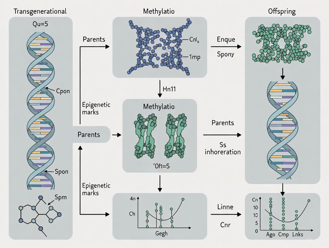
Transgenerational Epigenetic Inheritance (TEI) describes the phenomenon where phenotypic traits induced by environmental factors are transferred to subsequent generations through epigenetic mechanisms rather than DNA sequence changes [1]. This field has revitalized the centuries-old debate about the inheritance of acquired characteristics, strongly contested since the Lamarckian and Darwinian eras [2]. While evidence for TEI is well-established in plants and invertebrates, its existence in mammals remains highly controversial due to fundamental biological differences in how organisms handle epigenetic information across generations [2] [3]. This technical guide examines the core mechanisms, experimental evidence, and methodological approaches for studying TEI across these taxonomic groups, with particular emphasis on the challenges specific to mammalian systems within the broader context of epigenetic research.
The central controversy in mammalian TEI stems from two waves of epigenetic reprogramming that occur during development: first in primordial germ cells and later in the developing embryo after fertilization [2]. These reprogramming events are characterized by global erasure of DNA methylation and remodeling of histone modifications, presenting a significant biological barrier to the transmission of epigenetic information [2]. Despite this barrier, some genomic regions—including imprinted control regions and transposable elements—resist complete reprogramming, creating potential windows for TEI to occur [2]. Understanding how these mechanisms differ across mammals, plants, and invertebrates is crucial for advancing research in environmental epigenetics, evolutionary biology, and toxicology.
Fundamental Biological Differences in TEI Mechanisms
Epigenetic Reprogramming Landscapes
The capacity for transgenerational epigenetic inheritance varies dramatically across taxonomic groups, primarily due to differences in epigenetic reprogramming events. The following table summarizes the key biological differences that influence TEI potential:
Table 1: Fundamental Biological Differences Affecting TEI Potential
| Characteristic | Mammals | Plants | Invertebrates (C. elegans) |
|---|---|---|---|
| Reprogramming Events | Two waves: in primordial germ cells and post-fertilization [2] | Limited reprogramming; no defined germline [3] | No extensive reprogramming comparable to mammals [2] |
| DNA Methylation Erasure | Global erasure with exceptions at imprinted regions & transposons [2] | Context-dependent maintenance; RNA-directed DNA methylation [3] | Minimal DNA methylation; alternative mechanisms [2] |
| Primary Epigenetic Carriers | DNA methylation (5mC), histone modifications [2] | DNA methylation, histone modifications, small RNAs [3] | Small RNAs (siRNAs, piRNAs), histone modifications [2] [3] |
| Germline Development | Early segregation; protected from somatic influences [2] | No preformation; germline differentiates late [3] | Preformed germline with enhanced permeability [3] |
| Evidence Strength | Controversial with rare examples (Agouti) [3] | Strong; numerous well-established examples [3] | Strong; RNAi-based mechanisms [2] [3] |
Key Epigenetic Mechanisms in TEI
The molecular mechanisms facilitating TEI differ significantly across biological kingdoms, with each taxonomic group employing distinct epigenetic carriers:
Mammals: DNA methylation represents the most extensively studied epigenetic marker in mammals. Despite global reprogramming, certain regions like imprinted control regions and transposable elements resist demethylation, potentially allowing environmental exposures to create heritable epigenetic marks [2]. Histone modifications (e.g., H3K9me3, H3K27me) may also contribute, though their heritability faces greater challenges due to histone exchange during reprogramming [2].
Plants: Unlike mammals, plants display robust TEI through multiple interconnected mechanisms including RNA-directed DNA methylation (RdDM), histone modifications, and small RNA pathways [3]. The absence of an early segregated germline allows somatic epigenetic states to be more readily transmitted to subsequent generations. Plants also lack the comprehensive epigenetic erasure that characterizes mammalian development [3].
Invertebrates: C. elegans and other invertebrates utilize small RNA pathways as primary carriers of epigenetic information. Environmental double-stranded RNA (dsRNA) triggers the RNA interference (RNAi) pathway, generating small interfering RNAs (siRNAs) that silence complementary genes across generations [2]. These secondary siRNAs propagate RNAi effects to subsequent generations, mediated by germline epigenetic modifications including PIWI-interacting RNAs (piRNAs) and specific histone modifications like H3K9me3 and H3K36 [2].
Experimental Evidence and Model Systems
Quantitative Evidence for TEI Across Taxa
Research across different model systems has produced varying levels of evidence for TEI, with quantitative data highlighting the strength of findings in different organisms:
Table 2: Key Experimental Models and Evidence for TEI
| Organism/Model | Environmental Exposure | Generations Affected | Key Phenotypic Outcomes | Epigenetic Changes Observed |
|---|---|---|---|---|
| Agouti Mouse (Mammal) | Maternal methyl donor diet [2] | F1, F2 [2] | Coat color variation, obesity [2] | Increased methylation at 6 CpG sites in Avy retrotransposon [2] |
| Vinclozolin Rat (Mammal) | In utero fungicide exposure [2] | F1-F4 (claimed) [2] | Testicular abnormalities, infertility [2] | Altered DNA methylation patterns in sperm [2] |
| Dutch Famine Humans (Mammal) | Periconceptional malnutrition [2] | F1, F2 (neonatal adiposity) [2] | Metabolic disease, schizophrenia risk [2] | IGF2 hypomethylation (6 decades later) [2] |
| C. elegans (Invertebrate) | Pathogenic viruses/bacteria [3] | ≥ F3 [3] | Pathogen resistance [3] | siRNA-mediated silencing; H3K9me3 [2] |
| Arabidopsis (Plant) | Various abiotic stresses [3] | Multiple generations [3] | Flowering time, stress responses [3] | DNA methylation changes, histone modifications [3] |
Methodological Approaches for TEI Investigation
Mammalian TEI Research Protocols
Agouti Viable Yellow (Avy) Mouse Model Protocol
The Agouti mouse model represents one of the most robust mammalian systems for studying TEI. The following protocol outlines key methodological considerations:
Experimental Design:
- Expose pregnant female mice (F0 generation) to methyl donor-supplemented diet (e.g., extra folic acid, vitamin B12, choline) during specific gestational windows [2].
- Breed resulting offspring (F1) to unexposed partners to generate F2 generation, continuing to F3 without further exposure to establish transgenerational effects (F3 represents first truly transgenerational generation when considering maternal exposure) [2].
Phenotypic Assessment:
Molecular Analysis:
Critical Considerations: Sample size must be sufficient to detect modest effects, as early positive findings with small samples failed replication in larger studies [3]. The Agouti locus represents an exception rather than the rule in mammalian TEI, as screens for similar metastable epialleles have been largely negative [3].
Invertebrate TEI Research Protocols
C. elegans RNAi Inheritance Protocol
The C. elegans system provides a well-established model for studying RNA-based transgenerational inheritance:
Environmental Exposure:
Generational Tracking:
- Transfer exposed P0 animals to fresh plates without dsRNA or pathogen.
- Collect F1 embryos using standard bleaching protocols to ensure separation from parental environment.
- Continue tracking phenotypes through multiple generations (typically F3-F10) without further exposure [2].
Phenotypic Assessment:
- Monitor gene silencing through fluorescent reporters or phenotypic markers.
- For pathogen resistance assays, challenge offspring generations with pathogens and quantify survival rates [3].
Molecular Analysis:
Visualizing Key Biological Pathways and Experimental Designs
Comparative Epigenetic Reprogramming Pathways
Mammalian TEI Experimental Workflow
The Scientist's Toolkit: Essential Research Reagents and Methodologies
Table 3: Essential Research Reagents for TEI Investigation
| Reagent/Method | Application | Key Considerations |
|---|---|---|
| Bisulfite Conversion Reagents | Detection of DNA methylation patterns [2] | Distinguishes 5-methylcytosine from cytosine; optimized protocols needed for different species [2] |
| Methylation-Sensitive Restriction Enzymes | Methylation analysis at specific loci [2] | Requires careful controls for complete digestion; cost-effective for candidate regions [2] |
| Histone Modification Antibodies | Chromatin immunoprecipitation (ChIP) for histone marks [2] | Specificity validation critical; species compatibility important [2] |
| Small RNA Sequencing Kits | Analysis of siRNA, piRNA populations in TEI [2] | Specialized protocols for small RNA enrichment; essential for invertebrate and plant studies [2] |
| CRISPR/dCas9 Epigenetic Editors | Functional testing of specific epigenetic marks [3] | Enables targeted methylation/demethylation; distinguishes correlation from causation [3] |
| DNMT and TET Inhibitors | Manipulation of DNA methylation states [3] | Pharmacological modulation of methylation; specificity and toxicity concerns [3] |
| Methyl Donor Supplements | Dietary manipulation of methylation capacity (e.g., choline, folate) [2] | Used in Agouti mouse studies; timing critical for developmental windows [2] |
| Allobetulone | Allobetulone, CAS:28282-22-6, MF:C30H48O2, MW:440.7 g/mol | Chemical Reagent |
| Combretastatin A4 | Combretastatin A4, CAS:117048-59-6, MF:C18H20O5, MW:316.3 g/mol | Chemical Reagent |
Critical Challenges and Methodological Considerations
Key Controversies in Mammalian TEI
The investigation of transgenerational epigenetic inheritance in mammals faces several fundamental challenges that contribute to the ongoing controversy in the field:
Biological Barriers: The two waves of epigenetic reprogramming in primordial germ cells and early embryos present a substantial biological hurdle for maintaining environmentally-induced epigenetic marks across generations [2]. As noted by researchers, "it is unlikely for environmentally induced or programmed epigenetic marks to be inherited" due to these reprogramming events [2].
Confounding Effects: Disentangling true germline epigenetic inheritance from other factors remains challenging. Potential confounders include intrauterine environmental effects, cytoplasmic factors, and the difficulty in distinguishing epigenetic changes from selection for genetic variants [2] [3]. Studies must carefully differentiate between intergenerational effects (direct exposure of gametes or fetus) versus true transgenerational inheritance (transmission through multiple generations without direct exposure) [2].
Reproducibility Issues: Several high-profile TEI findings have faced replication challenges. For example, while initial reports suggested diet could induce heritable changes in Agouti mice, subsequent studies with larger sample sizes failed to validate these findings [3]. Similarly, the transgenerational effects of vinclozolin exposure demonstrated in some rat strains were not reproducible in inbred mouse strains [2].
Evolutionary Considerations: The potential adaptive value of TEI remains debated. While some invertebrate and plant examples show clear adaptive benefits (e.g., pathogen resistance in C. elegans), most putative mammalian examples appear non-adaptive and resemble random epigenetic mutations rather than programmed responses [3]. As noted in recent research, "rather than eagerly harnessing environmental inputs from the life experiences of their ancestors via epigenetic mechanisms, organisms strive to prevent contamination of a new generation with the accumulated epigenetic baggage of the previous one" [3].
Methodological Recommendations for Robust TEI Research
To address these challenges, researchers should implement rigorous methodological approaches:
Generational Study Design: Properly design studies to distinguish intergenerational from transgenerational effects. In maternal exposure models, F3 represents the first transgenerational generation [2].
Multiple Strain Validation: Test findings across different genetic backgrounds to control for strain-specific effects, as demonstrated by the variable vinclozolin effects in outbred versus inbred mice [2].
Comprehensive Epigenomic Profiling: Move beyond candidate loci approaches to genome-wide epigenetic analyses while implementing appropriate multiple testing corrections [2].
Functional Validation: Utilize epigenetic editing tools (e.g., dCas9-effector fusions) to test the functional consequences of specific epigenetic changes rather than relying solely on correlative evidence [3].
The investigation of transgenerational epigenetic inheritance reveals profound differences across taxonomic groups, with robust mechanisms well-established in plants and invertebrates but remaining controversial in mammals. The contrasting biological strategies—from the extensive reprogramming barriers in mammals to the RNA-based inheritance pathways in invertebrates—highlight the diverse evolutionary solutions to balancing environmental responsiveness with genomic integrity across generations.
Future research directions should prioritize overcoming the methodological challenges that have plagued the field, particularly in mammalian systems. This includes developing more sensitive tools for detecting rare epigenetic variants that survive reprogramming, implementing rigorous multi-generational study designs, and applying epigenetic editing technologies to establish causal relationships. Furthermore, exploring the potential intersection between TEI and other inheritance mechanisms, including the transmission of microbial symbionts and parental effects, may provide a more comprehensive understanding of non-genetic inheritance.
For drug development professionals and translational researchers, the current evidence suggests caution in extrapolating TEI findings from invertebrate and plant models to mammalian systems. While the Agouti locus demonstrates that TEI can occur in mammals in specific circumstances, these appear to be exceptions rather than the rule. Nevertheless, understanding these mechanisms remains crucial for comprehensive toxicological risk assessment and for exploring potential long-term impacts of environmental exposures on human health across generations.
Epigenetics is the study of heritable and stable changes in gene expression that occur through alterations in the chromosome rather than in the DNA sequence [4]. These mechanisms form a crucial layer of control that regulates gene expression and silencing without changing the underlying genetic code, enabling cells with identical DNA sequences to develop into distinct cell types and respond to environmental cues [4]. The significance of epigenetic regulation extends beyond cellular differentiation to encompass potentially transgenerational effects, wherein environmentally induced modifications can be transmitted to subsequent generations.
In the context of mammalian transgenerational epigenetic inheritance (TEI), research focuses on how acquired traits can be transmitted through the germline without changes to the DNA sequence itself [5]. True transgenerational inheritance in mammals requires transmission that extends to at least the F2 generation after F0 paternal exposure, and to the F3 generation after F0 maternal exposure, thereby excluding direct exposure effects on the developing embryo or its germ cells [5]. This distinction is critical for establishing genuine epigenetic inheritance across generations, as opposed to intergenerational effects that result from direct exposure.
The three primary epigenetic mechanisms—DNA methylation, histone modifications, and non-coding RNA-associated gene silencing—work in concert to regulate gene expression by modulating chromatin structure and accessibility [4]. These systems create a comprehensive network of regulatory pathways and feedback loops that can dynamically respond to environmental factors including age, diet, smoking, stress, and disease state [4] [6]. Understanding these core mechanisms provides the foundation for exploring how environmental experiences can potentially shape phenotypes across multiple generations, with profound implications for evolution, disease susceptibility, and therapeutic development.
DNA Methylation
Molecular Mechanisms and Enzymatic Regulation
DNA methylation represents a fundamental epigenetic mechanism involving the addition of a methyl group to cytosine nucleotides, predominantly within cytosine-guanine (CpG) dinucleotides [4]. This covalent modification is catalyzed by DNA methyltransferase (DNMT) enzymes, primarily DNMT1, DNMT3A, and DNMT3B, which establish and maintain methylation patterns throughout the genome [6] [4]. DNMT1 functions as the maintenance methyltransferase, faithfully copying methylation patterns during DNA replication, while DNMT3A and DNMT3B serve as de novo methyltransferases that establish new methylation patterns during embryonic development [6].
The distribution of DNA methylation across the genome is non-random, with CpG islands—stretches of DNA with high CpG density—being particularly important regulatory targets. Approximately 70% of gene promoter regions lie within CpG islands, making them crucial elements for transcriptional regulation [4]. When methylated, these promoter-associated CpG islands recruit methyl-binding proteins and associated gene suppressor complexes that promote a compact chromatin state, effectively silencing gene expression by preventing transcription factors from accessing their target sequences [4] [5]. This repressive function positions DNA methylation as a key regulator of tissue-specific gene expression, genomic imprinting, and X-chromosome inactivation [4].
The dynamic nature of DNA methylation is maintained through active demethylation processes mediated by ten-eleven translocation (TET) enzymes, which oxidize 5-methylcytosine (5mC) to 5-hydroxymethylcytosine (5hmC) and further derivatives [5]. This oxidation pathway facilitates both passive and active DNA demethylation, allowing for erasure and reprogramming of epigenetic marks during specific developmental windows, particularly in primordial germ cells and early embryos [7] [5]. The balance between methylation and demethylation activities enables the epigenome to remain responsive to environmental signals while maintaining transcriptional stability.
Role in Transgenerational Inheritance and Evidence from Mammalian Studies
In the context of transgenerational epigenetic inheritance, DNA methylation represents the most extensively studied mechanism, though evidence for its role in mammals remains subject to ongoing scientific debate [7] [3]. The potential for methylation patterns to escape the widespread epigenetic reprogramming that occurs during early mammalian development is central to this discourse. This reprogramming involves genome-wide demethylation in primordial germ cells and subsequent remethylation around the time of embryo implantation, a process thought to be required for resetting genomic imprints and reactivating genes essential for proper development [7]. However, research indicates that this reset is not entirely complete, with specific genomic regions, particularly imprinted genes and transposable elements, demonstrating resistance to demethylation and thus potentially carrying epigenetic information across generations [7].
The Agouti viable yellow (Avy) mouse model provides one of the most compelling examples of metastable epialleles that exhibit transgenerational inheritance of DNA methylation patterns [3]. In this system, a transposable element inserted upstream of the Agouti gene exhibits variable methylation that correlates with coat color and disease susceptibility, and this methylation state shows modest but measurable heritability [3]. Early studies suggested that maternal diet, particularly methyl donor supplementation, could influence the epigenetic status of this locus in offspring, though subsequent larger studies have yielded conflicting results regarding the environmental responsiveness and transgenerational stability of these epigenetic marks [3].
Human studies have provided correlative evidence for transgenerational DNA methylation patterns, particularly in the context of early life stress and nutritional status [7]. For instance, analysis of individuals prenatally exposed to the Dutch Hunger Winter revealed persistent DNA methylation changes at specific loci, such as the imprinted IGF2 gene, decades after the initial exposure [4]. However, distinguishing true transgenerational inheritance from intergenerational effects in human studies presents significant methodological challenges, as it requires demonstrating transmission through multiple generations without continued direct exposure.
Table 1: DNA Methylation Patterns in Transgenerational Studies
| Study Model | Environmental Exposure | Methylation Changes | Generational Persistence | Associated Phenotypes |
|---|---|---|---|---|
| Agouti Mice [3] | Maternal methyl donor diet | Avy locus hypomethylation | F1-F3 (with decreasing penetrance) | Yellow coat color, obesity, tumor susceptibility |
| Dutch Hunger Winter Cohort [4] | Prenatal famine | IGF2 hypomethylation | F1 only (intergenerational) | Metabolic disease, cardiovascular risk |
| Rat VOC Model [5] | Gestational toxicant exposure | Sperm DNA methylation changes | F1-F3 (transgenerational) | Testis disease, kidney disease, multiple disease states |
| Mouse High-Fat Diet [7] | Maternal high-fat diet | Neural stem cell methylation changes | F1-F3 (transgenerational) | Altered neurogenesis, metabolic changes |
Histone Modifications
The Histone Code Hypothesis and Major Modification Types
Histone modifications constitute a second major epigenetic mechanism that regulates gene expression by altering chromatin structure and DNA-histone interactions [4]. The "histone code" hypothesis proposes that covalent post-translational modifications to the N-terminal tails of histone proteins create a combinatorial language that determines chromosomal functional states [6]. These modifications occur primarily on the flexible histone tails that protrude from the nucleosome core and include acetylation, methylation, phosphorylation, ubiquitination, and other chemical alterations [4].
Each modification type demonstrates specific functions in regulating chromatin dynamics and gene expression. Histone acetylation, catalyzed by histone acetyltransferases (HATs), involves the addition of acetyl groups to lysine residues, neutralizing their positive charge and consequently weakening DNA-histone interactions [4]. This charge neutralization promotes an open chromatin configuration (euchromatin) that facilitates transcription factor binding and gene activation [4]. Conversely, histone deacetylases (HDACs) remove acetyl groups, strengthening DNA-histone interactions and promoting transcriptional repression. Key activating acetylation marks include H3K9ac and H3K27ac, both associated with active transcription [4].
Histone methylation represents a more complex regulatory system that can either activate or repress transcription depending on the specific residue modified and its methylation state [4]. Unlike acetylation, methylation does not alter the charge of histone tails but instead creates binding platforms for chromatin-modifying complexes. For example, H3K4me3 is strongly associated with transcription activation, while H3K27me3 and H3K9me3 correlate with transcription repression and heterochromatin formation [4]. The functional outcome depends on the specific lysine or arginine residue modified, the degree of methylation (mono-, di-, or tri-methylation), and the broader chromatin context.
Table 2: Major Histone Modifications and Their Functional Consequences
| Modification Type | Histone Site | Enzymes Responsible | Chromatin State | Functional Outcome |
|---|---|---|---|---|
| Acetylation | H3K9, H3K14, H3K27, H4K16 | HATs, HDACs | Open euchromatin | Transcriptional activation |
| Methylation (activating) | H3K4, H3K36, H3K79 | HMTs, KDMs | Open euchromatin | Transcriptional activation |
| Methylation (repressive) | H3K9, H3K27, H4K20 | HMTs, KDMs | Closed heterochromatin | Transcriptional repression |
| Phosphorylation | H3S10 | Kinases, phosphatases | Condensed chromatin | Mitosis, DNA damage response |
| Ubiquitination | H2AK119, H2BK123 | E3 ubiquitin ligases | Variable | Transcriptional regulation, DNA repair |
Experimental Methodologies for Histone Modification Analysis
The investigation of histone modifications employs a diverse array of molecular techniques that enable researchers to map modification patterns, quantify abundance, and determine functional consequences. Chromatin immunoprecipitation followed by sequencing (ChIP-seq) represents the gold standard for genome-wide mapping of histone modifications [6]. This methodology involves cross-linking proteins to DNA, chromatin fragmentation, immunoprecipitation with modification-specific antibodies, and high-throughput sequencing of associated DNA fragments. The resulting data provides comprehensive maps of histone modification distributions across the genome, enabling correlation with gene expression states and identification of regulatory elements.
Mass spectrometry-based approaches offer complementary quantitative information about histone modification states, allowing for precise measurement of modification abundances and combinatorial patterns without antibody-based biases [6]. These techniques typically involve histone extraction, chemical derivatization, protease digestion, and liquid chromatography-tandem mass spectrometry (LC-MS/MS) analysis. The resulting data provides quantitative information about the relative abundance of specific modifications and can reveal crosstalk between different modification types.
For functional validation, CRISPR/Cas9-based epigenetic editing systems enable targeted manipulation of histone modifications at specific genomic loci [7]. These approaches fuse catalytically dead Cas9 (dCas9) with histone-modifying domains to recruit specific enzymatic activities to defined genomic locations. For example, dCas9-p300 promotes histone acetylation and gene activation, while dCas9-LSD1 facilitates demethylation and gene repression. These tools allow researchers to establish causal relationships between specific histone modifications and transcriptional outcomes, moving beyond correlation to demonstrate functional significance.
Evidence for Transgenerational Transmission of Histone Modifications
While DNA methylation has traditionally been the focus of transgenerational epigenetic inheritance research, emerging evidence suggests that histone modifications may also contribute to epigenetic transmission across generations, particularly in mammalian systems [5]. This represents a significant conceptual challenge, as the majority of histones are replaced by protamines during spermatogenesis and extensively remodeled following fertilization. However, recent studies have identified a small but significant percentage of nucleosomes that are retained in sperm and may carry paternal epigenetic information [5].
Research in C. elegans has demonstrated that certain histone modifications, particularly H3K4me and H3K9me, can be transmitted across multiple generations and influence gene expression in progeny [3]. However, these effects are often dependent on mutant backgrounds that lack the histone modification erasure machinery present in wildtype organisms, suggesting that efficient removal mechanisms typically prevent transgenerational perpetuation of most histone marks [3]. Similarly, studies in S. pombe have shown that heterochromatic histone modifications can be maintained across generations but require specific environmental conditions or genetic backgrounds to escape resetting mechanisms.
In mammals, the evidence for transgenerational inheritance of histone modifications remains limited and controversial. Some studies have reported transmission of histone methylation patterns, such as H3K4me3 and H3K27me3, at specific developmental gene loci [5]. For example, paternal exposure to stress or toxicants has been associated with altered H3K4 methylation in sperm and corresponding behavioral and metabolic phenotypes in offspring [5]. However, these findings are complicated by the extensive epigenetic reprogramming that occurs during early mammalian development and the technical challenges of distinguishing true epigenetic inheritance from other forms of transmission.
Non-Coding RNAs
Classification and Functional Mechanisms
Non-coding RNAs (ncRNAs) represent a diverse class of functional RNA molecules that are transcribed from DNA but not translated into proteins, playing crucial roles in epigenetic regulation and gene silencing [4]. Once considered genomic "junk," ncRNAs are now recognized as essential components of the epigenetic machinery, potentially accounting for significant phenotypic variation despite similarity in protein-coding sequences [4]. The ncRNA family encompasses multiple classes distinguished by size, structure, biogenesis, and functional mechanisms.
MicroRNAs (miRNAs) constitute the most extensively studied class of small ncRNAs, comprising evolutionary conserved transcripts of 17-25 nucleotides that regulate gene expression at the post-transcriptional level [6]. miRNA biogenesis involves sequential processing of primary miRNA transcripts by Drosha and Dicer enzymes, resulting in mature miRNAs that guide the RNA-induced silencing complex (RISC) to complementary target mRNAs. Perfect complementarity leads to mRNA cleavage and degradation, while partial complementarity, more common in mammals, results in translational repression [6]. The specificity of miRNA targeting is determined primarily by nucleotides 2-7 from the 5' end, known as the "seed sequence," which must base-pair with the 3' untranslated region of target mRNAs [6].
Long non-coding RNAs (lncRNAs) represent a more heterogeneous class defined as transcripts exceeding 200 nucleotides with limited protein-coding potential [6]. These molecules exhibit lower evolutionary conservation than miRNAs and frequently display cell- and tissue-specific expression patterns [6]. lncRNAs function through diverse mechanisms, acting as scaffolds, guides, decoys, or signals in complex regulatory networks [6]. As molecular scaffolds, they assemble multi-protein complexes that coordinate biological processes; as guides, they direct chromatin-modifying enzymes to specific genomic loci; as decoys, they sequester transcription factors or miRNAs; and as signals, they mark specific developmental stages or chromosomal territories [6].
Additional ncRNA classes include small interfering RNAs (siRNAs), which share biogenesis and effector mechanisms with miRNAs but derive from different precursor molecules, and piwi-interacting RNAs (piRNAs), which are 26-31 nucleotides in length and associate with Piwi proteins to silence transposable elements in the germline [6]. Circular RNAs (circRNAs), produced through noncanonical back-splicing events, have emerged as important regulators that can function as miRNA sponges, protein decoys, or templates for translation [6].
Methodologies for ncRNA Analysis and Functional Characterization
The study of ncRNAs employs specialized methodologies tailored to their unique properties and functions. For miRNA profiling, quantitative reverse transcription PCR (qRT-PCR) arrays and small RNA sequencing represent the most common approaches for expression analysis [6]. These techniques enable comprehensive characterization of miRNA abundance across different tissues, developmental stages, or experimental conditions. Functional investigation typically involves miRNA inhibition using antisense oligonucleotides (antagomirs) or mimics for overexpression, followed by assessment of phenotypic consequences and target validation.
For lncRNA analysis, RNA sequencing provides the most powerful approach for discovery and expression profiling, while RNA fluorescence in situ hybridization (RNA-FISH) enables spatial localization within cells and tissues [6]. Functional characterization often employs antisense oligonucleotides (ASOs) or RNA interference for knockdown, complemented by CRISPR-based approaches for genomic deletion. To identify lncRNA interaction partners, techniques such as RNA immunoprecipitation (RIP) and cross-linking immunoprecipitation (CLIP) determine protein binding partners, while chromatin isolation by RNA purification (ChIRP) maps genomic association sites [6].
High-throughput screening approaches have been developed to systematically interrogate ncRNA function, including pooled CRISPR screens targeting lncRNA loci and combinatorial miRNA inhibitor libraries. These unbiased approaches facilitate discovery of functionally significant ncRNAs within specific biological contexts, providing insights into their roles in development, homeostasis, and disease processes.
Role in Transgenerational Epigenetic Inheritance
Non-coding RNAs have emerged as compelling candidates for mediators of transgenerational epigenetic inheritance, particularly in the context of environmentally induced phenotypes [7] [5]. The germline enrichment of specific ncRNA classes, their ability to shuttle between tissues, and their capacity to instruct epigenetic modifications position them as potential vectors for transmitting environmental information across generations.
In invertebrate models, compelling evidence demonstrates RNA-based transgenerational inheritance. In Caenorhabditis elegans, exposure to viral RNA triggers the production of small interfering RNAs that are transmitted to subsequent generations, conferring pathogen resistance in the absence of continued exposure [3]. Similarly, starvation-induced changes in small RNA profiles can be inherited for multiple generations, associated with corresponding alterations in gene expression and lifespan [5]. These RNA-based inheritance mechanisms represent evolved adaptive responses that provide progeny with pre-existing resistance to environmental challenges encountered by their ancestors.
Mammalian studies have provided more limited but suggestive evidence for ncRNA-mediated transgenerational inheritance. Paternal exposure to stress, toxins, or dietary manipulation has been associated with changes in sperm small RNA profiles, including miRNAs, tsRNAs, and piRNAs [5]. For instance, paternal trauma exposure alters sperm miRNA content, with corresponding behavioral and metabolic phenotypes in offspring [5]. Similarly, paternal obesity modifies sperm tsRNA profiles, and injection of these tsRNAs into normal zygotes recapitulates metabolic dysfunction in the resulting offspring [5]. These findings suggest that sperm-borne RNAs can carry paternal environmental information to the next generation.
The mechanistic basis for RNA-mediated inheritance likely involves regulation of early embryonic development, during which sperm-delivered RNAs may influence epigenetic reprogramming and gene expression patterns [5]. However, significant questions remain regarding the stability of RNA molecules across generations, the specificity of their effects, and the relative contribution of RNA-mediated mechanisms compared to other epigenetic pathways. Additionally, the potential for non-germline transmission through behavioral or cultural means complicates the interpretation of transgenerational phenomena in mammals.
Experimental Approaches for Studying Transgenerational Epigenetic Inheritance
Model Systems and Methodological Considerations
The investigation of transgenerational epigenetic inheritance requires carefully controlled experimental designs that account for species-specific biological considerations and distinguish true germline transmission from other forms of inheritance [5]. In mammalian systems, proper experimental design must account for the distinction between intergenerational and transgenerational effects [5]. For maternal exposures, transmission to the F3 generation represents the first transgenerational cohort, as the F2 generation germline was directly exposed in the developing F1 fetus [5]. For paternal exposures, the F2 generation is considered transgenerational, as only the F1 generation germline was directly exposed [5].
Invertebrate models, including C. elegans and D. melanogaster, offer significant advantages for TEI research due to their short generation times, well-characterized genetics, and simplified germline development [5]. These systems enable large-scale screening approaches and precise manipulation of epigenetic pathways. Plant models, particularly Arabidopsis thaliana, have also provided fundamental insights into TEI mechanisms, with well-documented examples of stress-induced epigenetic changes that persist across generations [7].
Mammalian studies predominantly utilize rodent models, with exposures typically administered during specific developmental windows to target germline epigenetic programming [7] [5]. Common exposure paradigms include gestational nutrient manipulation, toxin administration, stress protocols, and endocrine disruptor exposure. Subsequent generations are maintained without additional exposure to assess transgenerational transmission of epigenetic marks and associated phenotypes.
Molecular Assessment Techniques
Comprehensive assessment of TEI requires multi-level molecular profiling across generations. DNA methylation analysis typically employs whole-genome bisulfite sequencing (WGBS) for comprehensive mapping or reduced representation bisulfite sequencing (RRBS) for cost-effective profiling of CpG-rich regions [7]. These approaches enable identification of differentially methylated regions (DMRs) that persist across generations and correlate with phenotypic outcomes.
Histone modification analysis primarily utilizes ChIP-seq with modification-specific antibodies, though this approach is limited by antibody quality and availability [6]. Mass spectrometry-based methods provide complementary quantitative data but require specialized instrumentation and expertise [6].
Non-coding RNA profiling employs small RNA sequencing for comprehensive characterization, with particular focus on sperm RNA content in paternal transmission studies [5]. Integration of multiple datasets through multi-omics approaches provides the most powerful strategy for identifying conserved epigenetic signatures of transgenerational inheritance.
Table 3: Key Research Reagents and Methodologies for TEI Research
| Research Tool Category | Specific Examples | Primary Applications | Technical Considerations |
|---|---|---|---|
| Epigenome Editing Systems | dCas9-DNMT3A, dCas9-TET1, dCas9-p300, dCas9-LSD1 | Targeted epigenetic manipulation; causal validation | Potential for off-target genetic effects [7] |
| DNA Methylation Profiling | WGBS, RRBS, Illumina EPIC Array | Genome-wide methylation mapping; DMR identification | Bisulfite conversion efficiency; coverage depth |
| Histone Modification Analysis | ChIP-seq, CUT&RUN, mass spectrometry | Histone mark mapping; quantitative modification analysis | Antibody specificity; chromatin shearing optimization |
| Non-coding RNA Profiling | Small RNA-seq, miRNA arrays, single-cell RNA-seq | ncRNA expression quantification; biomarker discovery | RNA integrity; normalization strategies |
| Animal Models | C. elegans, Drosophila, mouse, rat | TEI pathway discovery; exposure effect characterization | Species-specific epigenetic reprogramming patterns |
The Scientist's Toolkit: Essential Research Reagents and Materials
The following table summarizes key research reagents and materials essential for investigating epigenetic mechanisms and transgenerational inheritance, compiled from current methodologies described in the scientific literature.
Table 4: The Scientist's Toolkit for Epigenetic Research
| Reagent/Material | Function/Application | Examples/Specifics |
|---|---|---|
| DNA Methyltransferase Inhibitors | Demethylating agents; cancer therapy | Azacytidine, Decitabine (FDA-approved) [4] |
| Histone Deacetylase Inhibitors | Increase histone acetylation; cancer therapy | Panobinostat, Romidepsin (FDA-approved) [4] |
| CRISPR-dCas9 Epigenetic Editors | Targeted epigenetic modification | dCas9-DNMT3A (methylation), dCas9-p300 (acetylation) [7] |
| Modification-Specific Antibodies | Histone mark detection; ChIP experiments | H3K4me3 (active), H3K27me3 (repressive) [6] [4] |
| Bisulfite Conversion Reagents | DNA methylation mapping | Sodium bisulfite for cytosine conversion [7] |
| Small RNA Inhibitors | Functional ncRNA characterization | Antagomirs, ASOs for miRNA inhibition [6] |
| Transgenerational Animal Models | TEI pathway discovery | Agouti mice, C. elegans RNAi models [5] [3] |
| Protriptyline Hydrochloride | Protriptyline Hydrochloride, CAS:1225-55-4, MF:C19H22ClN, MW:299.8 g/mol | Chemical Reagent |
| Cefoselis | Cefoselis, CAS:122841-10-5, MF:C19H22N8O6S2, MW:522.6 g/mol | Chemical Reagent |
Signaling Pathways and Experimental Workflows
The following diagrams illustrate key signaling pathways and experimental workflows in epigenetic research, created using Graphviz DOT language with adherence to the specified color palette and contrast requirements.
Diagram 1: Epigenetic Regulation of Gene Expression
Diagram 2: Transgenerational Inheritance Experimental Design
The field of epigenetics has revolutionized our understanding of gene regulation and inheritance, revealing sophisticated mechanisms that interface with environmental signals to shape phenotypes within and potentially across generations. The core epigenetic mechanisms—DNA methylation, histone modifications, and non-coding RNAs—function not in isolation but as integrated components of a comprehensive regulatory network that controls chromatin structure and gene expression [6]. While evidence for transgenerational epigenetic inheritance is well-established in plants and invertebrate animals, its significance in mammals remains a subject of ongoing investigation and debate [7] [3].
The clinical implications of epigenetic research are substantial, particularly in cancer biology, where epigenetic dysregulation represents a hallmark of carcinogenesis [4]. The FDA approval of epigenetic therapies such as azacytidine, decitabine, panobinostat, and romidepsin demonstrates the translational potential of targeting epigenetic mechanisms [4]. Furthermore, the reversible nature of epigenetic modifications offers promising therapeutic avenues for diverse conditions, including neurological disorders, metabolic diseases, and imprinting disorders [4].
Future research directions will likely focus on developing more precise epigenetic editing tools, resolving the functional relationships between different epigenetic layers, and elucidating the mechanisms that allow specific epigenetic marks to escape reprogramming during mammalian development [7] [5]. The potential contribution of epigenetic mechanisms to evolutionary processes also represents an exciting frontier, particularly regarding how environmental experiences might shape phenotypic variation and adaptation across generations [7]. As technologies for epigenome mapping and manipulation continue to advance, so too will our understanding of these fundamental regulatory processes and their roles in health, disease, and inheritance.
In mammalian development, the germline serves as a timeless bridge between generations, necessitating the resetting of epigenetic information to restore totipotency. Primordial germ cells (PGCs) undergo genome-wide epigenetic reprogramming, a process characterized by the extensive erasure of DNA methylation to establish a basal epigenetic state. However, this reprogramming is incomplete. Specific genomic regions, including those associated with evolutionarily young retrotransposons and certain metabolic and neurological disorder genes, demonstrate resistance to demethylation. This selective retention of epigenetic marks creates a "reprogramming barrier," forming a potential molecular substrate for transgenerational epigenetic inheritance. This whitepaper delves into the technical nuances of this barrier, detailing the mechanisms of erasure and retention, their implications for disease and evolution, and the advanced methodologies illuminating this critical biological process.
The germline is the custodian of genetic and epigenetic information that must be passed to the next generation. To ensure each offspring starts life without the accumulated epigenetic modifications of their parents, mammalian PGCs undergo a dramatic epigenetic reprogramming event [8]. This process involves the genome-wide erasure of DNA methylation, which resets genomic potential and is crucial for the acquisition of totipotency—the ability of a cell to give rise to any cell type of an organism [8] [9]. A pivotal feature of this reprogramming is the near-complete elimination of DNA methylation, with global levels dropping to less than 5% in human PGCs (hPGCs) by weeks 5-7 of development [9]. This demethylation encompasses the erasure of parental genomic imprints and facilitates X chromosome reactivation [8] [9].
Despite the global trend toward hypomethylation, certain genomic sequences resist this reprogramming, creating a barrier to complete erasure. These resistant loci provide a potential mechanism for the transmission of epigenetic information across generations, independent of DNA sequence changes—a phenomenon termed transgenerational epigenetic inheritance [7] [9]. The evidence for this in mammals remains a subject of intense debate and research, as distinguishing true transgenerational inheritance from intergenerational effects requires rigorous criteria [7] [10]. This whitepaper explores the mechanisms underlying this great reprogramming barrier, framing it within the context of its potential impact on mammalian biology and human disease.
Mechanisms of Epigenetic Erasure
Epigenetic reprogramming in PGCs is a coordinated process involving dynamic changes in both DNA methylation and histone modifications.
DNA Demethylation: Active and Passive Pathways
The erasure of DNA methylation in PGCs is driven by a combination of passive and active mechanisms.
- Passive Demethylation: This replication-dependent process occurs through the repression of key components of the DNA methylation machinery. In hPGCs, expression of the de novo DNA methyltransferases DNMT3A and DNMT3B and the maintenance methyltransferase cofactor UHRF1 is suppressed. Without these factors, DNA methylation is not maintained after successive cell divisions, leading to its gradual dilution [8].
- Active Demethylation: This replication-independent process is facilitated by the Ten-Eleven Translocation (TET) family of enzymes (TET1, TET2, TET3). These enzymes oxidize 5-methylcytosine (5mC) to 5-hydroxymethylcytosine (5hmC) and further to 5-formylcytosine (5fC) and 5-carboxycytosine (5caC). These oxidized derivatives can then be excised by thymine-DNA glycosylase (TDG) and replaced with an unmodified cytosine through base excision repair [8]. The expression of TET1 and TET2 is enriched in hPGCs, driving this active demethylation pathway [8].
Table 1: Key Enzymes and Factors in PGC DNA Methylation Erasure
| Factor | Role in DNA Methylation | Activity/Expression in PGCs | Effect on Methylation |
|---|---|---|---|
| DNMT3A/B | De novo methylation | Repressed [8] | Promotes passive demethylation |
| UHRF1 | Targets DNMT1 to hemi-methylated sites | Repressed [8] | Promotes passive demethylation |
| TET1/2 | Oxidizes 5mC to 5hmC, 5fC, 5caC | Enriched [8] | Drives active demethylation |
| TDG | Excises 5fC and 5caC | Implicated in active pathway [8] | Completes active demethylation cycle |
Histone Modification and Chromatin Reorganization
Concurrent with DNA demethylation, the chromatin landscape of PGCs is extensively reorganized. Histone modifications serve a critical regulatory function, particularly in compensating for the loss of DNA methylation-based transcriptional control [8]. Key changes include:
- Repressive Mark Dynamics: The repressive mark H3K9me2 is present at lower levels in PGCs compared to surrounding somatic cells. In contrast, H3K27me3 shows a more complex pattern, being stronger in migratory PGCs but becoming depleted by weeks 7-9 in humans. H3K27me3 is involved in chromatin compaction and may act as a temporary repressive mechanism during reprogramming [8].
- Chromatin State: The global changes in histone modifications contribute to a more open chromatin configuration, facilitating the large-scale transcriptional changes required for PGC development and the resetting of pluripotency.
The Reprogramming Barrier: Loci Resisting Erasure
The paradigm of global epigenetic erasure is challenged by the discovery of specific genomic sequences that demonstrate resilience to demethylation. These resistant loci constitute the "reprogramming barrier."
Classes of Resistant Sequences
Research has identified several categories of sequences that consistently retain DNA methylation in hPGCs despite the global hypomethylated state [9]:
- Evolutionarily Young Retrotransposons: Notably, SVA (SINE-VNTR-Alu) elements, which are primate-specific retrotransposons, show significant resistance to demethylation. These elements are considered potentially "hazardous" due to their ability to mobilize and cause genomic instability, suggesting that their sustained silencing is a protective genome-defense mechanism [9].
- Germline Gene-Associated Loci: Intriguingly, some resistant regions are single-copy sequences located near genes critical for germline development. The persistent methylation at these sites may fine-tune the transcriptional program of the germline [9].
- Metabolic and Neurological Disorder Loci: A finding with profound implications for human disease is that some loci associated with metabolic and neurological disorders resist demethylation. This identifies them as candidate regions for mediating epigenetic memory and transgenerational inheritance of disease susceptibility in humans [9].
Mechanisms Protecting Against Demethylation
The molecular basis for this resistance is an area of active investigation. The prevailing hypothesis is that these regions are protected by specific protein factors or local chromatin environments that either shield them from the demethylation machinery or actively promote maintenance methylation. The resistance is not absolute; rather, these regions exhibit slower demethylation kinetics compared to the rest of the genome, suggesting a nuanced and regulated process [8].
Technical and Experimental Approaches
Studying the ephemeral population of human PGCs presents significant technical challenges. Advances in technology and in vitro modeling have been instrumental in driving progress.
Key Methodologies for Profiling Epigenetic States
- Whole-Genome Bisulfite Sequencing (BS-seq): This is the gold-standard method for assessing DNA methylation at single-base resolution. Treatment of DNA with sodium bisulfite converts unmethylated cytosines to uracils (read as thymines in sequencing), while methylated cytosines remain unchanged. Applying this to purified hPGCs allows for the base-resolution mapping of the methylome during reprogramming [9]. A key limitation is that it cannot distinguish between 5mC and 5hmC.
- RNA-Sequencing (RNA-seq): This technique provides a comprehensive quantitative profile of the transcriptome. It has been critical for defining the unique gene regulatory network of hPGCs, which differs from that of mouse PGCs, and for tracking the expression of reprogramming factors like TET enzymes and DNMTs [8] [9].
- Fluorescence-Activated Cell Sorting (FACS): The isolation of pure populations of hPGCs from human embryonic tissues is paramount. A high-purity FACS protocol using cell-surface markers TNAP (tissue non-specific alkaline phosphatase) and c-KIT has been established, enabling the isolation of hPGC populations with >97% purity for downstream molecular analyses [9].
AnIn VitroModel: Human PGC-Like Cells (hPGCLCs)
Given the ethical and practical constraints of studying early human development, researchers have developed in vitro models. By differentiating human embryonic stem cells (hESCs) into human PGC-like cells (hPGCLCs), it is possible to recapitulate key aspects of PGC specification and epigenetic reprogramming [8] [9]. This model system allows for genetic manipulation and high-throughput screening that would be impossible in vivo.
Visualization of Key Concepts
Diagram 1: Epigenetic Reprogramming and Resistance in Human Primordial Germ Cells
Diagram 2: Molecular Pathways of DNA Methylation Erasure
Table 2: Key Reagents and Materials for Germline Reprogramming Research
| Reagent / Material | Function / Application | Specific Examples / Notes |
|---|---|---|
| FACS Antibodies | Isolation of pure PGC populations | Anti-TNAP and anti-c-KIT for high-purity hPGC sorting [9] |
| Bisulfite Conversion Kit | Preparing DNA for methylation sequencing | Critical for BS-seq; distinguishes methylated vs. unmethylated C [8] [9] |
| hESC Lines | In vitro model for PGC development | Used to differentiate into hPGCLCs [8] [9] |
| CRISPR/Cas9 System | Gene editing for functional studies | Used to interrogate the role of specific genes (e.g., TETs, DNMTs) [7] |
| Anti-5mC/5hmC Antibodies | Immunostaining for methylation/hydroxymethylation | Visualizing global epigenetic states in cells/tissues |
| RNA-seq Library Prep Kits | Transcriptome profiling | Analyzing gene expression networks in PGCs vs. soma [9] |
Implications for Transgenerational Epigenetic Inheritance and Disease
The incomplete erasure of epigenetic marks in the germline provides a plausible molecular substrate for transgenerational epigenetic inheritance in mammals.
The Debate and Evidence
The field is marked by ongoing debate regarding the robustness of evidence in mammals. A critical review of 80 articles found that many claims did not meet the necessary criteria, which include: inheritance of the same epimutations across generations, associated gene expression changes, and confirmation of the epimutation in the germ cells of each generation [7]. However, other studies provide compelling data. For instance, exposure of gestating rats to plastic-derived compounds led to specific DNA methylation biomarkers in the sperm of the F3 generation, which were linked to diseases like testis and kidney pathologies [7]. Similarly, a high-fat diet in F0 female mice induced epigenetic changes in neural stem cells that persisted into the F3 generation, despite subsequent generations consuming a standard diet [7].
Linking Resistant Loci to Human Disease
The discovery that loci associated with metabolic and neurological disorders are among those resistant to demethylation in hPGCs directly links the reprogramming barrier to human health [9]. This suggests that environmental exposures could potentially lead to epigenetic alterations in these resistant loci, which, if not fully erased in the germline, could be transmitted to offspring, influencing their disease susceptibility. This mechanism could contribute to the heritability of complex disorders like type 2 diabetes mellitus and obesity [7].
The journey of primordial germ cells through epigenetic reprogramming is one of profound renewal, yet it is punctuated by deliberate retention. The "Great Reprogramming Barrier" is not a failure of the reset mechanism but appears to be a regulated, functional feature of germline development. It serves to protect genome integrity by silencing hazardous repetitive elements while simultaneously creating a potential archive of epigenetic information that may influence the phenotype of subsequent generations. The resistance of specific disease-associated loci to demethylation underscores the profound implications of this biology for human health. Future research, leveraging increasingly sophisticated in vitro models and single-cell multi-omics technologies, will be essential to fully decipher the mechanisms governing this barrier and to unequivocally determine its role in transgenerational epigenetic inheritance in mammals.
Transgenerational epigenetic inheritance (TEI) represents a paradigm shift in our understanding of heritability, demonstrating that acquired phenotypic traits can be transmitted to subsequent generations without alterations to the primary DNA sequence [5]. This phenomenon challenges strictly Mendelian views of inheritance and suggests that environmental factors experienced by ancestors can shape the biology and health of their descendants through epigenetic mechanisms. In mammals, definitive evidence for TEI requires transmission beyond the first unexposed generation: to the F2 generation for paternal exposure, and to the F3 generation for maternal exposure, thereby excluding direct exposure effects on the germline or developing embryo [5] [2]. This whitepaper examines key historical and contemporary case studies that have shaped our understanding of TEI, focusing on the seminal Agouti mouse model and extending to more recent mammalian studies, with particular emphasis on their implications for biomedical research and therapeutic development.
The molecular executors of epigenetic information include DNA methylation, histone modifications, non-coding RNAs, and chromatin remodeling complexes [5]. While these mechanisms are well-established in regulating gene expression within an organism's lifetime, their persistence through the extensive epigenetic reprogramming events that occur during mammalian gametogenesis and embryogenesis presents a fundamental biological puzzle [11] [2]. Despite these reprogramming barriers, a growing body of evidence from multiple mammalian species indicates that certain epigenetic marks can escape erasure and influence phenotypes across generations.
The Agouti Viable Yellow (Avy) Mouse: A Foundational Model
Historical Context and Molecular Basis
The Agouti viable yellow (Avy) mouse model stands as one of the most extensively studied and historically significant examples of transgenerational epigenetic inheritance in mammals. This model originated decades ago with the discovery of a mouse exhibiting an unexpected yellow coat in a C3H/HeJ colony [12]. Molecular characterization revealed that the Avy allele resulted from the insertion of an intracisternal A particle (IAP), a murine retrotransposon, upstream of the transcription start site of the Agouti gene [13]. The wild-type Agouti gene encodes a paracrine signaling molecule that regulates melanin production, typically resulting in a brown ("agouti") coat color with a sub-apical yellow band on each hair shaft [13].
The critical mechanistic feature of the Avy allele is a cryptic promoter within the proximal end of the Avy IAP that drives constitutive, ectopic expression of the Agouti gene [13]. This ectopic expression not only affects coat color but also leads to pleiotropic effects including adult-onset obesity, diabetes, and increased tumor susceptibility [14]. The activity of this cryptic promoter is inversely correlated with the DNA methylation status of CpG sites within the IAP's long terminal repeat (LTR); hypomethylation permits Agouti expression (yellow coat), while hypermethylation silences the locus (pseudoagouti coat) [13] [15].
Epigenetic Inheritance and Environmental Modulation
The Avy locus is classified as a "metastable epiallele" – identical DNA sequences that can be variably expressed in a stable manner due to epigenetic modifications established early in development [13]. Isogenic Avy/a mice display a remarkable range of coat colors, from fully yellow through mottled to fully pseudoagouti (brown), reflecting their individual epigenetic mosaicism at this locus [14] [15].
Table 1: Environmental Influences on the Avy Epigenome
| Environmental Factor | Effect on Avy Methylation | Phenotypic Outcome | Generational Impact |
|---|---|---|---|
| Maternal Genistein Supplementation (Soy Phytoestrogen) | Increased methylation at 6 CpG sites within Avy IAP | Shift toward pseudoagouti coat color; protection from obesity | Persistent into adulthood (F1) [13] |
| Maternal Bisphenol A (BPA) Exposure | Decreased methylation at 9 CpG sites within Avy IAP | Shift toward yellow coat color | Fetal (F1) [13] |
| Maternal Methyl Donor Supplementation (Folic Acid, Betaine, Vitamin B12, Choline) | Counteracted BPA-induced hypomethylation | Protection from BPA-induced yellow shift | Fetal (F1) [13] |
| Maternal Genistein + Methyl Donors | Abolished BPA-induced hypomethylation | Protection from BPA-induced phenotypic changes | Fetal (F1) [13] |
The inheritance patterns at the Avy locus demonstrate clear non-Mendelian characteristics. The epigenetic state shows maternal transmission bias, with pseudoagouti mothers producing a higher proportion of pseudoagouti offspring, regardless of the sire's phenotype [14]. This inheritance occurs despite the extensive epigenetic reprogramming that takes place during mammalian development, suggesting that the epigenetic mark at this locus is incompletely erased in the female germline [14].
Research using the Avy model has been instrumental in demonstrating that nutritional and environmental exposures during critical developmental windows can permanently alter the epigenetic establishment and inheritance. Maternal dietary supplementation with the soy phytoestrogen genistein increases DNA methylation at the Avy IAP, shifting offspring coat color toward pseudoagouti and providing protection against obesity [13]. Conversely, maternal exposure to the endocrine disruptor bisphenol A (BPA) induces hypomethylation at the Avy locus, shifting coat color toward yellow [13]. Importantly, these BPA-induced effects can be counteracted by maternal nutritional supplementation with methyl donors or genistein, demonstrating the potential for dietary interventions to mitigate environmental toxicant effects [13].
Methodological Approaches
Key experimental methodologies employed in Avy mouse research include:
Southern Blot Analysis of DNA Methylation: This traditional approach utilizes methylation-sensitive restriction enzymes (e.g., HpaII) and probes specific to the Avy IAP region to assess methylation status. DNA is digested with BamHI plus either HpaII (methylation-sensitive) or its isoschizomer MspI (methylation-insensitive), followed by Southern transfer and hybridization with an Agouti-specific probe. Methylation levels are quantified by the ratio of uncut to cut fragments [15].
Bisulfite Sequencing: This gold-standard method provides single-base resolution DNA methylation data. Genomic DNA is treated with sodium bisulfite, which converts unmethylated cytosines to uracils (read as thymines in sequencing), while methylated cytosines remain unchanged. The target region is then PCR-amplified and sequenced, allowing precise mapping of methylated CpG sites [15].
Phenotypic Scoring: Coat color is classified along a continuum: yellow (unmethylated IAP), mottled (variegated methylation), and pseudoagouti (methylated IAP) [15]. This visible phenotype provides a non-invasive biomarker of the underlying epigenetic state.
Table 2: Comparative Analysis of Mammalian Metastable Epialleles
| Epiallele | Species | Associated Element | Phenotypic Manifestations | Inheritance Pattern |
|---|---|---|---|---|
| Agouti (Avy) | Mouse | IAP Retrotransposon | Coat color, obesity, diabetes, tumor susceptibility | Maternal bias [13] [14] |
| Axin Fused (AxinFu) | Mouse | IAP Retrotransposon | Tail kinking | Paternal transmission in 129 background [13] [15] |
| CabpIAP | Mouse | IAP Retrotransposon | Unknown | Responsive to BPA exposure [13] |
Beyond Agouti: Contemporary Mammalian Models
Paternal Methionine Supplementation in Sheep
Recent research has extended TEI investigations beyond rodent models to large mammals. A 2025 study examined the transgenerational effects of paternal methionine supplementation in sheep, providing compelling evidence for TEI across multiple unexposed generations [16]. Methionine serves as a crucial methyl group donor through its conversion to S-adenosylmethionine (SAM), the primary substrate for DNA methyltransferases [16].
This study implemented a robust experimental design using twin-pair F0 rams, with one twin receiving methionine supplementation and the other serving as a control. Researchers then tracked DNA methylation patterns and phenotypic traits across four generations (F0-F4) using whole-genome bisulfite sequencing (WGBS) to identify differentially methylated cytosines (DMCs) and genes (DMGs) [16].
The findings demonstrated that a moderate dietary intervention in F0 rams induced transgenerational effects on growth and fertility-related phenotypes that persisted into the F3 and F4 generations, including significant effects on birth weight, weaning weight, post-weaning weight, loin muscle depth, and scrotal circumference [16]. Most notably, the study identified 41 DMGs exhibiting transgenerational epigenetic inheritance across four generations (TEI-DMGs) and 11 TEI-DMGs across all five generations, providing unprecedented molecular evidence for stable transgenerational inheritance of diet-induced epigenetic changes in a mammalian system [16].
Environmental Toxicants and Stress Models
Other contemporary models have expanded the range of environmental exposures known to induce transgenerational effects:
Endocrine Disruptors: The fungicide vinclozolin, when administered to gestating rats during gonadal sex determination, induces adult-onset disease states including infertility, immune abnormalities, and tumorigenesis that persist transgenerationally [2]. However, reproducibility concerns and strain-specific effects have been noted, highlighting the complexity of these phenomena [2].
Traumatic Stress: Parental trauma exposure can alter stress responses and behavior in subsequent generations. Children of Holocaust survivors show altered cortisol levels and DNA methylation patterns in the FKBP5 gene [11]. Similarly, maternal separation in mice induces depression-like behaviors and epigenetic changes in genes like MeCP2 and CRFR2 that persist for multiple generations [11].
Molecular Mechanisms and Mediators of TEI
DNA Methylation
DNA methylation represents the most extensively studied epigenetic mechanism in TEI. Cytosine methylation at CpG dinucleotides provides a relatively stable epigenetic mark that can be maintained through cell divisions via the activity of DNA methyltransferase 1 (DNMT1) [11]. The resistance of certain genomic regions, particularly those associated with retrotransposons like IAPs, to epigenetic reprogramming during germ cell development and early embryogenesis provides a potential mechanism for transgenerational persistence [13] [15].
However, evidence from the Avy model suggests that DNA methylation itself may not be the primary inherited mark. Studies examining DNA methylation in mature gametes, zygotes, and blastocysts found that methylation is entirely absent from the Avy locus in blastocysts following maternal transmission, despite clear epigenetic inheritance [15]. This indicates that another epigenetic feature must be carrying the heritable information.
Histone Modifications and RNA Mediators
Other potential molecular carriers of transgenerational epigenetic information include:
Histone Modifications: Post-translational modifications of histone tails (e.g., methylation, acetylation) can influence chromatin structure and gene expression. Certain histone marks may evade reprogramming or be re-established based on templating mechanisms [17].
Non-coding RNAs: Small non-coding RNAs, particularly those present in gametes (e.g., piRNAs, miRNAs), represent compelling candidates for epigenetic mediators. Sperm RNA from environmentally-exposed males can recapitulate phenotypic effects in offspring, and specific miRNAs have been implicated in transmitting the effects of environmental enrichment [11].
Histone Retention: In sperm, some histones are retained and carry modifications that may influence embryonic gene expression, though the extent and functional significance remain debated [5].
The following diagram illustrates the potential mechanisms whereby epigenetic information flows across generations, integrating the various molecular mediators discussed:
Critical Windows and Reprogramming Evasion
The establishment of transgenerationally heritable epigenetic marks depends on exposure during critical developmental windows, particularly during germ cell development and early embryogenesis when the epigenome is most plastic [13]. The differential susceptibility of specific genomic regions to epigenetic reprogramming represents a key determinant of transgenerational inheritance potential. Imprinted genes, metastable epialleles, and repetitive elements appear particularly prone to retaining epigenetic memory across generations [17].
Experimental Protocols and Methodologies
Whole Genome Bisulfite Sequencing (WGBS)
For comprehensive methylation profiling, WGBS has become the gold standard:
Protocol Overview:
- DNA Extraction: High-quality genomic DNA is isolated from target tissues (e.g., liver, sperm).
- Bisulfite Conversion: DNA is treated with sodium bisulfite, converting unmethylated cytosines to uracils while leaving methylated cytosines unchanged.
- Library Preparation: Converted DNA is processed for next-generation sequencing with appropriate adapters.
- Sequencing: High-coverage sequencing is performed (typically >30x coverage for most CpGs).
- Bioinformatic Analysis: Reads are aligned to a reference genome, and methylation levels are calculated for each cytosine in a CpG context.
Applications in TEI Research: WGBS enables identification of inter-individual differentially methylated regions (iiDMRs) and metastable epialleles genome-wide, as demonstrated in studies of Avy mice and methionine-supplemented sheep [16] [12].
Germline Epigenome Analysis
Investigating TEI requires careful examination of germ cells to distinguish true germline transmission from somatic effects:
Sperm DNA Methylation Analysis: Protocol similar to WGBS but optimized for sperm DNA, which has unique packaging and methylation patterns.
Oocyte Epigenetic Profiling: Technically challenging due to limited material but critical for understanding maternal transmission.
Cross-Fostering Studies: Essential for distinguishing in utero effects from true germline transmission by transferring embryos or newborns to unexposed mothers.
The Scientist's Toolkit: Essential Research Reagents and Materials
Table 3: Essential Research Reagents for TEI Studies
| Reagent/Material | Function/Application | Example Use in TEI Research |
|---|---|---|
| Methylation-Sensitive Restriction Enzymes (HpaII) | Detection of methylated CpG sites | Southern blot analysis of Avy IAP methylation status [15] |
| Sodium Bisulfite | Chemical conversion of unmethylated cytosine to uracil | Bisulfite sequencing for single-base resolution methylation mapping [15] |
| DNA Methyltransferase Inhibitors | Experimental manipulation of DNA methylation | Investigating causal role of methylation in phenotype establishment |
| Histone Modification-Specific Antibodies | Immunoprecipitation of modified chromatin | ChIP-seq for genome-wide mapping of histone marks in germ cells |
| Small RNA Isolation Kits | Purification of piRNAs, miRNAs from limited samples | Analysis of sperm RNA cargo in paternal transmission models [11] |
| Methyl-Donor Compounds (Folic Acid, Betaine, Choline) | Nutritional manipulation of methylation capacity | Examining dietary protection against environmental toxicants [13] |
| Endocrine Disrupting Chemicals (BPA, Vinclozolin) | Environmental exposure models | Induction of epigenetic alterations in germ cells [13] [2] |
| Phytoestrogens (Genistein) | Natural compound exposure models | Demonstrating nutritional influence on fetal epigenome [13] |
| Whole Genome Bisulfite Sequencing Kits | Comprehensive methylation profiling | Identification of iiDMRs and metastable epialleles [16] [12] |
| Germ Cell Sorting Protocols | Isolation of pure populations of gametes | Cell-type specific epigenome analysis |
| ABT-702 dihydrochloride | ABT-702 dihydrochloride, CAS:1188890-28-9, MF:C22H21BrCl2N6O, MW:536.2 g/mol | Chemical Reagent |
| Biperiden Hydrochloride | Biperiden Hydrochloride, CAS:1235-82-1, MF:C21H30ClNO, MW:347.9 g/mol | Chemical Reagent |
The Agouti mouse model has served as a foundational biosensor in environmental epigenetics, providing critical insights into how nutritional and environmental factors during development can shape the epigenome with lifelong and transgenerational consequences [13]. Contemporary studies in sheep and other mammalian models have confirmed that TEI is a biologically significant phenomenon with potential implications for understanding disease etiology and adaptation.
Significant challenges remain in the TEI field, including the need for standardized criteria to distinguish true transgenerational inheritance from intergenerational effects, better understanding of the molecular mechanisms that allow epigenetic information to survive reprogramming, and the development of high-throughput assays for screening environmental epigenotoxicants [2] [1]. Furthermore, the potential for reversal or mitigation of adverse transgenerational epigenetic effects through nutritional or pharmacological interventions represents a promising area for therapeutic development.
For researchers and drug development professionals, understanding TEI mechanisms has profound implications for disease risk assessment, therapeutic target identification, and the design of safer interventions that consider potential multigenerational impacts. As the field advances, integrating epigenetic inheritance into pharmacological models may open new avenues for preventing and treating complex diseases with both genetic and environmental components.
Distinguishing Transgenerational from Intergenerational Inheritance
In mammalian research, the precise distinction between intergenerational and transgenerational inheritance is fundamental yet frequently misunderstood. This distinction hinges on whether subsequent generations were directly exposed to the original environmental stressor or its physiological effects, or whether the inheritance occurs in the absence of any direct exposure [18]. Within the context of a broader thesis on transgenerational epigenetic inheritance in mammals, clarifying this terminology is not merely semantic; it is critical for establishing rigorous experimental designs and interpreting data on the transmission of acquired traits, particularly those involving epigenetic mechanisms such as DNA methylation, histone modifications, and non-coding RNA signatures [7] [19].
The ongoing debate in the field concerns whether true transgenerational epigenetic inheritance, which must bypass two waves of epigenetic reprogramming in mammals, genuinely exists or whether most observed phenomena can be explained by intergenerational effects [2] [3]. This guide provides a technical framework for distinguishing these phenomena, detailing appropriate experimental models, and applying stringent evidence criteria essential for research and drug development professionals working in this contested area.
Core Conceptual Distinctions
Foundational Definitions
The transmission of phenotypic traits or molecular signatures across generations falls into distinct categories based on the route of exposure and the generations affected.
Intergenerational Inheritance: This describes the transmission of effects from a directly exposed parent (F0 generation) to its immediately subsequent offspring (F1 generation). The key characteristic is that the offspring were directly exposed to the ancestral environmental stressor, either in utero or via the germline [18] [19]. For example, if a pregnant female (F0) is exposed to a toxin, her developing embryo (F1) and the embryo's own primordial germ cells (which will form the F2 generation) are both directly exposed. Thus, observed effects in both the F1 and F2 generations are considered intergenerational [18].
Transgenerational Inheritance: This phenomenon is defined by the appearance of effects in generations that were never directly exposed to the original environmental stimulus [18] [20]. In the case of maternal exposure, the F3 generation is the first considered transgenerational because the F2 generation's germline (which produces the F3) was not directly exposed. When the paternal line is studied, an exposure affecting a male (F0) and his sperm (F1) means that the F2 generation is the first transgenerational generation, as its germline was not directly exposed [18].
Table 1: Defining Generational Exposure and Inheritance Types
| Exposure Scenario | Directly Exposed Generations | First Unexposed (Transgenerational) Generation | Inheritance Type in Immediate Offspring |
|---|---|---|---|
| Maternal (during gestation) | F0 (mother), F1 (fetus), F2 (fetal germ cells) | F3 | Intergenerational (F1 & F2) |
| Paternal (during adulthood) | F0 (father), F1 (sperm) | F2 | Intergenerational (F1) |
The Mammalian Challenge: Epigenetic Reprogramming
A significant biological challenge to transgenerational epigenetic inheritance in mammals is the process of epigenetic reprogramming. This involves two major waves of genome-wide erasure and re-establishment of epigenetic marks [2]:
- In Primordial Germ Cells (PGCs): During fetal development, PGCs undergo a dramatic loss of DNA methylation and histone modifications, resetting most epigenetic information.
- In the Pre-Implantation Embryo: Following fertilization, there is a second wave of demethylation, remodelling the epigenome for totipotency [2].
For a trait to be truly transgenerationally inherited, the epigenetic signature must escape both of these reprogramming events and persist in the absence of the original trigger. This has led some scientists to question the prevalence, and even the existence, of transgenerational epigenetic inheritance in mammals, suggesting that many reported cases may be intergenerational effects or confounded by genetic or in utero factors [2] [3].
Key Evidence and Methodological Criteria in Mammals
Evaluating Evidence for Transgenerational Inheritance
A critical analysis of 80 studies claiming or cited as evidence for transgenerational epigenetic inheritance in mammals revealed widespread confusion and a lack of consistent adherence to fundamental criteria [2]. To establish robust evidence for the phenomenon, the following key criteria are proposed:
Table 2: Essential Criteria for Demonstrating Transgenerational Epigenetic Inheritance (TEI) in Mammals
| Criterion | Description | Technical/Methodological Consideration |
|---|---|---|
| 1. Inheritance of Epimutations | The same specific epigenetic alterations (e.g., DNA methylation patterns) must be identified across multiple unexposed generations. | Requires bisulfite sequencing or other epigenomic profiling in F2/F3+ generations. Must control for genetic sequence variation. |
| 2. Associated Gene Expression | Observed epimutations should be linked to functional changes in gene expression in subsequent generations. | RNA sequencing and functional validation needed to link methylation changes to transcript levels. |
| 3. Germline Epimutations | The epigenetic marks must be detectable in the germ cells (sperm or eggs) of each generation. | Analysis of purified primordial germ cells or gametes is essential to rule out somatic inheritance. |
| 4. Exclusion of Genetic Causes | The heritable phenotype must not be explainable by DNA sequence mutations. | Whole-genome sequencing of transmitting ancestors and offspring is recommended to exclude confounders [2]. |
| 5. Control for Direct Exposure | The experimental design must rigorously control for and rule out any continued direct exposure to the stressor or its metabolites. | Critical for distinguishing intergenerational from transgenerational effects [18]. |
Prominent Mammalian Models and Studies
Several landmark studies and model systems have shaped the debate on transgenerational epigenetic inheritance in mammals. The evidence from these models varies in strength, as assessed against the criteria above.
Table 3: Key Mammalian Models in Transgenerational Epigenetics Research
| Model / Study | Environmental Exposure | Reported Transgenerational Phenotypes | Key Epigenetic Findings | Assessment Against TEI Criteria |
|---|---|---|---|---|
| Agouti Viable Yellow (Avy) Mouse | Maternal diet (methyl donors) | Coat color, obesity (F1, F2) [2] | Methylation of a retrotransposon upstream of Agouti gene [2] | Weak: Shows intergenerational, but limited transgenerational inheritance; considered a rare metastable epiallele rather than a general paradigm [3]. |
| Vinclozolin Exposure (Rat) | Gestational exposure to fungicide | Testis, kidney disease, tumorigenesis (F1-F4) [7] [2] | Altered DNA methylation patterns in sperm of F3 generation [7] | Controversial: Initial findings were not consistently reproducible across strains; evidence for transgenerational epimutations exists but is debated [7] [2]. |
| High-Fat Diet (Mouse) | Maternal high-fat diet (F0 only) | Altered metabolism, enhanced spinal cord injury recovery (F1-F3) [7] | Epigenetic changes in neural stem cells persisted to F3 despite standard diet in F1-F2 [7] | Supportive: Suggests persistence of epigenetic marks to the F3 transgenerational generation. |
| DNA Methylation-Edited Mice | Artificial epigenetic editing at a CpG island | Altered gene expression and metabolic phenotypes (obesity) transmitted over multiple generations [21] | Engineered methylation at a specific genomic region was heritable. | Strong: Provides direct experimental evidence that stable CpG island methylation can be transmitted transgenerationally in mice. |
Diagram 1: Generational Exposure and Inheritance Types. This diagram illustrates the critical distinction between intergenerational effects (red), where generations are directly exposed to the ancestral stressor, and transgenerational inheritance (green), which appears in generations that were never directly exposed. The first transgenerational generation differs between maternal and paternal exposure models [18].
Experimental Design and Protocols
Standardized Multigenerational Study Design
A robust protocol for investigating transgenerational epigenetic inheritance must meticulously control for exposure and track phenotypes and molecular marks across multiple generations.
1. Animal Model Selection:
- Isogenic Strains: Use inbred mouse or rat strains (e.g., C57BL/6 mice) to minimize genetic variability. However, be aware that findings in inbred strains may not always replicate in outbred populations [2].
- Outbred Strains: Can be used to assess generality but increase n-number requirements due to genetic heterogeneity.
2. The F0 Exposure Protocol:
- Preconception Exposure: For males, expose for a full spermatogenesis cycle (~50 days in mice). For females, exposure should occur before and during mating.
- Gestational Exposure: For studies on maternal effects, expose during specific windows of gestation (e.g., during primordial germ cell development). The F1 offspring born to these exposed mothers are used to generate the F2, and so on, with no further exposure.
3. Breeding Scheme to Isolate Effects:
- Always use cross-fostering to F0 unexposed mothers to rule out postnatal maternal care effects.
- For paternal lineage studies, breed exposed F0 males with naive females. Their offspring (F1) are bred with naive partners to produce F2, and so on.
- For maternal lineage studies, breed exposed F0 females with naive males. The F1 female offspring are then bred with naive males to produce F2. This is critical to separate the effect transmitted through the female germline from the in utero environment.
4. Generational Endpoints:
- Phenotypic Assessment: Track relevant physiological, metabolic, or behavioral phenotypes in F1 (intergenerational), F2 (intergenerational/maternal line; transgenerational/paternal line), and F3 (transgenerational/maternal line) [18].
- Tissue Collection: Collect germ cells (sperm, oocytes), relevant somatic tissues (e.g., liver, brain), and embryos at defined developmental stages for molecular analysis.
Core Molecular Methodologies
1. Epigenome-Wide Profiling:
- Whole-Genome Bisulfite Sequencing (WGBS): The gold standard for unbiased, genome-wide DNA methylation analysis. It quantitatively maps methylated cytosines at single-base resolution. Essential for identifying differential methylated regions (DMRs) in germ cells and somatic tissues across generations [21].
- Protocol: Fragment genomic DNA -> bisulfite conversion (converts unmethylated C to U, while methylated C remains) -> library prep and next-generation sequencing -> alignment to reference genome and methylation calling.
2. Targeted Epigenetic Analysis:
- Bisulfite Pyrosequencing: For quantitative, high-resolution validation of methylation levels at specific CpG sites identified by WGBS.
- ChIP-seq (Chromatin Immunoprecipitation followed by sequencing): Profiles genome-wide histone modifications (e.g., H3K27ac, H3K4me3, H3K9me3) or transcription factor binding. Critical for assessing the chromatin state in transmitted regions.
3. Transcriptomic Analysis:
- RNA-seq: To correlate inherited epigenetic marks with changes in gene expression patterns in relevant tissues of subsequent generations.
4. Integrated Data Analysis:
- Triangulate data from WGBS, ChIP-seq, and RNA-seq to identify loci where a heritable epigenetic change is linked to a stable transcriptional and phenotypic outcome. This multi-omics approach is necessary to meet the key criteria for TEI.
The Scientist's Toolkit: Research Reagent Solutions
Table 4: Essential Reagents and Tools for Transgenerational Epigenetics Research
| Reagent / Tool Category | Specific Examples | Critical Function in Research |
|---|---|---|
| Epigenetic Editing Tools | CRISPR/dCas9 fused to DNMT3A (methyltransferase) or TET1 (demethylase) [21] | Allows targeted installation or removal of DNA methylation at specific genomic loci to test causal relationships between an epimutation and a transgenerational phenotype. |
| DNA Methylation Analysis Kits | Bisulfite Conversion Kits (e.g., EZ DNA Methylation kits); Methylated DNA Immunoprecipitation (MeDIP) Kits | Essential for preparing samples for downstream methylation analysis via sequencing or arrays. Quality of conversion is critical for data accuracy. |
| Antibodies for Epigenetic Marks | Anti-5-methylcytosine (5mC); Anti-Histone Modification antibodies (e.g., H3K27me3, H3K9me3) | Used for techniques like MeDIP and ChIP-seq to map the genome-wide location of specific epigenetic marks in germ cells and somatic tissues. |
| Next-Generation Sequencing | WGBS, ChIP-seq, and RNA-seq Library Prep Kits | Enable genome-scale profiling of the epigenome and transcriptome. Vendor choice impacts coverage, sensitivity, and cost. |
| Bioinformatics Software | Bismark (for WGBS alignment); MEDIPS or MethylKit (for DMR analysis); ChIPseq pipelines | Specialized computational tools are mandatory for processing, analyzing, and interpreting high-throughput epigenetic data. |
| Pazufloxacin | Pazufloxacin, CAS:127045-41-4, MF:C16H15FN2O4, MW:318.30 g/mol | Chemical Reagent |
| Saquinavir | Saquinavir|CAS 127779-20-8|HIV Protease Inhibitor | Saquinavir is a potent HIV protease inhibitor for antiviral research. This product is For Research Use Only. Not for human or veterinary diagnostic or therapeutic use. |
Distinguishing between intergenerational and transgenerational epigenetic inheritance remains a cornerstone of rigorous research in mammalian epigenetics. While intergenerational effects are well-documented, the bar for demonstrating true transgenerational inheritance is high, requiring evidence that environmentally induced epigenetic signatures can escape profound reprogramming and manifest in completely unexposed generations [2] [3].
Future research must prioritize the adoption of standardized criteria [2], including the inheritance of specific epimutations in the germline, associated functional changes, and the rigorous exclusion of genetic and direct exposure confounders. The development of more sophisticated epigenetic editing tools [21] provides a powerful path forward for establishing causality. For drug development professionals, understanding these distinctions is critical. True transgenerational inheritance would have profound implications for assessing the long-term, cross-generational impacts of pharmaceuticals and environmental toxins, potentially influencing risk-benefit analyses and regulatory science. The field, while controversial, holds the promise of uncovering new layers of inheritance that could reshape our understanding of disease etiology and prevention.
Engineering and Harnessing TEI: From Models to Therapeutic Insights
Epigenome editing represents a transformative approach in genetic engineering, enabling precise, heritable alterations in gene expression without changing the underlying DNA sequence. This technical guide explores CRISPR-based platforms for establishing stable gene silencing through targeted epigenetic modifications. We examine the molecular mechanisms, experimental methodologies, and therapeutic applications of these tools, with particular emphasis on their implications for transgenerational epigenetic inheritance research in mammals. The content provides researchers and drug development professionals with a comprehensive framework for implementing these technologies, including detailed protocols, reagent specifications, and analytical workflows to advance epigenetic research and therapeutic development.
Epigenome editing refers to the targeted alteration of epigenetic marks—chemical modifications to DNA and histone proteins that regulate gene expression patterns without changing the DNA sequence itself. These modifications include DNA methylation, histone post-translational modifications, and chromatin remodeling, which collectively establish heritable gene expression states that can be maintained through cell divisions [22]. The emergence of CRISPR-based systems has revolutionized this field by providing programmable platforms that can precisely target specific genomic loci with unprecedented ease and accuracy.
The foundational technology for modern epigenome editing centers on the catalytically inactive Cas9 (dCas9) system. Unlike conventional CRISPR-Cas9 that creates double-strand breaks in DNA, dCas9 lacks nuclease activity while retaining its ability to bind specific DNA sequences guided by RNA molecules. This targetable scaffold serves as a platform for recruiting epigenetic effector domains to precise genomic locations [23]. When fused to epigenetic writer or eraser domains, dCas9-based systems can install or remove specific epigenetic marks, enabling researchers to directly manipulate the epigenetic landscape at gene regulatory elements and observe subsequent effects on gene transcription.
The potential for stable, heritable epigenetic modifications positions these tools as particularly valuable for investigating transgenerational epigenetic inheritance in mammals. This phenomenon involves the transmission of epigenetic states and associated phenotypic traits across generations without changes to DNA sequence, challenging traditional genetic inheritance models [7]. While well-documented in plants and invertebrates, evidence for transgenerational epigenetic inheritance in mammals remains an area of active investigation, with stringent criteria requiring epigenetic transmission to at least the F3 generation after maternal exposure or F2 after paternal exposure to exclude intergenerational effects [5]. CRISPR-based epigenome editing provides an unprecedented opportunity to systematically investigate these mechanisms by enabling precise installation of epigenetic marks in germline or early embryonic cells and tracking their persistence through subsequent generations.
Molecular Toolkit: CRISPR-Based Epigenetic Editors
The effectiveness of CRISPR-based epigenetic editing depends on the specific effector domains fused to the dCas9 scaffold. These effectors are typically catalytic domains from epigenetic enzymes that write, erase, or read specific epigenetic marks. Below, we systematically categorize the primary epigenetic editing platforms with demonstrated efficacy for stable gene silencing.
Table 1: Major CRISPR-dCas9 Epigenetic Editing Systems for Gene Silencing
| Editor System | Epigenetic Modification | Catalytic Domain | Expression Level | Silencing Efficiency | Duration |
|---|---|---|---|---|---|
| CRISPR-KRAB | H3K9me3, DNA methylation | KRAB (Krüppel-associated box) | High | >80% | Long-term (weeks) |
| CRISPR-DNMT3A | DNA methylation | DNMT3A/3L | Moderate | 60-90% | Stable through cell divisions |
| CRISPR-EZH2 | H3K27me3 | EZH2 (PRC2 subunit) | Moderate | 70-95% | Long-term (weeks) |
| CRISPR-Ring1b | H2AK119ub | Ring1b (PRC1 subunit) | Moderate | 50-80% | Medium-term |
| CRISPR-G9a | H3K9me2 | G9a (EHMT2) | Moderate | 60-85% | Long-term (weeks) |
| Dofequidar | Dofequidar, CAS:129716-58-1, MF:C30H31N3O3, MW:481.6 g/mol | Chemical Reagent | Bench Chemicals | ||
| Mafenide Acetate | Mafenide Acetate, CAS:13009-99-9, MF:C9H14N2O4S, MW:246.29 g/mol | Chemical Reagent | Bench Chemicals |
DNA Methylation Editors
DNA methylation, particularly at cytosine residues in CpG islands, represents one of the most stable epigenetic modifications and is strongly associated with long-term gene silencing. The primary editors for installing DNA methylation are based on DNA methyltransferases fused to dCas9. The most effective system combines the catalytic domains of DNMT3A and DNMT3L, which work synergistically to establish de novo DNA methylation patterns at targeted loci [23]. This system has demonstrated the ability to induce 60-90% methylation at previously unmethylated promoters, leading to stable, heritable silencing that persists through multiple cell divisions [23]. The DNMT3A-3L fusion is particularly valuable for establishing persistent epigenetic states that may be maintained across generations in transgenerational inheritance studies.
Histone Modification Editors
Histone modifications play crucial roles in chromatin organization and gene regulation, with several specific marks strongly associated with transcriptional repression:
H3K9me3 Editors: The KRAB-dCas9 system recruits endogenous histone methyltransferases that catalyze H3K9 trimethylation, leading to heterochromatin formation and stable gene silencing. This system has demonstrated particularly strong and persistent silencing effects, with some studies reporting >80% reduction in target gene expression that persists for weeks [22]. The KRAB system's effectiveness stems from its ability to initiate a self-reinforcing silent chromatin state that can spread beyond the initial target site.
H3K27me3 Editors: Fusion of dCas9 to the catalytic domain of EZH2 installs H3K27me3 marks, which are associated with facultative heterochromatin and long-term gene silencing. When co-targeted with H2AK119ub editors like Ring1b, the silencing penetrance maximizes across single cells, creating particularly stable repressed states [23]. This combinatorial approach mimics the natural Polycomb repression system and represents one of the most effective strategies for establishing heritable epigenetic silencing.
Diagram: Molecular Pathways to Epigenetic Silencing. This diagram illustrates how different dCas9-effector fusion proteins recruit distinct epigenetic modification systems that converge on heterochromatin formation and stable gene silencing.
Experimental Framework for Epigenome Editing
Implementing CRISPR-based epigenome editing requires careful experimental design, from vector selection to validation assays. Below we outline a comprehensive workflow for establishing stable epigenetic silencing, with particular attention to methodologies relevant for transgenerational inheritance studies.
Vector Design and Delivery Systems
Delivery efficiency critically determines the success of epigenome editing experiments. The choice of delivery system depends on the target cell type and application:
- Lentiviral vectors offer high delivery efficiency, stable integration, and sustained effector expression, making them ideal for establishing long-term epigenetic modifications. However, their integrating nature poses potential risks for clinical applications.
- Adeno-associated viruses (AAVs) provide efficient transduction with lower immunogenicity and predominantly episomal persistence, reducing the risk of insertional mutagenesis.
- Non-viral methods including electroporation and lipid nanoparticles enable transient delivery of ribonucleoprotein (RNP) complexes, minimizing off-target effects but typically offering lower editing efficiency.
For mammalian transgenerational studies, delivery to germ cells or early embryos is essential. Promuclear injection of CRISPR components into zygotes represents the most direct approach for establishing edited lineages. Recent advances have also demonstrated successful epigenetic editing in primordial germ cells, enabling the investigation of meiotic stability of installed epigenetic marks.
Guide RNA Design and Validation
gRNA selection significantly influences both on-target efficiency and off-target effects. Key considerations include:
- Targeting gRNAs to gene promoter regions, particularly within CpG islands for DNA methylation editors
- Avoiding repetitive genomic regions to minimize off-target editing
- Including multiple gRNAs against the same target to enhance editing efficiency
- Utilizing computational prediction tools (e.g., Cas-OFFinder) to identify potential off-target sites
Validation of gRNA binding efficiency through methods such as Chromatin Immunoprecipitation (ChIP) using dCas9-specific antibodies is recommended before proceeding with full epigenetic editing experiments.
Table 2: Experimental Parameters for Epigenome Editing in Mammalian Systems
| Parameter | In Vitro Systems | In Vivo Systems | Transgenerational Studies |
|---|---|---|---|
| Delivery Method | Lentivirus, Electroporation | AAV, Zygote Injection | Zygote Injection, Germline Targeting |
| Effector Expression | Doxycycline-inducible | Constitutive / Inducible | Transient (to avoid mosaicism) |
| Validation Timeline | 3-14 days post-treatment | 2-8 weeks | F0 to F3 generations |
| Key Controls | Catalytic dead effectors, Non-targeting gRNA | Tissue-specific controls, Wild-type littermates | Cross-fostering, Recipient embryos |
Validation and Analysis Methods
Rigorous validation of epigenetic editing outcomes requires multi-layered analysis:
- Epigenetic Mark Analysis: Bisulfite sequencing (whole-genome or reduced-representation) provides single-base resolution of DNA methylation changes [24]. For histone modifications, CUT&RUN or ChIP-seq offers genome-wide mapping of specific marks with lower input requirements than traditional ChIP [23].
- Transcriptional Analysis: RNA-seq or qRT-PCR quantifies changes in target gene expression, while single-cell RNA-seq can reveal heterogeneity in silencing penetrance.
- Phenotypic Validation: Functional assays appropriate to the target gene's function (e.g., proliferation assays for oncogenes, differentiation assays for developmental genes) confirm biological impact.
- Heritability Assessment: For transgenerational studies, tracking epigenetic marks and phenotypes through multiple generations (to F3 for maternal lineage, F2 for paternal) is essential to distinguish true transgenerational inheritance from intergenerational effects [5].
Diagram: Epigenome Editing Experimental Workflow. This sequential workflow outlines the key stages in designing, executing, and validating epigenome editing experiments, particularly for transgenerational inheritance studies.
Research Reagent Solutions
Successful implementation of epigenome editing requires access to specialized reagents and tools. The following table catalogs essential resources for establishing CRISPR-based epigenetic silencing platforms.
Table 3: Essential Research Reagents for Epigenome Editing
| Reagent Category | Specific Examples | Function | Considerations |
|---|---|---|---|
| dCas9 Effector Plasmids | dCas9-KRAB, dCas9-DNMT3A-3L, dCas9-EZH2 | Target epigenetic modifying activity to specific loci | Catalytic dead variants serve as critical controls |
| Guide RNA Cloning Systems | Lentiguide, lentiCRISPRv2, sgRNA expression cassettes | Program targeting specificity | Multiple gRNAs per target enhance efficiency |
| Delivery Vehicles | Lentiviral packaging plasmids, AAV helper plasmids, Lipid nanoparticles | Introduce editing components into cells | Choice affects persistence and safety profile |
| Validation Antibodies | Anti-5mC, Anti-H3K9me3, Anti-H3K27me3 | Detect specific epigenetic modifications | CUT&RUN validated antibodies preferred |
| Analysis Kits | Bisulfite conversion kits, Chromatin accessibility assays, RNA extraction kits | Characterize editing outcomes | Single-cell compatible kits for heterogeneity studies |
Applications in Transgenerational Epigenetic Inheritance Research
CRISPR-based epigenome editing tools provide unprecedented opportunities to investigate the mechanisms and extent of transgenerational epigenetic inheritance in mammals. Several key applications are emerging:
Modeling Environmentally-Induced Epigenetic Inheritance
Environmental exposures such as toxins, nutritional factors, and stress have been associated with epigenetic changes that potentially persist across generations. CRISPR editors enable direct testing of these associations by installing specific epigenetic marks that mimic those observed after environmental exposures and tracking their stability through generations. For example, editing systems can recreate the DNA methylation patterns observed in offspring of animals exposed to endocrine disruptors, enabling researchers to determine whether these patterns alone are sufficient to transmit phenotypic traits [5].
Dissecting Molecular Mechanisms of Epigenetic Transmission
The molecular basis for transgenerational epigenetic inheritance remains poorly understood, particularly in mammals where extensive epigenetic reprogramming occurs during gametogenesis and early development. CRISPR-based editors allow systematic investigation of which epigenetic marks resist this reprogramming. Recent studies suggest that histone modifications such as H3K4me3 and H3K27me3 may be more likely to persist through reprogramming than DNA methylation in some contexts [7] [5]. By installing specific combinations of modifications, researchers can determine their relative contributions to transgenerational inheritance.
Establishing Causal Relationships in Disease Transmission
Epidemiological studies have suggested that various disease susceptibilities can be transmitted epigenetically across generations. CRISPR epigenome editing enables direct establishment of causal relationships between specific epigenetic marks and disease phenotypes. For instance, installing repression marks on metabolic genes in germ cells can test whether this alone is sufficient to transmit obesity or diabetes susceptibility to subsequent generations, as suggested by some parental exposure studies [7].
Technical Challenges and Optimization Strategies
Despite considerable progress, several technical limitations must be addressed to fully realize the potential of epigenome editing for stable silencing and inheritance studies:
Editing Efficiency and Specificity
A primary challenge remains achieving consistent, high-efficiency epigenetic editing across all target cells. Current systems typically show variable efficiency depending on the target locus, cell type, and specific effector used. Optimization strategies include:
- Multiplexing gRNAs targeting the same regulatory region to enhance editing efficiency
- Utilizing optimized effector domains with enhanced catalytic activity
- Employing synergistic effector combinations (e.g., DNMT3A with KRAB) to establish more stable silent states
- Implementing feedback systems such as reporter constructs to isolate successfully edited cells
Off-Target Effects
Unintended epigenetic modifications at off-target sites represent a significant concern, particularly for therapeutic applications. Several approaches can minimize these effects:
- High-fidelity Cas9 variants with reduced off-target binding
- Transient delivery methods such as ribonucleoprotein (RNP) complexes rather than viral vectors
- Careful gRNA design using computational prediction tools to avoid off-target sites
- Epigenetic profiling methods like CUT&RUN to genome-widely assess editing specificity [23]
Stability and Heritability of Edits
The persistence of installed epigenetic marks through cell divisions and across generations remains variable. Strategies to enhance stability include:
- Combining multiple repressive epigenetic marks that reinforce each other
- Targeting early developmental stages when epigenetic patterns are being established
- Leveraging endogenous epigenetic maintenance systems through recruitment of native chromatin regulators
The rapid advancement of CRISPR-based epigenome editing tools is transforming our ability to precisely manipulate gene expression states and investigate fundamental biological processes. For the specific study of transgenerational epigenetic inheritance in mammals, these technologies offer unprecedented opportunities to move beyond correlation to causation by directly testing whether specific epigenetic marks are sufficient to transmit information across generations.
Future technology development will likely focus on several key areas: (1) enhancing the efficiency and specificity of epigenetic editors through improved effector domains and delivery systems; (2) developing temporal and spatial control systems that enable precise timing of epigenetic editing during development; and (3) creating multiplexed editing platforms that can simultaneously manipulate multiple epigenetic pathways to better mimic natural epigenetic states.
As these tools mature, they will not only advance our basic understanding of epigenetic inheritance but also open new therapeutic avenues for diseases driven by epigenetic dysregulation. The ability to stably silence pathogenic genes without altering DNA sequence represents a promising approach for addressing various genetic disorders and cancers while avoiding the permanent genetic changes associated with conventional gene editing.
In conclusion, CRISPR-based epigenome editing for stable gene silencing represents a powerful methodology with particular significance for transgenerational inheritance research. The systematic implementation of the tools, protocols, and validation methods outlined in this technical guide provides a foundation for researchers to advance this rapidly evolving field and contribute to our understanding of how epigenetic information shapes biology across generations.
The study of transgenerational epigenetic inheritance (TEI) in mammals investigates the transmission of gene expression patterns across generations without changes to the DNA sequence itself. This phenomenon challenges conventional genetic inheritance models and has profound implications for understanding disease etiology, evolution, and developmental biology. Engineered mouse embryonic stem cells (mESCs) and the isogenic animal models derived from them represent the most technologically advanced system for interrogating TEI mechanisms in controlled mammalian models. These systems enable researchers to move beyond correlation to establish causal relationships between specific epigenetic marks, their stability through germline transmission, and resulting phenotypic outcomes. The precision offered by epigenetic editing in mESCs provides an unparalleled platform for investigating how environmentally-induced epigenetic signatures might escape the extensive reprogramming that occurs during mammalian gametogenesis and early embryogenesis, thereby influencing subsequent generations.
Core Experimental Platform: Epigenetic Editing in mESCs
The establishment of a robust model for TEI requires precise manipulation of epigenetic states at specific genomic loci, followed by the derivation of animals to track inheritance. The following section details the key methodologies.
Detailed Experimental Protocol for Inducing and Tracking TEI
The foundational protocol, as established by Takahashi et al. (2023) and critiqued by subsequent analyses, involves a multi-stage process for creating and validating epigenetic inheritance [25] [21] [26].
Step 1: Target Selection and Epigenetic Vector Design
- Selection of Target Loci: The protocol typically focuses on CpG Island (CGI)-rich promoters of genes whose silencing produces a clear, heritable phenotype. The Takahashi et al. study targeted the Ankrd26 and Ldlr genes. Silencing Ankrd26 leads to obesity, while silencing Ldlr causes hypercholesterolemia, both of which are easily trackable metabolic phenotypes [21] [26].
- Construction of the CpG-Free Cassette: A fundamental design element is the use of a large, CpG-free DNA cassette. This cassette is designed to be devoid of the CpG dinucleotides that are the substrate for DNA methyltransferases. The hypothesis is that its insertion disrupts the native, transcription-factor-driven machinery that normally maintains the CGI in an unmethylated state, thereby inducing de novo DNA methylation [25] [26].
Step 2: CRISPR/Cas9-Mediated Targeted Epigenetic Editing
- Delivery System: The CpG-free cassette is inserted into the target CGI promoter in mESCs using CRISPR/Cas9-mediated homology-directed repair (HDR). The construct is flanked by specific recognition sites, such as inverted terminal repeats (ITRs) for the PiggyBac transposase, to allow for subsequent excision [25] [21].
- Isolation of Edited Clones: Following transfection, mESCs are screened for successful integration. Clones are expanded and validated through PCR and sequencing to confirm precise, mono-allelic or bi-allelic integration at the target site [21].
Step 3: Excision of the Cassette and Validation of Epigenetic States
- PiggyBac Transposase Excision: The integrated cassette is excised from the genome using PiggyBac transposase. This critical step removes the genetic trigger for methylation, allowing researchers to test whether the induced DNA methylation state is stable in the absence of the original genetic perturbation [25] [26].
- Post-Excision Analysis: The mESC clones are then rigorously analyzed post-excision.
- Whole Genome Sequencing (WGS) is performed to confirm precise excision and to screen for potential off-target CRISPR/Cas9 mutations. Critics note that the stringency of this off-target analysis is crucial, as undetected genetic changes could confound results [25].
- Bisulfite Sequencing is used to quantify DNA methylation levels at the targeted CGI, confirming that hypermethylation persists after cassette excision.
- RNA-Sequencing and qPCR assess the level of gene silencing of the target gene (Ankrd26 or Ldlr).
Step 4: Generation of Epigenetically Edited Mouse Models
- Blastocyst or Eight-Cell Stage Injection: The validated, methylation-edited mESCs are microinjected into host mouse blastocysts or eight-cell stage embryos [26].
- Germline Transmission: The resulting chimeric mice are bred to wild-type mates to test for germline transmission of the edited allele. Successful transmission produces F1 offspring that are heterozygous for the epimutation.
- Phenotypic Screening: F1 and subsequent generations are monitored for the expected metabolic phenotypes (obesity, hypercholesterolemia) and their tissues are analyzed to confirm maintenance of the CGI methylation and gene silencing [21] [26].
Step 5: Assessing Transgenerational Inheritance
- Defining Generations: To claim true TEI, the epigenetic trait and associated phenotype must be observed in generations that were not directly exposed to the original edited germline. When transmitting through the male germline, the F2 generation is considered the first transgenerational generation [27].
- Molecular Tracking: Offspring are tracked across multiple generations (e.g., up to F6 in the Ldlr model) via bisulfite sequencing of somatic tissues and germ cells to assess the stability of the methylation mark through rounds of epigenetic reprogramming [21] [26].
- Analysis of Reprogramming: A key finding of the Takahashi et al. study was that while the induced methylation at the CGI was erased in parental Primordial Germ Cells (PGCs), it was re-established in the post-implantation epiblast of the offspring, providing a potential mechanism for its stability across generations [26].
Table 1: Key Quantitative Findings from Takahashi et al. (2023)
| Model | Target Gene | Induced Phenotype | Methylation Inheritance | Phenotype Inheritance |
|---|---|---|---|---|
| Ankrd26 | Tumor suppressor & metabolism | Obesity | Up to F4 generation | Up to F3 generation |
| Ldlr | Low-density lipoprotein receptor | Hypercholesterolemia | Up to F6 generation | Up to F3 generation |
Critical Technical Considerations and Potential Confounders
While powerful, this engineered system requires careful controls to rule out alternative explanations for apparent inheritance [25] [27].
Genetic Confounds: The use of CRISPR/Cas9 and homologous recombination inherently risks introducing unintended genetic changes.
- Small Sequence Alterations: The excision of the cassette by PiggyBac transposase often leaves behind a small "footprint" mutation (e.g., a 2-bp change from TTCT to TTAA). This minor sequence alteration can itself function as a methylation Quantitative Trait Locus (mQTL) or expression QTL (eQTL), potentially driving the observed methylation and expression changes [25].
- Strain-Specific Allele Swaps: The use of homology arms from one mouse strain (e.g., C57BL/6J) in mESCs of a hybrid background (e.g., B6/129 Sv) can result in a local switch of the allele. Given that strain background is a major driver of gene expression and methylation variance, this can be a confounding factor [25].
- CRISPR/Cas9 Off-Target Effects: Whole-genome sequencing must be performed with a sufficiently broad and sensitive analysis to detect off-target mutations that could indirectly influence the epigenetic state of the target locus [25].
The "Seamless" Clone Control: To address the issue of genetic confounds, Takahashi et al. attempted to generate a "seamless" (SL) clone where the cassette was targeted to a pre-existing TTAA site to avoid leaving a mutation. However, critics note that this clone still contained a deletion on one allele, meaning a genetic change was still present [25]. This highlights the extreme technical difficulty of completely isolating epigenetic from genetic effects.
The experimental workflow for creating and validating these models, along with key decision points, is summarized in the diagram below.
Diagram: Experimental workflow for generating and validating transgenerational epigenetic inheritance in mouse models, highlighting key technical steps and critical considerations.
The Scientist's Toolkit: Essential Research Reagents
The execution of these sophisticated experiments relies on a suite of specialized reagents and tools, each with a critical function.
Table 2: Essential Research Reagents for Epigenetic Inheritance Models
| Research Reagent / Tool | Critical Function in the Protocol |
|---|---|
| CRISPR/Cas9 System | Enables precise, targeted integration of the epigenetic cassette into the specific CpG island promoter in mESCs via homology-directed repair. |
| CpG-Free DNA Cassette | The genetic "trigger" whose insertion disrupts the native state of the CpG island, leading to de novo DNA methylation. Its CpG-free nature is key. |
| PiggyBac Transposase | Excises the integrated cassette from the genome, allowing researchers to test the stability of the induced DNA methylation in the absence of the original genetic perturbation. |
| Mouse Embryonic Stem Cells (mESCs) | The cellular platform for initial epigenetic editing. Their pluripotency allows for the derivation of entire, viable mice carrying the epimutation. |
| Bisulfite Sequencing (WGBS/TE-BS) | The gold-standard method for quantifying DNA methylation levels at base resolution, both globally and at the targeted locus, across generations. |
| Next-Generation Sequencing (NGS) | Essential for Whole Genome Sequencing (WGS) to rule out off-target mutations and for RNA-Sequencing to confirm gene expression changes. |
| Tilmicosin Phosphate | Tilmicosin Phosphate, CAS:137330-13-3, MF:C46H83N2O17P, MW:967.1 g/mol |
| Deferoxamine Mesylate | Deferoxamine Mesylate, CAS:138-14-7, MF:C26H52N6O11S, MW:656.8 g/mol |
Engineered mESC and isogenic mouse models provide a powerful, causal framework for investigating TEI in mammals. The ability to induce specific epigenetic states and track their fidelity across generations, even after the removal of the initial genetic trigger, offers compelling evidence that DNA methylation patterns at certain genomic loci can serve as a transgenerational information carrier. However, the field must rigorously address the challenge of genetic confounders through increasingly precise editing techniques and comprehensive genomic analyses. Future work should focus on applying this epigenetic editing platform to additional loci to determine the generalizability of the phenomenon, and on elucidating the precise molecular mechanisms that allow specific methylation signatures to be re-established after the global reprogramming events in the germline and early embryo. The continued refinement of these model systems is paramount for understanding the potential impact of TEI on mammalian evolution, disease risk, and public health.
Linking Engineered Epialleles to Metabolic and Disease Phenotypes
The investigation of transgenerational epigenetic inheritance (TEI) in mammals seeks to decipher how environmentally induced biochemical modifications, transmitted independently of DNA sequence, can influence phenotypes across generations. A powerful approach to conclusively establish causality in this complex field is the creation of engineered epialleles—specific, targeted epigenetic modifications designed to probe the functional link between an epigenetic state and its phenotypic outcome. This guide details the methodologies for generating and validating such epialleles, with a specific focus on dissecting their role in metabolic pathways and disease phenotypes. Framing this work within the broader challenge of TEI is critical; while TEI has been well-documented in plants and invertebrates, its definitive demonstration in mammals remains a subject of intense research and requires unequivocal evidence, such as the inheritance of the same epimutations across generations and their presence in germ cells [7]. Engineered epialleles provide a controlled system to overcome these challenges, enabling researchers to move beyond correlation and toward a mechanistic understanding of how stable epigenetic states can influence health and disease transgenerationally.
Core Concepts and Key Challenges
Defining the Epigenetic Landscape and TEI
Epigenetic pathways encompass several mitotically heritable processes, including DNA methylation and histone modifications, that regulate gene expression in response to environmental stressors. Transgenerational Epigenetic Inheritance describes the phenomenon where these environmentally induced phenotypic traits are transferred to subsequent generations via the germline, even in the absence of the original trigger [1]. This is distinct from intergenerational effects, which involve direct exposure of the embryo or its germ cells in utero.
A significant challenge in the field is the need for rigorous evidence. As highlighted in a review of 80 studies, many claims of mammalian TEI lack key evidence, including the inheritance of the same epimutations across generations, associated gene expression changes, and confirmation of the epimutations in the germ cells of each generation [7].
The Critical Role of Engineered Epialleles
Engineered epialleles are fundamental for addressing these challenges. By creating specific, targeted epigenetic changes—such as inducing DNA methylation at a promoter or altering histone marks at an enhancer—researchers can directly test whether a particular epigenetic state is sufficient to cause a metabolic phenotype and whether that state can be stably transmitted. This moves the research from observational association to causal demonstration. However, a major caveat exists: techniques like CRISPR/Cas9-based epigenetic editing can sometimes introduce unintended genetic changes, which themselves could be responsible for observed heritable modifications. It is therefore paramount to confirm that any transgenerational phenotype is indeed due to the engineered epigenetic change and not to an underlying genetic mutation [7].
Experimental Protocols for Engineering and Validating Epialleles
Protocol 1: CRISPR/dCas9-Mediated Targeted DNA Methylation
This protocol details the induction of DNA methylation at a specific gene promoter to create a stable, transcriptionally silenced epiallele.
- Objective: To heritably silence a candidate gene (e.g., Mthfr) via targeted promoter hypermethylation and assess the transgenerational impact on formic acid levels and one-carbon metabolism [28].
- Materials:
- dCas9-DNMT3A fusion protein construct
- sgRNAs designed for the target promoter region
- Procedure:
- sgRNA Design: Design and validate 3-5 sgRNAs flanking the transcriptional start site of the target gene.
- Vector Construction: Clone sgRNA sequences into a mammalian expression vector and co-transfect with the dCas9-DNMT3A construct into mouse embryonic stem (ES) cells.
- Cell Screening: Select for transfected cells (e.g., via puromycin). Isolate single-cell clones and screen for targeted methylation using bisulfite sequencing (see Protocol 3.3) and for reduced gene expression via qRT-PCR.
- Animal Generation: Use positively screened ES cells to generate chimeric mice via blastocyst injection. Breed chimeras to establish a founder line (F0) carrying the engineered epiallele.
- Phenotypic Screening: In F0 and subsequent generations (F1-F3), perform metabolic phenotyping. This should include:
- Transgenerational Tracking: Outcross F0 founders to wild-type mates and track the epiallele's stability, associated methylation patterns, and phenotypes through the F3 generation (the first considered truly transgenerational when the F0 female is exposed) without any further genetic manipulation.
Protocol 2: High-Throughput Metabolic Phenotyping in Mouse Models
This protocol outlines the standardized, comprehensive phenotyping used to characterize metabolic alterations in models carrying engineered epialleles, based on the approaches of the International Mouse Phenotyping Consortium (IMPC) [29].
- Objective: To systematically identify strong metabolic phenotypes in mutant mouse strains.
- Materials:
- Age-matched mutant and wild-type control mice on a C57BL/6N background.
- Metabolic cages, glucometer, plasma triglyceride assay kit.
- Procedure:
- Husbandry: House mice under a standard 12-hour light/dark cycle with ad libitum access to food and water.
- Weekly Monitoring: From weaning (3 weeks) to 14 weeks, record body mass (BM) weekly.
- Fasting and Blood Collection: At 14 weeks, fast mice for 6 hours and collect blood via tail vein or retro-orbital bleed.
- Plasma Biochemistry:
- Measure fasting basal blood glucose (T0).
- Measure plasma triglyceride (TG) levels using a commercial enzymatic assay.
- Glucose Tolerance Test (ipGTT): Administer glucose intraperitoneally (2g/kg body weight) and measure blood glucose at 15, 30, 60, and 120 minutes. Calculate the area under the curve (AUC).
- Metabolic Rate Analysis: Place mice in comprehensive lab animal monitoring system (CLAMS) cages for 48 hours to measure:
- Metabolic Rate (MR)
- Oxygen Consumption (VO2)
- Respiratory Exchange Ratio (RER) - an indicator of whole-body fuel utilization (carbohydrate vs. fat).
- Data Analysis: Calculate mutant-to-wild-type ratios for each parameter. Define a "strong metabolic phenotype" as a value falling below the 5th or above the 95th percentile of the ratio distribution [29]. Analyze males and females separately to account for sexual dimorphism.
Protocol 3: Bisulfite Sequencing for DNA Methylation Analysis
This is the gold-standard method for quantifying DNA methylation at single-base resolution across a target locus.
- Objective: To validate the presence and stability of an engineered epiallele and track it transgenerationally.
- Materials:
- Sodium bisulfite conversion kit (e.g., EZ DNA Methylation-Lightning Kit)
- PCR reagents, primers designed for bisulfite-converted DNA
- Procedure:
- DNA Extraction: Isolate genomic DNA from target tissues (e.g., liver) and germ cells (sperm).
- Bisulfite Conversion: Treat 500 ng of DNA with sodium bisulfite, which deaminates unmethylated cytosines to uracils (read as thymine in sequencing), while methylated cytosines remain unchanged.
- PCR Amplification: Design primers that are specific to the bisulfite-converted sequence and lack CpG sites to avoid bias. Amplify the target region.
- Sequencing and Analysis: Clone the PCR product and sequence multiple clones (typically 10-20), or use next-generation sequencing. Calculate the percentage methylation for each CpG site by dividing the number of reads reporting a 'C' by the total reads at that position.
Table 1: Key Metabolic Parameters for Phenotyping Mice
| Parameter | Description | Physiological Significance | Measurement Method |
|---|---|---|---|
| Body Mass (BM) | Total body weight | Indicator of overall growth and energy balance; obesity link [29] | Scale |
| Fasting Blood Glucose (T0) | Glucose levels after a fast | Indicator of basal glycemic control; diabetes link [29] | Glucometer |
| Glucose AUC | Area under the curve during GTT | Measure of glucose clearance capacity and insulin sensitivity [29] | Calculated from GTT |
| Plasma Triglycerides (TG) | Concentration of triglycerides in plasma | Marker of lipid metabolism and cardiovascular disease risk [29] | Enzymatic assay |
| Respiratory Exchange Ratio (RER) | Ratio of COâ‚‚ produced to Oâ‚‚ consumed | Reveals primary fuel source (carbs ~1.0, fats ~0.7) [29] | Indirect calorimetry |
| Oxygen Consumption (VOâ‚‚) | Rate of oxygen consumption | Measure of overall metabolic rate [29] | Indirect calorimetry |
Data Analysis and Integration
Integrating Metabolome and Genome Data
Modern investigations into epigenetic-metabolic links rely on combining high-throughput data types. A Metabolome Genome-Wide Association Study (MGWAS) exemplifies this by pairing whole-genome sequencing with nontarget metabolome analysis (e.g., LC-MS and NMR) of plasma. This approach can identify novel associations between genetic (or epigenetic) loci and metabolite levels, revealing pathways influenced by the engineered epiallele [28]. This is crucial for annotating the function of genes/epialleles in metabolism, as demonstrated by the IMPC, which found that 429 of 974 genes with strong metabolic phenotypes had no previously known link to metabolism [29].
Statistical Considerations and False Discovery
In large-scale phenotyping, it is vital to evaluate the false discovery rate. One method is to benchmark results against a list of genes with established links to a disease (e.g., obesity and type 2 diabetes). The IMPC reported that 57.4% (58/101) of such candidate genes scored as "strong metabolic phenotype genes" in their screen, validating their approach [29]. For transgenerational studies, sample sizes must be sufficient to achieve statistical power, and the analysis must rigorously account for the multiple comparisons inherent in genome-wide or epigenome-wide studies.
Table 2: Research Reagent Solutions for Epigenetic Metabolic Research
| Reagent / Resource | Function and Application | Key Characteristics |
|---|---|---|
| dCas9-Epigenetic Effectors (dCas9-DNMT3A, dCas9-TET1) | Targeted alteration of DNA methylation states to create engineered epialleles. | Enables locus-specific epigenetic editing without cutting DNA; crucial for causality tests. |
| C57BL/6N Mouse Strain | Standardized in vivo model for metabolic phenotyping. | Well-characterized genetic background; reduces variability for high-throughput screens like the IMPC [29]. |
| Liquid Chromatography-Mass Spectrometry (LC-MS) | Nontargeted profiling of plasma metabolites (metabolomics). | Allows for the discovery of novel metabolite associations, including xenobiotics from gut microbiota [28]. |
| Bisulfite Sequencing Reagents | Conversion and sequencing of DNA to map methylation at single-base resolution. | Gold-standard for validating engineered epialleles and tracking epigenetic inheritance. |
| International Mouse Phenotyping Consortium (IMPC) Pipeline | Standardized protocols for comprehensive metabolic and physiological screening. | Ensures reproducibility and robust data comparison across studies; includes ipGTT, CLAMS, etc. [29]. |
Visualization of Workflows and Pathways
The following diagrams, created using Graphviz and adhering to the specified color and style guidelines, illustrate the core experimental and conceptual frameworks.
Engineered Epiallele Workflow
Metabolic Phenotyping Pipeline
Transgenerational Evidence Logic
The targeted engineering of epialleles represents a paradigm shift in the study of transgenerational epigenetic inheritance, providing the tools needed to move from observing correlations to proving causal relationships between epigenetic states, metabolic phenotypes, and disease. This guide has outlined the core methodologies—from epigenetic editing and high-throughput phenotyping to robust data analysis—required for this endeavor. The future of this field hinges on overcoming significant challenges, including the careful distinction between true epigenetic inheritance and genetic confounding, and the identification of the precise molecular drivers of TEI [1]. As these methodologies mature, they hold immense promise for deciphering the role of epigenetics in adaptation and evolution, and for identifying biomarkers and novel therapeutic targets for metabolically driven diseases like obesity and type 2 diabetes, the inheritance of which may have a profound epigenetic component [7] [29].
Sperm RNA Injections and Other Gamete-Based Transfer Techniques
The field of assisted reproduction is undergoing a profound transformation, evolving beyond its initial goal of overcoming infertility to becoming a pivotal platform for investigating and potentially modulating transgenerational epigenetic inheritance in mammals. At the forefront of this transformation are advanced gamete-based transfer techniques, including sperm RNA injections and other direct gamete manipulations. These methodologies enable researchers to directly intervene in the molecular composition of sperm and oocytes, thereby providing an unprecedented experimental pathway to study how acquired traits or environmental exposures can be transmitted to subsequent generations. The core premise rests on the understanding that gametes carry not only genetic but also epigenetic information in the form of RNA populations, DNA methylation patterns, and histone modifications, which can influence embryonic development and adult phenotypes in the offspring.
This technical guide details the current methodologies for manipulating gametes, with a specific focus on RNA injection techniques and their application in cutting-edge research on epigenetic inheritance. It provides a framework for researchers aiming to design experiments that probe the mechanisms by which parental experiences—such as diet, stress, or toxin exposure—become biologically embedded and transmitted. The content is structured to serve as a comprehensive resource, encompassing quantitative outcome data, detailed experimental protocols, visualization of key pathways, and a catalog of essential research reagents, thereby equipping scientists with the practical tools needed to advance this complex and rapidly evolving field.
Gamete manipulation encompasses a suite of techniques designed to facilitate fertilization, select optimal gametes, or directly introduce molecular agents for research or therapeutic purposes. The following table summarizes the primary techniques relevant to epigenetic research.
Table 1: Core Gamete Manipulation Techniques in Epigenetic Research
| Technique | Primary Application | Key Epigenetic Rationale | Commonly Used With |
|---|---|---|---|
| Intracytoplasmic Sperm Injection (ICSI) [30] | Treatment of severe male factor infertility; fundamental step for other manipulations. | Bypasses all natural selection barriers for sperm, allowing the use of sperm with specific epigenetic or RNA profiles. | mRNA supplementation, sperm selection techniques. |
| Advanced Sperm Selection (e.g., HA-ICSI, MACS) [31] | Selection of sperm with higher maturity and DNA integrity for ICSI. | Hyaluronic acid binding selects sperm with lower levels of DNA fragmentation and more mature nuclei, potentially carrying fewer epigenetic abnormalities. | ICSI |
| mRNA Delivery via Lipid Nanoparticles (LNPs) [32] [33] | Rescuing genetic infertility or manipulating gene expression in gametes/somatic cells. | Non-integrating delivery of mRNA to restore or alter protein expression in testes or ovaries, potentially correcting epigenetic deficits. | Testicular injection, ICSI |
| Gamete Intrafallopian Transfer (GIFT) [34] [35] | Assisted reproduction where fertilization occurs in vivo. | Less commonly used for epigenetic research due to lack of direct control over fertilization and early embryonic environment. | N/A |
The dominance of Intracytoplasmic Sperm Injection (ICSI) in this field is foundational. Originally developed to overcome male infertility, ICSI is now an indispensable tool for epigenetics research because it enables the precise injection of a single, characterized spermatozoon—or its contents—into an oocyte [30]. This allows researchers to directly test the functional consequences of specific sperm-borne factors, such as RNA populations, on embryonic development and offspring phenotype.
Building on ICSI, advanced sperm selection techniques like Hyaluronic Acid-selected ICSI (HA-ICSI) have been developed. The rationale is that sperm selected for their ability to bind to hyaluronic acid, a component of the oocyte's vestments, are more mature and possess better genomic integrity. A Cochrane review of multiple randomized controlled trials concluded that while HA-ICSI may not significantly increase live birth rates compared to conventional ICSI, it may reduce miscarriage rates (RR 0.61, 95% CI 0.45 to 0.83), suggesting an improvement in the epigenetic or genetic quality of the embryos formed [31].
The most innovative techniques involve the direct introduction of nucleic acids. The successful use of lipid nanoparticles (LNPs) to deliver mRNA to the testis in mouse models represents a paradigm shift. This approach has moved from concept to clinical relevance, demonstrating that a non-integrating, synthetic delivery system can restore spermatogenesis and produce viable, fertile offspring in genetic models of male infertility [32] [33]. This method minimizes the risks associated with permanent genome integration inherent to viral gene therapy.
Quantitative Data and Outcomes
Evaluating the efficacy of these techniques requires careful analysis of key reproductive outcomes. The data below, synthesized from controlled studies and clinical reviews, provides a benchmark for researchers.
Table 2: Quantitative Outcomes of Key Gamete Manipulation Techniques
| Technique / Study | Live Birth / Offspring Production | Clinical Pregnancy | Miscarriage | Key Findings |
|---|---|---|---|---|
| HA-ICSI (vs. ICSI) [31] | RR 1.09 (95% CI 0.97-1.23) | RR 1.00 (95% CI 0.92-1.09) | RR 0.61 (95% CI 0.45-0.83) | Little to no difference in live birth, but significant reduction in miscarriage. |
| LNP-mRNA in NOA Mice [32] [33] | 26 pups from 117 embryos (22.2% efficiency) | N/A | Not reported | Successfully rescued meiosis arrest; offspring were healthy and fertile. |
| Zeta Sperm Selection [31] | RR 2.48 (95% CI 1.34-4.56) | RR 1.82 (95% CI 1.20-2.75) | RR 0.73 (95% CI 0.16-3.37) | Single study with positive results, but evidence is very low quality. |
The data for HA-ICSI is derived from a meta-analysis of randomized controlled trials, providing a robust, population-level estimate of its effects. The significant reduction in miscarriage is a critical outcome, as it suggests that selecting sperm based on hyaluronic acid binding may prevent early embryonic loss potentially linked to epigenetic or DNA integrity issues [31].
In contrast, the data for LNP-mediated mRNA delivery comes from a pioneering preclinical study. The 22.2% efficiency in producing live pups via ICSI using recovered sperm is a powerful proof-of-concept. It demonstrates that a defined genetic defect causing meiotic arrest and non-obstructive azoospermia (NOA) can be overcome through transient, targeted mRNA expression in germ cells [32] [33]. This outcome paves the way for "gene-informed" therapies for currently untreatable forms of male infertility.
Detailed Experimental Protocols
Protocol: LNP-mRNA Delivery to the Testis for Spermatogenesis Rescue
This protocol is adapted from the groundbreaking work of Mashiko et al. (2025), which restored fertility in a murine model of genetic non-obstructive azoospermia [32] [33].
I. mRNA Preparation and LNP Formulation
- mRNA Template Design: Clone the cDNA of the target gene (e.g., Pdha2) into an in vitro transcription vector containing a strong promoter (e.g., T7). To achieve germ cell-biased expression, append the 3'-untranslated region (3'-UTR) of a gene like Dsc1, which contains target sequences for miRNAs (e.g., miR-471) that are abundant in Sertoli cells. This design ensures mRNA degradation in Sertoli cells and preferential translation in germ cells.
- mRNA Synthesis: Generate the mRNA via in vitro transcription, incorporating a 5' cap analog (e.g., CleanCap) and a poly(A) tail to enhance stability and translation. Purify the mRNA using standard methods (e.g., lithium chloride precipitation or chromatography).
- LNP Formulation: Formulate the mRNA into lipid nanoparticles using microfluidics. A standard lipid mixture includes an ionizable cationic lipid (e.g., DLin-MC3-DMA), cholesterol, a helper phospholipid (e.g., DSPC), and a PEG-lipid (e.g., DMG-PEG2000) at a defined molar ratio. The final formulation should be dialyzed against a sterile buffer (e.g., PBS, pH 7.4) to remove organic solvents and unencapsulated mRNA. Determine particle size, polydispersity index, and mRNA encapsulation efficiency.
II. Surgical Delivery and Analysis
- Animal Model: Utilize adult male mice with a defined genetic deficiency causing meiotic arrest (e.g., Pdha2 knockout).
- Testicular Injection: Anesthetize the mouse and perform a scrotal incision to expose the testes. Using a fine glass needle (30-35 µm tip) and a microinjection pump, inject approximately 10-15 µL of the LNP-mRNA formulation (e.g., at 1 µg/µL concentration) directly into the rete testis. This approach allows distribution throughout the seminiferous tubules. A successful injection is visualized by the filling of the tubules.
- Post-operative Care: Monitor animals for recovery and any signs of distress.
- Tissue and Outcome Analysis:
- Efficiency Check: Sacrifice a subset of animals 24-48 hours post-injection to analyze mRNA expression and protein translation via RT-qPCR and immunohistochemistry. Expect expression to last ~5 days and reach ~55% of tubules.
- Spermatogenesis Recovery: Analyze testes at weekly intervals (1-4 weeks) post-injection. Histological examination (H&E staining) should show the appearance of round spermatids by week 2 and fully formed, elongated sperm by week 3.
- Functional Assessment: Retrieve sperm from the cauda epididymis or testis at the peak of recovery. Use these sperm for ICSI into wild-type oocytes. Transfer the resulting viable embryos into pseudo-pregnant female mice and monitor for the birth of live, healthy offspring.
Protocol: Investigating RNA Transfer from Somatic Cells to Oocytes
This protocol, based on a 2025 preprint, details a method to identify RNA transmitted from granulosa cells to oocytes, a novel pathway of cellular communication [36].
I. Generation of Chimeric Follicle Reaggregates
- Isolation of Components: Isolate oocytes from preantral follicles of juvenile mice from one genetic background (e.g., C57BL/6J). Separately, isolate granulosa cells from preantral follicles of a distantly related strain (e.g., CAST/EiJ) with abundant single nucleotide polymorphisms (SNPs).
- Reaggregation: Enzymatically digest the follicles to separate oocytes from their granulosa cells. Reaggregate the C57BL/6J oocytes with the CAST/EiJ granulosa cells in vitro using a low-speed centrifugation step to form a pellet. Culture these chimeric reaggregates for several days (e.g., 6 days) in appropriate media to allow the granulosa cells to re-establish transzonal projections (TZPs) and functional connections with the oocyte.
II. Detection and Identification of Transferred RNA
- Oocyte Purification and RNA-seq: After the culture period, meticulously separate the oocytes from the surrounding granulosa cells. Purify total RNA from the pooled oocytes and perform bulk RNA-sequencing (RNA-seq).
- Bioinformatic Analysis: Align the RNA-seq reads to a combined reference genome of both mouse strains. Exploit the SNP differences to assign the cellular origin of each transcript. Transcripts harboring CAST/EiJ-specific SNPs that are found within the C57BL/6J oocytes are definitive evidence of RNA transfer from granulosa cells.
- Functional Enrichment Analysis: Perform pathway analysis (e.g., KEGG, Gene Ontology) on the identified transferred RNAs to determine if they are enriched for functions in oocyte maturation, early embryogenesis, or maternal effect (e.g., components of the Subcortical Maternal Complex).
Pathway and Workflow Visualization
Granulosa-to-Oocyte RNA Transfer Pathway
The following diagram illustrates the newly discovered cellular mechanism for RNA trafficking from granulosa cells to the developing oocyte, a pathway with significant implications for understanding the maternal contribution to the embryo [36].
Diagram Title: RNA Transfer via Transzonal Projections
This pathway shows that granulosa cells transcribe mRNAs critical for oocyte maturation and early development. These mRNAs are packaged into ribonucleoprotein particles (RNPs) with RNA-binding proteins like FMRP and TDP-43, which facilitate their transport along transzonal projections (TZPs) that penetrate the zona pellucida (ZP) and connect to the oocyte. This process ensures the oocyte is supplied with essential maternal-effect transcripts, such as those required for forming the Subcortical Maternal Complex (SCMC) [36].
LNP-mRNA Testicular Injection Workflow
The workflow for rescuing spermatogenesis using LNP-mediated mRNA delivery is a multi-stage process, summarized below.
Diagram Title: LNP-mRNA Therapy Workflow for NOA
This workflow begins with the formulation of mRNA-loaded LNPs, which are surgically injected into the rete testis for broad distribution. The mRNA is translated into functional protein in germ cells, rescuing meiotic arrest and allowing complete spermatogenesis to proceed. The resulting sperm are then used in ICSI to generate healthy offspring [32] [33].
The Scientist's Toolkit: Research Reagent Solutions
Table 3: Essential Reagents for Gamete-Based RNA and Epigenetic Research
| Reagent / Material | Function / Application | Technical Notes |
|---|---|---|
| Ionizable Cationic Lipid (e.g., DLin-MC3-DMA) | Key component of LNPs for encapsulating and delivering mRNA. | Enables endosomal escape and release of mRNA into the cytoplasm. Critical for in vivo efficacy. |
| Germ Cell-Specific 3' UTR (e.g., Dsc1 3'-UTR) | Directs translation of delivered mRNA preferentially to germ cells over somatic cells. | Contains target sites for miRNAs (e.g., miR-471) abundant in Sertoli cells, leading to mRNA degradation in those cells. |
| Hyaluronic Acid (HA) | Polymer used for in vitro sperm selection (PICSI). | Binds to the hyaluronic acid receptor on mature, capacitated sperm, aiding in the selection of sperm with better DNA integrity. |
| Cast/EiJ (CAST) and C57BL/6J (B6) Mice | Model organisms for creating chimeric tissue reaggregates. | Millions of SNP differences allow precise tracking of RNA origin via RNA-seq. |
| 5-Ethynyluridine (EU) | A uridine analog for metabolic labeling of newly synthesized RNA. | Allows visualization and tracking of RNA movement from granulosa cells to oocytes via Click Chemistry. |
| Single-Cell Whole-Genome Bisulfite Sequencing (scWGBS) | Profiling DNA methylation in single oocytes or sperm. | Crucial for identifying transgenerational epigenetic marks in germ cells with single-cell resolution. |
| Carzenide | Carzenide, CAS:138-41-0, MF:C7H7NO4S, MW:201.20 g/mol | Chemical Reagent |
| Clorgyline hydrochloride | Clorgyline hydrochloride, CAS:17780-75-5, MF:C13H16Cl3NO, MW:308.6 g/mol | Chemical Reagent |
Discussion and Future Perspectives
The techniques detailed in this guide, particularly sperm RNA injections and LNP-mediated mRNA delivery, are transforming our ability to interrogate the mechanisms of transgenerational epigenetic inheritance in mammals. The evidence that granulosa cells supply maternal-effect RNAs to the oocyte revolutionizes our understanding of oocyte competence and suggests that some forms of infertility may stem from defective somatic support rather than an intrinsic oocyte deficiency [36]. Simultaneously, the ability to rescue spermatogenesis via transient mRNA expression provides a powerful tool to dissect the genetic requirements of meiosis and a promising therapeutic platform for currently untreatable male infertility [32] [33].
These experimental approaches are converging with broader findings in epigenetic research. For instance, studies demonstrating the transgenerational inheritance of diminished ovarian reserve (DOR) in mice following prenatal exposure to the endocrine disruptor propylparaben provide a critical context [37]. In such models, these gamete manipulation techniques could be used to actively intervene—for example, by delivering mRNAs that correct epigenetic errors (e.g., methyltransferases) or by selecting sperm that have escaped epigenetic dysregulation.
Future research must focus on several key areas:
- Mechanistic Elucidation: While RNA transfer is documented, the precise signals that govern which RNAs are trafficked and how their delivery is regulated remain largely unknown.
- Safety and Efficacy Translation: The LNP-mRNA approach, while safer than viral integration, requires extensive optimization for human application, including dosing, long-term safety, and efficacy in diverse genetic models.
- Integration with Environmental Epigenetics: These techniques should be deployed in models of ancestral environmental exposure (e.g., toxins, diet) to test causal relationships between specific gamete-borne molecules and offspring phenotypes.
In conclusion, sperm RNA injections and associated gamete manipulation techniques have moved beyond assisted reproduction to become indispensable tools for basic epigenetic research. They offer a direct means to test the hypothesis that gametes function as vectors of hereditary information beyond the DNA sequence, carrying a rich, dynamic, and potentially modifiable epigenetic payload that shapes the biology of future generations.
Transgenerational epigenetic inheritance (TEI) presents a paradigm-shifting model for understanding the etiology of complex diseases. This in-depth technical review examines the compelling evidence for TEI in mammalian systems, focusing on its role in the transmission of obesity, hypercholesterolemia, and neurobehavioral traits. We synthesize findings from recent studies that demonstrate how ancestral environmental exposures—particularly dietary interventions—program phenotypic outcomes across multiple unexposed generations through stable epigenetic modifications in the germline. The review provides a critical analysis of methodological frameworks, detailed experimental protocols, and essential research tools for investigating TEI mechanisms, offering researchers a comprehensive resource for advancing this rapidly evolving field with significant implications for therapeutic development.
Transgenerational epigenetic inheritance (TEI) refers to the transmission of acquired phenotypic traits across generations through non-genetic, epigenetic mechanisms without changes to the underlying DNA sequence [5]. In mammals, this phenomenon requires demonstration of inherited traits beyond the directly exposed generations: to the F2 generation after paternal (F0) exposure, and to the F3 generation after maternal (F0) exposure, thereby excluding direct embryonic or germline exposure effects [5]. The molecular substrates of TEI include DNA methylation, histone modifications, chromatin remodeling, and non-coding RNA populations that can escape the extensive epigenetic reprogramming events during gametogenesis and early embryogenesis [7] [38].
The conceptual framework for TEI challenges traditional genetic inheritance models by proposing that environmental exposures—including diet, toxins, and stress—can induce epigenetic modifications in germ cells that persist across multiple generations, potentially contributing to the heritability of complex diseases [7] [5]. This review examines the evidence for TEI in three interrelated disease domains: obesity and metabolic syndrome, hypercholesterolemia and cardiometabolic traits, and neurobehavioral phenotypes, with particular focus on the mechanistic insights and methodological approaches driving this emerging field.
TEI of Obesity and Metabolic Syndrome Phenotypes
Evidence from Mammalian Models
Compelling evidence for TEI of obesity-related phenotypes comes from well-designed mammalian studies that satisfy stringent transgenerational criteria. A landmark sheep model investigation demonstrated that paternal methionine supplementation in F0 rams induced transgenerational effects on growth phenotypes persisting through the F3 and F4 generations [38]. The study documented significant effects on birth weight, weaning weight, and loin muscle depth in unexposed descendants, accompanied by hundreds of differentially methylated cytosines (DMCs) and genes (DMGs) across generations [38]. Importantly, researchers identified 41 DMGs exhibiting transgenerational inheritance across four generations and 11 TEI-DMGs across five generations, providing compelling molecular evidence for stable epigenetic transmission [38].
Rodent models similarly support TEI of metabolic traits. Maternal nutrient restriction in F0 rats programmed elevated blood pressure across F1, F2, and F3 generations [38], while perinatal exposure to Bisphenol A disrupted fertility traits in male offspring persisting to the F3 generation [38]. These phenotypic observations, while not always accompanied by identified epigenetic marks in earlier studies, strongly suggest TEI mechanisms.
Integrative Models of Metabolic Syndrome
Recent research has revealed that what is conventionally termed "metabolic syndrome" (MetS) may comprise distinct subtypes with different transgenerational transmission patterns. An integrative model of cardiometabolic traits identified two distinct types of metabolic syndrome: a classical MetS component and a novel non-classical MetS component that is decoupled from traditional diagnostic parameters [39] [40]. This non-classical MetS is characterized by dozens of parameters, including dysregulated lipoprotein parameters (e.g., low free cholesterol in small high-density lipoproteins) and attenuated cytokine responses of immune cells to ex vivo stimulations [39] [40]. Both components associate with disease states but demonstrate different association patterns, suggesting potentially distinct transgenerational transmission mechanisms.
Table 1: Transgenerational Inheritance of Obesity-Related Phenotypes in Mammalian Models
| Model System | F0 Exposure | Transgenerational Effects | Epigenetic Correlates |
|---|---|---|---|
| Sheep [38] | Paternal methionine supplementation | Altered birth weight, weaning weight, loin muscle depth in F3-F4 | 41 DMGs across 4 generations; 11 TEI-DMGs across 5 generations |
| Rat [38] | Maternal nutrient restriction | Elevated blood pressure in F1-F3 | Phenotypic observation only |
| Rat [38] | Perinatal Bisphenol A | Impaired male fertility in F3 | Phenotypic observation only |
| Mouse [3] | Maternal methyl donors | Coat color changes in F1-F2 (lost in F3) | DNA methylation at Agouti locus |
TEI of Hypercholesterolemia and Cardiometabolic Traits
Lipoprotein Metabolism and TEI
The regulation of cholesterol metabolism presents a compelling model for TEI research, as evidenced by integrative mapping of cardiometabolic traits. Sophisticated analyses of lipoprotein parameters reveal their central organization in metabolic networks, with systematic mapping demonstrating that lipoprotein subfractions follow a circular pattern corresponding to the known endogenous pathway of gradual maturation from large very-low-density lipoproteins (XXL-VLDLs) to small low-density lipoproteins (S-LDLs) [40]. This organization suggests coordinated regulation that may be susceptible to epigenetic programming.
The two-component model of whole-body metabolic state reveals that both classical and non-classical MetS components associate with disease states but through different pathways [39] [40]. The non-classical MetS component is characterized by specific dysregulated lipoprotein parameters, including low free cholesterol in small HDL particles and distinctive patterns of cytokine responsiveness [39] [40]. These findings suggest that transgenerational inheritance of hypercholesterolemic states may occur through both recognized metabolic syndrome pathways and through this newly identified non-classical pathway.
Molecular Mechanisms of Cardiometabolic TEI
The molecular basis for TEI of cardiometabolic traits involves DNA methylation changes at critical regulatory genes, particularly those governing lipid metabolism and adipogenesis. In the sheep model of paternal methionine supplementation, the persistence of hundreds of DMCs across unexposed generations indicates that specific genomic regions evade epigenetic reprogramming [38]. Methionine plays a central role in this process through its conversion to S-adenosylmethionine (SAM), the primary methyl group donor for DNA methylation reactions, providing a direct mechanistic link between dietary intervention and epigenetic modifications [38].
Table 2: Characteristics of Classical vs. Non-Classical Metabolic Syndrome Components
| Parameter | Classical MetS (IM1) | Non-Classical MetS (IM2) |
|---|---|---|
| Key Drivers | Traditional MetS criteria (lipids, waist circumference, blood pressure) | Dysregulated lipoprotein parameters (e.g., low free cholesterol in small HDL) |
| Immune Correlates | Standard cytokine responses | Attenuated cytokine responses to ex vivo stimulation |
| Association with Disease | Cardiovascular disease, diabetes | Distinct disease associations, opening avenues for personalized diagnosis |
| Potential for TEI | Established in some models | Emerging evidence, mechanisms under investigation |
TEI of Neurobehavioral Traits
Heritability of Neurobehavioral Correlates of Obesity
Neurobehavioral traits demonstrate substantial heritability that may involve TEI mechanisms. Research has established that obesity-associated genes are predominantly expressed in the central nervous system, and obesity correlates strongly with specific neurobehavioral phenotypes including brain morphology, cognitive performance, and personality traits [41]. Neuroimaging studies support the "right brain hypothesis" for obesity, with increased BMI associated with decreased cortical thickness in right frontal lobe and increased thickness in the left frontal lobe, particularly in lateral prefrontal cortex [41].
These neurobehavioral correlates show significant genetic overlap with obesity, with cognitive test scores and brain morphometry demonstrating 0.25-0.45 genetic correlations with BMI, and phenotypic correlations with BMI being 77-89% explained by genetic factors [41]. This substantial shared heritability suggests potential coordination through epigenetic mechanisms that could manifest as TEI.
Mechanisms for Neurobehavioral TEI
The mechanisms underlying potential TEI of neurobehavioral traits involve germline epigenetic modifications that program brain development and function in subsequent generations. Studies demonstrate that acquired nervous system phenotypes can be transmitted transgenerationally through epigenetic mechanisms including DNA methylation, histone modifications, and non-coding RNAs [5]. The nervous system presents particular challenges for TEI research due to its extraordinary cellular diversity and plasticity, with chromatin states dynamically shaped by experience and environmental exposures throughout the lifespan [5].
Experimental models provide mechanistic insights, with one study showing that folate supplementation in F0 mice enhanced axon regeneration in unsupplemented F1-F3 progeny following spinal cord injury [7]. Another study found that transmission of decreased miRNA-34/449 from sperm to preimplantation embryos caused anxiety and defective sociability in female offspring of mice [7]. These findings suggest specific molecular pathways for TEI of neurobehavioral traits.
Methodological Framework for TEI Research
Experimental Design Considerations
Rigorous TEI research requires careful experimental design to distinguish true transgenerational inheritance from intergenerational effects. For maternal exposure, studies must track phenotypes to the F3 generation to exclude in utero exposure effects on the F2 germline [5]. For paternal exposure, tracking to the F2 generation is sufficient, as the F1 embryo is not directly exposed [5]. This design accounts for the potential confound of direct embryonic exposure to maternal factors or toxins.
Comprehensive TEI studies should demonstrate five key criteria [38]:
- Phenotypic inheritance through unexposed generations
- Epigenetic marks that persist transgenerationally
- Germline transmission of epimutations
- Gene expression changes in subsequent generations
- Functional validation of epigenetic marks
Epigenomic Profiling Technologies
Advanced genomic technologies enable comprehensive mapping of epigenetic marks associated with TEI:
Whole-genome bisulfite sequencing (WGBS) provides single-base resolution of DNA methylation patterns across the entire genome, offering the most comprehensive assessment of differentially methylated cytosines (DMCs) and regions [38].
Reduced representation bisulfite sequencing (RRBS) offers a cost-effective alternative for profiling methylation patterns in CpG-rich regions, suitable for larger sample sizes [38].
Histone modification profiling through ChIP-seq and related methods maps post-translational histone modifications implicated in TEI.
Small RNA sequencing characterizes non-coding RNA populations in germ cells that may mediate TEI.
Figure 1: Experimental Design for Transgenerational Epigenetic Inheritance Studies
Detailed Experimental Protocols
Large Animal Model Protocol: Sheep Methionine Supplementation
The sheep model of paternal methionine supplementation provides a robust protocol for TEI investigation [38]:
F0 Generation Treatment:
- Select twin-pair F0 rams to control for genetic variability
- Implement methionine supplementation for treatment group (specific dosage determined by weight)
- Maintain control group on standard diet
- Collect semen samples for baseline epigenomic analysis
Breeding Scheme:
- Breed F0 rams with naive ewes (no methionine exposure) to generate F1
- Breed F1 offspring with naive partners to generate F2 (first unexposed generation for paternal line)
- Continue breeding through F3-F4 generations with naive partners at each generation
Phenotypic Assessment:
- Record birth weights, weaning weights, and post-weaning weights at standardized intervals
- Measure ultrasound loin muscle depth (LMD) at consistent developmental stages
- Document scrotal circumference (SC) in males at puberty
- Collect tissue samples for molecular analyses
Epigenomic Analysis:
- Perform WGBS on sperm DNA from each generation
- Identify differentially methylated cytosines (DMCs) and regions (DMRs)
- Conduct gene ontology analysis of differentially methylated genes (DMGs)
- Validate methylation patterns using bisulfite pyrosequencing
Integrated Cardiometabolic Phenotyping Protocol
Comprehensive cardiometabolic phenotyping for TEI studies involves [39] [40]:
Clinical Chemistry Panel:
- Standard lipid profile (triglycerides, LDL-C, HDL-C, total cholesterol)
- Extended lipoprotein subfraction analysis via NMR spectroscopy
- Glucose and insulin measurements with HOMA-IR calculation
- Liver function enzymes (ALT, AST, GGT)
Hemodynamic Parameters:
- Resting blood pressure and heart rate measurements
- Heart rate variability assessment
- Echocardiography for cardiac structure and function
Immune Phenotyping:
- Peripheral blood mononuclear cell (PBMC) isolation
- Ex vivo cytokine response profiling to microbial stimulation
- Immune cell population quantification via flow cytometry
Body Composition Analysis:
- DEXA scanning for fat and lean mass distribution
- MRI-based assessment of visceral and subcutaneous adipose tissue
- Liver fat quantification via proton density fat fraction
Research Reagent Solutions Toolkit
Table 3: Essential Research Reagents for TEI Investigation
| Reagent/Category | Specific Examples | Research Application | Technical Considerations |
|---|---|---|---|
| Epigenomic Profiling | WGBS kits, RRBS kits, Methylation arrays | Genome-wide DNA methylation analysis | WGBS provides single-base resolution; RRBS is cost-effective for large n |
| Histone Modification | ChIP-grade antibodies, ChIP-seq kits | Mapping histone modifications | Antibody specificity validation critical; low input protocols needed for germ cells |
| Non-coding RNA | Small RNA sequencing kits, miRNA inhibitors/ mimics | Characterizing RNA-mediated TEI | RNA stability challenges in germ cell samples |
| Genome Editing | CRISPR/dCas9 epigenome editors, ZFNs, TALENs | Functional validation of epigenetic marks | Off-target effects must be controlled; efficiency varies by system |
| Animal Models | Sheep, rats, mice | Transgenerational tracking | Species-specific generation intervals impact study duration |
| Bioinformatics | Bismark, MethylKit, SeSAMe | Epigenomic data analysis | Specialized pipelines required for bisulfite sequencing data |
Signaling Pathways and Molecular Mechanisms
The molecular mechanisms of TEI involve complex interactions between multiple epigenetic systems:
DNA Methylation Dynamics: DNA methylation represents the most extensively characterized mechanism in TEI, involving the addition of methyl groups to cytosine bases primarily at CpG dinucleotides [5]. This modification generally recruits methyl-binding proteins that promote chromatin compaction and transcriptional repression. The ten-eleven translocation (TET) family of enzymes can catalyze DNA demethylation through oxidation of 5-methylcytosine, making DNA methylation a dynamic and potentially reversible modification [5]. Specific genomic regions, including metastable epialleles and imprinted loci, demonstrate particular susceptibility to environmentally-induced methylation changes that can escape epigenetic reprogramming during development [5].
Histone Modification Systems: Histone modifications—including acetylation, methylation, phosphorylation, and ubiquitination—represent another key mechanism for TEI. Histone acetylation generally associates with open chromatin and active transcription, while histone methylation can either activate or repress transcription depending on the specific modified residue and its context [5]. For example, H3K4me3 typically marks active promoters, while H3K27me3 and H3K9me3 associate with facultative and constitutive heterochromatin, respectively [5]. The transmission of histone marks through the germline represents a potentially important TEI mechanism, though this process is less well characterized in mammals than in invertebrate models.
Non-coding RNA Pathways: Non-coding RNAs—including microRNAs, piRNAs, and long non-coding RNAs—serve as guides, scaffolds, or decoys to recruit or sequester transcription factors and chromatin-remodeling complexes at specific genomic loci [5]. Small RNAs in sperm have been implicated in the intergenerational transmission of metabolic and behavioral phenotypes, with demonstrated roles in conditions including anxiety, metabolic syndrome, and stress responses [7] [5]. RNA methylation modifications, particularly N6-methyladenosine (m6A), add another regulatory layer by influencing RNA splicing, stability, localization, and translation [5].
Figure 2: Molecular Mechanisms of Transgenerational Epigenetic Inheritance
Transgenerational epigenetic inheritance represents a transformative model for understanding the complex inheritance of obesity, hypercholesterolemia, and neurobehavioral traits. The evidence from mammalian systems, particularly the robust sheep model of paternal methionine supplementation, demonstrates that dietary exposures can program phenotypic and epigenetic outcomes across multiple unexposed generations [38]. The identification of distinct classical and non-classical metabolic syndrome components further refines our understanding of potential TEI pathways in cardiometabolic disease [39] [40].
Future research priorities include:
- Elucidating the full spectrum of molecular mechanisms that enable specific epigenetic marks to escape reprogramming
- Developing targeted epigenetic editing tools to functionally validate putative TEI mechanisms
- Establishing standardized criteria and protocols for rigorous TEI investigation across model systems
- Translating TEI findings from model systems to human populations with appropriate ethical considerations
The emerging recognition that environmental exposures can shape disease risk across generations through epigenetic mechanisms has profound implications for public health, preventive medicine, and therapeutic development. As research methodologies advance and our understanding of TEI deepens, this field promises to revolutionize our comprehension of disease etiology and inheritance patterns.
Confronting Controversy: Overcoming Major Hurdles in TEI Research
Within the controversial field of transgenerational epigenetic inheritance (TEI) in mammals, a core challenge persists: distinguishing true germline epigenetic transmission from effects caused by underlying genetic variation. This whitepaper details the critical methodological imperative of utilizing inbred strains and comprehensive sequencing to control for genetic confounders. We provide a technical guide outlining how these tools strengthen experimental design, ensure the validity of epigenetic data, and ultimately determine whether observed heredity is genetic or epigenetic in nature. Framed within our broader thesis that conclusive evidence for TEI in mammals remains scarce, this document arms researchers with the protocols and analytical frameworks necessary to build a more rigorous evidentiary standard in the field.
The quest to demonstrate transgenerational epigenetic inheritance (TEI) in mammals—the transmission of environmentally-induced phenotypic traits across generations via epigenetic mechanisms in the absence of the original trigger—is fundamentally plagued by the problem of genetic confounders. The core assumption that the environment can instruct the epigenome and that these acquired marks can be transmitted through the germline hinges on the ability to exclude all other forms of inheritance [3]. In practice, observed phenotypic or molecular differences across generations can be driven by several factors:
- True TEI: The inheritance of epigenetic marks (e.g., DNA methylation, histone modifications, small RNAs) through the gametes that influence offspring phenotype.
- Genetic Inheritance: Underlying DNA sequence variants, including single nucleotide variants (SNVs), copy number variations (CNVs), and retrotransposon insertions, which themselves can cause both the phenotype and associated epigenetic patterns (so-called "secondary epimutations") [27] [42].
- Intergenerational Effects: Direct exposure of the fetus (F1) and its primordial germ cells (the future F2) to the environmental stimulus in utero, which is a direct somatic effect and not germline inheritance [43].
- Ecological and Cultural Inheritance: The inheritance of a constructed environment, niche, or culture from parents, which is particularly relevant in human studies [27].
This guide focuses on the second and most insidious of these: ruling out genetic inheritance. Without controlling for genetics, any claim of TEI remains suspect.
The Necessity of Inbred Strains in TEI Research
The use of inbred animal models is the first and most powerful line of defense against genetic confounders.
Theoretical Basis: Achieving Genetic Uniformity
Inbred strains are generated via repeated sibling matings over 20 or more generations, resulting in individuals that are virtually isogenic [12]. This genetic homogeneity ensures that any phenotypic or epigenetic variation observed among littermates is not due to pre-existing genetic differences, thereby isolating non-genetic sources of variation.
The Pitfalls of Outbred Strains
While outbred strains may more closely mimic the genetic diversity of a human population, their use in TEI studies creates a profound confounding effect [42]. As stated in guidelines for germline-dependent epigenetic inheritance experiments, "unless there are clear reasons to choose an outbred strain, we recommend using inbred strains to remove genetic variability and aid the interpretation of epigenetic data" [42]. In an outbred strain, every animal is genetically unique. Therefore, an epigenetic difference observed after an environmental exposure could simply be a consequence of the random assortment of genetic alleles that influence epigenetic patterning, rather than a direct effect of the environment on the epigenome.
Table 1: Comparison of Inbred vs. Outbred Strains in TEI Research
| Feature | Inbred Strains | Outbred Strains |
|---|---|---|
| Genetic Uniformity | High; virtually isogenic | Low; genetically heterogeneous |
| Interpretation of TEI | Simplified; genetic confounders minimized | Complicated; genetic differences are a major confounder |
| Phenotypic Variability | Lower, allowing detection of subtle epigenetic effects | Higher, which can mask or mimic epigenetic effects |
| Recommendation for TEI | Strongly recommended for isolating epigenetic effects | Not recommended due to severe interpretative challenges |
Evidence of Genetic Variation Even in Inbred Strains
It is a misconception that inbred strains are perfectly identical. Whole-genome sequencing of two littermates from the widely used Avy mouse colony (on a C57BL/6J background) identified 985 single nucleotide variants (SNVs) that differed between them, including 11 in exons, 7 of which were predicted to cause amino acid changes [12]. A single large CNV and 10 polymorphic retrotransposon insertions were also identified. This demonstrates that even in controlled inbred colonies, spontaneous genetic variation arises. Crucially, this study also established that these particular genetic differences were not linked to the metastable epigenetic phenotype (agouti coat color) under investigation, a conclusion only possible because the genetic background was otherwise controlled [12]. This highlights that sequencing is necessary even when using inbred models.
The Role of Sequencing in Identifying Residual Genetic Variation
Sequencing provides the ultimate tool for detecting the genetic variants that can masquerade as epigenetic inheritance.
Whole Genome Sequencing (WGS)
WGS is the most comprehensive method for identifying genetic differences between experimental and control lineages.
- Protocol: High-molecular-weight DNA is extracted from tissue (e.g., tail clip). Sequencing libraries are prepared and sequenced on a platform such as Illumina to a minimum coverage of 30-40x. The resulting reads are aligned to a reference genome (e.g., mm10 for mice), and variants (SNVs, indels) are called using a pipeline like BWA-GATK. Copy number variants (CNVs) and structural variants require specialized callers [12].
- Application: In a TEI study, WGS should be performed on the founding generation (F0) and on multiple animals from subsequent generations (e.g., F3) of both control and exposed lineages. This allows researchers to identify and then exclude any heritable phenotype that co-segregates with a specific genetic variant.
Detecting Secondary Epimutations
A "secondary epimutation" occurs when a genetic variant causes an altered epigenetic state. For example, a mutation in a neighboring gene that disrupts a transcription termination signal can lead to extended transcription into a downstream gene, causing aberrant promoter methylation and silencing [27]. This epigenetic mark co-segregates with the genetic mutation and can mimic true TEI. If an epimutation always appears on the same genetic haplotype background, it is most likely a secondary effect [27]. Whole-genome sequencing is required to identify the causative genetic variant.
Table 2: Key Genetic Elements to Interrogate via Sequencing in TEI Studies
| Genetic Element | Potential Confounding Role | Sequencing/Method for Detection |
|---|---|---|
| Single Nucleotide Variants (SNVs) | Can alter transcription factor binding sites, create or destroy CpG sites, or change amino acid sequence. | Whole Genome Sequencing |
| Copy Number Variants (CNVs) | Gene dosage effects can directly cause phenotypic changes and alter epigenetic landscapes. | WGS with CNV-specific callers |
| Retrotransposon Insertions (IAP, L1) | Can introduce new promoters or enhancers that alter gene expression and local epigenetics (e.g., Agouti Avy allele) [12]. | WGS with repeat-specific analysis |
| Structural Variants | Large inversions or translocations can disrupt chromatin domains. | WGS, Hi-C |
| Haplotype Analysis | If an epimutation is always on the same haplotype, it suggests a genetic cause. | WGS followed by phasing |
Integrated Experimental Design: A Roadmap for Robust TEI Studies
To provide conclusive evidence for TEI, studies must be designed from the outset to rule out genetic confounders. The following workflow integrates the use of inbred strains and sequencing into a robust experimental pipeline.
Experimental Workflow for Controlling Genetic Confounders
The following diagram illustrates a rigorous experimental pathway that utilizes inbred strains and sequencing to control for genetic confounders.
Critical Breeding Schemes
The breeding design is paramount for distinguishing intergenerational from transgenerational effects.
- After exposing a pregnant female (F0): The fetus (F1) and its developing germ cells (the future F2) are directly exposed. Therefore, effects observed in the F1 and F2 are intergenerational. A true transgenerational effect is only seen in the F3 generation, which was never exposed to the initial stimulus [43].
- After exposing a male (F0): Only his germline (the F1 generation) is directly exposed. Thus, effects observed in the F2 generation are transgenerational [43].
- Breeding Strategy: To isolate germline effects, use patrilineal (exposed male x control naive female) or matrilineal (exposed female x control naive male) breeding. Avoid dual breeding groups (exposed male x exposed female) as they intertwine effects and make interpretation difficult [42].
The Scientist's Toolkit: Essential Research Reagents and Methods
Table 3: Key Research Reagent Solutions for Controlling Genetic Confounders
| Reagent / Method | Function in TEI Research | Key Consideration |
|---|---|---|
| Inbred Strains (e.g., C57BL/6J) | Provides a genetically uniform background to isolate epigenetic effects. | Spontaneous mutations still occur; requires monitoring via sequencing. |
| Whole Genome Sequencing (WGS) | Gold standard for identifying SNVs, CNVs, and structural variants that may confound results. | Cost has decreased; now considered an essential component of a rigorous TEI study. |
| Whole Genome Bisulfite Sequencing (WGBS) | Provides base-resolution maps of DNA methylation to identify differential methylation regions (DMRs). | Must be performed on animals (F3+) that are also sequenced to rule out genetic causes of DMRs. |
| Circle-Seq | Protocol for purifying and sequencing extrachromosomal circular DNA (eccDNA), a potential source of genomic instability and variation [44]. | Can be adapted for different tissues; requires careful bioinformatic analysis. |
| In Vitro Fertilization (IVF) & Embryo Transfer | Isolate germline transmission from other parental effects (e.g., seminal fluid, maternal care). | Technically demanding but provides the clearest evidence for a germline-based effect. |
| Foster Mothers | Controls for postnatal maternal effects by having pups reared by an unexposed dam. | A critical control for behavioral and metabolic phenotypes. |
The path to establishing credible evidence for transgenerational epigenetic inheritance in mammals is fraught with technical challenges, paramount among them being the exclusion of genetic confounders. The combined use of inbred strains and whole-genome sequencing is not merely a best practice but a fundamental requirement for attributing heritable phenotypic changes to epigenetic mechanisms. As the field progresses, studies that fail to incorporate these rigorous controls will continue to generate controversial and irreproducible results. By adopting the stringent experimental designs and analytical frameworks outlined in this whitepaper, researchers can significantly advance the field, either by providing robust evidence for TEI or by re-attributing purported epigenetic phenomena to their underlying genetic causes.
Differentiate True Germline Transmission from Direct Intrauterine and Somatic Effects
In mammalian research, particularly in the burgeoning field of transgenerational epigenetics, accurately differentiating the routes of phenotypic transmission is paramount. True germline transmission represents the inheritance of traits through epigenetic information carried within the gametes (sperm or egg cells) themselves, independent of the parent's somatic cells or direct environmental exposures [27]. This is often contrasted with two other significant phenomena: direct intrauterine effects, where the maternal environment during gestation permanently alters the developing fetus's somatic cells, and somatic effects, which are alterations acquired in non-reproductive cells during an organism's lifetime that are not passed to subsequent generations [45] [46]. The confusion between these pathways, especially between true germline and intergenerational effects, represents a major challenge in validating transgenerational epigenetic inheritance in mammals. This guide details the conceptual frameworks and experimental methodologies required to distinguish these mechanisms definitively.
Conceptual Framework and Key Definitions
The Germline and Its Role in Inheritance
The germline is a unique, potentially immortal cell lineage that transmits genetic and, potentially, epigenetic information from one generation to the next [47]. It originates from primordial germ cells (PGCs), which are distinct from somatic cells, and ultimately differentiates into mature gametes (sperm and oocytes) [47]. The principle of the "Weismann Barrier," though occasionally debated in the context of new evidence, conceptually enforces a separation between the somatic cells of the body and the germline, which is the sole conduit for heritable information [27] [47]. True germline transmission implies that an epigenetic mark or its initiating signal (e.g., a small RNA) is present in the gamete and can direct a phenotypic outcome in the offspring without requiring continued exposure to the original trigger.
Somatic Effects and Mutations
Somatic effects involve changes to an organism's DNA or epigenome that occur after conception in any cell that is not a germ cell [45] [48]. These changes can be triggered by environmental factors such as ultraviolet radiation, chemicals, dietary components, or random errors during cell division. A key characteristic of somatic mutations and epigenetic alterations is that they are not heritable; they are confined to the individual in which they occur and cannot be passed from parent to offspring [45] [49]. For example, the majority of cancers are caused by the accumulation of somatic mutations and are thus termed "sporadic" [49] [48].
Direct Intrauterine and Intergenerational Effects
Direct intrauterine effects are a specific form of intergenerational inheritance. This occurs when an environmental factor (e.g., maternal diet, stress, or toxin exposure) directly affects the developing embryo/fetus (F1 generation) and, crucially, its developing primordial germ cells (which will become the F2 generation) [27] [46]. The phenotype observed in the F2 generation is not a result of information encoded in the fertilized gamete from the F0 parent, but rather a direct exposure of the F1 fetus and its germline. Consequently, observing an effect in the F2 generation after exposing a pregnant F0 female is not evidence of transgenerational inheritance, as the F2 generation's germline was directly exposed [27].
Table 1: Key Characteristics of Transmission Types
| Transmission Type | Definition | Cells Affected | Heritable? | Key Differentiator |
|---|---|---|---|---|
| Somatic Effect | Alteration acquired after conception during an individual's lifetime. | Any cell except germ cells (egg/sperm). | No | Confined to the individual; not passed to offspring. |
| Direct Intrauterine Effect | Maternal exposure during pregnancy directly affects the fetus and its developing germ cells. | F1 somatic cells & F1 germ cells (future F2). | Appears in F1 & F2 only | Effect requires direct exposure of the fetus. |
| True Germline Transmission (Transgenerational) | Epigenetic information is transmitted via the gametes themselves. | Germ cells (sperm or egg). | Yes, to multiple generations | Persists in F3 (maternal lineage) or F2 (paternal lineage) without exposure. |
Diagram 1: Generational exposure map for maternal and paternal lineages.
Experimental Design for Differentiation
The Critical Importance of Generation Counting
The cornerstone of differentiating true germline transmission from intergenerational effects is rigorous generation counting [27] [46]. The goal is to demonstrate the persistence of a phenotype in a generation that was never directly exposed to the initial environmental trigger.
- After Maternal F0 Exposure: The F1 embryo and its F2 germline are directly exposed. Therefore, a true transgenerational effect, transmitted via the germline, can only be confirmed if the phenotype persists into the F3 generation [27] [46].
- After Paternal F0 Exposure: Only the F0 male and his sperm (the F1 germline) are directly exposed. Thus, observation of the phenotype in the F2 generation is sufficient to claim transgenerational inheritance, as the F2 generation was never exposed [27].
Methodologies to Isolate Germline Transmission
To provide conclusive evidence for true germline transmission, the following experimental controls and techniques are essential:
- In Vitro Fertilization (IVF) and Embryo Transfer: Using IVF with sperm or oocytes from exposed animals and implanting the embryos into unexposed, foster mothers is a critical control. This eliminates the possibility that the observed effect is due to post-fertilization influences, such as alterations in seminal fluid or the intrauterine environment of the exposed mother [27].
- Cross-Fostering Experiments: Having offspring born to an exposed mother nursed by an unexposed foster mother controls for the potential effects of altered maternal care or milk composition.
- Strictly Controlled Environments: For animal studies, the use of inbred strains and tightly controlled environmental conditions (diet, light cycles, etc.) is necessary to minimize confounding genetic and ecological variables [27].
- Genetic Controls: It is crucial to rule out that an observed inherited epigenetic pattern (epimutation) is not actually a secondary epimutation—that is, an epigenetic change caused by an underlying genetic mutation that is being co-inherited. Whole-genome sequencing and analysis of the haplotype background of the epimutation are required to rule this out [27].
Table 2: Key Experimental Controls and Their Functions
| Control Method | Protocol Summary | Function in Isolating Germline Transmission |
|---|---|---|
| In Vitro Fertilization (IVF) & Embryo Transfer | Collect gametes from exposed F0 generation; perform IVF and transfer resultant embryos into unexposed, wild-type surrogate dams. | Eliminates confounders from post-conception maternal influences (uterine environment, gestation). |
| Cross-Fostering | Newborn pups from an exposed birth mother are immediately given to an unexposed foster mother for nursing and rearing. | Controls for effects mediated by postnatal maternal care and milk composition. |
| Haplotype Analysis & Whole-Genome Sequencing | Analyze the genetic background of inherited epimutations; perform WGS to search for causative genetic variants. | Rules out secondary epimutations driven by underlying DNA sequence changes. |
| Use of Inbred Strains | Conduct experiments using genetically identical or highly inbred animal models. | Minimizes phenotypic variance due to genetic heterogeneity. |
Molecular Mechanisms and Detection
Putative Carriers of Germline Epigenetic Information
True germline transmission is hypothesized to be mediated by molecular factors that can escape the extensive epigenetic reprogramming that occurs during germ cell development and after fertilization [27] [50]. The primary candidates include:
- DNA Methylation: Certain genomic regions, such as imprinted control regions, transposable elements, and some specific gene promoters, are known to resist post-fertilization reprogramming. Altered methylation states at these loci can be transmitted through the germline, as demonstrated in the Agouti viable yellow (Avy) mouse model [3].
- Small Non-Coding RNAs (sncRNAs): Spermatozoa carry a diverse population of sncRNAs, including miRNAs, piRNAs, and tRNA fragments. Experiments, such as the injection of sperm RNAs from traumatized male mice into wild-type zygotes, have successfully reproduced paternal phenotypic traits in the resulting offspring, providing strong evidence for an RNA-mediated pathway [27].
- Histone Modifications: While histones are largely replaced by protamines in mammalian sperm, a small percentage of nucleosomes are retained. Specific histone modifications (e.g., H3K27me3) at developmental gene promoters have been identified as another potential carrier of epigenetic information across generations [50].
Analyzing the Germline
Direct analysis of germ cells is technically challenging but necessary. Key methodologies include:
- High-Purity Germ Cell Isolation: Techniques such as sperm swim-up or fluorescence-activated cell sorting (FACS) using germ cell-specific surface markers are used to obtain highly pure populations of germ cells, minimizing contamination from somatic cells [27].
- Molecular Profiling: Isolated germ cells can be profiled using whole-genome bisulfite sequencing (WGBS) for DNA methylation, RNA-Seq for transcriptome and sncRNA content, and ChIP-Seq for histone modifications.
- Functional Validation via Epigenome Editing: The most definitive proof involves directly manipulating the putative epigenetic factor in germ cells and assessing the outcome in offspring. CRISPR/Cas9-based epigenome editors (e.g., dCas9 fused to methyltransferases or demethylases) can be used to introduce or erase specific epigenetic marks to test their causal role [27].
Diagram 2: Workflow for establishing germline epigenetic carriers.
The Scientist's Toolkit: Research Reagent Solutions
Table 3: Essential Reagents and Kits for Germline Epigenetics Research
| Research Reagent / Kit | Function and Application |
|---|---|
| Germ Cell Isolation Kits | Immunomagnetic beads or FACS antibodies for specific germ cell markers (e.g., DDX4/MVH) to obtain high-purity populations of primordial germ cells, spermatogonia, or sperm. |
| Sperm Swim-Up Media | Specialized buffers for isolating motile, morphologically normal sperm from semen or epididymal fluid, reducing somatic cell contamination. |
| Low-Input WGBS Kits | Bisulfite conversion and library preparation kits optimized for the picogram-to-nanogram amounts of DNA obtainable from purified germ cells. |
| Small RNA Sequencing Kits | Library prep kits designed to capture and amplify the full spectrum of sncRNAs (miRNA, piRNA, tRNA fragments) from limited germ cell RNA. |
| CRISPR/dCas9 Epigenetic Editors | Plasmids or ribonucleoprotein complexes for targeted DNA methylation (dCas9-DNMT3A) or demethylation (dCas9-TET1) to functionally test epigenetic marks. |
| In Vitro Fertilization (IVF) Media | Sequential culture media systems that support the key steps of IVF: sperm capacitation, oocyte fertilization, and early embryo development. |
The unequivocal demonstration of true germline transmission of epigenetic information in mammals remains a significant technical and conceptual hurdle. The field requires a meticulous approach that rigorously separates this phenomenon from the more readily observed direct intrauterine and somatic effects. Success hinges on a combination of sophisticated experimental designs—most notably, the analysis of appropriately defined transgenerational generations and the use of IVF and foster mothers—coupled with direct molecular profiling of purified germ cells and, ultimately, functional validation through epigenetic editing. Adherence to this stringent framework is essential for advancing our understanding of transgenerational epigenetic inheritance and its potential implications for human health and disease etiology.
Addressing the Issue of Ecological and Cultural Inheritance in Human Studies
The concept that acquired traits can be inherited across generations, once a largely dismissed Lamarckian idea, has re-emerged as a subject of serious scientific inquiry within the framework of transgenerational epigenetic inheritance (TEI). For researchers investigating ecological and cultural inheritance in humans, this field presents both profound implications and substantial methodological challenges. TEI is defined as the germline transmission of epigenetic information between generations in the absence of continued direct environmental exposure, leading to phenotypic changes [5]. This phenomenon is distinct from intergenerational inheritance, which involves direct exposure of gestating individuals and their germ cells [2] [51].
In mammals, the plausibility of TEI must be reconciled with two extensive waves of epigenetic reprogramming that occur during development: first in primordial germ cells and later in the preimplantation embryo [2]. These reprogramming events are characterized by genome-wide erasure and subsequent re-establishment of DNA methylation patterns, presenting a significant biological barrier to the transmission of epigenetic marks [2] [52]. Despite this barrier, certain genomic regions, including imprinted genes, transposable elements, and metastable epialleles, demonstrate resistance to reprogramming, providing potential mechanisms for epigenetic inheritance [2] [5].
The investigation of TEI in human studies is particularly relevant for understanding the transmission of disease susceptibility and adaptive traits across generations. Evidence from animal models indicates that exposures to environmental factors such as toxins, dietary changes, and stress can induce epigenetic modifications that persist for multiple generations [7] [2]. This review addresses the key challenges in human TEI research and provides a technical framework for designing rigorous studies that can distinguish genuine transgenerational epigenetic effects from confounding factors.
Key Challenges in Human Transgenerational Epigenetic Research
Distinguishing Transgenerational from Intergenerational Effects
A fundamental challenge in human studies involves properly distinguishing transgenerational from intergenerational inheritance, each with different experimental design requirements:
- Maternal Exposures: For exposures affecting pregnant females (F0), demonstration of TEI requires tracking effects to at least the F3 generation. The directly exposed F1 embryo and its germline (F2) represent intergenerational inheritance, whereas effects in the unexposed F3 generation constitute true transgenerational inheritance [5].
- Paternal Exposures: For exposures affecting males only, TEI can be demonstrated in the F2 generation, as their germline (F1) is directly exposed, but the F2 generation is not [5].
This distinction is crucial because many studies claiming TEI actually document intergenerational effects where the exposure directly affected the germ cells or the in utero environment of subsequent generations [2].
Confounding from Shared Environments and Cultural Inheritance
Human studies face the significant challenge of controlling for multigenerational environmental exposures and shared cultural factors [51]. Families typically share similar lifestyles, diets, social stresses, and behavioral patterns across generations, all of which can independently influence epigenetic marks and disease risk. Disentangling true germline epigenetic inheritance from these ecological and cultural influences requires sophisticated study designs that can account for these confounding factors [51].
Biological Barriers: Epigenetic Reprogramming
The extensive epigenetic reprogramming during mammalian germ cell development and early embryogenesis presents a primary biological barrier to TEI [2]. This reprogramming involves genome-wide DNA demethylation and remodelling of histone modifications to establish totipotency in the embryo [2] [52]. While most epigenetic marks are erased during this process, specific regions escape complete reprogramming, including imprinted control regions, transposable elements, and some metastable epialleles [2]. The molecular mechanisms allowing certain marks to evade reprogramming remain an active area of investigation.
Molecular Mechanisms of Epigenetic Transmission
Several molecular mechanisms have been proposed to mediate TEI in mammals, though evidence for each remains actively debated:
- DNA Methylation: Persistent methylation at specific genomic regions that escape reprogramming [7] [5]
- Histone Modifications: Heritable chromatin states that can influence gene expression [53] [5]
- Non-coding RNAs: Sperm-borne RNAs, including miRNAs and tsRNAs, that can deliver epigenetic information to the embryo [53] [5]
- Chromatin Remodeling: Structural changes to chromatin organization that can be maintained across cell divisions [54]
Each of these mechanisms faces the challenge of surviving the extensive reprogramming events that occur between generations, though evidence suggests certain epigenetic marks can persist through these processes [7] [5].
Methodological Framework for Human Studies
Core Methodological Criteria
Based on critical evaluations of the TEI literature, several core criteria have been proposed for rigorous demonstration of transgenerational epigenetic inheritance in mammals [2]:
Table 1: Essential Criteria for Transgenerational Epigenetic Inheritance Studies
| Criterion | Description | Experimental Implementation |
|---|---|---|
| Generation Requirements | Effects must persist to F3 (maternal exposure) or F2 (paternal exposure) | Multi-generational cohort tracking with careful pedigree documentation |
| Germline Epimutation | Identification of specific epigenetic alterations in germ cells | Epigenetic profiling of sperm/oocytes across generations |
| Inheritance Pattern | Demonstration that epimutations are transmitted through meiosis | Tracking of specific epigenetic marks through generations |
| Phenotypic Association | Correlation between epimutations and observable phenotypes | Integrated analysis of epigenomic and phenotypic data |
| Genetic Control | Exclusion of genetic confounding | Genomic sequencing to rule out genetic mutations |
Recommended Experimental Designs for Human Studies
Given the lengthy timescales and ethical limitations of controlled human studies, several complementary approaches are recommended:
- Multigenerational Cohorts: Established family cohorts with detailed exposure histories and biological samples across multiple generations provide the most direct evidence [51]. These studies require careful documentation of exposures and health outcomes across decades.
- Genetic Confounding Controls: Use of methylation quantitative trait loci (meQTL) mapping and Mendelian randomization approaches to distinguish true epigenetic inheritance from genetic influences [51].
- Cell-Type Specific Analysis: Isolation and analysis of specific cell types to avoid confounding from shifts in cellular composition [51].
- Multi-omics Integration: Combined analysis of genomic, epigenomic, and transcriptomic data to establish mechanistic links [51].
Analysis of Putative Transgenerational Inheritance Pathways
Research suggests two potential pathways for transgenerational inheritance in humans:
Table 2: Proposed Pathways for Transgenerational Inheritance
| Pathway | Mechanism | Evidence Status |
|---|---|---|
| Direct Germline Transmission | Environmental exposure induces epimutations in germ cells that are transmitted to subsequent generations | Limited in mammals due to reprogramming; suggested by some animal studies [7] |
| Induced Epigenetic Transmission | Exposure induces phenotypic changes in F1 that create new environmental exposures leading to de novo epimutations in F1 germ cells | Proposed as alternative mechanism [51]; may explain some observations where direct germline transmission is unlikely |
The following diagram illustrates the key distinction between intergenerational and transgenerational inheritance, which is fundamental to proper study design:
Experimental Approaches and Reagent Solutions
Core Molecular Techniques for TEI Research
The investigation of transgenerational epigenetic inheritance requires specialized methodologies to detect and quantify epigenetic marks across generations:
Table 3: Key Methodologies for Transgenerational Epigenetic Research
| Methodology | Application | Considerations |
|---|---|---|
| Whole-genome bisulfite sequencing | Base-resolution DNA methylation analysis | Gold standard for methylation profiling; requires appropriate controls for oxidation derivatives [54] |
| Chromatin Immunoprecipitation (ChIP) | Mapping histone modifications and chromatin states | Antibody specificity is critical; challenging with limited germ cell material |
| Small RNA sequencing | Profiling sperm-borne miRNAs and tsRNAs | Important for studying RNA-mediated inheritance [53] [5] |
| Epigenome editing | Functional validation using CRISPR/Cas9-based systems | Enables causal testing but requires careful control for genetic off-target effects [7] |
| Multi-omics integration | Combined analysis of genomic, epigenomic, and transcriptomic data | Essential for distinguishing epigenetic from genetic effects [51] |
Research Reagent Solutions
The following table outlines essential research reagents and their applications in TEI studies:
Table 4: Essential Research Reagents for Transgenerational Epigenetic Studies
| Reagent Category | Specific Examples | Research Application |
|---|---|---|
| Epigenetic Enzyme Inhibitors | DNMT inhibitors (5-azacytidine), HDAC inhibitors (TSA) | Functional tests of epigenetic mechanisms [54] |
| Antibodies for Epigenetic Marks | Anti-5mC, anti-5hmC, anti-H3K27me3, anti-H3K4me3 | Detection and quantification of specific epigenetic modifications [52] |
| CRISPR Epigenetic Editors | dCas9-DNMT3A, dCas9-TET1, dCas9-p300 | Targeted epigenetic manipulation for functional validation [7] [54] |
| Transgenic Reporter Systems | Agouti viable yellow (Avy) mice | In vivo monitoring of epigenetic states and inheritance [2] |
| Germline Isolation Tools | Flow cytometry markers, microdissection protocols | Isolation of pure germ cell populations for epigenetic analysis [2] |
The experimental workflow for a comprehensive transgenerational epigenetic study typically involves multiple integrated approaches, as illustrated below:
Future Directions and Concluding Recommendations
The study of ecological and cultural inheritance through epigenetic mechanisms in humans remains challenging but holds significant promise for understanding disease etiology and adaptive evolution. Future research should prioritize:
- Standardized Criteria: Development and adoption of consensus criteria for demonstrating TEI in human studies, including appropriate generation requirements and controls for genetic confounding [2] [51].
- Advanced Multi-omics Approaches: Integration of genomic, epigenomic, and transcriptomic data across multiple generations to establish causal relationships [51].
- Epigenetic Editing Validation: Use of CRISPR-based epigenetic editing tools to functionally validate putative epimutations while controlling for genetic off-target effects [7].
- Cross-species Comparative Studies: Leveraging insights from well-established non-mammalian models while acknowledging important differences in epigenetic reprogramming between species [53] [3].
In conclusion, addressing ecological and cultural inheritance in human studies requires rigorous methodological approaches that can distinguish true transgenerational epigenetic effects from intergenerational exposures, genetic confounding, and shared environmental factors. By implementing the frameworks and methodologies outlined in this review, researchers can advance our understanding of how environmental experiences become biologically embedded across generations in human populations.
Identifying Primary vs. Secondary Epimutations Driven by Genetic Variants
Epimutations, characterized by heritable changes in gene expression without alterations to the DNA sequence itself, represent a crucial interface between genetics and epigenetics in disease etiology. In mammalian systems, particularly in the context of cancer predisposition syndromes like Lynch syndrome, epimutations are categorized as either primary or secondary, with this distinction having profound implications for inheritance patterns and clinical management. Primary epimutations arise de novo without an apparent genetic trigger, while secondary epimutations are directly driven by cis-acting genetic variants. This technical guide delineates the molecular mechanisms, distinguishing features, and advanced methodologies for discriminating between these two classes of epimutations, with a specific focus on the MLH1 gene as a paradigmatic model. The content is framed within the broader challenge of demonstrating authentic transgenerational epigenetic inheritance in mammals, a field where mechanistic insights from disease-associated epimutations provide critical evidence.
The concept of the inheritance of acquired characteristics has been debated for centuries, with Thomas Hunt Morgan once describing early attempts to establish it as a "veritable nightmare of false logic" [3]. In contemporary terms, transgenerational epigenetic inheritance (TEI) refers to the transmission of phenotypic traits across generations through the germline via epigenetic mechanisms, independent of changes to the underlying DNA sequence [5]. In mammals, conclusive evidence for TEI is notoriously challenging to obtain due to two extensive waves of epigenetic reprogramming—in primordial germ cells and in the pre-implantation embryo—which erase most epigenetic marks [2] [5]. True TEI requires that these marks evade erasure and persist; following a maternal exposure, effects must be observed in the F3 generation to be considered transgenerational, as the F2 generation germline was directly exposed in the F0 mother [5].
Within this complex landscape, epimutations associated with human disease provide compelling, mechanistically informed models. An epimutation is defined as a soma-wide, mitotically stable alteration in gene expression due to an epigenetic defect, most commonly promoter hypermethylation [55] [56]. The classification into primary and secondary types is fundamental:
- Primary Epimutations arise idiopathically or de novo, with no discernible cis-acting genetic alteration. They are often reversible between generations, showing non-Mendelian inheritance patterns [57] [58].
- Secondary Epimutations are caused by an underlying cis-acting genetic variant (e.g., in a promoter or regulatory element) that predisposes the allele to epigenetic silencing. These follow Mendelian inheritance and are a clear example of a genetic defect manifesting as an epigenetic phenotype [59] [58].
The following diagram illustrates the fundamental classification and key characteristics of primary and secondary epimutations.
Molecular Characterization and Distinguishing Features
The MLH1 gene, a DNA mismatch repair gene, serves as the best-characterized model for constitutional epimutations in humans. Its loss of function due to promoter hypermethylation predisposes individuals to Lynch syndrome-associated cancers.
Primary Constitutional MLH1 Epimutations
Primary MLH1 epimutations are focal events, typically restricted to a specific 1.6 kb CpG island encompassing the bidirectional promoter for MLH1 and its upstream neighbor, EPM2AIP1 [57]. They present a closed chromatin conformation characterized by reduced levels of the active histone mark H3K27ac on the methylated allele [57]. Advanced chromatin conformation capture techniques (UMI-4C) have revealed that the epimutant MLH1 promoter exhibits differential 3D chromatin interactions with distal regulatory elements compared to the wild-type allele, suggesting a potential mechanism for its establishment or maintenance [57]. A key feature is their tendency to arise de novo and undergo intergenerational erasure, meaning the methylation is not transmitted to offspring, who instead inherit the genetic haplotype in an unmethylated state [57].
Secondary Constitutional MLH1 Epimutations
Secondary MLH1 epimutations are directly caused by a variety of underlying genetic defects. The resulting methylation is consistently linked in cis to the variant allele and is transmitted in an autosomal dominant manner [59] [58]. A critical feature of many reported secondary epimutations is mosaicism, where the constitutional methylation is present in a fraction of somatic cells at varying levels (e.g., from <1% to ~16% in blood DNA) [59]. This mosaic pattern can vary between tissues within a single carrier and can also segregate with the variant allele across generations within a family [59].
Table 1: Underlying Genetic Variants Associated with Secondary MLH1 Epimutations
| Genetic Variant/Location | Variant Type | Key Characteristics and Evidence |
|---|---|---|
| MLH1: c.27G>A (p.Arg9=) [59] | Synonymous variant in Exon 1 | Mosaic constitutional methylation (≤1% to 16%) in blood; linked in cis to c.27A allele; segregates across generations. |
| MLH1: c.-11C>T [56] | Promoter variant | Tumour profile clusters with constitutional epimutation cases; low-level mosaic methylation detectable by ddPCR in blood/normal mucosa. |
| Alu Insertion [58] | Repetitive element insertion in coding sequence | Found via long-range PCR NGS; co-segregates with methylation; disrupts transcript. |
| Intronic & 5'-UTR variants [58] | Intron 1 variant; single-nucleotide deletion in 5'-UTR | Novel variants identified in epimutation carriers; segregation with methylation demonstrated. |
| Partial Gene Duplication [58] | Structural variant | Required MLPA for detection; implicates structural variation in epimutation etiology. |
Methodologies for Discrimination and Analysis
Distinguishing between primary and secondary epimutations requires a multi-faceted approach, combining highly sensitive methylation detection, exhaustive genetic analysis, and advanced molecular profiling.
Sensitive Methylation Detection
Mosaic, low-level methylation characteristic of secondary epimutations is often undetectable by conventional methods like Methylation-Specific Multiplex Ligation-Dependent Probe Amplification (MS-MLPA). Methylation-sensitive droplet digital PCR (ddPCR) is a critical technology that enables absolute quantification of low-level methylation (as low as 0.01%) in DNA from blood, normal mucosa, or buccal cells [55] [56]. This method's high sensitivity is essential for identifying mosaic carriers.
Exhaustive Genetic Interrogation
Ruling out a cis-acting genetic cause is paramount for classifying a primary epimutation. This requires techniques beyond standard Sanger sequencing of exons.
- Long-Range PCR and Next-Generation Sequencing (NGS): This strategy screens the entire MLH1 locus, including promoters, introns, and non-coding regions, for variants and structural alterations like Alu insertions [58].
- Linked-Read Whole Genome Sequencing (e.g., 10X Genomics): Provides phased whole-genome data, allowing for the detection of rare genetic alterations in promoter-contacting regions and other structural variants that might be missed by targeted approaches [57].
- Multiplex Ligation-dependent Probe Amplification (MLPA): Essential for identifying exon-level duplications or deletions that can underlie secondary epimutations [58].
Genome-Wide Molecular Profiling
Tumour molecular profiles can serve as a discriminant. Genome-wide DNA methylation profiling of colorectal cancers can cluster epimutation-associated tumours separately from sporadic MLH1-methylated cancers [55] [56]. Furthermore, epimutation-associated tumours typically show monoallelic MLH1 methylation and lack the BRAF p.V600E mutation and high CpG Island Methylator Phenotype (CIMP) that define sporadic cases [55] [56]. Allele-specific assays are crucial for confirming that methylation and transcriptional silencing are occurring on a single allele.
The following workflow outlines a comprehensive experimental strategy for identifying and characterizing MLH1 epimutations.
Advanced Chromatin and 3D Genomic Analysis
For mechanistic studies, profiling the chromatin landscape of lymphoblastoid cell lines (LCLs) or other patient-derived cells is highly informative.
- ATAC-seq: Assays transposase-accessible chromatin to identify open and closed regions.
- CUT&Tag for H3K27ac: Maps active enhancers and promoters, revealing allele-specific loss of active marks on the epimutant allele [57].
- UMI-4C: A variant of Chromosome Conformation Capture that maps allele-specific 3D interactions, identifying lost and gained contacts associated with the epimutant promoter [57].
Table 2: Key Research Reagent Solutions for Epimutation Analysis
| Reagent / Solution | Function in Analysis | Specific Application Example |
|---|---|---|
| Methylation-sensitive ddPCR Assays | Absolute quantification of methylated vs. unmethylated MLH1 promoter molecules in a DNA sample. | Detection of low-level (≤1%), mosaic constitutional methylation in blood DNA of MLH1 c.27G>A carriers [59]. |
| Linked-Read WGS Kits (e.g., 10X Genomics) | Generation of long-range, phased genomic information from short reads, enabling variant phasing and structural variant detection. | Identification of rare genetic alterations in promoter-contacting regions of primary CME carriers [57]. |
| CUT&Tag Kits | Mapping of histone modifications (e.g., H3K27ac) with low cell input requirements and high signal-to-noise ratio. | Revealing allele-specific closed chromatin and reduced H3K27ac on the epimutant MLH1 allele [57]. |
| UMI-4C Reagents | Unbiased mapping of all genomic regions interacting with a specific "bait" region, incorporating unique molecular identifiers (UMIs) for error correction. | Defining differential 3D chromatin interaction profiles of wild-type vs. epimutant MLH1 promoters [57]. |
The Concept of Tertiary Epimutations
A novel concept, the "tertiary epimutation," has been proposed from studies in rats. This describes a scenario where an initial primary epimutation, induced by an environmental exposure like the endocrine disruptor vinclozolin, subsequently promotes genomic instability, leading to an accelerated accumulation of genetic mutations in later generations [60]. This suggests that a purely epigenetic insult can, over generations, create a genetic landscape predisposing to disease, adding a further layer of complexity to the interplay between genetics and epigenetics in transgenerational inheritance.
The precise discrimination between primary and secondary epimutations is a critical endeavor in clinical epigenetics, with direct consequences for cancer risk assessment and genetic counseling for patients and their families. While secondary epimutations follow clear Mendelian inheritance, their frequent mosaic nature necessitates highly sensitive diagnostic tools. Primary epimutations, though often sporadic, provide a powerful model for studying how stable epigenetic states can be established and maintained in the absence of a genetic trigger, offering insights into the fundamental mechanisms that could underpin bona fide transgenerational epigenetic inheritance in mammals. The continued refinement of the methodologies outlined in this guide—particularly sensitive methylation assays, exhaustive genetic screening, and multi-omics profiling—will be essential for unraveling the full spectrum of epimutation-related diseases and their transmission.
Optimizing Germ Cell Purity and Analyzing Epigenetic Marks in Sperm and Oocytes
The study of transgenerational epigenetic inheritance (TEI) in mammals is predicated on the precise analysis of germ cells, which serve as the sole vectors for the transmission of non-genetic information across generations. The functional integrity of these specialized cells—sperm and oocytes—is intimately tied to their purity and the stability of their epigenetic landscapes. Environmental exposures, including diet, toxicants, and stress, can induce epigenetic alterations in parental germ cells that have the potential to affect phenotypes in unexposed offspring, challenging traditional views of inheritance [5] [61]. However, the efficient detection and mechanistic interpretation of such inherited epigenetic signals are critically dependent on robust methodologies for obtaining high-purity germ cell populations and for conducting subsequent epigenomic profiling. This guide provides a consolidated technical framework for optimizing these foundational procedures, thereby empowering research into the molecular basis of paternal and maternal epigenetic contributions to development and disease.
Establishing a Foundation: Germ Cell Biology and Epigenetic Principles
Mammalian Germline Development and Epigenetic Reprogramming
The mammalian germline is established after fertilization, undergoing two major waves of epigenetic reprogramming. The first wave occurs in the primordial germ cells (PGCs), the precursors to all gametes, and involves global erasure of DNA methylation marks, including those on genomic imprints. This is followed by a remethylation phase that establishes sex-specific patterns during gametogenesis [5]. A second reprogramming wave takes place after fertilization in the pre-implantation embryo. The extensive nature of this reprogramming presents a significant conceptual and technical challenge for the field of TEI, as it requires that environmentally-induced epigenetic marks not only be established in the parental germline but also somehow resist this erasure to influence development and phenotype in subsequent generations [5] [3].
Key Epigenetic Marks in Mammalian Gametes
The mature gametes, sperm and oocytes, possess distinct but complementary epigenetic profiles designed to support the initial stages of embryonic development.
Sperm Epigenome: The sperm chromatin is uniquely compacted, with the majority of histones (85-95%) replaced by protamines to facilitate dense packaging and protect genomic integrity during transit [62] [61]. The retained histones (1-15%) are not random; they are enriched at loci crucial for embryonic development, including gene promoters, enhancers, and imprinted genes. These histones carry important post-translational modifications (PTMs) such as H3K4me3 (associated with active promoters), H3K4me1 (associated with enhancers), and H3K27ac (associated with active enhancers and promoters) [61]. DNA methylation in sperm is another critical mark, particularly at imprinted loci, which must be faithfully transmitted to maintain appropriate gene dosage in the embryo [62].
Oocyte Epigenome: The oocyte contributes the maternal genome and a rich cytoplasmic environment to the embryo. Its epigenetic landscape is characterized by a hypomethylated state at most of the genome, with hypermethylation confined to imprinted control regions, which is the inverse pattern of the sperm for these specific loci [62]. The oocyte also provides the machinery for the protamine-to-histone exchange and the initial remodeling of the paternal genome post-fertilization [61]. Research into female germline stem cells (FGSCs) highlights their potential to express germ cell-specific markers like MVH, Dazl, and OCT4, and to differentiate into functional oocytes, offering a promising model for studying the female germline [63].
Technical Guide 1: Generating and Purifying Primordial Germ Cell-Like Cells (PGCLCs)
The in vitro differentiation of pluripotent stem cells (PSCs) into primordial germ cell-like cells (PGCLCs) provides a scalable and ethically accessible model for studying early human germline specification, development, and epigenetic regulation [64].
Monolayer Differentiation Protocol for PGCLCs
This protocol outlines a simplified, highly efficient 2D method for PGCLC differentiation, adapted from a 2023 Nature Communications study [64]. The key innovation is the precise temporal control of WNT signaling.
Day -2 to 0: Pre-differentiation Culture
- Maintain human PSCs (hPSCs) in a state of naive pluripotency using appropriate culture medium.
- Ensure cells are at a high viability (>95%) and ~70% confluency at the start of differentiation (Day 0).
Day 0 - Day 0.5 (12 hours): Induction of Posterior Epiblast-like State
- Key Signaling Pathway: Activate WNT and TGF-β signaling.
- Base Medium: Essential 8 (E8) medium.
- Small Molecules & Cytokines:
- CHIR99021 (WNT agonist): 3-6 µM
- Recombinant Human BMP4: 10-20 ng/mL
- Y-27632 (ROCK inhibitor): 10 µM
- Incubation: 12 hours in a standard cell culture incubator (37°C, 5% CO₂).
- Expected Outcome: Cells transition to a homogeneous population expressing pluripotency markers (OCT4, NANOG) alongside early posterior epiblast/primitive streak markers (MIXL1, BRACHYURY).
Day 0.5 - Day 3.5 (72 hours): PGCLC Specification
- Key Signaling Pathway: Inhibit WNT signaling.
- Base Medium: GK15 medium (or similar) supplemented with specific factors.
- Small Molecules & Cytokines:
- XAV939 (WNT inhibitor): 2-5 µM
- Recombinant Human BMP4: 50-100 ng/mL
- Recombinant Human SCF: 50-100 ng/mL
- Recombinant Human EGF: 50-100 ng/mL
- Recombinant Human LIF: 10-20 ng/mL
- Incubation: 72 hours. Do not aggregate cells; maintain in monolayer culture.
- Expected Outcome: Emergence of NANOS3-positive PGCLCs.
Purification and Validation of PGCLCs
After the 3.5-day differentiation, PGCLCs can be purified from the heterogeneous culture using fluorescence-activated cell sorting (FACS).
- Purification Strategy: Utilize the specific surface marker profile of human PGCLCs: CXCR4+ PDGFRA- GARP- [64].
- Procedure:
- Gently dissociate cells into a single-cell suspension using enzyme-free dissociation buffer.
- Stain cells with fluorescently-conjugated antibodies against CXCR4, PDGFRA, and GARP.
- Include viability dye (e.g., DAPI) to exclude dead cells.
- Perform FACS to isolate the live CXCR4+PDGFRA-GARP- population.
- Validation: Assess the purity and identity of the sorted population.
- Immunofluorescence/Flow Cytometry: Confirm expression of key PGCLC proteins like NANOS3 and BLIMP1.
- Single-Cell RNA-Sequencing (scRNA-seq): Transcriptomically compare the derived PGCLCs to in vivo human fetal PGCs to validate their similarity [64].
The following diagram illustrates the core workflow and critical signaling pathway dynamics of this monolayer PGCLC differentiation protocol:
Critical Insights and Technical Notes
- Temporal Dynamics of WNT are Crucial: A short 12-hour pulse of WNT activation is optimal. Prolonged WNT activation (e.g., 24 hours) drives cells irreversibly toward a primitive streak/mesoderm fate, thereby abrogating PGCLC potential [64].
- Monolayer vs. 3D Culture: This 2D system offers superior control over signal concentration and timing compared to complex 3D aggregates, improving reproducibility and simplifying downstream analysis [64].
- Pluripotency Factor Continuity: Pluripotency transcription factors OCT4 and NANOG are continuously expressed during the transition from pluripotency to PGCLC, bridging the pluripotent and germline states without an intervening phase of silencing [64].
Table 1: Key Reagents for Monolayer PGCLC Differentiation and Purification
| Reagent/Category | Specific Example | Function in Protocol |
|---|---|---|
| WNT Pathway Agonist | CHIR99021 | Induces posterior epiblast state during initial 12-hour pulse. |
| WNT Pathway Inhibitor | XAV939 | Critical for PGCLC specification; inhibits endogenous WNT signaling in second phase. |
| Key Cytokines | Recombinant Human BMP4, SCF, EGF, LIF | Provide essential signals for germ cell survival, proliferation, and specification. |
| Cell Surface Markers | Anti-CXCR4, Anti-PDGFRA, Anti-GARP Antibodies | Enable FACS-based purification of PGCLC population. |
| Reporter Cell Line | NANOS3-mCherry hPSCs | Allows real-time monitoring and quantification of PGCLC differentiation efficiency. |
Technical Guide 2: Analyzing the Sperm Epigenome
The sperm epigenome is a complex but critical information source for paternal epigenetic inheritance studies. Its unique, highly compacted nature requires specific isolation and analysis techniques.
Sperm Chromatin Isolation and Quality Control
- Sperm Collection and Lysis: Use purified sperm samples, typically obtained from swim-up or density gradient centrifugation to select for motile, morphologically normal sperm. Lyse cells using a buffer containing Triton X-100 and Dithiothreitol (DTT) to efficiently break down the disulfide-rich, protamine-packed chromatin structure [65].
- Chromatin Fragmentation: For techniques like ChIP-seq, fragment chromatin to an appropriate size (200-600 bp) using sonication (e.g., Covaris focused ultrasonicator) or enzymatic digestion (e.g., MNase).
- Quality Control Metrics:
- Histone-to-Protamine Ratio: Assess via chromomycin A3 (CMA3) staining or acidified aniline blue staining; an abnormal ratio correlates with infertility and potential epigenetic anomalies [65].
- DNA Integrity: Evaluate using the sperm chromatin dispersion (SCD) test or TUNEL assay.
- Sperm Histone Retention: Quantify the global level of histone retention, which varies from ~1% in mice to ~15% in humans [61].
Profiling Key Sperm Epigenetic Marks
The following table summarizes the core methodologies for analyzing the major components of the sperm epigenome.
Table 2: Core Methodologies for Sperm Epigenome Analysis
| Epigenetic Mark | Primary Assay | Key Technical Considerations | Functional Relevance |
|---|---|---|---|
| Histone Modifications | Chromatin Immunoprecipitation followed by sequencing (ChIP-seq) | Use of high-quality, validated antibodies (e.g., for H3K4me3, H3K27ac). Low input protocols are often needed due to low histone content. | Retained histones mark developmental gene promoters and enhancers. H3K4me3 is linked to gene activation in the embryo [61]. |
| DNA Methylation | Whole Genome Bisulfite Sequencing (WGBS) | Assess global and locus-specific patterns. Crucial for analyzing imprinted genes and transposable elements. | Paternal diet/obesity can alter sperm DNA methylation, linked to metabolic dysfunction in offspring [62]. |
| Protamine Mapping | ChIP-seq for Protamines | Protamines are heavily modified; specific crosslinking and immunoprecipitation conditions must be optimized. | Proper protamine incorporation is essential for paternal genome compaction and embryogenesis [61]. |
| Sperm sncRNA | Small RNA Sequencing (sncRNA-seq) | Isolve total RNA, size-select for small RNAs (18-40 nt). Profile miRNAs, piRNAs, tRNA-derived fragments. | sncRNAs can mediate intergenerational effects of paternal stress and diet on offspring metabolism and mental health [62] [5]. |
Advanced Integrative Analysis
After primary data acquisition, advanced bioinformatic integration is essential:
- Integrative Epigenomic Analysis: Overlap datasets (e.g., H3K4me3 ChIP-seq peaks with DNA methylation regions) to identify genomic loci where multiple epigenetic marks are co-localized. These are high-priority candidate regions for functional validation in TEI studies [61].
- Cross-Referencing with Public Data: Compare sperm epigenetic profiles with chromatin state data from early embryos (e.g., from mouse or human pre-implantation embryos) to identify paternally transmitted marks that persist post-fertilization and potentially influence gene expression [61].
The workflow for a comprehensive sperm epigenome analysis, from sample preparation to data integration, is depicted below:
The Scientist's Toolkit: Essential Research Reagents and Materials
This section catalogs crucial reagents and materials referenced in the featured protocols and broader field, providing a quick-reference resource for experimental design.
Table 3: Essential Research Reagent Solutions for Germ Cell and Epigenetics Work
| Reagent Category | Specific Examples | Primary Function/Application |
|---|---|---|
| Signaling Modulators | CHIR99021 (WNT agonist), XAV939/IWP2 (WNT inhibitors), Recombinant Human BMP4, SCF, EGF, LIF | Directing cell fate in stem cell differentiation protocols (e.g., PGCLC specification) [64]. |
| Cell Surface Markers | Anti-CXCR4, Anti-PDGFRA, Anti-GARP, Anti-CD38 | Identification and fluorescence-activated cell sorting (FACS) of specific germ cell populations [64]. |
| Germ Cell Reporter Lines | NANOS3-mCherry, MVH-GFP, DAZL-GFP hPSCs | Real-time, non-invasive monitoring of germ cell differentiation efficiency and live tracking [63] [64]. |
| Epigenetic Profiling Kits | ChIP-seq Kits (e.g., for H3K4me3, H3K27ac), WGBS Kits, Small RNA-seq Kits | Standardized workflows for genome-wide mapping of histone PTMs, DNA methylation, and sncRNA populations [62] [61]. |
| Histone/DNA Modification Antibodies | Anti-H3K4me3, Anti-H3K27ac, Anti-5-Methylcytosine (5mC), Anti-Protamine P1/P2 | Critical for detection, quantification, and immunoprecipitation of specific epigenetic marks in immunofluorescence, Western blot, and ChIP assays [61]. |
| Chromatin Assembly Factors | Recombinant Transition Proteins (TP1, TP2), Testis-Specific Histone Variants (H2A.L.2, TH2B, H3T) | In vitro study of the histone-to-protamine transition and sperm chromatin maturation [61]. |
The rigorous investigation of transgenerational epigenetic inheritance demands an unwavering commitment to technical precision at the level of the germline. The methodologies detailed herein—from the temporally-controlled, monolayer differentiation of PGCLCs to the multi-faceted analysis of the compacted sperm epigenome—provide a robust foundation for generating high-quality, interpretable data. As the field progresses, the integration of these optimized wet-lab protocols with advanced computational analyses will be paramount for deciphering how paternal and maternal life experiences are molecularly encoded and transmitted. By adhering to these stringent practices, researchers can illuminate the complex dialogue between the environment and the epigenome, ultimately clarifying the role of soft-inheritance in mammalian development, evolution, and disease etiology.
Establishing Causality: Validation Standards and Evolutionary Context
The concept of Transgenerational Epigenetic Inheritance (TEI) in mammals—where environmental exposures induce phenotypic traits that are transmitted to subsequent generations via the germline without changes to the DNA sequence—remains a contentious field of research. This controversy stems from profound biological challenges, primarily the two waves of epigenetic reprogramming in primordial germ cells (PGCs) and the developing embryo, which globally erase and reset DNA methylation and histone modification marks [2]. This whitepaper establishes a definitive, multi-phase roadmap to validate TEI, providing researchers with the stringent experimental protocols and validation criteria necessary to bridge the gap from gamete to phenotype and move this field from contested observation to established science.
Foundational Concepts and Core Challenges
1.1 Distinguishing Intergenerational from Transgenerational Inheritance A critical first step is precisely defining the object of study. Intergenerational inheritance occurs when the offspring (F1 generation) are directly exposed to the environmental stressor in utero or through lactation. In contrast, true Transgenerational Inheritance (TEI) requires demonstrating the phenomenon in the F3 generation for maternal exposures, and the F2 generation for paternal exposures, where the direct exposure to the initial environmental trigger is absent [1] [2]. Much of the reported evidence for TEI is confounded by this lack of clear distinction.
1.2 The Biological Hurdle: Epigenetic Reprogramming In mammals, the germline undergoes two major epigenetic resets. The first occurs in developing PGCs, and the second in the pre-implantation embryo. These waves are characterized by genome-wide DNA demethylation (except for imprinted control regions and certain transposable elements) and remodelling of histone modifications to establish totipotency [2]. For TEI to occur, environmentally-induced epigenetic marks must somehow evade this extensive reprogramming, a significant biological hurdle that any validation roadmap must address.
The Consensus Validation Roadmap: A Multi-Phase Approach
To overcome these challenges, a consensus is emerging on the fundamental criteria required for a definitive TEI study. The following roadmap, synthesized from critical evaluations of the field, outlines a phased experimental approach [2].
Table 1: The Five Consensus Criteria for Definitive TEI Evidence
| Criterion | Description | Common Pitfalls |
|---|---|---|
| 1. Transgenerational Design | Evidence must be shown in the unexposed F3 generation (for maternal exposure) or F2 (for paternal exposure) [2]. | Stopping at F1/F2, which only demonstrates intergenerational effects. |
| 2. Germline Epigenetic Change | Identification of specific, persistent epigenetic alterations (e.g., DNA methylation, histone mods) in purified gametes [2]. | Inferring germline changes from somatic tissue analysis alone. |
| 3. Phenotypic Concordance | The inherited epigenetic variant must be linked to a stable and measurable phenotype across generations [1]. | Vague or inconsistent phenotypic reporting. |
| 4. Exclusion of Genetic Confounders | Rigorous control for potential DNA sequence mutations that could explain the observed inheritance [2]. | Inadequate backcrossing or use of genetic sequencing controls. |
| 5. Functional & Mechanistic Evidence | Demonstration of a causal link between the epigenetic variant and the phenotype, not just correlation [2]. | Relying solely on association data without functional validation. |
Detailed Experimental Protocols for Each Phase
3.1 Phase 1: Initial Exposure and F1 Phenotyping
- Stressor Administration: Expose F0 adult animals (both males and females in separate cohorts) to a well-defined environmental stressor (e.g., specific endocrine disruptor like vinclozolin, nutritional challenge like a high-fat or low-protein diet, or chronic stress paradigm). Include vehicle/sham controls.
- F1 Generation Production: Breed exposed F0 animals with naive controls.
- F1 Analysis:
- Phenotyping: Conduct deep phenotyping of F1 offspring, including metabolic profiles, organ histology, and behavioral assays.
- Germline Collection: Purify PGCs or gametes from F1 embryos/adults for epigenetic analysis.
- Tissue Banking: Preserve somatic tissues (liver, brain, fat) for comparative analysis.
3.2 Phase 2: Intergenerational Check and F2 Breeding
- Breeding Scheme: Cross F1 animals (e.g., an F1 male from an exposed F0 father) with unexposed, wild-type partners to generate the F2 generation. This eliminates direct in utero exposure effects for paternal lineages.
- F2 Analysis: Repeat the comprehensive phenotyping and germline epigenomic analysis performed in the F1 generation. The persistence of the phenotype and epigenetic marks at this stage is a prerequisite for TEI but is still considered intergenerational for maternal exposures.
3.3 Phase 3: The Transgenerational Confirmation (F3)
- Definitive Breeding: Cross F2 animals to produce the F3 generation. In the F3, no individual is directly exposed to the initial F0 stressor, providing the definitive test for TEI.
- Comprehensive F3 Interrogation:
- Phenotype Stability: Confirm the phenotype observed in F1 and F2 is still present in F3.
- Multi-Omics Epigenetic Profiling:
- Whole-Genome Bisulfite Sequencing (WGBS) on F3 sperm/oocytes to identify Differentially Methylated Regions (DMRs).
- ChIP-Seq for histone modifications (e.g., H3K9me3, H3K27me3) in germ cells.
- small RNA-Seq to profile sperm-borne tRNA fragments, piRNAs, and miRNAs.
- Transcriptomics: RNA-Seq on relevant somatic tissues from F3 to link germline epigenetic marks to altered gene expression programs.
3.4 Phase 4: Functional Causal Validation This is the most critical and often missing phase. It moves from correlation to causation.
- Epigenetic Editing: Use dCas9-engineered systems (e.g., dCas9-DNMT3A to methylate, dCas9-TET1 to demethylate) targeted to the specific DMR identified in F3 gametes. Introduce this construct into wild-type zygotes and assess if the TEI phenotype is recapitulated in the resulting offspring.
- Model System Cross-Validation: Introduce the candidate epigenetic variant into a different, tractable model system (e.g., C. elegans) to test its sufficiency for inducing the phenotype.
- In Vitro Gametogenesis: Differentiate F3-derived induced Pluripotent Stem Cells (iPSCs) into gametes to confirm the stability of the epigenetic variant through a developmental bottleneck.
Quantitative Analysis and Data Interpretation
Table 2: Key Quantitative Measurements for TEI Validation
| Measurement | Technology/Method | Data Output & Validation Benchmark | ||
|---|---|---|---|---|
| DNA Methylation | Whole-Genome Bisulfite Sequencing (WGBS) | Identification of statistically significant DMRs (e.g., >10% methylation difference, FDR < 0.05) that persist from F1 to F3 gametes. | ||
| Histone Modifications | Chromatin Immunoprecipitation Sequencing (ChIP-Seq) | Peaks of specific histone marks (e.g., H3K9me3) that are consistently enriched/depleted in F3 germ cells at candidate loci. | ||
| Non-coding RNA | small RNA-Seq | Differential expression of specific piRNAs or tRNA fragments in F3 sperm compared to controls (e.g., | log2FC | > 1, p < 0.01). |
| Phenotypic Strength | Relevant assays (e.g., glucose tolerance, fertility tests) | Quantitative metrics (e.g., 20% increase in serum glucose, 30% reduction in litter size) that are consistent and significant in F3. |
The Scientist's Toolkit: Essential Research Reagents
Table 3: Key Research Reagent Solutions for TEI Investigation
| Reagent / Material | Function in TEI Research |
|---|---|
| Vinchlozolin / DDT | Well-studied endocrine disrupting chemicals used as model environmental stressors to induce epigenetic changes in the germline [2]. |
| Methyl-Donor Supplements (Folic Acid, Choline) | Dietary factors used to investigate the impact of nutrition on epigenetic reprogramming and TEI, as demonstrated in the agouti mouse model [2]. |
| Cas9-Engineered Epigenetic Editors (dCas9-DNMT3A, dCas9-TET1) | Crucial tools for functional validation, allowing targeted methylation or demethylation of specific genomic regions to test causality [2]. |
| Low-Cell Input WGBS & ChIP-Seq Kits | Enables high-resolution epigenomic profiling from the limited numbers of purified gametes or PGCs available from a single animal. |
| Genetic Lineage Tracing Barcodes | Lentiviral barcodes used to track cell relatedness and clonal dynamics, helping to distinguish between selection of pre-existing variants and de novo epigenetic change [66]. |
| Purified Gamete & PGC Isolation Kits (e.g., FACS antibodies) | Essential for obtaining pure germline cell populations for analysis, avoiding contamination and confounding signals from somatic tissues. |
Visualizing the Molecular Logic of TEI
The following diagram synthesizes the core molecular pathway a candidate epigenetic variant must traverse to achieve TEI, accounting for the hurdle of reprogramming.
The path to validating Transgenerational Epigenetic Inheritance in mammals is fraught with biological and methodological complexity. This roadmap provides a structured, rigorous framework to navigate these challenges. By adhering to the defined multi-generational experimental design, implementing comprehensive multi-omics profiling, and, most critically, fulfilling the requirement for functional causal validation, researchers can generate the definitive proof needed to solidify TEI as a fundamental biological process with profound implications for understanding disease etiology, evolution, and the long-term impact of our environment.
The Role of IVF, Embryo Transfer, and Foster Studies in Isolating Germline Transmission
In mammalian transgenerational epigenetic inheritance (TEI) research, a central challenge is disentangling germline-mediated transmission from confounding influences such as in utero exposures, postnatal care, and shared environments. The parental environment can directly shape offspring phenotype through these non-germline pathways, creating the illusion of inheritance. To unequivocally demonstrate that an acquired trait is transmitted via the germline—through epigenetic factors in sperm or oocytes—these confounding variables must be systematically eliminated. IVF (In Vitro Fertilization), embryo transfer, and foster studies constitute a powerful methodological triad that enables researchers to isolate germline transmission from other parental effects.
This technical guide details the experimental frameworks and protocols that leverage these technologies to provide definitive evidence for germline-based epigenetic inheritance in mammals, addressing a core requirement in the broader thesis of TEI research. The application of these techniques has revealed that paternal experiences, including diet, stress, and toxicant exposure, can be encoded in sperm as DNA methylation patterns, histone modifications, and small RNAs (e.g., microRNAs, tRFs), which can influence offspring metabolism, stress responses, and disease risk [5] [67].
Conceptual Framework and Key Definitions
Distinguishing Intergenerational from Transgenerational Inheritance
A precise understanding of inheritance timelines is critical for experimental design. The terms "intergenerational" and "transgenerational" are often confused but have distinct meanings [5]:
Intergenerational Effects: Occur when the offspring (F1 generation) is directly exposed to the parental environmental factor. In mammals, this includes:
- Maternal Exposure: The developing F1 fetus and its primordial germ cells (which will give rise to the F2 generation) are directly exposed in utero. Thus, effects seen in F1 and F2 progeny after maternal exposure can be intergenerational.
- Paternal Exposure: Only the sperm (F1 generation) is directly exposed. Therefore, effects seen in the F1 generation are intergenerational.
Transgenerational Effects (TEI): Occur when phenotypes persist in generations not directly exposed to the initial environmental trigger. This is the true test of germline epigenetic inheritance.
- After Maternal Exposure: The F3 generation and beyond are considered transgenerational, as the F2 germline was directly exposed.
- After Paternal Exposure: The F2 generation and beyond are considered transgenerational, as the F1 germline was directly exposed.
Table 1: Defining Inheritance Across Generations
| Initial Exposure | F1 Generation | F2 Generation | F3 Generation |
|---|---|---|---|
| Paternal (F0 Male) | Intergenerational | Transgenerational | Transgenerational |
| Maternal (F0 Female) | Intergenerational | Intergenerational | Transgenerational |
Source: Adapted from [5]
The Stoichiometric Problem in Germline Epigenetics
A significant mechanistic challenge in the field is the "stoichiometric problem." Sperm are orders of magnitude smaller in volume than oocytes, meaning any epigenetic cargo (e.g., RNAs, histones) is massively diluted upon fertilization [67]. For such factors to be functional in the zygote, highly potent and targeted mechanisms must exist. This underscores the need for rigorous, well-controlled experiments to validate any proposed epigenetic vector.
Core Methodologies for Isolating Germline Transmission
The following experimental designs are considered the gold standard for establishing germline-mediated TEI, as they control for the primary confounding factors of in utero exposure and postnatal care.
The Foster Study Design
Foster studies are employed to isolate the effects of postnatal maternal care from germline or in utero effects.
Experimental Workflow:
- Breed F0 Generation: Subject F0 animals to an environmental challenge (e.g., stress, diet, toxicant) or use as controls.
- Generate F1 Offspring: Allow F0 females to give birth naturally.
- Cross-Foster Newborns: Within 24-48 hours of birth, cross-foster the F1 pups between biological mothers and unexposed surrogate mothers. This creates four experimental groups:
- Biological exposed mother → Surrogate control mother
- Biological exposed mother → Surrogate exposed mother
- Biological control mother → Surrogate control mother
- Biological control mother → Surrogate exposed mother
- Analyze Phenotypes: Assess relevant behavioral, metabolic, or molecular phenotypes in the fostered F1 offspring.
Key Insight: If a phenotype persists in F1 offspring born to an exposed mother but raised by a control surrogate, it can be attributed to germline and/or in utero effects, but not postnatal care. This design was pivotal in classic studies showing that maternal care behaviors (e.g., licking and grooming) can induce epigenetic changes in offspring, but it alone cannot confirm germline transmission [5].
In Vitro Fertilization (IVF) and Embryo Transfer
IVF and embryo transfer represent the most stringent method for isolating germline transmission, as they completely bypass internal gestation and any associated maternal physiological influences during the experimental period.
Experimental Workflow for Paternal-Line Inheritance:
- Expose F0 Males: Subject F0 male animals to the experimental condition.
- Collect Gametes: Harvest sperm from exposed F0 males and unexposed control males.
- Perform IVF: Fertilize oocytes from naive, unexposed females with sperm from exposed or control males in vitro.
- Transfer Embryos: Implant the resulting F1 embryos into naive, unexposed surrogate mothers.
- Analyze F1 Offspring: Raise the offspring by the surrogate and analyze for phenotypes. Any observed differences must originate from the paternal germline (sperm).
Experimental Workflow for Maternal-Line Inheritance:
- Expose F0 Females: Subject F0 females to the experimental condition.
- Collect Gametes: Harvest oocytes from exposed F0 females and unexposed controls.
- Perform IVF: Fertilize the oocytes with sperm from naive, unexposed males.
- Transfer Embryos: Implant the embryos into naive surrogate mothers.
- Analyze F1 and F2 Offspring: The F1 generation is directly derived from the exposed oocyte. To demonstrate transgenerational inheritance (TEI), the F1 females must be bred naturally or via IVF to produce an F2 generation, whose germline was never directly exposed.
This methodology was crucial in demonstrating that sperm from male mice fed an altered diet, when used in IVF, could transmit metabolic phenotypes to offspring, strongly implicating sperm-borne factors like tRFs and miRNAs [67] [5].
Diagram 1: Isolating germline transmission with IVF/Embryo Transfer
Integrated Workflow for Definitive TEI Evidence
The most robust studies combine these approaches across multiple generations to provide irrefutable evidence for germline transmission.
Diagram 2: Multi-generation TEI experimental design
Key Experimental Findings and Data
The application of these rigorous methodologies has yielded compelling, though sometimes controversial, data supporting the phenomenon of TEI in mammals.
Table 2: Key Transgenerational Inheritance Studies Using Rigorous Designs
| Initial Exposure (F0) | Species | Key Phenotype in Unexposed Offspring | Epigenetic Vector / Marker Implicated | Experimental Method |
|---|---|---|---|---|
| Plastic-derived compounds [7] | Rat | Testis, kidney, and multi-system diseases in F3 | Specific DNA methylation biomarkers in sperm | Cross-generational exposure, F3 analysis |
| High-Fat Diet [7] | Mouse | Altered neural stem cells in F3 | Persistent epigenetic changes | F0 exposure only, IVF for subsequent gens |
| Childhood Adversity (ACE) [68] | Human | Differential DNA methylation in cord blood | 5 CpG sites in genes for mitochondrial function and neuronal development | Cohort study (ALSPAC), control for maternal mood |
| Early Life Stress [67] | Mouse | Altered metabolic function and stress response | Sperm tRFs (e.g., tRF-Gly-GCC) and miRNAs | IVF and embryo transfer |
| Thyroid Hormone Excess [7] | Mouse | Altered Dio3 expression in descendants | Altered epigenetic regulation | F0 genetic model, exposure in descendants |
Quantitative Data from Human Cohort Studies
Human studies face inherent limitations in controlling for confounding variables. However, large, well-designed cohort studies can provide correlative evidence.
Table 3: Epigenome-Wide Association Study (EWAS) on Maternal ACEs
| Metric | Maternal Antenatal Blood | Infant Cord Blood (Male) |
|---|---|---|
| Sample Size (n) | 896 mother-infant pairs | Subset of cohort (male offspring only) |
| CpG Sites Analyzed | >450,000 | >450,000 |
| Significant Sites (FDR<.05) | 0 | 5 |
| Effect Size (Partial η²) | N/A | 0.060 - 0.078 (Medium) |
| Gene Functions | N/A | Mitochondrial function, Neuronal development (cerebellum) |
| Mediation by Maternal Anxiety/Depression | Not tested (no direct effect) | No mediation found |
Source: Adapted from [68]. This study illustrates an intergenerational effect, where the mother's childhood environment (ACEs) is associated with the newborn's (F1) epigenome, independent of the mother's prenatal mood.
The Scientist's Toolkit: Essential Reagents and Methods
This section details the core reagents, technologies, and analytical tools required to conduct research in this field.
Table 4: Research Reagent Solutions for Germline TEI Studies
| Reagent / Solution | Function / Application | Specific Example / Kit |
|---|---|---|
| Illumina Infinium MethylationEPIC BeadChip | Genome-wide DNA methylation profiling at ~850,000 CpG sites. Standard for EWAS. | Used in human [68] and rodent [69] studies to identify DMRs. |
| ZymoResearch EZ DNA Methylation Kit | Bisulfite conversion of genomic DNA. Critical pre-step for methylation arrays and sequencing. | Used in protocols for preparing samples for the Infinium BeadChip [69]. |
| Small RNA Sequencing Library Prep Kits | Preparation of libraries for high-throughput sequencing of miRNAs, tRFs, and other small RNAs. | Used to profile sperm RNA cargo, identifying differentially abundant tRFs/miRNAs [67]. |
| CRISPR/dCas9 Epigenetic Editing Systems | Targeted manipulation of epigenetic marks (e.g., methylation, acetylation) without altering DNA sequence. | Used for causal validation; requires careful control for off-target genetic effects [7]. |
| Microinjection Systems | Micromanipulation of zygotes for IVF or injection of putative epigenetic vectors (e.g., RNA). | Critical for IVF protocols and causal tests where sperm RNAs are injected into naive zygotes [67]. |
| Antibodies for Histone Modifications | Immunoprecipitation of histone-bound DNA for ChIP-seq; analysis of retained histones in sperm. | Used to investigate H3K4me3, H3K27me3, etc., in sperm and their transmission [5] [67]. |
Mechanistic Insights and Proposed Pathways
The experimental approaches outlined above have helped researchers propose specific mechanistic pathways for how parental experience is embedded in the germline and transmitted to subsequent generations.
Diagram 3: Proposed pathway from exposure to inherited phenotype
The leading mechanistic candidates for germline transmission include:
- Sperm Small Non-Coding RNAs: This is the most causally supported mechanism. Paternal stressors alter the population of small RNAs (particularly tRNA fragments - tRFs - and miRNAs) in sperm. During IVF, injection of these specific RNAs into naive zygotes is sufficient to recapitulate the phenotype in the resulting offspring, confirming a causal role [67].
- DNA Methylation at Resilient Loci: While the genome undergoes widespread demethylation after fertilization, specific regions, such as imprinting control regions (ICRs) and metastable epialleles, resist this reprogramming. Environmental exposures can lead to stable changes in methylation at these loci, which can be transmitted [5] [7].
- Histone Modifications in Retained Sperm Nucleosomes: Although most histones are replaced by protamines in sperm, a small percentage (1-10%) are retained, often at gene promoters of developmental importance. Modifications on these histones (e.g., H3K4me3, H3K27me3) can serve as another source of heritable epigenetic information [67].
The integration of IVF, embryo transfer, and foster studies provides an indispensable and methodologically stringent framework for establishing germline-mediated transgenerational epigenetic inheritance in mammals. By systematically isolating the gamete from the soma and the parent from the offspring's early environment, these techniques allow researchers to move beyond correlation and toward causation. The data generated from such designs, while technically demanding, are pushing the field toward a more concrete understanding of the molecular vectors—be they RNA, DNA methylation, or histone modifications—that carry epigenetic information across generations. As these technologies and associated 'omics tools continue to advance, they will undoubtedly refine our understanding of this complex and paradigm-shifting biological process, with profound implications for understanding disease etiology and evolution.
Transgenerational epigenetic inheritance (TEI) represents a non-Mendelian mechanism for the transmission of phenotypic traits across generations through non-DNA sequence-based molecular modifications. This in-depth technical analysis examines the dual nature of TEI outcomes—adaptive and deleterious—within mammalian systems and model organisms. By synthesizing current empirical evidence, we demonstrate that TEI functions as a double-edged sword: it can facilitate rapid environmental adaptation or perpetuate disease states across generations. Our comprehensive assessment integrates quantitative trait analysis, detailed experimental methodologies, and molecular pathway visualization to provide researchers and drug development professionals with a rigorous framework for evaluating TEI's multifaceted roles in heredity, disease etiology, and potential therapeutic targeting.
Transgenerational epigenetic inheritance (TEI) describes the transmission of environmentally-induced phenotypic changes to subsequent generations through mechanisms that persist beyond the initial environmental exposure [7]. Unlike direct genetic inheritance, TEI involves the stable transmission of epigenetic marks—including DNA methylation patterns, histone modifications, and non-coding RNA populations—that can influence gene expression and phenotype in offspring without alterations to the underlying DNA sequence [70] [7].
The fundamental distinction between intergenerational and transgenerational inheritance is critical for rigorous experimental design. Intergenerational effects occur when the exposed pregnant female (F0) also exposes her developing fetus (F1) and the F1's primordial germ cells (which will form the F2 generation). True transgenerational inheritance is only demonstrable in the F3 generation and beyond when the initial exposure has ceased, as this represents inheritance without direct exposure [70] [7]. This temporal distinction is essential for attributing phenotypic changes to bona fide epigenetic inheritance rather than direct exposure effects.
In mammalian systems, the evidence for TEI remains contentious despite numerous claims in the literature. A rigorous review of 80 published studies found that most failed to provide unequivocal evidence for TEI in mammals according to key criteria: inheritance of the same epimutations across generations, corresponding gene expression changes, and demonstration of epimutations in germ cells each generation [7]. However, emerging studies that meet these stringent criteria suggest TEI may contribute to the inheritance of disease susceptibility and adaptive traits, presenting both challenges and opportunities for biomedical research and therapeutic development [7].
Quantitative Analysis of TEI Outcomes Across Model Systems
The phenotypic consequences of TEI manifest across diverse biological systems, with outcomes ranging from adaptive to maladaptive depending on the environmental stressor, biological system, and specific epigenetic mechanisms involved. The table below synthesizes key quantitative findings from empirical studies across model organisms.
Table 1: Quantitative Analysis of TEI Outcomes Across Experimental Systems
| Experimental System | Environmental Exposure/Condition | Generations Analyzed | Key Phenotypic Outcomes | Fitness Consequences |
|---|---|---|---|---|
| Daphnia spp. [70] | Toxic cyanobacterium (Microcystis) | F0-F3 | Survival: ↓ 19.7% (F3)Body Growth: ↓ 14.15% (F3)Time to First Brood: ↑ 1.52 days | Maladaptive |
| Rats [7] | Plastic-derived compounds | F0-F3 | Increased disease incidence: Testis, Kidney, Multiple organs | Maladaptive |
| Mice [7] | High-fat diet | F0-F3 | Altered DNA methylation in neural stem/progenitor cells | Context-dependent |
| Mice [7] | Folate supplementation | F0-F3 | Enhanced axon regeneration after spinal cord injury | Adaptive |
| Daphnia magna [7] | Toxic copper exposure | F0-F3 | Modified transcriptional patterns: DNA repair, oxidative stress mitigation, detoxification | Adaptive |
| Plants [7] | Lead contamination | Parent-Offspring | Altered growth patterns in contaminated vs. clean soil patches | Adaptive |
The data reveal several critical patterns. First, TEI outcomes are highly context-dependent, with the same mechanism producing adaptive or maladaptive consequences depending on the specific environmental challenge. Second, the direction of these effects does not consistently align with what might be predicted by adaptive hypotheses—in several cases, TEI produced clearly maladaptive outcomes that reduced organismal fitness [70]. Third, the magnitude of TEI effects can be substantial, with double-digit percentage changes in key life history traits such as survival and growth rates.
Table 2: Effect Size Variation in TEI Across Genetic Backgrounds
| Genotype | F0 Survival During Exposure | F3 Survival with TEI | Relative Fitness Impact |
|---|---|---|---|
| Genotype 5 [70] | 57% | Not specified | High negative impact |
| Genotype 7 [70] | 84% | Not specified | Moderate negative impact |
| Average across 8 genotypes [70] | 67% | 58.75% (with TEI) vs. 78.75% (without TEI) | Significant negative impact |
The variation in TEI effects across genetic backgrounds highlights the importance of gene-epigenome interactions in determining phenotypic outcomes. This genetic modulation of TEI has crucial implications for predicting individual susceptibility to environmental exposures in human populations and for designing personalized therapeutic approaches that account for epigenetic inheritance patterns.
Methodological Framework for TEI Investigation
Critical Experimental Design Considerations
Robust investigation of TEI requires stringent methodological approaches to distinguish true transgenerational inheritance from intergenerational effects and genetic confounding:
Generation Spanning Designs: For studies in mammals, analysis must extend to the F3 generation at minimum to establish true TEI. In the F0 generation, direct exposure affects not only the exposed individual but also the F1 embryo and the germ line that will give rise to the F2 generation. Only in the F3 generation are organisms completely free of direct exposure effects [7].
Germline Epimutation Analysis: Comprehensive evaluation of epigenetic marks (DNA methylation, histone modifications, non-coding RNA) in purified germ cells at each generation is essential to establish a mechanistic basis for inheritance [7]. Techniques should include bisulfite sequencing for DNA methylation, ChIP-seq for histone modifications, and RNA-seq for non-coding RNA expression.
Control for Genetic Confounding: The use of isogenic lines (in plants and invertebrates) or careful genetic tracking in outbred populations is necessary to control for potential genetic contributions to observed phenotypes. Emerging CRISPR/Cas9-based epigenetic editing approaches must be carefully controlled, as these tools can sometimes introduce genetic changes that confound interpretation of epigenetic inheritance [7].
Standardized Protocol for Mammalian TEI Assessment
The following protocol provides a standardized approach for assessing TEI in mammalian model systems:
F0 Exposure Generation
- Expose gestating F0 females to the environmental factor of interest during critical windows of germ cell development in the embryos they carry.
- Maintain appropriate control groups under identical conditions without the exposure.
- Collect tissues from F0 animals for molecular analysis, including sperm from F0 males for epigenetic profiling.
F1-F3 Generational Analysis
- Breed exposed F0 animals to produce F1 generation, maintaining careful pedigree records.
- Cross F1 animals to produce F2 generation, continuing without further exposure.
- Cross F2 animals to produce the first truly transgenerational cohort (F3).
- At each generation, assess phenotypic outcomes relevant to the exposure (e.g., metabolic parameters, organ function, behavior).
- Collect germ cells (sperm/oocytes) from each generation for epigenetic analysis.
Molecular Validation
- Perform whole-genome bisulfite sequencing on germ cell DNA from each generation to identify persistent methylation changes.
- Conduct transcriptomic analysis on relevant tissues to correlate epigenetic marks with gene expression changes.
- Utilize emerging techniques such as epigenetic editing to validate causal relationships between specific epimutations and phenotypic outcomes.
Visualization of TEI Mechanisms and Experimental Workflows
Molecular Pathways of Transgenerational Epigenetic Inheritance
Diagram 1: Molecular Pathways of TEI
Experimental Workflow for Transgenerational Studies
Diagram 2: Experimental Workflow for TEI Studies
Table 3: Essential Research Reagents for TEI Investigation
| Reagent/Resource | Application in TEI Research | Technical Considerations |
|---|---|---|
| Bisulfite Conversion Kits | DNA methylation analysis at single-base resolution | Optimize conversion conditions for different tissue types; include controls for incomplete conversion |
| Histone Modification Antibodies | ChIP-seq for histone mark transmission | Validate specificity for each modification; use appropriate positive and negative controls |
| Small RNA Sequencing Reagents | Analysis of sperm-borne tRNA fragments and other small RNAs | Implement protocols that preserve small RNA species; differentiate functional RNAs from degradation products |
| Germ Cell Isolation Kits | Purification of sperm/oocytes for epigenetic analysis | Minimize epigenetic changes during isolation; process samples rapidly after collection |
| CRISPR/dCas9 Epigenetic Editors | Targeted epigenetic modification to establish causality | Include controls for potential off-target genetic effects; use multiple guide RNAs for validation |
| Transgenerational Animal Models | Establishment of TEI phenotypes in controlled genetic backgrounds | Maintain careful pedigree records; control for cage effects and maternal care influences |
Discussion and Research Implications
Dual Nature of TEI Outcomes
The empirical evidence clearly demonstrates that TEI can produce both adaptive and deleterious outcomes depending on the specific environmental context, biological system, and genetic background. In some cases, such as copper exposure in Daphnia magna [7] or folate supplementation in mice [7], TEI appears to confer adaptive advantages by preparing offspring for similar environmental challenges. However, in other instances, including Microcystis exposure in Daphnia [70] and plastic-derived compound exposure in rats [7], TEI produces clearly maladaptive outcomes that reduce fitness components such as survival and growth.
This duality presents a conceptual challenge for understanding the evolutionary significance of TEI. While often hypothesized to be an adaptive mechanism that enhances population persistence in fluctuating environments [70], the evidence for consistent adaptive benefits is mixed. The observed increase in phenotypic variance associated with TEI [70] may represent a form of "heritable bet-hedging" that could be beneficial in unpredictable environments, even when the mean phenotypic effect is neutral or slightly deleterious.
Implications for Drug Development and Therapeutic Innovation
For pharmaceutical researchers and drug development professionals, TEI presents both challenges and opportunities:
Disease Risk Assessment: The potential for TEI to transmit disease susceptibility across generations necessitates expanded toxicological testing paradigms. Current safety assessment protocols typically evaluate direct exposure effects but may need to incorporate multigenerational studies to detect transgenerational epigenetic risks [7].
Epigenetic Therapy Development: Understanding TEI mechanisms opens new avenues for therapeutic intervention. Compounds that modify epigenetic marks could potentially reverse maladaptive transgenerational inheritance patterns. However, such approaches must be developed with caution due to the genome-wide effects of most epigenetic modifiers and the potential for unintended consequences across generations.
Biomarker Discovery: Transgenerational epigenetic marks associated with specific exposures or disease states may serve as valuable biomarkers for identifying at-risk individuals before disease manifestation. The discovery of DNA methylation biomarkers in sperm for specific transgenerational diseases represents a promising approach in this area [7].
Future Research Directions
Several critical knowledge gaps must be addressed to advance our understanding of TEI:
Mechanistic Resolution: While correlative associations between epigenetic marks and phenotypes are accumulating, causal relationships remain poorly established. The development of more precise epigenetic editing tools that avoid potential genetic confounding will be essential for establishing mechanism.
Interface with Genetic Variation: The modulation of TEI effects by genetic background [70] requires systematic investigation to understand gene-epigenome interactions. Genome-wide association studies of epigenetic variability may identify genetic modifiers of TEI.
Human Relevance: Extrapolating TEI findings from model organisms to humans remains challenging. Development of non-invasive methods for detecting potential TEI in human populations, such as the analysis of sperm epigenetic marks in relation to ancestral exposures [7], will be crucial for establishing human relevance.
In conclusion, TEI represents a conceptually transformative but methodologically challenging area of biological research. Its dual capacity to produce both adaptive and deleterious outcomes underscores the importance of rigorous, well-controlled multigenerational studies to elucidate its mechanisms, functional consequences, and potential therapeutic applications.
{# The Challenge of Transgenerational Epigenetic Inheritance}
Transgenerational epigenetic inheritance (TEI) in mammals—the transmission of acquired traits across multiple generations without changes to the DNA sequence—presents a significant challenge to classical genetics. A central paradox exists: how can epigenetic information be stably inherited when the mammalian genome undergoes extensive reprogramming events that erase most epigenetic marks during primordial germ cell development and early embryogenesis [5] [71].
The resolution to this paradox appears to lie in specific genomic loci that can somehow evade this reprogramming. Among these, Intracisternal A-particle (IAP) elements, a class of endogenous retroviruses, have emerged as the most robust and well-characterized facilitators of TEI in mice [72]. This whitepaper delves into the mechanisms by which IAPs and other resistant genomic loci escape reprogramming, synthesizing current research to provide a technical guide for scientists exploring this frontier.
{# Mechanisms of Reprogramming Escape}
The ability of IAPs to serve as vectors for epigenetic inheritance stems from a combination of their intrinsic sequence properties, their interaction with epigenetic machinery, and their genomic context.
{## Intrinsic Properties of IAP Elements}
IAPs are evolutionarily young, active endogenous retroviruses, which may contribute to their unique epigenetic status [72]. Key characteristics that enable their resistance include:
- Resistance to Demethylation: Unlike most of the genome, IAPs have been reported to resist the widespread demethylation that characterizes pre-implantation embryonic development [72].
- Structured Nature: Computational analyses have refined IAP annotations, distinguishing between fully structured elements (with tandem LTRs) and fragmented ones. Studies indicate that constitutive Variably Methylated IAPs (cVM-IAPs), which show inter-individual methylation variability across all tissues, are more likely to be fully structured, suggesting that intact sequences may be important for maintaining epigenetic variability [72].
{## Molecular and Cellular Interplay}
The resistance of IAPs is not solely intrinsic but is enforced and modulated through dynamic interactions with the cellular machinery:
- CTCF Binding: The multifunctional transcription factor CTCF is enriched at VM-IAPs. An inverse correlation has been observed between CTCF binding levels and DNA methylation at these loci, positioning CTCF as a potential mediator of their variable epigenetic states [72].
- Intercellular Communication in Gametogenesis: A plant-specific, but conceptually informative, mechanism involves small interfering RNAs (siRNAs). In Arabidopsis, siRNAs transcribed from hypermethylated transposable elements in nurse cells are transported into meiocytes to reconstitute germline DNA methylation patterns [71]. A analogous, though less defined, process may involve mobile signals in mammalian gametogenesis.
- Histone Modification Crosstalk: Distinct patterns of chromatin interactions have been identified at IAPs with low methylation, which correlate with levels of the repressive mark H3K9me3 [72]. This suggests that a complex interplay between DNA methylation and histone modifications helps maintain the silenced or variable state of these elements.
{## The Sperm Epigenome Template}
The transmission of paternal epigenetic information involves specialized packaging. During spermatogenesis, most histones are replaced by protamines to achieve extreme chromatin compaction. However, approximately 1% of histones in mice and up to 15% in humans are retained at specific genomic loci [61]. These retained histones are enriched at important regulatory genes, including those involved in development and homeostasis. Notably, sperm histones carry active marks such as H3K4me2 and H3K4me3, and these marks are often found at gene promoters that are highly expressed in the early embryo [61]. This strategic retention provides a potential pathway for epigenetic information, potentially including states at IAPs, to be transmitted to the next generation and influence embryonic development.
{# Quantitative Profiling of Resistant Loci}
The systematic identification and characterization of variably methylated IAPs (VM-IAPs) have revealed their diversity and functional implications. Research has categorized them based on their methylation patterns across tissues.
Table 1: Classification and Characteristics of Variably Methylated IAPs (VM-IAPs) in Mice
| Feature | Constitutive VM-IAPs (cVM-IAPs) | Tissue-Specific VM-IAPs (tsVM-IAPs) |
|---|---|---|
| Definition | IAPs with inter-individual methylation variation that is consistent across all tested tissues [72] | IAPs with methylation variability restricted to specific tissues (e.g., B cells) but not others [72] |
| Prevalence | 51 validated elements identified in one C57BL/6J mouse study [72] | 26 validated elements (16 in B-cells only; 10 in multiple tissues) [72] |
| Methylation Stability | High inter-individual variation, stable across tissues within an individual [72] | Lower average methylation variability compared to cVM-IAPs; patterns can be tissue-concordant or tissue-restricted [72] |
| IAP Structure | More likely to be fully structured [72] | Less consistent IAP structure [72] |
| Functional Impact | ~10% show inverse correlation between IAP methylation and expression of a nearby gene in a tissue-specific manner [72] | Can regulate host gene expression when located within introns; e.g., Acss2 expression inversely correlates with IAP methylation [72] |
The Agouti viable yellow (Avy) and Axin Fused (AxinFu) alleles are classic examples of metastable epialleles driven by IAP insertions. The methylation state of the IAP dictates phenotypic outcomes: in Avy, low methylation leads to yellow coat color and obesity, while high methylation results in a pseudoagouti brown coat and normal weight [73] [72]. Furthermore, these alleles demonstrate TEI, as the parental methylation state can be transmitted, with varying fidelity, to the offspring [72].
{# Experimental Protocols for Analysis}
Studying reprogramming-resistant loci requires a combination of advanced genomic and molecular techniques.
Table 2: Key Experimental Methods for Analyzing Resistant Epigenetic Loci
| Method | Application | Key Insight from Methodology |
|---|---|---|
| Whole Genome Bisulfite Sequencing (WGBS) | Genome-wide identification of differentially methylated regions (DMRs) at single-base resolution [72] | Enabled the initial systematic screen that identified 104 candidate VM-IAPs in mouse B and T cells [72]. |
| Bisulfite Pyrosequencing | Targeted, quantitative validation of DNA methylation levels at specific candidate loci across multiple tissues and individuals [72] | Confirmed constitutive and tissue-specific methylation variability in VM-IAPs and allowed for high-throughput validation [72]. |
| Chromatin Immunoprecipitation Sequencing (ChIP-seq) | Mapping of histone modifications (e.g., H3K9me3) and transcription factor binding (e.g., CTCF) at VM-IAPs [72] | Revealed the inverse correlation between CTCF binding and DNA methylation, and the association of H3K9me3 with low-methylation IAP interactions [72]. |
| Chromatin Conformation Assays (e.g., Hi-C) | Investigating long-range chromatin interactions mediated by VM-IAPs [72] | Uncovered that cVM-IAPs with low methylation can dynamically interact with other genomic loci, suggesting a role in long-range gene regulation [72]. |
{# The Scientist's Toolkit}
A robust experimental approach to TEI requires specific reagents and model systems. The following table details key resources for investigating IAP-driven epigenetic inheritance.
Table 3: Research Reagent Solutions for IAP and TEI Studies
| Reagent / Model | Function & Application | Key Characteristic |
|---|---|---|
| C57BL/6 J Inbred Mice | The primary model organism for discovering and validating VM-IAPs; provides a genetically uniform background to isolate epigenetic variation [72]. | Isogenic genome allows epigenetic differences to be studied without genetic confounding. |
| Avy and AxinFu Mouse Models | Classic in vivo models for studying metastable epialleles and transgenerational inheritance of IAP methylation states [73] [72]. | Visible phenotypic readouts (coat color, tail kinking) directly linked to IAP methylation status. |
| KDM1A Transgenic Mouse Model | Model for perturbing the sperm epigenome (specifically H3K4me2/me3) to study intergenerational and transgenerational effects on development [61]. | Demonstrates that sperm histone modification disruption can cause developmental defects propagated across generations. |
| Anti-5mC / 5hmC Antibodies | Essential for immunoprecipitation-based methods to map DNA methylation (e.g., MeDIP) and its oxidative derivatives [74]. | Enable precise mapping of the canonical repressive mark and its intermediates during active demethylation. |
| Anti-H3K4me3 / H3K27me3 Antibodies | Used in ChIP-seq to map the genomic localization of activating (H3K4me3) and repressive (H3K27me3) histone marks in sperm and embryonic tissues [61]. | Reveal retention of paternal histone marks at developmental gene promoters in sperm. |
| Anti-CTCF Antibodies | Critical for ChIP-seq experiments to investigate the relationship between CTCF binding and DNA methylation stability at VM-IAPs [72]. | Helps elucidate the role of architectural proteins in maintaining epigenetic variability at repetitive elements. |
{# Conclusion and Future Directions}
IAP elements represent a paradigm for how specific genomic loci can circumvent epigenetic reprogramming to facilitate transgenerational epigenetic inheritance in mammals. Their resistance is a multifactorial phenomenon, involving their inherent sequence properties, dynamic interactions with DNA methylation machinery, transcription factors like CTCF, and the strategic packaging of the paternal germline epigenome.
Future research must focus on elucidating the precise molecular cues that designate an IAP for variable methylation and resistance. Furthermore, while IAPs are the most prominent players, the potential for other genomic features, such as tandem repeats or other classes of transposable elements, to act in a similar capacity warrants deeper investigation [3]. A more complete understanding of these "epigenetic safe houses" will not only resolve fundamental questions in heredity but also illuminate novel pathways and targets for diagnosing and treating complex diseases with transgenerational components.
Transgenerational Epigenetic Inheritance (TEI) presents a paradigm shift in our understanding of heredity, proposing the transmission of acquired phenotypic traits across generations without changes to the DNA sequence. While well-documented in plants and invertebrates, its prevalence and mechanistic basis in mammals remain a subject of intense debate. This review posits that the robust epigenetic reprogramming events inherent to mammalian development are not merely a biological oversight but a sophisticated evolutionary adaptation. We argue that these reprogramming barriers actively suppress widespread TEI to maintain genomic integrity, ensure developmental fidelity, and prevent the maladaptive inheritance of environmentally induced epimutations. By synthesizing recent findings on genomic regions that escape reprogramming, we explore the evolutionary trade-offs involved and the specific contexts in which TEI may nevertheless confer an adaptive advantage.
Transgenerational Epigenetic Inheritance (TEI) is defined as the germline-mediated transmission of epigenetic information between generations in the absence of direct environmental influences, leading to phenotypic variation [75]. For a trait to be considered transgenerational, it must persist beyond the directly exposed generation: in mammals, evidence must extend to the F2 generation after paternal (F0) exposure, or the F3 generation after maternal (F0) exposure, to rule out direct exposure effects on the fetus or its germ cells [5].
The molecular substrates of TEI include:
- DNA methylation: The addition of a methyl group to cytosine, primarily at CpG dinucleotides, which generally represses gene transcription [5] [76].
- Histone modifications: Post-translational modifications such as acetylation and methylation that alter chromatin structure and gene accessibility [76].
- Non-coding RNAs (ncRNAs): Small RNA molecules that can guide transcriptional and post-transcriptional silencing mechanisms [5].
Despite robust evidence for TEI in plants and invertebrate animals, its occurrence in mammals is less acknowledged and a topic of ongoing controversy [7] [5]. This discrepancy suggests the existence of potent biological mechanisms that suppress its widespread occurrence in mammals. The central thesis of this review is that the extensive epigenetic reprogramming events in the mammalian germline and early embryo have evolved as a dominant mechanism to safeguard the generational reset of the epigenome, thereby ensuring the fidelity of developmental programs across generations. This review will explore the mechanistic bases for this suppression and the evolutionary rationale behind it.
The Mammalian Epigenetic Reprogramming Barrier
The mammalian lifecycle incorporates two major waves of global epigenetic reprogramming, which serve as powerful barriers to TEI.
The Two Waves of Reprogramming
- In Primordial Germ Cells (PGCs): During gametogenesis, the precursors of sperm and egg undergo global DNA demethylation, erasing most epigenetic marks acquired from the parents. This includes the erasure of genomic imprints, which are subsequently re-established in a sex-specific manner [77] [78] [76].
- In the Pre-implantation Embryo: Following fertilization, the paternal and maternal genomes undergo another round of extensive demethylation prior to implantation, resetting the epigenome for totipotency and new developmental programming [77] [78].
The purpose of this reprogramming is believed to prevent the transfer of acquired epigenetic signatures imposed by the environment from being permanently transmitted to the offspring, thus restoring developmental plasticity [78].
Exceptions to the Rule: Genomic Regions Escaping Reprogramming
Despite this global erasure, specific genomic regions are known to resist reprogramming, providing a potential conduit for TEI. Imprinted genes and transposable elements (TEs) are the classic examples that maintain their DNA methylation signatures to ensure normal gene dosage and genome stability, respectively [77] [78].
Emerging evidence suggests that other genomic regions can also evade clearance. A seminal study in sheep identified 107 transgenerationally inherited differentially methylated cytosines (DMCs) in sperm across the F1 and F2 generations after paternal exposure to a methionine-supplemented diet [77] [78]. Table 1 summarizes the genomic locations of these escaping regions.
Table 1: Genomic Locations of Transgenerationally Inherited DMCs in Sheep
| Genomic Location | Percentage of TEI DMCs | Postulated Functional Role |
|---|---|---|
| Intergenic Regions | 65% | May regulate genomic stability, microRNA expression, and long-range gene control [78]. |
| Intronic Regions | 33% | Gene body methylation; can show a positive correlation with gene expression [78]. |
| Promoter Regions | 2% | Classical regulatory role; methylation is typically associated with gene silencing [78]. |
| Repetitive Elements (LINEs, etc.) | 63.5% | Prevents reactivation of transposable elements to maintain genomic stability [78]. |
This data indicates that genomic regions outside the well-established imprinted genes and TEs have a propensity to escape reprogramming and are potential candidates for mediating TEI in mammals.
Evolutionary Rationale for Suppressing Widespread TEI
The evolution of robust reprogramming mechanisms in mammals suggests a strong selective pressure against unregulated TEI. The primary evolutionary advantages of suppressing widespread TEI are:
- Maintenance of Genomic Integrity: The mammalian genome is densely populated with transposable elements (TEs). The primary function of DNA methylation in TEs is to silence them and prevent their replication and insertion into functional genomic regions, which can cause mutations and genomic instability [78]. Widespread TEI could lead to the uncontrolled inheritance of epimutations at TEs, increasing the risk of genomic destabilization across generations.
- Developmental Fidelity and Canalization: Mammalian development is a complex, highly orchestrated process. Permitting extensive inheritance of acquired epigenetic marks could disrupt the precise spatiotemporal gene expression patterns required for normal embryogenesis. Reprogramming ensures that each generation starts development with a largely clean epigenetic slate, buffering against phenotypic variation induced by parental environment and ensuring the reliable expression of species-specific traits [76].
- Prevention of Maladaptive Inheritance: Not all environmentally induced epigenetic changes are adaptive. A parent's exposure to transient, pathological, or stressful conditions could lead to the inheritance of maladaptive phenotypes. For instance, exposure to toxins has been linked to epigenetic changes associated with diseases in subsequent generations [7] [5]. Suppressing this transmission prevents the accumulation of deleterious epimutations in the population.
The following diagram illustrates this evolutionary trade-off and the limited contexts in which TEI can occur.
Methodologies for Investigating TEI and Reprogramming Escapees
Research into TEI requires carefully controlled experimental designs and sophisticated molecular techniques to distinguish true germline transmission from intergenerational effects.
Key Experimental Workflow
A standard protocol for establishing TEI in a mammalian model (e.g., mouse, rat, sheep) involves the steps outlined below. This workflow is critical for isolating germline-mediated effects from those driven by the maternal environment or direct fetal exposure.
The Scientist's Toolkit: Core Reagents and Assays
Investigating TEI and reprogramming escapees relies on a suite of specialized reagents and molecular assays. Table 2 details the essential tools for this research.
Table 2: Key Research Reagent Solutions for TEI Studies
| Reagent / Assay | Function in TEI Research | Specific Examples / Notes |
|---|---|---|
| Bisulfite Sequencing | Gold-standard for mapping DNA methylation at single-base resolution. | RRBS (Reduced Representation Bisulfite Sequencing) for cost-effective profiling; Whole-genome bisulfite sequencing for comprehensive coverage [78]. |
| Chromatin Immunoprecipitation (ChIP) | Identifies genomic locations of specific histone modifications. | Antibodies against H3K4me3 (activating), H3K27me3 (repressive), etc. Critical for linking histone marks to TEI [76]. |
| small RNA Sequencing | Profiles miRNA and other small non-coding RNAs in sperm and oocytes. | Identifies potential RNA-mediated TEI vectors. Example: miRNA-34/449 in sperm linked to offspring anxiety [7]. |
| DNMT Inhibitors | Chemical tools to probe the functional role of DNA methylation. | 5-Aza-2'-deoxycytidine (decitabine) inhibits DNMT1, causing DNA demethylation [76]. |
| HDAC Inhibitors | Chemical tools to probe the role of histone acetylation. | Sodium butyrate, Trichostatin A. Butyrate effects studied in bovine kidney cells [75]. |
| Epigenetic Editing (CRISPR/dCas9) | Targeted manipulation of epigenetic marks to establish causality. | dCas9 fused to DNMT3A (for methylation) or TET1 (for demethylation). Requires careful control for off-target genetic effects [7]. |
Discussion and Future Directions
The evolutionary perspective that mammals suppress widespread TEI reconciles the observed phenomena of specific, limited TEI with the overwhelming evidence for powerful reprogramming barriers. The inheritance of epigenetic states appears to be the exception rather than the rule, confined to metastable epialleles, specific repetitive elements, and other resilient genomic regions [78] [5].
Future research must focus on:
- Identifying Escape Signatures: Systematically mapping the genetic and chromatin features that define genomic regions capable of evading reprogramming.
- Elucidating Mechanisms: Determining the molecular players (e.g., specific DNA-binding factors, histone variants, or ncRNAs) that protect these regions from demethylation.
- Assessing Adaptive Value: In the contexts where TEI is observed, such as in response to diet or toxicants, rigorous studies are needed to test whether these inherited changes are truly adaptive or merely bystander effects of a disrupted epigenome.
The implications for drug development are profound. If certain diseases have a transgenerational epigenetic component, understanding the rules of reprogramming escape could open new avenues for epigenetic therapeutics aimed at preventing the hereditary transmission of disease risk [79]. Furthermore, in agricultural science, harnessing beneficial TEI could improve livestock breeding programs [75].
The suppression of widespread Transgenerational Epigenetic Inheritance in mammals is not a failure to observe the phenomenon but is likely a deeply evolved adaptive strategy. The extensive epigenetic reprogramming during germ cell development and embryogenesis acts as a guardian of the epigenome, ensuring the stability and fidelity of the developmental program across generations. While specific genomic regions can bypass this barrier—allowing for limited TEI that may be crucial for rapid adaptation in certain scenarios—the overarching evolutionary perspective confirms that mammalian biology is fundamentally geared towards generational reset. This framework provides a coherent explanation for the TEI paradox and establishes a robust foundation for future research into the mechanisms and consequences of epigenetic inheritance.
Conclusion
The study of transgenerational epigenetic inheritance in mammals stands at a pivotal juncture. While the field has moved from controversial anecdote to engineered reality in laboratory settings, the evidence for widespread, environmentally-induced TEI in humans remains inconclusive. The key takeaway is that true TEI is a rare but demonstrable phenomenon, often involving specific genomic contexts like retrotransposons or engineered loci that can evade or re-establish epigenetic marks post-reprogramming. For clinical and drug development, this suggests that while TEI may contribute to the heritability of complex diseases like metabolic syndrome and mental health disorders, it is unlikely to be a primary driver. Future research must focus on identifying the precise molecular carriers of epigenetic memory—be they RNA, residual methylation, or chromatin structure—and determining the full scope of loci susceptible to such inheritance. This knowledge will be crucial for assessing transgenerational risks from environmental toxins and for potentially harnessing epigenetic mechanisms for novel therapeutic strategies that can reset or rewrite detrimental epigenetic legacies.
References
- 1. Present and future challenges for the investigation of ... [https://www.sciencedirect.com/science/article/pii/S0160412023000491]
- 2. Calling the question: what is mammalian transgenerational ... [https://pmc.ncbi.nlm.nih.gov/articles/PMC10965103/]
- 3. Transgenerational epigenetic inheritance: a critical ... [https://www.frontiersin.org/journals/epigenetics-and-epigenomics/articles/10.3389/freae.2024.1434253/full]
- 4. Genetics, Epigenetic Mechanism - StatPearls - NCBI Bookshelf [https://www.ncbi.nlm.nih.gov/books/NBK532999/]
- 5. Transgenerational Non-Genomic Transmission of Acquired ... [https://www.sciencedirect.com/science/article/pii/S0197018625001615]
- 6. Histone Modifications and Non-Coding RNAs - PubMed Central [https://pmc.ncbi.nlm.nih.gov/articles/PMC9146199/]
- 7. Adaptation and changing phenotypes through ... [https://pmc.ncbi.nlm.nih.gov/articles/PMC12351697/]
- 8. Epigenetic reprogramming in mouse and human primordial ... [https://www.nature.com/articles/s12276-024-01359-z]
- 9. A Unique Gene Regulatory Network Resets the Human ... [https://pmc.ncbi.nlm.nih.gov/articles/PMC4459712/]
- 10. Editorial: Epigenetics and genomic causality in ... [https://www.frontiersin.org/journals/epigenetics-and-epigenomics/articles/10.3389/freae.2025.1564074/full]
- 11. Transgenerational Epigenetic Inheritance: From Phenomena ... [https://pmc.ncbi.nlm.nih.gov/articles/PMC6889819/]
- 12. Genetic and epigenetic variation among inbred mouse ... [https://epigeneticsandchromatin.biomedcentral.com/articles/10.1186/s13072-015-0047-z]
- 13. The agouti mouse model: an epigenetic biosensor for nutritional and environmental alterations on the fetal epigenome - PMC [https://pmc.ncbi.nlm.nih.gov/articles/PMC2822875/]
- 14. Epigenetic inheritance at the agouti locus in the mouse - PubMed [https://pubmed.ncbi.nlm.nih.gov/10545949/]
- 15. Dynamic Reprogramming of DNA Methylation at an Epigenetically Sensitive Allele in Mice | PLOS Genetics [https://journals.plos.org/plosgenetics/article?id=10.1371/journal.pgen.0020049]
- 16. Transgenerational Epigenetic and Phenotypic Inheritance ... [https://pmc.ncbi.nlm.nih.gov/articles/PMC12250259/]
- 17. Mediators of maternal intergenerational epigenetic ... [https://pubmed.ncbi.nlm.nih.gov/40607792/]
- 18. Inter- or transgenerational epigenetic inheritance? [https://thisisepigenetics.ca/about-epigenetics/intergenerational-versus-transgenerational-epigenetic-inheritance-parental]
- 19. Inter- and transgenerational epigenetic inheritance [https://pmc.ncbi.nlm.nih.gov/articles/PMC4456695/]
- 20. Models of Intergenerational and Transgenerational ... [https://www.nature.com/articles/npp2015249]
- 21. Transgenerational inheritance of acquired epigenetic ... [https://www.sciencedirect.com/science/article/pii/S0092867422016300]
- 22. Precision scalpels for the epigenome: next-gen editing tools in ... [https://pmc.ncbi.nlm.nih.gov/articles/PMC12209235/]
- 23. Systematic epigenome editing captures the context ... [https://www.nature.com/articles/s41588-024-01706-w]
- 24. A Comparative Overview of Epigenomic Profiling Methods [https://www.frontiersin.org/journals/cell-and-developmental-biology/articles/10.3389/fcell.2021.714687/full]
- 25. Genetic confounds of transgenerational epigenetic ... [https://pmc.ncbi.nlm.nih.gov/articles/PMC10878023/]
- 26. Transgenerational Epigenetic Inheritance in Mammals [https://epigenie.com/dna-methylation-editing-provides-evidence-for-transgenerational-epigenetic-inheritance-in-mammals/]
- 27. A critical view on transgenerational epigenetic inheritance ... [https://www.nature.com/articles/s41467-018-05445-5]
- 28. Identification of critical genetic variants associated with ... [https://pmc.ncbi.nlm.nih.gov/articles/PMC7659008/]
- 29. Identification of genetic elements in metabolism by high ... [https://www.nature.com/articles/s41467-017-01995-2]
- 30. The pioneering of intracytoplasmic sperm injection: historical perspectives - PubMed [https://pubmed.ncbi.nlm.nih.gov/29046342/]
- 31. Advanced sperm selection techniques for assisted reproduction - PMC [https://pmc.ncbi.nlm.nih.gov/articles/PMC6699650/]
- 32. New mRNA therapy could inform future male infertility ... [https://www.drugtargetreview.com/news/189727/new-mrna-therapy-could-inform-future-male-infertility-treatments/]
- 33. mRNA therapy restores sperm production and fertility in mice [https://resou.osaka-u.ac.jp/en/research/2025/mRNA-therapy-restores-sperm-production-and-fertility-in-mice]
- 34. Gamete Transplantation Navigating Dynamics ... [https://www.archivemarketresearch.com/reports/gamete-transplantation-142230]
- 35. Gamete Transplantation Strategic Insights: Analysis 2025 ... [https://www.datainsightsmarket.com/reports/gamete-transplantation-560447]
- 36. Mammalian oocytes receive maternal-effect RNAs from ... [https://pmc.ncbi.nlm.nih.gov/articles/PMC11844425/]
- 37. Transgenerational inheritance of diminished ovarian ... [https://www.nature.com/articles/s41467-025-63440-z]
- 38. Transgenerational Epigenetic and Phenotypic Inheritance ... [https://www.mdpi.com/1422-0067/26/13/6412]
- 39. An integrative model of cardiometabolic traits identifies two ... [https://pubmed.ncbi.nlm.nih.gov/33507147/]
- 40. An integrative model of cardiometabolic traits identifies two ... [https://elifesciences.org/articles/61710]
- 41. Neurobehavioral correlates of obesity are largely heritable [https://pubmed.ncbi.nlm.nih.gov/30154161/]
- 42. Part 2: Guidelines for studying epigenetic inheritance [https://biofortified.org/2020/01/14/studying-epigenetic-inheritance/]
- 43. Elusive inheritance: Transgenerational effects and ... [https://pmc.ncbi.nlm.nih.gov/articles/PMC4784256/]
- 44. EccDNA atlas in male mice reveals features protecting ... [https://pmc.ncbi.nlm.nih.gov/articles/PMC11845583/]
- 45. Somatic Mutation vs. Germline Mutation [https://my.clevelandclinic.org/health/body/23067-somatic--germline-mutations]
- 46. Epigenetic Inheritance: Concepts, Mechanisms and ... [https://pmc.ncbi.nlm.nih.gov/articles/PMC6172332/]
- 47. Germline - an overview [https://www.sciencedirect.com/topics/biochemistry-genetics-and-molecular-biology/germline]
- 48. The Difference Between Germline and Somatic Cells [https://www.carislifesciences.com/the-difference-between-germline-and-somatic-cells/]
- 49. Germline and Somatic Variants: What Is the Difference? [https://www.ons.org/publications-research/voice/news-views/01-2020/germline-and-somatic-variants-what-difference]
- 50. Transgenerational Epigenetic Inheritance: myths and ... [https://pmc.ncbi.nlm.nih.gov/articles/PMC4020004/]
- 51. Transgenerational epigenetic inheritance: Perspectives ... [https://pmc.ncbi.nlm.nih.gov/articles/PMC10795545/]
- 52. Epigenetic mechanisms in mammals - PMC - PubMed Central [https://pmc.ncbi.nlm.nih.gov/articles/PMC2780668/]
- 53. Functions and mechanisms of epigenetic inheritance in ... [https://www.nature.com/articles/s41580-018-0074-2]
- 54. Epigenetics-targeted drugs: current paradigms and future ... [https://www.nature.com/articles/s41392-024-02039-0]
- 55. Identifying primary and secondary MLH1 epimutation carriers ... [https://pmc.ncbi.nlm.nih.gov/articles/PMC10239107/]
- 56. Identifying primary and secondary MLH1 epimutation carriers ... [https://clinicalepigeneticsjournal.biomedcentral.com/articles/10.1186/s13148-023-01511-y]
- 57. Altered chromatin landscape and 3D interactions associated ... [https://clinicalepigeneticsjournal.biomedcentral.com/articles/10.1186/s13148-024-01770-3]
- 58. Diversity of genetic events associated with MLH1 promoter ... [https://www.sciencedirect.com/science/article/pii/S1098360021000204]
- 59. MLH1 c.27G>A (p.Arg9=) is a synonymous likely/ ... [https://pmc.ncbi.nlm.nih.gov/articles/PMC12330223/]
- 60. Tertiary Epimutations – A Novel Aspect of Epigenetic ... [https://pmc.ncbi.nlm.nih.gov/articles/PMC5167269/]
- 61. Emerging evidence that the mammalian sperm epigenome ... [https://www.nature.com/articles/s41467-023-37820-2]
- 62. How do lifestyle and environmental factors influence the ... [https://clinicalepigeneticsjournal.biomedcentral.com/articles/10.1186/s13148-025-01815-1]
- 63. Female germline stem cells: recent advances ... [https://pmc.ncbi.nlm.nih.gov/articles/PMC12343424/]
- 64. Monolayer platform to generate and purify primordial germ ... [https://www.nature.com/articles/s41467-023-41302-w]
- 65. Sperm chromatin: Evaluation, epigenetic signatures and ... [https://www.sciencedirect.com/science/article/pii/S0171933524000463]
- 66. Quantitative measurement of phenotype dynamics during ... [https://pmc.ncbi.nlm.nih.gov/articles/PMC12181405/]
- 67. The Transmission of Intergenerational Epigenetic Information ... [https://pmc.ncbi.nlm.nih.gov/articles/PMC9036291/]
- 68. Epigenetic Intergenerational Transmission: Mothers ... [https://pmc.ncbi.nlm.nih.gov/articles/PMC10594411/]
- 69. Intergenerational transmission of complex traits and the ... [https://www.nature.com/articles/s41380-025-02981-7]
- 70. Transgenerational epigenetic inheritance increases trait ... [https://pmc.ncbi.nlm.nih.gov/articles/PMC11042258/]
- 71. Transgenerational Transmission of Epigenetic Marks During ... [https://pmc.ncbi.nlm.nih.gov/articles/PMC12204037/]
- 72. Genomic properties of variably methylated retrotransposons in ... [https://mobilednajournal.biomedcentral.com/articles/10.1186/s13100-021-00235-1]
- 73. Transgenerational Epigenetic Inheritance of Traumatic ... [https://www.mdpi.com/2073-4425/14/1/120]
- 74. Epigenetic Echoes: Bridging Nature, Nurture, and Healing ... [https://www.mdpi.com/1422-0067/26/7/3075]
- 75. Epigenetics and transgenerational inheritance in domesticated farm animals | Journal of Animal Science and Biotechnology | Full Text [https://jasbsci.biomedcentral.com/articles/10.1186/2049-1891-5-48]
- 76. Transgenerational Inheritance of Environmentally Induced Epigenetic Alterations during Mammalian Development [https://www.mdpi.com/2073-4409/8/12/1559]
- 77. Characterization of genomic regions escaping epigenetic reprogramming in sheep - PubMed [https://pubmed.ncbi.nlm.nih.gov/38496251/]
- 78. Characterization of genomic regions escaping epigenetic ... [https://pmc.ncbi.nlm.nih.gov/articles/PMC10944287/]
- 79. Epigenetic Inheritance of Trauma Across Generations [https://www.oxjournal.org/epigenetic-inheritance-of-trauma-across-generations/]