Histone Lactylation Mapping: A Comprehensive Guide to ChIP-seq Protocol, Applications, and Data Analysis for Epigenetic Researchers
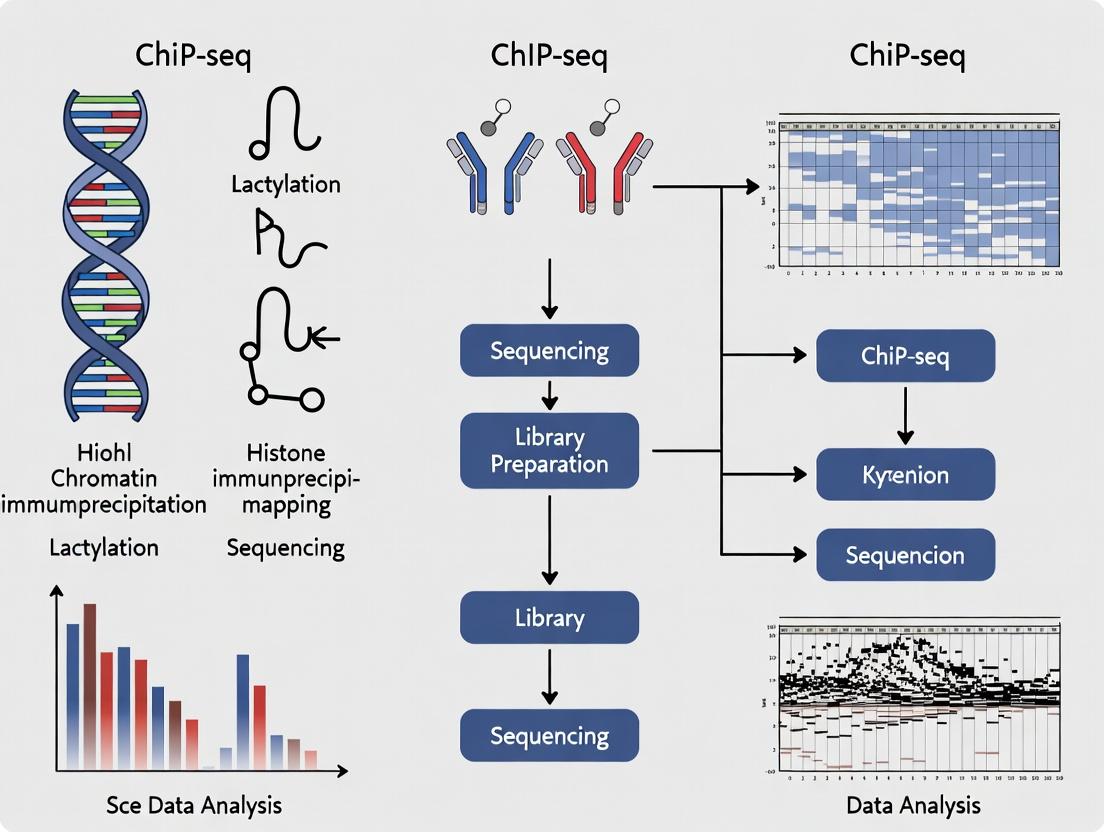
This guide provides a comprehensive framework for researchers and drug development professionals to successfully implement and interpret ChIP-seq for histone lactylation mapping.
Histone Lactylation Mapping: A Comprehensive Guide to ChIP-seq Protocol, Applications, and Data Analysis for Epigenetic Researchers
Abstract
This guide provides a comprehensive framework for researchers and drug development professionals to successfully implement and interpret ChIP-seq for histone lactylation mapping. We cover the foundational biology linking cellular metabolism to epigenetic regulation via lactate-derived modifications, detail step-by-step methodologies from antibody validation to library preparation, address common technical challenges and optimization strategies, and establish rigorous validation and comparative analysis protocols. This resource aims to standardize practices in this emerging field, enabling robust investigation of lactylation's role in immunity, cancer, neurology, and beyond.
Understanding Histone Lactylation: From Metabolic Signal to Epigenetic Regulator
Histone lactylation is a novel post-translational modification (PTM) where a lactyl group is conjugated to a lysine ε-amino group on histone proteins. This modification occurs via a non-enzymatic or enzyme-driven mechanism, linking cellular metabolism—specifically lactate production—to epigenetic regulation. The chemical structure and properties distinguish it from other histone acylations.
- Chemical Structure:
- Lactylation (Kla): Addition of a lactyl group (
-CO-CH(OH)-CH3). The presence of a hydroxyl group on the β-carbon creates a chiral center and increases hydrophilicity. - Acetylation (Kac): Addition of an acetyl group (
-CO-CH3). A simple, small, and hydrophobic modification. - Crotonylation (Kcr): Addition of a crotonyl group (
-CO-CH=CH-CH3). The presence of a carbon-carbon double bond introduces planarity and potential for distinct reader protein interactions.
- Lactylation (Kla): Addition of a lactyl group (
Table 1: Comparative Analysis of Histone Acylations
| Property | Lactylation (Kla) | Acetylation (Kac) | Crotonylation (Kcr) |
|---|---|---|---|
| Chemical Formula | -CO-CH(OH)-CH3 | -CO-CH3 | -CO-CH=CH-CH3 |
| Molecular Weight | ~72 Da | ~42 Da | ~70 Da |
| Key Feature | β-hydroxyl group | Small, hydrophobic | Planar, unsaturated bond |
| Precursor Metabolite | Lactate (Lactyl-CoA) | Acetyl-CoA | Crotonyl-CoA |
| Writer Enzymes | p300 (demonstrated); other KATs proposed | p300/CBP, GCN5, etc. | p300, CBP |
| Eraser Enzymes | HDAC1-3 (SIRT1-3 in vitro) | HDAC1-11; SIRT1-7 | HDAC1-3; SIRT1-3 |
| Established Function | Promotes M2 macrophage polarization; links glycolysis to gene activation | Transcriptional activation; chromatin opening | Associated with active transcription, particularly at enhancers and sex chromosomes |
Application Notes for ChIP-seq in Histone Lactylation Mapping
Within a thesis focused on ChIP-seq for histone lactylation mapping, understanding these chemical distinctions is critical for experimental design and data interpretation.
- Antibody Specificity: The anti-histone Kla antibody must not cross-react with Kac, Kcr, or other structurally similar acylations (e.g., β-hydroxybutyrylation). Validation via peptide array or dot blot is essential.
- Metabolic Perturbation: Modulating lactate levels (e.g., using glycolysis inhibitors like 2-DG, or lactate dehydrogenase inhibitors) is a key strategy to manipulate histone lactylation states before ChIP-seq.
- Data Integration: ChIP-seq peaks for H3K18la must be compared with existing datasets for H3K18ac and H3K18cr to identify unique and shared genomic loci, revealing modification-specific gene regulation.
Detailed Experimental Protocols
Protocol 1: Cell Culture and Metabolic Modulation for Lactylation Induction Objective: Generate cells with high histone lactylation levels for ChIP-seq.
- Culture RAW 264.7 macrophages in DMEM with 10% FBS.
- Polarize cells to an M2 state with IL-4 (20 ng/mL) for 24 hours.
- Key Step: Treat cells with 20 mM sodium lactate (or 10 mM exogenous lactate) for the final 6-12 hours of polarization. Control: Use equimolar sodium chloride.
- Harvest cells, wash with PBS, and proceed to cross-linking for ChIP.
Protocol 2: Chromatin Immunoprecipitation (ChIP) for Histone Lactylation Objective: Immunoprecipitate lactylated histone-DNA complexes.
- Cross-link cells with 1% formaldehyde for 10 min at room temperature. Quench with 125 mM glycine.
- Lysate Preparation: Lyse cells in ChIP lysis buffer (50 mM HEPES-KOH pH 7.5, 140 mM NaCl, 1 mM EDTA, 1% Triton X-100, 0.1% Na-deoxycholate, 0.1% SDS, protease inhibitors). Sonicate chromatin to ~200-500 bp fragments.
- Pre-clear & Immunoprecipitation: Incubate 50-100 µg chromatin with protein A/G magnetic beads for 1h. Incubate pre-cleared supernatant with 2-5 µg of validated anti-H3K18la antibody overnight at 4°C. Isotype IgG serves as negative control.
- Wash & Elute: Wash beads sequentially with Low Salt, High Salt, LiCl, and TE buffers. Elute chromatin in ChIP elution buffer (1% SDS, 0.1M NaHCO3).
- Reverse Cross-linking: Add NaCl to 200 mM and incubate at 65°C overnight. Treat with Proteinase K, recover DNA with SPRI beads.
Protocol 3: ChIP-seq Library Preparation and Data Analysis Objective: Generate sequencing libraries from immunoprecipitated DNA.
- Use a commercial high-sensitivity DNA library prep kit.
- Perform end-repair, A-tailing, and adapter ligation.
- Amplify libraries with 10-12 cycles of PCR using indexed primers.
- Sequence on an Illumina platform (≥ 20 million reads/sample).
- Bioinformatics Analysis:
- Align reads to reference genome (e.g., mm10) using Bowtie2.
- Call peaks with MACS2 (
-g mm -B --nomodel --extsize 200). - Annotate peaks to genomic features with HOMER.
- Perform differential binding analysis with DiffBind.
- Integrate with public Kac/Kcr datasets and pathway analysis (GO, KEGG).
Visualizations
Title: Metabolic Pathway to Histone Lactylation
Title: ChIP-seq Workflow for Histone Lactylation Mapping
The Scientist's Toolkit: Research Reagent Solutions
Table 2: Essential Reagents for Histone Lactylation Research
| Item | Function & Application | Example / Note |
|---|---|---|
| Validated Anti-Histone Lactylation Antibody | Primary antibody for detection (WB, IF) and enrichment (ChIP). Critical for specificity. | Anti-H3K18la (PTM Biolabs, Active Motif). Validate with modified peptide competition. |
| Sodium Lactate | Direct metabolic precursor to increase intracellular lactate and induce histone lactylation. | Use at 10-20 mM in cell culture. Prepare fresh in PBS. |
| LDHA Inhibitor | Modulates endogenous lactate production, used as a negative control. | GSK2837808A (1-10 µM) to reduce lactylation. |
| HDAC1/2/3 Inhibitor | Potential eraser inhibitor; may stabilize histone lactylation signals. | MS-275 (Entinostat, 1-5 µM). |
| Pan-Anti-Acetyllysine Antibody | Comparator for ChIP-seq to distinguish lactylation-specific vs. general acylation sites. | For sequential or parallel ChIP. |
| ChIP-validated p300/CBP Antibody | To investigate the writer enzyme's genomic localization relative to Kla sites. | For co-localization studies. |
| Control Peptides | Unmodified, Lac, Ac, and Cr modified histone tail peptides for antibody validation. | Essential for testing antibody cross-reactivity via dot blot. |
| Magnetic Protein A/G Beads | For efficient immunoprecipitation of antibody-chromatin complexes in ChIP. | Reduce non-specific background vs. agarose beads. |
| High-Sensitivity DNA Library Prep Kit | For constructing sequencing libraries from low-input ChIP DNA. | KAPA HyperPrep or NEB Next Ultra II. |
| Glycolysis/Gluconeogenesis Assay Kit | To biochemically confirm metabolic state perturbations in treated cells. | Correlate lactate production with Kla levels. |
Within the framework of ChIP-seq for histone lactylation mapping research, a paradigm-shifting concept has emerged: the metabolic-epigenetic axis. This axis directly links cellular metabolic flux, particularly the glycolytic production of lactate, to epigenetic regulation via histone lysine lactylation (Kla). This post-translational modification (PTM) functions as a "metabolite-sensing" mechanism, directly translating shifts in cellular energy metabolism into gene expression programs. This application note details the protocols and methodologies for investigating this axis, with a focus on quantitative lactylation mapping under controlled metabolic perturbations.
Key Quantitative Data on Histone Lactylation
Table 1: Core Histone Lysine Residues Subject to Lactylation
| Histone | Lysine Residue (Human) | Associated Function/Context | Key Reference |
|---|---|---|---|
| H3 | K9 | Promotes M2 macrophage polarization; marks active chromatin | Zhang et al., 2019 |
| H3 | K14 | Linked to glycolytic gene activation; common acetylation site | Irizarry-Caro et al., 2020 |
| H3 | K18 | Associated with hypoxia response and tumorigenesis | Yang et al., 2022 |
| H3 | K23 | Implicated in neural stem cell fate determination | Zhang et al., 2019 |
| H4 | K5, K8, K12 | Often modified in tandem; marks highly active promoters | Dai et al., 2020 |
| H2B | K5, K20 | Less characterized; potential role in metabolic stress response | Gaffney et al., 2020 (MS data) |
Table 2: Metabolic Interventions to Modulate Histone Lactylation Levels
| Intervention | Target/Mechanism | Effect on Intracellular [Lactate] | Expected Effect on Histone Kla | Typical Concentration/Duration |
|---|---|---|---|---|
| High Glucose (e.g., 25 mM) | Enhances glycolytic flux | ↑↑↑ | ↑↑↑ | 24-48 hours |
| Oligomycin (ATP synthase inhibitor) | Shifts metabolism to glycolysis | ↑↑ | ↑↑ | 1-10 µM, 4-16 hours |
| Dichloroacetate (DCA) | Inhibits PDK, promotes oxidative metabolism | ↓↓ | ↓↓ | 5-10 mM, 24 hours |
| LDH-A siRNA/Inhibitor (e.g., GNE-140) | Blocks pyruvate-to-lactate conversion | ↓↓↓ | ↓↓↓ | siRNA: 48-72h; Inhibitor: 1-10 µM, 24h |
| Exogenous Sodium Lactate | Directly increases lactate pool | ↑↑↑ | ↑↑↑ | 10-20 mM, 4-12 hours |
| Hypoxia (1% O₂) | Stabilizes HIF-1α, enhances glycolysis | ↑↑↑ | ↑↑↑ | 16-48 hours |
Experimental Protocols
Protocol 1: Inducing and Validating Histone Lactylation in Cell Culture
Aim: To establish a cellular model with high histone Kla levels for subsequent ChIP-seq analysis.
- Cell Treatment: Plate cells (e.g., macrophages, cancer cells) and allow to adhere. Treat with a lactylation-inducing condition (e.g., 20 mM sodium lactate + 25 mM glucose in DMEM) for 12-16 hours. Include a control (normal glucose, no lactate).
- Histone Extraction: Harvest cells. Use an acid extraction kit (e.g., EpiQuik Total Histone Extraction Kit). Resuspend purified histones in neutralization buffer.
- Western Blot Validation:
- Load 2-5 µg histone extract per lane on a 4-20% gradient SDS-PAGE gel.
- Transfer to PVDF membrane.
- Block with 5% BSA in TBST for 1 hour.
- Incubate with primary antibodies: Pan anti-histone lactylation (PTM-1401, 1:1000) and H3 (loading control, 1:5000) overnight at 4°C.
- Incubate with HRP-conjugated secondary antibody (1:5000) for 1 hour.
- Develop with ECL reagent and image. Compare band intensity between treated and control samples.
Protocol 2: Chromatin Immunoprecipitation for Histone Lactylation (Lac-ChIP)
Aim: To isolate lactylated histone-bound DNA fragments for sequencing.
- Crosslinking & Cell Lysis: Treat cells with 1% formaldehyde for 10 min at RT. Quench with 125 mM glycine. Wash with cold PBS. Lyse cells in SDS Lysis Buffer (1% SDS, 10 mM EDTA, 50 mM Tris-HCl, pH 8.1) with protease inhibitors.
- Chromatin Shearing: Sonicate lysate to shear chromatin to an average size of 200-500 bp. Confirm fragment size on a 2% agarose gel.
- Immunoprecipitation: Dilute sheared chromatin 10-fold in ChIP Dilution Buffer. Pre-clear with protein A/G beads for 1 hour. Incubate 10-50 µg chromatin with 2-5 µg of validated anti-histone Kla antibody (e.g., H3K9la, H3K18la) or IgG control overnight at 4°C with rotation.
- Bead Capture & Washes: Add protein A/G beads for 2 hours. Wash sequentially: Low Salt Wash Buffer (once), High Salt Wash Buffer (once), LiCl Wash Buffer (once), TE Buffer (twice).
- Elution & De-crosslinking: Elute chromatin from beads twice with 250 µL Fresh Elution Buffer (1% SDS, 0.1M NaHCO3). Add NaCl to 200 mM and reverse crosslinks at 65°C for 4-6 hours.
- DNA Purification: Add RNase A (30 min, 37°C) then Proteinase K (2 hours, 45°C). Purify DNA using a PCR purification kit. Quantify via Qubit.
Protocol 3: Library Prep and Bioinformatic Analysis for Lac-ChIP-seq
Aim: To generate sequencing libraries and define lactylation peaks.
- Library Preparation: Use 1-10 ng of purified ChIP-DNA with a ThruPLEX DNA-seq or NEBNext Ultra II DNA Library Prep Kit. Follow manufacturer's protocol, including end-repair, A-tailing, adapter ligation, and size selection (150-300 bp). Amplify with 8-12 PCR cycles.
- Sequencing: Pool libraries and sequence on an Illumina platform (e.g., NovaSeq 6000) to achieve 20-40 million paired-end 150 bp reads per sample.
- Bioinformatic Pipeline:
- Quality Control: FastQC on raw reads.
- Alignment: Map reads to reference genome (e.g., hg38) using BWA or Bowtie2.
- Peak Calling: Call significant enrichment peaks from Lac-ChIP sample vs. IgG control using MACS2 (
-g hs -q 0.05 --broadfor broad histone marks). - Annotation & Motif Analysis: Annotate peaks to nearest TSS using ChIPseeker. Perform de novo motif discovery with HOMER.
- Integration: Overlap peaks with public ChIP-seq datasets (e.g., H3K27ac, H3K4me3) and RNA-seq data from matched conditions to infer functional impact.
Visualizations
Title: Metabolic Flow to Epigenetic Modification
Title: Lac-ChIP-seq Experimental Pipeline
Title: Multi-Omics Data Integration Logic
The Scientist's Toolkit: Key Research Reagent Solutions
Table 3: Essential Materials for Lactylation Research
| Item | Function/Description | Example Product/Catalog # |
|---|---|---|
| Pan anti-Kla Antibody | Broad detection of lactylated lysines on histones and other proteins. Critical for initial Western Blot screening. | PTM-1401 (Pan-Kla) |
| Site-Specific Kla Antibodies | Validated antibodies for ChIP-seq targeting specific histone marks (e.g., H3K9la, H3K18la). Essential for locus-specific mapping. | PTM-1406 (H3K9la), PTM-1411 (H3K18la) |
| LDH-A Inhibitor | Pharmacological tool to block lactate production from pyruvate, establishing causality in the axis. | GNE-140 (MedChemExpress) |
| Sodium Lactate (isotope-labeled) | Direct lactylation precursor. ¹³C or D-labeled lactate enables tracking of lactate-derived lactyl groups via MS. | Sodium L-Lactate-¹³C₃ (Sigma) |
| Histone Extraction Kit | Rapid, standardized acid extraction of histones from cells/tissues, minimizing protease activity. | EpiQuik Total Histone Extraction Kit |
| ChIP-Validated Antibody Beads | Magnetic beads pre-coupled to Protein A/G for efficient and clean immunoprecipitation in ChIP protocols. | Dynabeads Protein A/G |
| ChIP-seq Library Prep Kit | Optimized for low-input DNA from ChIP, ensuring high-complexity libraries for sequencing. | NEBNext Ultra II DNA Library Prep Kit |
| HDAC Class I/IIa Inhibitors | Controls to distinguish lactylation from acetylation, as some HDACs (e.g., HDAC1-3) may also delactylate. | Trichostatin A (TSA), Nicotinamide |
Application Notes: Histone Lactylation in Disease and Physiology
Histone lysine lactylation (Kla) is a novel post-translational modification (PTM) derived from lactate, linking cellular metabolism to epigenetic regulation. Its discovery has provided a mechanistic framework for understanding lactate's role as a signaling molecule beyond a metabolic byproduct. This note details key findings and protocols within the broader context of ChIP-seq for lactylation mapping research.
Lactylation in M1 Macrophage Polarization
M1 macrophage polarization, typically induced by LPS and IFN-γ, leads to a metabolic shift towards glycolysis and lactate production. This lactate serves as a substrate for histone lactylation, which promotes a "homeostatic" gene expression program that temporally follows the initial pro-inflammatory response.
Key Quantitative Findings:
Table 1: Lactylation Dynamics in M1 Macrophages
| Target | Lactylation Change | Functional Outcome | Key Readout |
|---|---|---|---|
| H3K18la | Increases ~4-6 fold at 16-24h post-LPS | Drives expression of Arg1, VEGF, other reparative genes | ChIP-qPCR signal enrichment |
| Promoter Lactylation | Positively correlates with gene activation (R² >0.7) | Transitions macrophage phenotype from pro-inflammatory to pro-healing | RNA-seq & ChIP-seq correlation |
| Lactate Concentration | Extracellular [Lactate] > 10mM required for significant histone Kla in vitro | Threshold for epigenetic reprogramming | LC-MS/MS quantification |
Research Reagent Solutions:
- Pan anti-Kla antibody: For immunoblotting to detect global lactylation levels.
- Histone H3K18la-specific antibody: Essential for ChIP-seq and IF to map locus-specific modification.
- Lactate Dehydrogenase (LDH) Inhibitor (GSK2837808A): To deplete intracellular lactate and validate Kla dependence.
- Sodium Lactate (isotope-labeled, e.g., 13C3): To trace lactate incorporation into histones.
Lactylation in the Tumor Microenvironment (TME)
In the TME, tumor cells and stromal cells often exhibit the Warburg effect, creating a lactate-rich milieu. This lactate lactylates histones in tumor-associated macrophages (TAMs), cancer-associated fibroblasts (CAFs), and tumor cells themselves, promoting immune evasion and tumor progression.
Key Quantitative Findings:
Table 2: Lactylation in the Tumor Microenvironment
| Cell Type | Primary Target | Effect on Gene Expression | Impact on Tumor Phenotype |
|---|---|---|---|
| TAMs | H3K18la at Arg1 promoter | Increases ARG1 expression >3-fold | Suppresses T-cell function, promotes immunosuppression |
| CAFs | Histone lactylation at pro-fibrotic genes | Upregulates ECM protein production | Enhances tumor stiffness & metastasis |
| Tumor Cells | H3K9la at PD-L1 promoter | Elevates PD-L1 levels ~2.5 fold | Increases immune checkpoint expression |
Research Reagent Solutions:
- MCT1/4 Inhibitors (e.g., AZD3965): To block lactate transport and disrupt lactylation signaling.
- p300/CBP Inhibitor (A485): p300 is a putative lactyltransferase; inhibitor validates enzyme role.
- Class I HDAC Inhibitors (e.g., TSA): HDAC1-3 are potential "delactylases"; inhibitors increase Kla.
- Recombinant Human Lactate (high purity): For in vitro TME simulation experiments.
Neuronal Function and Lactylation
In the brain, lactate produced by astrocytes is shuttled to neurons (ANLS). Neuronal histone lactylation links this metabolic coupling to adaptive gene expression involved in long-term memory and disease states.
Key Quantitative Findings:
Table 3: Neuronal Lactylation in Physiology and Disease
| Context | Modification Site | Gene Regulation | Functional Consequence |
|---|---|---|---|
| Memory Formation | H4K12la in hippocampus | Activates Plasticity-related genes (e.g., c-Fos, Egr1) | Essential for fear conditioning memory consolidation |
| Ischemic Stroke | Global histone Kla increase in penumbra | Represses neuroinflammatory pathways | Neuroprotective effect observed in mouse models |
| Major Depressive Disorder | Decreased H3K18la in prefrontal cortex | Downregulates synaptic function genes | Correlates with depressive-like behavior in rodents |
Research Reagent Solutions:
- Neuron-Specific Lactate Dehydrogenase Knockdown (AAV-shLDHA): To cell-specifically modulate neuronal lactate metabolism.
- Lactate Sensor (pHLARE): For real-time imaging of lactate flux in astrocyte-neuron co-cultures.
- Bromodomain-containing protein 4 (BRD4) Inhibitor (JQ1): BRD4 may "read" Kla; inhibitor probes functional output.
Detailed Protocols
Protocol 1: ChIP-seq for Histone Lactylation (H3K18la)
Objective: To map genome-wide occupancy of histone H3 lysine 18 lactylation.
Workflow:
- Cell Crosslinking & Lysis: Treat cells (e.g., LPS-stimulated macrophages) with 1% formaldehyde for 10 min at room temperature. Quench with 125mM glycine. Lyse cells in SDS Lysis Buffer.
- Chromatin Shearing: Sonicate lysate to shear DNA to 200-500 bp fragments. Verify fragment size by agarose gel electrophoresis.
- Immunoprecipitation: Dilute sonicated chromatin in ChIP Dilution Buffer. Pre-clear with Protein A/G beads for 1h. Incubate supernatant with 2-5 µg of validated anti-H3K18la antibody overnight at 4°C. Use species-matched IgG as negative control. Add beads and incubate for 2h.
- Washing & Elution: Wash beads sequentially with Low Salt, High Salt, LiCl, and TE buffers. Elute chromatin with fresh elution buffer (1% SDS, 0.1M NaHCO3).
- Reverse Crosslinking & Purification: Add NaCl to 200mM and incubate at 65°C overnight to reverse crosslinks. Treat with Proteinase K, then purify DNA with a PCR purification kit.
- Library Prep & Sequencing: Prepare sequencing library from ChIP and Input DNA using a commercial kit (e.g., NEBNext Ultra II). Sequence on an Illumina platform (≥ 20 million reads/sample).
- Data Analysis: Align reads to reference genome (e.g., GRCh38). Call peaks using MACS2. Annotate peaks to nearest TSS. Integrate with RNA-seq data for correlation.
Diagram Title: ChIP-seq Workflow for Histone Lactylation Mapping
Protocol 2: Validating Functional Role of Kla using Metabolite Manipulation
Objective: To establish causality between lactate, Kla, and gene expression.
Procedure: A. Lactate Augmentation: Culture target cells in medium supplemented with 10-20 mM sodium lactate (pH-adjusted) for 12-24h. Include sodium chloride as an osmotic control. B. Lactate Depletion: Treat cells with 10 µM GSK2837808A (LDH A inhibitor) for 24h to inhibit endogenous lactate production. C. Downstream Analysis: * Immunoblot: Extract histones via acid extraction. Use pan-Kla and site-specific (H3K18la) antibodies for detection. * ChIP-qPCR: Perform ChIP as in Protocol 1 on key target gene promoters (e.g., Arg1). Analyze by qPCR. * RT-qPCR: Isolate RNA, synthesize cDNA, and measure expression of target genes.
Diagram Title: Validating Lactate-Lactylation Causality
The Scientist's Toolkit: Essential Research Reagents
Table 4: Key Reagents for Lactylation Research
| Reagent Name | Category | Primary Function |
|---|---|---|
| Validated Anti-Histone Lactylation Antibodies | Antibody | Critical for detection (WB, IF) and enrichment (ChIP) of Kla. Site-specific (H3K18la) and pan-specific available. |
| Sodium Lactate (13C3-labeled) | Metabolic Tracer | Traces the incorporation of lactate into histone lysine residues via LC-MS/MS. |
| LDH A Inhibitor (GSK2837808A) | Small Molecule Inhibitor | Inhibits glycolysis-derived lactate production, used to test necessity of endogenous lactate for Kla. |
| p300/CBP Inhibitor (A485) | Epigenetic Enzyme Inhibitor | Inhibits putative lactyltransferase activity of p300, used to validate enzyme-substrate relationship. |
| HDAC1-3 Inhibitor (e.g., TSA) | Deacetylase/Delactylase Inhibitor | Potentially inhibits "delactylase" activity, leading to Kla accumulation for mechanistic studies. |
| MCT1/4 Inhibitor (AZD3965) | Transporter Inhibitor | Blocks lactate import/export, disrupting lactylation signaling in co-culture or TME models. |
| Acid Extraction Kit | Histone Purification | Isolates histones from cells/tissues with minimal PTM loss for downstream immunoblot or MS analysis. |
| ChIP-seq Grade Protein A/G Magnetic Beads | Immunoprecipitation | For efficient and clean pull-down of lactylated chromatin complexes. |
Within the paradigm of epigenetic regulation, histone post-translational modifications (PTMs) are central to core biological functions. Recent research has identified histone lactylation, derived from lactate—a metabolite linked to cellular metabolism and signaling—as a novel epigenetic mark. This application note situates histone lactylation within a broader thesis on ChIP-seq mapping, detailing its role in regulating gene expression, influencing cell fate decisions, and modulating immune responses. Protocols for the systematic study of this modification are provided for researchers and drug development professionals.
The Role of Histone Lactylation in Core Biological Functions
Regulating Gene Expression
Histone lactylation, particularly on lysine residues such as H3K9la and H3K18la, is associated with active gene transcription. It functions as a "metabolo-epigenetic" link, where elevated lactate levels, often from glycolysis or the tumor microenvironment, drive lactyl-CoA production and subsequent histone modification.
- Mechanism: Lactylation adds a lactyl group to histone tails, neutralizing positive charge and loosening chromatin structure, facilitating transcriptional activator recruitment.
- Key Findings: In macrophages, H3K18la marks promote polarization to a pro-healing, M2-like state by activating homeostatic and Arg1 genes. In cancer cells, lactylation drives oncogene expression.
Table 1: Key Histone Lactylation Marks and Transcriptional Outcomes
| Histone Mark | Associated Context | Primary Transcriptional Effect | Example Target Genes/Phenotype |
|---|---|---|---|
| H3K18la | M2 Macrophage Polarization | Activation | Arg1, Ym1; Tissue Repair |
| H3K9la | Tumor Microenvironment | Activation | PDK1, SOX5; Metabolic Reprogramming |
| H3K56la | Embryonic Stem Cells | Activation | Pluripotency Network; Self-renewal |
Determining Cell Fate
Lactylation serves as a metabolic sensor influencing cell differentiation and identity.
- Stem Cells: In embryonic stem cells, lactate-induced H3K56la promotes self-renewal and pluripotency gene expression.
- Reprogramming: Modulating lactylation levels can impact somatic cell reprogramming efficiency.
- Cancer: Sustained lactylation in tumor-initiating cells promotes stem-like properties and chemoresistance.
Modulating Immune Responses
Immune cell function is intimately linked to metabolic state. Lactylation is a key regulator.
- Macrophage Polarization: Lactate from glycolysis drives H3K18la, shifting macrophages from pro-inflammatory (M1) to anti-inflammatory, pro-repair (M2) states.
- T Cell Function: Early evidence suggests lactylation may influence T cell exhaustion in chronic infection/tumors.
- Inflammatory Diseases: Aberrant lactylation is implicated in the pathophysiology of sepsis, fibrosis, and autoimmune conditions.
Table 2: Lactylation in Immune Cell Modulation
| Immune Cell Type | Metabolic Trigger | Lactylation Role | Functional Outcome |
|---|---|---|---|
| Macrophage | High Lactate (Warburg) | H3K18la at M2 genes | Polarization to Pro-Repair Phenotype |
| Dendritic Cell | TLR Activation | Promotes tolerogenic gene expression | Immune Tolerance |
| Tumor-Infiltrating Myeloid Cells | Tumor Microenvironment | Sustains immunosuppressive programs | Inhibition of Anti-Tumor Immunity |
Detailed Application Notes & Protocols for ChIP-seq of Histone Lactylation
Protocol 1: Cell Culture & Lactylation Induction
Aim: To generate cells with enriched histone lactylation for downstream ChIP-seq.
- Culture target cells (e.g., RAW 264.7 macrophages, cancer cell lines) under standard conditions.
- Induction: Treat cells with sodium lactate (e.g., 20 mM) for 12-24 hours. Include sodium chloride control.
- Inhibition (Optional): Pre-treat with glycolysis inhibitor 2-DG (10 mM, 2h) to deplete lactate.
- Harvest cells by gentle scraping. Wash 2x with cold PBS.
- Proceed to crosslinking or snap-freeze pellet at -80°C.
Protocol 2: Crosslinking & Chromatin Preparation for Lactylation ChIP
Note: Lactylation is a relatively labile PTM. Use light crosslinking.
- Crosslinking: Resuspend cell pellet in 1% formaldehyde in PBS. Incubate for 5-8 minutes at RT with gentle rotation.
- Quenching: Add glycine to 125 mM final concentration. Incubate 5 min at RT.
- Wash cells 2x with ice-cold PBS.
- Lysis & Sonication: Lyse cells in SDS lysis buffer. Sonicate chromatin to ~200-500 bp fragments using a focused ultrasonicator (e.g., Covaris). Optimize settings to prevent overheating.
- Clarify lysate by centrifugation (15 min, 13,000 rpm, 4°C). Aliquot supernatant.
Protocol 3: Immunoprecipitation with Anti-Lactylation Antibodies
Critical: Antibody specificity is paramount.
- Pre-clear: Incubate chromatin aliquot (50-100 µg) with Protein A/G magnetic beads for 1h at 4°C.
- Immunoprecipitation: Transfer pre-cleared chromatin to a new tube. Add validated anti-histone lactylation antibody (e.g., anti-H3K18la, PTM Bio). Incubate overnight at 4°C with rotation.
- Capture: Add fresh Protein A/G beads. Incubate 2-4h at 4°C.
- Wash: Wash beads sequentially with: Low Salt Wash Buffer, High Salt Wash Buffer, LiCl Wash Buffer, and TE Buffer (twice).
- Elution & Decrosslinking: Elute complexes in freshly prepared Elution Buffer (1% SDS, 0.1M NaHCO3). Add NaCl to 200 mM final and reverse crosslinks at 65°C overnight.
- DNA Purification: Treat with RNase A and Proteinase K. Purify DNA using silica spin columns. Quantify by Qubit.
Protocol 4: Library Preparation & Sequencing
- Use a high-sensitivity library preparation kit (e.g., NEBNext Ultra II) for low-input DNA.
- Perform size selection (150-300 bp) to isolate nucleosome-associated fragments.
- Amplify with limited PCR cycles (≤12).
- Validate library quality with Bioanalyzer.
- Sequence on an Illumina platform (PE 50bp or longer). Aim for 20-40 million non-duplicate reads per sample.
Protocol 5: Bioinformatics & Data Analysis Pipeline
- Quality Control: FastQC for read quality. Trim adapters with Trimmomatic.
- Alignment: Map reads to reference genome (e.g., mm10, hg38) using Bowtie2 or BWA.
- Peak Calling: Call significant lactylation peaks using MACS2 with input DNA as control. Use a stringent p-value (e.g., 1e-5).
- Annotation & Visualization: Annotate peaks to genomic features (HOMER). Generate bigWig files for visualization in IGV or UCSC Genome Browser.
- Integration: Integrate with RNA-seq data (e.g., from lactate-treated vs. control) to correlate lactylation marks with gene expression changes.
Visualizations
Title: Metabolic Pathway to Histone Lactylation and Gene Activation
Title: ChIP-seq Workflow for Histone Lactylation Mapping
The Scientist's Toolkit: Key Research Reagent Solutions
Table 3: Essential Materials for Histone Lactylation Research
| Item | Function & Role in Lactylation Research | Example Product/Catalog |
|---|---|---|
| Validated Anti-Lactylation Antibodies | Critical for specific detection and ChIP of histone lactylation marks. | PTM Biolabs Anti-H3K18la; Active Motif Anti-H3K9la |
| Pan Anti-Lactyllysine Antibody | Detects global protein lactylation levels via western blot. | PTM Biolabs Pan-Kla |
| Lactate (Sodium Salt) | Used to induce histone lactylation in cell culture models. | Sigma-Aldrich L7022 |
| Lactate Dehydrogenase (LDH) Inhibitor | Tool to reduce lactate production and study lactylation dependency. | GSK2837808A |
| p300/CBP Inhibitor | Probing the role of putative histone lactyltransferases. | A-485 |
| Histone Deacetylase (HDAC) Inhibitors | Certain HDACs (e.g., HDAC1-3) may remove lactylation; used to study turnover. | Trichostatin A (TSA) |
| Glycolysis Inhibitor (2-DG) | Reduces glycolytic flux and lactate, serving as a negative control. | Sigma-Aldrich D8375 |
| Magnetic Protein A/G Beads | For efficient immunoprecipitation in ChIP assays. | Thermo Fisher Scientific 10002D/10004D |
| Covaris MicroTubes & AFA Fiber | For consistent, controlled chromatin shearing via sonication. | Covaris 520045 |
| Low-Input Library Prep Kit | Essential for preparing sequencing libraries from ChIP DNA. | NEBNext Ultra II DNA Library Prep Kit |
| Lactate Assay Kit | Quantifying intracellular/extracellular lactate levels. | Abcam ab65331 |
| ChIP-seq Grade Control Antibodies | Positive (H3K27ac) and negative (IgG) controls for assay validation. | Diagenode C15410196; Millipore 12-370 |
ChIP-seq (Chromatin Immunoprecipitation followed by sequencing) is the established gold standard for mapping protein-DNA interactions in vivo, with its most prominent application being the genome-wide profiling of histone post-translational modifications (PTMs). Within the context of a thesis exploring the novel histone mark histone lactylation, ChIP-seq represents the indispensable methodological cornerstone. This lactylation mark, derived from cellular lactate as a metabolic signaling molecule, links cellular metabolism to epigenetic gene regulation. Precise mapping of its genomic localization is critical for understanding its role in immunology, cancer, and neurobiology, and for identifying potential therapeutic targets in drug development.
ChIP-seq: Core Principles and Workflow
The fundamental principle of ChIP-seq involves the selective immunoprecipitation of chromatin fragments bound by a protein of interest—in this case, a lactylated histone—using a specific antibody. The co-precipitated DNA is then purified, converted into a sequencing library, and subjected to high-throughput sequencing. The resulting reads are aligned to a reference genome to identify enriched regions (peaks), which represent the genomic loci associated with the histone modification.
Experimental Protocol: Standard ChIP-seq for Histone Lactylation
Key Materials & Reagents:
- Crosslinking Agent (1% Formaldehyde): Fixes protein-DNA interactions.
- Cell Lysis Buffers: Series of buffers (e.g., LB1, LB2, LB3) to isolate nuclei and prepare chromatin.
- Micrococcal Nuclease (MNase) or Sonication: For chromatin shearing to ~150-300 bp fragments.
- Protein A/G Magnetic Beads: For antibody capture.
- Validated Anti-Histone Lactylation Antibody (e.g., anti-H3K9la, anti-H4K12la): Specificity is paramount.
- ChIP Elution Buffer (1% SDS, 0.1M NaHCO3): Releases immunoprecipitated complexes.
- DNA Purification Kit: For cleanup of crosslink-reversed DNA.
- Library Preparation Kit (e.g., ThruPLEX or NEBNext): For NGS library construction.
- High-Sensitivity DNA Assay Kit (e.g., Qubit, Bioanalyzer): For DNA quantification and quality control.
Detailed Protocol Steps:
- Crosslinking & Quenching: Treat cells with 1% formaldehyde for 8-10 min at RT. Quench with 125mM Glycine.
- Cell Lysis & Chromatin Preparation: Wash cells, resuspend in lysis buffer. Isolate nuclei and shear chromatin using optimized MNase digestion or sonication (e.g., 4x 10 min cycles, 30 sec ON/30 sec OFF, high setting).
- Immunoprecipitation: Clear chromatin lysate with beads. Incubate supernatant with 1-5 µg of validated anti-lactyl-antibody overnight at 4°C. Add Protein A/G beads for 2 hours.
- Washing & Elution: Wash beads stringently with low-salt, high-salt, LiCl, and TE buffers. Elute bound complexes in elution buffer.
- Reverse Crosslinking & DNA Purification: Incubate eluate at 65°C overnight with 200mM NaCl. Treat with RNase A and Proteinase K. Purify DNA using a silica-membrane column.
- Library Preparation & Sequencing: Convert 1-10 ng of ChIP DNA into a sequencing library following kit protocol. Perform paired-end sequencing (e.g., 2x75 bp) on an Illumina platform to a depth of 20-40 million reads.
Experimental Workflow Diagram
Diagram Title: Standard ChIP-seq Experimental Workflow for Histone Lactylation Mapping
The Scientist's Toolkit: Essential Reagents for Histone Lactylation ChIP-seq
| Reagent / Material | Function & Critical Consideration |
|---|---|
| Validated Anti-Histone Lactylation Antibody | Core reagent for specific pulldown. Must be validated by dot-blot, western, and peptide competition against other acylations (e.g., acetylation). |
| MNase (Micrococcal Nuclease) | Preferred for histone ChIP; digests linker DNA to yield mononucleosomes, preserving PTM context. |
| Protein A/G Magnetic Beads | Facilitate antibody-antigen complex capture and efficient washing. Choice depends on antibody species/isotype. |
| Protease/Phosphatase Inhibitors | Preserve the lactylation epitope and overall chromatin state during extraction. |
| Sodium Butyrate (in buffers) | Inhibits histone deacetylases (HDACs) to prevent loss of related marks, but may indirectly affect lactylation. |
| DNA Clean & Concentrator Kit | For efficient recovery of low-abundance ChIP DNA post-purification. |
| High-Sensitivity DNA Kit (Bioanalyzer) | Assess shearing efficiency (150-300 bp smear) and final library quality. |
| Spike-in Control DNA/Chromatin | Normalizes for technical variation (e.g., Drosophila chromatin in human samples). |
Data Analysis and Key Metrics
Following sequencing, raw data undergoes a standardized bioinformatic pipeline: quality control (FastQC), alignment (Bowtie2/BWA), duplicate removal, peak calling (MACS2), and annotation. Key quantitative metrics for a successful ChIP-seq experiment are summarized below.
Table 1: Standard ChIP-seq Quality Control Metrics and Benchmarks
| Metric | Target/Expected Value | Significance for Histone Lactylation Studies |
|---|---|---|
| Sequencing Depth | 20-40 million mapped reads | Sufficient for robust peak detection, given lactylation's potential low abundance. |
| Fraction of Reads in Peaks (FRiP) | > 1-5% for histone marks | Indicator of IP efficiency. Lower may signal antibody specificity issues. |
| Peak Number | Project-dependent; 10,000 - 100,000 | Reflects genomic distribution of the mark. Context-specific (cell type, stimulation). |
| Peak Distribution | Promoters/Enhancers/Intergenic | Lactylation is enriched at active promoters and enhancers. Profile validates biological expectation. |
| Replicate Correlation (RSC) | > 0.8 (Pearson's) | Indicates high reproducibility between biological replicates. |
| Non-Redundant Fraction (NRF) | > 0.8 | Measures library complexity; low values suggest over-amplification or PCR duplicates. |
Pathway: Linking Metabolism to Epigenetics via Histone Lactylation
The discovery of histone lactylation establishes a direct molecular pathway from cellular metabolism to chromatin regulation, which ChIP-seq is uniquely positioned to map.
Diagram Title: Metabolic Pathway to Histone Lactylation and Functional Readouts
Advanced Protocol: Sequential ChIP-seq (Re-ChIP) for Co-localization Studies
To investigate the interplay between histone lactylation and other modifications (e.g., acetylation), sequential ChIP-seq is employed.
Protocol Steps:
- Perform first ChIP as standard using Antibody A (e.g., anti-H3K9la).
- After the final wash, elute the bound complexes not with SDS buffer, but with 10mM DTT at 37°C for 30 min.
- Dilute the eluate 1:50 with Re-ChIP buffer (1% Triton, 2mM EDTA, 150mM NaCl, 20mM Tris pH 8.1).
- Perform a second immunoprecipitation on the diluted eluate using Antibody B (e.g., anti-H3K9ac).
- Wash, elute with standard SDS buffer, reverse crosslinks, purify DNA, and prepare library.
Table 2: Comparison of Standard vs. Sequential ChIP-seq
| Parameter | Standard ChIP-seq | Sequential ChIP-seq (Re-ChIP) |
|---|---|---|
| Primary Objective | Map single histone mark genome-wide. | Identify genomic loci co-modified by two distinct marks. |
| Antibody Requirement | One highly specific antibody. | Two highly specific antibodies, different species/isotypes preferred. |
| Input Material | High (≥ 1x10^6 cells). | Very high (≥ 5x10^6 cells). |
| Data Complexity | Single peak set. | Overlapping peak sets; requires stringent statistical overlap analysis. |
| Application to Lactylation | Define lactylation landscape. | Test hypothesis of "lactylation-acetylation crosstalk" on same histone tail. |
ChIP-seq remains the definitive method for mapping the genomic landscape of histone modifications, including the emergent mark of histone lactylation. Its robustness, scalability, and compatibility with complex samples make it essential for linking metabolic shifts to epigenetic outcomes in disease and therapeutic contexts. The protocols and considerations outlined here provide a framework for integrating histone lactylation mapping into a broader thesis on metabolism-driven epigenetic regulation.
A Step-by-Step ChIP-seq Protocol for Robust Histone Lactylation Profiling
The mapping of histone lactylation (Kla) via chromatin immunoprecipitation followed by sequencing (ChIP-seq) has emerged as a pivotal methodology for understanding the role of lactate, a metabolite and signaling molecule, in epigenetic regulation. The success of these studies is critically dependent on the specificity and affinity of the antibodies used to capture lactylated histones. Two primary classes of antibodies are employed: pan-Kla antibodies, which recognize the lactyl-lysine modification irrespective of the protein context, and site-specific antibodies, such as those targeting H3K18la. This application note provides a detailed comparison, validation protocols, and workflow integration for selecting the optimal antibody for histone lactylation mapping research.
The selection between pan-Kla and site-specific antibodies involves trade-offs in specificity, signal strength, and applicability. The following table summarizes key characteristics based on recent literature and commercial product specifications.
Table 1: Comparison of Pan-Kla and Site-Specific (H3K18la) Antibodies for ChIP-seq
| Feature | Pan-Kla Antibody | H3K18la-Specific Antibody |
|---|---|---|
| Epitope Target | Lactyl-lysine modification on any histone/non-histone protein. | Lactyl-lysine specifically on histone H3 at lysine 18. |
| Primary Application | Discovery-based profiling of novel lactylation sites; immunoblot. | Targeted investigation of a specific, biologically validated locus. |
| ChIP-seq Specificity | Lower; may pull down all lactylated chromatin regions. | Higher; maps only H3K18la-associated genomic loci. |
| Signal-to-Noise in ChIP | Potentially higher background due to broader capture. | Generally cleaner, more interpretable peaks. |
| Validation Requirement | High; must confirm histone origin via pre-clearing or mass spectrometry. | High; must confirm specificity via peptide competition assays. |
| Common Validation Metrics | Peptide microarray/ELISA, Knockdown of lactylation writers (p300), Lactate stimulation assays. | Site-mutant peptide competition (H3K18la vs. H3K18ac), Specificity dot blots. |
| Reported ChIP-seq Peak Count (MCF-7 cells, lactate stimulated) | ~15,000 - 25,000 broad peaks | ~5,000 - 10,000 sharp peaks |
Detailed Experimental Protocols
Protocol 3.1: Dot Blot Validation for Antibody Specificity
Purpose: To rapidly assess cross-reactivity with other acylations (e.g., acetylation, crotonylation).
Materials:
- Nitrocellulose membrane.
- Synthetic histone peptides (1 µg/µL): Unmodified, Kla (Pan-Kla or H3K18la), Kac (H3K18ac), Kcr.
- Blocking buffer (5% BSA in TBST).
- Primary antibody (Pan-Kla or H3K18la, 1:1000).
- HRP-conjugated secondary antibody.
- Chemiluminescent substrate.
Procedure:
- Spot 1 µL of each peptide onto a dry nitrocellulose membrane. Air dry for 15 min.
- Block the membrane with 5% BSA for 1 hour at room temperature (RT).
- Incubate with primary antibody diluted in blocking buffer overnight at 4°C.
- Wash 3x for 5 min with TBST.
- Incubate with HRP-conjugated secondary antibody (1:5000) for 1 hour at RT.
- Wash 3x for 5 min with TBST.
- Develop using a chemiluminescent imager. A valid antibody should show a strong signal only for its target lactylated peptide.
Protocol 3.2: Peptide Competition ChIP-seq (for Site-Specific Antibodies)
Purpose: To confirm that ChIP-seq signals are specifically derived from the target lactylation mark.
Materials:
- Crosslinked chromatin (from ~1x10⁶ cells).
- Validated H3K18la antibody.
- Competing peptides: H3K18la (specific) and H3K18ac (non-specific), at 10x and 100x molar excess.
- Protein A/G magnetic beads.
- ChIP-seq library preparation kit.
Procedure:
- Prepare standard ChIP buffer and aliquot chromatin into four tubes.
- Pre-incubate the H3K18la antibody with either: a) no peptide, b) 10x specific peptide, c) 100x specific peptide, d) 100x non-specific peptide, for 2 hours at 4°C on a rotator.
- Add each antibody-peptide mixture to the chromatin aliquots and perform the standard ChIP protocol (overnight incubation, bead capture, washes, elution, reverse crosslinking).
- Purify DNA and prepare sequencing libraries.
- Analysis: The 100x specific peptide competition should abolish >90% of peaks, while the non-specific peptide should have minimal effect, confirming antibody specificity in situ.
Protocol 3.3: Cellular Stimulation & Inhibition Control for Pan-Kla Specificity
Purpose: To demonstrate that ChIP signals using a pan-Kla antibody are dependent on cellular lactylation levels.
Materials:
- Cell culture (e.g., MCF-7, macrophages).
- Sodium lactate (e.g., 20 mM, 24h stimulation).
- p300/CBP inhibitor (e.g., A-485, 10 µM, 24h inhibition).
- Pan-Kla antibody for ChIP.
Procedure:
- Treat cells in three conditions: a) Control, b) Lactate-stimulated, c) Lactate + A-485 inhibitor.
- Harvest cells and perform ChIP-seq using the pan-Kla antibody in parallel for all conditions.
- Validate via parallel immunoblotting: increased pan-Kla signal with lactate, decreased with inhibitor.
- Sequencing data should show a global increase in pan-Kla ChIP-seq peaks with lactate stimulation and a significant reduction upon p300 inhibition, linking the captured signal to enzymatically regulated lactylation.
Visualization of Workflows & Pathways
Diagram Title: Antibody Selection and Validation Decision Pathway
Diagram Title: Histone Lactylation ChIP-seq Experimental Workflow
The Scientist's Toolkit: Key Research Reagent Solutions
Table 2: Essential Materials for Histone Lactylation ChIP-seq Research
| Item | Function & Importance | Example/Notes |
|---|---|---|
| Validated Anti-Histone Lactylation Antibody | Core reagent for specific capture of lactylated chromatin. Pan-Kla for discovery, site-specific (e.g., H3K18la) for targeted studies. | Critical to obtain validation data (dot blots, competition). Commercial sources vary in quality. |
| Synthetic Lactylated Peptides | Gold standard for antibody validation via competition assays and dot blots. | Unmodified, Kla (pan or site-specific), and other acylated (Kac, Kcr) control peptides are essential. |
| p300/CBP Inhibitor (e.g., A-485) | Pharmacological tool to inhibit major histone lactyltransferases, providing a negative control for specificity. | Confirms that ChIP signal is enzymatically regulated. |
| Sodium Lactate (Cell Culture Grade) | Used to stimulate endogenous histone lactylation levels in cell models, enhancing ChIP signal. | Typical working concentration 10-20 mM for 12-24h. |
| Chromatin Shearing System | To fragment crosslinked chromatin to optimal size (200-500 bp) for ChIP. | Covaris sonicator or Bioruptor is standard. Consistency is key for reproducibility. |
| Protein A/G Magnetic Beads | For efficient capture of antibody-chromatin complexes. | Preferred over agarose beads for easier handling and lower background. |
| ChIP-seq Grade DNA Library Prep Kit | To prepare immunoprecipitated DNA for next-generation sequencing. | Must be compatible with low-input DNA (1-10 ng). |
| Control Antibodies | Essential for assay quality control. | Positive: H3K27ac; Negative: Normal Rabbit IgG. |
1. Introduction and Thesis Context
This document outlines key application notes and protocols for modulating histone lactylation in cell culture, a critical preparatory step for subsequent Chromatin Immunoprecipitation followed by sequencing (ChIP-seq) experiments. Within the broader thesis on mapping histone lactylation landscapes via ChIP-seq, controlling the cellular lactylation state through metabolic priming is essential for generating comparative datasets (e.g., high vs. low lactylation). These protocols enable researchers to probe the functional consequences of lactylation on gene regulation by altering the availability of its precursor, lactate.
2. Quantitative Data Summary: Metabolic Primers and Their Effects
Table 1: Common Metabolic Priming Agents for Modulating Histone Lactylation
| Agent | Typical Concentration Range | Primary Target / Mechanism | Expected Effect on Global Histone Lactylation | Key Considerations |
|---|---|---|---|---|
| Sodium Lactate (Exogenous) | 10 - 40 mM | Directly increases intracellular lactate pool, substrate for lactylation. | Increase | Concentration- and time-dependent. Monitor for pH changes; use sodium pyruvate as osmolarity/control. |
| 2-Deoxy-D-Glucose (2-DG) | 5 - 20 mM | Competitive inhibitor of glycolysis (hexokinase). Reduces pyruvate/lactate production. | Decrease | Can induce ER stress and ATP depletion. Use shorter treatments (<24h). |
| Oxamate | 20 - 50 mM | Competitive inhibitor of Lactate Dehydrogenase (LDH). Blocks pyruvate-to-lactate conversion. | Decrease | Directly inhibits lactate generation. High concentrations may be required. |
| FK866 (Daporinad) | 10 - 100 nM | Inhibits NAMPT (Nicotinamide phosphoribosyltransferase), depleting NAD+. | Decrease | Inhibits glycolytic flux & LDH activity by reducing NADH/NAD+ ratio. Potent and specific. |
| Glucose Deprivation | 0 mM Glucose | Limits glycolytic flux, reducing pyruvate and lactate generation. | Decrease | Use with dialyzed serum. Can activate strong stress pathways. |
| Hypoxia (1% O₂) | Low Oxygen | Stabilizes HIF-1α, upregulates glycolysis and LDH-A, increasing lactate. | Increase | Complex pleiotropic effects. Requires hypoxia chamber/workstation. |
Table 2: Example Experimental Outcomes from Metabolic Priming (Representative Data)
| Cell Line | Priming Condition (Duration) | Measured Output | Result (vs. Control) | Citation Context |
|---|---|---|---|---|
| RAW 264.7 Macrophages | 20mM Lactate, 16h | Pan-Kla ChIP-seq (H3K18la) | ~2-5 fold increase in peak intensity & number | Zhang et al., Nature, 2019 |
| HEK293T | 10mM 2-DG, 24h | Western Blot (H3K9la) | ~60-70% reduction in signal | Irizarry-Caro et al., Mol Cell, 2020 |
| MCF-7 | 50mM Oxamate, 12h | LC-MS/MS (Histone pan-Kla) | ~40% reduction in abundance | TTM et al., Cell Metabolism, 2021 |
| Bone Marrow-Derived Macrophages | 100nM FK866, 24h | Immunofluorescence (H4K12la) | Marked decrease in nuclear signal | Meng et al., Cell Reports, 2022 |
3. Detailed Experimental Protocols
Protocol 3.1: Metabolic Priming of Adherent Cells for Lactylation Modulation
A. Materials & Reagents
- Complete cell culture medium (e.g., DMEM high glucose, RPMI).
- Dialyzed Fetal Bovine Serum (dFBS) for glucose deprivation studies.
- Metabolic priming agents: Sodium Lactate (sterile, pH 7.4), 2-Deoxy-D-Glucose, Oxamate, FK866.
- Phosphate-Buffered Saline (PBS).
- Cell culture plates (e.g., 15 cm dishes for ChIP-seq scale).
- Trypsin-EDTA or appropriate detachment reagent.
- Hemocytometer or automated cell counter.
- Controls: Sodium Pyruvate (osmolarity control for lactate), DMSO (vehicle for FK866).
B. Procedure
- Cell Seeding: Seed adherent cells at an appropriate density to reach 70-80% confluence at the time of harvest. Culture overnight in standard complete medium.
- Preparation of Priming Media:
- For lactate treatment: Add sterile sodium lactate from a 1M stock (pH adjusted to 7.4) to complete medium to final desired concentration (e.g., 20mM). Include a sodium pyruvate (equimolar) control.
- For glycolytic inhibition: Prepare fresh treatment medium containing 2-DG (e.g., 10mM), Oxamate (e.g., 40mM), or FK866 (e.g., 50nM from a DMSO stock) in complete medium. Include a vehicle control (e.g., equal volume DMSO).
- For glucose deprivation: Use glucose-free base medium supplemented with dFBS.
- Treatment:
- Aspirate the standard culture medium from cells.
- Wash cells gently once with pre-warmed PBS.
- Add the prepared priming medium. Ensure even distribution.
- Incubate cells for the determined duration (typically 12-24h) under standard culture conditions (37°C, 5% CO₂), except for hypoxia experiments.
- Harvest:
- For direct protein analysis (Western Blot): Harvest cells by scraping in ice-cold PBS. Pellet and snap-freeze or lyse immediately.
- For ChIP-seq: Cross-link cells directly on the plate with 1% formaldehyde for 10 min at room temperature. Quench with glycine, wash with cold PBS, and scrape. Pelleted chromatin is now ready for sonication and immunoprecipitation.
Protocol 3.2: Validation of Priming Efficacy via Western Blotting
A. Materials
- RIPA Lysis Buffer (with protease inhibitors).
- BCA Protein Assay Kit.
- SDS-PAGE gel, transfer apparatus.
- Primary Antibodies: Anti-pan-Kla (e.g., PTM Biolabs), Anti-Histone H3 (loading control).
- HRP-conjugated secondary antibodies.
- Chemiluminescent substrate.
B. Procedure
- Lyse primed and control cells in RIPA buffer on ice for 30 min.
- Clarify lysates by centrifugation (14,000 x g, 15 min, 4°C).
- Determine protein concentration using BCA assay.
- Load equal protein amounts (10-20 µg) onto an SDS-PAGE gel.
- Transfer to PVDF membrane.
- Block membrane with 5% non-fat milk in TBST for 1h.
- Incubate with primary antibody (pan-Kla, 1:1000; H3, 1:5000) diluted in blocking buffer overnight at 4°C.
- Wash and incubate with appropriate HRP-secondary antibody for 1h at RT.
- Develop using chemiluminescence. Compare band intensities to confirm lactylation modulation before proceeding to ChIP-seq.
4. Visualizations
Diagram 1: Metabolic Pathways and Priming Targets for Lactylation Control.
Diagram 2: Workflow for Metabolic Priming Before Lactylation ChIP-seq.
5. The Scientist's Toolkit: Key Research Reagent Solutions
Table 3: Essential Materials for Metabolic Priming and Lactylation Analysis
| Item | Example Product / Specification | Function in Context |
|---|---|---|
| Sodium L-Lactate | Sterile, cell culture tested, 1M solution (pH 7.4). | Directly elevates intracellular lactate to stimulate histone lactylation. Primary agent for "gain-of-function" studies. |
| 2-Deoxy-D-Glucose (2-DG) | High-purity (>98%), prepared as 500mM stock in PBS or water. | Glycolysis inhibitor used to deplete endogenous lactate, reducing lactylation ("loss-of-function"). |
| FK866 (Daporinad) | >98% purity, supplied as DMSO stock. Aliquot and store at -80°C. | Highly specific NAMPT inhibitor that depletes cellular NAD+, indirectly and potently inhibiting glycolysis and lactylation. |
| Pan-Kla Antibody | Validated for ChIP-seq (e.g., PTM Biolabs PTM-1401). | Critical reagent for immunoprecipitating lactylated histones in downstream ChIP-seq protocols. |
| Dialyzed FBS | 10kDa molecular weight cut-off, glucose-free. | Removes small metabolites like glucose, essential for conducting clean glucose deprivation/starvation experiments. |
| Hypoxia Chamber | Modular incubator chamber with gas regulator (1% O₂, 5% CO₂, balance N₂). | Creates a low-oxygen environment to induce endogenous lactate production via HIF-1α stabilization. |
| Acid Extraction Kit | Histone purification kit (e.g., Abcam ab113476). | For clean histone isolation prior to western blot or mass spectrometry analysis of lactylation modifications. |
Optimized Crosslinking and Chromatin Fragmentation for Lactylated Histone Capture
Histone lactylation, a novel post-translational modification (PTM) linking cellular metabolism to gene regulation, presents unique challenges for chromatin immunoprecipitation sequencing (ChIP-seq). Standard ChIP-seq protocols for canonical PTMs (e.g., acetylation, methylation) are suboptimal for lactylation due to its lower abundance, potential lability, and dynamic nature tied to glycolytic flux. This application note details optimized methods for formaldehyde crosslinking and chromatin fragmentation specifically tailored for the capture of lactylated histone marks (H3K9la, H3K18la, H3K27la, etc.), within the broader thesis of mapping lactylomes to understand gene regulation in immunology, cancer, and metabolic disease.
Optimization focused on balancing epitope preservation, chromatin accessibility, and fragment size distribution. Data from recent studies are summarized below.
Table 1: Optimization of Crosslinking Conditions for Histone Lactylation ChIP
| Condition | Formaldehyde Concentration (%) | Crosslinking Time (min) | Quenching Agent | Relative ChIP-qPCR Signal (vs 1% 10min) | Fragment Size Post-Sonication (avg bp) |
|---|---|---|---|---|---|
| Standard for H3K27ac | 1.0 | 10 | 125 mM Glycine | 1.0 ± 0.2 | 200-500 |
| Optimized for H3K9la | 1.5 | 8 | 250 mM Glycine | 3.5 ± 0.4 | 150-400 |
| Extended Fixation | 1.5 | 15 | 125 mM Glycine | 1.8 ± 0.3 | 100-300 |
| Double Crosslink (DSG+FA) | 0.1% DSG + 1% FA | 45 (DSG), 10 (FA) | 250 mM Glycine | 2.1 ± 0.5 | 300-700 |
Table 2: Comparison of Chromatin Fragmentation Methods
| Method | Device / Settings | Time (min) | Peak Fragment Size (bp) | H3K18la ChIP Efficiency (% Input) | Histone Recovery (μg) |
|---|---|---|---|---|---|
| Probe Sonicator | 30% amplitude, 15 sec ON/45 sec OFF | 20-25 | 250 | 2.5% ± 0.3 | 15 ± 2 |
| Covaris Focused-ultrasonicator | Peak Incident Power: 175, Duty Factor: 10%, Cycles/Burst: 200 | 18 | ~200 | 4.8% ± 0.6 | 22 ± 3 |
| Water Bath Sonicator | High Setting, 30 sec ON/30 sec OFF | 30-40 | 400 | 1.8% ± 0.4 | 12 ± 2 |
| Enzymatic (MNase) | 0.5 U/μL, 37°C | 15 | 150 (mono-nucleosome) | 3.2% ± 0.5 | 18 ± 2 |
Detailed Experimental Protocols
Protocol 1: Optimized Formaldehyde Crosslinking for Adherent Cells
Goal: Preserve lactylation-specific protein-DNA interactions while maintaining chromatin accessibility.
- Grow cells to 70-80% confluence in appropriate media.
- Add 1X PBS containing 1.5% formaldehyde (v/v) directly to culture media for a final concentration of ~1.0-1.2%. Swirl gently.
- Incubate for 8 minutes at room temperature with gentle rocking.
- Quench crosslinking by adding 2.5M glycine to a final concentration of 250 mM. Incubate for 5 min at RT.
- Aspirate media, wash cells twice with ice-cold 1X PBS.
- Scrape cells in PBS containing protease inhibitors (including 10mM Nicotinamide to inhibit de-lactylases) and pellet at 500 x g for 5 min at 4°C. Flash-freeze pellet or proceed to lysis.
Protocol 2: Covaris-based Chromatin Shearing for Lactylated Histone ChIP
Goal: Generate consistent 150-400 bp chromatin fragments with high yield.
- Resuspend fixed cell pellet (~1x10^7 cells) in 1 mL Lysis Buffer 1 (50 mM HEPES-KOH pH 7.5, 140 mM NaCl, 1 mM EDTA, 10% Glycerol, 0.5% NP-40, 0.25% Triton X-100, plus protease inhibitors). Rotate 10 min at 4°C. Pellet nuclei.
- Resuspend in 1 mL Lysis Buffer 2 (10 mM Tris-HCl pH 8.0, 200 mM NaCl, 1 mM EDTA, 0.5 mM EGTA, plus inhibitors). Rotate 10 min at 4°C. Pellet nuclei.
- Resuspend pellet in 1 mL Shearing Buffer (0.1% SDS, 1 mM EDTA, 10 mM Tris-HCl pH 8.0). Transfer to a Covaris microTUBE.
- Shear on a Covaris S220/E220 or equivalent with the following settings:
- Peak Incident Power (W): 175
- Duty Factor: 10%
- Cycles per Burst: 200
- Treatment Time: 18 minutes
- Temperature: 4-6°C (maintained by water bath)
- Centrifuge sheared lysate at 16,000 x g for 10 min at 4°C. Transfer supernatant (sheared chromatin) to a new tube. Assess fragment size using a Bioanalyzer/TapeStation (target: 150-400 bp smear).
Protocol 3: Immunoprecipitation for Lactylated Histones
- Dilute sheared chromatin 1:10 in ChIP Dilution Buffer (0.01% SDS, 1.1% Triton X-100, 1.2 mM EDTA, 16.7 mM Tris-HCl pH 8.0, 167 mM NaCl). Use ~50 μg chromatin per IP.
- Pre-clear with Protein A/G beads for 1 hour at 4°C.
- Incubate supernatant with 2-5 μg of validated anti-histone lactylation antibody (e.g., PTM Bio anti-H3K9la, anti-H3K18la) overnight at 4°C with rotation.
- Add pre-blocked Protein A/G beads and incubate for 2 hours.
- Wash beads sequentially:
- Wash Buffer I (Low Salt): 0.1% SDS, 1% Triton X-100, 2 mM EDTA, 20 mM Tris-HCl pH 8.0, 150 mM NaCl.
- Wash Buffer II (High Salt): 0.1% SDS, 1% Triton X-100, 2 mM EDTA, 20 mM Tris-HCl pH 8.0, 500 mM NaCl.
- Wash Buffer III (LiCl): 0.25 M LiCl, 1% NP-40, 1% Na-deoxycholate, 1 mM EDTA, 10 mM Tris-HCl pH 8.0.
- TE Buffer: 10 mM Tris-HCl pH 8.0, 1 mM EDTA.
- Elute chromatin, reverse crosslinks, and purify DNA for qPCR or library preparation.
Visualized Workflows & Pathways
Title: Workflow for Lactylated Histone ChIP-seq
Title: Metabolic Pathway to Histone Lactylation & Function
The Scientist's Toolkit: Research Reagent Solutions
Table 3: Essential Reagents and Materials for Lactylation ChIP
| Item | Function/Justification | Example Product/Catalog |
|---|---|---|
| High-Quality Formaldehyde (FA) | Primary crosslinker. Must be fresh (<1 year old) for efficient, reversible protein-DNA crosslinking. | Thermo Fisher, 28906; Methanol-free, ultrapure grade. |
| Covaris Focused-ultrasonicator & microTUBEs | Provides consistent, adjustable, and reproducible acoustic shearing with low heat generation, critical for labile PTMs. | Covaris S220/E220, microTUBE, 520045. |
| Validated Anti-Lactyllysine Antibodies | Specificity is paramount. Antibodies must be validated for ChIP-seq application. | PTM Biolabs PTM-1401 (H3K9la), PTM-1406 (H3K18la); Abcam ab238356. |
| Nicotinamide (NAM) | Inhibits Sirtuin family deacetylases, some of which (e.g., SIRT1/2) may possess de-lactylase activity. Add to all buffers post-crosslinking. | Sigma-Aldrich, N3376. |
| Glycine (Quenching Solution) | Efficiently quenches formaldehyde to prevent over-crosslinking. Higher concentration (250 mM) recommended. | Sigma-Aldrich, G7126. |
| Magnetic Protein A/G Beads | For efficient antibody capture and easy washing. Reduce non-specific background. | Pierce ChIP-grade Protein A/G Magnetic Beads, 26162. |
| Chromatin Size/Quality Analyzer | Essential for verifying shearing efficiency (target: 150-400 bp smear). | Agilent Bioanalyzer High Sensitivity DNA Kit, 5067-4626. |
| ChIP-seq Library Prep Kit | For next-generation sequencing library construction from low-input, low-complexity ChIP DNA. | NEBNext Ultra II DNA Library Prep Kit, E7645. |
| Lactate (Sodium Salt) | Positive control. Treating cells with exogenous lactate (e.g., 20 mM, 12-24h) can induce histone lactylation. | Sigma-Aldrich, L7022. |
Within a broader thesis on mapping histone lactylation via ChIP-seq, the immunoprecipitation (IP) step is critical. The specificity and yield of the IP directly determine the accuracy and signal-to-noise ratio of subsequent sequencing data. This application note details optimized strategies for bead coupling, wash stringency, and elution specifically for lactylated histone peptide and nucleosome IPs.
Bead Coupling Strategy
The choice of bead and coupling chemistry affects antibody orientation, binding capacity, and non-specific background.
Bead Types Comparison
| Bead Type | Coupling Chemistry | Binding Capacity (µg IgG/mg bead) | Recommended Use Case for Histone Lactylation |
|---|---|---|---|
| Protein A | Binds Fc region of IgG | ~50-60 | Standard IP with rabbit polyclonal antibodies. |
| Protein G | Binds Fc region of IgG | ~30-40 | Superior for mouse monoclonal or goat antibodies. |
| Protein A/G | Mixed recombinant | ~40-50 | Broad species reactivity; good for screening. |
| Magnetic, Streptavidin | Biotin-Antibody linkage | ~10-20 | Ultra-low background applications; stringent washes. |
| Agarose, NHS-activated | Covalent amine coupling | Varies by resin | For custom antibody immobilization; reduces heavy/light chain leak. |
Protocol: Covalent Coupling to NHS-Activated Magnetic Beads
Objective: To covalently couple a pan anti-lactyl-lysine antibody to beads for repeated use in ChIP-seq. Materials:
- NHS-activated magnetic beads (e.g., Sera-Mag)
- Anti-lactyl-lysine monoclonal antibody (e.g., PTM-1401)
- Coupling Buffer: 0.1 M NaPhosphate, 0.15 M NaCl, pH 7.2
- Blocking Buffer: 0.1 M Tris-HCl, pH 7.4
- Wash Buffer A: 0.1 M Acetate, 0.5 M NaCl, pH 4.0
- Wash Buffer B: 0.1 M Tris, 0.5 M NaCl, pH 8.0
- Storage Buffer: 1x PBS, 0.1% BSA, 0.02% NaN3, pH 7.2
Procedure:
- Wash Beads: Suspend 10 mg NHS-activated beads in 1 mL cold 1 mM HCl for 5 minutes on a rotator. Separate on a magnet and discard supernatant.
- Antibody Coupling: Resuspend beads in 1 mL Coupling Buffer. Add 100 µg of antibody in a total volume ≤ 1 mL. Rotate for 4 hours at 4°C.
- Block Remaining Sites: Separate beads, remove supernatant. Resuspend in 1 mL Blocking Buffer. Rotate for 1 hour at RT.
- Wash: Sequentially wash beads with 1 mL Wash Buffer A, then 1 mL Wash Buffer B. Repeat twice.
- Store: Resuspend beads in 1 mL Storage Buffer at 4°C. Concentration is now ~10 mg/mL.
Wash Stringency Optimization
Stringency controls non-specific binding. For histone lactylation, which may have lower abundance than acetylation, balancing specificity and sensitivity is key.
Wash Buffer Formulations & Effects
| Buffer Name | Composition | Stringency | Purpose in Histone Lactylation IP |
|---|---|---|---|
| Low Salt Wash | 20 mM Tris-HCl, 150 mM NaCl, 2 mM EDTA, 1% Triton X-100, pH 8.0 | Low | Initial removal of unbound chromatin; standard for most histone PTM ChIP. |
| High Salt Wash | 20 mM Tris-HCl, 500 mM NaCl, 2 mM EDTA, 1% Triton X-100, pH 8.0 | Medium | Removes chromatin bound via electrostatic interactions. |
| LiCl Wash | 10 mM Tris-HCl, 250 mM LiCl, 1% NP-40, 1% Na-Deoxycholate, 1 mM EDTA, pH 8.0 | High | Disrupts hydrophobic & protein-protein interactions; reduces background. |
| RIPA Wash | 50 mM HEPES, 500 mM LiCl, 1 mM EDTA, 1% NP-40, 0.7% Na-Deoxycholate, pH 7.6 | Very High | High-stringency wash for transcription factor IPs; can be used for lactylation if background is high. |
| TE Buffer Wash | 10 mM Tris-HCl, 1 mM EDTA, pH 8.0 | Final | Removes detergent and salt before elution. |
Protocol: Stepwise Stringency Wash for Lactylation ChIP
Procedure after overnight IP at 4°C:
- Pellet beads and carefully remove supernatant.
- Wash 1: Add 1 mL Low Salt Wash Buffer. Rotate for 5 minutes at 4°C. Pellet, discard supernatant.
- Wash 2: Add 1 mL High Salt Wash Buffer. Rotate for 5 minutes at 4°C. Pellet, discard supernatant.
- Wash 3: Add 1 mL LiCl Wash Buffer. Rotate for 5 minutes at 4°C. Pellet, discard supernatant.
- Wash 4: Add 1 mL TE Buffer. Rotate for 2 minutes at 4°C. Pellet, discard supernatant.
- Proceed to elution. Note: For high-abundance marks, 2x Low Salt + 1x TE may suffice. For lactylation, including the High Salt and LiCl steps is recommended.
Elution Conditions
Efficient elution of bound chromatin-antibody complexes from beads is required for high yield.
Elution Method Comparison
| Elution Method | Conditions | Efficiency (%) | Pros/Cons for ChIP-seq |
|---|---|---|---|
| Thermal Denaturation | 1% SDS, 0.1 M NaHCO3, 65°C, 15 min with shaking. | ~85-95 | Standard, efficient. May co-elute non-specific binds. |
| Acidic Elution | 0.2 M Glycine, pH 2.5, RT, 5 min. Neutralize quickly. | ~70-80 | Gentle on epitopes. Lower efficiency; pH critical. |
| Competitive Elution (Peptide) | 1 mg/mL lactyl-lysine peptide in PBS, 30°C, 1 hr. | ~60-75 | Highly specific; preserves antibody-bead coupling. Costly. |
| Alkaline Elution | 50 mM Tris, 10 mM EDTA, 1% SDS, pH 10.0, 65°C, 10 min. | ~80-90 | High efficiency. Harsh conditions. |
Protocol: Standard SDS Elution for Downstream Sequencing
Materials: Elution Buffer (1% SDS, 0.1 M NaHCO3), 5M NaCl, Proteinase K, RNase A. Procedure:
- After final TE wash, resuspend beads in 150 µL Elution Buffer.
- Incubate at 65°C for 15 minutes with constant shaking (1000 rpm). Briefly spin and place on magnet. Transfer supernatant (eluate) to a new tube.
- Repeat elution with another 150 µL Elution Buffer. Combine eluates (~300 µL total).
- Reverse Crosslinks: Add 12 µL of 5M NaCl (final ~0.2 M) to combined eluate. Incubate at 65°C overnight (or ≥6 hours).
- Add 10 µL of 0.5M EDTA, 20 µL of 1M Tris-HCl (pH 6.5), and 2 µL of 10 mg/mL Proteinase K. Incubate at 55°C for 2 hours.
- Purify DNA using a PCR purification kit. Elute in 20-30 µL EB buffer. Proceed to library prep.
The Scientist's Toolkit: Key Reagents for Lactylation ChIP-seq
| Reagent / Material | Function in Lactylation ChIP-seq |
|---|---|
| Pan anti-lactyl-lysine antibody (e.g., PTM-1401) | Primary IP reagent to recognize lactylation modification on histones. |
| Protein A/G Magnetic Beads | Solid support for antibody immobilization; enables easy washing. |
| Formaldehyde (37%) | Crosslinking agent to fix protein-DNA interactions in cells. |
| Glycine (2.5 M) | Quenches formaldehyde to stop crosslinking. |
| Micrococcal Nuclease (MNase) | Digests chromatin to yield mononucleosomes for fine mapping. |
| Protease Inhibitor Cocktail | Prevents protein degradation during cell lysis and chromatin prep. |
| Sodium Butyrate (500 mM) | HDAC inhibitor to preserve histone acetylation/lactylation states. |
| Lactyl-lysine Competitor Peptide | For competitive elution or antibody validation in blocking experiments. |
| DNA Purification Kit (PCR clean-up) | To purify eluted, decrosslinked DNA for library construction. |
| ChIP-seq Library Prep Kit | For preparing sequencing libraries from low-input ChIP DNA. |
Visualizations
Title: Workflow for Lactylation-Specific Chromatin Immunoprecipitation
Title: Effect of Wash Stringency on Binding During IP
Library Preparation and Sequencing Depth Recommendations for NGS
This application note details library preparation and sequencing requirements for Next-Generation Sequencing (NGS) applications, specifically within the context of a broader thesis investigating histone lactylation mapping via ChIP-seq. Accurate mapping of this novel post-translational modification (PTM) requires stringent optimization of pre-sequencing workflows and sequencing depth to ensure the detection of genuine, low-abundance signals amidst background noise. These protocols are designed for researchers, scientists, and drug development professionals aiming to characterize epigenetic landscapes driven by metabolic reprogramming.
Key Considerations for Histone Lactylation ChIP-seq
Histone lactylation, linking cellular metabolism to gene regulation, presents unique challenges for ChIP-seq. The modification is often lower in abundance compared to canonical marks like H3K27ac. Furthermore, antibody quality and specificity are paramount. These factors directly influence library complexity and the required sequencing depth.
Library Preparation Protocols
ChIP DNA End Repair, A-tailing, and Adapter Ligation
This protocol begins with immunoprecipitated and purified DNA (typically 1-10 ng).
Materials:
- Purified ChIP DNA
- End Repair & A-tailing Module (e.g., NEBNext Ultra II FS)
- Ligation Module with Blunt/TA Ligase
- User-defined or multiplexing-compatible adapters
- SPRIselect beads
Procedure:
- End Repair: Combine ChIP DNA, End Prep Reaction Buffer, and End Prep Enzyme Mix. Incubate at 20°C for 30 minutes, then 65°C for 30 minutes.
- Adapter Ligation: Add Ligation Buffer, diluted adapter (1:10 to 1:50), and DNA Ligase to the reaction. Incubate at 20°C for 15 minutes.
- Cleanup: Add SPRIselect beads at a 1:1 ratio. Pellet, wash twice with 80% ethanol, elute in nuclease-free water or Tris buffer.
Size Selection and PCR Enrichment
Critical for removing adapter dimers and selecting optimal fragment sizes.
Materials:
- Adapter-ligated DNA
- Universal PCR primers, Index primers
- High-fidelity PCR Master Mix
- SPRIselect beads
Procedure:
- Double-Sided Size Selection: Perform sequential bead cleanups. First, add beads at a ratio (e.g., 0.5x) to bind and discard large fragments. Recover supernatant. Then, add beads to the supernatant at a higher ratio (e.g., 1.5x) to bind the target size range (150-300 bp for histones). Elute.
- PCR Amplification: Amplify the library using a limited cycle PCR (8-15 cycles, depending on input). Use a polymerase suited for GC-rich regions.
- Final Cleanup: Perform a final 1x bead cleanup. Quantify using qPCR and profile on a Bioanalyzer/TapeStation.
Sequencing Depth Recommendations
Appropriate sequencing depth balances cost with statistical power for peak calling, especially for broad or low-signal marks.
Table 1: Recommended Sequencing Depth for Various NGS Applications
| Application | Minimum Recommended Depth | Optimal Depth (for lactylation thesis) | Key Rationale |
|---|---|---|---|
| Histone Mark ChIP-seq (Canonical, high-abundance) | 20 million reads | 30-40 million reads | Standard for marks like H3K4me3, H3K27ac. |
| Histone Lactylation ChIP-seq | 40 million reads | 50-60 million reads | Accounts for lower antibody efficiency and potentially lower abundance of the mark. |
| Transcription Factor ChIP-seq | 20 million reads | 30-50 million reads | Sharp, localized peaks require less depth than broad marks. |
| Input/Control DNA | Equivalent to deepest ChIP sample | Equivalent to deepest ChIP sample | Essential for accurate peak calling and background subtraction. |
| RNA-seq (Bulk) | 20-30 million reads | 30-50 million reads | Sufficient for gene-level expression quantification. |
Table 2: Key Quality Control Metrics Post-Sequencing
| Metric | Target Value | Importance for Lactylation Mapping |
|---|---|---|
| Fraction of Reads in Peaks (FRiP) | > 1% (≥ 5% is good) | Primary indicator of ChIP success. Low values may signal antibody or enrichment issues. |
| Non-Redundant Fraction (NRF) | > 0.8 | Measures library complexity. Low complexity requires greater depth. |
| PCR Bottleneck Coefficient (PBC) | > 0.8 (ideal) | Indicates over-amplification. Critical for assessing library quality pre-peak calling. |
| Cross-Correlation (NSC/ RSC) | NSC > 1.05, RSC > 0.8 | Measures signal-to-noise. Vital for validating weak or broad enrichment patterns. |
Experimental Workflow Diagram
Workflow for Histone Lactylation ChIP-seq Sequencing
Signaling Pathway Context: Lactate to Epigenetic Regulation
Lactate to Chromatin Signaling Pathway
The Scientist's Toolkit: Research Reagent Solutions
Table 3: Essential Reagents for Lactylation ChIP-seq
| Reagent / Material | Function & Importance | Example/Note |
|---|---|---|
| Validated Anti-Lactyllysine Antibody | Specifically enriches lactylated histones. The most critical and variable reagent. | Rabbit monoclonal (e.g., PTM-1406). Validate via peptide array or Western. |
| Protein A/G Magnetic Beads | Efficient capture of antibody-antigen complexes for ChIP. | Enable low-background, rapid washes crucial for low-abundance marks. |
| Micrococcal Nuclease (MNase) or Sonication | Fragments chromatin to optimal size for IP. | MNase preferred for histone marks for precise nucleosome resolution. |
| SPRIselect Beads | Solid-phase reversible immobilization for DNA size selection and cleanup. | Enables reproducible double-sided size selection for library prep. |
| Ultra II FS DNA Library Prep Kit | Prepares sequencing-compatible libraries from low-input ChIP DNA. | Optimized for <10 ng input, includes all enzymes/buffers for end prep/ligation. |
| Unique Dual Index (UDI) Primers | Allows multiplexing of many samples without index hopping bias. | Essential for pooling samples to achieve recommended sequencing depth cost-effectively. |
| High Sensitivity DNA/RNA Assay | Accurate quantification of low-concentration ChIP DNA and final libraries. | Bioanalyzer/TapeStation profiles fragment size distribution. |
Histone lysine lactylation (Kla) is a novel post-translational modification linking cellular metabolism to epigenetic regulation. Mapping Kla sites via ChIP-seq provides a critical foundation; however, its functional interpretation requires integration with complementary multi-omics datasets. This document outlines application notes and detailed protocols for integrating histone lactylation maps with RNA-seq and ATAC-seq to decipher the functional role of lactylation in gene regulation, cellular states, and disease pathophysiology, framed within a broader ChIP-seq-based lactylation mapping thesis.
Rationale for Multi-Omics Integration
Lactylation marks, particularly at histone H3 lysine 18 (H3K18la) and H3K9la, are associated with active gene expression. Isolated ChIP-seq data identifies genomic loci of lactylation enrichment but cannot establish causal relationships with transcriptional output or chromatin accessibility. Integrative analysis addresses this by:
- Correlating Lactylation with Transcriptional Output: RNA-seq integration identifies genes where lactylation changes correlate with mRNA expression changes under specific metabolic conditions (e.g., high lactate).
- Linking Lactylation to Chromatin State: ATAC-seq integration determines if lactylated regions coincide with open chromatin, suggesting a role in maintaining or promoting an accessible state.
- Defining Lactylation-Specific Regulatory Programs: Joint analysis with other histone marks (e.g., H3K27ac, H3K4me3) distinguishes lactylation-unique roles from general active mark functions.
- Identifying Master Regulators: Triangulation of data can reveal key transcription factors whose binding motifs are enriched in lactylated, open, and transcriptionally active regions.
Core Data Integration Workflows & Protocols
Experimental Design for Coordinated Multi-Omics
Objective: Generate matched ChIP-seq (H3K18la, pan-Kla), RNA-seq, and ATAC-seq datasets from the same biological system under consistent conditions (e.g., M1 macrophages treated with LPS vs. LPS + lactate).
Protocol 3.1: Coordinated Sample Preparation
- Cell Culture & Treatment: Plate cells (e.g., primary murine macrophages) in triplicate. Treat according to experimental design (e.g., Control, 10 mM Sodium Lactate for 24h, LPS 100 ng/mL for 24h, LPS + Lactate).
- Harvesting: Trypsinize or scrape cells. Perform a single-cell count.
- Aliquot for Multi-Omics:
- ChIP-seq: Fix 2-4 million cells per sample in 1% formaldehyde for 10 min at RT. Quench with 125 mM glycine. Pellet, wash with PBS, and freeze at -80°C.
- RNA-seq: Lysate 1-2 million cells per sample in TRIzol. Store at -80°C.
- ATAC-seq: Keep 50,000-100,000 live cells per sample in cold PBS. Process immediately for transposition.
- Key Consideration: Maintain identical passage numbers, treatment durations, and harvesting procedures across all replicates.
Protocol 3.2: Bioinformatic Integration Pipeline
Software: Snakemake/Nextflow for workflow management. R/Bioconductor (ChIPseeker, DiffBind, DESeq2, edgeR, ChIPpeakAnno, rtracklayer) and Python (pyBigWig, deeptools) for analysis.
Primary Data Processing:
- ChIP-seq: Align reads (Bowtie2/BWA), call peaks (MACS2), generate bigWig files for visualization.
- RNA-seq: Align (STAR/HISAT2), quantify gene counts (featureCounts), perform differential expression (DESeq2).
- ATAC-seq: Align (Bowtie2), filter for nucleosome-free fragments, call peaks (MACS2), calculate insertion profiles.
Peak-to-Gene Association:
- Assign lactylation peaks to nearest transcriptional start sites (TSS) or use regulatory domain maps (e.g., from PROMO). A typical window is ±5 kb from the TSS for promoters and ±100 kb for enhancers.
Integrative Correlation Analysis:
- For genes associated with a lactylation peak, compare the ChIP-seq signal fold-change with the RNA-seq expression fold-change. Statistical significance is assessed via rank-based methods (Spearman correlation) or linear modeling.
Overlap & Motif Enrichment Analysis:
- Use Bedtools to find genomic intersections between lactylation peaks, ATAC-seq peaks, and other histone mark peaks.
- Perform de novo motif discovery (HOMER, MEME-ChIP) on unique or overlapping peak sets to identify candidate regulatory factors.
Key Data Outputs & Quantitative Summaries
Table 1: Example Integration Output from Macrophage Lactylation Study
| Integrated Dataset | Key Metric | Control | LPS | LPS + Lactate | Interpretation |
|---|---|---|---|---|---|
| H3K18la ChIP-seq | Total Peaks | 5,120 ± 205 | 8,745 ± 312 | 12,540 ± 455 | Lactate synergistically increases lactylation with LPS. |
| RNA-seq | DE Genes (vs. Control) | - | 2,105 Up | 3,888 Up | Lactate amplifies the inflammatory transcriptional program. |
| Overlap (Peak-Gene) | Genes with ↑H3K18la & ↑Expression | - | 488 | 1,422 | Subset of LPS response genes are co-regulated by lactylation. |
| ATAC-seq | Accessible Regions | 45,201 | 58,990 | 72,345 | Global chromatin opening correlates with lactylation increase. |
| Triple Overlap | Regions with ↑Acc., ↑Kla, ↑Expr. | - | 215 | 792 | Defines high-confidence lactylation-driven regulatory loci. |
| Motif Analysis | Top Enriched TF in Triple Overlaps | - | AP-1 | HIF-1α | Metabolic stress pathways linked to lactylation-dependent regulation. |
Table 2: Essential Research Reagent Solutions for Integrated Lactylation Studies
| Reagent/Material | Supplier Examples | Function in Protocol |
|---|---|---|
| Anti-H3K18la Antibody | PTM Biolabs, Active Motif | Specific immunoprecipitation of lactylated chromatin for ChIP-seq. |
| Pan Anti-Kla Antibody | Cell Signaling Technology, Merck | Detection of total lysine lactylation in western blot validation. |
| Tn5 Transposase | Illumina (Nextera), Diagenode | Enzymatic tagmentation of open chromatin in ATAC-seq protocol. |
| Lactate (Sodium Salt) | Sigma-Aldrich | Cell culture supplement to modulate intracellular lactate pool. |
| LDH Inhibitor (GSK2837808A) | Tocris | Tool to inhibit lactate production, validating lactylation dependency. |
| Magnetic Protein A/G Beads | Dynabeads, Millipore | Capture of antibody-chromatin complexes during ChIP. |
| Nuclei Isolation Kit | 10x Genomics, Nuclei EZ Prep | Preparation of intact nuclei for ATAC-seq and nuclear RNA-seq. |
| Dual Index Kit for NGS | Illumina, IDT | Multiplexed sequencing of ChIP, RNA, and ATAC libraries. |
Visualization of Integrative Analysis Pathways
Diagram Title: Integrative Analysis of Lactylation Regulation
Diagram Title: Multi-Omics Data Integration Workflow
Solving Common Challenges in Lactylation ChIP-seq: Antibody Specificity, Signal-to-Noise, and Reproducibility
Within a research program focused on mapping histone lactylation (Kla) using ChIP-seq, achieving high immunoprecipitation (IP) efficiency is paramount. This epigenetic mark, linked to metabolic reprogramming and gene activation, often yields low signal-to-noise ratios due to antibody challenges and chromatin complexity. Low IP efficiency directly compromises data quality, leading to inconclusive or non-reproducible peaks. This guide provides a systematic troubleshooting framework centered on three core pillars: antibody titer validation, chromatin quality assessment, and bead capacity optimization, with specific considerations for lactylation mapping.
Quantitative Assessment Tables
Table 1: Diagnostic Tests for Low IP Efficiency
| Symptom | Primary Suspect | Confirmatory Test | Expected Outcome for Valid System |
|---|---|---|---|
| High background, non-specific peaks | Antibody Titer / Specificity | Dot blot with acetylated vs. lactylated peptides | Strong signal only for lactyl-lysine peptide. |
| Low fragment yield post-sonication | Chromatin Quality / Fragmentation | Agarose gel electrophoresis | Smear centered at 200-500 bp. |
| Poor recovery in IP supernatant | Bead Capacity / Binding Kinetics | Pre- & post-IP supernatant Western blot | Significant depletion of target in post-IP sample. |
| Inconsistent replicates | All of the above | qPCR on positive/negative control loci | High enrichment at positive control locus (>10-fold). |
Table 2: Optimized Reagent Ratios for Kla ChIP
| Component | Typical Starting Amount | Troubleshooting Range | Key Consideration |
|---|---|---|---|
| Chromatin (DNA mass) | 5-25 µg | 1-50 µg | Scale antibody & beads proportionally. |
| Anti-Kla Antibody | 1-5 µg per 25 µg chromatin | 0.5-10 µg | Critical: Must be validated for IP. |
| Protein A/G Beads | 20-50 µL slurry | 10-100 µL slurry | Must be blocked with BSA/sheared salmon sperm DNA. |
| Incubation Time | Overnight (Ab+Chromatin) | 2 hr – Overnight | Longer incubation can improve low-affinity Ab binding. |
| Wash Stringency | Standard (Low Salt) | Varied (Low to High Salt) | Increase salt concentration progressively to reduce noise. |
Detailed Experimental Protocols
Protocol 1: Antibody Titer Validation via Peptide Dot Blot
- Objective: Confirm specificity of anti-histone lactylation antibody against lactyl-lysine versus other short-chain acylations (e.g., acetyl, crotonyl).
- Materials: Nitrocellulose membrane, synthetic histone tail peptides (unmodified, Kla, Kac), blocking buffer (5% BSA/TBST), primary antibody, HRP-conjugated secondary antibody, chemiluminescent substrate.
- Steps:
- Spot 1 µL of each peptide (100 ng/µL) onto a nitrocellulose membrane. Air dry.
- Block membrane with 5% BSA in TBST for 1 hour at room temperature (RT).
- Incubate with your anti-Kla antibody (at ChIP concentration) in blocking buffer for 2 hours at RT.
- Wash 3x with TBST, 5 minutes each.
- Incubate with appropriate HRP-secondary antibody for 1 hour at RT.
- Wash 3x with TBST. Develop using chemiluminescence.
- Interpretation: Signal should be robust for the Kla peptide and minimal for Kac and unmodified peptides. High cross-reactivity necessitates antibody replacement.
Protocol 2: Chromatin Quality Control and Fragmentation
- Objective: Generate optimally sized chromatin (200-500 bp) for high-resolution Kla mapping.
- Materials: Cross-linked cells, lysis buffers, micrococcal nuclease (MNase) or sonicator (e.g., Covaris), agarose gel, DNA purification kit.
- Steps (MNase Digestion for Nucleosome Positioning):
- Prepare nuclei from fixed cells.
- Resuspend nuclei in MNase digestion buffer. Aliquot.
- Titrate MNase enzyme (e.g., 0.5, 2, 8 U) across aliquots. Incubate 15 min at 37°C.
- Stop reaction with EDTA/SDS. Reverse cross-links and purify DNA.
- Run DNA on 1.5% agarose gel. Select condition yielding majority of fragments as mononucleosomes (~150 bp core + linkers = ~200 bp).
- Note: For sonication, optimize for time/power/cycle to achieve a 200-500 bp smear. Over-sonication can destroy epitopes.
Protocol 3: Bead Capacity and Blocking Optimization
- Objective: Ensure quantitative capture of antibody-chromatin complexes.
- Materials: Protein A/G magnetic beads, BSA, sheared salmon sperm DNA (sssDNA), PBS.
- Steps:
- Wash 100 µL bead slurry 2x with PBS.
- Block beads with 0.5% BSA and 0.1 mg/mL sssDNA in PBS for 1-2 hours at 4°C on a rotator.
- Wash beads 2x with ChIP incubation buffer.
- After the primary IP incubation (antibody + chromatin), add the blocked beads to the mixture. Incubate 2 hours at 4°C.
- Test capacity: Split a pre-qualified chromatin sample into multiple IPs with varying bead volumes (10, 25, 50 µL). qPCR on a positive control locus identifies the point of diminishing returns.
Visualization: Workflow & Diagnostic Pathways
Diagram 1: Low IP Efficiency Diagnostic Decision Tree
Diagram 2: ChIP-seq Workflow for Histone Lactylation Mapping
The Scientist's Toolkit: Key Reagent Solutions
| Item | Function in Kla ChIP-seq | Key Consideration |
|---|---|---|
| Dual Crosslinker (Formaldehyde + Disuccinimidyl Glutarate) | Stabilizes labile lactate-lysine interaction; improves epitope retention. | Requires optimization of concentration and time. |
| Validated Anti-Histone Lactylation Antibody | Specifically recognizes lactyl-lysine moiety on histones. | Must be validated for ChIP (not just IF/WB). Check for cross-reactivity with acetyl-lysine. |
| Protein A/G Magnetic Beads | Solid-phase support for antibody capture. | Block thoroughly with BSA/sssDNA to reduce non-specific DNA binding. |
| MNase Enzyme | Enzymatic fragmentation for nucleosome-resolution mapping. | Preferable over sonication for preserving labile modifications and precise nucleosome positioning. |
| Synthetic Lactylated Peptide | For competition assays to confirm signal specificity. | Can be used to block antibody, abolishing specific IP signal as a negative control. |
| Control Primers (qPCR) | For positive (e.g., active promoter) and negative (e.g., gene desert) genomic loci. | Essential for calculating % input and fold-enrichment to objectively measure IP efficiency. |
This application note details protocols for optimizing signal-to-noise ratios in chromatin immunoprecipitation (ChIP) assays, specifically for the challenging detection of histone lactylation marks. High background is a pervasive issue in ChIP-seq for low-abundance epigenetic marks like histone lactylation (Kla), which is central to metabolic regulation and gene expression. This work supports a broader thesis on mapping lactylation landscapes in disease models, where stringent washing and blocking are prerequisites for high-fidelity data.
- Non-specific antibody binding: Antibodies, especially polyclonals, may bind to unrelated histone epitopes or protein complexes.
- Non-optimal chromatin fragmentation: Over- or under-sonication leads to non-specific protein-DNA interactions.
- Inefficient bead blocking: Unblocked magnetic Protein A/G beads adsorb chromatin non-specifically.
- Insufficiently stringent washes: Failure to remove loosely bound complexes post-immunoprecipitation.
Quantitative Comparison of Blocking Agents
Table 1: Efficacy of Common Blocking Agents in Histone Lactylation ChIP-seq
| Blocking Agent | Typical Working Concentration | Primary Mechanism | Pros for Kla ChIP | Cons for Kla ChIP |
|---|---|---|---|---|
| BSA (Bovine Serum Albumin) | 0.1-0.5% (w/v) | Occupies non-specific protein binding sites on beads & plastic. | Inexpensive, stable. | May contain trace lactylations, risking increased background. |
| Non-Fat Dry Milk | 1-5% (w/v) | Contains casein proteins that block non-specific sites. | Highly effective, cheap. | Contains endogenous bioamines & potential modifying enzymes; high batch variability. |
| Sheared Salmon Sperm DNA | 0.1-0.5 mg/mL | Competes for non-specific DNA-binding proteins. | Critical for reducing DNA-protein background. | Does not block protein-protein interactions alone. |
| Recombinant Protein Blockers (e.g., SUPERase•In RNase Inhibitor) | As per manufacturer | Specifically inhibits RNases that can degrade chromatin. | Protects RNA-associated chromatin integrity. | Expensive; does not block general protein interactions. |
| Combination: BSA + SS DNA | 0.5% BSA + 0.1 mg/mL SS DNA | Dual blocking of protein and DNA binding sites. | Gold standard for most histone ChIP; balanced. | BSA quality is critical; potential for animal-source contamination. |
Quantitative Comparison of Wash Buffer Stringency
Table 2: Impact of Wash Buffer Composition on Signal-to-Noise Ratio in Kla ChIP
| Wash Buffer (in order of use) | Key Components | Ionic Strength | Purpose & Effect on Background | Recommended Duration |
|---|---|---|---|---|
| Low Salt Wash Buffer | 20 mM Tris-HCl (pH 8.0), 150 mM NaCl, 2 mM EDTA, 1% Triton X-100, 0.1% SDS | Low | Removes non-specific interactions while preserving specific antibody-antigen binding. First-line noise reduction. | 5 min, rotating, 4°C |
| High Salt Wash Buffer | 20 mM Tris-HCl (pH 8.0), 500 mM NaCl, 2 mM EDTA, 1% Triton X-100, 0.1% SDS | High | Disrupts ionic protein-DNA interactions. Critical for removing weakly bound chromatin. | 5 min, rotating, 4°C |
| LiCl Wash Buffer | 10 mM Tris-HCl (pH 8.0), 250 mM LiCl, 1% NP-40, 1% Na-Deoxycholate, 1 mM EDTA | Moderate (but chaotropic) | Removes protein aggregates and non-specific protein-protein complexes. Effective for histone marks. | 5 min, rotating, 4°C |
| TE Buffer (Final Wash) | 10 mM Tris-HCl (pH 8.0), 1 mM EDTA | Very Low | Removes detergent and salt residues before elution. Prevents carryover into elution buffer. | 1 min, rotating, 4°C |
Detailed Protocols
Protocol 1: Optimized Bead Blocking and Pre-Clearance
Objective: To minimize non-specific adsorption of chromatin to magnetic beads. Materials: Protein A/G magnetic beads, PBS, BSA, Sheared Salmon Sperm DNA (ssDNA), 0.5% BSA/PBS. Procedure:
- Wash 50 µL bead slurry per IP twice with 1 mL cold PBS.
- Resuspend beads in 1 mL blocking solution (0.5% BSA + 0.1 mg/mL ssDNA in PBS).
- Rotate at 4°C for a minimum of 1 hour (overnight is optimal).
- Before use, wash blocked beads twice with 1 mL ice-cold RIPA Buffer (for pre-clearing) or ChIP Dilution Buffer.
- For pre-clearance, incubate blocked beads with 500 µg of sonicated chromatin for 1 hour at 4°C. Pellet beads and use supernatant for IP.
Protocol 2: Staged Stringency Wash for Histone Lactylation ChIP
Objective: To remove non-specifically bound chromatin after immunoprecipitation with maximal specificity. Materials: Low Salt, High Salt, LiCl, and TE Wash Buffers (see Table 2), magnetic rack. Procedure:
- After overnight IP and bead capture, gently resuspend bead-antibody-chromatin complex in 1 mL Low Salt Wash Buffer.
- Rotate for 5 minutes at 4°C. Place on magnetic rack, discard supernatant.
- Repeat Step 1 & 2 with 1 mL High Salt Wash Buffer.
- Repeat Step 1 & 2 with 1 mL LiCl Wash Buffer.
- Perform a final, quick wash with 1 mL TE Buffer for 1 minute.
- Proceed to DNA elution (e.g., with Chelex-100 or elution buffer).
Diagram Title: Optimization Strategy for High Background in Lactylation ChIP-seq
Diagram Title: Optimized ChIP-seq Protocol Workflow for Histone Lactylation
The Scientist's Toolkit: Key Reagent Solutions
Table 3: Essential Reagents for Low-Noise Histone Lactylation ChIP-seq
| Reagent Category | Specific Product/Example | Function in Kla ChIP-seq Optimization |
|---|---|---|
| Validated Antibody | Anti-Histone Lysine Lactylation (Kla) Rabbit pAb (e.g., PTM-1406) | Primary immunoprecipitation reagent. Specificity is paramount; validation by dot-blot or peptide competition is essential. |
| Magnetic Beads | Protein A/G UltraLink Magnetic Beads | Solid support for antibody capture. Low non-specific binding surface is critical. |
| Blocking Agent | Molecular Biology Grade BSA (Acetylated, Protease-Free) | Primary blocking protein. Acetylated form reduces risk of lactylation contamination. |
| DNA Competitor | Sheared, Denatured Salmon Sperm DNA (10 mg/mL) | Competes for non-specific DNA-binding sites on beads and antibodies. |
| Wash Buffer Additives | Triton X-100, SDS, Sodium Deoxycholate, LiCl | Detergents and salts that modulate wash stringency to remove non-specific complexes. |
| Chromatin Shearing Reagent | Covaris microTUBES & Shearing Buffer | For consistent, optimized ultrasonic fragmentation to ~200-500 bp, reducing non-specific pull-down. |
| Nuclease Inhibitor | SUPERase•In RNase Inhibitor (Optional) | Protects chromatin-associated RNA, which may be relevant for certain lactylation functions. |
| DNA Purification | SPRI Beads (e.g., AMPure XP) or Phenol-Chloroform | For clean post-elution DNA purification before library construction. |
Within the broader thesis on ChIP-seq for histone lactylation mapping, a critical challenge is the reliable distinction of lysine lactylation (Kla) from other structurally similar acylations, such as crotonylation (Kcr), 2-hydroxyisobutyrylation (Khib), and succinylation (Ksucc). All involve the addition of a carbonyl-containing moiety of similar mass and chemical properties, leading to potential cross-reactivity in antibody-based detection. This document outlines specific strategies and protocols to ensure experimental specificity.
Key Challenges and Distinguishing Features
The primary challenge stems from the near-isobaric nature of these modifications. For example, lactyllysine (C9H16N2O4), crotonyllysine (C10H16N2O3), and 2-hydroxyisobutyryllysine (C10H18N2O4) have very similar molecular weights (see Table 1). Specificity must be validated using orthogonal methods.
Table 1: Key Structural and Mass Properties of Similar Lysine Modifications
| Modification | Chemical Formula | Approximate Monoisotopic Mass Shift (Da) | Key Structural Feature |
|---|---|---|---|
| Lactylation (Kla) | C9H16N2O4 | +72.021129 | Hydroxy group on β-carbon |
| Crotonylation (Kcr) | C10H16N2O3 | +70.041865 | C=C double bond (α,β-unsaturated) |
| 2-Hydroxyisobutyrylation (Khib) | C10H18N2O4 | +86.036779 | Two methyl groups on α-carbon |
| Succinylation (Ksucc) | C10H16N2O5 | +100.016044 | Free carboxylic acid terminus |
| Acetylation (Kac) | C8H14N2O3 | +42.010565 | Reference standard |
Strategy 1: Chemical Derivatization and Mass Spectrometry
The most definitive method for distinguishing modifications is liquid chromatography-tandem mass spectrometry (LC-MS/MS) with prior chemical derivatization.
Protocol 1.1: Dimethylation-Based Distinction of Kla from Khib
- Principle: Differential labeling via reductive dimethylation with isotopically labeled formaldehyde (CH2O vs. CD2O) and cyanoborohydride reacts with primary amines. The free hydroxy group on Kla can be subsequently derivatized, altering its mass signature uniquely compared to Khib.
- Procedure:
- Digest histone or protein samples with trypsin/Lys-C.
- Split digest into two aliquots.
- Light Labeling: React Aliquot A with formaldehyde (CH2O, 30mM final) and sodium cyanoborohydride (20mM final) in 50mM HEPES pH 7.5 for 1 hour at 25°C.
- Heavy Labeling: React Aliquot B with deuterated formaldehyde (CD2O) under identical conditions.
- Quench reactions with 100mM ammonium bicarbonate for 30 min.
- Combine aliquots A and B.
- Optional: Derivative hydroxy groups with propionic anhydride.
- Analyze by high-resolution LC-MS/MS (e.g., Orbitrap). Kla and Khib peptides will show distinct mass doublet patterns due to the differential reactivity of their side chains.
Strategy 2: Antibody Validation and Cross-Reactivity Testing
A mandatory step before any ChIP-seq or immunofluorescence experiment.
Protocol 2.1: Peptide Dot Blot Assay for Antibody Specificity
- Materials: Commercial or custom-synthesized histone H3 or H4 peptides bearing single, defined modifications (Kla, Kcr, Khib, Ksucc, Kac). Unmodified peptide serves as control.
- Procedure:
- Spot 1 µL of each peptide solution (100 µM in PBS) onto a nitrocellulose membrane. Air dry.
- Block membrane with 5% BSA in TBST for 1 hour.
- Incubate with primary anti-lactylation antibody (e.g., PTM-1401) at recommended dilution in blocking buffer, overnight at 4°C.
- Wash 3x with TBST, 5 min each.
- Incubate with HRP-conjugated secondary antibody for 1 hour at RT.
- Develop with ECL reagent and image.
- Interpretation: A specific antibody will show a strong signal only for the Kla peptide. Any signal for Kcr, Khib, or other peptides indicates cross-reactivity, necessitating antibody pre-clearing or alternative validation.
Strategy 3: Enzymatic and Metabolic Interference
Exploiting the unique metabolic precursor (lactate) of lactylation.
Protocol 3.1: Lactate Depletion/Supplementation Control for ChIP-seq
- Principle: Histone lactylation levels are directly influenced by cellular lactate concentration. This dependence is not shared by crotonylation (linked to crotonyl-CoA) or succinylation (linked to succinyl-CoA).
- Procedure:
- Treat Cells: Culture cells (e.g., macrophages, HeLa) under three conditions for 12-24h:
- Condition A: Standard medium.
- Condition B: Medium with 20mM sodium lactate (supplementation).
- Condition C: Glucose-free medium + 10µM FX11 (lactate dehydrogenase inhibitor) or medium with substituted energy source (depletion).
- Harvest and Process: Cross-link cells (1% formaldehyde, 10 min), quench with glycine, and perform standard ChIP-seq protocol using the validated anti-Kla antibody.
- Analysis: Peak calling and comparison across conditions. Genuine Kla ChIP-seq signals should show significant, site-specific increases in Condition B and decreases in Condition C. Peaks unchanged across conditions are likely artifacts or cross-reactivity.
- Treat Cells: Culture cells (e.g., macrophages, HeLa) under three conditions for 12-24h:
Visualizing the Specificity Verification Workflow
Workflow for Validating Lactylation Specificity
The Scientist's Toolkit: Key Research Reagent Solutions
Table 2: Essential Reagents for Lactylation-Specific Research
| Reagent | Function & Specificity Consideration | Example Product/Catalog |
|---|---|---|
| Pan Anti-Lactyllysine Antibody | Primary detection for WB, IF, IP. Must be validated via Protocol 2.1. | PTM Biolabs PTM-1401; Merck MABC1155 |
| Histone H3K18la Site-Specific Antibody | For locus-specific ChIP-seq. Subject to same validation as pan antibodies. | Active Motif 61459 |
| Synthetic Modified Histone Peptides | Essential controls for antibody validation (Kla, Kcr, Khib, Ksucc, Kac). | Custom synthesis from JPT Peptides, Abcam |
| Lactate Dehydrogenase Inhibitor (FX11) | Pharmacologically reduces intracellular lactate to test modification dependence. | Tocris 4425 |
| Sodium Lactate (13C3 labeled) | Isotope tracer for metabolic LC-MS studies to confirm lactyl group origin. | Cambridge Isotope CLM-1579 |
| Recombinant p300/CBP | Reported to have lactyltransferase activity; used in in vitro modification assays. | Active Motif 31157 |
| HDAC1/2/3 (Sirt1/2/3) Inhibitors | Tool compounds to probe potential eraser involvement and stabilize modifications. | Nicotinamide (Sirt inh.), Trichostatin A (Class I/II HDAC inh.) |
| Pan Anti-Crotonyllysine Antibody | Negative control antibody to test cross-reactivity in parallel experiments. | PTM Biolabs PTM-501 |
Integrating these strategies—chemical MS validation, rigorous antibody testing, and metabolic dependency checks—creates a robust framework to distinguish lactylation from similar modifications. This specificity is paramount for generating reliable data in histone lactylation mapping via ChIP-seq and for translating these findings into drug discovery contexts targeting epigenetic metabolic signaling.
Within the context of a ChIP-seq thesis focused on mapping novel histone lactylation marks, robust quality control (QC) is non-negotiable. Unlike well-characterized modifications like H3K27ac, lactylation mapping presents unique challenges due to its dynamic, metabolism-linked nature and potential lower antibody specificity. This document outlines an integrated QC strategy employing spike-in controls and genomic region assessments to validate assay performance, ensure reproducibility, and enable accurate cross-condition comparisons essential for drug development research.
The Scientist's Toolkit: Research Reagent Solutions
| Item | Function in Lactylation ChIP-seq QC |
|---|---|
| Drosophila melanogaster Chromatin Spike-in (e.g., Active Motif #61686) | Provides an exogenous reference for normalizing sample-to-sample variation in IP efficiency and sequencing depth, enabling quantitative comparisons between conditions. |
| Anti-Histone Lactylation Antibody (e.g., PTM Biolabs PTM-1401) | Primary antibody for immunoprecipitation of lactylated histones. Lot-to-lot validation against QC genomic regions is critical. |
| Species-Matched Non-Immune IgG | Negative control antibody for assessing non-specific background enrichment. |
| PCR Primers for Positive & Negative Control Genomic Regions | Validate IP specificity in vivo via qPCR. Positive regions are known lactylation sites; negative regions are devoid of marks. |
| Synthetic Nucleosome Spike-in with Defined Modification | Alternative/complement to chromatin spike-ins; assesses IP efficiency quantitatively. |
| Crosslinking Reversal & DNA Clean-up Kit | Ensures consistent recovery of immunoprecipitated DNA prior to library prep. |
Quantitative QC Metrics & Data Tables
Table 1: Interpretation of Key QC Metrics from Spike-in and Genomic Region Analysis
| Metric | Calculation Method | Target Range | Indication of Problem |
|---|---|---|---|
| Spike-in Normalization Factor | Ratio of mapped reads from spike-in genome in experimental vs. reference sample. | 0.5 - 2.0 for most samples | Extreme values indicate major technical variance, precluding direct comparison. |
| FRiP (Spike-in Adjusted) | Fraction of Reads in Peaks, calculated after aligning to host genome using spike-in-normalized read depth. | >1% for broad histone marks | Low FRiP suggests poor IP enrichment or high background. |
| Positive Control Region Enrichment (qPCR) | Fold-Enrichment over IgG at predefined positive loci. | >10-fold | Low enrichment indicates failed IP or antibody issue. |
| Negative Control Region Enrichment (qPCR) | Fold-Enrichment over IgG at predefined negative loci. | <2-fold | High enrichment indicates excessive non-specific background. |
| Peak Overlap with Positive Regions | % of predefined positive genomic regions called as peaks. | >70% | Low overlap signals loss of sensitivity. |
| Peak Overlap with Negative Regions | % of predefined negative genomic regions erroneously called as peaks. | <1% | High overlap signals loss of specificity. |
Experimental Protocols
Protocol 1: Chromatin Preparation and Spike-in Addition for Lactylation ChIP-seq
- Cell Fixation & Harvesting: Treat cells per experimental condition (e.g., varying lactate levels). Fix with 1% formaldehyde for 10 min at room temperature. Quench with 125 mM glycine.
- Chromatin Isolation: Lysate cells in SDS Lysis Buffer. Sonicate chromatin to an average fragment size of 200-500 bp. Centrifuge to clear debris.
- Spike-in Addition: Critical Step. For every 10 µg of host chromatin, add 0.5 ng of pre-characterized D. melanogaster chromatin spike-in. Mix thoroughly by gentle vortexing and pulse-spin.
- Chromatin Aliquot & Storage: Aliquot spiked-in chromatin and store at -80°C. Use one aliquot per IP reaction.
Protocol 2: Immunoprecipitation and qPCR Validation with Control Regions
- IP Setup: For each sample, set up three parallel IPs: a) Anti-lactylation antibody, b) Species-matched IgG (negative control), c) Input reference (no IP).
- Immunoprecipitation: Dilute chromatin in IP Dilution Buffer. Pre-clear with Protein A/G beads for 1 hour. Incubate supernatant with primary antibody (2-5 µg) overnight at 4°C with rotation.
- Bead Capture & Washes: Add pre-blocked Protein A/G beads for 2 hours. Wash sequentially with: Low Salt Wash Buffer (1x), High Salt Wash Buffer (1x), LiCl Wash Buffer (1x), and TE Buffer (2x).
- Elution & Decrosslinking: Elute chromatin with Fresh Elution Buffer (1% SDS, 100 mM NaHCO3). Add NaCl to 200 mM and reverse crosslinks at 65°C overnight.
- DNA Purification: Treat with RNase A and Proteinase K. Purify DNA using a spin column kit. Elute in 50 µL TE buffer.
- qPCR QC Checkpoint: Dilute 1 µL of purified IP and Input DNA 1:10. Perform qPCR in triplicate using primers for 3 positive control loci (e.g., lactylation-enriched gene promoters) and 3 negative control loci (e.g., gene deserts, silent heterochromatin). Calculate % Input for each primer set. The anti-lactylation IP must show strong enrichment at positive loci and minimal signal at negative loci versus the IgG control.
Protocol 3: Library Preparation, Sequencing & Data Normalization
- Library Prep: Construct sequencing libraries from the purified ChIP DNA (and corresponding Input) using a standard kit (e.g., NEBNext Ultra II). Use a minimum of 6 PCR cycles to minimize bias.
- Sequencing: Pool libraries and sequence on an Illumina platform. Aim for 20-30 million host-aligned reads plus sufficient depth to obtain ~100,000 spike-in-aligned reads per sample.
- Spike-in Based Normalization: Map reads to a combined host (e.g., human) + spike-in (e.g., D. melanogaster) reference genome. Calculate the scaling factor:
(Spike-in read count in Reference Sample) / (Spike-in read count in Experimental Sample). Use this factor to down-sample or scale the host genome aligned reads from the experimental sample before peak calling.
Mandatory Visualizations
Experimental Workflow with QC Checkpoints
Spike-in Normalization Logic
Within the broader thesis investigating histone lactylation mapping via ChIP-seq, a critical pillar is the generation of reproducible and statistically robust data. Metabolic epigenetics, which examines the interplay between cellular metabolism and epigenetic regulation, introduces unique variability from both biological (inter-individual metabolic states) and technical (assay sensitivity for labile modifications) sources. This document provides application notes and protocols to standardize replicate design, ensuring reliable interpretation of how nutrient availability or metabolic inhibitors alter histone lactylation landscapes.
Definitions and Replicate Strategy Table
A clear distinction between replicate types is fundamental for experimental design and data analysis.
Table 1: Replicate Definitions and Recommendations for Histone Lactylation Studies
| Replicate Type | Definition | Purpose | Recommended Minimum N | Primary Source of Variance |
|---|---|---|---|---|
| Biological Replicate | Independent biological samples derived from separate animals, cell culture passages, or patient specimens. | Capture biological variation within a population or condition. | 3 (in vitro), 5-10 (in vivo/clinical) | Genotype, metabolic heterogeneity, microenvironment. |
| Technical Replicate | Multiple measurements/aliquots from the same homogenized biological sample. | Assess precision and noise of the assay procedure. | 2-3 | Cell lysis efficiency, antibody binding, sequencing library prep, instrument noise. |
| Experimental Replicate | Independent repetition of the entire experiment from the start (cell seeding/treatment). | Confirm the overall robustness and reproducibility of findings. | 2 | Combines biological and technical variability. |
Detailed Experimental Protocols
Protocol: Cell Culture and Metabolic Perturbation for Lactylation Studies
Objective: To generate biologically replicated samples for studying the effects of metabolic modulation on histone lactylation.
- Cell Seeding & Treatment: Seed cells at a standardized density (e.g., 2.5 x 10^5 cells/cm²) in biological triplicate culture dishes/vessels. After attachment, treat with:
- Condition A: High Glucose (25 mM) DMEM + 10% FBS.
- Condition B: Low Glucose (1 mM) DMEM + 10% FBS.
- Condition C: Condition A + 20 mM Sodium Oxamate (LDHA inhibitor).
- Incubate for 16-24 hours in a 37°C, 5% CO₂ incubator.
- Metabolite Extraction (Parallel Sample): Harvest one set of replicates in ice-cold 80% methanol for LC-MS analysis of intracellular lactate and ATP levels. Snap-freeze and store at -80°C.
- Nuclear Extraction: Wash remaining cells with cold PBS. Scrape and pellet cells. Resuspend in Hypotonic Lysis Buffer (10 mM HEPES pH 7.9, 1.5 mM MgCl₂, 10 mM KCl, protease inhibitors) on ice for 15 min. Centrifuge. Pellet contains crude nuclei.
- Acid Extraction of Histones: Suspend nuclear pellet in 0.2 M H₂SO₄ and rotate at 4°C for 4 hours. Centrifuge. Precipitate histones from supernatant with 33% TCA. Wash with acetone and air-dry.
- Histone Quantification: Redissolve in water. Quantify via Bradford assay. Aliquot and store at -80°C.
Protocol: ChIP-seq for Histone Lactylation with Technical Replicates
Objective: To perform chromatin immunoprecipitation for histone lactylation (e.g., H3K9la, H3K18la) with integrated technical replication.
- Chromatin Preparation: Fix ~1x10^6 cells per biological sample with 1% formaldehyde for 10 min. Quench with glycine. Sonicate chromatin to 200-500 bp fragments (verify on agarose gel). Technical Replicate Note: Split each sonicated chromatin sample into 2-3 aliquots before immunoprecipitation.
- Immunoprecipitation: For each technical aliquot:
- Pre-clear chromatin with Protein A/G beads.
- Incubate with 2-5 µg of validated anti-histone lactylation antibody (e.g., PTM Biolabs) or IgG control overnight at 4°C.
- Add beads, incubate, and perform stringent washes.
- Elution & Decrosslinking: Elute complexes, reverse crosslinks at 65°C overnight.
- DNA Purification: Treat with RNase A and Proteinase K. Purify DNA using silica-membrane columns.
- Library Preparation & Sequencing: Prepare sequencing libraries from each technical replicate ChIP DNA using a kit (e.g., NEB Next Ultra II). Barcode libraries. Pool and sequence on an Illumina platform (minimum 20 million non-duplicate reads per library).
Diagrams
Title: Replicate Strategy Workflow for Lactylation ChIP-seq
Title: Core Logic of Metabolic Epigenetics Signaling
The Scientist's Toolkit: Research Reagent Solutions
Table 2: Essential Reagents for Metabolic Epigenetics and Histone Lactylation Studies
| Reagent/Material | Supplier Examples | Function & Critical Note |
|---|---|---|
| Validated Anti-Histone Lactylation Antibodies | PTM Biolabs, Cell Signaling Technology, Active Motif | Primary antibody for ChIP or immunofluorescence. Critical: Must be validated for specificity via peptide competition or use in KO cells. |
| LDHA Inhibitors (e.g., Sodium Oxamate, GSK2837808A) | Sigma-Aldrich, Tocris | Pharmacologically reduces intracellular lactate production, a key precursor for lactylation. Essential for causality experiments. |
| Sodium Lactate (isotope-labeled, e.g., 13C3-Lactate) | Cambridge Isotope Laboratories | Tracks the metabolic fate of lactate and its incorporation into histones. Used in pulse-chase or metabolic tracing studies. |
| HDAC Class I/II Inhibitors (e.g., Trichostatin A) | Sigma-Aldrich, Cayman Chemical | Pan-HDAC inhibitor; used to test interplay between lactylation and acetylation. |
| SIRT1/2/3 Inhibitors (e.g., AGK2, SirReal2) | Cayman Chemical, Sigma-Aldrich | SIRT2 is a reported histone delactylase. Inhibitors help stabilize lactylation marks. |
| ChIP-validated Histone Modification Antibodies (Acetylation, Methylation) | Abcam, Diagenode | Used in sequential or co-ChIP experiments to study crosstalk between lactylation and other marks. |
| Chromatin Shearing Kit (Covaris microTUBEs & Buffer) | Covaris | Ensures reproducible and efficient chromatin fragmentation to ideal size for ChIP-seq. |
| Magnetic Protein A/G Beads | Thermo Fisher Scientific, Millipore | For efficient immunoprecipitation with low background. Crucial for technical reproducibility. |
| NEB Next Ultra II DNA Library Prep Kit | New England Biolabs | High-efficiency library preparation from low-input ChIP DNA for next-generation sequencing. |
| LC-MS Grade Solvents & Columns for Metabolomics | Fisher Chemical, Agilent | Essential for accurate quantification of metabolites (lactate, ATP, acyl-CoAs) from parallel samples. |
Validating Your Lactylation Data: From Peak Calling to Functional Confirmation
This application note is a component of a broader thesis investigating the role of histone lactylation in epigenetic regulation using ChIP-seq (Chromatin Immunoprecipitation followed by sequencing). Histone lactylation marks, such as H3K18la and H4K12la, often exhibit broad, low-intensity enrichment patterns across genomic regions, distinct from the sharp peaks of marks like H3K27ac. Accurate bioinformatic identification of these broad domains is critical for downstream analysis. This document details the comparative application and parameterization of two primary peak calling algorithms, MACS2 and SICER2, for optimal lactylation mark detection.
MACS2 (Model-based Analysis of ChIP-Seq 2)
MACS2 is widely used for sharp peak calling but can be adapted for broad marks. Key parameters must be adjusted to sensitively capture diffuse signals.
Critical Parameters for Broad Marks:
--broad: Enables broad peak calling, outputting both narrow peaks and broad regions.--broad-cutoff: The cutoff value for broad region detection (less stringent than-q).--extsize: The extension size should be set to approximate the average fragment length of your library. This helps in building the shift model.--nolambda: For broad marks with high background, this can be considered to avoid local bias correction, though it increases stringency.
SICER2 (Spatial Clustering for Identification of ChIP-Enriched Regions 2)
SICER2 is explicitly designed to identify broad domains by statistically assessing the spatial clustering of reads, making it inherently suited for histone lactylation data.
Critical Parameters:
redundancy threshold: Maximum allowed identical tags at the same genomic position.window size: The size of the window to scan for reads (e.g., 200 bp). Smaller windows increase sensitivity.fragment size: The average length of sequenced fragments.effective genome fraction: The fraction of the genome that is mappable.gap size: The maximum allowed gap (in bp) between significant windows to be merged into a domain (e.g., 600 bp). Crucial for defining broad regions.FDR: False Discovery Rate threshold.
Table 1: Recommended Parameter Comparison for Histone Lactylation ChIP-seq
| Algorithm | Key Parameter | Recommended Setting for Lactylation | Rationale |
|---|---|---|---|
| MACS2 | --broad |
Enabled | Switches algorithm to broad peak calling mode. |
-q / --broad-cutoff |
0.05 (narrow) / 0.1 (broad) | Use a relaxed cutoff for broad regions to capture diffuse signal. | |
--extsize |
Estimated fragment length (e.g., 150) | Proper modeling of the ChIP-seq fragment distribution. | |
| SICER2 | window size |
200 bp | Balances resolution and sensitivity for diffuse enrichment. |
gap size |
600 bp | Allows merging of neighboring enriched windows into a coherent broad domain. | |
FDR |
0.01 | Standard stringent statistical threshold. |
Detailed Protocol for Peak Calling Analysis
Protocol 1: Peak Calling with MACS2 for H3K18la ChIP-seq
- Input: Processed, aligned (BAM) files for IP and Input control samples.
- Command:
- Output: Files include
_peaks.narrowPeak,_peaks.broadPeak, and_summits.bed. The_peaks.broadPeakfile is primary for downstream analysis.
Protocol 2: Peak Calling with SICER2 for H4K12la ChIP-seq
- Input: Processed, aligned (BAM) files. Convert BAM to BED using
bedtools bamtobed. - Command (Step-by-step using SICER2 suite):
- Output: The final file (
Final_BroadDomains) lists coordinates of identified broad lactylation domains.
Visualization and Workflow
(Diagram Title: ChIP-seq Pipeline for Lactylation Peak Calling)
The Scientist's Toolkit: Research Reagent Solutions
Table 2: Essential Materials for ChIP-seq of Histone Lactylation
Item / Reagent
Function / Role
Anti-Histone Lactylation Antibodies (e.g., anti-H3K18la, anti-H4K12la)
Primary antibodies for specific immunoprecipitation of lactylated chromatin. Validation for ChIP-grade specificity is paramount.
Protein A/G Magnetic Beads
Solid-phase support for antibody-chromatin complex immobilization and purification.
Cell Fixation Reagent (e.g., 1% Formaldehyde)
Crosslinks proteins to DNA to preserve in vivo protein-DNA interactions.
Chromatin Shearing Kit (Enzymatic or Sonication-based)
Fragments crosslinked chromatin to optimal size (200-600 bp) for sequencing.
ChIP-seq DNA Library Prep Kit
Prepares the immunoprecipitated DNA for next-generation sequencing (end-repair, adapter ligation, PCR amplification).
High-Sensitivity DNA Assay (e.g., Bioanalyzer/Qubit kits)
Quantifies DNA concentration and quality after ChIP and library preparation.
MACS2 Software
Algorithm for peak calling, adaptable for broad marks with --broad flag.
SICER2 Software
Algorithm specifically designed for identifying broad epigenetic domains via spatial clustering.
Genome Annotation File (GTF/GFF)
Used to annotate called peaks to genomic features (promoters, enhancers, etc.).
This Application Note provides a comparative framework for investigating the nascent epigenetic mark, histone lactylation, against well-established active acetylation marks (H3K27ac, H3K9ac). The research is situated within a broader thesis employing Chromatin Immunoprecipitation followed by sequencing (ChIP-seq) to map lactylation landscapes. A core thesis objective is to determine the unique genomic localization, functional consequences, and regulatory roles of lysine lactylation (Kla) by contrasting it with canonical acetylation (Kac), which shares the same lysine residue modification site but derives from distinct metabolic pathways (lactate vs. acetyl-CoA). This direct comparison is crucial for deconvoluting lactate-driven gene regulation from general active chromatin states.
Table 1: Comparative Properties of Histone Lactylation vs. Acetylation Marks
| Property | Histone Lactylation (e.g., H3K18la, H3K9la) | Histone Acetylation (H3K27ac, H3K9ac) |
|---|---|---|
| Precursor Metabolite | Lactate | Acetyl-CoA |
| Writer Enzyme | Acyltransferase p300/CBP; others under investigation | Histone Acetyltransferases (HATs) e.g., p300/CBP, PCAF |
| Eraser Enzyme | HDAC1-3; Sirtuin 1-3 (SIRT1-3) | Histone Deacetylases (HDACs); Sirtuins (SIRTs) |
| Chemical Structure | Adds a lactyl group (-CO-CH(OH)-CH3) | Adds an acetyl group (-CO-CH3) |
| Primary Functional Role | Links cellular metabolism (glycolysis, lactate) to epigenetic regulation; promotes gene expression in M1 macrophage polarization, wound healing, neural function. | Neutralizes lysine charge, loosens chromatin, facilitates transcription; hallmark of active enhancers (H3K27ac) and promoters. |
| Typical Genomic Context | Promoters and enhancers of genes involved in homeostasis, repair, and glycolysis. Predominantly post-stimulation (e.g., LPS, hypoxia). | H3K27ac: Active enhancers and super-enhancers. H3K9ac: Active gene promoters. |
| Signal Dynamics | Induced by high lactate (e.g., Warburg effect, hypoxia), slower turnover? | Rapidly responsive to cellular signaling and metabolic flux (acetyl-CoA). |
| Cross-talk with other PTMs | May compete with acetylation for same lysine residues. Interplay with methylation under investigation. | Well-documented interplay with methylation (e.g., H3K27ac antagonizes H3K27me3). |
Table 2: Example Quantitative ChIP-seq Peak Overlap Data (Hypothetical Model Study)
| Comparison | Total Peaks (Lactylation) | Overlapping Peaks (% of Lactylation Peaks) | Unique Lactylation Peaks | Genomic Context of Unique Peaks (Enrichment) |
|---|---|---|---|---|
| H3K18la vs. H3K27ac | 12,500 | 7,500 (60%) | 5,000 (40%) | Intronic regions, specific metabolic gene promoters |
| H3K9la vs. H3K9ac | 8,200 | 5,330 (65%) | 2,870 (35%) | Promoters of hypoxia-inducible genes |
| H3K18la vs. H3K4me3 | 12,500 | 6,250 (50%) | 6,250 (50%) | Enhancer regions (predicted) |
Experimental Protocols
Protocol A: Sequential ChIP-seq (Re-ChIP) for Co-localization Analysis
Purpose: To identify genomic regions co-modified by lactylation and acetylation on the same histone molecule.
- Crosslinking & Cell Lysis: Treat cells (e.g., macrophages under M1 polarization) with 1% formaldehyde for 10 min. Quench with 125mM glycine. Lyse cells in SDS Lysis Buffer.
- Chromatin Shearing: Sonicate lysate to achieve 200-500 bp DNA fragments. Centrifuge to clear debris.
- First Immunoprecipitation (IP): Incubate chromatin supernatant with anti-lactylation antibody (e.g., anti-H3K18la) conjugated to Protein A/G magnetic beads overnight at 4°C.
- Elution of Immune Complexes: Wash beads extensively. Elute the lactylated chromatin complexes using 50µL of 10mM DTT at 37°C for 30 min. Centrifuge and collect supernatant (Eluate 1).
- Second Immunoprecipitation (Re-ChIP): Dilute Eluate 1 1:10 in Re-ChIP Dilution Buffer. Perform a second IP with anti-acetylation antibody (e.g., anti-H3K27ac) overnight at 4°C.
- Final Elution & Decrosslinking: Wash, then elute complexes in Elution Buffer (1% SDS, 0.1M NaHCO3). Add NaCl to 200mM and reverse crosslinks at 65°C overnight.
- DNA Purification & Sequencing: Treat with Proteinase K and RNase A. Purify DNA using spin columns. Prepare sequencing library for Illumina platforms.
Protocol B: Metabolic Perturbation & PTM Extraction for Immunoblot Comparison
Purpose: To contrast the induction dynamics of lactylation vs. acetylation in response to metabolic stimuli.
- Cell Stimulation:
- Lactylation Induction: Treat cells with 20mM Sodium Lactate + 10µM L-Malate (inhibits mitochondrial lactate oxidation) for 6-24h. Include hypoxic conditions (1% O2) as positive control.
- Acetylation Induction: Treat cells with 1µM Trichostatin A (TSA, HDAC inhibitor) or 5mM Sodium Butyrate for 6h.
- Histone Acid Extraction: Harvest cells. Pellet and wash in PBS. Lyse in Triton Extraction Buffer on ice. Centrifuge. Resuspend nuclear pellet in 0.2N HCl and incubate overnight at 4°C.
- Histone Precipitation & Quantification: Centrifuge acid extract. Precipitate histones from supernatant with 33% TCA. Wash with acetone. Air dry and resuspend in water. Quantify via Bradford assay.
- Immunoblotting: Load equal histone amounts (2-4 µg) on 15% SDS-PAGE gel. Transfer to PVDF membrane. Perform parallel immunoblotting with:
- Primary Antibodies: Anti-H3K18la, Anti-H3K27ac, Anti-H3K9ac, Pan-H3 (loading control).
- Secondary Antibodies: HRP-conjugated anti-rabbit/mouse IgG.
- Detection: Use chemiluminescent substrate and image.
Mandatory Visualizations
Diagram 1 Title: Metabolic Pathways to Histone Lactylation vs Acetylation
Diagram 2 Title: Sequential ChIP-seq Workflow for Co-modification Mapping
The Scientist's Toolkit: Research Reagent Solutions
Table 3: Essential Reagents for Comparative Lactylation/Acetylation Studies
| Reagent Category | Specific Item/Product Example | Function & Application Note |
|---|---|---|
| Critical Antibodies | Anti-H3K18la (Rabbit Monoclonal, PTM-1406RM) | Specific detection of histone H3 lactylated at K18 for ChIP-seq and WB. Validate for species. |
| Anti-H3K27ac (Rabbit Monoclonal, C15410196) | Gold-standard marker for active enhancers; essential for comparison in Re-ChIP. | |
| Pan-acetyl-H3 (Rabbit Polyclonal) | Useful immunoblot control for global acetylation changes. | |
| ChIP-seq Grade Reagents | Protein A/G Magnetic Beads | For efficient antibody-chromatin complex pulldown. |
| ChIP-seq Buffer Kit (includes Lysis, Wash, Elution buffers) | Ensures consistent, low-background ChIP performance. | |
| Next-Generation Sequencing Library Prep Kit for Illumina | Converts immunoprecipitated DNA to sequencer-ready libraries. | |
| Metabolic Modulators | Sodium Lactate (L- or D-,L-) | Cell culture additive to directly elevate intracellular lactate pools. |
| Trichostatin A (TSA) | Potent HDAC inhibitor; positive control for elevating acetylation. | |
| Oligomycin & 2-Deoxy-D-glucose (2-DG) | Inhibitors to manipulate glycolytic flux and indirectly affect lactate/acetyl-CoA. | |
| Histone Extraction & Analysis | Histone Extraction Kit (Acid-Based) | Standardized protocol for clean histone isolation from cells/tissues. |
| HDAC/Sirtuin Activity Assay Kit | To measure eraser enzyme activity changes under different conditions. | |
| Bioinformatics Tools | ChIP-seq Peak Caller (e.g., MACS2) | Identifies genomic regions enriched for lactylation or acetylation signals. |
| Genome Browser (e.g., IGV, UCSC) | Visualizes aligned sequencing reads and compares track profiles. | |
| Motif Discovery Suite (e.g., HOMER) | Finds transcription factor binding motifs enriched under unique lactylation peaks. |
This document provides detailed application notes and protocols for orthogonal validation within a thesis research project focused on mapping histone lactylation (Kla) via ChIP-seq. While ChIP-seq identifies genomic loci enriched for specific histone modifications, orthogonal techniques are critical to validate antibody specificity, confirm cellular localization, and verify target engagement. These protocols for Western Blot (WB), Immunofluorescence (IF), and Cleavage Under Targets & Tagmentation (CUT&Tag) are optimized for the unique challenges of detecting protein lactylation, a dynamic post-translational modification linked to cellular metabolism and gene regulation.
Table 1: Comparison of Orthogonal Validation Techniques for Lactylation Research
| Technique | Primary Application in Lactylation Validation | Key Quantitative Outputs | Key Advantages for Kla | Key Limitations |
|---|---|---|---|---|
| Western Blot (WB) | Specificity validation of anti-lactyl-lysine antibodies; semi-quantitative measurement of global or protein-specific Kla levels. | Band intensity (Absorbance Units, AU) normalized to loading control (e.g., β-actin, Histone H3). Fold-change vs. control (e.g., Lactate stimulation vs. Baseline). | Confirms antibody specificity via competitive peptide blocking. Assesses global lactylation changes in response to metabolic perturbations (e.g., lactate, glycolysis inhibitors). | Low throughput. Requires high-quality, specific antibodies. Semi-quantitative. Loses spatial information. |
| Immuno-fluorescence (IF) | Subcellular localization of lactylation; validation of cell-type-specific patterns observed in ChIP-seq. | Fluorescence intensity per cell/nucleus (Mean Gray Value). Co-localization coefficients (e.g., Pearson's with DAPI or specific organelle markers). | Provides spatial context (nuclear, cytoplasmic). Confirms cell-specific patterns from heterogeneous samples. Visual co-localization with other marks. | Qualitative to semi-quantitative. Autofluorescence can interfere. Antibody cross-reactivity concerns. |
| CUT&Tag | Genome-wide profiling of histone lactylation with low cell input; orthogonal validation of ChIP-seq peaks. | Sequencing reads, peak calls (e.g., MACS2). Peak overlap with ChIP-seq data (e.g., % of CUT&Tag peaks overlapping ChIP-seq peaks). Correlation of signal at called peaks (R² value). | Low cell number requirement (500-50k cells). Low background. Excellent for rare cell types or samples limited after ChIP-seq. Complements ChIP-seq findings. | Requires sequencing. Protocol complexity. Antibody quality is paramount. |
Table 2: Expected Outcomes from Orthogonal Validation of H3K18la ChIP-seq
| Validation Target | WB Result | IF Result | CUT&Tag Result | Interpretation for Thesis |
|---|---|---|---|---|
| Antibody Specificity | Single band at ~17 kDa (H3); abolished by lactyl-lysine peptide block. | Nuclear signal; abolished by peptide block. | High signal-to-noise; enrichment at positive control loci. | Anti-H3K18la antibody is specific for ChIP-seq. |
| Response to Lactate | >2-fold increase in H3K18la band intensity after 24h sodium lactate (20mM) treatment. | Significant increase in nuclear fluorescence intensity (p<0.01). | Significant increase in number of called peaks (e.g., from 5k to 15k) and read depth. | Lactate induces genome-wide H3K18la, validating ChIP-seq stimulus model. |
| Peak Validation | N/A | N/A | >70% overlap of high-confidence ChIP-seq peaks with CUT&Tag peaks; R² > 0.8 at shared loci. | ChIP-seq peak calls are reproducible by an orthogonal genomics method. |
Detailed Experimental Protocols
Protocol 3.1: Western Blot for Histone Lactylation Objective: Validate anti-lactyl-lysine antibody specificity and measure global histone lactylation changes.
- Sample Preparation: Harvest 1x10⁶ cells. Isolate acid-extracted histones using a commercial histone extraction kit (e.g., Abcam ab113476). Determine concentration via BCA assay.
- Electrophoresis: Load 2-5 µg histone extract per lane on a 4-20% Tris-Glycine SDS-PAGE gel. Run at 120V for 90 minutes.
- Transfer: Wet transfer to PVDF membrane at 100V for 60 minutes on ice.
- Blocking & Antibody Incubation: Block membrane in 5% BSA/TBST for 1h. Incubate with primary antibody (e.g., anti-H3K18la, PTM-1406, 1:1000 in 5% BSA/TBST) overnight at 4°C. Critical Step: Include a peptide blocking control: pre-incubate antibody with 10x molar excess of lactyl-lysine peptide for 1h at RT before application.
- Detection: Wash, incubate with HRP-conjugated secondary antibody (1:5000) for 1h at RT. Develop with ECL reagent and image.
- Reprobing: Strip membrane (e.g., mild stripping buffer) and re-probe for total Histone H3 (1:5000) as a loading control.
- Analysis: Quantify band intensity using ImageJ. Normalize Kla signal to total H3.
Protocol 3.2: Immunofluorescence for Lactylation Localization Objective: Visualize subcellular localization of lactylation in adherent cells.
- Cell Culture & Fixation: Plate cells on poly-L-lysine-coated coverslips. After treatment, fix with 4% paraformaldehyde for 15 min at RT. Permeabilize with 0.5% Triton X-100 for 10 min.
- Blocking & Staining: Block with 5% normal goat serum/1% BSA/PBS for 1h. Incubate with primary antibody (anti-pan-Kla, PTM-1401, 1:500; or site-specific) in blocking buffer overnight at 4°C in a humid chamber.
- Detection & Mounting: Wash, incubate with Alexa Fluor 594-conjugated secondary antibody (1:1000) for 1h at RT in the dark. Counterstain nuclei with DAPI (1 µg/mL) for 5 min. Mount with antifade mounting medium.
- Imaging & Analysis: Acquire images using a confocal microscope with consistent settings. Quantify nuclear fluorescence intensity (Mean Gray Value in DAPI-positive areas) using Fiji/ImageJ. Perform co-localization analysis if using a second channel.
Protocol 3.3: CUT&Tag for Histone Lactylation Profiling Objective: Generate genome-wide histone lactylation profiles from low cell inputs as an orthogonal method to ChIP-seq. Based on the *EpiCypher CUT&Tag v2.1 protocol.*
- Cell Preparation: Harvest and wash 100,000 cells. Adhere cells to pre-activated Concanavalin A-coated magnetic beads.
- Permeabilization & Antibody Incubation: Permeabilize cells in Dig-wash buffer (0.05% Digitonin) for 5 min. Incubate with primary antibody (e.g., anti-H3K18la) in Dig-wash buffer overnight at 4°C with rotation.
- Secondary Antibody & pA-Tn5 Assembly: Wash. Incubate with secondary antibody (anti-rabbit IgG) for 1h at RT. Wash. Incubate with pre-assembled pA-Tn5 adapter complex (1:250 dilution) for 1h at RT.
- Tagmentation: Wash to remove unbound pA-Tn5. Resuspend beads in Tagmentation buffer. Incubate at 37°C for 1h.
- DNA Extraction & Library Prep: Stop reaction with EDTA/SDS/Proteinase K. Extract DNA using SPRI beads. Amplify library via PCR (12-15 cycles) using indexed primers.
- Quality Control & Sequencing: Assess library size (~150-500 bp) on a Bioanalyzer. Pool and sequence on an Illumina platform (2x75 bp, 5-10M reads/sample).
- Bioinformatic Analysis: Process reads (trimming, alignment to reference genome). Call peaks using MACS2. Compare peak sets and signal to ChIP-seq data.
Visualization: Diagrams & Workflows
Title: Orthogonal Validation Workflow for a Lactylation ChIP-seq Thesis
Title: Western Blot Protocol Flow with Specificity Control
The Scientist's Toolkit: Research Reagent Solutions
Table 3: Essential Reagents for Lactylation Validation Studies
| Reagent / Kit | Vendor Examples (For Reference) | Function in Lactylation Research |
|---|---|---|
| Pan anti-Lactyllysine (Kla) Antibody | PTM Bio PTM-1401, ImmuneChem ICP0380 | Detects lactylation across multiple protein targets. Essential for initial WB and IF screening. |
| Site-specific Anti-Histone Lactylation Antibodies | PTM Bio (e.g., H3K18la, H4K12la), Abcam (developing) | Validates specific histone marks identified in ChIP-seq. Critical for CUT&Tag. |
| Competing Lactyl-lysine Peptide | Custom synthesis (e.g., GenScript) | Serves as a blocking control to confirm antibody specificity in WB and IF. |
| Histone Extraction Kit | Abcam ab113476, Active Motif 40025 | Isolates clean histone fractions from cells, removing interfering proteins for WB. |
| CUT&Tag Assay Kit v2 | EpiCypher 14-1047, Cell Signaling Technologies | Provides optimized buffers, pA-Tn5, and beads for robust CUT&Tag profiling. |
| High Sensitivity DNA Assay Kits | Agilent High Sensitivity DNA Kit, Qubit dsDNA HS Assay | Accurately quantifies low-concentration DNA libraries from CUT&Tag prior to sequencing. |
| Mounting Medium with DAPI | Vector Laboratories H-1200, Invitrogen P36931 | For IF: preserves fluorescence and provides nuclear counterstain for localization analysis. |
| ECL Western Blotting Substrate | Thermo Fisher SuperSignal West Pico/Femto | Provides chemiluminescent signal for detection of lactylated proteins on WB membranes. |
Within the broader thesis on ChIP-seq for histone lactylation mapping, a critical step is moving beyond correlation to establish causative links between specific lactylation (Kla) marks and the transcriptional regulation of putative target genes. This document provides detailed application notes and protocols for the functional validation of candidate lactylation-regulated genes and enhancers identified via lactylation-specific ChIP-seq. The integration of siRNA/CRISPR-mediated perturbation with luciferase reporter assays forms a robust framework to confirm that a lactylated genomic locus directly modulates the expression of an associated gene.
Key Research Reagent Solutions
Table 1: Essential Reagents and Materials for Functional Validation
| Item | Function & Explanation |
|---|---|
| Lactylation-Specific Antibodies (e.g., anti-H3K18la, anti-H4K12la) | Validated ChIP-grade antibodies for initial peak identification and subsequent validation of lactylation loss after perturbation. |
| siRNA Pools / sgRNA CRISPR/Cas9 Systems | For targeted knockdown (siRNA) or knockout/editing (CRISPR) of the lactylate writer (e.g., p300), eraser (e.g., HDAC1-3, SIRT1-3), or the target gene itself to disrupt the lactylation-transcription axis. |
| D/L-Lactate Isotopes (¹³C-labeled) | To trace lactyl-CoA derivation and modulate endogenous histone lactylation levels in cell culture. |
| Dual-Luciferase Reporter Assay System | Quantifies transcriptional activity. The candidate lactylated genomic region is cloned upstream of a minimal promoter driving Firefly luciferase. Renilla luciferase controls for transfection efficiency. |
| qPCR Primers | Designed for ChIP-qPCR (to validate peak regions) and RT-qPCR (to measure changes in endogenous target gene expression post-perturbation). |
| Chromatin Immunoprecipitation (ChIP) Kit | For validating lactylation occupancy at the candidate cis-regulatory element before and after experimental perturbations. |
Application Notes & Protocols
Protocol 1: Candidate Selection and Prioritization from ChIP-seq Data
- Peak-Gene Linking: Using lactylation ChIP-seq data, associate significant peaks (e.g., H3K18la) with putative target genes based on:
- Proximity: The nearest transcription start site (TSS).
- Chromatin Interaction Data: Hi-C or H3K27ac HiChIP data to link distal enhancer peaks to target promoters.
- Correlation Analysis: Correlate lactylation peak signal intensity with target gene expression (RNA-seq) across conditions (e.g., high vs. low lactate).
- Prioritization: Select top candidates for validation where strong lactylation peaks correlate with high gene expression.
Table 2: Example Candidate Loci from Lactylation ChIP-seq
| Candidate ID | Genomic Locus (Peak) | Associated Gene | Peak Intensity (Fold Change) | Gene Expression Correlation (r) |
|---|---|---|---|---|
| Kla-Enh1 | chr6: 52,100,450-52,101,200 | MYC | 12.5 | 0.89 |
| Kla-Enh2 | chr11: 65,234,100-65,234,900 | IL1B | 8.7 | 0.92 |
| Kla-Prom1 | chr2: 215,789,300-215,790,100 | LDHA | 15.2 | 0.95 |
Protocol 2: siRNA/CRISPR-Mediated Perturbation of the Lactylation Pathway
A. siRNA Knockdown of Target Gene or Regulatory Enzymes
- Objective: To assess the dependency of the lactylation mark and gene expression on a specific enzyme or the target gene product.
- Procedure:
- Seed relevant cells (e.g., macrophages, cancer cells) in 6-well plates.
- Transfect with siRNA targeting:
- The lactylation-associated gene (e.g., MYC).
- A putative lactyltransferase/writer (e.g., EP300).
- A putative delactylase/eraser (e.g., SIRT2).
- Non-targeting siRNA (negative control).
- After 48-72 hours, harvest cells for:
- Western Blot: Confirm protein knockdown.
- RT-qPCR: Measure expression changes of the target gene.
- ChIP-qPCR: Quantify lactylation signal at the candidate peak locus using locus-specific primers.
B. CRISPR/Cas9-Mediated Deletion of the Lactylated Cis-Regulatory Element
- Objective: To directly test the necessity of the specific genomic locus for target gene expression.
- Procedure:
- Design two sgRNAs flanking the candidate lactylation peak region (e.g., Kla-Enh1).
- Co-transfect cells with a Cas9 expression plasmid and the two sgRNAs.
- Generate and isolate monoclonal cell lines. Screen by genomic PCR for homozygous deletion of the locus.
- In deletion-positive clones, perform:
- RT-qPCR: Assess expression of the putative target gene.
- Dual-Luciferase Reporter Assay: As below, using wild-type and deleted sequence constructs.
Protocol 3: Luciferase Reporter Assay for Enhancer/Promoter Validation
- Objective: To test the sufficiency of the candidate genomic sequence to drive transcription.
- Procedure:
- Clone: Insert the wild-type genomic sequence spanning the lactylation peak (e.g., 500-1000 bp) into a pGL4.23[luc2/minP] vector upstream of a minimal promoter.
- Mutate: Generate a control construct with mutations in critical residues or a scrambled sequence.
- Co-transfect: Seed cells in 24-well plates. Co-transfect each reporter construct (Firefly) with a Renilla luciferase control plasmid (e.g., pRL-TK).
- Stimulate: Treat cells with lactate (20 mM) or vehicle control for 18-24 hours to modulate lactylation.
- Measure: Use the Dual-Luciferase Reporter Assay System. Lyse cells, sequentially measure Firefly and Renilla luminescence. Calculate the normalized ratio (Firefly/Renilla).
- Interpret: A lactate-dependent increase in the wild-type reporter activity, abolished in the mutant construct, supports the lactylation-responsiveness of the sequence.
Table 3: Expected Results from Integrated Validation Experiments
| Experimental Perturbation | Expected Effect on Lactylation at Locus (ChIP-qPCR) | Expected Effect on Endogenous Gene (RT-qPCR) | Expected Effect on Reporter Activity |
|---|---|---|---|
| siRNA: Knockdown of Writer (p300) | Decrease | Decrease | N/A |
| siRNA: Knockdown of Eraser (SIRT2) | Increase | Increase | N/A |
| CRISPR: Deletion of Kla-Peak Locus | N/A (Locus deleted) | Decrease | Basal activity lost |
| Reporter: WT Construct + Lactate | N/A (Exogenous) | N/A (Exogenous) | Increase |
| Reporter: Mutant Construct + Lactate | N/A (Exogenous) | N/A (Exogenous) | No Change |
Visualizations
Diagram 1: Functional validation workflow for lactylation peaks.
Diagram 2: Lactylation signaling to gene expression pathway.
Within the broader thesis on advancing ChIP-seq methodologies for histone lactylation (Kla) mapping, benchmarking against public datasets is a critical pillar. This emerging post-translational modification, linking cellular metabolism to chromatin state, requires robust analytical frameworks. Public repositories provide essential ground truth data for validating novel antibody specificities, optimizing peak-calling parameters, and comparing experimental systems. This Application Note details current resources and standardized protocols for leveraging these datasets to enhance the rigor and reproducibility of histone lactylation research.
Key Public Data Repositories
The following table summarizes primary repositories hosting histone lactylation ChIP-seq datasets, as identified via current search.
Table 1: Public Repositories for Histone Lactylation ChIP-seq Data
| Repository Name | Primary Focus/Description | Example Accession IDs/Studies | Data Type Available |
|---|---|---|---|
| Gene Expression Omnibus (GEO) | Archive of functional genomics datasets; primary source for published Kla ChIP-seq. | GSE178841, GSE160763, GSE130091, GSE140703 | Raw FASTQ, processed BED/peak files, signal tracks. |
| Cistrome Data Browser | Integrative platform for ChIP-seq, ATAC-seq, and DNase-seq; includes Kla datasets. | DB IDs linked to GEO/SRA accessions. Tool suite for analysis. | Harmonized peak calls, quality metrics, genome browser tracks. |
| ENCODE (Encyclopedia of DNA Elements) | Reference catalog of functional elements; may include lactylation data from participating labs. | (Emerging) Search via experiment type "ChIP-seq" and target "histone lactylation". | Standardized processed data (IDR thresholded peaks, signal p-value tracks). |
| NIH SRA (Sequence Read Archive) | Primary repository for raw sequencing data linked to GEO/BioProject records. | SRR numbers associated with GEO SuperSeries. | Raw sequencing reads (FASTQ). |
Protocol: Benchmarking Novel Kla ChIP-seq Data Against Public Datasets
Protocol: Data Acquisition and Preprocessing from Repositories
Objective: To uniformly download and process public Kla ChIP-seq data for use as a benchmark.
Materials & Software: Unix/Linux environment, SRA Toolkit (fastq-dump or fasterq-dump), Trimmomatic or Cutadapt, Bowtie2 or BWA, SAMtools, deepTools.
Procedure:
- Identify Datasets: Search GEO using queries ["histone lactylation" ChIP-seq] and ["Kla" ChIP-seq]. Note GEO Series (GSE) and Sample (GSM) IDs.
- Download Raw Data:
Quality Control & Trimming:
Alignment to Reference Genome:
Generate Normalized Signal Tracks: Use deepTools
bamCoverageto create BigWig files for visualization.Consolidate Metadata: Record key experimental parameters (cell type, treatment, antibody catalog, sequencing depth) in a local database.
Protocol: Comparative Peak Analysis and Validation
Objective: To compare peaks from a new experiment with public dataset peaks to assess consistency and identify novel loci.
Materials & Software: BEDTools, MEME Suite, R/Bioconductor (ChIPpeakAnno, diffBind), UCSC Genome Browser/IGV.
Procedure:
- Peak Overlap Analysis:
- Motif Enrichment Analysis: Perform de novo motif discovery on overlapping vs. unique peak sets using MEME-ChIP to check for shared transcription factor binding motifs.
- Functional Annotation: Use tools like ChIPseeker in R to annotate peaks to genomic features (promoters, enhancers). Compare distributions between benchmark and novel data.
- Correlation of Signal Profiles: Compute genome-wide correlation of signal (e.g., using
multiBigwigSummaryandplotCorrelationfrom deepTools) between biological replicates of public data and your novel dataset. - Visual Validation: Load public and novel BigWig tracks simultaneously in IGV to manually inspect concordance at high-signal loci (e.g., promoter regions of known lactylation-sensitive genes like HIF1α or IL-1β).
Visualization: Kla ChIP-seq Benchmarking Workflow
Diagram Title: Kla ChIP-seq Public Data Benchmarking Workflow
The Scientist's Toolkit: Key Research Reagent Solutions
Table 2: Essential Reagents and Tools for Histone Lactylation ChIP-seq
| Item | Function/Description | Example Supplier/Catalog (Illustrative) |
|---|---|---|
| Anti-histone Kla Antibody | Immunoprecipitation of lactylated histones; critical for specificity. | PTM Bio Lactyllysine mAb (PTM-1401), Abcam (ab238356). |
| ChIP-Validated Histone H3/H4 Antibody | Positive control for ChIP efficiency. | Cell Signaling Technology (CST#4620 for H3). |
| Protein A/G Magnetic Beads | Efficient capture of antibody-chromatin complexes. | Thermo Fisher Scientific (10001D/10003D). |
| Crosslinking Agent (Formaldehyde) | Fixes protein-DNA interactions in situ. | Thermo Fisher Scientific (PI28906). |
| Chromatin Shearing Enzymes (e.g., MNase) or Sonicator | Fragments chromatin to optimal size (200-500 bp). | Covaris S2 sonicator, Micrococcal Nuclease (Worthington). |
| Lactate (Sodium Salt) / LDH Inhibitor (GSK2837808A) | Cell culture modulators to induce or inhibit histone lactylation. | Sigma-Aldrich (L7022), MedChemExpress (HY-101966). |
| ChIP-seq Library Prep Kit | Prepares sequencing libraries from immunoprecipitated DNA. | Illumina TruSeq ChIP Library Prep Kit, NEB Next Ultra II. |
| Peak Calling Software | Identifies enriched genomic regions from aligned reads. | MACS2, HOMER. |
| Public Data Access Tools | Command-line tools for dataset retrieval. | SRA Toolkit, GEOquery (R package). |
Application Notes
- Antibody Specificity is Paramount: Always benchmark public data generated with a specific antibody against your own. Cross-reactivity with other acylations (e.g., acetylation) is a known concern.
- Context Matters: Note the biological context (cell type, lactate treatment, disease model) of public datasets. Benchmarking is most meaningful within similar contexts.
- Process Raw Data When Possible: Re-process public FASTQ files through your standardized pipeline to eliminate batch effects from different bioinformatic processing methods.
- Use IDR Thresholds: For rigorous benchmarking, use high-confidence peak sets from public data that have been processed with Irreproducible Discovery Rate (IDR) analysis, as often provided by ENCODE or Cistrome.
Conclusion
ChIP-seq for histone lactylation mapping has emerged as a vital technique for deciphering the dynamic interface between cellular metabolism and the epigenome. Success hinges on a foundational understanding of the modification's biology, a meticulously optimized and validated methodological pipeline, proactive troubleshooting to ensure specificity, and rigorous bioinformatic and functional validation. As the field matures, standardized protocols will be crucial for comparing findings across studies and model systems. Future directions point toward single-cell lactylation profiling, understanding lactyltransferases and 'erasers', and developing small-molecule modulators of lactylation for therapeutic intervention in cancer, inflammatory diseases, and metabolic disorders. By adopting the comprehensive framework outlined here, researchers can generate high-quality, interpretable data to advance this exciting frontier of epigenetic research.