CRISPR-dCas9 Epigenetic Editing: A Comprehensive Guide for Functional Validation in Biomedical Research
This article provides a detailed overview of CRISPR-dCas9-based epigenetic editing for the functional validation of genetic targets.
CRISPR-dCas9 Epigenetic Editing: A Comprehensive Guide for Functional Validation in Biomedical Research
Abstract
This article provides a detailed overview of CRISPR-dCas9-based epigenetic editing for the functional validation of genetic targets. Aimed at researchers, scientists, and drug development professionals, it covers the foundational principles of using catalytically dead Cas9 fused to epigenetic modifiers like Tet1 and p300 to precisely manipulate gene expression without altering the DNA sequence. The scope extends from core mechanisms and diverse methodological applications to critical troubleshooting for minimizing off-target effects and rigorous validation strategies. By synthesizing current research and comparative analyses, this guide serves as a resource for leveraging epigenetic editing to deconvolute disease mechanisms, identify novel drug targets, and advance therapeutic development.
Demystifying the Core Principles: From CRISPR-Cas9 to Epigenome Editing
The repurposing of the native, DNA-cleaving Cas9 (CRISPR-associated protein 9) into a nuclease-deactivated Cas9 (dCas9) represents a foundational shift in the capabilities of CRISPR technology. By introducing point mutations (D10A and H840A for Streptococcus pyogenes Cas9) to inactivate the RuvC and HNH nuclease domains, researchers transformed a precise molecular scissor into a versatile, programmable DNA-binding platform [1] [2]. This evolution has unlocked a new realm of applications that move beyond permanent gene knockout towards the reversible and precise control of gene expression and epigenetic states, a capability of paramount importance for functional validation research in drug discovery.
This shift is encapsulated by the emergence of the "CRISPR-Epigenetics Regulatory Circuit," a model highlighting the bidirectional relationship where dCas9 systems can rewrite epigenetic states, and the pre-existing epigenetic landscape, in turn, influences the efficiency of dCas9 binding and function [1]. This paradigm is central to using dCas9 for functional genomics, as it allows researchers to probe the causal relationships between epigenetic marks, gene expression, and cellular phenotypes without altering the underlying DNA sequence, thereby providing a powerful tool for validating disease-relevant gene targets.
The dCas9 Toolkit: From Transcriptional to Epigenetic Control
The core dCas9 protein serves as a scaffold that can be fused to a diverse array of effector domains, enabling multifaceted control over genomic function. The primary applications fall into three key categories:
- CRISPR Interference (CRISPRi): dCas9 fused to transcriptional repressor domains like the KRAB (Krüppel-associated box) domain silences target gene expression by inducing heterochromatin formation [2].
- CRISPR Activation (CRISPRa): dCas9 fused to transcriptional activators such as VP64, p65, or Rta (VPR system) or recruited to more complex systems like the Synergistic Activation Mediator (SAM), upregulates endogenous gene expression [3] [2].
- Epigenetic Editing (Epi-CRISPR): dCas9 targeted to specific loci can rewrite the local epigenetic code. For example, fusions with the TET1 demethylase catalyze DNA demethylation to activate genes, while fusions with DNA methyltransferases (DNMTs) or histone modifiers (e.g., p300) can introduce repressive or activating marks, respectively [4] [1].
Table 1: Core dCas9 Systems for Functional Genomics
| System | Key Effector Domains | Primary Function | Main Application in Functional Validation |
|---|---|---|---|
| CRISPRi | dCas9-KRAB | Transcriptional Repression | Gene knockdown studies; essential gene validation |
| CRISPRa | dCas9-VP64, dCas9-SAM, dCas9-VPR | Transcriptional Activation | Gain-of-function screens; target gene validation |
| Epi-CRISPR | dCas9-TET1, dCas9-p300, dCas9-DNMT3A | Targeted DNA Demethylation/Methylation | Causal link between epigenetic state and gene expression |
| Goserelin Acetate | Goserelin Acetate, CAS:145781-92-6, MF:C61H88N18O16, MW:1329.5 g/mol | Chemical Reagent | Bench Chemicals |
| Amastatin hydrochloride | Amastatin hydrochloride, CAS:100938-10-1, MF:C21H39ClN4O8, MW:511.0 g/mol | Chemical Reagent | Bench Chemicals |
These systems form the basis of perturbomics, a functional genomics approach that systematically annotates gene function by observing phenotypic changes resulting from targeted perturbations, with dCas9-based CRISPR screens becoming the method of choice for this purpose [2].
Application Note 1: Targeted Reactivation of a Tumor Suppressor via Epigenetic Editing
Objective and Rationale
A common hallmark of cancer is the epigenetic silencing of tumor suppressor genes via promoter hypermethylation. This protocol outlines the use of a CRISPR/dCas9-TET1 system to reactivate the tumor-suppressive microRNA, miR-200c, which is frequently silenced in aggressive breast cancer cells and is a key regulator of epithelial-to-mesenchymal transition (EMT) [4].
Experimental Workflow and Protocol
Step 1: gRNA Design and Vector Construction
- gRNA Design: Design two sgRNAs to flank the CpG-rich region of the miR-200c promoter (e.g., within -343 to -115 bp upstream of the transcription start site). Use tools like CHOPCHOP for design and off-target prediction [4].
- Vector Construction: Clone the sgRNA sequences into a plasmid containing the dCas9-TET1 fusion protein. Validate all constructs using colony PCR and Sanger sequencing (see Fig. 2 in the original study) [4].
Step 2: Cell Transfection and Delivery
- Cell Lines: Use appropriate cancer cell models (e.g., MCF-7 and MDA-MB-231 breast cancer lines).
- Transfection: Transfect cells with the dCas9-TET1 construct along with one or both sgRNAs. A control group should be transfected with a catalytically inactive mutant (dCas9Mut-TET1). Transfection efficiency can be monitored using a co-delivered GFP reporter vector [4].
Step 3: Validation of Epigenetic and Transcriptional Changes
- DNA Methylation Analysis: 48 hours post-transfection, extract genomic DNA. Perform bisulfite sequencing or methylation-specific PCR for the targeted region to quantify demethylation efficiency.
- Gene Expression Analysis: Extract total RNA and measure miR-200c expression levels using RT-qPCR. Co-transfection with both sgRNAs has been shown to have a synergistic effect on reactivation [4].
Step 4: Functional Phenotypic Assays
- Downstream Target Analysis: Evaluate the expression of miR-200c target genes (e.g., ZEB1, ZEB2, KRAS) via RT-qPCR or Western blot to confirm functional restoration of the pathway.
- Cell Viability Assay: Perform an MTT assay to assess the impact of miR-200c reactivation on cell proliferation.
- Apoptosis Assay: Use Annexin V/PI staining and flow cytometry to quantify apoptosis induction. The original study reported an increase in apoptosis from 1.5% (control) to 35.07% in MDA-MB-231 cells [4].
Application Note 2: High-Throughput CRISPRa Screening for Gene Discovery
Objective and Rationale
Gain-of-function (GOF) screens using CRISPRa are powerful for identifying novel genes that control specific cellular processes, such as pluripotency or disease resistance. This protocol details the establishment of a CRISPRa library to identify transcription factors co-regulating the OCT4 gene in pig cells, a methodology adaptable to human drug target discovery [3].
Experimental Workflow and Protocol
Step 1: Library and Reporter Cell Line Engineering
- sgRNA Library Design: Design a library of sgRNAs targeting the promoter regions of transcription factors (e.g., 5,056 sgRNAs for 1,264 factors). Cloning involves synthesizing tandem tRNA–sgRNA sequences into a lentiviral backbone [3].
- Reporter Cell Line Generation: Engineer a stable reporter cell line (e.g., PK15) with a single-copy knock-in of an EGFP reporter gene driven by the OCT4 promoter at a safe-harbor locus (e.g., ROSA26). Also, stably express the dCas9-SAM system in these cells [3].
Step 2: High-Throughput Screening and Hit Identification
- Library Transduction: Transduce the reporter cell population with the sgRNA lentiviral library at a low MOI to ensure most cells receive a single sgRNA.
- Fluorescence-Activated Cell Sorting (FACS): After a suitable expression period, use FACS to isolate the top and bottom percentiles of EGFP-expressing cells (e.g., high vs. low OCT4 activation).
- Hit Deconvolution: Extract genomic DNA from sorted populations, amplify the integrated sgRNA sequences via PCR, and identify enriched or depleted sgRNAs through high-throughput sequencing [3].
Step 3: Validation of Hits
- Individual Validation: Select candidate hits (e.g., MYC, SOX2 as activators; OTX2 as a repressor) and perform individual sgRNA transfections to confirm their specific effect on OCT4-EGFP expression.
- Synergistic Validation: Test combinations of hits (e.g., GATA4 with SALL4) to identify synergistic regulatory relationships [3].
Table 2: Key Reagents for CRISPR/dCas9 Functional Genomics
| Reagent Category | Specific Example | Function in Experiment |
|---|---|---|
| dCas9 Effector System | dCas9-TET1, dCas9-SAM | Programmable DNA-binding scaffold fused to epigenetic/transcriptional modulators. |
| Delivery Vector | Lentiviral sgRNA library | Enables efficient, stable delivery of sgRNAs for high-throughput screening. |
| Reporter System | OCT4-promoter-EGFP knock-in | Provides a fluorescent readout for target gene activity for easy sorting and quantification. |
| Validation Tools | qPCR primers (ZEB1, ZEB2), MTT assay kit | Validates molecular and phenotypic outcomes post-perturbation. |
The Scientist's Toolkit: Essential Reagents and Considerations
Successful implementation of dCas9-based protocols requires careful selection of core reagents and attention to common experimental hurdles.
Table 3: Research Reagent Solutions for dCas9 Experiments
| Essential Material | Function/Description | Key Considerations for Selection |
|---|---|---|
| dCas9 Effector Plasmid | Expresses the dCas9-effector (e.g., TET1, VP64) fusion protein. | Choose the effector appropriate for the goal (activation, repression, epigenetic editing). Verify nuclear localization signals. |
| sgRNA Expression Vector | Delivers the sequence-specific guide RNA. | For libraries, use lentiviral backbones with selection markers. For single guides, consider tRNA-sgRNA arrays for multiplexing. |
| Lentiviral Packaging System | Produces lentivirus for efficient, stable delivery of sgRNAs/dCas9. | Essential for hard-to-transfect cells and pooled screens. Monitor titer and MOI to ensure single guide delivery. |
| Validated Cell Line Model | The cellular context for the functional assay. | Use reporter lines (e.g., OCT4-EGFP) where possible. Ensure robust expression of dCas9 and sgRNAs. Check epigenetic context of target. |
| Analysis & Validation Kits | Bisulfite sequencing kits, qPCR assays, flow cytometry antibodies. | Use highly specific and sensitive kits for detecting subtle changes in methylation or expression. |
| Enzastaurin | Enzastaurin, CAS:170364-57-5, MF:C32H29N5O2, MW:515.6 g/mol | Chemical Reagent |
| Tenofovir Disoproxil | Tenofovir Disoproxil|CAS 201341-05-1|RUO | Tenofovir disoproxil is a nucleotide reverse transcriptase inhibitor (NtRTI) prodrug for HIV and HBV research. This product is for Research Use Only (RUO). Not for human use. |
The transition from DNA-cleaving Cas9 to programmable dCas9 has provided research and drug development professionals with an unparalleled toolkit for functional gene validation. The protocols outlined herein—from targeted epigenetic reactivation of a single tumor suppressor to high-throughput GOF screening—demonstrate the power of dCas9 to establish causal gene-phenotype relationships in a reversible and precise manner.
For successful implementation, researchers must strategically select the dCas9 system (CRISPRi, CRISPRa, or Epi-CRISPR) that best addresses their biological question. Critical success factors include rigorous gRNA design to minimize off-target effects, the use of efficient delivery systems, and a comprehensive validation pipeline that links epigenetic or transcriptional changes to functional phenotypic outcomes. As the field evolves, the integration of these tools with single-cell omics and advanced bioinformatics will further solidify dCas9's role as an indispensable asset for functional genomics and target discovery.
This application note details the functional properties and experimental implementation of two pivotal epigenetic effector domains—Tet1 for DNA demethylation and p300 for histone acetylation—within the context of CRISPR-dCas9 epigenetic editing systems. These tools enable precise, sequence-specific manipulation of the epigenome for functional validation studies in drug discovery and basic research. We provide structured quantitative data, optimized protocols, and visual workflows to facilitate the integration of these effectors into target validation pipelines, allowing researchers to establish causal relationships between epigenetic marks and gene expression outcomes.
Domain Architectures and Functional Mechanisms
TET1: DNA Demethylation Machinery
The Ten-eleven translocation 1 (TET1) protein functions as a DNA demethylase through iterative oxidation of 5-methylcytosine (5mC). Its primary structure features a C-terminal catalytic domain that is essential for its enzymatic activity [5] [6].
- Catalytic Core: The C-terminal domain contains a cysteine-rich region (CRD) and a double-stranded β-helix (DSBH) fold that coordinates Fe(II) and α-ketoglutarate cofactors, which are indispensable for the oxidation reaction [6].
- Mechanism of Action: TET1 initiates the DNA demethylation pathway by converting 5mC to 5-hydroxymethylcytosine (5hmC), then to 5-formylcytosine (5fC), and finally to 5-carboxycytosine (5caC). The latter intermediates (5fC and 5caC) are excised and replaced with unmethylated cytosine via thymine-DNA glycosylase (TDG) and base excision repair (BER) pathways, completing active DNA demethylation [6].
- Targeting Specificity: The natural CXXC zinc finger domain of TET1 confers binding preference for CpG-rich sequences, particularly in gene promoters. In CRISPR-dCas9 applications, this inherent targeting is replaced by the guide RNA programmability [6].
Table 1: TET1 Catalytic Oxidation Pathway Products
| Oxidation Step | Intermediate | Detection Method | Repair Mechanism |
|---|---|---|---|
| Initial Oxidation | 5hmC | oxBS-Seq, specific antibodies | Passive dilution through replication |
| Second Oxidation | 5fC | fCAB-Seq | TDG-BER pathway |
| Third Oxidation | 5caC | CAB-Seq | TDG-BER pathway |
p300/CBP: Histone Acetylation Machinery
The histone acetyltransferase p300 (EP300/KAT3B) and its paralog CBP (CREBBP/KAT3A) catalyze lysine acetylation on histone tails, promoting chromatin relaxation and transcriptional activation [7] [8].
- Multidomain Architecture: The catalytic core of p300 contains several functional domains including bromodomain (reader), RING, PHD, and HAT (writer) domains, which cooperate for optimal nucleosome recognition and modification [7].
- Reader-Writer Mechanism: p300 employs a unique mechanism where its bromodomain recognizes existing acetylated marks on histone H4 (specifically H4K12ac/K16ac), directing its catalytic HAT domain to acetylate other histone tails within the same nucleosome, particularly H2B N-terminal tails [7].
- Nucleosome Remodeling: p300-mediated acetylation, especially of H2B N-terminal tails, promotes the dissociation of H2A-H2B dimers, leading to local nucleosome destabilization and facilitating transcription factor access to DNA [7].
- Catalytic Mechanism: p300 operates via a "hit-and-run" (Theorell-Chance) catalytic mechanism where the ternary complex of enzyme, acetyl-CoA, and substrate exists only transiently, with key residues Y1394, D1507, and a conserved water molecule facilitating proton transfer during acetylation [9].
Table 2: p300-Mediated Histone Acetylation Targets and Functional Consequences
| Histone Target | Primary Sites | Chromatin Effect | Transcriptional Role |
|---|---|---|---|
| H2B | K11, K12, K15, K16, K20 | Nucleosome destabilization | Enhancer activation |
| H3 | K14, K18, K23, K27 | Chromatin opening | Promoter activation |
| H4 | K5, K8, K12, K16 | Chromatin relaxation | Transcriptional elongation |
| H2A | K5 | Unknown | Context-dependent |
Experimental Implementation for Functional Validation
CRISPR-dCas9-TET1 Targeted Demethylation Protocol
The following protocol adapts the methodology from published studies using CRISPR-dCas9-TET1 for reactivation of the hypermethylated miR-200c promoter in breast cancer cells [4].
Reagent Preparation
- Plasmid Construct: dCas9-TET1 catalytic domain fusion (Addgene #83340 or similar)
- Guide RNA Design: Design two sgRNAs flanking the CpG-rich region of the target promoter:
- sgRNA-1: 5'-GGGGCAGGAGGCGGAGGC-3' (miR-200c promoter example)
- sgRNA-2: 5'-GTCGCCAGCCATCGCAGC-3' (miR-200c promoter example)
- Control Constructs: Include dCas9-only and catalytically dead dCas9-TET1mut as negative controls
Transfection and Processing
- Day 1: Seed MCF-7 or MDA-MB-231 cells in 6-well plates at 2.5 × 10^5 cells/well
- Day 2: Transfect with 2.5 μg dCas9-TET1 and 1.25 μg of each sgRNA plasmid using Lipofectamine 3000
- Day 3: Change media 6-8 hours post-transfection
- Day 4: Harvest cells 48 hours post-transfection for downstream analysis
Validation and Functional Assessment
- Methylation Analysis: Perform bisulfite sequencing on extracted genomic DNA focusing on the target region
- Expression Analysis: Quantify target gene expression (e.g., miR-200c) via RT-qPCR
- Phenotypic Assays:
- MTT assay at 48-72 hours to assess cell viability impact
- Annexin V/PI staining with flow cytometry for apoptosis detection
- Western blotting for downstream targets (e.g., ZEB1/ZEB2 for miR-200c)
Expected Outcomes
Based on published data, effective TET1-mediated demethylation should yield [4]:
- 40-60% reduction in promoter methylation by bisulfite sequencing
- 3-5 fold increase in target gene expression (miR-200c)
- 20-35% reduction in viability in aggressive cell lines (MDA-MB-231)
- 2-4 fold increase in apoptosis compared to controls
CRISPR-dCas9-p300 Targeted Acetylation Protocol
This protocol leverages p300's core catalytic domain (BRPH) for targeted histone acetylation based on structural insights of p300-nucleosome interactions [7].
System Configuration
- Effector Construction: dCas9-p300 core (BRPHZT domain, residues 1048-1836) for optimal nucleosome recognition and catalytic activity
- sgRNA Targeting: Design sgRNAs to position dCas9-p300 at enhancer or promoter regions (150-500bp upstream of TSS for promoters)
- Critical Controls: Include dCas9-only and catalytically inactive HAT domain mutant (autoacetylation site mutations)
Transfection and Analysis
- Day 1: Seed HEK293T or other relevant cells in 6-well plates at 3.0 × 10^5 cells/well
- Day 2: Transfect with 3 μg dCas9-p300 and 1.5 μg sgRNA plasmid using appropriate transfection reagent
- Day 3: Refresh media after 6 hours
- Day 4: Harvest cells at 48-72 hours for molecular analyses
Validation Methods
- Histone Acetylation Mapping:
- ChIP-qPCR/seq using H2BK15ac, H3K27ac, or H4K8ac antibodies
- Compare target vs. non-target regions for specificity assessment
- Transcriptional Output:
- RT-qPCR for target gene expression
- RNA-seq for genome-wide expression profiling
- Chromatin Accessibility: ATAC-seq to confirm chromatin opening at targeted loci
Expected Outcomes
Based on structural and functional studies [7]:
- 5-20 fold increase in H2B acetylation at targeted sites
- 3-8 fold increase in target gene expression
- Increased chromatin accessibility by ATAC-seq
- Synergistic effects when multiple sgRNAs target the same regulatory region
The Scientist's Toolkit: Research Reagent Solutions
Table 3: Essential Reagents for Epigenome Editing Applications
| Reagent Category | Specific Product | Function/Application | Key Considerations |
|---|---|---|---|
| Effector Plasmids | dCas9-TET1 (CDS) | Targeted DNA demethylation | Include catalytic domain only (residues 1369-2139) for reduced size |
| dCas9-p300 core (BRPH) | Targeted histone acetylation | BRPH domain (1048-1836) provides optimal nucleosome binding | |
| Control Systems | dCas9 alone | Baseline transcriptional effects | Accounts for dCas9 binding-induced effects |
| Catalytic dead mutants | Control for catalytic activity | TET1mut (H1672Y/D1674A), p300mut (Y1394A/D1507A) | |
| Validation Tools | 5hmC-specific antibodies | TET1 activity validation | Distinguish from 5mC in immunostaining |
| H2BK15ac antibodies | p300 activity readout | Primary target of p300 reader-writer mechanism | |
| Bisulfite conversion kits | DNA methylation quantification | Use oxidative bisulfite for 5hmC discrimination | |
| Delivery Reagents | Lipofectamine 3000 | Standard plasmid transfection | Optimize for cell type-specific efficiency |
| Lentiviral packaging system | Difficult-to-transfect cells | Enables stable expression systems | |
| Vincristine | Vincristine | High-purity Vincristine for cancer research. Explore its mechanism as a microtubule polymerization inhibitor. For Research Use Only. Not for human consumption. | Bench Chemicals |
| Ifenprodil | Ifenprodil | Ifenprodil is a potent, selective NMDA receptor antagonist targeting the GluN2B subunit. For Research Use Only. Not for human or veterinary diagnostic or therapeutic use. | Bench Chemicals |
Troubleshooting and Optimization Guidelines
Common Challenges and Solutions
Low Editing Efficiency:
- Verify sgRNA binding efficiency using dCas9-GFP and fluorescence quantification
- Test multiple sgRNAs targeting different regions of the regulatory element
- Consider transiently overexpressing wild-type TET1 or p300 to saturate endogenous inhibitors
Off-Target Effects:
- Include multiple control sgRNAs targeting non-functional genomic regions
- Perform whole-genome bisulfite sequencing or ChIP-seq to assess genome-wide specificity
- Use inducible systems to limit duration of effector expression
Variable Phenotypic Outcomes:
- Account for cell type-specific epigenetic backgrounds that influence responsiveness
- Consider the existing chromatin state (H3K4me3-marked promoters respond better to p300)
- Optimize transfection efficiency and measure protein expression directly by Western blot
Applications in Drug Development
The integration of these epigenetic editors into target validation pipelines provides critical functional evidence for:
- Establishing causal relationships between specific epigenetic marks and disease-relevant gene expression
- Prioritizing targets for epigenetic drug development
- Validating mechanisms of action for small molecule epigenetic inhibitors
- Developing patient stratification strategies based on epigenetic markers
These applications accelerate the transition from association studies to functional validation, ultimately supporting more informed decisions in epigenetic drug discovery pipelines.
A central challenge in modern functional genomics is conclusively establishing that observed correlations between specific epigenetic marks, gene expression changes, and phenotypic outcomes represent causal relationships rather than secondary consequences. Traditional pharmacological inhibitors of epigenetic modifiers affect the entire genome, making it difficult to attribute phenotypic changes to the modification of a specific locus. The development of CRISPR-dCas9-based epigenetic editing systems has revolutionized this pursuit by enabling precise, targeted manipulation of individual epigenetic marks at single genetic loci, thereby providing the tools necessary for direct functional validation.
This Application Note details a protocol for employing CRISPR-dCas9-mediated targeted DNA demethylation to establish causality between promoter DNA methylation status, gene reactivation, and subsequent phenotypic consequences. The methodology is framed within a broader research strategy for the functional validation of epigenetically silenced candidate genes, providing a robust experimental framework for researchers and drug development professionals aiming to validate novel epigenetic therapeutic targets.
Conceptual Framework and Key Principles
Epigenetics represents the study of heritable changes in gene expression that do not involve alterations to the underlying DNA sequence [10]. These changes, including DNA methylation and histone modifications, form a critical interface between the genotype and the resulting phenotype, and can be dynamically influenced by environmental factors [10] [11].
- DNA Methylation: In mammals, this typically involves the covalent addition of a methyl group to a cytosine base in a CpG dinucleotide context. Promoter-associated CpG island methylation is frequently associated with transcriptional silencing, as it can lead to chromatin condensation and prevent transcription factor binding [10].
- Establishing Causality: To move beyond correlation, one must demonstrate that directed manipulation of a specific epigenetic mark at a defined locus directly precipitates a change in gene expression, which in turn produces a predictable phenotypic output. The reversible nature of epigenetic marks makes them particularly suitable for such functional interrogation [4].
Featured Methodology: CRISPR-dCas9-TET1 for Targeted Demethylation
The following section outlines a specific application of the dCas9 epigenetic editing system, as demonstrated in a recent study that successfully reactivated the epigenetically silenced tumor suppressor miR-200c in breast cancer cells [4].
Protocol: Targeted Reactivation of miR-200c via Promoter Demethylation
Objective: To reactivate the silenced miR-200c gene in breast cancer cell lines (MCF-7 and MDA-MB-231) by specifically demethylating its promoter using the CRISPR-dCas9-TET1 system and to quantify the subsequent effects on gene expression and tumor-associated phenotypes.
Step 1: sgRNA Design and Plasmid Construction
sgRNA Design:
- Identify the CpG-rich region within the promoter of your target gene (e.g., the miR-200c promoter, particularly the island spanning -343 to -115 bp upstream) [4].
- Design two sgRNAs (gRNA1 and gRNA2) to flank this CpG-dense region. This allows for synergistic demethylation by covering a broader area.
- Use bioinformatic tools like CHOPCHOP to select sgRNA sequences with high on-target efficiency and minimal predicted off-target effects [4].
- Control: Design a non-targeting sgRNA (scrambled sequence) as a negative control.
Plasmid Assembly:
Step 2: Delivery of the CRISPR-dCas9-TET1 System
- Cell Culture: Culture relevant cell lines (e.g., MCF-7 and MDA-MB-231) under standard conditions.
- Co-transfection:
- Transfect cells with the following components:
- Plasmid expressing the dCas9-TET1 fusion protein (catalytically inactive Cas9 fused to the TET1 demethylase domain).
- The constructed sgRNA plasmid(s) (gRNA1, gRNA2, or both).
- Control Groups must include:
- Cells transfected with dCas9-TET1 alone (no sgRNA).
- Cells transfected with a catalytically inactive mutant TET1 construct (dCas9Mut-TET1) with sgRNAs.
- A GFP-expression vector can be co-transfected to monitor and confirm transfection efficiency via fluorescence microscopy [4].
- Transfect cells with the following components:
Step 3: Validation of Targeted Demethylation and Gene Reactivation
DNA Methylation Analysis (48-72 hours post-transfection):
- Extract genomic DNA from transfected and control cells.
- Perform bisulfite sequencing (or a quantitative method like pyrosequencing) on the extracted DNA to analyze the methylation status of the target promoter region.
- Expected Outcome: A significant reduction in methylation levels at the miR-200c promoter in cells co-transfected with dCas9-TET1 and the specific sgRNAs, compared to control groups [4].
Gene Expression Analysis (48 hours post-transfection):
- Extract total RNA.
- Quantify the expression of the target gene (e.g., mature miR-200c) using RT-qPCR.
- Expected Outcome: A significant increase in miR-200c expression, with the highest upregulation observed in cells co-transfected with both gRNA1 and gRNA2, demonstrating a synergistic effect [4].
Step 4: Assessment of Downstream Transcriptional and Phenotypic Effects
Downstream Target Gene Analysis:
- Analyze the expression of known direct targets of the reactivated gene. For miR-200c, this includes genes like ZEB1, ZEB2 (EMT transcription factors), and KRAS.
- Use RT-qPCR or Western blotting to confirm the downregulation of these target proteins.
- Assess the re-expression of epithelial markers like E-cadherin, a hallmark of Mesenchymal-to-Epithelial Transition (MET) [4].
Functional Phenotypic Assays:
- Cell Viability (MTT Assay): Measure cell viability 3-5 days post-transfection. Expect reduced viability in cells with reactivated miR-200c [4].
- Apoptosis Assay (Annexin V/Propidium Iodide Staining): Analyze apoptosis via flow cytometry 72-96 hours post-transfection. Expect a significant increase in the percentage of apoptotic cells, particularly in aggressive cell lines like MDA-MB-231 [4].
The table below summarizes expected quantitative outcomes based on the referenced study [4], providing a benchmark for researchers.
Table 1: Expected Quantitative Outcomes from miR-200c Reactivation
| Experimental Parameter | MCF-7 Cells | MDA-MB-231 Cells | Experimental Notes |
|---|---|---|---|
| Promoter Methylation | Marked decrease | Marked decrease | Effect more pronounced from a higher baseline in MDA-MB-231 |
| miR-200c Expression | Significant increase (synergistic with 2 gRNAs) | Significant increase (primarily with gRNA1) | gRNA efficiency is context-dependent; test multiple guides |
| ZEB1/ZEB2 Expression | Downregulation | Downregulation | Confirms miR-200c target engagement |
| E-cadherin Expression | Minimal change | Significant increase | Phenotypic effect is cell context-dependent |
| Cell Viability | Reduced | Reduced | Effect more pronounced in MDA-MB-231 cells |
| Apoptosis Rate | Increase (~1.98% to 10.5%) | Increase (~1.5% to 35.07%) | Stronger effect in more aggressive, mesenchymal-like cells |
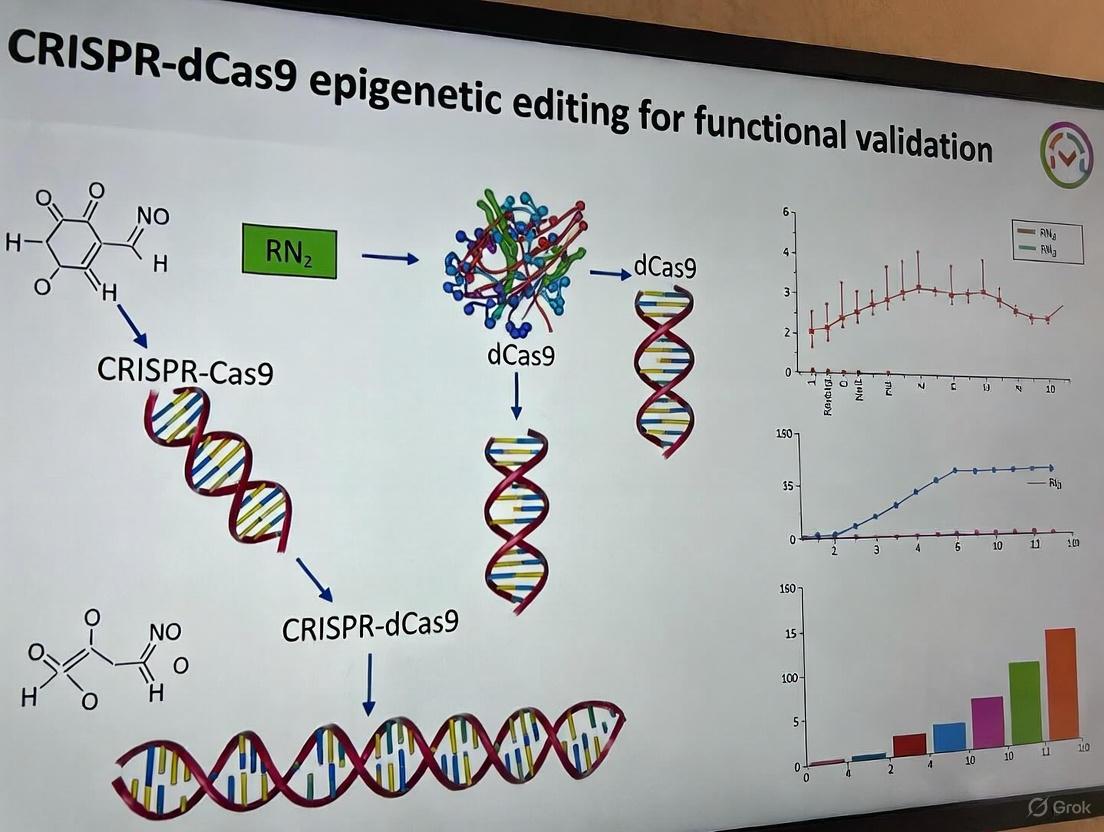
Experimental Workflow and Pathway Visualization
Experimental Workflow Diagram
The following diagram illustrates the complete experimental workflow from system design to phenotypic validation.
Signaling Pathway Diagram
This diagram outlines the core signaling pathway reactivated by miR-200c promoter demethylation, demonstrating the link from epigenetic editing to phenotypic outcome.
The Scientist's Toolkit: Essential Research Reagents
A successful CRISPR-dCas9 epigenetic editing experiment requires the following key reagents and controls, the importance of which is emphasized in the cited literature [4] [12] [13].
Table 2: Essential Research Reagents and Controls for dCas9 Epigenetic Editing
| Reagent / Control | Function & Purpose | Example / Specification |
|---|---|---|
| dCas9-Effector Fusion | Catalytic core for targeted epigenetic modification. | dCas9-TET1: For targeted DNA demethylation. |
| Target-Specific sgRNAs | Guides the dCas9-effector to the target genomic locus. | 2+ sgRNAs flanking the CpG-rich promoter region. Validated with tool like CHOPCHOP [4]. |
| Delivery Vector | Introduces genetic constructs into cells. | Lentivirus, plasmid, or RNP complexes for primary cells [13]. |
| Methylation Analysis Kit | Quantifies DNA methylation changes at the target locus. | Bisulfite Conversion Kit & Primers for Pyrosequencing. |
| Expression Assay | Measures mRNA/miRNA expression changes post-editing. | RT-qPCR Assay for target gene (e.g., miR-200c) and downstream targets (e.g., ZEB1). |
| Phenotypic Assay Kits | Evaluates functional biological consequences. | MTT Kit (viability) & Annexin V/FITC Kit (apoptosis) [4]. |
| Critical Negative Controls | Distracts specific from non-specific effects. | dCas9-only (no sgRNA): Controls for dCas9 toxicity. Catalytic Mutant (dCas9mut-TET1): Controls for non-catalytic effects [4]. Non-targeting sgRNA: Controls for off-target sgRNA effects. |
| Positive Control | Validates entire experimental system is functional. | sgRNA and assay for a previously validated target locus. |
| S-Isopropylisothiourea hydrobromide | S-Isopropylisothiourea hydrobromide, CAS:4269-97-0, MF:C4H11BrN2S, MW:199.12 g/mol | Chemical Reagent |
| 10-Debc hydrochloride | 10-Debc hydrochloride, CAS:925681-41-0, MF:C20H26Cl2N2O, MW:381.3 g/mol | Chemical Reagent |
Technical Considerations and Best Practices
- gRNA Design and Validation: The efficiency of specific gRNAs can vary significantly between cell lines, as observed with gRNA2 having a minimal effect in MDA-MB-231 cells but a significant one in MCF-7 cells [4]. It is critical to design and empirically test multiple gRNAs.
- Cell Context Dependence: Phenotypic outcomes, such as E-cadherin upregulation, can be minimal in some cellular contexts (e.g., MCF-7) but pronounced in others (e.g., MDA-MB-231), highlighting the importance of the initial epigenetic and transcriptional state of the cell [4].
- Specificity Controls: The use of multiple control conditions, including a catalytically dead mutant of TET1 (dCas9Mut-TET1), is essential to confirm that observed effects are due to targeted demethylation and not unrelated to dCas9 binding or cellular stress responses [4].
- Beyond DNA Methylation: This protocol focuses on DNA methylation, but the dCas9 system can be adapted to target other epigenetic marks, such as histone modifications, by fusing dCas9 to histone acetyltransferases (HATs) or histone methyltransferases, forming a broader "CRISPR-Epigenetics Regulatory Circuit" for functional discovery [14].
The advent of catalytically dead Cas9 (dCas9) has fundamentally expanded the CRISPR toolkit beyond permanent genome editing, enabling precise transcriptional and epigenetic control without altering the underlying DNA sequence [15] [16]. By mutating the RuvC and HNH nuclease domains of Cas9, researchers have created dCas9, which retains its ability to bind DNA target sites specified by a guide RNA (gRNA) but cannot create double-strand breaks [17] [18]. This core protein serves as a programmable platform for recruiting effector domains to specific genomic loci, facilitating reversible gene modulation [15]. This approach presents a paradigm shift from traditional knockout and knockin techniques, which rely on error-prone non-homologous end joining (NHEJ) or homology-directed repair (HDR) to make irreversible changes to the DNA sequence [19] [20]. For functional validation research and drug development, the ability to reversibly tune gene expression and epigenetic states offers a more nuanced and physiologically relevant method for probing gene function and identifying therapeutic targets.
Core Advantages of dCas9 Systems
The dCas9 platform offers several distinct advantages over conventional gene editing for functional genomics and pre-clinical research, primarily centered on its reversibility and temporal control.
Reversible Modulation: Unlike traditional knockout/knockin strategies that introduce permanent, heritable changes to the DNA sequence, dCas9-mediated modulation operates at the transcriptional or epigenetic level [15]. The changes induced by systems like CRISPR interference (CRISPRi) or CRISPR activation (CRISPRa) do not alter the genetic code and are often reversible upon removal of the dCas9-effector complex [12] [16]. This is crucial for studying essential genes, modeling transient cellular states, and developing potential therapeutic strategies that require temporary gene expression alteration.
Sequential and Temporal Control: The activity of dCas9 systems can be finely controlled over time using inducible expression systems, such as doxycycline-inducible promoters, or chemically induced proximity systems [15] [18]. This allows researchers to initiate gene modulation at a specific point in a differentiation protocol or disease model, enabling the study of gene function at precise developmental or disease stages. This temporal resolution is difficult to achieve with traditional methods where the genetic change is present from the outset [18].
Precise Epigenetic Engineering: dCas9 can be fused to epigenetic writer and eraser domains, such as DNA methyltransferases (e.g., DNMT3A) or demethylases (e.g., TET1), enabling targeted editing of the epigenome [16] [21]. This allows for the functional validation of specific epigenetic marks at single-gene resolution without the confounding effects of global epigenetic drugs.
Reduced Off-Target and Genotoxic Risks: Since dCas9 systems lack nuclease activity, they do not introduce double-strand breaks (DSBs), thereby eliminating the genotoxic stress associated with the NHEJ and HDR pathways and reducing the risk of chromosomal translocations and large deletions [16] [18].
Multiplexability: The simplicity of designing gRNAs allows for the easy targeting of multiple genomic loci simultaneously. dCas9 can be guided by several sgRNAs to bind to different target sites, enabling the coordinated regulation of entire gene networks or pathways in a single experiment [15] [17].
Table 1: Quantitative Comparison of Gene Modulation Techniques
| Feature | Traditional CRISPR Knockout/Knockin | dCas9-Mediated Modulation |
|---|---|---|
| DNA Cleavage | Yes (Creates DSBs) | No (Catalytically inactive) [16] |
| Change to DNA Sequence | Permanent (Indels or insertions) | None (Epigenetic/transcriptional) [15] |
| Reversibility | Irreversible | Reversible [12] [15] |
| Key Repair Pathway | NHEJ (Knockout) / HDR (Knockin) [20] | N/A |
| Typical Timeline for Mouse Model Generation | 1–2 years [19] | 1–2 months [19] |
| Primary Application | Gene disruption, gene insertion | Gene activation (CRISPRa), repression (CRISPRi), epigenome editing [12] [16] |
Key Experimental Protocols
The following protocols outline core methodologies for implementing reversible gene modulation using the dCas9 system.
Protocol: CRISPR/dCas9-Tet1-Mediated DNA Demethylation
This protocol enables targeted demethylation of specific CpG islands to activate gene expression, using a dCas9-Tet1 fusion system [21].
Step 1: sgRNA Design and Cloning
- Identify Target Sequence: Design sgRNAs to target promoter regions or other regulatory elements rich in CpG sites. The target should be adjacent to a PAM sequence (NGG for SpCas9) [21].
- Clone into sgRNA Scaffold: Anneal and phosphorylate oligonucleotides encoding the sgRNA target sequence. Ligate them into a BsmBI-linearized sgRNA expression vector (e.g., Addgene #84477) [21].
- Validate Clones: Transform the ligation product into Stbl3 competent cells. Screen positive colonies by PCR and Sanger sequencing to confirm correct insertion.
Step 2: Delivery of the dCas9-Tet1 System
- Cell Culture: Maintain relevant cells (e.g., HEK293T, MEFs, or hESCs) in their appropriate culture medium [21].
- Co-transfection: Co-transfect the Fuw-dCas9-Tet1-P2A-BFP plasmid (Addgene #108245) and the constructed sgRNA plasmid into target cells using a suitable transfection reagent (e.g., X-tremeGENE). For difficult-to-transfect cells, use lentiviral delivery.
- Selection and Sorting: After 48-72 hours, harvest cells and use FACS to isolate BFP-positive cells, indicating successful transfection and expression of the dCas9-Tet1 construct [21].
Step 3: Validation of Demethylation Efficiency
- DNA Extraction: Harvest transfected cells and extract genomic DNA using a commercial kit (e.g., DNeasy Blood & Tissue Kit) [21].
- Bisulfite Conversion: Treat 500 ng of genomic DNA with the EZ DNA Methylation-Gold kit to convert unmethylated cytosines to uracils.
- Pyrosequencing: Amplify the target region by PCR using bisulfite-converted DNA as a template. Analyze the PCR product by pyrosequencing to quantify the percentage of methylation at individual CpG sites [21].
Protocol: dCas9-Based Transcriptional Repression (CRISPRi)
This protocol describes gene knockdown using dCas9 fused to a transcriptional repressor domain, such as KRAB [16] [18].
Step 1: System Assembly
- Select Repressor Domain: Clone the dCas9-KRAB fusion protein into an expression vector. The KRAB domain recruits repressive complexes that promote histone methylation (H3K9me3), leading to stable gene silencing [16].
- Design sgRNAs: Design sgRNAs to bind the template strand within -50 to +300 bp relative to the transcription start site (TSS) of the target gene to effectively block RNA polymerase [18].
Step 2: Delivery and Induction
- Transduction: Deliver the dCas9-KRAB and sgRNA constructs to cells via lentiviral transduction.
- Doxycycline Induction: If using an inducible system, treat cells with doxycycline (e.g., 1 µg/mL) to initiate expression of the dCas9-effector complex, allowing for temporal control over repression [18].
Step 3: Validation of Knockdown
- RT-qPCR: Isolve total RNA 72-96 hours post-induction and perform reverse transcription followed by quantitative PCR to measure mRNA expression levels of the target gene.
- Western Blot: Analyze protein levels 5-7 days post-induction to confirm functional knockdown.
Visualization of dCas9 Systems
The following diagrams illustrate the core mechanisms and experimental workflow for dCas9-mediated gene modulation.
dCas9 Gene Modulation Mechanisms
Experimental Workflow for DNA Methylation Editing
The Scientist's Toolkit: Research Reagent Solutions
Table 2: Essential Reagents for dCas9-Mediated Gene Modulation
| Reagent / Tool | Function / Description | Example Items & Sources |
|---|---|---|
| dCas9 Effector Plasmids | Core protein that binds DNA without cleavage; fused to activator, repressor, or epigenetic effector domains. | dCas9-VP64 (Addgene), dCas9-KRAB (Addgene), Fuw-dCas9-Tet1 (Addgene #108245) [16] [21] |
| sgRNA Expression Vectors | Delivers the targeting component; can be cloned into single or multiplexed backbones. | pgRNA-modified (Addgene #84477), pLenti-sgRNA(MS2)_Zeo [22] [21] |
| Delivery Systems | Introduces genetic constructs into target cells; choice depends on cell type and efficiency required. | Lentiviral particles, X-tremeGENE transfection reagent, PiggyBac transposon system [21] |
| Validation Kits | Essential for confirming the success and specificity of the epigenetic or transcriptional modulation. | EZ DNA Methylation-Gold Kit (Zymo Research), PyroMark PCR Master Mix (Qiagen), RT-qPCR reagents [21] |
| Cell Lines | Models for functional validation; can include primary cells, stem cells, or established cell lines. | HEK293T (ATCC CRL-11268), PK15 pig kidney cells, human embryonic stem cells (hESCs) [22] [21] |
| Hki-357 | (E)-N-(4-((3-Chloro-4-((3-fluorobenzyl)oxy)phenyl)amino)-3-cyano-7-ethoxyquinolin-6-yl)-4-(dimethylamino)but-2-enamide | High-purity (E)-N-(4-((3-Chloro-4-((3-fluorobenzyl)oxy)phenyl)amino)-3-cyano-7-ethoxyquinolin-6-yl)-4-(dimethylamino)but-2-enamide for research. This covalent EGFR inhibitor is For Research Use Only. Not for human or veterinary diagnosis or therapeutic use. |
| Lapatinib | Lapatinib, CAS:388082-78-8, MF:C29H26ClFN4O4S, MW:581.1 g/mol | Chemical Reagent |
A Toolkit for Discovery: Methodologies and Real-World Applications in Functional Genomics
Catalytically deactivated Cas9 (dCas9) serves as a programmable DNA-binding scaffold that can be fused with various epigenetic effector domains to manipulate the epigenome without altering the underlying DNA sequence. This technology enables precise functional validation of epigenetic marks in gene regulation, offering significant advantages over global pharmacological epigenetic modifiers that cause genome-wide changes and confounding effects [23] [24]. The three primary dCas9 fusion constructs—dCas9-Tet1CD for DNA demethylation, dCas9-DNMT3A for DNA methylation, and dCas9-p300 for histone acetylation—provide researchers with powerful tools to establish causal relationships between specific epigenetic modifications and gene expression outcomes in functional genomics research and drug discovery.
Molecular Biology of dCas9 Fusion Constructs
dCas9-Tet1CD Demethylation Construct
The dCas9-Tet1CD fusion protein combines the DNA-targeting capability of dCas9 with the catalytic domain of Ten-Eleven Translocation 1 (TET1), an α-ketoglutarate and Fe²âº-dependent dioxygenase. TET1 catalyzes the conversion of 5-methylcytosine (5mC) to 5-hydroxymethylcytosine (5hmC), which can be further oxidized to 5-formylcytosine (5fC) and 5-carboxylcytosine (5caC), initiating the DNA demethylation pathway through base excision repair [23]. This construct enables targeted DNA demethylation when guided to specific genomic loci by sgRNAs. In Arabidopsis, overexpression of TET1cd reduced genome-wide methylation, while targeted demethylation of the hypermethylated NMR19-4 region in the PPH gene promoter increased PPH expression and accelerated leaf senescence [23]. The demethylated state and associated phenotypic effects demonstrated Mendelian inheritance in progeny, indicating stable transgenerational transmission of the edited epiallele [23].
dCas9-p300 Acetylation Construct
The dCas9-p300 fusion links dCas9 to the core catalytic domain of human acetyltransferase p300 (amino acids 1048-1664 or 1284-1673), which acetylates histone H3 at lysine 27 (H3K27ac) [25] [26]. This modification is strongly associated with active gene regulatory elements and enhancers. The dCas9-p300 system functions through a mechanism distinct from conventional dCas9-activators like dCas9-VP64. While VP64 and similar activation domains recruit transcriptional machinery, p300 directly modifies chromatin structure by adding acetyl groups to histones, potentially creating a more permissive chromatin environment for transcription [26]. This system effectively activates genes from promoters, proximal enhancers, and distal enhancers, with studies demonstrating significant transactivation of endogenous genes including IL1RN, MYOD, and OCT4 (POU5F1) [25]. Notably, dCas9-p300 Core induced significantly higher transcription levels than dCas9-VP64 when targeted to the IL1RN and MYOD promoters and successfully activated gene expression from enhancer regions where dCas9-VP64 was ineffective [25].
dCas9-DNMT3A Methylation Construct
The dCas9-DNMT3A fusion couples dCas9 to the de novo DNA methyltransferase DNMT3A, enabling targeted DNA methylation at CpG islands. This construct catalyzes the transfer of methyl groups from S-adenosyl-l-methionine to the C-5 position of cytosine to form 5-methylcytosine (5mC) [27]. DNMT3L is often co-expressed as it enhances de novo methylation activity by forming heterotetramers with DNMT3A [28]. The CRISPR/dCas9-Dnmt3a system has been applied for targeted methylation of the amyloid precursor protein (APP) gene promoter in Alzheimer's disease research, significantly reducing APP mRNA expression, decreasing amyloid-beta peptide levels, and attenuating cognitive impairments in mouse models [27]. Similar approaches have successfully silenced oncogenes like CDKN2A and BACH2 in cancer contexts [27] [28]. Optimization strategies include using the SunTag system to recruit multiple DNMT3A molecules to specific loci, achieving hypermethylation across regions up to 4.5 kb [28].
Table 1: Core Characteristics of dCas9 Fusion Constructs
| Construct | Catalytic Domain | Epigenetic Modification | Primary Effect on Transcription | Key Applications |
|---|---|---|---|---|
| dCas9-Tet1CD | TET1 catalytic domain | 5mC → 5hmC → 5fC → 5caC → C (DNA demethylation) | Activation | Functional validation of hypomethylated regions, gene reactivation [23] |
| dCas9-p300 | p300 HAT core domain | Histone H3K27 acetylation | Activation | Gene activation from promoters and enhancers, chromatin opening [25] [26] |
| dCas9-DNMT3A | DNMT3A methyltransferase | Cytosine → 5-methylcytosine (DNA methylation) | Repression | Gene silencing, studying hypermethylation effects, therapeutic repression [27] [28] |
| BAY-678 | BAY-678, CAS:675103-36-3, MF:C20H15F3N4O2, MW:400.4 g/mol | Chemical Reagent | Bench Chemicals | |
| NVP-ADW742 | NVP-ADW742, CAS:475489-15-7, MF:C28H31N5O, MW:453.6 g/mol | Chemical Reagent | Bench Chemicals |
Quantitative Performance Comparison
Table 2: Quantitative Performance Metrics of dCas9 Fusion Constructs
| Construct | Target Region | Editing Efficiency | Expression Change | Persistence/Inheritance | Key Experimental System |
|---|---|---|---|---|---|
| dCas9-Tet1CD | NMR19-4 (PPH promoter) | Significant demethylation of hypermethylated region | Increased PPH expression, accelerated leaf senescence | Mendelian inheritance over generations (F1, F2) [23] | Arabidopsis natural accessions |
| dCas9-p300 | IL1RN promoter | N/A | Significant activation vs. dCas9-VP64 (P=0.01924) [25] | Transient (duration dependent on delivery method) | HEK293T cells |
| dCas9-p300 | MYOD distal regulatory region | N/A | Significant activation (P=0.0009); dCas9-VP64 ineffective [25] | Transient | HEK293T cells |
| dCas9-p300 | OCT4 (POU5F1) | 6 of 9 gRNAs increased transcription ≥2-fold [26] | 2-fold or more activation | Transient | HEK293T cells |
| dCas9-DNMT3A | APP promoter | Altered DNA methylation pattern | Significantly reduced APP mRNA | Durable (weeks to months) [27] | APP-KI mouse primary neurons |
| dCas9-DNMT3A | BACH2 locus | Up to 60% CpG methylation [28] | Decreased gene expression | Durable (weeks to months) | HEK293T cells |
| dCas9-DNMT3A | CDKN2A locus | Up to 50% DNA methylation [28] | Decreased gene expression | Durable (weeks to months) | HEK293T cells |
Experimental Protocols
Targeted DNA Demethylation Using dCas9-Tet1CD
Vector Design and Assembly
- Clone the TET1 catalytic domain (Tet1CD) into your dCas9 expression backbone using appropriate restriction sites or Gibson assembly, creating a C-terminal fusion to dCas9
- Design sgRNAs targeting 20-base pair sequences adjacent to the methylation site(s) of interest
- For plant systems, use binary vectors for Agrobacterium-mediated transformation; for mammalian cells, use lentiviral or other appropriate delivery vectors [23]
Delivery and Analysis
- Transform Arabidopsis hypermethylated ecotypes using floral dip method with Agrobacterium carrying the dCas9-Tet1CD construct and sgRNA expression cassette
- Select transgenic plants using appropriate antibiotics or other selection markers
- Analyze methylation status using bisulfite sequencing of the targeted NMR19-4 region
- Evaluate phenotypic consequences (e.g., PPH expression via RT-qPCR, leaf senescence assessment)
- Conduct inheritance studies through genetic crosses and analyze F1 and F2 progeny for stable transmission of demethylated epialleles [23]
Gene Activation Using dCas9-p300
Construct Delivery and Validation
- Co-transfect HEK293T cells with dCas9-p300 Core fusion plasmid and sgRNA expression vectors (typically 4 sgRNAs per target promoter) using your preferred transfection method
- Include controls: dCas9-VP64 for comparison, and catalytically inactive dCas9-p300 Core (D1399Y)
- Harvest cells 48-72 hours post-transfection for analysis
- Validate protein expression via western blot using anti-Cas9 or anti-p300 antibodies [25] [26]
Assessment of Activation
- Quantify gene expression of target genes (e.g., IL1RN, MYOD, OCT4) using RT-qPCR with gene-specific primers
- Assess histone acetylation at target sites using chromatin immunoprecipitation (ChIP) with anti-H3K27ac antibody
- For enhancer targeting, analyze expression of genes regulated by the targeted enhancer region
- Compare activation efficiency between dCas9-p300 and conventional activators like dCas9-VP64 [25]
Targeted DNA Methylation Using dCas9-DNMT3A
In Vitro and Cell Culture Applications
- Co-transfect HEK293T cells with dCas9-DNMT3A and sgRNA plasmids targeting the APP promoter region
- For enhanced methylation efficiency, include DNMT3L in the system
- For primary neurons, transfer lentiviral particles containing dCas9-Dnmt3a and APP-targeting sgRNA (e.g., APP -189 sgRNA)
- Culture transfected/transduced neurons for 2 weeks before analysis [27]
In Vivo Application in Mouse Models
- Perform stereotaxic injection of dCas9-Dnmt3a and sgRNA lentivirus into the dentate gyrus region of APP-KI mice (coordinates: AP -2 mm, ML ±1.1 mm, DV -2 mm)
- Inject 10 µl of lentivirus into each hemisphere
- Allow 4 weeks for expression and methylation establishment before behavioral and biochemical analysis [27]
Molecular and Phenotypic Analysis
- Analyze DNA methylation patterns at the APP promoter using bisulfite sequencing
- Quantify APP mRNA levels using RT-qPCR
- Measure amyloid-beta peptide levels and Aβ42/40 ratio via ELISA
- Assess neuronal cell death using TUNEL or other apoptosis assays
- Evaluate cognitive function using behavioral tests (Y-maze, fear conditioning, water maze) [27]
The Scientist's Toolkit: Essential Research Reagents
Table 3: Essential Research Reagents for dCas9 Epigenome Editing
| Reagent/Solution | Function/Purpose | Example Applications | Key Considerations |
|---|---|---|---|
| dCas9-Tet1CD plasmid | Targeted DNA demethylation | Functional validation of hypomethylated regions, gene reactivation studies [23] | Catalytic domain requires α-ketoglutarate and Fe²⺠cofactors |
| dCas9-p300 Core plasmid | Targeted histone acetylation, gene activation | Activation from promoters and enhancers, chromatin dynamics studies [25] [26] | More effective than dCas9-VP64 for enhancer activation |
| dCas9-DNMT3A plasmid | Targeted DNA methylation, gene silencing | Therapeutic repression, methylation functional studies [27] [28] | Co-expression with DNMT3L enhances efficiency |
| Guide RNA vectors | Target specificity determination | All targeted epigenome editing applications | Multiple sgRNAs often needed for robust effects |
| Bisulfite conversion kit | DNA methylation analysis | Validation of methylation changes at target loci [23] [27] | Essential for assessing DNA methylation editing efficiency |
| H3K27ac antibody | Histone acetylation detection | ChIP validation of dCas9-p300 activity [25] | Confirms targeted chromatin modification |
| Lentiviral packaging system | Efficient delivery to difficult cells | Primary neurons, in vivo applications [27] | Enables transduction of non-dividing cells |
| Off-target prediction tools | Specificity assessment | Cas-OFFinder for identifying potential off-target sites [27] | Critical for experimental validation |
| Oxantel | Oxantel, CAS:36531-26-7, MF:C13H16N2O, MW:216.28 g/mol | Chemical Reagent | Bench Chemicals |
| PF-04217903 | PF-04217903, CAS:1159490-85-3, MF:C19H16N8O, MW:372.4 g/mol | Chemical Reagent | Bench Chemicals |
Advanced Delivery Methods: RENDER Platform
Recent advances in delivery methods have addressed challenges associated with the large size of CRISPR-based epigenome editors. The RENDER (Robust ENveloped Delivery of Epigenome-editor Ribonucleoproteins) platform utilizes engineered virus-like particles (eVLPs) to transiently deliver CRISPR epigenome editors as ribonucleoprotein complexes into human cells [29]. This system offers several advantages:
- Transient delivery: Minimizes off-target editing risks associated with prolonged editor expression
- Size flexibility: eVLPs can accommodate large epigenome editors that exceed AAV packaging capacity
- Versatility: Successfully delivered CRISPRi, DNMT3A-3L-dCas9, CRISPRoff, and TET1-dCas9 editors
- Broad applicability: Effective across various human cell types, including primary T cells and stem cell-derived neurons
- Durability: DNMT3A-3L-dCas9 and CRISPRoff eVLPs maintained robust epigenetic silencing for at least 14 days post-treatment [29]
The RENDER platform enables reversible epigenome editing, with TET1-dCas9 eVLPs successfully reversing CRISPRoff-mediated repression of CLTA-GFP in ~6% of treated cells, with reactivation remaining stable for 15 days [29].
CRISPR activation (CRISPRa) represents a powerful approach in functional genomics, enabling researchers to precisely upregulate endogenous gene expression without altering the underlying DNA sequence. These systems utilize a catalytically inactive Cas9 (dCas9) fused or recruited to transcriptional activators, which are guided to specific genomic loci by single-guide RNAs (sgRNAs). This technology has revolutionized gain-of-function studies by allowing targeted transcriptional modulation within native chromosomal contexts, preserving natural splice variants, regulatory feedback, and epigenetic states that are often disrupted by traditional cDNA overexpression. For functional validation research, CRISPRa provides a more physiologically relevant method for establishing gene function and modeling gene dosage effects, making it particularly valuable for drug target identification and validation [30] [31].
The evolution of CRISPRa systems has progressed from first-generation constructs to increasingly sophisticated architectures that enhance activation potency. The fundamental dCas9-VP64 system serves as the foundation, while subsequent developments like the MS2-MCP-scaffolded VP64 and Synergistic Activation Mediator (SAM) systems employ multi-component recruitment strategies to achieve stronger transcriptional activation. Understanding the comparative advantages, limitations, and optimal applications of these systems is essential for their effective implementation in epigenetic editing and functional validation research [32] [31].
Comparative Analysis of CRISPRa Systems
System Architectures and Activation Mechanisms
dCas9-VP64: This first-generation system employs dCas9 directly fused to a VP64 activator domain (a tandem tetramer of the VP16 peptide). It represents the simplest CRISPRa architecture, where the dCas9-VP64 fusion protein is recruited to target sites via sgRNAs, bringing the VP64 domain to promote transcription initiation. While simple in design, its activation potency is limited due to the delivery of only a single activator complex per dCas9 molecule [32] [31].
MS2-MCP-scaffolded VP64: This enhanced system incorporates an engineered sgRNA containing MS2 RNA aptamers in its stem loop. These aptamers recruit MCP (MS2 coat protein) fused to additional VP64 domains. This creates a dual-recruitment mechanism where activation comes from both the dCas9-VP64 fusion and the MS2-MCP-VP64 recruits, significantly enhancing the local concentration of activators at the target locus without requiring larger dCas9 fusion proteins [32].
SAM (Synergistic Activation Mediator): The SAM system represents a third-generation CRISPRa platform that further amplifies the recruitment strategy. It utilizes three components: (1) dCas9-VP64; (2) an engineered sgRNA containing two MS2 aptamers; and (3) MCP fused to a heterologous activation domain composed of p65 and HSF1 (often abbreviated as MPH). This creates a three-pronged activation complex that recruits VP64, p65, and HSF1 activation domains simultaneously, generating a synergistic effect that drives robust transcriptional activation [32] [33].
Table 1: Comparative Architecture of CRISPRa Systems
| System | Core Components | Activation Domains | Recruitment Mechanism |
|---|---|---|---|
| dCas9-VP64 | dCas9-VP64 fusion protein | VP64 | Direct fusion of VP64 to dCas9 |
| MS2-MCP-scaffolded VP64 | dCas9-VP64 + sgRNA-MS2 + MCP-VP64 | VP64 (multiple copies) | dCas9 fusion + MS2 aptamer-mediated recruitment |
| SAM | dCas9-VP64 + sgRNA-MS2 + MCP-p65-HSF1 | VP64 + p65 + HSF1 | dCas9 fusion + MS2 aptamer-mediated recruitment of heterologous activators |
Performance and Efficiency Comparison
Quantitative assessments reveal significant differences in activation potency across these systems. Direct comparisons show that MS2-MCP-scaffolded VP64 consistently outperforms simple dCas9-VP64 fusions across multiple endogenous gene targets. The SAM system typically demonstrates the highest activation efficacy, achieving substantially greater fold-increases in target gene expression compared to both dCas9-VP64 and MS2-MCP-scaffolded VP64 systems [32].
The superior performance of SAM stems from its ability to recruit diverse activation domains that work synergistically. The combination of VP64, p65, and HSF1 engages multiple facets of the transcriptional machinery, leading to more robust and sustained gene activation. However, this enhanced potency comes with practical limitations, including increased cytotoxicity associated with expressing the potent p65-HSF1 activator fusion, which can lead to cell death and confounding selection pressures in experimental systems [33].
Table 2: Performance Characteristics of CRISPRa Systems
| System | Activation Efficiency | Cytotoxicity Concerns | Ideal Applications |
|---|---|---|---|
| dCas9-VP64 | Moderate (2-10 fold) | Low | Basic gene activation, sensitive cell models |
| MS2-MCP-scaffolded VP64 | High (10-50 fold) | Low to moderate | Balanced performance needs |
| SAM | Very high (50-1000+ fold) | Significant | Maximal activation, robust cell lines |
Recent advances in CRISPRa have highlighted the importance of condensate dynamics in effective gene activation. Systems that form liquid-like transcriptional condensates with high dynamicity and liquidity demonstrate superior activation compared to those forming solid-like condensates that sequester co-activators. This understanding provides a mechanistic framework for why systems with optimized activator multiplicity, such as SunTag3xVPR, can outperform systems with excessive scaffolding [34].
Application Notes for Functional Validation Research
System Selection Guidelines
Choosing the appropriate CRISPRa system depends on multiple factors, including the target gene's baseline expression, chromatin environment, and the desired level of activation. For genes with low basal expression or those embedded in repressive chromatin, the SAM system often provides sufficient activation potency. However, researchers must carefully consider the cellular context, as the cytotoxicity associated with strong activators like p65-HSF1 can limit application in sensitive systems such as primary cells [33].
For functional validation in drug discovery, establishing a dose-response relationship between gene activation and phenotypic outcome is crucial. The MS2-MCP-scaffolded VP64 system often provides an optimal balance between potency and practicality for such applications. When working with silent loci or highly methylated gene promoters, combining CRISPRa with epigenetic modifiers such as TET1 can enhance activation by remodeling the chromatin landscape to a more permissive state [35].
Addressing Technical Challenges
Cytotoxicity Management: The pronounced cytotoxicity of potent activator domains like those in the SAM system presents a significant challenge. Strategies to mitigate this include using inducible expression systems for activators, employing lower-efficacy promoters to reduce expression levels, and implementing careful titration experiments to identify the minimal effective dose. When using lentiviral delivery, toxicity can manifest as low viral titers during production and cell death post-transduction, potentially confounding experimental results through selective pressure [33].
Multiplexed Activation: A key advantage of CRISPRa systems is their ability to activate multiple genes simultaneously through delivery of multiple sgRNAs. For the MS2-MCP-scaffolded VP64 and SAM systems, multiplexing gRNA expression significantly enhances endogenous gene activation, with some studies reporting levels comparable to SAM with a single gRNA. This capability is particularly valuable for validating multi-gene pathways or modeling polygenic diseases in functional validation research [32].
Experimental Protocols
Protocol 1: Lentiviral Delivery of SAM System for Endogenous Gene Activation
Principle: This protocol describes the implementation of the three-component SAM system for robust gene activation in mammalian cells, suitable for functional validation studies.
Reagents and Materials:
- Lenti-dCas9-VP64 (Addgene #61425)
- Lenti-MPH (MS2-p65-HSF1) (Addgene #89308)
- Lenti-sgRNA(MS2) backbone (Addgene #89308)
- HEK293T packaging cells
- Polyethylenimine (PEI) transfection reagent
- Target cells of interest
- Puromycin and hygromycin selection antibiotics
Procedure:
- sgRNA Design and Cloning: Design sgRNAs targeting regions -200 to -50 bp upstream of the transcription start site (TSS) of your target gene. Clone annealed sgRNA oligos into the BsmBI-digested lenti-sgRNA(MS2) backbone via Golden Gate assembly.
- Lentivirus Production:
- Day 1: Seed HEK293T cells in 10 cm plates to reach 70-80% confluency the next day.
- Day 2: Transfect with 3.3 µg transfer vector (sgRNA, dCas9-VP64, or MPH), 2.5 µg psPAX2, and 1.2 µg pMD2.G using PEI transfection reagent.
- Day 3: Replace medium with fresh DMEM + 10% FBS.
- Day 4: Collect viral supernatant, filter through 0.45 µm membrane, and concentrate if necessary.
- Cell Transduction:
- Day 1: Seed target cells at 30-50% confluency.
- Day 2: Transduce with dCas9-VP64 lentivirus in the presence of 8 µg/mL polybrane.
- Day 4: Begin selection with 2 µg/mL puromycin for 5-7 days.
- Day 7: Transduce selected cells with MPH lentivirus.
- Day 9: Begin selection with 100 µg/mL hygromycin for 5-7 days.
- Day 12: Transduce with sgRNA lentivirus.
- Day 14: Begin selection with appropriate antibiotic for the sgRNA vector.
- Validation and Analysis:
- Day 17: Harvest cells for RNA extraction and qRT-PCR analysis of target gene expression.
- Day 18: Perform functional assays based on the expected phenotypic outcomes.
Troubleshooting Notes:
- Low activation efficiency: Optimize sgRNA positioning relative to TSS; test multiple sgRNAs per gene.
- High cytotoxicity: Titrate viral MOI to minimize activator expression while maintaining efficacy; consider inducible systems.
- Variable results: Ensure stable polyclonal populations by maintaining selection antibiotics throughout experiments.
Protocol 2: MS2-MCP-scaffolded VP64 for Multiplexed Gene Activation
Principle: This protocol enables simultaneous activation of multiple genes using the MS2-MCP-scaffolded VP64 system, ideal for pathway validation studies.
Reagents and Materials:
- dCas9-VP64 expression plasmid
- MCP-VP64 expression plasmid
- sgRNA expression vector with MS2 aptamers
- Lipofectamine 3000 or similar transfection reagent
- Target cells
Procedure:
- Multiplexed sgRNA Design: Design 3-5 sgRNAs per target gene, focusing on regions within 500 bp upstream of TSS. For multiplexing, clone up to 6 sgRNAs as tandem tRNA-sgRNA arrays using BsmBI restriction sites.
- Transient Transfection:
- Day 1: Seed cells in 12-well plates to reach 70-80% confluency at transfection.
- Day 2: Prepare transfection complex with 500 ng dCas9-VP64, 500 ng MCP-VP64, and 750 ng sgRNA plasmid(s) per well using Lipofectamine 3000 according to manufacturer's protocol.
- Day 3: Replace with fresh medium.
- Day 4: Harvest cells for analysis or continue with functional assays.
- Validation:
- Quantify mRNA expression of target genes via qRT-PCR 48-72 hours post-transfection.
- Assess protein expression changes via Western blot or immunofluorescence 72-96 hours post-transfection.
- Perform functional assays relevant to the activated pathway (e.g., proliferation, differentiation, reporter assays).
Optimization Tips:
- For difficult-to-activate genes, include multiple sgRNAs targeting both promoter and potential enhancer regions.
- When activating multiple genes in a pathway, balance the sgRNA ratios to achieve physiological expression levels.
- Include non-targeting sgRNA controls and target known easily-activated genes (e.g., housekeeping genes) as positive controls.
Figure 1: CRISPRa Experimental Workflow. This flowchart outlines the key decision points and steps in implementing CRISPRa systems for functional validation studies.
The Scientist's Toolkit: Essential Research Reagents
Table 3: Essential Reagents for CRISPRa Implementation
| Reagent Category | Specific Examples | Function | Notes |
|---|---|---|---|
| dCas9 Activators | dCas9-VP64, dCas9-VPR, dCas9-p300 | Core DNA-binding moiety fused to activation domains | VP64 provides basic activation; VPR offers enhanced potency |
| sgRNA Scaffolds | MS2, PP7, com RNA aptamers | Recruit additional activator proteins to target locus | MS2 most commonly used for MCP recruitment |
| Recruited Activators | MCP-VP64, MCP-p65-HSF1 (MPH), PCP-p65-HSF1 (PPH) | Secondary activation components | p65-HSF1 fusions highly potent but cytotoxic |
| Delivery Vectors | Lentiviral, piggyBac, episomal plasmids | Introduce CRISPRa components into cells | Lentiviral enables stable integration; episomal less toxic |
| Selection Markers | Puromycin, hygromycin, blasticidin, GFP | Enrich for successfully transduced cells | Multiple markers needed for multi-component systems |
| Validation Tools | qPCR primers, antibody panels, functional assay kits | Confirm target gene activation and phenotypic outcomes | Essential for measuring system efficacy |
| 1,3-PBIT dihydrobromide | 1,3-PBIT dihydrobromide, MF:C12H20Br2N4S2, MW:444.3 g/mol | Chemical Reagent | Bench Chemicals |
| PTP Inhibitor IV | PTP Inhibitor IV, CAS:329317-98-8, MF:C26H26F6N2O4S2, MW:608.6 g/mol | Chemical Reagent | Bench Chemicals |
The strategic selection and implementation of CRISPRa systems is paramount for successful functional validation research. While the SAM system offers maximum activation potency, its associated cytotoxicity may limit utility in sensitive experimental systems. The MS2-MCP-scaffolded VP64 system represents a robust middle ground, providing substantial activation with more manageable toxicity profiles. The foundational dCas9-VP64 system remains valuable for applications requiring moderate gene upregulation or when working with cytotoxicity-sensitive models.
As CRISPRa technology continues to evolve, emerging considerations such as transcriptional condensate dynamics and activator-induced cytotoxicity will inform the development of next-generation systems. By matching system capabilities to experimental requirements and carefully managing technical challenges, researchers can effectively leverage these powerful tools for comprehensive functional validation in epigenetic editing and drug development workflows.
CRISPR-dCas9 epigenetic editing has emerged as a transformative tool for functional genomics, enabling researchers to directly manipulate gene expression without altering the underlying DNA sequence. By fusing a catalytically inactive Cas9 (dCas9) to epigenetic effector domains, this technology allows for precise, programmable modification of the epigenome—including DNA methylation and histone modifications—to establish causal relationships between epigenetic states, gene transcription, and cellular phenotypes [36] [37]. This approach provides powerful capabilities for target deconvolution and disease modeling by moving beyond correlation to demonstrate causation in epigenetic regulation.
The reversibility of epigenetic modifications makes them particularly attractive for therapeutic development, and CRISPR-dCas9 systems offer unprecedented precision for manipulating these dynamic marks [36] [38]. Unlike traditional CRISPR-Cas9 that creates DNA double-strand breaks, dCas9-based epigenetic editors provide a reversible, non-mutagenic means of controlling gene expression, making them especially valuable for modeling disease states and validating therapeutic targets in functional genomics research [37].
Key Applications in Target Deconvolution and Validation
Reactivating Silenced Tumor Suppressors in Cancer Models
A primary application of CRISPR-dCas9 epigenetic editing in functional genomics involves reactivating epigenetically silenced tumor suppressor genes to validate their functional roles in cancer progression. In a recent study investigating breast cancer mechanisms, researchers employed a CRISPR/dCas9-TET1 system to specifically demethylate and reactivate the promoter of miR-200c, a microRNA known to regulate epithelial-to-mesenchymal transition (EMT) and metastasis [4]. This targeted approach enabled precise deconvolution of miR-200c's specific functional contributions within the complex regulatory network of breast cancer progression.
The experimental workflow demonstrated that targeted demethylation of the miR-200c promoter using two specifically designed gRNAs resulted in synergistic reactivation of miR-200c expression, leading to downstream suppression of key EMT-related transcription factors ZEB1 and ZEB2, as well as the oncogene KRAS [4]. This epigenetic reactivation functionally impaired tumor cell aggressiveness, as evidenced by reduced cell viability and increased apoptosis in breast cancer cell lines—providing direct causal evidence for miR-200c's role as a metastasis suppressor and validating its potential as a therapeutic target [4].
Dissecting Histone Modification Functions in Development
Beyond DNA methylation, CRISPR-dCas9 systems have been deployed to elucidate the functional consequences of specific histone modifications in developmental processes. In plant developmental biology, researchers designed a CRISPR-dCas9-based tool to specifically remove the repressive H3K27me3 mark from the CUP SHAPED COTYLEDON 3 (CUC3) boundary gene in Arabidopsis [39]. By recruiting the JMJ13 H3K27me3 demethylase catalytic domain to the CUC3 locus via a dCas9-SunTag system, they achieved targeted demethylation that induced ectopic transcription and altered gene expression patterns [39].
This precise epigenetic manipulation resulted in measurable phenotypic changes including altered leaf morphology and compromised meristem integrity, directly establishing the causal role of H3K27me3-mediated repression in developmental outcomes [39]. This approach provides a powerful template for deconvoluting the specific functions of histone modifications in developmental gene regulation across diverse biological systems.
Modeling Neurological Disorders through Epigenetic Manipulation
CRISPR-dCas9 epigenetic editing platforms show significant promise for modeling neurological disorders influenced by epigenetic dysregulation. Researchers have developed protein-based delivery systems using virus-like particles (VLPs) to deliver CRISPR epigenome editors to neurons derived from induced pluripotent stem cells (iPSCs) [37]. This approach has enabled targeted epigenetic silencing of disease-relevant genes such as tau, which forms neurofibrillary tangles in Alzheimer's disease [37].
The platform demonstrates the versatility of dCas9 systems for both gene silencing (through fusion to DNMT3A DNA methyltransferase) and gene reactivation (through fusion to TET demethylase), providing a bidirectional tool for modeling disease states associated with both epigenetic silencing and inappropriate gene activation [37]. The protein-based delivery method offers particular advantages for neuronal applications, where traditional delivery methods face challenges, and provides transient editing activity that may enhance safety profiles for functional genomics research [37].
Quantitative Data from Key Studies
Table 1: Quantitative Outcomes of CRISPR-dCas9-Mediated Epigenetic Editing in Functional Genomics Studies
| Study Model | Epigenetic Editor | Target Gene/Locus | Editing Efficiency | Functional Outcomes |
|---|---|---|---|---|
| Breast cancer cells (MDA-MB-231) [4] | dCas9-TET1 | miR-200c promoter | Significant reduction in promoter methylation; ~35% apoptosis (vs. 1.5% in controls) | Reduced cell viability; Downregulation of ZEB1, ZEB2, KRAS; Increased E-cadherin |
| Breast cancer cells (MCF-7) [4] | dCas9-TET1 | miR-200c promoter | Marked decrease in methylation; ~10.5% apoptosis (vs. 1.98% in controls) | Reduced cell viability; Downregulation of ZEB1, ZEB2; Minimal E-cadherin change |
| Arabidopsis plants [39] | dCas9-JMJ13 (H3K27me3 demethylase) | CUC3 boundary gene | Specific H3K27me3 removal; Ectopic transcription induction | Altered leaf morphology (smaller, rounder leaves); Compromised meristem integrity |
| Bladder cancer model [40] | dCas9-SAM | Multiple target genes | Efficient multi-gene activation with minimal off-target effects | Inhibited proliferation and migration; Promoted apoptosis in cancer cells |
Table 2: Comparison of Epigenetic Effector Domains for CRISPR-dCas9 Applications
| Effector Domain | Epigenetic Modification | Effect on Transcription | Key Applications | Considerations |
|---|---|---|---|---|
| TET1 [4] [37] | DNA demethylation | Activation | Reactivating silenced tumor suppressors; Disease modeling | Requires targeting to CpG-rich promoter regions |
| DNMT3A [37] [38] | DNA methylation | Repression | Silencing oncogenes; Modeling loss-of-function states | Can spread beyond target site; Potentially heritable |
| JMJ13 [39] | H3K27me3 demethylation | Activation | Studying developmental genes; Chromatin function | Highly specific to particular histone marks |
| KRAB [41] [38] | Histone modification (H3K9me3) | Repression | Gene silencing studies; Functional knockout alternatives | Potent repression; may affect larger chromatin domains |
Detailed Experimental Protocols
Protocol for Targeted DNA Demethylation and Functional Validation
Objective: Reactivate epigenetically silenced miR-200c via targeted demethylation and evaluate functional consequences in breast cancer cells [4].
Materials:
- Plasmid constructs: dCas9-TET1 fusion, sgRNA expression vectors (pUC19-U6-gRNA)
- Cell lines: MDA-MB-231 and MCF-7 breast cancer cells
- Culture media and transfection reagents
- Antibodies for Western blot (E-cadherin, ZEB1, ZEB2, KRAS)
- qPCR reagents for miR-200c and mRNA quantification
- Apoptosis detection kit (Annexin V/PI)
- Methylation-specific PCR reagents
Procedure:
sgRNA Design and Validation:
Cell Culture and Transfection:
- Maintain MDA-MB-231 and MCF-7 cells in standard conditions.
- Co-transfect cells with dCas9-TET1 and validated sgRNA constructs using preferred transfection method.
- Include controls: empty vector, dCas9 without sgRNA, and catalytically inactive dCas9Mut-TET1.
- Assess transfection efficiency using GFP-containing vector at 24h post-transfection [4].
Efficiency Validation (48-72h post-transfection):
- Extract genomic DNA and perform methylation-specific PCR to assess demethylation at miR-200c promoter.
- Isolate total RNA and quantify miR-200c expression via qRT-PCR.
- Analyze expression changes in downstream targets (ZEB1, ZEB2, KRAS) by qRT-PCR and Western blot.
Functional Assays (5-7 days post-transfection):
- Cell Viability: Perform MTT assay to measure proliferation changes.
- Apoptosis: Analyze apoptosis rates using Annexin V/PI staining and flow cytometry.
- EMT Markers: Evaluate E-cadherin expression changes via immunofluorescence and Western blot.
Data Analysis:
- Compare methylation levels, gene expression, and functional outcomes between experimental and control groups.
- Perform statistical analyses to determine significance (typically p<0.05).
- Correlate degree of demethylation with functional consequences.
Protocol for Targeted Histone Demethylation in Plant Systems
Objective: Remove H3K27me3 marks from specific developmental gene loci to establish causal relationship with morphological phenotypes [39].
Materials:
- dCas9-SunTag system with JMJ13 catalytic domain (H3K27me3 demethylase)
- Plant expression vectors with appropriate promoters
- Arabidopsis plants (pCUC3::CFP reporter line)
- Agrobacterium tumefaciens for plant transformation
- Chromatin immunoprecipitation (ChIP) reagents
- Imaging systems for phenotypic documentation
Procedure:
Tool Assembly:
- Clone JMJ13 catalytic domain into dCas9-SunTag system for effector amplification.
- Design multiple sgRNAs targeting promoter and gene body regions of CUC3 locus.
- Assemble final construct in plant binary vector.
Plant Transformation and Selection:
- Transform Arabidopsis plants via floral dip method using Agrobacterium.
- Select primary transformants on appropriate antibiotics.
- Advance to T2 and T3 generations for stable lines.
Molecular Validation:
- Perform ChIP-qPCR using H3K27me3-specific antibodies to verify targeted demethylation.
- Analyze gene expression patterns via RT-qPCR and reporter fluorescence.
- Assess ectopic transcription through specialized assays.
Phenotypic Analysis:
- Document growth rates and rosette areas under controlled conditions.
- Quantify leaf morphology parameters (length-to-width ratios).
- Evaluate meristem integrity and developmental phenotypes.
Data Correlation:
- Correlate degree of H3K27me3 reduction with transcriptional changes.
- Establish connection between epigenetic changes and morphological outcomes.
- Compare with known overexpression phenotypes for validation.
The Scientist's Toolkit: Essential Research Reagents
Table 3: Essential Research Reagents for CRISPR-dCas9 Epigenetic Editing Studies
| Reagent/Category | Specific Examples | Function & Application | Key Considerations |
|---|---|---|---|
| dCas9 Effector Fusions | dCas9-TET1, dCas9-DNMT3A, dCas9-JMJ13, dCas9-KRAB | Catalytic domains for specific epigenetic modifications; TET1/DNMT3A for DNA methylation editing, JMJ13 for histone demethylation | Select based on target modification; Consider potential off-target effects [4] [37] [39] |
| Delivery Systems | PLACS Nanoparticles [40], Virus-like Particles (VLPs) [37], Lentiviral vectors | Transport CRISPR components into cells; Nanoparticles offer targeted delivery with reduced toxicity | Choice affects efficiency, specificity, and duration of editing; VLPs optimal for neuronal cells [40] [37] |
| sgRNA Design Tools | CHOPCHOP [4], CRISPOR [42] | In silico prediction of optimal sgRNA sequences with minimal off-target effects | Critical for success; Always validate multiple sgRNAs experimentally [4] [42] |
| Validation Assays | Methylation-specific PCR, ChIP-qPCR, RNA-seq, Flow cytometry | Confirm epigenetic changes, gene expression alterations, and functional consequences | Use multiple orthogonal methods; Include appropriate controls [4] [39] |
| Control Constructs | dCas9 only, Catalytically dead effectors (dCas9Mut), Non-targeting sgRNAs | Essential for establishing specificity of observed effects | Must be included in every experiment to rule out non-specific effects [4] |
Critical Considerations for Experimental Design
Addressing Off-Target Effects
A paramount concern in CRISPR-dCas9 epigenetic editing is the potential for off-target effects, which can confound functional genomics data [42]. Unlike nuclease-active CRISPR systems that create double-strand breaks, dCas9 epigenetic editors can cause off-target epigenetic modifications through non-specific binding, potentially altering gene expression at unintended loci [42]. Several strategies can mitigate this risk:
- Careful gRNA Design: Utilize computational tools like CRISPOR to select gRNAs with high on-target to off-target activity ratios, prioritizing those with higher GC content and minimal similarity to other genomic sites [42].
- Epigenetic Editor Selection: Consider that high-fidelity Cas9 variants reduce off-target cleavage but not necessarily off-target binding—a crucial distinction for epigenetic editing applications [42].
- Delivery Optimization: Use transient delivery methods ( nanoparticles, VLPs, mRNA) to limit exposure time and reduce off-target potential [40] [37].
- Comprehensive Validation: Employ whole-genome or targeted sequencing approaches (GUIDE-seq, CIRCLE-seq) to assess off-target editing, particularly for therapeutic applications [42].
Navigating Cell-Type Specific Considerations
The functional outcomes of epigenetic editing can vary significantly across cell types due to differences in basal epigenetic states, chromatin accessibility, and cellular context [4]. For instance, the same miR-200c reactivation produced more pronounced E-cadherin changes and apoptosis in MDA-MB-231 cells compared to MCF-7 cells, highlighting the importance of cellular context in functional genomics studies [4]. Researchers should:
- Characterize baseline epigenetic status at target loci before editing
- Test multiple relevant cell models when possible
- Consider species-specific differences in epigenetic machinery
- Account for potential heterogeneity in cellular responses
Delivery Optimization for Challenging Systems
Efficient delivery remains a significant challenge, particularly for difficult-to-transfect cells like neurons [37]. Recent advances in non-viral delivery systems show promise for overcoming these limitations:
- Nanoparticle Systems: PLACS nanoparticles demonstrated efficient co-delivery of multi-component CRISPR/dCas9-SAM systems to bladder cancer cells, with superior lysosomal escape capability and reduced toxicity compared to viral vectors [40].
- Protein-Based Delivery: Virus-like particles (VLPs) delivering preassembled CRISPR-dCas9-epigenetic editor proteins enable efficient editing in neurons with transient activity that reduces off-target risks [37].
The integration of artificial intelligence approaches is further advancing the field by accelerating the optimization of gene editors, guiding engineering of existing tools, and supporting discovery of novel genome-editing enzymes [43]. These computational methods can predict structure-function relationships and optimize editing systems for specific applications in functional genomics.
Table 4: Troubleshooting Common Challenges in CRISPR-dCas9 Epigenetic Editing
| Challenge | Potential Causes | Solutions | Preventive Measures |
|---|---|---|---|
| Low Editing Efficiency | Inefficient delivery; Poor sgRNA design; Inaccessible chromatin | Optimize delivery method; Test multiple sgRNAs; Use chromatin-opening agents | Validate sgRNAs in reporter systems; Use efficient delivery systems [40] |
| Variable Outcomes Between Cell Types | Differences in epigenetic machinery; Chromatin state; Expression levels | Characterize baseline states; Adjust effector domains; Optimize expression levels | Include multiple cell models; Use cell-type specific promoters [4] |
| Off-Target Effects | Non-specific gRNA binding; Prolonged editor expression | Use high-fidelity systems; Transient delivery; Chemical gRNA modifications | Careful gRNA design; Include appropriate controls; Limit exposure time [42] |
| Incomplete Phenotypic Penetrance | Epigenetic redundancy; Compensation mechanisms | Target multiple regulatory elements; Combine epigenetic editors | Understand regulatory network; Use synergistic approaches [4] [39] |
CRISPR-dCas9-based epigenetic editing has emerged as a powerful tool for functional genomics, allowing for precise modulation of gene expression without altering the underlying DNA sequence. This technology circumvents the issue of DNA damage and provides a more flexible means of epigenetic regulation, making it ideal for investigating gene function and regulatory mechanisms [44]. While extensively applied in mammalian models and biomedical research, its use in aquatic species for functional validation remains less explored.
This case study details the application of a CRISPR/dCas9-TET1 catalytic domain (tet1CD) system to epigenetically activate the fgf2 (fibroblast growth factor 2) gene in the teleost model, Indian medaka (Oryzias melastigma). The work establishes a framework for using epigenetic editors to dissect the role of growth-associated genes in aquatic species, validating fgf2 as a key regulator of cellular growth processes [44].
Key Experimental Findings and Quantitative Data
The targeted epigenetic demethylation of the fgf2 promoter yielded significant molecular and phenotypic outcomes, summarized in the table below.
Table 1: Summary of Key Experimental Results from fgf2 Epigenetic Editing
| Parameter Investigated | Experimental Result | Significance / Implication |
|---|---|---|
| DNA Demethylation Efficiency | Successful demethylation of the CpG island in the fgf2 promoter region. | Confirms system's ability to target and alter the epigenetic state of a specific genomic locus [44]. |
| fgf2 Expression Level | ~2-fold increase in gene expression. | Demonstrates functional reactivation of the gene as a direct consequence of promoter demethylation [44]. |
| Off-Target Effects (Epigenomic) | Minimal off-target effects as assessed by Whole-Genome Bisulfite Sequencing (WGBS). | Highlights the high specificity of the CRISPR/dCas9-tet1CD system for the intended target [44]. |
| Off-Target Effects (Transcriptomic) | Minimal off-target effects as assessed by transcriptome analysis. | Further confirms the precision of the epigenetic editing tool and supports its safety for functional studies [44]. |
| Phenotypic Outcome | Durable increase in cell growth. | Links the epigenetic activation of fgf2 to a relevant biological function, underscoring its role in growth regulation [44]. |
Detailed Experimental Protocol
This section provides a step-by-step methodology for the epigenetic activation of fgf2 in Indian medaka, from system construction to functional validation.
Molecular Cloning and Vector Construction
- Clone tet1 Catalytic Domain (tet1CD): Amplify the coding sequence for the tet1 catalytic domain (amino acids Ala1352–Tyr2034) from Indian medaka cDNA. Clone the fragment into a suitable expression vector.
- Construct dCas9-tet1CD Fusion: Fuse the tet1CD in-frame to the C-terminus of a catalytically dead dCas9 gene via a flexible linker sequence (e.g., GSG linker) in a single expression vector. This creates the core epigenetic editor, dCas9-tet1CD.
- Design and Clone sgRNAs: Design single-guide RNAs (sgRNAs) targeting the CpG island identified in the promoter region of the fgf2 gene. The target site should be located within 50 bp of the transcription start site for optimal effect. Clone the sgRNA sequence(s) into a delivery vector containing the U6 promoter.
Identification of Target CpG Island
- Treat Cells with Decitabine: Culture Indian medaka cells or treat embryos with the DNA methyltransferase inhibitor decitabine (5-aza-2'-deoxycytidine). This induces global DNA demethylation and can lead to the unsilencing of methylated genes.
- Screen for Responsive Genes: Perform RNA sequencing or quantitative PCR (qPCR) on decitabine-treated samples to identify genes with significantly increased expression, which suggests their regulation by promoter methylation.
- Map CpG Islands: Analyze the promoter region of the responsive gene fgf2 using bioinformatic tools to identify and pinpoint the specific CpG island responsible for its methylation-sensitive expression.
Delivery and Validation of Epigenetic Editing
- Co-transfect Cells: Co-transfect the constructed dCas9-tet1CD vector and the fgf2-targeting sgRNA vector into Indian medaka cells or embryos. Include control groups transfected with a non-targeting sgRNA (NT-sgRNA).
- Assess DNA Methylation (Post-Transfection):
- Genomic DNA Extraction: Harvest cells 72-96 hours post-transfection and extract genomic DNA.
- Bisulfite Conversion: Treat the DNA with sodium bisulfite, which converts unmethylated cytosines to uracils (and subsequently to thymidines during PCR), while methylated cytosines remain unchanged.
- Pyrosequencing or Bisulfite Sequencing PCR (BSP): Amplify the targeted fgf2 promoter region and subject the product to pyrosequencing or clone and sequence multiple PCR products to determine the percentage of methylation at each CpG site.
- Quantify Gene Expression (Post-Transfection):
- RNA Extraction: Isolate total RNA from transfected cells 96-120 hours post-transfection.
- cDNA Synthesis: Synthesize complementary DNA (cDNA) using a reverse transcriptase enzyme.
- Quantitative PCR (qPCR): Perform qPCR using primers specific to fgf2 and a reference housekeeping gene (e.g., β-actin, gapdh). Calculate the relative fold-change in expression using the 2^(-ΔΔCt) method, comparing the dCas9-tet1CD + fgf2 sgRNA group to the control groups.
Phenotypic and Functional Analysis
- Cell Growth / Viability Assay: Seed transfected cells at a low density and monitor their growth over several days. Use assays like MTT, AlamarBlue, or simply count cells using an automated cell counter to quantify the durable increase in cell proliferation and viability.
- Transcriptome Analysis: To comprehensively assess off-target effects, perform RNA sequencing (RNA-seq) on edited cells and compare the global gene expression profile to that of control cells.
Signaling Pathway Diagram
The following diagram illustrates the logical workflow and the core molecular mechanism of the CRISPR/dCas9-tet1CD system for activating fgf2.
The Scientist's Toolkit: Essential Research Reagents
The following table lists the key reagents and their functions required to implement this epigenetic editing protocol.
Table 2: Research Reagent Solutions for CRISPR/dCas9 Epigenetic Editing
| Research Reagent / Material | Function / Role in the Experiment |
|---|---|
| dCas9-tet1CD Fusion Plasmid | Core epigenetic editor; the dCas9 provides target DNA binding, while tet1CD catalyzes the conversion of 5-methylcytosine (5-mC) to 5-hydroxymethylcytosine (5-hmC) and other oxidation products, initiating DNA demethylation [44]. |
| sgRNA Expression Vector (U6 promoter) | Delivers the guide RNA that directs the dCas9-tet1CD complex to the specific CpG island in the fgf2 promoter [44]. |
| Indian Medaka (O. melastigma) Cells/Embryos | The model organism and biological system for functional validation of the epigenetic editing system [44]. |
| Decitabine (5-aza-2'-deoxycytidine) | A DNA methyltransferase inhibitor used in preliminary experiments to identify genes, like fgf2, that are silenced by promoter methylation [44]. |
| Bisulfite Conversion Kit | Essential reagent for preparing genomic DNA to assess DNA methylation status by converting unmethylated cytosines to uracils [44]. |
| Cell Growth/Viability Assay Kit (e.g., MTT) | A colorimetric assay used to quantitatively measure the phenotypic outcome of fgf2 activation, specifically the increase in cell proliferation and metabolic activity [44]. |
| Next-Generation Sequencing (NGS) Services | For conducting Whole-Genome Bisulfite Sequencing (WGBS) to assess genome-wide off-target DNA methylation changes and RNA-seq for transcriptome analysis [44]. |
CRISPR activation (CRISPRa) is a cutting-edge technology that has revolutionized functional genomics and crop improvement strategies. Unlike conventional CRISPR-Cas systems that introduce double-stranded DNA breaks to create permanent gene knockouts, CRISPRa employs a nuclease-deficient Cas9 (dCas9) fused to transcriptional activators. This system allows for precise, targeted upregulation of endogenous genes without altering the underlying DNA sequence, effectively creating gain-of-function mutations in their native genomic context [45]. The ability to quantitatively and reversibly control gene expression makes CRISPRa particularly valuable for studying gene function and enhancing desirable traits in plants, including disease resistance [45].
In the broader context of CRISPR-dCas9 epigenetic editing for functional validation research, CRISPRa represents a powerful tool for establishing causal relationships between gene expression and phenotypic outcomes. By directly manipulating transcriptional activity through programmable targeting, researchers can move beyond correlation to demonstrate functional requirement, thereby validating potential candidates identified through omics approaches [45]. This case study explores the application of CRISPRa technology for enhancing disease resistance in plants, detailing specific experimental implementations, quantitative outcomes, and practical protocols for researchers in functional genomics and therapeutic development.
Core CRISPRa Architecture
The foundational CRISPRa system consists of several key components: a deactivated Cas9 (dCas9) protein lacking endonuclease activity, single-guide RNA (sgRNA) for target specificity, and transcriptional activation domains fused to dCas9 [45] [46]. The dCas9 serves as a programmable DNA-binding module that can be directed to specific genomic loci through complementary base pairing between the sgRNA and target DNA sequence. Once bound to the target site, the fused transcriptional activators recruit the cellular machinery necessary to initiate or enhance transcription [46].
The most widely adopted CRISPRa systems differ primarily in their effector recruitment strategies:
- dCas9-VP64: The first-generation system featuring dCas9 fused to four copies of the VP16 transactivation domain from herpes simplex virus [46].
- dCas9-SAM (Synergistic Activation Mediator): Incorporates dCas9-VP64 along with modified sgRNAs containing MS2 RNA aptamers that recruit additional activation domains (MS2-P65-HSF1) [46].
- dCas9-SunTag: Utilizes dCas9 fused to a peptide array that recruits multiple copies of antibody-fused VP64 domains, enabling stronger activation through avidity effects [46].
- dCas9-VPR: Combines three potent activation domains - VP64, p65, and Rta - in a single fusion protein to create a highly effective transcriptional activator [46].
Table 1: Comparison of Major CRISPRa Systems
| System | Key Components | Activation Strength | Advantages | Limitations |
|---|---|---|---|---|
| dCas9-VP64 | dCas9 fused to 4xVP64 domains | Moderate | Simple architecture; small size | Lower activation efficiency |
| dCas9-SAM | dCas9-VP64 + MS2-P65-HSF1 via modified sgRNA | High | Strong synergistic activation | Larger size; more complex delivery |
| dCas9-SunTag | dCas9 with peptide array + scFv-VP64 | High | Avidity effects; reduced steric hindrance | Multiple components required |
| dCas9-VPR | dCas9 fused to VP64-p65-Rta | High | Single component; potent across cell types | Potentially higher off-target effects |
Molecular Mechanisms of Transcriptional Activation
CRISPRa systems function through multiple mechanisms to enhance transcription. When targeted to gene promoters or enhancers, the recruited activation domains facilitate the assembly of transcription pre-initiation complexes, recruit RNA polymerase II, and modify the local chromatin environment to a more open, transcriptionally permissive state [47]. The dCas9-VPR system, for instance, has been shown to induce enhancer RNA (eRNA) production at targeted enhancers, with eRNA induction positively correlated with mRNA expression from cognate promoters [47]. This suggests that CRISPRa can harness endogenous enhancer-promoter communication mechanisms to drive sustained transcriptional activation.
Recent advances have enabled bidirectional epigenetic editing through systems like CRISPRai, which allows simultaneous activation and repression of different genetic loci in the same cell [48]. This capability is particularly valuable for studying genetic interactions and signaling networks in disease resistance pathways, where balanced expression of multiple components may be necessary for an effective immune response.
Application Case Studies in Plant Disease Resistance
Enhanced Blight Resistance in Staple Crops
CRISPRa has demonstrated remarkable success in enhancing resistance to bacterial and fungal pathogens in staple crops. In a groundbreaking application, researchers employed CRISPRa to upregulate endogenous defense genes, resulting in significantly enhanced blight resistance without introducing foreign DNA sequences [45]. This approach maintained the native regulation and expression patterns of defense genes while boosting their transcriptional output above threshold levels required for effective pathogen defense. The targeted activation of multiple defense-related genes created a synergistic effect that provided broader and more durable resistance compared to single-gene manipulations.
Tomato Defense Against Clavibacter michiganensis
In Micro-Tom tomato, a model system for Solanaceae research, CRISPRa was successfully deployed to enhance resistance against Clavibacter michiganensis through two distinct mechanisms. First, targeted upregulation of the PATHOGENESIS-RELATED GENE 1 (SlPR-1) significantly improved plant defense against bacterial infection [45]. Second, epigenetic reprogramming of the SlPAL2 gene through CRISPRa-mediated targeted epigenetic modifications led to enhanced lignin accumulation and increased physical barriers against pathogen invasion [45]. These complementary approaches demonstrate how CRISPRa can be strategically employed to activate different components of the plant immune system for enhanced protection.
Activation of Antimicrobial Peptides in Phaseolus vulgaris
A recent study in Phaseolus vulgaris (common bean) hairy roots utilized a CRISPR-dCas9-6×TAL-2×VP64 (TV) system to upregulate defense genes encoding antimicrobial peptides, including PvD1, Pv-thionin, and Pv-lectin [45]. This approach resulted in substantial increases in target gene expression, with a remarkable 6.97-fold upregulation observed for Pv-lectin [45]. The successful activation of these defense genes highlights the potential of CRISPRa for enhancing innate immunity in legume crops, which are vulnerable to numerous fungal and bacterial pathogens.
Table 2: Quantitative Outcomes of CRISPRa-Mediated Disease Resistance Applications
| Plant Species | Target Gene | Pathogen System | Activation System | Fold Change | Resistance Phenotype |
|---|---|---|---|---|---|
| Solanum lycopersicum (Tomato) | SlPR-1 | Clavibacter michiganensis | CRISPRa-epigenetic editing | Significant upregulation | Enhanced defense response |
| Solanum lycopersicum (Tomato) | SlPAL2 | Clavibacter michiganensis | CRISPRa-epigenetic editing | Significant upregulation | Enhanced lignin accumulation |
| Phaseolus vulgaris (Common Bean) | Pv-lectin | Not specified | dCas9-6×TAL-2×VP64 (TV) | 6.97-fold | Increased antimicrobial peptides |
| Phaseolus vulgaris (Common Bean) | PvD1, Pv-thionin | Not specified | dCas9-6×TAL-2×VP64 (TV) | Significant upregulation | Increased antimicrobial peptides |
Experimental Design and Workflow
Comprehensive Experimental Pipeline
A standardized workflow for implementing CRISPRa to enhance disease resistance involves multiple stages from target identification to phenotypic validation. The process begins with the selection of candidate genes based on functional genomics data, followed by sgRNA design and vector construction, plant transformation, molecular validation of gene activation, and finally, assessment of disease resistance phenotypes.
Target Selection and sgRNA Design
The first critical step in CRISPRa experimental design is the identification of appropriate target genes and the design of effective sgRNAs. Target genes should be selected based on evidence from multiple sources, including genome-wide association studies (GWAS), transcriptomic analyses of pathogen responses, and known defense signaling pathways [45]. Genes encoding transcription factors that regulate broad defense networks, pattern recognition receptors, key enzymes in phytoalexin biosynthesis, and antimicrobial peptides are particularly promising targets.
For sgRNA design, computational tools should be employed to identify target sites within 200 base pairs upstream of the transcription start site, as this region typically provides the strongest activation [49]. The optimized sgRNA libraries should maximize on-target activity while minimizing off-target effects through careful application of design rules that account for both sequence composition and epigenetic context [49]. Recent studies have demonstrated that epigenetic landscapes substantially influence CRISPR editing efficiency, necessitating consideration of chromatin accessibility in target site selection [14].
Detailed Experimental Protocols
Protocol 1: Vector Construction for Plant CRISPRa
Materials:
- Plant-optimized dCas9-VPR, dCas9-SAM, or dCas9-SunTag backbone vectors
- U6 or U3 promoters for sgRNA expression
- Gateway or Golden Gate cloning components
- Agrobacterium tumefaciens strain GV3101
- Plant selection markers (hygromycin, kanamycin, or BASTA resistance)
Procedure:
sgRNA Design and Oligonucleotide Annealing
- Design sgRNAs targeting the promoter region (200 bp upstream) of your gene of interest using computational tools
- Synthesize complementary oligonucleotides with appropriate overhangs for your chosen vector system
- Anneal oligonucleotides by mixing 1 μL of each 100 μM oligonucleotide with 48 μL of annealing buffer (10 mM Tris, 50 mM NaCl, 1 mM EDTA, pH 8.0)
- Heat mixture to 95°C for 5 minutes, then gradually cool to 25°C at 1°C per minute
Golden Gate Assembly
- Set up Golden Gate reaction: 50 ng destination vector, 1 μL annealed sgRNA oligonucleotides, 1 μL T4 DNA ligase, 1 μL BsaI restriction enzyme, 2 μL 10× T4 DNA ligase buffer, and nuclease-free water to 20 μL
- Perform thermocycling: 30 cycles of (37°C for 2 minutes, 16°C for 5 minutes), followed by 60°C for 10 minutes and 80°C for 10 minutes
Transformation and Vector Validation
- Transform assembly reaction into competent E. coli cells and plate on selective media
- Screen colonies by colony PCR and verify positive clones by Sanger sequencing
- Introduce validated plasmids into Agrobacterium tumefaciens via electroporation or freeze-thaw method
Protocol 2: Plant Transformation and Selection
Materials:
- Sterilized plant seeds or explant tissues
- Plant growth media appropriate for your species
- Selection agents (antibiotics or herbicides)
- Tissue culture facilities with controlled environment
Procedure:
Plant Material Preparation
- Surface-sterilize seeds or explants using 70% ethanol (1 minute) followed by 20% commercial bleach (10 minutes) and three rinses with sterile distilled water
- Germinate seeds on half-strength MS media or prepare explants (cotyledons, hypocotyls, or leaf disks) for transformation
Agrobacterium-Mediated Transformation
- Grow Agrobacterium containing your CRISPRa construct overnight in YEP medium with appropriate antibiotics at 28°C with shaking
- Pellet bacteria by centrifugation and resuspend in liquid co-cultivation medium to OD600 = 0.5-0.8
- Immerse explants in bacterial suspension for 15-30 minutes with gentle agitation
- Blot dry explants and transfer to solid co-cultivation medium for 2-3 days in the dark at 22-25°C
Selection and Regeneration
- Transfer explants to selection media containing appropriate antibiotics to eliminate Agrobacterium and select for transformed plant cells
- Subculture every 2 weeks to fresh selection media until shoot regeneration occurs
- Transfer regenerated shoots to rooting medium containing selection agents
- Acclimate rooted plantlets to greenhouse conditions
Protocol 3: Molecular Validation of Gene Activation
Materials:
- RNA extraction kit
- cDNA synthesis kit
- qPCR reagents and system
- Antibodies for target protein detection (if available)
- Primers for target genes and reference genes
Procedure:
RNA Extraction and cDNA Synthesis
- Harvest plant tissue (100 mg) and immediately freeze in liquid nitrogen
- Extract total RNA using commercial kits with DNase I treatment to remove genomic DNA contamination
- Quantify RNA concentration and purity using spectrophotometry
- Synthesize cDNA using 1 μg total RNA with reverse transcriptase and oligo(dT) or random primers
Quantitative PCR Analysis
- Design qPCR primers that amplify 100-200 bp fragments of target genes, preferably spanning exon-exon junctions
- Perform qPCR reactions in triplicate using SYBR Green chemistry on a real-time PCR system
- Use the 2^(-ΔΔCt) method to calculate relative expression levels normalized to appropriate reference genes
- Include non-transformed plants and empty vector controls as references
Phenotypic Validation of Disease Resistance
- Inoculate CRISPRa plants and controls with target pathogen using standardized infection protocols
- For fungal pathogens, apply spore suspensions of consistent concentration (e.g., 10^5 spores/mL)
- For bacterial pathogens, use syringe infiltration or dipping methods with standardized bacterial concentrations
- Monitor disease progression using established scoring systems specific to the pathogen
- Quantify pathogen biomass using qPCR with pathogen-specific primers or by plating tissue homogenates on selective media
Research Reagent Solutions
Table 3: Essential Research Reagents for CRISPRa Plant Research
| Reagent Category | Specific Examples | Function | Considerations |
|---|---|---|---|
| CRISPRa Systems | dCas9-VPR, dCas9-SAM, dCas9-SunTag | Transcriptional activation | VPR offers single-component simplicity; SAM provides high activation; SunTag enables avidity effects |
| Plant Codon Optimization | Plant-optimized dCas9 sequences | Enhanced expression in plant systems | Improves translation efficiency and protein accumulation |
| sgRNA Scaffolds | Modified sgRNAs with MS2, PP7, or com aptamers | Recruit additional activators | Essential for systems like SAM that require RNA aptamers for effector recruitment |
| Promoter Systems | UBQ10, 35S, RPS5a for dCas9; U6/U3 for sgRNAs | Drive expression of system components | Constitutive vs. inducible promoters offer temporal control; tissue-specific promoters enable spatial control |
| Selection Markers | Hygromycin phosphotransferase (hpt), BASTA resistance (bar), Kanamycin resistance (nptII) | Select transformed plant tissue | Choice depends on plant species and transformation efficiency |
| Vector Systems | Golden Gate-compatible modules, Gateway system | Facilitate modular cloning | Enable rapid assembly of multigene constructs and sgRNA libraries |
| Transformation Tools | Agrobacterium tumefaciens GV3101, biolistic particle delivery | Deliver CRISPRa constructs into plant cells | Agrobacterium preferred for dicots; biolistics often used for monocots |
Integration with Functional Genomics
CRISPRa technology is most powerful when integrated with comprehensive functional genomics approaches. Genome-wide association studies (GWAS) can identify natural variation in gene expression associated with disease resistance, providing candidate genes for CRISPRa intervention [45]. Similarly, transcriptomic analyses of plant-pathogen interactions can reveal key nodes in defense networks whose enhanced expression might provide broader resistance.
The combination of CRISPRa with multi-omics data enables a systematic approach to identifying and validating master regulators of plant immunity. Recent advances in single-cell transcriptomics coupled with CRISPR perturbation screening (Perturb-seq) in mammalian systems offer a roadmap for similar applications in plants [48]. Although not yet widely implemented in plant systems, the adaptation of these technologies would enable high-resolution mapping of gene regulatory networks in plant immunity.
Furthermore, the development of bidirectional epigenetic editing systems like CRISPRai opens possibilities for simultaneously activating defense genes while repressing negative regulators of immunity [48]. This balanced approach could fine-tune defense responses to minimize fitness costs while maximizing protective effects—a crucial consideration for developing commercially viable disease-resistant crops.
Challenges and Future Perspectives
Despite its considerable promise, the widespread adoption of CRISPRa in plant biology faces several technical and regulatory challenges. Efficient delivery of the relatively large CRISPRa constructs remains a bottleneck, particularly for polyploid crops and recalcitrant transformation systems [45]. The use of optimal sgRNA design is critical, as recent studies have shown that epigenetic landscapes substantially influence CRISPR editing efficiency, necessitating consideration of chromatin accessibility in target site selection [14].
Future developments will likely focus on creating more compact CRISPRa systems compatible with viral delivery vectors, engineering improved transcriptional activators with plant-specific optimization, and developing tissue-specific or inducible systems for precise spatiotemporal control of gene activation [46]. The exploration of CRISPR-epigenetic crosstalk represents a particularly promising direction, as evidenced by the emerging "CRISPR-Epigenetics Regulatory Circuit" model that highlights the bidirectional interplay between CRISPR systems and epigenetic modifications [14].
As regulatory frameworks evolve, the non-transgenic nature of CRISPRa-mediated gene activation (when delivered as transient reagents or edited to remove foreign DNA) may facilitate smoother paths to commercialization compared to traditional transgenic approaches. This technical characteristic, combined with its precision and reversibility, positions CRISPRa as a transformative tool for sustainable crop improvement and functional validation research in the coming decade.
Navigating Challenges: A Guide to Troubleshooting and Enhancing Editing Efficiency
The advent of CRISPR-dCas9 systems for targeted epigenetic editing represents a transformative approach for functional validation research, allowing for precise modulation of gene expression without creating double-strand breaks. Unlike nuclease-active CRISPR-Cas9, which introduces permanent genetic alterations, CRISPR-dCas9 leverages catalytically dead Cas9 fused to epigenetic effector domains to institute reversible changes to the epigenome, such as DNA methylation or histone modifications [50]. This capability is particularly valuable for investigating causal relationships between epigenetic states and gene expression outcomes in fields like cellular differentiation, disease modeling, and drug development.
However, the therapeutic promise of CRISPR-dCas9 is tempered by concerns about off-target effects. These unintended alterations may occur when the dCas9-epigenetic effector complex binds to genomic sites with sequence similarity to the intended target, potentially leading to aberrant epigenetic modifications and consequent misinterpretation of functional validation data [50]. Such off-target events pose significant safety risks in therapeutic contexts and can compromise experimental validity in research settings. Consequently, comprehensive and sensitive assessment methods are paramount for the responsible development and application of these technologies.
Whole-genome bisulfite sequencing (WGBS) has emerged as a powerful method for evaluating the specificity of CRISPR-dCas9 epigenetic editors. WGBS provides a base-resolution map of DNA methylation across the entire genome, enabling researchers to detect both intended on-target methylation changes and unwanted off-target alterations at any cytosine in the genome [51] [52]. This application note details how WGBS can be strategically implemented within CRISPR-dCas9 functional validation workflows to ensure reliable interpretation of epigenetic editing outcomes and support the development of safer epigenetic therapeutics.
The Critical Role of WGBS in CRISPR-dCas9 Workflows
WGBS offers several distinct advantages for profiling CRISPR-dCas9 editing specificity. The technique provides single-base resolution, allowing researchers to pinpoint exact cytosine positions that have undergone methylation changes, and offers comprehensive genome-wide coverage, enabling unbiased discovery of off-target effects without prior knowledge of potential off-target sites [51]. This is particularly valuable for CRISPR-dCas9 applications, as off-target binding can occur at sequences with limited homology to the guide RNA.
The fundamental principle underlying WGBS involves treating DNA with sodium bisulfite, which converts unmethylated cytosines to uracils (read as thymines during sequencing) while leaving methylated cytosines unchanged [52]. Subsequent high-throughput sequencing and comparison to a reference genome allows for quantitative assessment of methylation levels at each cytosine position. This precise measurement enables researchers to distinguish between successful on-target editing and potentially confounding off-target events when assessing CRISPR-dCas9 activity.
For CRISPR-dCas9 functional validation studies, integrating WGBS provides critical empirical evidence regarding editing specificity. This is especially important when correlating epigenetic changes with phenotypic outcomes, as off-target methylation could lead to misinterpretation of results. Furthermore, regulatory agencies increasingly expect comprehensive off-target assessment for therapeutic development, making WGBS an essential component of the preclinical safety package [50].
Table 1: Comparison of Key Methods for Assessing CRISPR-dCas9 Off-Target Effects
| Method | Principle | Resolution | Pros | Cons |
|---|---|---|---|---|
| WGBS | Bisulfite conversion & whole-genome sequencing | Single-base | Genome-wide, unbiased detection | Higher cost, computationally intensive |
| CIRCLE-seq | In vitro circularization & Cas9 cleavage | Sequence-specific | High sensitivity, works with purified DNA | Lacks cellular context |
| GUIDE-seq | Integration of oligonucleotide tags at DSB sites | Sequence-specific | Works in living cells | Only applicable to nuclease-active systems |
| Digenome-seq | In vitro Cas9 cleavage & whole-genome sequencing | Sequence-specific | Sensitive, defined cut sites | Not ideal for epigenetic editors |
| In Silico Prediction | Computational homology searching | Predictive | Rapid, inexpensive | High false positive/negative rates |
Experimental Protocol: WGBS for CRISPR-dCas9 Off-Target Assessment
Sample Preparation and Experimental Design
Materials:
- CRISPR-dCas9 epigenetic editor (e.g., CRISPRoff for methylation [53])
- Appropriate guide RNA(s) targeting gene(s) of interest
- Primary cells or cell lines relevant to research question
- DNA extraction kit (high molecular weight)
- Bisulfite conversion kit
- Library preparation kit compatible with bisulfite-converted DNA
- Sequencing platform (Illumina recommended for WGBS)
Procedure:
- Cell Transfection/Treatment:
- Deliver CRISPR-dCas9 epigenetic editor components (dCas9-effector fusion and guide RNA) to target cells using an appropriate method (electroporation for primary T cells, lipofection for cell lines).
- Include appropriate controls: non-targeting guide RNA and untreated cells.
- Culture cells for sufficient duration to allow epigenetic changes to stabilize (typically 3-7 days post-transfection).
Genomic DNA Extraction:
- Harvest cells at appropriate time points.
- Extract high-quality, high-molecular-weight genomic DNA using a method that preserves methylation patterns (avoid phenol-chloroform extraction).
- Quantify DNA using fluorometry and assess quality via agarose gel electrophoresis or Bioanalyzer.
Library Preparation for WGBS:
- Subject 100-500ng of genomic DNA to bisulfite treatment using a commercial kit (e.g., EZ DNA Methylation kits).
- Prepare sequencing libraries from bisulfite-converted DNA using kits specifically designed for WGBS applications.
- Incorporate unique dual indexing to enable sample multiplexing.
- Validate library quality and size distribution using Bioanalyzer or TapeStation.
- Quantify libraries by qPCR for accurate sequencing pool normalization.
Sequencing Recommendations
For comprehensive genome coverage, aim for:
- 30x average coverage for human genomes
- Paired-end sequencing (2x100bp or 2x150bp) on Illumina platforms
- Adjust coverage based on genome size and biological replication (3 replicates per condition recommended)
Computational Analysis of WGBS Data
The analysis of WGBS data requires specialized bioinformatic pipelines to accurately map bisulfite-converted reads and quantify methylation levels. Multiple tools and workflows have been developed for this purpose, with msPIPE and Bismark representing two widely-used approaches [51] [52].
WGBS Data Analysis Workflow
Primary Analysis Steps
Quality Control and Adapter Trimming:
Alignment to Reference Genome:
Methylation Calling:
- Extract methylation information for each cytosine context (CpG, CHG, CHH).
- Calculate methylation percentage as (methylated reads / total reads) × 100 at each position.
- Generate genome-wide methylation files (e.g., bedGraph format) for visualization.
Downstream Analysis for CRISPR-dCas9 Specificity Assessment
Differential Methylation Analysis:
Off-Target Identification:
- Compare DMRs with intended on-target sites to confirm editing efficiency.
- Identify off-target DMRs by cross-referencing with in silico predicted off-target sites (using Cas-OFFinder or similar tools) [50].
- Annotate all DMRs with genomic features (promoters, enhancers, gene bodies) to assess potential functional impact.
Visualization and Interpretation:
- Generate genome browser tracks to visualize methylation patterns.
- Create methylation profile plots around target sites.
- Perform pathway enrichment analysis on genes associated with off-target DMRs.
Table 2: Key Bioinformatics Tools for WGBS Analysis
| Tool | Function | Application in CRISPR-dCas9 Validation |
|---|---|---|
| FastQC | Quality control | Assess sequencing data quality pre- and post-trimming |
| Trim Galore! | Adapter trimming | Remove adapters and low-quality bases |
| Bismark | Alignment & methylation calling | Map bisulfite-converted reads, extract methylation calls |
| methylKit | Differential methylation | Identify DMRs between treated and control samples |
| MethylSeekR | Methylome segmentation | Identify functional methylation states |
| Cas-OFFinder | Off-target prediction | Generate list of potential off-target sites for comparison |
| MultiQC | Report generation | Consolidate quality metrics from multiple tools |
Case Study: Validating CRISPRoff Specificity in Human T Cells
A recent study exemplifies the application of WGBS for assessing CRISPR-dCas9 off-target effects. Researchers utilized CRISPRoff to epigenetically silence specific genes in primary human T cells, followed by WGBS to comprehensively evaluate editing specificity [53].
Experimental Approach:
- Primary human T cells from multiple donors were transfected with CRISPRoff mRNA and sgRNAs targeting specific genes (e.g., CD81, CD55).
- Control cells received non-targeting control (NTC) sgRNAs.
- Genomic DNA was harvested and subjected to WGBS analysis.
- 16 WGBS libraries were sequenced, generating 5.4 Gb of data.
Results and Interpretation: WGBS analysis confirmed highly specific on-target methylation at the intended genomic loci, with minimal off-target methylation observed genome-wide. The study demonstrated that CRISPRoff-mediated silencing was maintained through multiple cell divisions without evidence of widespread off-target effects. This WGBS validation provided critical evidence for the specificity of epigenetic programming in T cells, supporting the potential therapeutic application of this approach for cell therapies without the safety risks associated with nuclease-based editing [53].
Table 3: Research Reagent Solutions for WGBS-Based Off-Target Assessment
| Reagent/Resource | Function | Example Products/Sources |
|---|---|---|
| CRISPR-dCas9 Epigenetic Editors | Targeted DNA methylation | CRISPRoff, SunTag-DNMT3A |
| Guide RNA Design Tools | Optimal sgRNA selection | CRISPOR, CHOPCHOP |
| Bisulfite Conversion Kits | Convert unmethylated C to U | EZ DNA Methylation kits, Qiagen Epitect |
| WGBS Library Prep Kits | Library construction from converted DNA | Illumina Epicentre, NEB Next Ultra II |
| Specialized Alignment Software | Map bisulfite-converted reads | Bismark, BS Seeker2, Bison |
| DMR Calling Tools | Identify differentially methylated regions | methylKit, MethylSig, Metilene |
| Off-Target Prediction Algorithms | Computational off-target site identification | Cas-OFFinder, CasOT, CCTop |
Whole-genome bisulfite sequencing provides an indispensable method for comprehensively assessing the specificity of CRISPR-dCas9 epigenetic editors in functional validation research. By enabling base-resolution, genome-wide profiling of DNA methylation patterns, WGBS empowers researchers to confidently distinguish intended on-target editing from potentially confounding off-target effects. The integration of robust experimental protocols with specialized bioinformatic analyses, as outlined in this application note, creates a rigorous framework for validating the precision of epigenetic editing tools. As CRISPR-dCas9 technologies continue to advance toward therapeutic applications, WGBS will play an increasingly critical role in ensuring both experimental accuracy and patient safety, ultimately supporting the development of more reliable and effective epigenetic therapies.
Optimizing Guide RNA (gRNA) Design and Delivery for Improved Specificity
The CRISPR-dCas9 system has revolutionized functional genomics by enabling precise epigenetic editing without altering the underlying DNA sequence. The efficacy and specificity of these experiments are fundamentally dependent on two critical factors: the optimal design of the guide RNA (gRNA) and its efficient delivery into target cells. gRNA design dictates both the efficiency of the epigenetic modifier's recruitment and the minimization of off-target effects, while delivery methods determine the practical feasibility and translational potential of the research. This application note provides a detailed, step-by-step protocol for designing high-specificity gRNAs and delivering CRISPR-dCas9 epigenetic editing systems, specifically framed within the context of functional validation research. We synthesize recent advances in computational gRNA design tools, empirical design rules, and innovative delivery strategies to provide researchers with a comprehensive framework for conducting robust and reproducible epigenetic studies.
Core Principles of gRNA Design for Epigenetic Editing
For CRISPR-dCas9-based epigenetic editing, where the goal is transcriptional modulation (CRISPRa or CRISPRi) rather than DNA cleavage, gRNA design requires special considerations. The target location is paramount, as efficacy is highly dependent on proximity to the transcriptional start site (TSS). For CRISPR activation (CRISPRa), the most efficacious window is approximately 100 nucleotides upstream of the TSS, while for CRISPR inhibition (CRISPRi), targeting within ~100 nucleotides downstream of the TSS yields optimal activity [55]. Accurate TSS annotation is crucial; databases like FANTOM, which use CAGE-seq data, provide the most reliable TSS mapping [55].
Beyond location, the gRNA sequence itself must be optimized for both on-target efficiency and off-target minimization. Unlike gene knockout experiments where many gRNA options are available per gene, the narrower targeting window for epigenetic editing means fewer gRNAs are available, making the balance between location and sequence optimality more critical [55].
Table 1: Key Parameters for gRNA Design Evaluation
| Parameter | Evaluation Method | Description & Purpose |
|---|---|---|
| On-Target Efficiency | Rule Set 3 [56] | Most updated algorithm; uses gradient boosting to predict gRNA activity based on sequence features and tracrRNA identity. |
| On-Target Efficiency | CRISPRscan [56] | Predictive model based on in vivo activity data in zebrafish; useful for considering biological context. |
| Off-Target Risk | Cutting Frequency Determination (CFD) [56] | Scores potential off-target sites using a weighted matrix; scores <0.05-0.023 indicate low risk. |
| Off-Target Risk | MIT Specificity Score [56] | Early established score assessing indel mutation levels from gRNAs with mismatches. |
| Off-Target Risk | Homology Analysis [56] | Genome-wide search for sequences with few mismatches; prioritizes gRNAs with no zero-mismatch off-targets. |
Computational Tools for gRNA Design and Specificity Analysis
Leveraging sophisticated computational tools is essential for modern, high-specificity gRNA design. These platforms integrate the scoring parameters listed above and provide user-friendly interfaces for comprehensive gRNA selection.
GuideScan2 represents a significant advancement in gRNA design technology. It uses a novel search algorithm based on the Burrows-Wheeler transform for indexing the genome, enabling memory-efficient, parallelizable construction of high-specificity gRNA databases [57]. GuideScan2 is particularly valuable for analyzing potential confounding effects in CRISPR screens, as it can identify gRNAs with low specificity that may produce false-positive or false-negative results in genetic screens [57]. The tool is accessible through both a command-line interface and a user-friendly web portal.
Table 2: Comparison of Public gRNA Design Tools
| Tool Name | On-Target Scoring | Off-Target Scoring | Key Features |
|---|---|---|---|
| CRISPick (Broad Institute) | Rule Set 3 [56] | CFD [56] | Simple interface; provides balanced scores; developed by a leading CRISPR research institute. |
| GuideScan2 | Proprietary Algorithm | Proprietary Algorithm | Highly memory-efficient; enables analysis of gRNAs not in its database; identifies confounding effects in screens [57]. |
| CHOPCHOP | Rule Set, CRISPRscan [56] | Homology Analysis [56] | Versatile; supports various CRISPR-Cas systems; provides visual off-target representations. |
| CRISPOR | Rule Set 2, CRISPRscan [56] | MIT, CFD [56] | Detailed off-target analysis with position-specific mismatch scoring; includes cloning aids. |
| GenScript sgRNA Tool | Rule Set 3 [56] | CFD [56] | User-friendly; integrates with downstream ordering; displays gRNAs on transcript maps. |
The experimental strategy should always employ multiple gRNAs targeting the same gene to control for potential off-target effects. A phenotype observed with multiple distinct gRNAs provides strong evidence for an on-target effect [55].
Delivery Strategies for CRISPR-dCas9 Epigenetic Editors
Efficient delivery of the CRISPR-dCas9 system, including the large dCas9-effector fusion protein and the gRNA, into target cells is a critical barrier, especially for in vivo applications. The choice of delivery vector can determine the success of an epigenetic editing experiment.
Table 3: Delivery Methods for CRISPR-dCas9 Systems
| Delivery Method | Key Features | Best Use Cases |
|---|---|---|
| Lipid Nanoparticles (LNPs) | High loading capacity, scalable, low immunogenicity, customizable surface modifications [58]. Co-encapsulation of dCas9-effector mRNA and gRNA is possible [58]. | In vivo therapeutic delivery, particularly to solid tumors [58]; in vitro delivery to difficult-to-transfect cells. |
| Viral Vectors | High transduction efficiency. Limitations include limited packaging capacity for large dCas9 fusions, potential for genomic integration, and immunogenicity [58]. | In vitro studies where high efficiency is paramount; ex vivo editing of patient cells. |
| Extracellular Vesicles (EVs) | Naturally occurring, potential for low immunogenicity, and ability to package biomolecules [59]. | An emerging alternative for in vivo delivery, potentially offering improved biocompatibility. |
A detailed protocol for LNP-mediated delivery to solid tumors, for instance, involves first designing optimized dCas9-effector mRNAs and gRNAs, formulating LNPs suitable for mRNA delivery, co-encapsulating the dCas9-effector mRNA and gRNA into these LNPs, and finally delivering the formulations to cell lines in vitro or via intravenous or intratumoral injection in mouse models of cancer [58].
Application Protocol: Reactivating a Tumor Suppressor via Targeted Demethylation
The following detailed protocol is adapted from a recent study that successfully reactivated the epigenetically silenced tumor suppressor miR-200c in breast cancer cells using a CRISPR-dCas9-TET1 system [4]. This serves as a practical example for functional validation of an epigenetic target.
Stage 1: gRNA Design and Vector Construction
- Step 1: Target Site Selection. Identify the CpG-rich promoter region of the target gene (e.g., the miR-200c promoter). Design 2-4 gRNAs flanking the methylated CpG islands. For miR-200c, two gRNAs were designed to cover a key regulatory region upstream of the transcription start site [4].
- Step 2: In Silico Validation. Use a design tool like CHOPCHOP [56] or GuideScan2 [57] to confirm the gRNAs have no predicted off-targets. The gRNA sequences can be: gRNA1:
CACGGCCCCCGGCCCGand gRNA2:TCAGCTCGCACTTCGACCCC(as examples from a related epigenetic editing study) [60]. - Step 3: Molecular Cloning.
- Use a CRISPR-dCas9 vector, such as px549 that has been engineered to be nuclease-dead (dCas9) [60].
- Clone the designed gRNA oligos into the BbsI sites of the dCas9 vector.
- Fuse the dCas9 to an epigenetic effector domain. For targeted demethylation, use a plasmid like pcDNA-dCas9-TET1 (available from Addgene, #61357 is a related p300 core activator) [60].
Diagram 1: Epigenetic Reactivation Workflow
Stage 2: Delivery and Transfection
- Step 4: In Vitro Transfection.
- Culture the target cell lines (e.g., MCF-7 and MDA-MB-231 for breast cancer models).
- Co-transfect the dCas9-TET1 effector plasmid and the gRNA construct(s) using a standard transfection reagent. A GFP-containing vector can be used in parallel to monitor transfection efficiency via fluorescence microscopy [4].
- Include controls: cells transfected with dCas9-TET1 without a gRNA, and a mutant dCas9-TET1 construct.
Stage 3: Validation and Functional Assays
- Step 5: Validate Epigenetic and Transcriptional Changes.
- Methylation Analysis: 48-72 hours post-transfection, extract genomic DNA. Analyze promoter methylation status using bisulfite sequencing or pyrosequencing. Successful editing will show a marked decrease in methylation levels compared to controls [4].
- Gene Expression Analysis: Extract total RNA and perform RT-qPCR to measure the expression of the reactivated gene (e.g., miR-200c). Co-transfection with multiple gRNAs often yields a synergistic increase in expression [4].
- Step 6: Assess Downstream Molecular and Phenotypic Effects.
- Downstream Target Analysis: Evaluate the expression of key downstream targets. For miR-200c, this includes measuring the downregulation of ZEB1, ZEB2, and KRAS, and the upregulation of E-cadherin via western blot or qPCR [4].
- Functional Phenotypic Assays:
- Cell Viability: Perform an MTT assay 3-5 days post-transfection. Expect reduced viability in treated cells compared to controls [4].
- Apoptosis: Use Annexin V/PI staining and flow cytometry to quantify apoptosis. The study reported an increase in apoptotic cells from 1.5% (control) to 35.07% in treated aggressive cancer cells [4].
Diagram 2: CRISPR-dCas9-TET1 Anti-Tumor Mechanism
The Scientist's Toolkit: Research Reagent Solutions
Table 4: Essential Reagents for CRISPR-dCas9 Epigenetic Editing
| Reagent / Tool | Function / Application | Example Sources / Identifiers |
|---|---|---|
| dCas9-Effector Plasmids | Core protein component for targeted epigenetic modification without DNA cleavage. | pcDNA-dCas9-p300 Core (#61357), pdCas9-DNMT3A (#100090), Ezh2[SET]-dCas9 (#100087) from Addgene [60]. |
| gRNA Cloning Vector | Backbone for expressing custom-designed guide RNAs. | px549 (adapted for dCas9) [60]. |
| Lipid Nanoparticles (LNPs) | For efficient in vivo or in vitro delivery of CRISPR components as RNA. | Custom formulations for co-encapsulating dCas9 mRNA and gRNA [58]. |
| Validated gRNA Sequences | Pre-tested gRNAs for specific epigenetic targets to accelerate research. | Addgene's Validated gRNA Sequence Datatable [55]. |
| HDR Enhancer Protein | Boosts homology-directed repair efficiency in knock-in experiments, useful for stable cell line generation. | Alt-R HDR Enhancer Protein (Integrated DNA Technologies) [61]. |
Optimizing gRNA design and delivery is paramount for achieving specific and potent epigenetic editing in functional validation research. By adhering to the principles and protocols outlined in this document—leveraging modern computational tools like GuideScan2 for high-specificity gRNA design, selecting appropriate delivery vectors such as LNPs for in vivo application, and employing rigorous validation workflows—researchers can robustly connect epigenetic modifications to functional phenotypic outcomes. This structured approach provides a reliable path for deconvoluting complex epigenetic pathways and identifying novel therapeutic targets.
The complexity of polygenic traits and disease states in functional validation research often necessitates interventions beyond single-gene targeting. Multiplexed CRISPR-dCas9 systems, which enable simultaneous epigenetic regulation at multiple genomic loci, have emerged as a transformative approach for comprehensive functional genomics. These systems leverage multiple guide RNAs (gRNAs) operating in concert with catalytically dead Cas9 (dCas9) to recruit epigenetic effectors to specific DNA sequences, enabling precise manipulation of the epigenome without altering the underlying genetic code [1]. The integration of advanced scaffold systems has further enhanced the efficiency and specificity of these tools, addressing previous limitations in transcriptional activation and chromatin remodeling.
For researchers and drug development professionals, these technological advances provide an unprecedented capacity to validate gene functions and dissect complex biological networks. By engineering synthetic gRNA arrays and optimizing their expression and processing, scientists can now perform sophisticated perturbations that mimic the multifactorial nature of disease processes, thereby generating more physiologically relevant models for therapeutic development [62]. This protocol details the implementation of these systems for robust epigenetic editing in functional validation research.
Quantitative Enhancements from Multiplexing and Scaffold Engineering
The efficiency gains from implementing multiplexed gRNAs and advanced scaffold systems have been rigorously quantified across multiple studies. The table below summarizes key performance metrics from recent research, demonstrating the substantial improvements in gene activation and editing outcomes.
Table 1: Quantitative Performance Metrics of Advanced CRISPR Systems
| System Component | Experimental Context | Efficiency Gain | Key Metric | Reference |
|---|---|---|---|---|
| CRISPR-dCas9-SunTag | Aspergillus nidulans (emodin production) | 20-fold enhancement | Transcriptional activation vs. dCas9-VPR | [63] |
| PLZ4-Lip@AMSN Nanoparticles | Bladder cancer (in vivo delivery) | Superior lysosomal escape | Transfection efficiency vs. Lipofectamine 3000 | [40] |
| dCas9-SunTag-VP64 | Human K562 cells (CXCR4 activation) | 10- to 50-fold enhancement | Transcriptional activation vs. dCas9-VP64 | [63] |
| tRNA-gRNA Array | Plant systems (multiplex editing) | 0-93% efficiency | Mutation rates across 8 genes | [64] |
| Dual gRNA Knockout | Human functional genomics | Efficient large deletions | Gene and noncoding element disruption | [65] |
Beyond these quantitative metrics, scaffold engineering has delivered qualitative improvements in operational specificity. The CRISPR-SunTag system employs a multivalent recruitment strategy where dCas9 is fused to multiple GCN4-derived SunTag epitopes, while a SunTag-specific single-chain antibody (scFv) is fused to a transcriptional activator such as VP64 [63]. This architecture forms localized activator clusters that significantly enhance transcriptional activation compared to single activator fusions. Similarly, the CRISPR/dCas9-SAM (Synergistic Activation Mediator) system demonstrates high specificity with low off-target effects, enabling multiplexed gene activation at minimal cost—requiring just 20 nucleotides per additional target [40].
Table 2: Advanced Scaffold Systems and Their Functional Attributes
| Scaffold System | Core Mechanism | Primary Application | Key Advantage | Reported Biosafety Profile |
|---|---|---|---|---|
| SunTag | GCN4 peptide array recruits multiple scFv-VP64 molecules | Transcriptional activation, epigenetic editing | Signal amplification (19-fold brightness in imaging) | Reduced genetic toxicity vs. viral vectors |
| SAM | MS2-P65-HSF1 recruitment to sgRNA scaffold | Multiplexed gene activation | High specificity, low off-target effects | Minimal liver targeting in vivo |
| Casilio | Pumilio/FBF (PUF) binding sites on sgRNA | Chromatin imaging, epigenetic regulation | High-resolution multiplexed imaging | Improved signal-to-noise ratio |
| dCas9-VPR | VP64-p65-Rta tripartite activator fusion | Gene activation in challenging contexts | Robust activation in fungal systems | Standard biosafety considerations |
Experimental Protocols for Multiplexed Epigenetic Editing
Protocol 1: Implementing a CRISPR-dCas9-SunTag System for Enhanced Transcriptional Activation
The CRISPR-dCas9-SunTag system significantly enhances transcriptional activation through its engineered peptide scaffold architecture, which employs a multivalent recruitment strategy [63]. This protocol outlines the implementation for multiplexed epigenetic activation in mammalian cell systems.
Reagents and Equipment:
- dCas9-GCN4 fusion construct
- scFv-VP64 transcriptional activator
- Target-specific sgRNAs with optimized scaffolds
- Plasmid assembly components (Golden Gate Assembly recommended)
- Appropriate cell culture reagents and transfection reagents
- RT-qPCR reagents for validation
- Western blot equipment for protein expression confirmation
Procedure:
- System Design and Codon Optimization:
- Perform codon optimization of dCas9-GCN4 fusion for your target cell type
- Engineer GCN4 repeat architecture (typically 10-24 repeats) for optimal recruitment
- Design scFv-VP64 construct with appropriate nuclear localization signals
Vector Assembly:
- Utilize a P2A-mediated multi-gene expression system for coordinated expression
- For prokaryotic stability, implement an intron-stabilization strategy to block leaky expression in bacterial systems
- Assemble using Golden Gate Assembly to manage repetitive sequences [62]
sgRNA Array Design:
- Design sgRNAs targeting promoter regions of interest with high specificity
- For multiplexed applications, implement a tRNA-gRNA array architecture for efficient processing [62]
- Validate sgRNA specificity using computational tools (e.g., CRISPOR)
Delivery and Transfection:
- For in vivo delivery, consider nanoparticle-based systems (e.g., PLZ4-Lip@AMSN) for enhanced targeting and lysosomal escape [40]
- For in vitro applications, use optimized transfection protocols specific to your cell type
- Implement appropriate selection markers for stable cell line generation
Validation and Optimization:
- Confirm system component expression via Western blotting
- Assess transcriptional activation of target genes via RT-qPCR 72-96 hours post-transfection
- Optimize sgRNA combinations for synergistic effects
Troubleshooting Tips:
- If experiencing low activation efficiency, verify the SunTag-scFv interaction integrity
- For multiplexed targets, validate individual sgRNA efficacy before combinatorial testing
- If observing cellular toxicity, titrate component expression levels and consider inducible systems
Protocol 2: Multiplexed Epigenetic Editing for Functional Validation Studies
This protocol leverages multiplexed gRNA delivery for simultaneous epigenetic regulation at multiple loci, enabling comprehensive functional validation of gene networks implicated in disease processes.
Reagents and Equipment:
- dCas9-epigenetic effector fusions (e.g., dCas9-p300, dCas9-TET1, dCas9-KRAB)
- Multiplexed gRNA expression system (tRNA-gRNA array recommended)
- Target cells with relevant disease model characteristics
- Next-generation sequencing reagents for assessing epigenetic modifications
- Functional assays relevant to your biological question
Procedure:
- Target Selection and gRNA Design:
Multiplexed gRNA Array Assembly:
- Select appropriate architecture based on application:
- Assemble array using Golden Gate Assembly or similar method to manage repetitive elements
Delivery System Selection:
- For in vitro studies: Use lentiviral delivery for sustained expression
- For in vivo applications: Implement nanoparticle systems (e.g., PLZ4-Lip@AMSN) for targeted delivery [40]
- Consider AAV delivery for specific tissue targeting with size-optimized editors
Epigenetic Editing Implementation:
- Co-deliver dCas9-effector and gRNA array to target cells
- Include appropriate controls (dCas9-only, non-targeting gRNAs)
- Allow 5-7 days for epigenetic establishment and cellular response
Validation and Functional Assessment:
- Assess epigenetic modifications via ChIP-qPCR or CUT&Tag for histone modifications
- Evaluate DNA methylation changes via bisulfite sequencing
- Measure transcriptional outcomes via RT-qPCR or RNA-seq
- Perform functional assays relevant to your disease model
Applications in Functional Validation:
- Polygenic Trait Engineering: Simultaneously target multiple genes controlling complex traits [64]
- Pathway Analysis: Activate or repress entire signaling pathways to validate network interactions
- Drug Target Validation: Epigenetically modulate putative drug targets to assess phenotypic consequences
- Combinatorial Screening: Identify synthetic lethal interactions through multiplexed epigenetic perturbations
Visualizing Multiplexed gRNA Systems: Mechanisms and Workflows
Diagram 1: Multiplexed gRNA system architecture for epigenetic editing. This illustrates how a single array is processed into multiple gRNAs that direct dCas9-epigenetic effectors to distinct genomic loci.
Diagram 2: Experimental workflow for multiplexed epigenetic editing functional validation. This outlines the key steps from system design to functional phenotyping, with critical considerations at each stage.
The Scientist's Toolkit: Essential Research Reagents
Table 3: Essential Research Reagents for Multiplexed Epigenetic Editing
| Reagent Category | Specific Examples | Function | Implementation Notes |
|---|---|---|---|
| dCas9-Effector Fusions | dCas9-p300, dCas9-TET1, dCas9-KRAB, dCas9-LSD1 | Targets epigenetic machinery to specific loci | Select based on desired modification (acetylation, methylation, etc.) |
| Advanced Scaffold Systems | SunTag, SAM, Casilio | Amplifies recruitment of epigenetic effectors | SunTag provides 19-fold signal enhancement in imaging applications [66] |
| gRNA Expression Systems | tRNA-gRNA arrays, Ribozyme-gRNA arrays, Cas12a crRNA arrays | Enables multiplexed targeting | tRNA arrays leverage endogenous processing enzymes [62] |
| Delivery Technologies | PLZ4-Lip@AMSN nanoparticles, Lentiviral vectors, AAV | Efficient in vivo/in vitro delivery | Nanoparticles show superior lysosomal escape and targeting [40] |
| Validation Tools | ChIP-qPCR kits, Bisulfite sequencing kits, RNA-seq | Confirms epigenetic and transcriptional changes | Essential for quantifying editing efficiency and functional outcomes |
| Computational Design Tools | CRISPOR, EPIGuide, Cas-OFFinder | gRNA design and specificity analysis | EPIGuide incorporates epigenetic features for improved predictions [1] |
Mitigating Cellular Toxicity and Immune Responses to Editing Components
CRISPR-dCas9 epigenetic editing has emerged as a powerful tool for functional validation research, enabling precise modulation of gene expression without creating double-strand breaks (DSBs). Unlike traditional CRISPR-Cas9 systems that introduce permanent DNA breaks, epigenetic editing utilizes catalytically dead Cas9 (dCas9) fused to epigenetic effector domains to rewrite epigenetic marks, thereby altering gene expression states. However, the clinical translation of these technologies faces significant challenges related to cellular toxicity and immune responses against the editing components. The primary sources of toxicity include prolonged expression of bacterial-derived Cas proteins, delivery-associated stress, and off-target epigenetic modifications, while immune responses predominantly involve host recognition of foreign CRISPR components as antigens.
Research demonstrates that dCas9-based epigenetic editors present a favorable safety profile compared to nuclease-active systems. A landmark study on primary human T cells revealed that CRISPRoff epigenetic silencing, which utilizes dCas9 fused to DNMT3A and other repressive domains, "eliminates the cytotoxicity and chromosomal translocations observed with multiplexed Cas9 gene editing" [67]. This finding is particularly significant for therapeutic applications where genomic instability poses substantial safety concerns. Furthermore, the transient delivery of editing components via mRNA rather than persistent viral expression can substantially reduce both cytotoxic and immunogenic responses.
Quantitative Assessment of Editing-Related Toxicity
Table 1: Comparative Toxicity Profiles of CRISPR Editing Platforms
| Editing Platform | Genomic Integrity Impact | Cellular Toxicity | Immunogenicity | Persistence Concerns |
|---|---|---|---|---|
| CRISPR-Cas9 (DSB) | High (chromosomal abnormalities, translocations) [67] | Significant (p53-mediated apoptosis) [68] | Substantial (immune recognition) [68] | Permanent genetic alterations |
| Base Editing | Moderate (single-strand breaks, point mutations) [68] | Reduced compared to Cas9 | Similar to Cas9 | Permanent sequence changes |
| dCas9 Epigenetic Editing | Minimal (no DSBs) [67] [69] | Low (no DNA damage response) [67] | Addressable via mRNA delivery [67] | Reversible with potential for memory [67] |
Table 2: Efficiency and Durability of Epigenetic Editing in Primary Human T Cells [67]
| Target Gene | Silencing Efficiency | Duration of Effect | Maintenance Through Cell Divisions | Specificity (Off-target Effects) |
|---|---|---|---|---|
| CD55 | >93% | ≥28 days | ~30-80 cell divisions | Highly specific (RNA-seq confirmed) |
| CD81 | >93% | ≥28 days | ~30-80 cell divisions | No differentially expressed genes |
| CD151 | 85-99% | ≥28 days | ~30-80 cell divisions | Minimal off-target methylation |
The quantitative data demonstrates that dCas9 epigenetic editors can achieve highly efficient and durable gene silencing while maintaining genomic integrity. The persistence of epigenetic memory through multiple cell divisions and T cell restimulation events highlights the potential for lasting therapeutic effects without permanent genetic alteration [67].
Mechanisms of Toxicity and Strategic Mitigation Approaches
Delivery Vector-Associated Toxicity
The choice of delivery vector significantly influences both toxicity and immunogenicity. Viral vectors, particularly lentiviruses and adenoviruses, can trigger robust immune responses and lead to persistent expression of CRISPR components, increasing the risk of immunogenicity and off-target effects. Non-viral delivery methods, particularly mRNA electroporation, have emerged as a superior approach for reducing toxicity in primary cells [67] [68].
Protocol: RNA-Based Electroporation for Primary T Cells [67]
- mRNA Preparation: Utilize CRISPRoff mRNA with Cap1 modification and 1-MepS-UTP base substitutions to enhance stability and reduce immunogenicity.
- sgRNA Complexation: Combine dCas9 mRNA with synthetic sgRNAs targeting specific genomic loci.
- Electroporation Parameters: Use Lonza 4D Nucleofector with pulse code DS-137 for optimal delivery efficiency.
- Post-Transfection Culture: Maintain cells in appropriate media (e.g., DMEM/F12 with knockout serum replacement for stem cells) [21].
- Validation Timeline: Assess editing efficiency at 24-48 hours post-transfection and monitor persistence over subsequent cell divisions.
This approach enables transient expression of editing components, eliminating the risk of persistent immune stimulation while maintaining high editing efficiency [67].
Editor Design Optimization
The architecture of dCas9-effector fusions significantly impacts both functionality and cellular stress responses. Research indicates that smaller fusion proteins generally exhibit reduced cellular toxicity, though this must be balanced with editing efficiency.
Diagram 1: Toxicity mechanisms and mitigation strategies for dCas9 epigenetic editors. The all-RNA platform eliminates DNA integration risks, while transient delivery reduces prolonged immune exposure [67] [70].
Immune Recognition and Evasion Strategies
Bacterial-derived Cas proteins can elicit host immune responses that both reduce editing efficiency and pose safety risks. Several strategies have been developed to address this challenge:
- Humanized Cas Variants: Engineering Cas proteins with human-derived sequences to reduce immunogenicity.
- Codon Optimization: Adapting the coding sequence to human codon usage patterns improves expression and reduces cellular stress [67].
- Effector Domain Selection: Choosing human-derived epigenetic effector domains (e.g., human DNMT3A, TET1) rather than bacterial or synthetic domains minimizes immune recognition.
Protocol: Immune Evasion through Transient Epigenetic Programming [67]
- Approach: Implement an all-RNA platform for epigenetic programming in primary human T cells using CRISPRoff and CRISPRon editors.
- Procedure: Electroporate mRNA encoding dCas9-effector fusions with optimized cap structures (Cap1) and base modifications (1-Me ps-UTP).
- Validation: Assess immune activation markers (e.g., cytokine secretion, immune cell activation) following editing compared to unedited controls.
- Advantage: Avoids sustained expression of CRISPR systems, preventing immune recognition and rejection of edited cells.
This approach has demonstrated that "epigenetic programming of diverse targeted genomic elements" can be achieved "without the need for sustained expression of CRISPR systems," thereby circumventing immune recognition [67].
Experimental Protocols for Toxicity Assessment
Comprehensive Viability and Genotoxicity Testing
Protocol: Assessment of Cellular Viability and Genomic Integrity [67] [68]
- Cell Viability Assay: Perform MTT assay at 24, 48, and 72 hours post-editing to quantify metabolic activity.
- Apoptosis Detection: Use Annexin V/PI staining with flow cytometry at 48-hour intervals to assess programmed cell death.
- Genomic Stability Assessment:
- Karyotyping: Analyze chromosomal abnormalities and large-scale rearrangements.
- Off-Target Analysis: Conduct whole-genome bisulfite sequencing (WGBS) for epigenetic editors to assess off-target methylation [67].
- Translocation Detection: Utilize PCR-based assays or sequencing to identify chromosomal translocations.
Protocol: Specificity Validation for Epigenetic Editors [21] [67]
- Targeted Analysis: Perform pyrosequencing of the edited locus to quantify methylation changes.
- Genome-Wide Specificity: Conduct whole-genome bisulfite sequencing (WGBS) to identify off-target methylation changes.
- Transcriptomic Analysis: Implement RNA sequencing (RNA-seq) to assess off-target gene expression changes.
- Comparison: Contrast the specificity profiles of dCas9-epigenetic editors with nuclease-active Cas9.
Research findings confirm that "CRISPRoff gene silencing was highly specific, with robust repression of the target gene and no other differentially expressed genes at 28 days after electroporation" [67].
Immune Response Profiling
Protocol: Immunogenicity Assessment of Edited Cells
- Surface Marker Analysis: Profile HLA and costimulatory molecule expression via flow cytometry.
- Cytokine Secretion: Quantify pro-inflammatory cytokine release (IFN-γ, TNF-α, IL-6) using ELISA.
- Immune Cell Activation: Co-culture edited cells with allogeneic lymphocytes to assess T-cell activation and proliferation.
- In Vivo Immune Monitoring: For preclinical models, monitor immune cell infiltration and cytokine storms following adoptive transfer.
Research Reagent Solutions for Toxicity Mitigation
Table 3: Essential Reagents for Reducing Editing-Associated Toxicity
| Reagent Category | Specific Examples | Function in Toxicity Mitigation | Application Notes |
|---|---|---|---|
| dCas9 Effector Plasmids | Fuw-dCas9-Tet1-P2A-BFP [21], dCas9-KRAB-MeCP2 [70] | Targeted epigenetic modification without DSBs | Select human-derived effector domains to reduce immunogenicity |
| Delivery Materials | X-tremeGENE DNA transfection reagent [21], mRNA with Cap1/1-Me ps-UTP [67] | Efficient delivery with minimal cellular stress | mRNA electroporation prevents genomic integration |
| Specificity Validation Kits | EZ DNA Methylation-Gold kit [21], PyroMark PCR Master Mix [21] | Assessment of on-target efficiency and off-target effects | Essential for quantifying editing specificity |
| Cell Culture Media | mTeSR1 for stem cells [21], DMEM/F12 with supplements [21] | Maintain cell viability during and after editing | Optimized formulations reduce editing-associated stress |
| Control Reagents | Non-targeting sgRNAs [67] [70], Catalytically dead TET1 [4] | Distinguish specific from non-specific effects | Critical for experimental rigor |
The strategic implementation of dCas9 epigenetic editing technologies, coupled with the mitigation approaches outlined in this application note, provides a pathway to harness the power of epigenetic programming while minimizing associated toxicity and immune responses. The growing toolkit of epigenetic editors, including CRISPRoff, CRISPRon, dCas9-TET1, and dCas9-VPR, offers researchers diverse options for functional gene validation with improved safety profiles.
Future directions in the field include the development of more compact epigenetic effectors to ease delivery constraints, improved predictive algorithms for gRNA specificity, and the integration of anti-CRISPR systems for enhanced control over editing persistence. As these technologies continue to evolve, the comprehensive assessment and mitigation of cellular toxicity and immune responses will remain paramount to their successful application in functional validation research and therapeutic development.
The convergence of artificial intelligence (AI) and CRISPR-dCas9-based epigenetic editing is revolutionizing functional genomics research, enabling unprecedented precision in probing gene regulation mechanisms. CRISPR-dCas9 systems, which utilize a nuclease-deactivated Cas9 protein, function as programmable DNA-targeting platforms that can be fused with various epigenetic effector domains to modulate gene expression without altering the underlying DNA sequence [16]. The primary challenge in deploying these powerful tools has been the predictable design of highly effective guide RNAs (gRNAs) and editors, a challenge now being overcome through advanced machine learning (ML) methodologies [71] [72]. By analyzing complex patterns within vast biological datasets, ML models can accurately forecast the outcomes of epigenetic interventions, thereby accelerating the functional validation of non-coding genomic elements and potential therapeutic targets [71] [73]. This integration of computational prediction with experimental epigenome editing represents a paradigm shift in biological research, offering researchers a more efficient and reliable path from target identification to functional characterization.
Machine Learning Frameworks for Predicting Editing Outcomes
Key Prediction Models and Their Architectures
Current machine learning approaches for predicting CRISPR-dCas9 outcomes primarily employ two sophisticated computational frameworks: convolutional neural networks (CNNs) and gradient boosting models (XGBoost). The launch-dCas9 framework exemplifies this dual approach, systematically integrating diverse input features to predict gRNA efficacy from multiple perspectives, including impacts on cell fitness, wild-type abundance, and gene expression in single cells [71]. In this unified framework, the CNN architecture processes gRNA sequences using one-hot encoding and employs filters of varying sizes to capture k-mer information, while XGBoost models utilize manually engineered features including mononucleotide and dinucleotide sequence patterns [71]. Both approaches then concatenate extracted sequence features with extensive functional annotations before passing them through fully connected layers to generate predictions.
These models demonstrate remarkable predictive accuracy, achieving Area Under the Curve (AUC) values of up to 0.81 in classifying effective versus ineffective gRNAs [71]. This represents a significant advancement over traditional design methods, with method-prioritized top gRNAs being 4.6-fold more likely to exert measurable biological effects compared to other gRNAs targeting the same cis-regulatory region [71]. The integration of both sequence-based and annotation-based features proves critical to model performance, as ablation studies reveal that models utilizing only one feature type show significantly reduced predictive capability [71].
Critical Predictive Features for gRNA Design
Machine learning models for epigenetic editing outcome prediction rely on a comprehensive set of sequence and functional features that collectively determine gRNA efficacy:
Table: Essential Features for Predicting gRNA Efficacy in CRISPR-dCas9 Epigenetic Editing
| Feature Category | Specific Features | Biological Significance | Impact on Efficacy |
|---|---|---|---|
| Epigenetic Annotations | H3K27ac, H3K4me3 signals | Marks active enhancers and promoters | Higher signals increase probability of impactful perturbations [71] |
| Thermodynamic Properties | ΔGH (gRNA-DNA hybridization free energy) | Measures binding efficiency between gRNA and target DNA | Lower values indicate more efficient binding [71] |
| Genomic Context | Essentiality of nearest genes | Proportion of cell lines where nearest gene is essential | Targeting essential gene regions increases impact on cell fitness [71] |
| Sequence Features | Mononucleotide/dinucleotide patterns, k-mer information | Influences gRNA binding specificity and stability | Captured through CNN filters and XGBoost engineered features [71] |
Feature importance analyses using SHapley Additive exPlanations (SHAP) reveal that functional annotation features constitute the most critical predictors, with the top six most important features all being functional annotations rather than sequence characteristics [71]. This highlights the essential role of epigenetic context in determining the success of dCas9-based interventions and underscores the limitations of sequence-only design approaches.
Experimental Protocols for ML-Guided Epigenetic Editing
Protocol: Validating ML Predictions Using dCas9-p300 Epigenetic Editing
Objective: To experimentally validate machine learning predictions of gRNA efficacy for targeted gene activation using the dCas9-p300 epigenetic editing system.
Principle: The dCas9-p300 fusion protein recruits histone acetyltransferase activity to specific genomic loci, catalyzing acetylation of histone H3 at lysine 27 (H3K27ac), an epigenetic mark associated with active transcription [74] [73]. This system enables direct manipulation of the epigenetic landscape to test computational predictions of gRNA effectiveness.
Materials:
- dCas9-p300 expression vector (e.g., Addgene #61357)
- MS2-VP64 expression vector (for enhanced activation)
- sgRNA expression backbone with MS2 hairpins
- HEK293T and K562 cell lines (or other relevant models)
- Lipidation nanoparticles (LNPs) for delivery [58]
- Antibodies for H3K27ac ChIP-seq
- RNA extraction kit and qPCR reagents
Methods:
gRNA Selection and Vector Preparation:
- Input target genomic regions of interest into the launch-dCas9 prediction platform [71]
- Select top-ranked gRNAs based on predicted impact scores for cell fitness and gene expression changes
- Clone selected gRNA sequences into MS2-modified sgRNA expression vectors
- Prepare control gRNAs with low prediction scores
Delivery of CRISPR-dCas9 System:
- Co-encapsulate dCas9-p300 mRNA and sgRNA into lipid nanoparticles (LNPs) using microfluidic mixing [58]
- For LNP formulation: combine ionizable lipid, phospholipid, cholesterol, and PEG-lipid at molar ratios of 50:10:38.5:1.5
- Transfect target cells (HEK293T or K562) using LNP formulations at optimized concentrations
- Include controls: empty LNPs, dCas9-only, and non-targeting gRNAs
Validation of Epigenetic and Transcriptional Changes:
- Harvest cells 72 hours post-transfection for molecular analyses
- Perform Chromatin Immunoprecipitation (ChIP) for H3K27ac at target loci
- Extract total RNA and conduct qRT-PCR for genes associated with targeted regulatory elements
- Analyze genome-wide expression changes by RNA-seq for top-performing gRNAs
Data Analysis and Model Refinement:
- Correlate observed gene expression changes with predicted efficacy scores
- Calculate Spearman's rank correlation between predicted and observed fold-changes
- Use validation data to retrain and refine machine learning models
Expected Outcomes: ML-prioritized gRNAs should demonstrate significantly higher rates of successful target gene activation (>4-fold improvement over non-prioritized gRNAs) and stronger enrichment of H3K27ac at target loci [71] [74].
Workflow Visualization: ML-Guided Epigenetic Editing Pipeline
Machine Learning-Guided Epigenetic Editing Workflow
Performance Benchmarks of ML Models in Epigenome Editing
Quantitative Assessment of Prediction Accuracy
Machine learning models for predicting CRISPR-dCas9 editing outcomes have demonstrated consistently strong performance across multiple validation studies and experimental systems:
Table: Performance Metrics of Machine Learning Models in Predicting CRISPR-dCas9 Outcomes
| Model/System | Prediction Task | Performance Metric | Result | Experimental Validation |
|---|---|---|---|---|
| launch-dCas9 (XGBoost) | gRNA impact on cell fitness (promoters) | AUC | 0.803 | K562 leukemia cells [71] |
| launch-dCas9 (CNN) | gRNA impact on cell fitness (promoters) | AUC | 0.800 | K562 leukemia cells [71] |
| PTM-based Expression Model | Gene expression from histone modifications | Transcriptome-wide correlation | 0.70-0.79 | 13 ENCODE cell types [73] |
| launch-dCas9 Prioritization | Top vs. other gRNAs in same DHS | Fraction with FDR < 0.05 | 36.4% vs. 14.5% | >1 million gRNAs screened [71] |
| AI-Designed OpenCRISPR-1 | Gene editing efficiency | Comparative activity | Comparable or improved vs. SpCas9 | Human cells [75] |
The exceptional performance of these models is further evidenced by their ability to generalize across cell types, maintaining predictive power when applied to different biological contexts [71] [73]. For enhancer-targeting gRNAs, models successfully identify functional elements, though with slightly reduced accuracy compared to promoter-targeting applications, reflecting the greater complexity of enhancer-gene relationships [71].
Feature Importance Analysis in Predictive Models
Comprehensive feature importance analyses reveal the relative contribution of different input classes to model predictions:
Key Predictive Features for gRNA Efficacy
SHAP value analysis of the launch-dCas9 XGBoost model demonstrates that epigenetic annotation features constitute the most influential predictors, with the top six most important features all being functional annotations rather than sequence characteristics [71]. Specifically, H3K27ac signal (an active enhancer mark), H3K4me3 (an active promoter mark), and ΔGH (gRNA-DNA hybridization free energy) emerge as consistently critical features across model iterations [71]. The essentiality of the nearest gene also significantly influences predictions, particularly for cell fitness outcomes, highlighting the biological context in which the editing occurs.
Advanced Applications: AI-Designed CRISPR Editors and Delivery Systems
De Novo Generation of CRISPR Editors with Machine Learning
Beyond predicting gRNA efficacy, machine learning now enables the de novo design of entirely novel CRISPR-Cas proteins with optimized properties for epigenetic editing. Researchers have successfully generated artificial intelligence-designed gene editors by training large language models on massive datasets of CRISPR operons mined from microbial genomes and metagenomes [75]. One such approach curated the CRISPR-Cas Atlas through systematic mining of 26 terabases of assembled genomes and metagenomes, uncovering 1,246,088 CRISPR-Cas operons to train generative models [75].
These AI-designed editors, such as OpenCRISPR-1, exhibit remarkable properties: they demonstrate comparable or improved activity and specificity relative to the natural SpCas9 editor, despite being 400 mutations away in sequence from any known natural protein [75]. The AI-generated proteins represent a 4.8-fold expansion of diversity compared to natural CRISPR-Cas proteins, with Cas9-like generated sequences showing an average identity of only 56.8% to any natural sequence [75]. This demonstrates the capacity of ML models to move beyond the constraints of natural evolution and create optimized editors for specific research and therapeutic applications.
Delivery Systems for CRISPR-dCas9 Epigenetic Editors
Effective delivery of CRISPR-dCas9 systems represents a critical translational challenge, particularly for in vivo applications. Recent advances have focused on lipid nanoparticles (LNPs) as non-viral delivery vectors, offering high loading capacity, customizable surface modification, and low immunogenicity [58]. Standardized protocols now exist for co-encapsulating dCas9-effector mRNAs and guide RNAs into LNPs optimized for delivery to solid tumors and other target tissues [58].
Key advancements in delivery methodology include:
- Ionizable lipid formulations that enable efficient encapsulation and endosomal escape
- Microfluidic mixing techniques for reproducible LNP preparation
- mRNA optimization for enhanced translation efficiency and reduced immunogenicity
- Cell-specific targeting motifs conjugated to LNP surfaces
These delivery solutions have proven particularly valuable for epigenetic editing applications in cancer models, enabling efficient modulation of oncogene expression and tumor suppressor reactivation in challenging in vivo contexts [58].
Research Reagent Solutions for ML-Guided Epigenetic Editing
Table: Essential Research Reagents for Machine Learning-Guided CRISPR-dCas9 Studies
| Reagent Category | Specific Examples | Function/Application | Key Features |
|---|---|---|---|
| AI-Designed Editors | OpenCRISPR-1 [75] | High-specificity epigenetic editing platform | Comparable to SpCas9 with 400 mutations difference |
| Epigenetic Effectors | dCas9-p300, dCas9-KRAB, PPAD-dCas9 [74] [76] | Targeted histone modification | p300 acetylates H3K27; KRAB represses; PPAD citrullinates histones |
| Delivery Systems | RNA-encapsulating LNPs [58] | In vivo delivery of CRISPR components | Co-encapsulates dCas9 mRNA and sgRNA; targets solid tumors |
| gRNA Design Tools | launch-dCas9 prediction platform [71] | Computational gRNA selection | Integrates sequence and epigenetic features; AUC ~0.80 |
| Validation Systems | enCRISPRa/enCRISPRi [74] | Enhancer validation | Dual-effector systems with dCas9-p300 + MCP-VP64 |
The integration of machine learning with CRISPR-dCas9 epigenetic editing has transformed the landscape of functional genomics, providing researchers with powerful predictive tools to navigate the complexity of gene regulation. The frameworks and protocols detailed in this application note represent the current state-of-the-art in computational-guided experimental design, enabling more efficient and reliable functional validation of non-coding genomic elements. As these technologies continue to evolve, we anticipate further refinements in prediction accuracy through the incorporation of additional data types, including single-cell epigenomic profiles and spatial genomic information. The emerging capability to design entirely novel CRISPR editors through generative AI approaches promises to expand the toolbox available for epigenetic interrogation, while advances in delivery methodologies will enhance translational applications. Together, these developments establish a robust foundation for accelerated discovery in functional genomics and epigenetic therapeutics.
Ensuring Rigor: Validation Strategies and Comparative Analysis of Editing Tools
The transition from traditional enzymatic assays to sophisticated sequencing-based validation represents a critical evolution in CRISPR-dCas9 epigenetic editing research. While mismatch cleavage assays, such as the T7 Endonuclease I (T7E1) assay, once served as initial validation tools, they possess significant limitations for comprehensive functional validation. These assays suffer from poor detection of low-frequency mutations, heterogeneous sensitivities for different variant types, and an inability to reliably detect editing frequencies beyond 30-40% [77]. Furthermore, they can produce false positives in the presence of heterozygous germline mutations and provide no information about specific sequence alterations [77].
Targeted Next-Generation Sequencing (NGS), particularly amplicon sequencing, has emerged as the gold-standard validation method, enabling precise quantification of editing efficiency and accurate characterization of epigenetic editing outcomes at single-base resolution. This technological shift is especially crucial for dCas9-based epigenetic editing, where successful functional validation requires not just confirming target site binding, but precisely measuring the resulting chromatin modifications and their transcriptional consequences without introducing double-strand breaks. The implementation of robust, NGS-based validation protocols provides the sensitivity and quantitative accuracy necessary to establish causal relationships between targeted epigenetic modifications and functional phenotypes in drug development research.
Comparative Analysis of Validation Methods
Table 1: Comparison of CRISPR Validation Methodologies
| Method | Detection Principle | Sensitivity | Quantitative Capability | Information Depth | Suitable for dCas9 Epigenetic Editing |
|---|---|---|---|---|---|
| T7E1/Surveyor Assay | Heteroduplex cleavage by mismatch enzymes | Limited (>5% variant frequency) | Semi-quantitative, underestimates efficiency | Binary (edited/not edited) | Limited - detects only sequence changes, not epigenetic states |
| Sanger Sequencing | Chain termination sequencing | Moderate (requires clonal isolation) | Limited in mixed populations | Full sequence for isolated clones | Moderate - can detect epigenetic modifications with specialized methods |
| Targeted Amplicon NGS | Parallel sequencing of amplified targets | High (<0.1% variant frequency) | Fully quantitative with precise allele frequency | Complete sequence information for all alleles | Excellent - enables precise quantification of epigenetic modifications |
| Whole Genome Sequencing | Comprehensive genome sequencing | Very high (0.1-1% with sufficient depth) | Fully quantitative across entire genome | Panoramic view of on/off-target effects | Complementary - assesses genomic safety but not direct epigenetic readout |
Targeted NGS Workflow for CRISPR-dCas9 Validation
Experimental Design Considerations
For dCas9 epigenetic editing validation, experimental design must account for the specific epigenetic readouts beyond simple sequence alteration. The choice between mixed cell populations and single-cell clones depends on the research objective: mixed populations reveal the heterogeneity of epigenetic responses, while clonal populations provide a uniform genetic background for assessing epigenetic stability [78]. Proper control samples are essential, including unedited cells, cells expressing dCas9 without effector domains, and appropriate epigenetic state controls. For functional validation studies, include positive and negative control loci with known epigenetic states to calibrate the detection system.
Sample Preparation and Library Construction
The wet-lab workflow begins with genomic DNA extraction, with critical attention to DNA quality and integrity, particularly when assessing chromatin accessibility or DNA methylation states. For comprehensive epigenetic validation, the protocol typically involves:
Target Amplification: Design primers flanking the dCas9 target site(s) with overhangs containing partial Illumina sequencing adapters [79]. Amplicon size should be optimized for the specific epigenetic mark being investigated (typically 200-500bp).
Indexing and Library Preparation: A second PCR amplification adds complete Illumina adapters with sample-specific dual indices to enable multiplexing [79]. For DNA methylation analysis, bisulfite conversion should precede library preparation.
Quality Control and Normalization: Validate amplicon size distribution using capillary electrophoresis and quantify using fluorometric methods before pooling equimolar amounts for sequencing.
Sequencing Parameters: Sequence with sufficient depth (typically >10,000x coverage per amplicon) to detect low-frequency epigenetic changes and ensure statistical robustness [77].
Bioinformatic Analysis Pipeline
The computational workflow for targeted NGS data requires specialized processing to accurately quantify epigenetic editing outcomes:
Demultiplexing and Quality Control: Separate sequenced reads by sample using dual indexing and assess read quality using FastQC or similar tools.
Read Alignment and Processing: Map reads to the reference genome using bisulfite-aware aligners for methylation studies or standard aligners for other epigenetic marks.
Variant and Epigenetic State Calling: For base editing, use specialized tools like CRISPResso2 to quantify base conversion efficiencies [79]. For epigenetic modifications, employ appropriate analysis pipelines for the specific mark (e.g., Bismark for methylation analysis).
Visualization and Interpretation: Generate visualization reports showing editing efficiency, allele-specific patterns, and correlation with functional readouts.
Table 2: Essential Research Reagent Solutions for NGS Validation
| Reagent/Category | Specific Examples | Function in Validation Workflow | Application Notes |
|---|---|---|---|
| Library Prep Kits | NEBNext Ultra II DNA Library Prep Kit [80] | Converts amplicons to sequencing-ready libraries | Optimized for amplicon sequencing; automation compatible |
| Enzymatic Assay Kits | EnGen Mutation Detection Kit [80] | Rapid preliminary assessment of editing | Useful for quick checks before NGS; lower sensitivity |
| Specialized Nucleases | Authenticase [80] | Improved mismatch detection over T7E1 | Broad detection range for CRISPR-induced mutations |
| Bioinformatic Tools | CRISPR-detector [81] | Comprehensive analysis of editing outcomes | Handles WGS data; integrates functional annotation |
| PCR Reagents | High-fidelity DNA polymerases | Amplification of target regions without introducing errors | Critical for maintaining sequence accuracy in amplicons |
Advanced Applications in Functional Validation Research
Off-Target Analysis for Epigenetic Editors
Comprehensive validation requires assessment of both on-target efficiency and off-target specificity. While dCas9-based epigenetic editors don't create double-strand breaks, they can still influence epigenetic states at off-target sites with sequence similarity to the guide RNA. Several advanced methods have been developed for this purpose:
- DISCOVER-seq: Identifies off-target binding sites by recruiting endogenous DNA repair machinery, even for catalytically dead Cas9 [77]
- Cas-ChIP: Utilizes chromatin immunoprecipitation with Cas9-specific antibodies to map binding sites genome-wide [77]
- Whole Genome Sequencing (WGS): Considered the "gold standard" for unbiased off-target discovery, capable of detecting epigenetic changes across the entire genome without prior hypothesis [78]
For therapeutic applications, a tiered approach is recommended: computational prediction followed by targeted validation, with final confirmation using WGS for clinical candidates [78].
Integration with Functional Readouts
True functional validation requires correlating epigenetic editing with phenotypic outcomes. Targeted NGS enables multi-omics integration by:
- Parallel Transcriptomic Analysis: Correlate epigenetic changes with gene expression profiles using RNA-seq from the same samples
- Chromatin State Mapping: Integrate with ATAC-seq or ChIP-seq data to understand broader chromatin context
- Single-Cell Multiomics: Employ technologies that capture both epigenetic state and transcriptome in the same cell
This integrated approach moves beyond simple validation of target engagement to establish causal relationships between specific epigenetic modifications and functional consequences—a critical requirement for drug development applications.
The migration from T7E1 to targeted NGS represents a fundamental shift in CRISPR validation paradigms, enabling researchers to move from simple confirmation of editing to comprehensive characterization of editing outcomes. For dCas9 epigenetic editing in functional validation research, this transition is particularly crucial, as the nuanced nature of epigenetic modifications demands higher sensitivity, quantitative accuracy, and the ability to detect heterogeneous responses across cell populations.
As CRISPR-based epigenetic therapies advance toward clinical applications, robust validation methodologies will play an increasingly critical role in establishing safety and efficacy profiles. The continued development of integrated validation platforms that combine targeted NGS with functional readouts will further accelerate the translation of CRISPR-dCas9 technologies from basic research to therapeutic applications, ultimately fulfilling the promise of epigenetic editing for precision medicine.
CRISPR-dCas9 epigenetic editing tools have revolutionized functional genomics, enabling precise manipulation of epigenetic states without altering the underlying DNA sequence. These technologies provide a powerful means to establish causal relationships between epigenetic marks and gene expression changes, moving beyond observational multi-omics correlations to direct functional validation. This Application Note provides detailed protocols for integrating CRISPR-dCas9-based epigenetic editing with multi-omics readouts to validate epigenetic regulation mechanisms, with particular emphasis on experimental design, implementation, and data analysis strategies relevant to drug discovery and development research.
The integration of epigenetic editing with transcriptomic profiling allows researchers to directly test hypotheses generated from association studies, such as epigenome-wide association studies (EWAS) that identify numerous CpG sites linked to pathological conditions [82]. This multi-omics validation approach is particularly valuable for investigating disease mechanisms and identifying novel therapeutic targets, as it establishes causal links between specific epigenetic modifications and functional transcriptional outcomes.
Background: The CRISPR-dCas9 Epigenetic Editing Toolkit
CRISPR-dCas9 systems have been engineered to target various epigenetic modifiers to specific genomic loci, enabling precise manipulation of the epigenome. The core system utilizes a catalytically dead Cas9 (dCas9) fused to epigenetic effector domains, which can be programmed with guide RNAs (gRNAs) to specific DNA sequences [83]. These tools have evolved beyond simple gene knockout approaches to enable sophisticated epigenetic programming, including targeted DNA methylation/demethylation and histone modification.
Recent advances include the development of CRISPRoff and CRISPRon systems for heritable epigenetic programming. CRISPRoff creates stable gene silencing by recruiting DNA methyltransferases (DNMT3A/DNMT3L) and KRAB repressive domains to target genes, while CRISPRon reverses silencing through targeted demethylation via TET1 catalytic domains [67]. These systems enable persistent epigenetic changes that are maintained through numerous cell divisions without permanent genetic alterations, making them particularly valuable for therapeutic applications.
The relationship between CRISPR systems and epigenetics is bidirectional—while CRISPR tools can manipulate epigenetic states, the existing epigenetic landscape also substantially influences CRISPR editing efficiency, creating a dynamic "CRISPR-Epigenetics Regulatory Circuit" [14]. This interplay must be considered when designing multi-omics validation experiments.
Experimental Design and Workflow
Core Experimental Framework
A robust multi-omics validation experiment requires careful planning across three key phases: (1) targeted epigenetic perturbation, (2) multi-omics data generation, and (3) integrative computational analysis. The complete workflow integrates laboratory protocols and computational analysis into a seamless pipeline for establishing causal relationships between epigenetic modifications and transcriptomic changes.
Key Considerations for Experimental Design
Appropriate Controls: Include multiple control conditions: non-targeting gRNA controls, untreated cells, and targeting controls with dCas9 alone (without effector domains). These controls help distinguish specific editing effects from non-specific responses.
Temporal Design: Epigenetic changes and their transcriptional consequences may unfold across different timescales. Include multiple time points for analysis (e.g., 2-3 days, 7 days, 14+ days post-editing) to capture both immediate and sustained effects [67].
Replication: Biological replicates are essential for robust statistical analysis. Aim for at least 3-4 independent biological replicates per condition to account for biological variability and technical noise.
Multi-omics Integration Strategy: Plan data integration methods during experimental design rather than as an afterthought. Consider whether the analysis will focus on targeted validation of specific loci or genome-wide discovery, as this affects sequencing depth and computational requirements.
Detailed Protocols
Protocol 1: Targeted DNA Methylation Editing with Validation
This protocol describes targeted DNA methylation using dCas9-DNMT3L-DNMT3A systems and subsequent multi-omics validation, adapted from recent studies in breast cancer models and primary human T cells [84] [67].
Reagents and Equipment:
- dCas9-DNMT3L-DNMT3A expression vector (Addgene #174169) or mRNA
- Guide RNA expression vectors or synthetic gRNAs
- Delivery system: Nucleofector device and appropriate kits for primary cells or lipid nanoparticles for cell lines
- DNA extraction kit (e.g., DNeasy Blood & Tissue Kit, Qiagen)
- RNA extraction kit (e.g., RNeasy Plus Mini Kit, Qiagen)
- Bisulfite conversion kit (e.g., EZ DNA Methylation-Gold Kit, Zymo Research)
- Whole-genome bisulfite sequencing (WGBS) or targeted bisulfite sequencing services
- RNA-seq library preparation kit
Step-by-Step Procedure:
gRNA Design and Validation
- Design 3-6 gRNAs targeting within 250 bp downstream of the transcription start site (TSS) of your gene of interest [67]
- For CpG island promoters, target gRNAs to regions with high CpG density
- Validate gRNA efficiency using predictive algorithms (e.g., CRISPRon or ChopChop)
- Clone validated gRNAs into appropriate expression vectors
CRISPR-dCas9 Delivery
- For primary T cells: Use mRNA electroporation with optimized pulse codes (e.g., Lonza 4D Nucleofector with DS-137 pulse code) [67]
- Prepare CRISPRoff mRNA (codon-optimized design with Cap1 structure and 1-Me-ps-UTP modification)
- Co-electroporate 2-5 µg of CRISPRoff mRNA with 1 µg of each gRNA (pool of 3 gRNAs recommended) per 10^6 cells
- For cell lines: Use lentiviral delivery with appropriate multiplicity of infection (MOI 3-10) and antibiotic selection
Efficiency Validation and Cell Culture
- Assess editing efficiency 3-5 days post-delivery
- Maintain cells for at least 14-28 days with periodic restimulation (for immune cells) to evaluate persistence
- Culture cells under standard conditions appropriate for the cell type
Multi-Omics Sample Collection
- DNA Extraction: Harvest 1×10^6 cells for DNA extraction using DNeasy kit according to manufacturer's protocol
- Bisulfite Conversion: Convert 500 ng DNA using EZ DNA Methylation-Gold Kit
- RNA Extraction: Harvest 0.5-1×10^6 cells for RNA extraction using RNeasy Plus Kit with DNase treatment
- Store samples at -80°C until processing
Downstream Analysis
- WGBS Library Preparation: Use commercial WGBS library prep kit following manufacturer's instructions
- RNA-seq Library Preparation: Use stranded mRNA-seq library prep kit with ribosomal RNA depletion
- Sequence libraries appropriately: 30-50M read pairs for RNA-seq, 10-20x coverage for WGBS
Troubleshooting Tips:
- Low editing efficiency: Optimize gRNA design, increase mRNA/gRNA ratio, or try different delivery methods
- Poor cell viability after electroporation: Optimize pulse code parameters, reduce mRNA amount, or use different electroporation buffer
- Incomplete methylation: Use pooled gRNAs targeting multiple sites within the promoter, or perform repeated editing
Protocol 2: Epigenetic Editing with dCas9-p300 for Histone Acetylation
This protocol enables targeted histone acetylation using the dCas9-p300 system to investigate the functional impact of activating histone marks on gene expression [85].
Reagents and Equipment:
- dCas9-p300 core activator (Addgene #89421)
- Guide RNA expression vectors targeting enhancer or promoter regions
- HEK293T or K562 cell lines
- Lipofectamine 3000 or similar transfection reagent
- ChIP-seq kit (e.g., Magna ChIP A/G Kit, Millipore)
- H3K27ac antibody (e.g., Abcam ab4729)
- RNA extraction and RNA-seq library preparation kits
Step-by-Step Procedure:
Target Identification and gRNA Design
- Identify putative enhancer regions using H3K27ac ChIP-seq data from similar cell types
- Design gRNAs targeting within 500 bp of enhancer regions or promoter elements
- Include negative control gRNAs targeting intergenic regions with no known regulatory function
Cell Transfection and Editing
- Plate HEK293T or K562 cells at 50-60% confluence 24 hours before transfection
- Transfect with 1 µg dCas9-p300 plasmid and 0.5 µg of each gRNA plasmid using Lipofectamine 3000
- Include controls: dCas9-only and non-targeting gRNA
- Harvest cells 72 hours post-transfection for analysis
Multi-Omics Validation
- H3K27ac ChIP-seq: Crosslink 1×10^7 cells with 1% formaldehyde for 10 minutes, quench with glycine, sonicate to 200-500 bp fragments, immunoprecipitate with H3K27ac antibody
- RNA-seq: Extract total RNA from parallel samples, prepare stranded RNA-seq libraries
Data Integration
- Align H3K27ac ChIP-seq reads to reference genome, call peaks, and compare signal at target loci
- Analyze RNA-seq data for differential expression of genes near targeted enhancers
- Correlate H3K27ac changes with transcriptional changes
Quantitative Data Analysis and Integration
The table below summarizes expected outcomes and validation metrics from successful epigenetic editing experiments, based on recent publications [84] [67]:
Table 1: Expected Outcomes and Validation Metrics for Epigenetic Editing Experiments
| Editing Type | Target Locus | Expected Methylation Change | Expected Expression Change | Validation Method | Timeframe |
|---|---|---|---|---|---|
| CRISPRoff silencing | CpG island promoters | 40-80% increase [84] | 70-99% reduction [67] | WGBS + RNA-seq | Persistent >28 days [67] |
| dCas9-p300 activation | Enhancer regions | N/A | 2-10 fold increase [85] | H3K27ac ChIP-seq + RNA-seq | Transient (3-7 days) |
| dCas9-DNMT3A targeted methylation | Specific CpG sites | 50-98% increase [84] | 60-90% reduction [84] | Targeted bisulfite sequencing + qPCR | Varies by system |
Table 2: Essential Research Reagent Solutions for Multi-Omics Validation
| Reagent Category | Specific Product | Function/Application | Key Considerations |
|---|---|---|---|
| Epigenetic Editors | CRISPRoff-V2.3 mRNA [67] | Targeted gene silencing via DNA methylation | Codon-optimized with Cap1 and 1-Me-ps-UTP for enhanced stability |
| dCas9-DNMT3L-DNMT3A system [84] | Targeted DNA methylation | High efficiency (up to 98% methylation increase at specific sites) | |
| dCas9-p300 system [85] | Targeted histone acetylation | Primarily increases H3K27ac at enhancers/promoters | |
| Delivery Tools | Lonza 4D Nucleofector [67] | RNA delivery to primary cells | DS-137 pulse code optimal for T cells |
| Lipofectamine 3000 [85] | Plasmid delivery to cell lines | Suitable for HEK293T and K562 cells | |
| Analysis Kits | EZ DNA Methylation-Gold Kit [84] | Bisulfite conversion of DNA | Maintains DNA integrity during conversion |
| Magna ChIP A/G Kit [85] | Chromatin immunoprecipitation | For H3K27ac and other histone modifications | |
| Illumina RNA Prep with Enrichment | RNA-seq library preparation | Capties protein-coding transcriptome |
Data Analysis and Interpretation
Computational Integration Methods
Successful multi-omics validation requires sophisticated computational integration of epigenetic and transcriptomic data. The following approaches have proven effective:
Differential Analysis Pipeline:
- Process WGBS data using Bismark or similar tools for alignment and methylation calling
- Identify differentially methylated regions (DMRs) using tools like methylSig or DSS
- Analyze RNA-seq data with standard alignment (STAR, HISAT2) and differential expression tools (DESeq2, edgeR)
- Integrate results by correlating DMRs with differentially expressed genes (DEGs) in genomic context
Machine Learning Approaches: Recent studies have employed convolutional neural networks (CNNs) and ridge regression models trained on endogenous histone PTM patterns to predict gene expression changes following epigenetic editing [85]. These models can achieve transcriptome-wide correlations of 0.70-0.79 when predicting gene expression from histone modification data.
Causal Inference Analysis: For establishing causal relationships, employ Mendelian randomization or similar causal inference methods on the integrated datasets to distinguish causal epigenetic changes from reactive alterations [82].
Interpretation Framework
When interpreting multi-omics validation data, consider the following framework:
Direct Targets vs. Secondary Effects: Distinguish between directly targeted genes and secondary transcriptional changes. Direct targets should show epigenetic changes at the targeted locus, while secondary effects may indicate downstream consequences or off-target editing.
Temporal Dynamics: Epigenetic changes may precede transcriptional changes. Analyze time-course data to establish temporal relationships that support causal inference.
Dose-Response Relationship: Evaluate whether the magnitude of epigenetic change correlates with the degree of transcriptional response, which strengthens evidence for a direct functional relationship.
Context Specificity: Consider that epigenetic editing effects may vary across cell types and developmental states due to differences in chromatin accessibility, co-factor availability, and cellular context [83].
Applications in Drug Discovery and Development
The integration of CRISPR-dCas9 epigenetic editing with multi-omics validation has significant applications throughout the drug discovery and development pipeline:
Target Identification and Validation: Epigenetic editing enables direct functional validation of targets identified through association studies. For example, identifying methylation-related differentially expressed genes (mrDEGs) in breast cancer progression models and validating their functional role through targeted epigenetic editing [84].
Mechanism of Action Studies: For epigenetic drugs in development, CRISPR-dCas9 systems can help elucidate precise mechanisms of action by recreating specific epigenetic states and observing downstream transcriptional consequences.
Biomarker Development: Multi-omics validation can identify robust epigenetic biomarkers of drug response by establishing causal relationships between specific epigenetic modifications and treatment-sensitive transcriptional programs.
Cell Therapy Engineering: Epigenetic editing enables improved engineering of therapeutic cells. Recent work demonstrates how CRISPRoff can silence therapeutic targets in CAR-T cells while CRISPRon can activate beneficial genes, resulting in enhanced tumor control in preclinical models [67].
The integration of CRISPR-dCas9 epigenetic editing with multi-omics profiling provides a powerful framework for establishing causal relationships between epigenetic modifications and transcriptional outcomes. The protocols outlined in this Application Note enable researchers to move beyond correlative observations to direct functional validation of epigenetic regulation mechanisms. As these technologies continue to evolve, with improvements in editing efficiency, specificity, and persistence, they will play an increasingly important role in target validation, drug discovery, and therapeutic development across a wide range of diseases.
The successful implementation of these approaches requires careful experimental design, appropriate controls, and sophisticated computational integration of multi-omics datasets. When properly executed, this integrated validation strategy provides compelling evidence for causal epigenetic mechanisms and accelerates the translation of epigenetic discoveries into therapeutic applications.
The advent of nuclease-deactivated CRISPR/Cas9 (dCas9) systems has revolutionized functional genomics by enabling precise transcriptional and epigenetic control without altering the underlying DNA sequence. These technologies fall into two primary categories: transient transcriptional modulators (CRISPRa and CRISPRi) and persistent epigenetic editors (e.g., CRISPRoff, CRISPRon). Understanding the fundamental distinctions between these systems is crucial for selecting the appropriate tool for functional validation research, particularly in the context of drug development. CRISPRa and CRISPRi provide reversible, transient control over gene expression by leveraging activator or repressor domains, whereas epigenetic editors install or erase stable methylation marks on DNA, creating durable, heritable changes in gene expression that persist through cell division [86] [87]. This Application Note provides a comparative analysis and detailed protocols to guide researchers in deploying these powerful technologies.
Comparative Mechanisms and Applications
Core Mechanisms of Action
The fundamental distinction lies in the persistence of the epigenetic effect. CRISPRa (CRISPR activation) and CRISPRi (CRISPR interference) function through transient transcriptional modulation. CRISPRa typically uses dCas9 fused to transcriptional activators like VP64 or VPR to recruit the cellular transcription machinery to gene promoters, thereby enhancing gene expression. Conversely, CRISPRi often employs dCas9 fused to repressor domains such as the KRAB (Krüppel-associated box) to sterically hinder RNA polymerase or recruit chromatin-compacting factors that suppress gene expression [16] [86] [87]. The effects of both CRISPRa and CRISPRi are transient, lasting only as long as the dCas9-effector protein is present and bound to the DNA, and gene expression typically returns to baseline once the system is removed [88] [87].
In contrast, epigenetic editors such as CRISPRoff and CRISPRon are designed to create persistent, heritable changes. CRISPRoff fuses dCas9 to domains like DNMT3A (a DNA methyltransferase) and KRAB to write durable DNA methylation marks at CpG islands, leading to stable gene silencing that is maintained through numerous cell divisions and even in vivo adoptive transfer in T-cell therapies. The silencing can be reversed by CRISPRon, which fuses dCas9 to the TET1 demethylase to actively remove methylation marks and reactivate gene expression [88]. This system achieves stable programming without double-strand breaks, avoiding the genotoxic risks associated with conventional CRISPR-Cas9 editing [88].
Quantitative Comparison of Key Characteristics
Table 1: Direct comparison of key operational characteristics between epigenetic editors and CRISPRa/i.
| Characteristic | Epigenetic Editors (e.g., CRISPRoff/on) | CRISPRa/i |
|---|---|---|
| Persistence of Effect | Long-term (e.g., maintained over 28 days in T cells through multiple stimulations) [88] | Transient (requires sustained effector presence) [88] [87] |
| Primary Mechanism | Writing/erasing DNA methylation marks (e.g., via DNMT3A, TET1) [88] [4] | Recruitment of transcriptional activators/repressors (e.g., VP64, KRAB) [16] [86] |
| Heritability | Yes, mitotically heritable [88] | No |
| Genotoxicity | Avoids double-strand breaks, reducing chromosomal abnormalities [88] | No double-strand breaks, but potential for immune recognition of sustained Cas protein [88] [86] |
| Typical Applications | Permanent gene silencing/activation for cell therapies, durable phenotypic studies [88] | Reversible gene modulation, essential gene studies, dynamic pathway analysis [87] [48] |
Experimental Workflow for Technology Selection
The following diagram outlines a decision-making workflow for selecting between persistent epigenetic editing and transient transcriptional modulation, based on the research objective.
Figure 1: Decision workflow for selecting CRISPR-based gene regulation technology.
Detailed Experimental Protocols
Protocol for Persistent Epigenetic Editing in Primary Human T Cells
This protocol, adapted from [88], details how to achieve durable gene silencing using the CRISPRoff system in primary human T cells, a therapeutically relevant cell type.
Key Reagent Solutions
- Epigenetic Effector mRNA: Use an all-RNA platform for clinical compatibility. The optimized CRISPRoff-V2.3 effector mRNA should include:
- Codon Optimization: "Design 1" algorithm.
- mRNA Cap Structure: Cap1.
- Nucleotide Modification: 1-Me-ps-UTP substitution to enhance stability and reduce immunogenicity [88].
- sgRNA Design: For optimal silencing, design a pool of 3 sgRNAs targeting within a 250-base pair region immediately downstream of the transcription start site (TSS) of the target gene. Use prediction algorithms without the need for prior T-cell validation [88].
- Delivery Method: Nucleofection of mRNA and synthetic sgRNAs.
Step-by-Step Procedure
- T Cell Activation: Isolate primary human T cells from donor blood and activate them using anti-CD2/CD3/CD28 soluble antibodies.
- Nucleofection: On day 0 or 2 post-activation, co-electroporate the optimized CRISPRoff mRNA and the pool of synthetic sgRNAs into the T cells using a 4D-Nucleofector (Lonza) with pulse code DS-137.
- Cell Culture and Restimulation: Culture the transfected cells in appropriate media (e.g., RPMI-1640 with IL-2). Perform restimulation with anti-CD2/CD3/CD28 soluble antibodies every 9-10 days to promote cell division.
- Validation and Monitoring:
- Flow Cytometry: Monitor cell surface protein levels of the target gene over a time course (e.g., 28 days) to confirm durable silencing.
- Bisulfite Sequencing: Perform whole-genome bisulfite sequencing (WGBS) on day 28 to confirm high-specificity DNA methylation at the target locus.
- RNA-seq: Conduct RNA sequencing to verify on-target silencing and assess transcriptome-wide specificity.
Protocol for Bidirectional Perturbation with CRISPRai
The CRISPRai system enables simultaneous activation of one locus and repression of another in the same cell, which is powerful for studying genetic interactions and enhancer networks [48].
Key Reagent Solutions
- Orthogonal dCas9 Effectors:
- For Activation (CRISPRa): Use a dSaCas9 (deactivated S. aureus Cas9) fused to a strong activator like VPR.
- For Interference (CRISPRi): Use a dSpCas9 (deactivated S. pyogenes Cas9) fused to a repressor domain like KRAB.
- Expression System: A Tet-On doxycycline-inducible system to control the timing and level of dCas9 expression, minimizing potential toxicity.
- gRNA Design: Design species-matched gRNAs with distinct scaffold sequences for dSaCas9 and dSpCas9 to ensure orthogonal targeting.
Step-by-Step Procedure
- Stable Cell Line Generation: Generate a stable cell line (e.g., K562, Jurkat) expressing both the VPR-dSaCas9 and dSpCas9-KRAB constructs under a doxycycline-inducible promoter.
- Lentiviral Library Transduction: Deliver a pooled lentiviral library containing the species-specific gRNA pairs for the target gene/enhancer pairs of interest at a low MOI to ensure most cells receive only one gRNA pair.
- Induction of Perturbations: Add doxycycline to the culture media to induce the expression of the dCas9 effectors.
- Single-Cell Readout with CRISPRai Perturb-seq:
- After a suitable perturbation period (e.g., 5-7 days), prepare single-cell suspensions.
- Use a droplet-based single-cell RNA-sequencing platform (e.g., 10x Genomics).
- gRNA Detection: During the reverse transcription (RT) step, spike in oligonucleotides complementary to the specific gRNA scaffold regions to capture the gRNA identity in each cell.
- Data Analysis: Process the single-cell RNA-seq data to associate each cell's transcriptome with its specific single or dual gRNA perturbation, enabling the analysis of genetic interactions and co-regulation.
The Scientist's Toolkit: Essential Research Reagents
Table 2: Key reagents and their functions for implementing epigenetic editing and transcriptional modulation studies.
| Reagent Category | Specific Examples | Function & Importance |
|---|---|---|
| dCas9 Effectors | dSpCas9, dSaCas9, dCas9-VPR, dCas9-KRAB, dCas9-DNMT3A, dCas9-TET1 | The core programmable DNA-binding protein. Orthogonal variants (Sp/Sa) enable multiplexing. Effector domains (VPR, KRAB, DNMT3A) determine the functional outcome [88] [16] [48]. |
| Guide RNAs (gRNAs) | Synthetically produced sgRNAs with target-specific 20nt spacers | Directs the dCas9-effector complex to the specific genomic locus. Synthetic guides offer faster production and higher accuracy than plasmid-based ones [87]. |
| Delivery Vectors | All-in-one mRNA, Lentiviral vectors (Tet-On inducible), Ribonucleoprotein (RNP) complexes | Method for introducing CRISPR components into cells. mRNA and RNP allow transient expression, while lentivirus enables stable integration for long-term studies [88] [89]. |
| Validation Tools | Flow Cytometry, Whole-Genome Bisulfite Sequencing (WGBS), RNA-Sequencing (RNA-seq) | Critical for confirming on-target efficacy (gene expression changes, methylation status) and assessing specificity (transcriptome-wide off-target effects) [88]. |
The choice between persistent epigenetic editing and transient CRISPRa/i is fundamental to experimental design in functional genomics and therapeutic development. Epigenetic editors offer a path to durable, heritable changes in gene expression, making them ideal for creating stable cell lines for long-term studies or for programming therapeutic cells for adoptive therapies where persistent modulation is desired. In contrast, CRISPRa/i provides a reversible and tunable system, perfect for studying essential genes, modeling transient biological processes, or conducting dynamic pathway analyses. By leveraging the protocols, workflows, and reagent toolkit outlined in this Application Note, researchers can strategically deploy these technologies to deconvolute complex gene regulatory networks and accelerate drug discovery.
The advent of sophisticated gene-editing and silencing technologies has fundamentally transformed functional genomics and therapeutic target validation. For decades, RNA interference (RNAi) and transcription activator-like effector nucleases (TALENs) served as pioneering tools for loss-of-function studies. However, the emergence of clustered regularly interspaced short palindromic repeats (CRISPR)-based systems, particularly those utilizing catalytically dead Cas9 (dCas9) for epigenetic editing, represents a paradigm shift in research capabilities. While RNAi achieves gene silencing at the mRNA level through post-transcriptional mechanisms, and TALENs enable DNA cleavage via protein-DNA interactions, CRISPR-dCas9 systems operate at the epigenetic level, allowing for precise transcriptional control without altering the underlying DNA sequence. This application note benchmarks these technologies, highlighting the distinct advantages of CRISPR-dCas9 within the context of functional validation research for drug discovery and development.
Comparative Technology Analysis
Fundamental Mechanisms of Action
RNAi (RNA interference): This method silences gene expression at the mRNA level through the introduction of small interfering RNAs (siRNAs) or short hairpin RNAs (shRNAs). These molecules associate with the RNA-induced silencing complex (RISC), leading to the degradation or translational inhibition of complementary mRNA sequences. The result is a knockdown effect, which is often transient and incomplete due to residual protein expression [90] [91].
TALENs (Transcription Activator-Like Effector Nucleases): As a second-generation genome-editing tool, TALENs are engineered proteins that fuse a customizable DNA-binding domain to the FokI nuclease domain. They function in pairs to create double-strand breaks (DSBs) at specific genomic loci, which are then repaired by non-homologous end joining (NHEJ), often resulting in disruptive insertions or deletions (indels) and permanent gene knockouts. A significant limitation is that for every new gene target, an entirely new protein must be designed and synthesized, making the process cumbersome and time-consuming [92] [91].
CRISPR-dCas9 (Catalytically dead Cas9) for Epigenetic Editing: The CRISPR-dCas9 system repurposes the bacterial adaptive immune system for programmable gene regulation. A key innovation is the use of a catalytically dead Cas9 (dCas9), which lacks nuclease activity but retains its ability to bind DNA based on guide RNA (gRNA) complementarity. When fused to epigenetic effector domains (e.g., repressors like KRAB or activators like VP64), the dCas9 complex can be targeted to genomic regulatory sequences to modulate chromatin states and precisely control transcription without creating DNA DSBs, thereby reducing genotoxic stress [93] [91].
Quantitative Performance Benchmarking
Table 1: Comparative analysis of key performance metrics for gene silencing and editing technologies.
| Parameter | RNAi | TALENs | CRISPR-dCas9 |
|---|---|---|---|
| Mechanism of Action | Post-transcriptional mRNA degradation (Knockdown) [90] | Protein-DNA binding; DSB creation (Knockout) [92] | RNA-DNA binding; transcriptional interference (Epigenetic editing) [93] [91] |
| Targeting Specificity | Moderate; high sequence-independent and dependent off-target effects [90] | High; less predictable off-target effects [92] | High; highly predictable off-targets, improvable with high-fidelity variants [92] [91] |
| Efficiency | Variable; often incomplete knockdown with residual protein [90] | Moderate (0–76%) [92] | High (0–81% for CRISPR-Cas9); more homogeneous response for CRISPRi [92] [93] |
| Ease of Design & Multiplexing | Easy to design, compatible with high-throughput workflows [94] | Difficult; new protein required for each target, less feasible for multiplexing [92] | Very easy; only gRNA sequence needs changing, highly feasible for multiplexing [92] |
| Permanence of Effect | Transient / Reversible [90] | Permanent knockout [92] | Stable but reversible transcriptional modulation [93] [91] |
| Primary Applications | High-throughput screens, transient gene silencing, functional genomics [94] [90] | Precise gene knockout in smaller-scale studies [92] | Transcriptional regulation, epigenetic marking, functional validation, high-resolution screening [93] [95] |
Advantages of CRISPR-dCas9 in Functional Validation
Enhanced Specificity and Reduced Off-Target Effects
CRISPR-dCas9 offers superior predictability regarding off-target effects compared to RNAi. RNAi is plagued by sequence-dependent off-targets where siRNAs may target mRNAs with partial complementarity, and sequence-independent off-targets such as the activation of interferon pathways [90]. While TALENs are specific, their off-target effects are less predictable. In contrast, the off-target profile of CRISPR-dCas9 is highly predictable computationally and can be significantly mitigated through optimal gRNA design, truncated gRNAs, and the use of high-fidelity dCas9 variants [91]. Furthermore, the CRISPR-dCas9 system avoids the genotoxic stress associated with the DSBs created by native Cas9 or TALENs, as dCas9 does not cut DNA [93].
Unparalleled Experimental Flexibility and Multiplexing Capacity
The simplicity of the CRISPR-dCas9 system, where targeting is dictated by a short, easily synthesized gRNA, provides a monumental advantage over protein-based technologies like TALENs. For every new gene target, TALENs require the complex and costly engineering of new protein sequences [92]. With CRISPR-dCas9, targeting a new gene requires only a change in the ~20 nucleotide gRNA sequence. This streamlined process makes large-scale, genome-wide screens significantly more feasible and cost-effective. This simplicity also enables facile multiplexing—the simultaneous regulation of multiple genes in a single cell—by introducing several gRNAs, a capability that is highly challenging with TALENs [92].
Superior Epigenetic Control for Complex Phenotypes
CRISPR-dCas9 enables a level of transcriptional control that is unattainable with RNAi or traditional nucleases. By fusing dCas9 to various effector domains, researchers can go beyond simple knockout or transient knockdown to achieve precise epigenome engineering. The dCas9-KRAB fusion is a powerful repressor (CRISPR interference, or CRISPRi), while fusions to activators like VP64 (CRISPR activation, or CRISPRa) can upregulate gene expression [91]. This allows for the modeling of dosage-sensitive diseases and the functional validation of non-coding genomic regions, providing a more nuanced understanding of gene regulatory networks in health and disease [95].
Advanced systems like CRISPRgenee exemplify the power of this approach by combining gene knockout and epigenetic repression within a single cell. This dual-action system uses two sgRNAs: one for CRISPR nuclease activity to disrupt the gene body and another with a truncated guide to recruit a dCas9-repressor fusion (e.g., ZIM3-KRAB) to the promoter. This strategy achieves robust and synergistic loss-of-function, improving depletion efficiency, reducing guide RNA performance variance, and accelerating phenotypic onset in screens [93].
Application Note: Protocol for CRISPR-dCas9-KRAB Mediated Transcriptional Repression
Experimental Workflow for Target Gene Silencing
Detailed Stepwise Methodology
Step 1: gRNA Design and Selection
- Objective: Design gRNAs that specifically target the transcription start site (TSS) or promoter region of your gene of interest.
- Protocol:
- Identify the TSS of the target gene using genomic databases (e.g., UCSC Genome Browser, ENSEMBL).
- Design 3-5 gRNAs targeting regions within -50 to +300 bp relative to the TSS for optimal repression efficiency with dCas9-KRAB [93].
- Utilize bioinformatics tools like CHOPCHOP or CRISPOR to select gRNAs with high on-target scores and minimal off-target potential [96] [97].
- Control: Design a non-targeting control (scrambled) gRNA.
Step 2: Vector Construction
- Objective: Clone selected gRNA sequences into a plasmid expressing both the gRNA and the dCas9-KRAB fusion protein.
- Protocol:
- Use a lentiviral backbone plasmid suitable for your cell model (e.g., pLV-dCas9-KRAB).
- Perform site-directed mutagenesis or golden gate assembly to insert the annealed oligos encoding your gRNA spacer sequence into the gRNA expression scaffold of the plasmid.
- Verify the final plasmid construct by Sanger sequencing.
Step 3: Delivery into Target Cells
- Objective: Efficiently deliver the CRISPR-dCas9-KRAB construct into the target cells.
- Protocol:
- Lentiviral Production: Co-transfect HEK293T cells with the transfer plasmid (pLV-dCas9-KRAB-gRNA), packaging plasmid (psPAX2), and envelope plasmid (pMD2.G) using a standard transfection reagent.
- Viral Harvesting: Collect the viral supernatant at 48 and 72 hours post-transfection, concentrate if necessary, and titrate.
- Cell Transduction: Transduce your target cells with the lentivirus in the presence of a transduction enhancer like Polybrene (e.g., 8 µg/mL). Include a mock transduction control.
Step 4: Selection and Validation of Edited Cells
- Objective: Generate a stable polyclonal cell population with robust target repression.
- Protocol:
- Begin antibiotic selection (e.g., Puromycin, Blasticidin) 48 hours post-transduction. Maintain selection for at least 5-7 days.
- Validation of Repression: After selection, harvest cells and assess knockdown efficiency.
- mRNA level: Extract total RNA, synthesize cDNA, and perform RT-qPCR using primers for the target gene. Normalize to housekeeping genes (e.g., GAPDH, ACTB). Expect >70% reduction in mRNA levels for effective gRNAs.
- Protein level: Perform Western blotting or immunocytochemistry to confirm reduction of the target protein.
Step 5: Functional Phenotyping
- Objective: Investigate the phenotypic consequences of target gene repression.
- Protocol:
- Conduct relevant functional assays based on the expected biological role of the target gene.
- Proliferation: Use MTT, CellTiter-Glo, or live-cell imaging.
- Migration/Invasion: Perform Transwell or wound-healing assays.
- Differentiation: Analyze differentiation markers via flow cytometry or immunofluorescence.
- Drug Sensitivity: Treat cells with relevant therapeutic compounds and assess IC~50~ shifts.
The Scientist's Toolkit: Essential Reagents for CRISPR-dCas9 Experiments
Table 2: Key research reagent solutions for CRISPR-dCas9 epigenetic editing workflows.
| Reagent / Tool Category | Specific Examples | Function & Utility |
|---|---|---|
| dCas9 Effector Plasmids | pLV dCas9-KRAB, pcDNA dCas9-VP64, pHR dCas9-SunTag | Lentiviral or transient expression vectors for dCas9 fused to transcriptional repressors (KRAB) or activators (VP64). The KRAB domain, particularly ZIM3-KRAB, shows superior silencing efficiency [93]. |
| gRNA Design & Analysis Tools | CRISPOR, CHOPCHOP, CRISPRidentify | Bioinformatics platforms for designing highly specific gRNAs, predicting on-target efficiency, and identifying potential off-target sites using integrated algorithms and machine learning [96] [97]. |
| Delivery Vehicles | Lentiviral particles, Lipofectamine CRISPRMAX, Electroporation | Methods for introducing CRISPR-dCas9 components into cells. Lentivirus enables stable integration, while synthetic guides (sgRNAs) with RNP formats can increase editing efficiency and reduce off-target effects [90] [89]. |
| Validation Assays | RT-qPCR primers, Western antibodies, Flow cytometry antibodies | Reagents to quantitatively measure the efficiency of transcriptional repression at the mRNA (RT-qPCR) and protein (Western Blot, Flow Cytometry) levels [93] [90]. |
| Advanced Screening Libraries | Minimal genome-wide CRISPRi libraries (e.g., 2-4 sgRNAs/gene) | Compact, highly active dual-guide libraries that enable high-throughput loss-of-function screens with improved consistency and reduced false positives, ideal for limited cell numbers [93] [97]. |
Assessing Durability and Heritability of Epigenetically Induced Gene Expression States
Epigenetic editing using CRISPR-dCas9 technology represents a transformative approach for functional validation research, enabling precise modulation of gene expression without altering the underlying DNA sequence. This technology leverages a catalytically deactivated Cas9 (dCas9) protein fused to various epigenetic effector domains, which can be targeted to specific genomic loci via guide RNAs to install or remove epigenetic marks [16]. Unlike conventional CRISPR-Cas9 systems that create DNA double-strand breaks, epigenetic editing operates through reversible mechanisms that more closely mimic natural gene regulation processes, making it particularly valuable for investigating long-term gene expression states and their heritability [16] [98].
The programmable nature of CRISPR-dCas9 systems allows researchers to explore causal relationships between specific epigenetic marks and transcriptional outcomes, moving beyond correlation studies to direct functional validation [99]. This capability is especially relevant for drug development, where understanding the persistence and stability of induced epigenetic states can inform therapeutic strategies for complex diseases, including cancer, autoimmune disorders, and neurological conditions [100] [98]. Recent advances have demonstrated that certain epigenetic modifications can establish durable, heritable gene expression states that persist through cell division long after the initial editing machinery has degraded [29] [98].
Key Epigenetic Editing Systems and Their Durability Characteristics
DNA Methylation-Based Systems
DNA methylation represents one of the most stable epigenetic modifications, providing exceptional durability for long-term gene silencing applications. The CRISPRoff system, which combines dCas9 with DNA methyltransferase domains (DNMT3A-DNMT3L) and the KRAB repressor domain, has demonstrated remarkable persistence in maintaining silenced states [29] [98]. In primary human T cells, CRISPRoff-induced silencing has been shown to persist through approximately 30-80 cell divisions in vitro, maintaining robust repression of target genes for at least 28 days despite repeated cellular stimulation [98]. This durability extends through in vivo adoptive transfer, with highly efficient knockdown maintained in CAR-T cells isolated from tumors and spleens 14 days after transfer [98].
The heritability of DNA methylation-based silencing stems from the maintenance methylation activity of endogenous DNMT1, which perpetuates the programmed methylation patterns through cell divisions [100]. This creates a self-sustaining epigenetic memory that does not require continuous expression of the editing machinery. The reversibility of this system has been demonstrated through CRISPRon, which utilizes TET1 demethylase to remove repressive DNA methylation and reactivate gene expression [29] [98].
Histone Modification Systems
Histone modifications offer varying degrees of durability, with some marks demonstrating considerable stability while others produce more transient effects. CRISPRi systems utilizing dCas9-KRAB recruit endogenous repressive complexes that establish H3K9me3 marks, resulting in heterochromatin formation and gene silencing [16] [101]. However, this silencing is generally less durable than DNA methylation-based approaches, with progressive loss of repression observed over time, particularly upon cellular stimulation [29] [98].
More sophisticated systems that combine multiple epigenetic effector domains can enhance both the potency and durability of histone modification-based editing. For example, recent research has demonstrated that co-targeting H3K27me3 and H2AK119ub maximizes silencing penetrance across single cells, creating more stable repressive chromatin states [99]. The development of modular epigenome editing platforms that program nine key chromatin modifications has enabled systematic quantification of the magnitude and heterogeneity of transcriptional responses elicited by each specific modification [99].
Table 1: Comparison of Major CRISPR-dCas9 Epigenetic Editing Systems
| System | Epigenetic Effectors | Key Modifications | Durability | Primary Applications |
|---|---|---|---|---|
| CRISPRoff | DNMT3A-3L + KRAB | DNA methylation, H3K9me3 | Long-term (weeks-months, heritable) | Durable gene silencing, cellular reprogramming |
| CRISPRi | KRAB domain | H3K9me3 | Medium-term (days-weeks) | Transient gene knockdown, functional screening |
| TET1-dCas9 | TET1 demethylase | DNA demethylation | Variable (days-weeks) | Gene reactivation, erasing epigenetic silencing |
| CRISPRa | p300, VP64, VPR | H3K27ac, H3K4me3 | Short to medium-term | Gene activation, enhancer studies |
| Combinatorial | Multiple effectors | Mixed modifications | Enhanced durability | Stable cell fate reprogramming |
Quantitative Assessment of Editing Durability
Rigorous assessment of epigenetic editing durability requires longitudinal tracking of gene expression and epigenetic marks across multiple cell divisions. Flow cytometry provides quantitative measurement of gene silencing efficiency at the population level, while single-cell RNA sequencing captures heterogeneity in transcriptional responses [99]. Bisulfite sequencing enables precise mapping of DNA methylation patterns at target loci, confirming the establishment and maintenance of programmed epigenetic states [98].
Recent studies implementing the RENDER (Robust ENveloped Delivery of Epigenome-editor Ribonucleoproteins) platform have demonstrated that a single treatment with CRISPRoff-eVLPs can maintain robust epigenetic silencing for over 14 days, with complete reactivation occurring only when using transient repressors like CRISPRi [29]. The durability of epigenetic editing outcomes appears to be influenced by multiple factors, including the specific genomic context, cell type, proliferation status, and the combination of epigenetic effectors employed [29] [98] [99].
Table 2: Quantitative Durability Metrics for Epigenetic Editing Systems
| Experimental System | Cell Type | Target Gene | Silencing Efficiency | Duration | Persistence Through Cell Divisions |
|---|---|---|---|---|---|
| CRISPRoff mRNA | Primary human T cells | RASA2 | >99% at 14 days | 28+ days | 30-80 divisions |
| CRISPRoff eVLPs | HEK293T | CLTA | >75% at 3 days | 14+ days | Not specified |
| CRISPRi | HEK293T | CLTA | >75% at 3 days | 7 days (full reactivation) | Not persistent |
| dCas9-DNMT3A-3L | HEK293T | CLTA | >75% at 3 days | 14+ days | Heritable |
| Multiplexed CRISPRoff | Primary human T cells | 3-5 genes | 65.8-93.5% at 30 days | 30 days | Maintained through stimulation |
Experimental Protocols for Assessing Epigenetic Heritability
Protocol 1: Longitudinal Tracking of Epigenetic Memory in dividing Cells
Objective: To quantify the stability and heritability of epigenetically induced gene expression states through multiple cell divisions.
Materials:
- Cells expressing targetable epigenetic editors (CRISPRoff, CRISPRi, etc.)
- Selection markers or fluorescent reporters for tracking edited populations
- Flow cytometry equipment for longitudinal monitoring
- Bisulfite conversion kit for DNA methylation analysis
- qPCR reagents for gene expression quantification
Procedure:
- Transient Editor Delivery: Introduce epigenetic editing machinery via eVLPs, mRNA electroporation, or transient transfection. Include appropriate controls (catalytically dead editors, non-targeting guides).
- Baseline Measurement: At 48-72 hours post-delivery, assess initial editing efficiency using flow cytometry (for reporter systems) and collect samples for baseline bisulfite sequencing and RNA expression analysis.
- Longitudinal Monitoring: Passage cells regularly while maintaining subconfluent conditions to ensure continuous proliferation. At each passage, track the percentage of cells maintaining the edited phenotype using flow cytometry and record population doubling times.
- Endpoint Molecular Analysis: After 14-28 days (or once editing efficiency stabilizes/declines), perform comprehensive molecular characterization including:
- Whole-genome bisulfite sequencing to assess methylation stability at target loci
- RNA-seq to evaluate transcriptome-wide effects and specificity
- CUT&RUN or ChIP-seq to analyze histone modifications at target sites
- Data Analysis: Calculate the rate of epigenetic memory loss by fitting decay curves to the longitudinal silencing efficiency data. Compare maintenance of different epigenetic marks over time.
Troubleshooting: Low durability may indicate inadequate initial editing or unstable genomic context. Consider testing different effector combinations or target sites. Rapid loss of editing may suggest active demethylation processes; include DNMT inhibitors as controls.
Protocol 2: Assessing Stability Through Cellular Activation and Differentiation
Objective: To evaluate whether epigenetically induced states withstand major cellular transitions including activation, differentiation, and reprogramming.
Materials:
- Primary T cells or stem cells with epigenetically edited loci
- Activation stimuli (anti-CD3/CD28 antibodies for T cells)
- Differentiation media for lineage-specific differentiation
- Antibodies for cell surface markers of differentiation
- Single-cell RNA sequencing capabilities
Procedure:
- Establish Edited Populations: Generate stably edited cell populations using CRISPRoff or similar durable editors. Confirm initial editing efficiency before proceeding.
- Activation/Stimulation Phase: Subject edited cells to appropriate activation protocols:
- For T cells: Multiple rounds of anti-CD3/CD28 stimulation
- For stem cells: Induce differentiation into relevant lineages
- Track Persistence: After each stimulation/differentiation round, assess:
- Maintenance of target gene expression states (flow cytometry, qPCR)
- Stability of epigenetic marks at target loci (bisulfite sequencing, CUT&RUN)
- Global transcriptomic changes (bulk or single-cell RNA-seq)
- Functional Assessment: Evaluate whether the edited epigenetic state confers functional consequences during activation/differentiation through functional assays appropriate to the cell type.
- Clonal Analysis: For stem cell systems, isolate single-cell clones after differentiation and assess heterogeneity in editing persistence across clones.
Troubleshooting: If editing is lost during differentiation, consider whether the target locus undergoes natural epigenetic remodeling during the differentiation process. Early developmental genes may be particularly resistant to stable editing due to protective mechanisms.
Experimental Workflow for Assessing Epigenetic Editing Durability
Protocol 3: Multiplexed Epigenetic Editing and Heritability Assessment
Objective: To evaluate the durability and stability of simultaneously editing multiple epigenetic targets and assess potential interference between editing events.
Materials:
- Multiple sgRNAs targeting different genomic loci
- Multiplexed delivery system (eVLP packaging multiple guides, polycistronic vectors)
- High-throughput sequencing capabilities
- Computational tools for analyzing multiplexed editing outcomes
Procedure:
- Guide Design and Validation: Design sgRNAs for 3-5 target genes with varying genomic contexts (CpG island promoters, non-CpG island promoters, enhancers). Include individual controls for each target.
- Multiplexed Delivery: Co-deliver all epigenetic editing components with the multiplexed guide system using optimized delivery methods (eVLPs show particular promise for this application).
- Efficiency Assessment: At 7 days post-editing, assess individual target silencing efficiency using:
- Flow cytometry (if fluorescent reporters are available)
- Multiplexed qPCR for all target genes
- High-throughput bisulfite sequencing for methylation analysis
- Longitudinal Stability: Monitor the persistence of multiplexed editing over 28+ days, comparing the stability of different target loci.
- Interference Analysis: Evaluate whether multiplexing affects durability by comparing the stability of singly-edited versus multiply-edited targets. Assess cellular toxicity and global epigenetic perturbations.
Troubleshooting: If certain targets show reduced durability in multiplexed format, consider staggered editing approaches or adjusting the relative expression levels of different guides. High toxicity may indicate excessive epigenetic perturbation; reduce the number of simultaneously targeted loci.
The Scientist's Toolkit: Essential Research Reagents
Table 3: Key Research Reagent Solutions for Epigenetic Editing Studies
| Reagent Category | Specific Examples | Function | Considerations for Durability Studies |
|---|---|---|---|
| Epigenetic Editors | CRISPRoff, CRISPRi, TET1-dCas9, dCas9-p300 | Install or remove specific epigenetic marks | Choose based on desired durability: CRISPRoff for long-term, CRISPRi for transient |
| Delivery Systems | eVLPs, mRNA electroporation, lipid nanoparticles, AAV | Deliver editing machinery to cells | eVLPs offer transient delivery that minimizes immune recognition while enabling editing |
| Tracking Tools | Fluorescent reporters (GFP, RFP), surface markers | Monitor editing efficiency and persistence | Dual reporter systems can track both initial and persistent editing |
| Analytical Reagents | Bisulfite conversion kits, anti-methylcytosine antibodies, ChIP-grade antibodies | Assess epigenetic mark establishment and maintenance | Use multiple complementary methods for validation |
| Cell Culture Supplements | Cytokines, growth factors, small molecule inhibitors | Maintain cell viability during extended culture | Include appropriate controls for cellular activation states |
Critical Factors Influencing Epigenetic Heritability
The durability and heritability of epigenetically induced gene expression states are influenced by multiple biological and technical factors. Understanding these variables is essential for designing robust functional validation experiments.
Genomic Context: Target loci with established stability in their epigenetic states tend to maintain edits more reliably. CpG island promoters often show more stable silencing compared to non-CpG island regions when targeted with DNA methylation-based editors [98]. The presence of underlying DNA sequence motifs can create switch-like or attenuative effects on how chromatin marks influence transcription [99].
Cellular Environment: The complement of endogenous epigenetic regulators in different cell types significantly impacts editing persistence. Proliferating cells may dilute epigenetic marks more rapidly than quiescent cells, though DNA methylation-based edits demonstrate remarkable stability through cell divisions [29] [98]. Cellular activation and differentiation states can either reinforce or erase programmed epigenetic states depending on the natural epigenetic remodeling that occurs during these processes.
Editor Design: The choice of epigenetic effector domains significantly impacts durability. Multi-domain editors like CRISPRoff that simultaneously establish DNA methylation and repressive histone modifications create more stable silencing than single-mechanism editors [29] [98]. The development of engineered KRAB domains such as ZIM3 has shown improved silencing efficiency compared to traditional KOX1 KRAB domains in some contexts [29].
Delivery Method: Transient delivery methods like eVLPs or mRNA electroporation minimize immune recognition and potential toxicity while still enabling durable editing [29] [98]. The RENDER platform demonstrates that transient ribonucleoprotein delivery can establish persistent epigenetic states that outlast the editing machinery by weeks [29].
Factors Influencing Epigenetic Editing Durability
The assessment of durability and heritability represents a critical frontier in epigenetic editing research, with significant implications for both fundamental biology and therapeutic development. Current evidence demonstrates that carefully designed epigenetic editing approaches can establish surprisingly persistent gene expression states that withstand cellular division and activation, particularly when leveraging DNA methylation-based mechanisms [29] [98]. The development of delivery platforms like RENDER that enable transient delivery of editing machinery while establishing durable epigenetic outcomes addresses key safety concerns for potential therapeutic applications [29].
Future advances in epigenetic editing will likely focus on enhancing the precision and stability of programmed epigenetic states while minimizing off-target effects. The systematic quantification of how different chromatin modifications influence transcriptional outputs, as enabled by recently developed modular platforms [99], will provide crucial design principles for achieving predictable, durable epigenetic control. As these technologies mature, they offer unprecedented opportunities for functional validation of epigenetic mechanisms in disease and the development of novel epigenetic therapies that maintain stable effects after transient treatment.
For drug development professionals, these advances suggest a path toward epigenetic medicines that combine the reversibility of small molecule drugs with the durability of gene therapies. The successful application of epigenetic editing in primary human T cells for CAR-T cell engineering demonstrates the near-term translational potential of these approaches [98]. As the field progresses, standardized protocols for assessing epigenetic editing durability will become increasingly important for comparing different platforms and building confidence in the long-term stability of engineered epigenetic states.
Conclusion
CRISPR-dCas9 epigenetic editing has matured into a powerful and precise platform for functional validation, enabling researchers to establish direct causal links between epigenetic states, gene expression, and cellular phenotypes. By leveraging tools like dCas9-Tet1 for targeted demethylation and dCas9-p300 for acetylation, scientists can probe gene function with minimal off-target effects and achieve durable changes in gene expression. The successful application of this technology across diverse models, from aquatic species to plants and human cell lines, underscores its broad utility in functional genomics and target validation for drug discovery. Future directions will focus on improving predictive models for editing outcomes, developing more efficient and specific effector systems, and translating these precise epigenetic manipulations into viable therapeutic strategies for genetic and epigenetic diseases. The continued integration of CRISPR epigenetic editing with multi-omics approaches and machine learning promises to unlock a new era of programmable biology.
References
- 1. CRISPR-epigenetic crosstalk: From bidirectional regulation ... [https://pmc.ncbi.nlm.nih.gov/articles/PMC12595283/]
- 2. Perturbomics: CRISPR–Cas screening-based functional ... [https://www.nature.com/articles/s12276-025-01487-0]
- 3. Establishment of a CRISPR/dCas9 Activation Library for ... [https://www.mdpi.com/2073-4409/14/17/1330]
- 4. CRISPR/dCas9-TET1–mediated epigenetic editing ... [https://www.nature.com/articles/s41598-025-16911-8]
- 5. TET1: The epigenetic architect of clinical disease progression [https://www.sciencedirect.com/science/article/pii/S2352304225000029]
- 6. TET (Ten-eleven translocation) family proteins: structure, ... [https://www.nature.com/articles/s41392-023-01537-x]
- 7. Epigenetic mechanisms to propagate histone acetylation ... [https://www.nature.com/articles/s41467-023-39735-4]
- 8. EP300 [https://en.wikipedia.org/wiki/EP300]
- 9. The Catalytic Mechanism of Histone Acetyltransferase p300 [https://pmc.ncbi.nlm.nih.gov/articles/PMC4037238/]
- 10. Epigenetics: Connecting Environment and Genotype to ... [https://pmc.ncbi.nlm.nih.gov/articles/PMC3317936/]
- 11. From genotype to phenotype. What do epigenetics and ... [https://www.nature.com/articles/hdy201066]
- 12. CRISPR/dCas9-Based Systems: Mechanisms and ... [https://pmc.ncbi.nlm.nih.gov/articles/PMC8540305/]
- 13. Cornerstones of CRISPR-Cas in drug development and therapy [https://pmc.ncbi.nlm.nih.gov/articles/PMC5459481/]
- 14. CRISPR-epigenetic crosstalk: from bidirectional regulation ... [https://www.sciencedirect.com/science/article/pii/S2001037025004325]
- 15. CRISPR/dCas9 platforms in plants - PubMed Central - NIH [https://pmc.ncbi.nlm.nih.gov/articles/PMC6920162/]
- 16. CRISPR/dCas9 Tools: Epigenetic Mechanism and ... [https://pmc.ncbi.nlm.nih.gov/articles/PMC10573330/]
- 17. CRISPR Guide [https://www.addgene.org/guides/crispr/]
- 18. Spatiotemporal control of CRISPR/Cas9 gene editing [https://www.nature.com/articles/s41392-021-00645-w]
- 19. Advantages of using the CRISPR/Cas9 system of genome ... [https://pmc.ncbi.nlm.nih.gov/articles/PMC4492054/]
- 20. CRISPR Knockouts vs. Knockins: Key Differences [https://www.zeclinics.com/blog/what-are-knockouts-and-knockins]
- 21. CRISPR/dCas9-Tet1-Mediated DNA Methylation Editing [https://en.bio-protocol.org/en/bpdetail?id=4976&type=0]
- 22. Establishment of a CRISPR/dCas9 Activation Library for ... [https://pmc.ncbi.nlm.nih.gov/articles/PMC12427770/]
- 23. Transgenerationally Transmitted DNA Demethylation of a ... [https://pmc.ncbi.nlm.nih.gov/articles/PMC9504898/]
- 24. Unraveling the functional role of DNA demethylation at ... [https://www.nature.com/articles/s41467-021-25991-9]
- 25. Epigenome editing by a CRISPR/Cas9-based ... [https://pmc.ncbi.nlm.nih.gov/articles/PMC4430400/]
- 26. CRISPR-based Gene Activation [https://www.sigmaaldrich.com/US/en/technical-documents/technical-article/genomics/gene-expression-and-silencing/dcas9-p300-crispr-gene-activator?srsltid=AfmBOop9sO6xteMU_0qubBvTPLFtg4pB6VzMMC_lND_kCiXu2C8E5C_Q]
- 27. CRISPR/dCas9-Dnmt3a-mediated targeted DNA ... [https://pmc.ncbi.nlm.nih.gov/articles/PMC9476342/]
- 28. DNA methylation and de-methylation using hybrid site ... [https://genomebiology.biomedcentral.com/articles/10.1186/s13059-018-1566-2]
- 29. Programmable epigenome editing by transient delivery of ... [https://www.nature.com/articles/s41467-025-63167-x]
- 30. CRISPR activation: identifying and using novel genes for ... [https://pmc.ncbi.nlm.nih.gov/articles/PMC12450905/]
- 31. Recent Progress and Future Prospect of CRISPR/Cas ... [https://www.mdpi.com/2073-4409/11/19/3045]
- 32. analysis of Comparative - dCas variants and multiplexed guide... 9 VP 64 [https://pubmed.ncbi.nlm.nih.gov/35763517/]
- 33. Cytotoxicity of activator expression in... | Nature Communications [https://www.nature.com/articles/s41467-025-63570-4?error=cookies_not_supported&code=c757f802-fcd9-4944-b7ee-2baa51dfa2b6]
- 34. Dynamic properties of transcriptional condensates ... [https://www.nature.com/articles/s41467-025-56735-8]
- 35. Leveraging CRISPR activation for rapid assessment of ... [https://www.sciencedirect.com/science/article/pii/S2213671125001031]
- 36. Targeting cancer epigenetics with CRISPR-dCAS9 [https://pmc.ncbi.nlm.nih.gov/articles/PMC7572534/]
- 37. CRISPR epigenome editor offers potential gene therapies [https://www.asbmb.org/asbmb-today/science/042525/crispr-epigenome-editor-treat-neuro-disorders]
- 38. Precision scalpels for the epigenome: next-gen editing tools in ... [https://pmc.ncbi.nlm.nih.gov/articles/PMC12209235/]
- 39. CRISPR/dCas9-targeted H3K27me3 demethylation at the ... [https://pmc.ncbi.nlm.nih.gov/articles/PMC12153052/]
- 40. Effective delivery of CRISPR/dCas9-SAM for multiplex ... [https://www.sciencedirect.com/science/article/pii/S1742706125002053]
- 41. Quantitative and modularized CRISPR/dCas9-dCpf1 dual ... [https://pmc.ncbi.nlm.nih.gov/articles/PMC10666755/]
- 42. CRISPR Off-Target Editing: Prediction, Analysis, and More [https://www.synthego.com/blog/crispr-off-target-editing/]
- 43. Harnessing artificial intelligence to advance CRISPR ... [https://www.nature.com/articles/s41576-025-00907-1]
- 44. Epigenetic editing of Indian Medaka fgf2 using CRISPR/ ... [https://www.zoores.ac.cn/article/doi/10.24272/j.issn.2095-8137.2025.089]
- 45. CRISPR activation: identifying and using novel genes for ... [https://www.frontiersin.org/journals/genome-editing/articles/10.3389/fgeed.2025.1596600/full]
- 46. Maybe you can turn me on: CRISPRa-based strategies for ... [https://pmc.ncbi.nlm.nih.gov/articles/PMC8840918/]
- 47. Systematic comparison of CRISPR-based transcriptional ... [https://pmc.ncbi.nlm.nih.gov/articles/PMC9371918/]
- 48. Bidirectional epigenetic editing reveals hierarchies in gene ... [https://www.nature.com/articles/s41587-024-02213-3]
- 49. Optimized sgRNA design to maximize activity and minimize ... [https://pmc.ncbi.nlm.nih.gov/articles/PMC4744125/]
- 50. evaluating and mitigating off-target effects in CRISPR gene ... [https://pmc.ncbi.nlm.nih.gov/articles/PMC10883050/]
- 51. msPIPE: a pipeline for the analysis and visualization of whole ... [https://bmcbioinformatics.biomedcentral.com/articles/10.1186/s12859-022-04925-2]
- 52. Whole-genome bisulfite sequencing data analysis learning ... [https://pmc.ncbi.nlm.nih.gov/articles/PMC11264297/]
- 53. GSE306917 - GEO Accession viewer - NIH [https://www.ncbi.nlm.nih.gov/geo/query/acc.cgi?acc=GSE306917]
- 54. Whole Genome Bisulfite Sequencing (WGBS) Data ... [https://www.cd-genomics.com/whole-genome-bisulfite-sequencing-wgbs-data-analysis-pipeline.html]
- 55. How to Design Your gRNA for CRISPR Genome Editing [https://blog.addgene.org/how-to-design-your-grna-for-crispr-genome-editing]
- 56. A Guide to Efficient CRISPR gRNA Design: Principles and ... [https://www.genscript.com/a-guide-to-efficient-crispr-grna-design-principles-and-design-tools.html]
- 57. Genome-wide CRISPR guide RNA design and specificity ... [https://genomebiology.biomedcentral.com/articles/10.1186/s13059-025-03488-8]
- 58. Protocol for Delivery of CRISPR/dCas9 Systems ... [https://pubmed.ncbi.nlm.nih.gov/39012601/]
- 59. Overcoming the Delivery Challenges in CRISPR/Cas9 Gene ... [https://pmc.ncbi.nlm.nih.gov/articles/PMC12434829/]
- 60. Epigenetic editing via CRISPR-dCas9 [https://bio-protocol.org/exchange/minidetail?id=18193901&type=30]
- 61. CMN Weekly (26 September 2025) [https://crisprmedicinenews.com/news/cmn-weekly-26-september-2025-your-weekly-crispr-medicine-news/]
- 62. Multiplexed CRISPR technologies for gene editing and ... [https://www.nature.com/articles/s41467-020-15053-x]
- 63. Establishment of an efficient CRISPR-dCas9-SunTag ... [https://www.sciencedirect.com/science/article/abs/pii/S1385894725086796]
- 64. harnessing multiplex editing for polygenic trait engineering ... [https://pmc.ncbi.nlm.nih.gov/articles/PMC12527382/]
- 65. Applications of multiplexed CRISPR–Cas for genome ... [https://www.nature.com/articles/s12276-025-01500-6]
- 66. Live genome imaging by CRISPR engineering: progress ... [https://www.nature.com/articles/s12276-025-01498-x]
- 67. Integrated epigenetic and genetic programming of primary ... [https://www.nature.com/articles/s41587-025-02856-w]
- 68. CRISPR Tools for T cells: Targeting the Genome, Epigenome ... [https://pmc.ncbi.nlm.nih.gov/articles/PMC12397615/]
- 69. Epigenetic mechanisms and next-gen editing platforms in ... [https://www.sciencedirect.com/science/article/abs/pii/S104084282500304X]
- 70. Cell-type- and locus-specific epigenetic editing of memory ... [https://www.nature.com/articles/s41588-025-02368-y]
- 71. Machine learning methods for predicting guide RNA effects ... [https://pmc.ncbi.nlm.nih.gov/articles/PMC11042384/]
- 72. Convergence of CRISPR and artificial intelligence [https://www.sciencedirect.com/science/article/abs/pii/S277304412400041X]
- 73. Predicting the effect of CRISPR-Cas9-based epigenome ... [https://elifesciences.org/reviewed-preprints/92991]
- 74. Interrogation of enhancer function by enhancer-targeting ... [https://www.nature.com/articles/s41467-020-14362-5]
- 75. Design of highly functional genome editors by modelling ... [https://www.nature.com/articles/s41586-025-09298-z]
- 76. A programmable CRISPR/dCas9-based epigenetic editing ... [https://www.sciencedirect.com/science/article/pii/S1673852724001231]
- 77. Technologies and Computational Analysis Strategies for ... [https://pmc.ncbi.nlm.nih.gov/articles/PMC7497852/]
- 78. The Gold Standard: Using WGS for CRISPR Analysis [https://www.cd-genomics.com/biomedical-ngs/resource/crispr-cas9-wgs-off-target.html]
- 79. Next Generation Sequencing Validating Your CRISPR ... [https://www.cd-genomics.com/next-generation-sequencing-validating-your-crispr-cas9-edit.html]
- 80. Validation of CRISPR-based Gene Editing [https://www.neb.com/en-us/products/crispr-cas-gene-editing/crispr-validation?srsltid=AfmBOoqIKRDXMikXOu_LTarZAnwQZPFxDWwvgxAvbU9qmAMvOMAJQ4np]
- 81. CRISPR-detector: fast and accurate detection, visualization ... [https://www.sciencedirect.com/science/article/pii/S1673852723000747]
- 82. Epigenome-wide DNA methylation association study of ... [https://www.nature.com/articles/s41467-025-59333-w]
- 83. Unraveling the future of genomics: CRISPR, single-cell ... [https://www.frontiersin.org/journals/genome-editing/articles/10.3389/fgeed.2025.1565387/full]
- 84. Integration of CRISPR/dCas9-Based methylation editing ... [https://pubmed.ncbi.nlm.nih.gov/39833630/]
- 85. Predicting the effect of CRISPR-Cas9-based epigenome ... [https://pmc.ncbi.nlm.nih.gov/articles/PMC10592942/]
- 86. CRISPR-Cas-mediated transcriptional modulation [https://pmc.ncbi.nlm.nih.gov/articles/PMC10362391/]
- 87. CRISPRa and CRISPRi [https://www.synthego.com/guide/crispr-methods/crispri-crispra/]
- 88. Integrated epigenetic and genetic... | Nature Biotechnology [https://www.nature.com/articles/s41587-025-02856-w?error=cookies_not_supported&code=ff6090fa-f75a-4475-9ac1-203c426d1b19]
- 89. CRISPR/Cas9 therapeutics: progress and prospects [https://www.nature.com/articles/s41392-023-01309-7]
- 90. RNAi vs. CRISPR: Guide to Selecting the Best Gene ... [https://www.synthego.com/blog/rnai-vs-crispr-guide/]
- 91. CRISPR/dCas9-Based Systems: Mechanisms and ... [https://www.mdpi.com/2223-7747/10/10/2055]
- 92. Exploring CRISPR-Cas: The transformative impact of gene ... [https://pmc.ncbi.nlm.nih.gov/articles/PMC12506487/]
- 93. CRISPR GENome and epigenome engineering improves loss ... [https://pmc.ncbi.nlm.nih.gov/articles/PMC12272254/]
- 94. Will CRISPR Cut Out the Competition or Can RNAi Help ... [https://www.sciencedirect.com/science/article/pii/S2472555222072720]
- 95. CRISPR screening redefines therapeutic target ... [https://www.sciencedirect.com/science/article/pii/S2095177925001741]
- 96. Unlocking the potential of CRISPR tools and databases for ... [https://www.frontiersin.org/journals/plant-science/articles/10.3389/fpls.2025.1563711/full]
- 97. A benchmark comparison of CRISPRn guide-RNA design ... [https://pubmed.ncbi.nlm.nih.gov/40011813/]
- 98. CRISPR Epigenetic Editing Delivers Durable Multiplexed ... [https://crisprmedicinenews.com/news/crispr-epigenetic-editing-delivers-durable-multiplexed-gene-silencing-in-t-cells/]
- 99. Systematic epigenome editing captures the context ... [https://www.nature.com/articles/s41588-024-01706-w]
- 100. Epigenetic editing - PubMed Central - NIH [https://pmc.ncbi.nlm.nih.gov/articles/PMC6078821/]
- 101. CRISPR/dCas9-KRAB mediated transcriptional ... [https://www.sciencedirect.com/science/article/abs/pii/S0168945225000676]